EP2369936B1 - Zusammensetzungen und verfahren zur behandlung und prävention von fibrotischen linsenerkrankungen - Google Patents
Zusammensetzungen und verfahren zur behandlung und prävention von fibrotischen linsenerkrankungen Download PDFInfo
- Publication number
- EP2369936B1 EP2369936B1 EP09831240.8A EP09831240A EP2369936B1 EP 2369936 B1 EP2369936 B1 EP 2369936B1 EP 09831240 A EP09831240 A EP 09831240A EP 2369936 B1 EP2369936 B1 EP 2369936B1
- Authority
- EP
- European Patent Office
- Prior art keywords
- cells
- lens
- pos
- cell
- skeletal muscle
- Prior art date
- Legal status (The legal status is an assumption and is not a legal conclusion. Google has not performed a legal analysis and makes no representation as to the accuracy of the status listed.)
- Not-in-force
Links
Images




Classifications
-
- C—CHEMISTRY; METALLURGY
- C07—ORGANIC CHEMISTRY
- C07K—PEPTIDES
- C07K16/00—Immunoglobulins [IG], e.g. monoclonal or polyclonal antibodies
- C07K16/18—Immunoglobulins [IG], e.g. monoclonal or polyclonal antibodies against material from animals or humans
- C07K16/28—Immunoglobulins [IG], e.g. monoclonal or polyclonal antibodies against material from animals or humans against receptors, cell surface antigens or cell surface determinants
-
- A—HUMAN NECESSITIES
- A61—MEDICAL OR VETERINARY SCIENCE; HYGIENE
- A61P—SPECIFIC THERAPEUTIC ACTIVITY OF CHEMICAL COMPOUNDS OR MEDICINAL PREPARATIONS
- A61P27/00—Drugs for disorders of the senses
- A61P27/02—Ophthalmic agents
-
- A—HUMAN NECESSITIES
- A61—MEDICAL OR VETERINARY SCIENCE; HYGIENE
- A61P—SPECIFIC THERAPEUTIC ACTIVITY OF CHEMICAL COMPOUNDS OR MEDICINAL PREPARATIONS
- A61P27/00—Drugs for disorders of the senses
- A61P27/02—Ophthalmic agents
- A61P27/12—Ophthalmic agents for cataracts
-
- A—HUMAN NECESSITIES
- A61—MEDICAL OR VETERINARY SCIENCE; HYGIENE
- A61P—SPECIFIC THERAPEUTIC ACTIVITY OF CHEMICAL COMPOUNDS OR MEDICINAL PREPARATIONS
- A61P43/00—Drugs for specific purposes, not provided for in groups A61P1/00-A61P41/00
-
- C—CHEMISTRY; METALLURGY
- C07—ORGANIC CHEMISTRY
- C07K—PEPTIDES
- C07K16/00—Immunoglobulins [IG], e.g. monoclonal or polyclonal antibodies
- C07K16/18—Immunoglobulins [IG], e.g. monoclonal or polyclonal antibodies against material from animals or humans
- C07K16/28—Immunoglobulins [IG], e.g. monoclonal or polyclonal antibodies against material from animals or humans against receptors, cell surface antigens or cell surface determinants
- C07K16/30—Immunoglobulins [IG], e.g. monoclonal or polyclonal antibodies against material from animals or humans against receptors, cell surface antigens or cell surface determinants from tumour cells
-
- A—HUMAN NECESSITIES
- A61—MEDICAL OR VETERINARY SCIENCE; HYGIENE
- A61K—PREPARATIONS FOR MEDICAL, DENTAL OR TOILETRY PURPOSES
- A61K39/00—Medicinal preparations containing antigens or antibodies
- A61K2039/505—Medicinal preparations containing antigens or antibodies comprising antibodies
Definitions
- the present invention relates to the field of lens fibrotic disease, and the invention is as defined in the accompanying claims.
- Myofibroblasts are the likely cause of lens fibrotic diseases that result in loss of vision, such as posterior capsule opacification (PCO) and anterior subcapsular cataract (ASC).
- PCO posterior capsule opacification
- ASC anterior subcapsular cataract
- PCO is a common complication of cataract surgery.
- the origin of myofibroblasts in ASC and PCO is unknown.
- WO 2006/128152 relates to methods to treat or prevent posterior capsular opacification.
- WO 2004/072117 relates to antibodies specific for c-Met, a protein tyrosine kinase whose ligand is hepatocyte growth factor (HGF).
- HGF hepatocyte growth factor
- Gerhart et al J Cell Biol. 2006 Oct 23; 175(2): 283-292 relates to MyoD-positive epiblast cells regulate skeletal muscle differentiation in the embryo.
- Strony et al Gene Expression Patterns 1 Feb 2005, 5(3):387-395 , relates to NeuroM and MyoD are expressed in separate subpopulations of cells in the pregastrulating epiblast.
- Schmitt-Graff et al Differentiation. 1990 Apr;43(2):115-22 relates to the appearance of alpha-smooth muscle actin in human eye lens cells of anterior capsular cataract and in cultured bovine lens-forming cells.
- Gerhart et al Dev Biol. 2009 Dec 1;336(1):30-41 relates to Noggin producing, MyoD-positive cells are crucial for eye development.
- the invention is as defined in the claims. Also disclosed but not claimed are methods of inhibiting fibrotic disease, particularly lens fibrotic disease.
- the methods comprise administering to the lens of a patient in need of such treatment a therapeutically effective amount of a composition comprising at least one skeletal muscle stem cell targeting molecule conjugated with at least one cytotoxic molecule, and at least one pharmaceutically acceptable carrier.
- the lens fibrotic disease is posterior capsular opacification or anterior subcapsular cataract.
- the composition of the instant invention is administered directly to the lens or the surrounding tissue.
- compositions for use in treating lens fibrotic disease as defined in the accompanying claims are provided.
- the compositions for use in treating lens fibrotic disease comprise a skeletal muscle stem cell targeting molecule conjugated to at least one cytotoxic molecule and at least one pharmaceutically acceptable carrier, wherein the skeletal muscle stem cell targeting molecule is a G8 antibody which specifically binds G8 antigen, and wherein the composition is administered directly to the lens or the surrounding tissue.
- the lens is presumed to consist of a single cell type, epithelial cells. Accordingly, it may be hypothesized that myofibroblasts within the lens develop from lens epithelial cells that undergo an epithelial to mesenchymal transition. This trans-differentiation can be identified by the expression of ⁇ -smooth muscle actin ( ⁇ -SMA).
- ⁇ -SMA ⁇ -smooth muscle actin
- a model for lens fibrotic disease is provided herein in which cultured chick embryo lenses recapitulate the major features of the lens fibrotic disorder post capsular opacification, i.e., proliferation, migration across the posterior capsule, and expression of mesenchymal markers.
- This model was used to identify the cells responsible for causing fibrosis.
- skeletal muscle stem cells skm stem cells
- skm stem cells a newly identified population of cells in the lens that expresses MyoD mRNA and the G8 antigen, were determined to play a major role in the development of lens fibrotic disease.
- the lens contains a unique subpopulation of stem cells that express markers of the skeletal muscle lineage. These skm stem cells are present in niches within the equatorial epithelium. Upon injury, skm stem cells are activated to emerge from the epithelium and express ⁇ -smooth muscle actin ( ⁇ -SMA). Skm stem cells are responsible for the expression of ⁇ -smooth muscle actin (a-SMA), a marker of myofibroblasts. These results indicate that the subpopulation of skm stem cells is the cause of fibrotic disease of the lens.
- ⁇ -SMA ⁇ -smooth muscle actin
- Posterior capsule opacification is a disease that develops in 20-40% of patients following cataract surgery.
- Lens epithelial cells that are left behind following cataract surgery migrate onto the cleared posterior capsule of the lens.
- a subpopulation of cells that emerges from the epithelium is responsible for fibrotic changes that impair vision during the development of PCO.
- the cells with myofibroblast-like properties originate from skeletal muscle stem cells embedded in the epithelium of the lens.
- Skeletal muscle stem cells within the lens can be identified based on their expression of the skeletal muscle specific transcription factor MyoD (e.g., MyoD mRNA) and the G8 antigen. Skeletal muscle stem cells may also express Noggin.
- Methods of inhibiting, preventing, reducing the risk of, and/or treating fibrotic diseases/fibrosis, particularly ocular fibrotic diseases are disclosed.
- those methods inhibit fibrosis in an organ or tissue (e.g., in the heart, skin, intestine, lungs, liver, and/or kidney).
- the methods comprise reducing and/or eliminating the skeletal muscle stem cells in the eye, particularly the lens.
- the ocular fibrotic disease is a lens fibrotic disease.
- Ocular fibrotic diseases include, without limitation, congenital ocular fibrosis syndrome, ocular allergic diseases (e.g., ocular allergic inflammation), fibrosis of the cornea, fibrosis of the trabecular meshwork, fibrosis of the retina, and lens fibrotic diseases.
- Lens fibrotic diseases include, without limitation, posterior capsule opacification and cataract (e.g., anterior subcapsular cataract).
- Ablation of skeletal muscle stem cells can be achieved by various approaches.
- the methods of the disclosure take advantage of the fact that skeletal muscle stem cells possess different molecules (e.g., at the plasma membrane) than other cells, particularly lens epithelial cells.
- the methods of inhibiting, preventing, reducing the risk of, and/or treating lens fibrotic disease comprise the administration of at least one molecule comprising at least one skeletal muscle stem cell targeting moiety optionally with (e.g., conjugated to) at least one cytotoxic molecule.
- the molecule(s) for use in the instant invention are as defined in the claims.
- the targeting moiety is covalently linked to the cytotoxic molecule, optionally through a linking domain.
- the cytotoxic molecule is attached to a molecule having specific affinity for the targeting moiety (e.g., an antibody which recognizes the G8 antibody).
- the targeting moiety and cytotoxic molecule are not covalently linked. Also disclosed herein is a method comprising the administration of the G8 antibody (a skm stem cell targeting molecule) and complement in a pharmaceutical composition or separate pharmaceutical compositions administered sequentially or concurrently.
- Specific targeting of skeletal muscle stem cells can be achieved by specifically binding cell surface molecules, e.g., with ligands and/or antibodies, which are unique to skeletal muscle stem cells compared to lens epithelial cells.
- Cell surface targets for skeletal muscle stem cells include, without limitation, G8 antigen, syndecans (syndecans 1-4), c-Met (mesenchymal-epithelial transition factor; hepatocyte growth factor receptor (HGFR)), CD34, and M-cadherin.
- the cell surface target is G8 antigen.
- the targeting moiety specifically binds a cell surface target of skeletal muscle stem cells to the general exclusion of other lens cells, e.g., lens epithelial cells.
- Skeletal muscle stem cells can be eliminated/reduced by coupling the targeting moiety to reagents that induce cell death (e.g., a cytotoxic molecule).
- Cytotoxic molecules include, without limitation, complement (e.g., mouse, rat, rabbit, guinea pig, cow, horse, and human; e.g., blood/serum fractions containing complement; e.g., complement component(s)/protein(s); e.g., activators of complement), nanoparticles and nanotubes (e.g., heat sensitive carbon nanocrystals; see e.g., Chakravarty et al. (2008) PNAS 105:8697-8702 ) and Cho et al. (2008) Clin.
- complement e.g., mouse, rat, rabbit, guinea pig, cow, horse, and human
- blood/serum fractions containing complement e.g., complement component(s)/protein(s); e.g., activators
- cytotoxic antibiotics e.g., calicheamicin
- cationic amphipathic lytic peptides e.g., KLA (amino acid sequence: KLAKLAKKLAKLAK (SEQ ID NO: 2)
- PTP prostate-specific membrane antigen-targeting peptide: CQKHHNYLC (SEQ ID NO: 3)
- radionuclides and toxins.
- Toxins can be derived from various sources, such as plants, bacteria, animals, and humans or be synthetic toxins (drugs), and include, without limitation, saprin, ricin (e.g., ricin A), abrin, ethidium bromide, diptheria toxin, Pseudomonas exotoxin, PE40, PE38, saporin, gelonin, RNAse, peptide nucleic acids (PNAs), ribosome inactivating protein (RIP) type-1 or type-2, pokeweed anti-viral protein (PAP), bryodin, momordin, chemotherapeutic agents, and bouganin.
- saprin e.g., ricin A
- abrin ethidium bromide
- diptheria toxin diptheria toxin
- Pseudomonas exotoxin PE40, PE38
- saporin saporin
- gelonin RNAse
- PNAse
- Radionuclides of the instant invention include, without limitation, positron-emitting isotopes and alpha-, beta-, gamma-, Auger- and low energy electron-emitters.
- the radionuclides are alpha-emitters or auger-emitters.
- the radioisotopes include, without limitation: 13 N, 18 F, 32 P, 64 Cu, 66 Ga, 67 Ga, 68 Ga, 67 Cu, 77 Br, 80m Br, 82 Rb, 86 Y, 90 Y, 95 Ru, 97 Ru, 99m Tc, 103 Ru, 105 Ru, 111 In, 113m In, 113 Sn, 121m Te, 122m Te, 125m Te, 123 I, 124 I, 125 I, 126 I, 131 I, 133 I, 165 Tm, 167 Tm, 168 Tm, 177 Lu, 186 Re, 188 Re, 195m Hg, 211 At, 212 Bi, 213 Bi, and 225 Ac.
- the radionuclide containing molecule can be administered with a radiosensitizer.
- compositions for use as defined in the claims may be administered, in a therapeutically effective amount, to a patient in need thereof for the treatment of a fibrotic disease, particularly lens fibrotic disease.
- composition(s) for use of the instant invention may be contained within a kit.
- compositions for use of the present invention can be administered by any suitable route, for example, by injection (e.g., for local (direct, including intraocular lens) or systemic administration), oral, pulmonary, topical, nasal or other modes of administration.
- the composition for use may be administered by any suitable means, including parenteral, intramuscular, intravenous, intraarterial, intraperitoneal, subcutaneous, topical, inhalatory, transdermal, intraocular, intrapulmonary, intraareterial, intrarectal, intramuscular, and intranasal administration.
- the composition for use is administered directly to the lens or the surrounding tissue.
- the pharmaceutically acceptable carrier of the composition is selected from the group of diluents, preservatives, solubilizers, emulsifiers, adjuvants and/or carriers.
- the compositions can include diluents of various buffer content (e.g., Tris HCl, acetate, phosphate), pH and ionic strength; and additives such as detergents and solubilizing agents (e.g., Tween 80, Polysorbate 80), anti oxidants (e.g., ascorbic acid, sodium metabisulfite), preservatives (e.g., Thimersol, benzyl alcohol) and bulking substances (e.g., lactose, mannitol).
- buffer content e.g., Tris HCl, acetate, phosphate
- pH and ionic strength e.g., Tris HCl, acetate, phosphate
- additives e.g., Tween 80, Polysorbate 80
- compositions can also be incorporated into particulate preparations of polymeric compounds such as polyesters, polyamino acids, hydrogels, polylactide/glycolide copolymers, ethylenevinylacetate copolymers, polylactic acid, polyglycolic acid, etc., or into liposomes.
- Such compositions may influence the physical state, stability, rate of in vivo release, and rate of in vivo clearance of components of a pharmaceutical composition of the present invention. See, e.g., Remington's Pharmaceutical Sciences, 18th Ed. (1990, Mack Publishing Co., Easton, PA 18042 ) pages 1435 1712.
- the pharmaceutical composition of the present invention can be prepared, for example, in liquid form, or can be in dried powder form (e.g., lyophilized for later reconstitution).
- compositions for use of the instant invention comprise eye drops, injectable solutions, or eye ointments.
- Injectable solutions can be directly injected into the lens or the adjacent tissues using a fine needle.
- the composition may also be administered to the eye via a contact lens or, more preferably, an intraocular lens (e.g., the intraocular lens used in the cataract surgery).
- the lens may be coated and/or embedded with the composition or may be soaked in the composition.
- compositions for use of the instant invention can be administered at the time of cataract surgery, after cataract surgery, or prior to cataract surgery in order to prevent fibrosis (e.g., the fibrosis that causes PCO).
- compositions for use of the instant invention can be injected directly into the lens capsule capsular bag following removal of the fiber cell mass. Since the lens is avascular, the molecules of the instant invention (e.g., coupled antibodies) are not expected to enter the systemic circulation.
- the procedures described herein are used to specifically target and eliminate/reduce skeletal muscle stem cells in the lens epithelium.
- the methods described herein could be used to ablate skeletal muscle stem cells in other tissues to treat, inhibit, and/or prevent other diseases and disorders associated with aberrant behavior and/or amounts of skeletal muscle stem cells and/or aberrant fibrosis.
- diseases and disorders include, for example, scar tissue formation (e.g., of the skin, heart, or liver).
- antibody or antibody molecule is any immunoglobulin, including antibodies and fragments thereof, that binds to a specific antigen.
- the term includes polyclonal, monoclonal, chimeric, single domain (Dab) and bispecific antibodies.
- antibody or antibody molecule contemplates recombinantly generated intact immunoglobulin molecules and immunologically active portions of an immunoglobulin molecule such as, without limitation: Fab, Fab', F(ab') 2 , F(v), scFv, scFv 2 , scFv-Fc, minibody, diabody, tetrabody, single variable domain (e.g., variable heavy domain, variable light domain), and bispecific.
- Dabs can be composed of a single variable light or heavy chain domain.
- the instant invention also encompasses Affibody® molecules (Affibody, Bromma, Sweden) and peptabodies ( Terskikh et al. (1997) PNAS 94:1663-1668 ).
- the variable light domain and/or variable heavy domain specific for target molecule on skeletal muscle stem cells are inserted into the backbone of the above mentioned antibody constructs. Methods for recombinantly producing antibodies are well-known in the art.
- “Fv” is an antibody fragment which contains an antigen-recognition and -binding site. This region consists of a dimer of one heavy- and one light-chain variable domain in tight, non-covalent association.
- variable domain interacts to define an antigen-binding site on the surface of the V H -V L dimer.
- the six CDRs confer antigen-binding specificity to the antibody.
- a single variable domain or half of an Fv comprising only three CDRs specific for an antigen has the ability to recognize and bind antigen, although often at a lower affinity than the entire binding site.
- Single-chain Fv or “scFv” antibody fragments comprise the V H and V L domains of an antibody, wherein these domains are present in a single polypeptide chain.
- the Fv polypeptide further comprises a polypeptide linker between the V H and V L domains which enables the scFv to form the desired structure for antigen binding.
- diabodies refers to small antibody fragments with two antigen-binding sites, which fragments comprise a heavy-chain variable domain (V H ) connected to a light-chain variable domain (V L ) on the same polypeptide chain (V H -V L ).
- the domains are forced to pair with the complementary domains of another chain and create two antigen-binding sites.
- Diabodies are described more fully in, for example, EP 404,097 ; WO 93/11161 ; and Holliger et al., (1993) Proc. Natl. Acad. Sci. USA, 90: 6444-6448 .
- the antibody is humanized.
- the term “immunologically specific” refers to antibodies that bind to one or more epitopes of a protein or compound of interest, but which do not substantially recognize and bind other molecules in a sample containing a mixed population of antigenic biological molecules.
- the term “specifically binds” refers to the binding of a polypeptide or compound of interest to a target polypeptide or compound while not substantially recognizing and binding other molecules in a sample containing a mixed population of biological molecules.
- a “specific binding pair” comprises a specific binding member and a binding partner which have a particular specificity for each other and which in normal conditions bind to each other in preference to other molecules.
- conjugated refers to the joining by covalent or noncovalent means of two molecules or compounds of the invention.
- the molecules may be joined by a linker domain.
- linker domain refers to a chemical moiety comprising a covalent bond or a chain of atoms that covalently attaches the targeting moiety to the cytotoxin.
- the linker may contain from 0 (i.e., a bond) to about 500 atoms, about 1 to about 100 atoms, or about 1 to about 50 atoms.
- Exemplary linkers may comprise at least one optionally substituted; saturated or unsaturated; linear, branched or cyclic alkyl, alkenyl, or aryl group.
- the linker may also be a polypeptide (e.g., from about 1 to about 20 amino acids).
- radiosensitizer is defined as a molecule administered to animals in therapeutically effective amounts to increase the sensitivity of the cells to radiation. Radiosensitizers are known to increase the sensitivity of cells to the toxic effects of radiation.
- Radiosensitizers include, without limitation, 2-nitroimidazole compounds, and benzotriazine dioxide compounds, halogenated pyrimidines, metronidazole, misonidazole, desmethylmisonidazole, pimonidazole, etanidazole, nimorazole, mitomycin C, RSU 1069, SR 4233, EO9, RB 6145, nicotinamide, 5-bromodeoxyuridine (BUdR), 5-iododeoxyuridine (IUdR), bromodeoxycytidine, fluorodeoxyuridine (FudR), hydroxyurea, cisplatin, and therapeutically effective analogs and derivatives of the same.
- “Pharmaceutically acceptable” indicates approval by a regulatory agency of the Federal or a state government or listed in the U.S. Pharmacopeia or other generally recognized pharmacopeia for use in animals, and more particularly in humans.
- a “carrier” refers to, for example, a diluent, adjuvant, preservative (e.g., Thimersol, benzyl alcohol), anti-oxidant (e.g., ascorbic acid, sodium metabisulfite), solubilizer (e.g., Tween 80, Polysorbate 80), emulsifier, buffer (e.g., Tris HCl, acetate, phosphate), antimicrobial, bulking substance (e.g., lactose, mannitol), excipient, auxilliary agent or vehicle with which an active agent of the present invention is administered.
- Pharmaceutically acceptable carriers can be sterile liquids, such as water and oils, including those of petroleum, animal, vegetable or synthetic origin.
- Water or aqueous saline solutions and aqueous dextrose and glycerol solutions are preferably employed as carriers, particularly for injectable solutions.
- Suitable pharmaceutical carriers are described in " Remington's Pharmaceutical Sciences” by E.W. Martin (Mack Publishing Co., Easton, PA ); Gennaro, A. R., Remington: The Science and Practice of Pharmacy, 20th Edition, (Lippincott, Williams and Wilkins), 2000 ; Liberman, et al., Eds., Pharmaceutical Dosage Forms, Marcel Decker, New York, N.Y., 1980 ; and Kibbe, et al., Eds., Handbook of Pharmaceutical Excipients (3rd Ed.), American Pharmaceutical Association, Washington, 1999 .
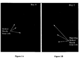
- Skeletal muscle stem cells were identified by immunofluorescence localization of the G8 antigen ( Figure 1 ), in situ hybridization for MyoD mRNA ( Figure 3 ), and confocal and epifluorescence microscopy.
- G8 antibody recognizes a surface antigen specifically expressed in cells that express MyoD mRNA in the chick embryo and fetus ( Gerhart et al. (2001) J. Cell Biol., 155:381-391 ; Gerhart et al. (2004) J.
- RNAs for MyoD were detected with DNA dendrimers (see, e.g., Gerhart et al. (2004) Biol. Proced. Online, 6:149-156 ) conjugated with Cy3 and the following antisense oligonucleotide sequence: chicken MyoD, 5'- TTCTCAAGAGCAAATACTCACCATTTGGTGATTCCGTGTAGTA-3' (L34006; Dechesne et al. (1994) Mol. Cell. Biol., 14:5474-5486 ).
- Fluorescent dendrimers were obtained from Genisphere, Inc. (Hatfield, PA). Double labeling with dendrimers and antibodies was performed as previously described Gerhart et al. (2001) J. Cell Biol., 155:381-391 ; Gerhart et al. (2004) J. Cell Biol., 164:739-746 ; Strony et al. (2005) Gene Expr. Patterns, 5:387-395 ; Gerhart et al. (2004) Biol. Proced. Online, 6:149-156 ).
- a subpopulation of G8/MyoD mRNA positive skm stem cells were detected within a niche located in the equatorial zone (EQ) of the lens nestled among the lens epithelial cells. This finding demonstrates for the first time that there is a population of cells within the lens epithelium with stem cell properties and a phenotype distinct from lens epithelial cells.
- EQ equatorial zone
- skm cells Upon injury induced by mock cataract surgery, skm cells quickly emerge from their niche with a mesenchymal morphology. These skm stem cells crawl over the top of the cuboidal lens epithelial cell monolayer and migrate to the leading edge of the collectively migrating epithelial cell sheet. Ablation of skm stem cells suppresses the expression of ⁇ -SMA expression in posterior capsular opacification cultures.
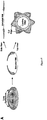
- Skeletal muscle stem cells were also identified by immunofluorescence localization of the G8 antigen, in situ hybridization for MyoD mRNA, and epifluorescence microscopy ( Figure 3 ). Accordingly, it is evident that the presence of skeletal muscle stem cells in lenses is not limited to chickens as skm stem cells are also present in other animals including mammals.
- EMT epithelial to mesenchymal transition
- Wounding of the epithelium is the result of mock cataract surgery in embryonic day 15 chick lenses.
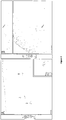
- This microsurgical procedure involves removal of the lens fiber cell mass from within the lens capsule, a thick basement membrane that surrounds the entire lens, which leaves the posterior aspects of the lens capsule denuded of cells (diagrammed in Fig. 5A ).
- the lens epithelium remains intact and attached to the capsule with its principal wound edge bordering the area where the fiber cells had been attached (leading edge, Fig. 5A ).
- leading edge, Fig. 5A By making a few cuts in its anterior regions, creating additional wound edges (cut edge, Fig. 5A ), the tissue is flattened, pinned to the culture dish cell side up and cultured as an ex vivo explant (modeled in Fig. 5A ).
- the hypotheses examined in this study were: 1) that a subpopulation of mesenchymal precursors was present among the epithelial cells of the mature lens, 2) that these cells could be activated upon injury to modulate the wound healing process, and 3) that the progeny of these cells have the potential to become myofibroblasts, a phenotype associated with the development of fibrotic disease.
- the cell type investigated as a candidate for the mesenchymal precursor cell in the lens injury model is identified by its expression of the cell surface antigen G8.
- Cells that label with the G8 monoclonal antibody (mAb) are a subpopulation of the epiblast, a tissue that gives rise to all three germ layers of the embryo ( Bellairs, R.
- G8 pos cells also express mRNA for the myogenic transcription factor myoD ( George-Weinstein, et al. (1996) Dev. Biol., 173:279-91 ; Gerhart et al. (2000) J. Cell. Biol., 149:825-34 ).
- myogenic transcription factor myoD George-Weinstein, et al. (1996) Dev. Biol., 173:279-91 ; Gerhart et al. (2000) J. Cell. Biol., 149:825-34 .
- During development most of the G8 pos /MyoD pos epiblast cells become integrated into the somites where their function is to regulate muscle differentiation ( Gerhart et al. (2006) J. Cell.
- G8 pos /MyoD pos epiblast cells have myogenic potential themselves when isolated and grown in a culture dish ( Gerhart et al. (2001) J. Cell. Biol., 155:381-92 ; Strony et al. (2005) Gene Expr. Patterns, 5:387-95 ; Gerhartet al. (2004) J. Cell. Biol., 164:739-46 ).
- subpopulations of both G8 antigen and MyoD expressing cells also have been detected amongst the cells of non-muscle tissues ( Gerhart et al. (2001) J. Cell. Biol., 155:381-92 ; Asakura et al.
- epithelial explants were immunostained as described previously ( Walker et al. (2007) Invest. Ophthalmol. Vis. Sci., 48:2214-23 ). Briefly, explants were fixed in 3.7% formaldehyde in PBS and permeabilized in 0.25% Triton X-100 in PBS before immunostaining. Cells were incubated with primary antiserum followed by rhodamine- (Jackson Laboratories, West Chester, PA and Millipore Corp., Bedford, MA), fluorescein- (Jackson Laboratories, West Chester, PA) or Alexa Fluor 488-(Invitrogen-Molecular Probes; Eugene, OR) conjugated secondary antibodies.
- rhodamine- Jackson Laboratories, West Chester, PA and Millipore Corp., Bedford, MA
- fluorescein- Jackson Laboratories, West Chester, PA
- RNAs for MyoD were detected with DNA dendrimers conjugated with Cy3 and the following antisense oligonucleotide sequence: chicken MyoD, 5'-TTCTCAAGAGCAAATACTCACCATTTGGTGA TTCCGTGTAGTA-3' (SEQ ID NO: 1) (Genisphere, Inc., Hatfield, PA) ( Gerhart et al. (2006) J. Cell. Biol., 175:283-92 ; Gerhart et al. (2004) Biol. Proced. Online, 6:149-156 ). Explants were double labeled for the G8 antigen and MyoD mRNA, and counterstained with Hoechst as previously described ( Gerhart et al. (2001) J.
- G8 cells were labeled for tracking according to a previously described procedure ( Gerhart et al. (2006) J. Cell. Biol., 175:283-92 ). Briefly, the lens ex vivo epithelial explants were incubated at T0 in Media 199 containing the G8 mAb (1:40) for 45 minutes at room temperature, rinsed in Media 199, and then incubated in rhodamine-conjugated IgM antibody (Millipore Corp., Bedford, MA) for 30 minutes at room temperature. The labeled explants were rinsed in Media 199, placed in serum free media (SFM: Media 199 containing 1% Pen strep and L-glutamine) and incubated at 37°C.
- SFM serum free media
- G8 pos cell ablation in epithelial explants followed a procedure previously described for ablation of G8 pos cells within the epiblast of the chick embryo ( Gerhart et al. (2006) J. Cell. Biol., 175:283-92 ).
- ex vivo lens epithelial explants were prepared in SFM. On culture day 1, epithelial explants were incubated with G8 antigen (1:20) diluted in Hanks Buffered Saline for 1 hour at 37°C followed by incubation in baby rabbit complement (1:40; Cedar Lane, Inc, Burlington, Ontario, Canada) diluted in Hanks buffered saline containing 0.1% BSA for 30 minutes at room temperature.
- Baby rabbit complement was prepared according to manufactures protocol (Cedar Lane, Inc, Burlington, Ontario, Canada). Control explants were left untreated or incubated with G8 mAb or complement alone. After treatment, explants were rinsed and incubated with SFM. The presence of lysed G8 cells was determined immediately after treatment by incubating the ex vivo epithelial explants in 0.2% trypan blue in PBS for 15 minutes at 37°C and visualized with a dissecting microscope (SMZ800; Nikon, Tokyo, Japan) and a Nikon Digital Sight DS-Fi1 camera and the images captured using Nikon NIS-Elements imaging software.
- SMZ800 dissecting microscope
- Nikon Nikon, Tokyo, Japan
- a Nikon Digital Sight DS-Fi1 camera the images captured using Nikon NIS-Elements imaging software.
- epithelial explants were extracted and lysed in OGT buffer (44.4 mM n-Octyl ⁇ -D glucopyranoside, 1% Triton X-100, 100 mM NaCl, 1 mM MgCl 2 , 5 mM EDTA, 10 mM imidazole) containing a protease inhibitor cocktail (Sigma, St. Louis, MO). Protein concentrations were determined with the BCA assay (Pierce, Rockford, IL). Proteins were separated on Tris-glycine gels (Novex, San Diego, CA), electrophoretically transferred to membrane (Immobilon-P; Millipore Corp., Bedford, MA), and immunoblotted.
- OGT buffer 44.4 mM n-Octyl ⁇ -D glucopyranoside, 1% Triton X-100, 100 mM NaCl, 1 mM MgCl 2 , 5 mM EDTA, 10 mM
- ECL reagent (Amersham Life Sciences, Arlington Heights, IL) was used. All gels were run under reducing conditions. Antibodies used for western blotting included ⁇ -actin and ⁇ -smooth muscle actin (Sigma, St Louis, MO).
- G8 pos cells were present in mature lenses (E15) and determined their localization, in situ, focusing on their association with the lens epithelium. In order to preserve the position of G8 pos cells as exists in vivo, lenses were fixed prior to the preparation of lens epithelial explants. G8 pos cells within the explants were labeled with a mAb to the G8 antigen followed by a rhodamine-conjugated secondary antibody. The cultures were co-stained with fluorescein-conjugated phalloidin to tag filamentous actin (F-actin) and reveal the cytoarchitecture of cells within the explant. The labeled explants were examined by high resolution confocal microscopy ( Fig. 4 ).
- G8 pos cells were discovered localized in niches that were nestled among the lens epithelial cells ( Fig. 4A ).
- a typical niche of G8 pos cells (shown at higher magnification in Fig. 4B , lower panel) contained, on average, 7 cells (7.57 +/- .66; Avg +/- SEM). Up to 14 such niches were detected in an epithelial explant.
- the positioning of the G8 pos cell niches within the lens epithelium corresponded to the equatorial region of the intact lens. Previous studies show that G8 pos cells often co-express mRNA for the myogenic transcription factor MyoD.
- a central aspect of this study was to examine the response of G8 pos precursor cells to injury of the lens epithelium.
- the lens epithelium was wounded by mock cataract surgery and placed in culture as ex vivo explants ( Fig. 5A ).
- the response of G8 pos precursor cells to injury of the epithelium was determined after one hour in culture in media containing serum. At this time the cultures were fixed, immunostained with antibody to the G8 antigen, co-stained for F-actin and examined by confocal microscopy ( Figure 5B-F ). Image analysis revealed that within this short time post-injury the G8 pos precursor cells had emerged from their niches and their population size had expanded ( Figure 5B, C ).
- G8 pos cells had rapidly migrated to the wound edges ( Fig. 5D-F ), both the leading edge adjacent to where the fiber cells had been removed ( Fig 5D, E ), and the cut edge where the epithelium had been flattened ( Figure 5F ).
- An orthogonal section through Z-stacks collected by confocal imaging revealed that the G8 pos cells traveled to the wound edges by migrating along the apical surfaces of the epithelium ( Fig. 5E , arrow, top panel).
- the migrating G8 pos cells exhibited a mesenchymal morphology, a phenotype confirmed by their expression of the mesenchymal marker vimentin ( Fig. 5G-I ).
- a myofibroblast is defined as a mesenchymal cell that has organized ⁇ -SMA into stress fibers ( Tomasek et al. (2002) Nat. Rev. Mol. Cell. Biol., 3:349-63 ; Hinz et al. (2007) Am. J. Pathol., 170:1807-16 ), a feature that provides these cells with the contractile function that links them to fibrotic diseases like PCO.
- G8 pos precursor cell to myofibroblast within small clusters of G8 pos cells associated with the epithelium.
- the transitional cell types included G8 pos cells with little to no expression of ⁇ SMA (white arrowhead), G8 pos cells that expressed ⁇ SMA not yet organized into stress fibers (arrow), and G8 pos cells containing ⁇ SMA positive stress fibers (open arrowhead), the G8 expressing myofibroblasts.
- This study also demonstrated that a final step in the differentiation of G8 cells to myofibroblasts was loss of the precursor cell antigen G8 (broken arrow).
- the G8 pos subpopulation emerged from their niches, expanded in population size, exhibited a mesenchymal phenotype and migrated to the wound edge.
- the presence of mesenchymal cells at the wound edge is a characteristic of many epithelial wound healing models, but their appearance is typically attributed to an EMT.
- the instant studies of the wounded lens epithelium suggested an alternate paradigm where G8 pos /MyoD pos precursor cells are the progenitors of the mesenchymal cells that responded to injury and localized to the wound edge. This same precursor population can differentiate into myofibroblasts, whose appearance following wounding is associated with the development of fibrotic disease.
- the eye of an adult mouse was embedded in paraffin, sectioned and stained with hematoxylin and eosin Figure 8A .
- the arrows indicate the areas shown at higher magnification in the fluorescence photomicrographs in Figs. 8B and 8C .
- Cells near the equator of the lens are labeled with the G8 antibody (arrows in Figs. 8B and 8C ).
- Cell nuclei are stained with a dye.
- Rhabdomyosarcomas are tumors containing cells that resemble skeletal muscle cells ( Figs. 9A-9D ).
- the G8 antibody also recognizes its antigen in cultures of human rhabdomyosarcoma cells.
- the rhabdomyosarcoma cells synthesize molecules that also are present in myofibroblasts, including the G8 antigen, alpha smooth muscle actin (SMA), myosin and MyoD protein. These results indicate that the G8 antibody can be used to detect myofibroblasts in the human lens.
- SMA alpha smooth muscle actin
Landscapes
- Health & Medical Sciences (AREA)
- Chemical & Material Sciences (AREA)
- Organic Chemistry (AREA)
- Life Sciences & Earth Sciences (AREA)
- Immunology (AREA)
- General Health & Medical Sciences (AREA)
- Medicinal Chemistry (AREA)
- Proteomics, Peptides & Aminoacids (AREA)
- Molecular Biology (AREA)
- Genetics & Genomics (AREA)
- Biophysics (AREA)
- Biochemistry (AREA)
- Veterinary Medicine (AREA)
- Chemical Kinetics & Catalysis (AREA)
- Engineering & Computer Science (AREA)
- Animal Behavior & Ethology (AREA)
- Pharmacology & Pharmacy (AREA)
- Nuclear Medicine, Radiotherapy & Molecular Imaging (AREA)
- General Chemical & Material Sciences (AREA)
- Public Health (AREA)
- Bioinformatics & Cheminformatics (AREA)
- Ophthalmology & Optometry (AREA)
- Cell Biology (AREA)
- Medicines Containing Antibodies Or Antigens For Use As Internal Diagnostic Agents (AREA)
- Peptides Or Proteins (AREA)
- Medicines That Contain Protein Lipid Enzymes And Other Medicines (AREA)
- Pharmaceuticals Containing Other Organic And Inorganic Compounds (AREA)
- Medicinal Preparation (AREA)
Claims (4)
- Zusammensetzung zur Verwendung beim Behandeln einer fibrotischen Linsenerkrankung, wobei die Zusammensetzung ein Skelettmuskelstammzellenzielmolekül und wenigstens einen pharmazeutisch unbedenklichen Träger umfasst, wobei das Skelettmuskelstammzellenzielmolekül ein Antikörper ist, der spezifisch an G8-Antigen bindet,
wobei das Skelettmuskelstammzellenzielmolekül an wenigstens ein zytotoxisches Molekül konjugiert ist, und wobei die Zusammensetzung direkt in die Linse oder das umgebende Gewebe verabreicht wird. - Verwendung eines Skelettmuskelstammzellenzielmoleküls beim Herstellen eines Arzneimittels zum direkten Verabreichen an die Linse oder das umgebende Gewebe zum Behandeln einer fibrotischen Linsenerkrankung, wobei das Skelettmuskelstammzellenzielmolekül ein Antikörper ist, der spezifisch an G8-Antigen bindet,
wobei das Skelettmuskelstammzellenzielmolekül an wenigstens ein zytotoxisches Molekül konjugiert ist. - Zusammensetzung zur Verwendung nach Anspruch 1 oder Verwendung nach Anspruch 2, wobei es sich bei der fibrotischen Linsenerkrankung um eine hintere Kapseltrübung oder Cataracta subcapsularis anterior handelt.
- Zusammensetzung zur Verwendung nach Anspruch 1 oder Verwendung nach Anspruch 2, wobei das zytotoxische Molekül ausgewählt ist aus der Gruppe bestehend aus zytotoxischen Antibiotika, kationischen amphipathischen lytischen Peptiden, Radionukliden und Toxinen.
Applications Claiming Priority (2)
| Application Number | Priority Date | Filing Date | Title |
|---|---|---|---|
| US11975008P | 2008-12-04 | 2008-12-04 | |
| PCT/US2009/066859 WO2010065920A1 (en) | 2008-12-04 | 2009-12-04 | Compositions and methods for the treatment and prevention of lens fibrotic diseases |
Publications (3)
| Publication Number | Publication Date |
|---|---|
| EP2369936A1 EP2369936A1 (de) | 2011-10-05 |
| EP2369936A4 EP2369936A4 (de) | 2013-01-09 |
| EP2369936B1 true EP2369936B1 (de) | 2018-01-24 |
Family
ID=42233638
Family Applications (1)
| Application Number | Title | Priority Date | Filing Date |
|---|---|---|---|
| EP09831240.8A Not-in-force EP2369936B1 (de) | 2008-12-04 | 2009-12-04 | Zusammensetzungen und verfahren zur behandlung und prävention von fibrotischen linsenerkrankungen |
Country Status (8)
| Country | Link |
|---|---|
| US (1) | US9273133B2 (de) |
| EP (1) | EP2369936B1 (de) |
| JP (1) | JP5761653B2 (de) |
| KR (1) | KR101846554B1 (de) |
| CN (1) | CN102333447B (de) |
| AU (1) | AU2009322184B2 (de) |
| CA (1) | CA2746511C (de) |
| WO (1) | WO2010065920A1 (de) |
Families Citing this family (7)
| Publication number | Priority date | Publication date | Assignee | Title |
|---|---|---|---|---|
| CA2676946A1 (en) | 2009-08-28 | 2011-02-28 | Lucie Peduto | Adam12 inhibitors and their use against inflammation-induced fibrosis |
| WO2013134768A1 (en) * | 2012-03-09 | 2013-09-12 | Lankenau Institute For Medical Research | Compositions and methods for treating cancer |
| US20160051693A1 (en) * | 2013-03-21 | 2016-02-25 | Genisphere, Llc | Cellular Delivery of DNA Intercalating Agents |
| GB201312010D0 (en) * | 2013-07-04 | 2013-08-21 | Univ Birmingham | Receptor Agonists |
| IL260983B (en) | 2016-02-19 | 2022-07-01 | Genisphere Llc | Nucleic acid carriers and methods of medical use |
| US11400099B2 (en) | 2016-09-23 | 2022-08-02 | Thomas Jefferson University | Drug-induced epigenetic remodeling to prevent fibrosis |
| HU231426B1 (hu) * | 2018-12-05 | 2023-08-28 | Pharmacoidea Kft. | Syndecan-3 45-384 aminosav közti régiójára specifikus ligandok alkalmazása neurodegenerativ kórképek kezelésére |
Family Cites Families (6)
| Publication number | Priority date | Publication date | Assignee | Title |
|---|---|---|---|---|
| DE3920358A1 (de) | 1989-06-22 | 1991-01-17 | Behringwerke Ag | Bispezifische und oligospezifische, mono- und oligovalente antikoerperkonstrukte, ihre herstellung und verwendung |
| DE69233528T2 (de) | 1991-11-25 | 2006-03-16 | Enzon, Inc. | Verfahren zur Herstellung von multivalenten antigenbindenden Proteinen |
| US6180357B1 (en) * | 1999-10-08 | 2001-01-30 | Arius Research, Inc. | Individualized patient-specific anti-cancer antibodies |
| EP1592713A2 (de) | 2003-02-13 | 2005-11-09 | Pharmacia Corporation | Antikörper gegen c-met zur behandlung von krebs |
| CA2611102A1 (en) * | 2005-05-26 | 2006-11-30 | Thomas Jefferson University | Method to treat and prevent posterior capsule opacification |
| US20090297479A1 (en) * | 2008-03-28 | 2009-12-03 | Kiyoshi Ariizumi | Dc-hil conjugates for treatment of t-cell disorders |
-
2009
- 2009-12-04 EP EP09831240.8A patent/EP2369936B1/de not_active Not-in-force
- 2009-12-04 JP JP2011539760A patent/JP5761653B2/ja active Active
- 2009-12-04 KR KR1020117015441A patent/KR101846554B1/ko not_active Expired - Fee Related
- 2009-12-04 WO PCT/US2009/066859 patent/WO2010065920A1/en not_active Ceased
- 2009-12-04 US US13/132,745 patent/US9273133B2/en active Active
- 2009-12-04 CA CA2746511A patent/CA2746511C/en not_active Expired - Fee Related
- 2009-12-04 CN CN200980156374.XA patent/CN102333447B/zh not_active Expired - Fee Related
- 2009-12-04 AU AU2009322184A patent/AU2009322184B2/en not_active Ceased
Non-Patent Citations (1)
| Title |
|---|
| None * |
Also Published As
| Publication number | Publication date |
|---|---|
| WO2010065920A1 (en) | 2010-06-10 |
| JP5761653B2 (ja) | 2015-08-12 |
| JP2012511025A (ja) | 2012-05-17 |
| EP2369936A1 (de) | 2011-10-05 |
| EP2369936A4 (de) | 2013-01-09 |
| US20110256054A1 (en) | 2011-10-20 |
| KR20110101186A (ko) | 2011-09-15 |
| CN102333447B (zh) | 2018-07-31 |
| AU2009322184A1 (en) | 2011-06-30 |
| CA2746511C (en) | 2019-06-18 |
| CN102333447A (zh) | 2012-01-25 |
| AU2009322184B2 (en) | 2016-03-31 |
| KR101846554B1 (ko) | 2018-04-06 |
| CA2746511A1 (en) | 2010-06-10 |
| US9273133B2 (en) | 2016-03-01 |
Similar Documents
| Publication | Publication Date | Title |
|---|---|---|
| Van Den Born et al. | Distribution of GBM heparan sulfate proteoglycan core protein and side chains in human glomerular diseases | |
| US9273133B2 (en) | Compositions and methods for the treatment and prevention of lens fibrotic diseases | |
| Schlosshauer et al. | Neurothelin: an inducible cell surface glycoprotein of blood-brain barrier-specific endothelial cells and distinct neurons. | |
| DE60132475T2 (de) | Neue tumorassoziierte marker | |
| JP5219827B2 (ja) | invivoで抗腫瘍活性を有する抗ヒトDlk−1抗体 | |
| JP5989299B2 (ja) | リゾホスファチジン酸結合のための組成物と方法 | |
| US12162937B2 (en) | Methods of treating an inflammatory disorder by administering an antibody which binds to a connexin CX43 hemichannel | |
| KR102494798B1 (ko) | Tau에 대한 항체 | |
| EP2033971A1 (de) | Bone Morphogenetic Protein (BMP)-bindende Domänen von Proteinen der Repulsive Guidance Molecule (RGM) Proteinfamilie und funktionale Fragmente davon sowie deren Verwendung | |
| JPWO2009116670A1 (ja) | invivoで抗腫瘍活性を有する抗ヒトDlk−1抗体 | |
| Rupp et al. | Matrix metalloproteinase 2-integrin αvβ3 binding is required for mesenchymal cell invasive activity but not epithelial locomotion: a computational time-lapse study | |
| EP1928905B1 (de) | Bindungsdomänen von proteinen der repulsive guidance molecule (rgm) proteinfamilie und funktionale fragmente davon sowie deren verwendung | |
| TWI609692B (zh) | 新穎stip1多肽及其用途 | |
| Assmann et al. | The organ distribution of gp-330 (Heymann antigen) and gp-90 in the mouse and the rat | |
| JP2022130488A (ja) | Pdgf-ccの阻害のための方法および組成物 | |
| TWI878967B (zh) | 抗間皮素抗體 | |
| CN106554418B (zh) | 炭疽毒素受体2单克隆抗体及制备方法和应用 | |
| US20240368259A1 (en) | Antibodies against lefty proteins | |
| JP7064203B2 (ja) | 抗ヒトcd26モノクローナル抗体 | |
| US20230265212A1 (en) | Compositions and methods for oocyte-specific contraception | |
| HK40076091B (zh) | 用於靶向连接蛋白半通道的组合物和方法 | |
| US20210253732A1 (en) | Antigen-binding fragment and/or aptamer for binding to an extracellular part of cd9 and therapeutic uses | |
| Michael | Alfred J. Fish, Kathleen M. Carmody, Mark S. Schiffer |
Legal Events
| Date | Code | Title | Description |
|---|---|---|---|
| PUAI | Public reference made under article 153(3) epc to a published international application that has entered the european phase |
Free format text: ORIGINAL CODE: 0009012 |
|
| 17P | Request for examination filed |
Effective date: 20110701 |
|
| AK | Designated contracting states |
Kind code of ref document: A1 Designated state(s): AT BE BG CH CY CZ DE DK EE ES FI FR GB GR HR HU IE IS IT LI LT LU LV MC MK MT NL NO PL PT RO SE SI SK SM TR |
|
| DAX | Request for extension of the european patent (deleted) | ||
| RAP1 | Party data changed (applicant data changed or rights of an application transferred) |
Owner name: THOMAS JEFFERSON UNIVERSITY Owner name: LANKENAU INSTITUTE FOR MEDICAL RESEARCH |
|
| RIC1 | Information provided on ipc code assigned before grant |
Ipc: A61K 39/395 20060101ALI20121128BHEP Ipc: A01N 63/02 20060101AFI20121128BHEP |
|
| A4 | Supplementary search report drawn up and despatched |
Effective date: 20121207 |
|
| 17Q | First examination report despatched |
Effective date: 20150506 |
|
| GRAP | Despatch of communication of intention to grant a patent |
Free format text: ORIGINAL CODE: EPIDOSNIGR1 |
|
| STAA | Information on the status of an ep patent application or granted ep patent |
Free format text: STATUS: GRANT OF PATENT IS INTENDED |
|
| INTG | Intention to grant announced |
Effective date: 20170801 |
|
| GRAS | Grant fee paid |
Free format text: ORIGINAL CODE: EPIDOSNIGR3 |
|
| GRAA | (expected) grant |
Free format text: ORIGINAL CODE: 0009210 |
|
| STAA | Information on the status of an ep patent application or granted ep patent |
Free format text: STATUS: THE PATENT HAS BEEN GRANTED |
|
| AK | Designated contracting states |
Kind code of ref document: B1 Designated state(s): AT BE BG CH CY CZ DE DK EE ES FI FR GB GR HR HU IE IS IT LI LT LU LV MC MK MT NL NO PL PT RO SE SI SK SM TR |
|
| REG | Reference to a national code |
Ref country code: GB Ref legal event code: FG4D |
|
| REG | Reference to a national code |
Ref country code: CH Ref legal event code: EP |
|
| REG | Reference to a national code |
Ref country code: AT Ref legal event code: REF Ref document number: 965260 Country of ref document: AT Kind code of ref document: T Effective date: 20180215 |
|
| REG | Reference to a national code |
Ref country code: IE Ref legal event code: FG4D |
|
| REG | Reference to a national code |
Ref country code: DE Ref legal event code: R096 Ref document number: 602009050604 Country of ref document: DE |
|
| REG | Reference to a national code |
Ref country code: CH Ref legal event code: NV Representative=s name: ISLER AND PEDRAZZINI AG, CH |
|
| REG | Reference to a national code |
Ref country code: NL Ref legal event code: MP Effective date: 20180124 |
|
| REG | Reference to a national code |
Ref country code: LT Ref legal event code: MG4D |
|
| REG | Reference to a national code |
Ref country code: AT Ref legal event code: MK05 Ref document number: 965260 Country of ref document: AT Kind code of ref document: T Effective date: 20180124 |
|
| PG25 | Lapsed in a contracting state [announced via postgrant information from national office to epo] |
Ref country code: NL Free format text: LAPSE BECAUSE OF FAILURE TO SUBMIT A TRANSLATION OF THE DESCRIPTION OR TO PAY THE FEE WITHIN THE PRESCRIBED TIME-LIMIT Effective date: 20180124 |
|
| PG25 | Lapsed in a contracting state [announced via postgrant information from national office to epo] |
Ref country code: FI Free format text: LAPSE BECAUSE OF FAILURE TO SUBMIT A TRANSLATION OF THE DESCRIPTION OR TO PAY THE FEE WITHIN THE PRESCRIBED TIME-LIMIT Effective date: 20180124 Ref country code: HR Free format text: LAPSE BECAUSE OF FAILURE TO SUBMIT A TRANSLATION OF THE DESCRIPTION OR TO PAY THE FEE WITHIN THE PRESCRIBED TIME-LIMIT Effective date: 20180124 Ref country code: NO Free format text: LAPSE BECAUSE OF FAILURE TO SUBMIT A TRANSLATION OF THE DESCRIPTION OR TO PAY THE FEE WITHIN THE PRESCRIBED TIME-LIMIT Effective date: 20180424 Ref country code: ES Free format text: LAPSE BECAUSE OF FAILURE TO SUBMIT A TRANSLATION OF THE DESCRIPTION OR TO PAY THE FEE WITHIN THE PRESCRIBED TIME-LIMIT Effective date: 20180124 Ref country code: CY Free format text: LAPSE BECAUSE OF FAILURE TO SUBMIT A TRANSLATION OF THE DESCRIPTION OR TO PAY THE FEE WITHIN THE PRESCRIBED TIME-LIMIT Effective date: 20180124 Ref country code: LT Free format text: LAPSE BECAUSE OF FAILURE TO SUBMIT A TRANSLATION OF THE DESCRIPTION OR TO PAY THE FEE WITHIN THE PRESCRIBED TIME-LIMIT Effective date: 20180124 |
|
| PG25 | Lapsed in a contracting state [announced via postgrant information from national office to epo] |
Ref country code: GR Free format text: LAPSE BECAUSE OF FAILURE TO SUBMIT A TRANSLATION OF THE DESCRIPTION OR TO PAY THE FEE WITHIN THE PRESCRIBED TIME-LIMIT Effective date: 20180425 Ref country code: PL Free format text: LAPSE BECAUSE OF FAILURE TO SUBMIT A TRANSLATION OF THE DESCRIPTION OR TO PAY THE FEE WITHIN THE PRESCRIBED TIME-LIMIT Effective date: 20180124 Ref country code: BG Free format text: LAPSE BECAUSE OF FAILURE TO SUBMIT A TRANSLATION OF THE DESCRIPTION OR TO PAY THE FEE WITHIN THE PRESCRIBED TIME-LIMIT Effective date: 20180424 Ref country code: IS Free format text: LAPSE BECAUSE OF FAILURE TO SUBMIT A TRANSLATION OF THE DESCRIPTION OR TO PAY THE FEE WITHIN THE PRESCRIBED TIME-LIMIT Effective date: 20180524 Ref country code: LV Free format text: LAPSE BECAUSE OF FAILURE TO SUBMIT A TRANSLATION OF THE DESCRIPTION OR TO PAY THE FEE WITHIN THE PRESCRIBED TIME-LIMIT Effective date: 20180124 Ref country code: SE Free format text: LAPSE BECAUSE OF FAILURE TO SUBMIT A TRANSLATION OF THE DESCRIPTION OR TO PAY THE FEE WITHIN THE PRESCRIBED TIME-LIMIT Effective date: 20180124 Ref country code: AT Free format text: LAPSE BECAUSE OF FAILURE TO SUBMIT A TRANSLATION OF THE DESCRIPTION OR TO PAY THE FEE WITHIN THE PRESCRIBED TIME-LIMIT Effective date: 20180124 |
|
| REG | Reference to a national code |
Ref country code: DE Ref legal event code: R097 Ref document number: 602009050604 Country of ref document: DE |
|
| PG25 | Lapsed in a contracting state [announced via postgrant information from national office to epo] |
Ref country code: EE Free format text: LAPSE BECAUSE OF FAILURE TO SUBMIT A TRANSLATION OF THE DESCRIPTION OR TO PAY THE FEE WITHIN THE PRESCRIBED TIME-LIMIT Effective date: 20180124 Ref country code: IT Free format text: LAPSE BECAUSE OF FAILURE TO SUBMIT A TRANSLATION OF THE DESCRIPTION OR TO PAY THE FEE WITHIN THE PRESCRIBED TIME-LIMIT Effective date: 20180124 Ref country code: RO Free format text: LAPSE BECAUSE OF FAILURE TO SUBMIT A TRANSLATION OF THE DESCRIPTION OR TO PAY THE FEE WITHIN THE PRESCRIBED TIME-LIMIT Effective date: 20180124 |
|
| PG25 | Lapsed in a contracting state [announced via postgrant information from national office to epo] |
Ref country code: CZ Free format text: LAPSE BECAUSE OF FAILURE TO SUBMIT A TRANSLATION OF THE DESCRIPTION OR TO PAY THE FEE WITHIN THE PRESCRIBED TIME-LIMIT Effective date: 20180124 Ref country code: SK Free format text: LAPSE BECAUSE OF FAILURE TO SUBMIT A TRANSLATION OF THE DESCRIPTION OR TO PAY THE FEE WITHIN THE PRESCRIBED TIME-LIMIT Effective date: 20180124 Ref country code: DK Free format text: LAPSE BECAUSE OF FAILURE TO SUBMIT A TRANSLATION OF THE DESCRIPTION OR TO PAY THE FEE WITHIN THE PRESCRIBED TIME-LIMIT Effective date: 20180124 Ref country code: SM Free format text: LAPSE BECAUSE OF FAILURE TO SUBMIT A TRANSLATION OF THE DESCRIPTION OR TO PAY THE FEE WITHIN THE PRESCRIBED TIME-LIMIT Effective date: 20180124 |
|
| PLBE | No opposition filed within time limit |
Free format text: ORIGINAL CODE: 0009261 |
|
| STAA | Information on the status of an ep patent application or granted ep patent |
Free format text: STATUS: NO OPPOSITION FILED WITHIN TIME LIMIT |
|
| 26N | No opposition filed |
Effective date: 20181025 |
|
| PG25 | Lapsed in a contracting state [announced via postgrant information from national office to epo] |
Ref country code: SI Free format text: LAPSE BECAUSE OF FAILURE TO SUBMIT A TRANSLATION OF THE DESCRIPTION OR TO PAY THE FEE WITHIN THE PRESCRIBED TIME-LIMIT Effective date: 20180124 |
|
| PG25 | Lapsed in a contracting state [announced via postgrant information from national office to epo] |
Ref country code: LU Free format text: LAPSE BECAUSE OF NON-PAYMENT OF DUE FEES Effective date: 20181204 Ref country code: MC Free format text: LAPSE BECAUSE OF FAILURE TO SUBMIT A TRANSLATION OF THE DESCRIPTION OR TO PAY THE FEE WITHIN THE PRESCRIBED TIME-LIMIT Effective date: 20180124 |
|
| REG | Reference to a national code |
Ref country code: IE Ref legal event code: MM4A |
|
| REG | Reference to a national code |
Ref country code: BE Ref legal event code: MM Effective date: 20181231 |
|
| PG25 | Lapsed in a contracting state [announced via postgrant information from national office to epo] |
Ref country code: IE Free format text: LAPSE BECAUSE OF NON-PAYMENT OF DUE FEES Effective date: 20181204 |
|
| REG | Reference to a national code |
Ref country code: DE Ref legal event code: R079 Ref document number: 602009050604 Country of ref document: DE Free format text: PREVIOUS MAIN CLASS: A01N0063020000 Ipc: A01N0063200000 |
|
| PG25 | Lapsed in a contracting state [announced via postgrant information from national office to epo] |
Ref country code: BE Free format text: LAPSE BECAUSE OF NON-PAYMENT OF DUE FEES Effective date: 20181231 |
|
| PG25 | Lapsed in a contracting state [announced via postgrant information from national office to epo] |
Ref country code: MT Free format text: LAPSE BECAUSE OF NON-PAYMENT OF DUE FEES Effective date: 20181204 |
|
| PG25 | Lapsed in a contracting state [announced via postgrant information from national office to epo] |
Ref country code: TR Free format text: LAPSE BECAUSE OF FAILURE TO SUBMIT A TRANSLATION OF THE DESCRIPTION OR TO PAY THE FEE WITHIN THE PRESCRIBED TIME-LIMIT Effective date: 20180124 |
|
| PGFP | Annual fee paid to national office [announced via postgrant information from national office to epo] |
Ref country code: CH Payment date: 20191213 Year of fee payment: 11 |
|
| PG25 | Lapsed in a contracting state [announced via postgrant information from national office to epo] |
Ref country code: PT Free format text: LAPSE BECAUSE OF FAILURE TO SUBMIT A TRANSLATION OF THE DESCRIPTION OR TO PAY THE FEE WITHIN THE PRESCRIBED TIME-LIMIT Effective date: 20180124 |
|
| PG25 | Lapsed in a contracting state [announced via postgrant information from national office to epo] |
Ref country code: MK Free format text: LAPSE BECAUSE OF NON-PAYMENT OF DUE FEES Effective date: 20180124 Ref country code: HU Free format text: LAPSE BECAUSE OF FAILURE TO SUBMIT A TRANSLATION OF THE DESCRIPTION OR TO PAY THE FEE WITHIN THE PRESCRIBED TIME-LIMIT; INVALID AB INITIO Effective date: 20091204 |
|
| REG | Reference to a national code |
Ref country code: CH Ref legal event code: PL |
|
| PG25 | Lapsed in a contracting state [announced via postgrant information from national office to epo] |
Ref country code: LI Free format text: LAPSE BECAUSE OF NON-PAYMENT OF DUE FEES Effective date: 20201231 Ref country code: CH Free format text: LAPSE BECAUSE OF NON-PAYMENT OF DUE FEES Effective date: 20201231 |
|
| PGFP | Annual fee paid to national office [announced via postgrant information from national office to epo] |
Ref country code: DE Payment date: 20211207 Year of fee payment: 13 Ref country code: FR Payment date: 20211217 Year of fee payment: 13 Ref country code: GB Payment date: 20211209 Year of fee payment: 13 |
|
| REG | Reference to a national code |
Ref country code: DE Ref legal event code: R119 Ref document number: 602009050604 Country of ref document: DE |
|
| GBPC | Gb: european patent ceased through non-payment of renewal fee |
Effective date: 20221204 |
|
| PG25 | Lapsed in a contracting state [announced via postgrant information from national office to epo] |
Ref country code: GB Free format text: LAPSE BECAUSE OF NON-PAYMENT OF DUE FEES Effective date: 20221204 Ref country code: DE Free format text: LAPSE BECAUSE OF NON-PAYMENT OF DUE FEES Effective date: 20230701 |
|
| PG25 | Lapsed in a contracting state [announced via postgrant information from national office to epo] |
Ref country code: FR Free format text: LAPSE BECAUSE OF NON-PAYMENT OF DUE FEES Effective date: 20221231 |