EP2386312A2 - Intrathekale und intratumorale Superantigene zur Behandlung von malignen Erkrankungen - Google Patents
Intrathekale und intratumorale Superantigene zur Behandlung von malignen Erkrankungen Download PDFInfo
- Publication number
- EP2386312A2 EP2386312A2 EP10012618A EP10012618A EP2386312A2 EP 2386312 A2 EP2386312 A2 EP 2386312A2 EP 10012618 A EP10012618 A EP 10012618A EP 10012618 A EP10012618 A EP 10012618A EP 2386312 A2 EP2386312 A2 EP 2386312A2
- Authority
- EP
- European Patent Office
- Prior art keywords
- superantigen
- composition
- tumor
- homologue
- native
- Prior art date
- Legal status (The legal status is an assumption and is not a legal conclusion. Google has not performed a legal analysis and makes no representation as to the accuracy of the status listed.)
- Granted
Links
- 231100000617 superantigen Toxicity 0.000 title claims abstract description 80
- 238000007913 intrathecal administration Methods 0.000 title claims abstract description 37
- 230000003211 malignant effect Effects 0.000 title claims description 14
- 230000002601 intratumoral effect Effects 0.000 title abstract description 84
- 201000010099 disease Diseases 0.000 title description 16
- 208000037265 diseases, disorders, signs and symptoms Diseases 0.000 title description 16
- 206010028980 Neoplasm Diseases 0.000 claims abstract description 321
- 239000000203 mixture Substances 0.000 claims abstract description 144
- 101000686985 Mouse mammary tumor virus (strain C3H) Protein PR73 Proteins 0.000 claims abstract description 80
- 238000000034 method Methods 0.000 claims abstract description 79
- 239000002246 antineoplastic agent Substances 0.000 claims abstract description 62
- 239000012634 fragment Substances 0.000 claims abstract description 46
- 108020001507 fusion proteins Proteins 0.000 claims abstract description 34
- 102000037865 fusion proteins Human genes 0.000 claims abstract description 34
- 229940044683 chemotherapy drug Drugs 0.000 claims abstract description 33
- 239000012530 fluid Substances 0.000 claims abstract description 22
- 201000011510 cancer Diseases 0.000 claims abstract description 20
- 210000000056 organ Anatomy 0.000 claims abstract description 17
- 238000009825 accumulation Methods 0.000 claims abstract description 16
- 206010048962 Brain oedema Diseases 0.000 claims abstract description 4
- 208000006752 brain edema Diseases 0.000 claims abstract description 4
- 238000002648 combination therapy Methods 0.000 claims abstract 3
- 108090000623 proteins and genes Proteins 0.000 claims description 81
- 239000003795 chemical substances by application Substances 0.000 claims description 73
- 102000004169 proteins and genes Human genes 0.000 claims description 68
- 108090000765 processed proteins & peptides Proteins 0.000 claims description 61
- 239000003814 drug Substances 0.000 claims description 48
- 210000001744 T-lymphocyte Anatomy 0.000 claims description 45
- 102000004196 processed proteins & peptides Human genes 0.000 claims description 44
- 229940079593 drug Drugs 0.000 claims description 41
- 229920001184 polypeptide Polymers 0.000 claims description 38
- 238000009472 formulation Methods 0.000 claims description 29
- 231100000655 enterotoxin Toxicity 0.000 claims description 26
- 230000004927 fusion Effects 0.000 claims description 26
- 108091008874 T cell receptors Proteins 0.000 claims description 23
- 125000003275 alpha amino acid group Chemical group 0.000 claims description 23
- 102000016266 T-Cell Antigen Receptors Human genes 0.000 claims description 21
- 206010026673 Malignant Pleural Effusion Diseases 0.000 claims description 19
- 238000004422 calculation algorithm Methods 0.000 claims description 19
- 238000013270 controlled release Methods 0.000 claims description 18
- 238000004458 analytical method Methods 0.000 claims description 15
- 239000002095 exotoxin Substances 0.000 claims description 14
- 231100000776 exotoxin Toxicity 0.000 claims description 14
- 206010025538 Malignant ascites Diseases 0.000 claims description 12
- 206010061289 metastatic neoplasm Diseases 0.000 claims description 12
- 230000004071 biological effect Effects 0.000 claims description 10
- OTLLEIBWKHEHGU-UHFFFAOYSA-N 2-[5-[[5-(6-aminopurin-9-yl)-3,4-dihydroxyoxolan-2-yl]methoxy]-3,4-dihydroxy-6-(hydroxymethyl)oxan-2-yl]oxy-3,5-dihydroxy-4-phosphonooxyhexanedioic acid Chemical compound C1=NC=2C(N)=NC=NC=2N1C(C(C1O)O)OC1COC1C(CO)OC(OC(C(O)C(OP(O)(O)=O)C(O)C(O)=O)C(O)=O)C(O)C1O OTLLEIBWKHEHGU-UHFFFAOYSA-N 0.000 claims description 9
- 201000009030 Carcinoma Diseases 0.000 claims description 9
- 241000193468 Clostridium perfringens Species 0.000 claims description 8
- 206010003246 arthritis Diseases 0.000 claims description 8
- 208000005228 Pericardial Effusion Diseases 0.000 claims description 7
- 241000607477 Yersinia pseudotuberculosis Species 0.000 claims description 7
- 238000007912 intraperitoneal administration Methods 0.000 claims description 5
- 230000001394 metastastic effect Effects 0.000 claims description 5
- 241000204031 Mycoplasma Species 0.000 claims description 2
- 230000004936 stimulating effect Effects 0.000 claims description 2
- 230000002325 super-antigenic effect Effects 0.000 claims 6
- 108010022946 erythrogenic toxin Proteins 0.000 claims 1
- 238000007920 subcutaneous administration Methods 0.000 claims 1
- 230000004083 survival effect Effects 0.000 abstract description 56
- 238000002347 injection Methods 0.000 abstract description 52
- 239000007924 injection Substances 0.000 abstract description 52
- 208000002151 Pleural effusion Diseases 0.000 abstract description 30
- 206010003445 Ascites Diseases 0.000 abstract description 13
- 230000002829 reductive effect Effects 0.000 abstract description 13
- 230000009467 reduction Effects 0.000 abstract description 11
- 230000002035 prolonged effect Effects 0.000 abstract description 6
- 230000002459 sustained effect Effects 0.000 abstract description 5
- 238000011521 systemic chemotherapy Methods 0.000 abstract description 5
- 231100000252 nontoxic Toxicity 0.000 abstract description 3
- 230000003000 nontoxic effect Effects 0.000 abstract description 3
- 230000006872 improvement Effects 0.000 abstract description 2
- 238000011282 treatment Methods 0.000 description 125
- 235000018102 proteins Nutrition 0.000 description 61
- 230000004044 response Effects 0.000 description 53
- 238000012360 testing method Methods 0.000 description 48
- 210000004027 cell Anatomy 0.000 description 44
- 238000002560 therapeutic procedure Methods 0.000 description 44
- 241001465754 Metazoa Species 0.000 description 42
- 241000699670 Mus sp. Species 0.000 description 40
- 206010063045 Effusion Diseases 0.000 description 39
- DQLATGHUWYMOKM-UHFFFAOYSA-L cisplatin Chemical compound N[Pt](N)(Cl)Cl DQLATGHUWYMOKM-UHFFFAOYSA-L 0.000 description 39
- 229960004316 cisplatin Drugs 0.000 description 39
- 230000000694 effects Effects 0.000 description 39
- 102000043131 MHC class II family Human genes 0.000 description 38
- 108091054438 MHC class II family Proteins 0.000 description 38
- FAPWRFPIFSIZLT-UHFFFAOYSA-M Sodium chloride Chemical compound [Na+].[Cl-] FAPWRFPIFSIZLT-UHFFFAOYSA-M 0.000 description 29
- 238000006467 substitution reaction Methods 0.000 description 29
- 210000000038 chest Anatomy 0.000 description 28
- 229940127089 cytotoxic agent Drugs 0.000 description 28
- -1 β and γ Proteins 0.000 description 28
- 241000282414 Homo sapiens Species 0.000 description 27
- 235000001014 amino acid Nutrition 0.000 description 27
- 238000002512 chemotherapy Methods 0.000 description 25
- 239000011780 sodium chloride Substances 0.000 description 25
- 150000001413 amino acids Chemical class 0.000 description 23
- 210000004910 pleural fluid Anatomy 0.000 description 23
- 206010027476 Metastases Diseases 0.000 description 22
- 229940024606 amino acid Drugs 0.000 description 22
- 239000000427 antigen Substances 0.000 description 22
- 108091007433 antigens Proteins 0.000 description 22
- 102000036639 antigens Human genes 0.000 description 22
- 150000007523 nucleic acids Chemical class 0.000 description 22
- 208000002154 non-small cell lung carcinoma Diseases 0.000 description 21
- 102000005962 receptors Human genes 0.000 description 21
- 108020003175 receptors Proteins 0.000 description 21
- 210000004881 tumor cell Anatomy 0.000 description 21
- AOJJSUZBOXZQNB-TZSSRYMLSA-N Doxorubicin Chemical compound O([C@H]1C[C@@](O)(CC=2C(O)=C3C(=O)C=4C=CC=C(C=4C(=O)C3=C(O)C=21)OC)C(=O)CO)[C@H]1C[C@H](N)[C@H](O)[C@H](C)O1 AOJJSUZBOXZQNB-TZSSRYMLSA-N 0.000 description 20
- 239000007943 implant Substances 0.000 description 20
- 210000004072 lung Anatomy 0.000 description 20
- 239000013641 positive control Substances 0.000 description 20
- 208000029729 tumor suppressor gene on chromosome 11 Diseases 0.000 description 20
- 230000003442 weekly effect Effects 0.000 description 20
- 238000002474 experimental method Methods 0.000 description 18
- 239000000454 talc Substances 0.000 description 18
- 229910052623 talc Inorganic materials 0.000 description 18
- 229940033134 talc Drugs 0.000 description 18
- 230000001225 therapeutic effect Effects 0.000 description 18
- 238000002360 preparation method Methods 0.000 description 17
- 102000004127 Cytokines Human genes 0.000 description 16
- 108090000695 Cytokines Proteins 0.000 description 16
- 150000001875 compounds Chemical class 0.000 description 16
- 102000039446 nucleic acids Human genes 0.000 description 16
- 108020004707 nucleic acids Proteins 0.000 description 16
- 230000000259 anti-tumor effect Effects 0.000 description 15
- 238000004364 calculation method Methods 0.000 description 15
- 230000001988 toxicity Effects 0.000 description 14
- 231100000419 toxicity Toxicity 0.000 description 14
- 238000012546 transfer Methods 0.000 description 14
- 230000034994 death Effects 0.000 description 13
- 231100000517 death Toxicity 0.000 description 13
- 238000011156 evaluation Methods 0.000 description 13
- 201000001441 melanoma Diseases 0.000 description 13
- 239000000243 solution Substances 0.000 description 13
- 238000002604 ultrasonography Methods 0.000 description 13
- 206010025323 Lymphomas Diseases 0.000 description 12
- 238000006243 chemical reaction Methods 0.000 description 12
- 238000001802 infusion Methods 0.000 description 12
- 210000003281 pleural cavity Anatomy 0.000 description 12
- 230000000306 recurrent effect Effects 0.000 description 12
- 102100023804 Coagulation factor VII Human genes 0.000 description 11
- 108010023321 Factor VII Proteins 0.000 description 11
- 206010058467 Lung neoplasm malignant Diseases 0.000 description 11
- NWIBSHFKIJFRCO-WUDYKRTCSA-N Mytomycin Chemical compound C1N2C(C(C(C)=C(N)C3=O)=O)=C3[C@@H](COC(N)=O)[C@@]2(OC)[C@@H]2[C@H]1N2 NWIBSHFKIJFRCO-WUDYKRTCSA-N 0.000 description 11
- 230000035508 accumulation Effects 0.000 description 11
- 238000003556 assay Methods 0.000 description 11
- 238000012217 deletion Methods 0.000 description 11
- 230000037430 deletion Effects 0.000 description 11
- 230000001419 dependent effect Effects 0.000 description 11
- 239000000147 enterotoxin Substances 0.000 description 11
- 229940012413 factor vii Drugs 0.000 description 11
- 238000001727 in vivo Methods 0.000 description 11
- 230000003902 lesion Effects 0.000 description 11
- 208000020816 lung neoplasm Diseases 0.000 description 11
- 230000036961 partial effect Effects 0.000 description 11
- 239000000126 substance Substances 0.000 description 11
- 230000008685 targeting Effects 0.000 description 11
- 239000004971 Cross linker Substances 0.000 description 10
- 101710104662 Enterotoxin type C-3 Proteins 0.000 description 10
- 102100030844 Exocyst complex component 1 Human genes 0.000 description 10
- 108091028043 Nucleic acid sequence Proteins 0.000 description 10
- 206010037660 Pyrexia Diseases 0.000 description 10
- 238000007792 addition Methods 0.000 description 10
- 125000000539 amino acid group Chemical class 0.000 description 10
- 239000003446 ligand Substances 0.000 description 10
- 125000005647 linker group Chemical group 0.000 description 10
- 239000002773 nucleotide Substances 0.000 description 10
- 125000003729 nucleotide group Chemical group 0.000 description 10
- 210000005166 vasculature Anatomy 0.000 description 10
- 208000026310 Breast neoplasm Diseases 0.000 description 9
- 238000011740 C57BL/6 mouse Methods 0.000 description 9
- 102000008186 Collagen Human genes 0.000 description 9
- 108010035532 Collagen Proteins 0.000 description 9
- 206010009944 Colon cancer Diseases 0.000 description 9
- 238000011765 DBA/2 mouse Methods 0.000 description 9
- 238000013459 approach Methods 0.000 description 9
- 229920001436 collagen Polymers 0.000 description 9
- 230000021615 conjugation Effects 0.000 description 9
- 238000010348 incorporation Methods 0.000 description 9
- 239000000463 material Substances 0.000 description 9
- 210000004379 membrane Anatomy 0.000 description 9
- 239000012528 membrane Substances 0.000 description 9
- 230000009401 metastasis Effects 0.000 description 9
- 238000001959 radiotherapy Methods 0.000 description 9
- 125000006850 spacer group Chemical group 0.000 description 9
- 102000002627 4-1BB Ligand Human genes 0.000 description 8
- 108010082808 4-1BB Ligand Proteins 0.000 description 8
- GHASVSINZRGABV-UHFFFAOYSA-N Fluorouracil Chemical compound FC1=CNC(=O)NC1=O GHASVSINZRGABV-UHFFFAOYSA-N 0.000 description 8
- 108010067060 Immunoglobulin Variable Region Proteins 0.000 description 8
- 241001529936 Murinae Species 0.000 description 8
- 241000699666 Mus <mouse, genus> Species 0.000 description 8
- 101150055297 SET1 gene Proteins 0.000 description 8
- 230000004913 activation Effects 0.000 description 8
- 210000004369 blood Anatomy 0.000 description 8
- 239000008280 blood Substances 0.000 description 8
- 239000006185 dispersion Substances 0.000 description 8
- 229960004679 doxorubicin Drugs 0.000 description 8
- 229960002949 fluorouracil Drugs 0.000 description 8
- 230000003993 interaction Effects 0.000 description 8
- 201000005202 lung cancer Diseases 0.000 description 8
- 208000037841 lung tumor Diseases 0.000 description 8
- 239000011159 matrix material Substances 0.000 description 8
- 210000003819 peripheral blood mononuclear cell Anatomy 0.000 description 8
- 230000035755 proliferation Effects 0.000 description 8
- 238000000746 purification Methods 0.000 description 8
- 230000002476 tumorcidal effect Effects 0.000 description 8
- 108010054265 Factor VIIa Proteins 0.000 description 7
- 102100040837 Galactoside alpha-(1,2)-fucosyltransferase 2 Human genes 0.000 description 7
- 101000893710 Homo sapiens Galactoside alpha-(1,2)-fucosyltransferase 2 Proteins 0.000 description 7
- 102000017727 Immunoglobulin Variable Region Human genes 0.000 description 7
- 206010039491 Sarcoma Diseases 0.000 description 7
- 230000008901 benefit Effects 0.000 description 7
- 229940012414 factor viia Drugs 0.000 description 7
- 238000003780 insertion Methods 0.000 description 7
- 230000037431 insertion Effects 0.000 description 7
- 210000004698 lymphocyte Anatomy 0.000 description 7
- 239000002609 medium Substances 0.000 description 7
- 230000004048 modification Effects 0.000 description 7
- 238000012986 modification Methods 0.000 description 7
- BASFCYQUMIYNBI-UHFFFAOYSA-N platinum Substances [Pt] BASFCYQUMIYNBI-UHFFFAOYSA-N 0.000 description 7
- 230000001698 pyrogenic effect Effects 0.000 description 7
- 238000002644 respiratory therapy Methods 0.000 description 7
- 238000001356 surgical procedure Methods 0.000 description 7
- 210000001519 tissue Anatomy 0.000 description 7
- 108010006654 Bleomycin Proteins 0.000 description 6
- 108010039209 Blood Coagulation Factors Proteins 0.000 description 6
- 102000015081 Blood Coagulation Factors Human genes 0.000 description 6
- 206010006187 Breast cancer Diseases 0.000 description 6
- 101710204419 Enterotoxin type G Proteins 0.000 description 6
- 241000282412 Homo Species 0.000 description 6
- 101000651178 Homo sapiens Striated muscle preferentially expressed protein kinase Proteins 0.000 description 6
- 102000008394 Immunoglobulin Fragments Human genes 0.000 description 6
- 108010021625 Immunoglobulin Fragments Proteins 0.000 description 6
- KDXKERNSBIXSRK-YFKPBYRVSA-N L-lysine Chemical compound NCCCC[C@H](N)C(O)=O KDXKERNSBIXSRK-YFKPBYRVSA-N 0.000 description 6
- KDXKERNSBIXSRK-UHFFFAOYSA-N Lysine Natural products NCCCCC(N)C(O)=O KDXKERNSBIXSRK-UHFFFAOYSA-N 0.000 description 6
- ZDZOTLJHXYCWBA-VCVYQWHSSA-N N-debenzoyl-N-(tert-butoxycarbonyl)-10-deacetyltaxol Chemical compound O([C@H]1[C@H]2[C@@](C([C@H](O)C3=C(C)[C@@H](OC(=O)[C@H](O)[C@@H](NC(=O)OC(C)(C)C)C=4C=CC=CC=4)C[C@]1(O)C3(C)C)=O)(C)[C@@H](O)C[C@H]1OC[C@]12OC(=O)C)C(=O)C1=CC=CC=C1 ZDZOTLJHXYCWBA-VCVYQWHSSA-N 0.000 description 6
- 206010029260 Neuroblastoma Diseases 0.000 description 6
- 229930012538 Paclitaxel Natural products 0.000 description 6
- 102100027659 Striated muscle preferentially expressed protein kinase Human genes 0.000 description 6
- IQFYYKKMVGJFEH-XLPZGREQSA-N Thymidine Chemical compound O=C1NC(=O)C(C)=CN1[C@@H]1O[C@H](CO)[C@@H](O)C1 IQFYYKKMVGJFEH-XLPZGREQSA-N 0.000 description 6
- 101710101607 Toxic shock syndrome toxin-1 Proteins 0.000 description 6
- 230000009471 action Effects 0.000 description 6
- 239000013543 active substance Substances 0.000 description 6
- 239000004037 angiogenesis inhibitor Substances 0.000 description 6
- 229960001561 bleomycin Drugs 0.000 description 6
- OYVAGSVQBOHSSS-UAPAGMARSA-O bleomycin A2 Chemical compound N([C@H](C(=O)N[C@H](C)[C@@H](O)[C@H](C)C(=O)N[C@@H]([C@H](O)C)C(=O)NCCC=1SC=C(N=1)C=1SC=C(N=1)C(=O)NCCC[S+](C)C)[C@@H](O[C@H]1[C@H]([C@@H](O)[C@H](O)[C@H](CO)O1)O[C@@H]1[C@H]([C@@H](OC(N)=O)[C@H](O)[C@@H](CO)O1)O)C=1N=CNC=1)C(=O)C1=NC([C@H](CC(N)=O)NC[C@H](N)C(N)=O)=NC(N)=C1C OYVAGSVQBOHSSS-UAPAGMARSA-O 0.000 description 6
- 239000003114 blood coagulation factor Substances 0.000 description 6
- 230000000139 costimulatory effect Effects 0.000 description 6
- 230000003013 cytotoxicity Effects 0.000 description 6
- 231100000135 cytotoxicity Toxicity 0.000 description 6
- 229960003722 doxycycline Drugs 0.000 description 6
- XQTWDDCIUJNLTR-CVHRZJFOSA-N doxycycline monohydrate Chemical compound O.O=C1C2=C(O)C=CC=C2[C@H](C)[C@@H]2C1=C(O)[C@]1(O)C(=O)C(C(N)=O)=C(O)[C@@H](N(C)C)[C@@H]1[C@H]2O XQTWDDCIUJNLTR-CVHRZJFOSA-N 0.000 description 6
- 206010016256 fatigue Diseases 0.000 description 6
- 230000005764 inhibitory process Effects 0.000 description 6
- 210000000265 leukocyte Anatomy 0.000 description 6
- 238000004519 manufacturing process Methods 0.000 description 6
- 229960004857 mitomycin Drugs 0.000 description 6
- 230000035772 mutation Effects 0.000 description 6
- 229960001592 paclitaxel Drugs 0.000 description 6
- 150000003141 primary amines Chemical group 0.000 description 6
- 239000000047 product Substances 0.000 description 6
- RCINICONZNJXQF-MZXODVADSA-N taxol Chemical compound O([C@@H]1[C@@]2(C[C@@H](C(C)=C(C2(C)C)[C@H](C([C@]2(C)[C@@H](O)C[C@H]3OC[C@]3([C@H]21)OC(C)=O)=O)OC(=O)C)OC(=O)[C@H](O)[C@@H](NC(=O)C=1C=CC=CC=1)C=1C=CC=CC=1)O)C(=O)C1=CC=CC=C1 RCINICONZNJXQF-MZXODVADSA-N 0.000 description 6
- 239000003981 vehicle Substances 0.000 description 6
- CMSMOCZEIVJLDB-UHFFFAOYSA-N Cyclophosphamide Chemical compound ClCCN(CCCl)P1(=O)NCCCO1 CMSMOCZEIVJLDB-UHFFFAOYSA-N 0.000 description 5
- 108010054477 Immunoglobulin Fab Fragments Proteins 0.000 description 5
- 102000001706 Immunoglobulin Fab Fragments Human genes 0.000 description 5
- BKAYIFDRRZZKNF-VIFPVBQESA-N N-acetylcarnosine Chemical compound CC(=O)NCCC(=O)N[C@H](C(O)=O)CC1=CN=CN1 BKAYIFDRRZZKNF-VIFPVBQESA-N 0.000 description 5
- 101000882403 Staphylococcus aureus Enterotoxin type C-2 Proteins 0.000 description 5
- 108060008682 Tumor Necrosis Factor Proteins 0.000 description 5
- 102000000852 Tumor Necrosis Factor-alpha Human genes 0.000 description 5
- 125000003295 alanine group Chemical group N[C@@H](C)C(=O)* 0.000 description 5
- 230000004075 alteration Effects 0.000 description 5
- 230000006907 apoptotic process Effects 0.000 description 5
- 230000037396 body weight Effects 0.000 description 5
- 210000000481 breast Anatomy 0.000 description 5
- 125000003178 carboxy group Chemical group [H]OC(*)=O 0.000 description 5
- 230000008859 change Effects 0.000 description 5
- 230000000973 chemotherapeutic effect Effects 0.000 description 5
- 238000010367 cloning Methods 0.000 description 5
- 230000015271 coagulation Effects 0.000 description 5
- 238000005345 coagulation Methods 0.000 description 5
- 238000009096 combination chemotherapy Methods 0.000 description 5
- 210000001151 cytotoxic T lymphocyte Anatomy 0.000 description 5
- 230000001472 cytotoxic effect Effects 0.000 description 5
- 239000003085 diluting agent Substances 0.000 description 5
- 239000012636 effector Substances 0.000 description 5
- VJJPUSNTGOMMGY-MRVIYFEKSA-N etoposide Chemical compound COC1=C(O)C(OC)=CC([C@@H]2C3=CC=4OCOC=4C=C3[C@@H](O[C@H]3[C@@H]([C@@H](O)[C@@H]4O[C@H](C)OC[C@H]4O3)O)[C@@H]3[C@@H]2C(OC3)=O)=C1 VJJPUSNTGOMMGY-MRVIYFEKSA-N 0.000 description 5
- 230000002496 gastric effect Effects 0.000 description 5
- 238000000338 in vitro Methods 0.000 description 5
- 230000001965 increasing effect Effects 0.000 description 5
- 239000004615 ingredient Substances 0.000 description 5
- 239000002054 inoculum Substances 0.000 description 5
- 238000005259 measurement Methods 0.000 description 5
- 230000006510 metastatic growth Effects 0.000 description 5
- 230000008747 mitogenic response Effects 0.000 description 5
- 210000005259 peripheral blood Anatomy 0.000 description 5
- 239000011886 peripheral blood Substances 0.000 description 5
- 150000003839 salts Chemical group 0.000 description 5
- 241000894007 species Species 0.000 description 5
- 239000000725 suspension Substances 0.000 description 5
- 230000009885 systemic effect Effects 0.000 description 5
- 229940063683 taxotere Drugs 0.000 description 5
- 239000003053 toxin Substances 0.000 description 5
- 231100000765 toxin Toxicity 0.000 description 5
- 108700012359 toxins Proteins 0.000 description 5
- XLYOFNOQVPJJNP-UHFFFAOYSA-N water Substances O XLYOFNOQVPJJNP-UHFFFAOYSA-N 0.000 description 5
- 206010001052 Acute respiratory distress syndrome Diseases 0.000 description 4
- 101100005765 Arabidopsis thaliana CDF1 gene Proteins 0.000 description 4
- 101100007579 Arabidopsis thaliana CPP1 gene Proteins 0.000 description 4
- 208000001333 Colorectal Neoplasms Diseases 0.000 description 4
- 101000764263 Homo sapiens Tumor necrosis factor ligand superfamily member 4 Proteins 0.000 description 4
- 206010021143 Hypoxia Diseases 0.000 description 4
- 108090000171 Interleukin-18 Proteins 0.000 description 4
- 108010002350 Interleukin-2 Proteins 0.000 description 4
- 208000008839 Kidney Neoplasms Diseases 0.000 description 4
- WHUUTDBJXJRKMK-VKHMYHEASA-N L-glutamic acid Chemical compound OC(=O)[C@@H](N)CCC(O)=O WHUUTDBJXJRKMK-VKHMYHEASA-N 0.000 description 4
- 206010054107 Nodule Diseases 0.000 description 4
- 241000700159 Rattus Species 0.000 description 4
- 101710084578 Short neurotoxin 1 Proteins 0.000 description 4
- 101710182532 Toxin a Proteins 0.000 description 4
- 102100026890 Tumor necrosis factor ligand superfamily member 4 Human genes 0.000 description 4
- 208000007097 Urinary Bladder Neoplasms Diseases 0.000 description 4
- HCHKCACWOHOZIP-UHFFFAOYSA-N Zinc Chemical compound [Zn] HCHKCACWOHOZIP-UHFFFAOYSA-N 0.000 description 4
- RJURFGZVJUQBHK-UHFFFAOYSA-N actinomycin D Natural products CC1OC(=O)C(C(C)C)N(C)C(=O)CN(C)C(=O)C2CCCN2C(=O)C(C(C)C)NC(=O)C1NC(=O)C1=C(N)C(=O)C(C)=C2OC(C(C)=CC=C3C(=O)NC4C(=O)NC(C(N5CCCC5C(=O)N(C)CC(=O)N(C)C(C(C)C)C(=O)OC4C)=O)C(C)C)=C3N=C21 RJURFGZVJUQBHK-UHFFFAOYSA-N 0.000 description 4
- 239000004480 active ingredient Substances 0.000 description 4
- 239000002671 adjuvant Substances 0.000 description 4
- 201000000028 adult respiratory distress syndrome Diseases 0.000 description 4
- 238000002266 amputation Methods 0.000 description 4
- 208000022531 anorexia Diseases 0.000 description 4
- 210000003567 ascitic fluid Anatomy 0.000 description 4
- 239000003124 biologic agent Substances 0.000 description 4
- 238000013276 bronchoscopy Methods 0.000 description 4
- DQXBYHZEEUGOBF-UHFFFAOYSA-N but-3-enoic acid;ethene Chemical compound C=C.OC(=O)CC=C DQXBYHZEEUGOBF-UHFFFAOYSA-N 0.000 description 4
- 230000001684 chronic effect Effects 0.000 description 4
- 239000000701 coagulant Substances 0.000 description 4
- 229920001577 copolymer Polymers 0.000 description 4
- 229960004397 cyclophosphamide Drugs 0.000 description 4
- 239000002254 cytotoxic agent Substances 0.000 description 4
- 206010061428 decreased appetite Diseases 0.000 description 4
- 239000003405 delayed action preparation Substances 0.000 description 4
- 238000009826 distribution Methods 0.000 description 4
- 239000002158 endotoxin Substances 0.000 description 4
- 239000005038 ethylene vinyl acetate Substances 0.000 description 4
- 229960005420 etoposide Drugs 0.000 description 4
- 239000000499 gel Substances 0.000 description 4
- 230000012010 growth Effects 0.000 description 4
- 208000018875 hypoxemia Diseases 0.000 description 4
- 230000003907 kidney function Effects 0.000 description 4
- 230000003908 liver function Effects 0.000 description 4
- 230000004807 localization Effects 0.000 description 4
- 201000005296 lung carcinoma Diseases 0.000 description 4
- 230000007246 mechanism Effects 0.000 description 4
- 230000002297 mitogenic effect Effects 0.000 description 4
- 230000003472 neutralizing effect Effects 0.000 description 4
- 210000000440 neutrophil Anatomy 0.000 description 4
- 238000007911 parenteral administration Methods 0.000 description 4
- 210000004303 peritoneum Anatomy 0.000 description 4
- 239000008194 pharmaceutical composition Substances 0.000 description 4
- 229920001200 poly(ethylene-vinyl acetate) Polymers 0.000 description 4
- 229920000642 polymer Polymers 0.000 description 4
- 108091033319 polynucleotide Proteins 0.000 description 4
- 102000040430 polynucleotide Human genes 0.000 description 4
- 239000002157 polynucleotide Substances 0.000 description 4
- 238000003259 recombinant expression Methods 0.000 description 4
- 238000011084 recovery Methods 0.000 description 4
- 238000013268 sustained release Methods 0.000 description 4
- 239000012730 sustained-release form Substances 0.000 description 4
- 208000024891 symptom Diseases 0.000 description 4
- 231100000331 toxic Toxicity 0.000 description 4
- 230000002588 toxic effect Effects 0.000 description 4
- 230000005909 tumor killing Effects 0.000 description 4
- 239000011701 zinc Substances 0.000 description 4
- 229910052725 zinc Inorganic materials 0.000 description 4
- JWDFQMWEFLOOED-UHFFFAOYSA-N (2,5-dioxopyrrolidin-1-yl) 3-(pyridin-2-yldisulfanyl)propanoate Chemical compound O=C1CCC(=O)N1OC(=O)CCSSC1=CC=CC=N1 JWDFQMWEFLOOED-UHFFFAOYSA-N 0.000 description 3
- UCTWMZQNUQWSLP-VIFPVBQESA-N (R)-adrenaline Chemical compound CNC[C@H](O)C1=CC=C(O)C(O)=C1 UCTWMZQNUQWSLP-VIFPVBQESA-N 0.000 description 3
- 229930182837 (R)-adrenaline Natural products 0.000 description 3
- STQGQHZAVUOBTE-UHFFFAOYSA-N 7-Cyan-hept-2t-en-4,6-diinsaeure Natural products C1=2C(O)=C3C(=O)C=4C(OC)=CC=CC=4C(=O)C3=C(O)C=2CC(O)(C(C)=O)CC1OC1CC(N)C(O)C(C)O1 STQGQHZAVUOBTE-UHFFFAOYSA-N 0.000 description 3
- UHOVQNZJYSORNB-UHFFFAOYSA-N Benzene Chemical group C1=CC=CC=C1 UHOVQNZJYSORNB-UHFFFAOYSA-N 0.000 description 3
- 208000003174 Brain Neoplasms Diseases 0.000 description 3
- 229920002134 Carboxymethyl cellulose Polymers 0.000 description 3
- 108020004414 DNA Proteins 0.000 description 3
- 208000002699 Digestive System Neoplasms Diseases 0.000 description 3
- 101710146739 Enterotoxin Proteins 0.000 description 3
- 108010014173 Factor X Proteins 0.000 description 3
- 102000018713 Histocompatibility Antigens Class II Human genes 0.000 description 3
- 108010027412 Histocompatibility Antigens Class II Proteins 0.000 description 3
- 102000014150 Interferons Human genes 0.000 description 3
- 108010050904 Interferons Proteins 0.000 description 3
- 108010065805 Interleukin-12 Proteins 0.000 description 3
- 108010002386 Interleukin-3 Proteins 0.000 description 3
- 108010002586 Interleukin-7 Proteins 0.000 description 3
- FBOZXECLQNJBKD-ZDUSSCGKSA-N L-methotrexate Chemical compound C=1N=C2N=C(N)N=C(N)C2=NC=1CN(C)C1=CC=C(C(=O)N[C@@H](CCC(O)=O)C(O)=O)C=C1 FBOZXECLQNJBKD-ZDUSSCGKSA-N 0.000 description 3
- 208000006552 Lewis Lung Carcinoma Diseases 0.000 description 3
- 239000004472 Lysine Substances 0.000 description 3
- 206010027458 Metastases to lung Diseases 0.000 description 3
- 241000283973 Oryctolagus cuniculus Species 0.000 description 3
- 206010061535 Ovarian neoplasm Diseases 0.000 description 3
- 208000002193 Pain Diseases 0.000 description 3
- 102000007079 Peptide Fragments Human genes 0.000 description 3
- 108010033276 Peptide Fragments Proteins 0.000 description 3
- 208000013616 Respiratory Distress Syndrome Diseases 0.000 description 3
- 206010041067 Small cell lung cancer Diseases 0.000 description 3
- HEMHJVSKTPXQMS-UHFFFAOYSA-M Sodium hydroxide Chemical compound [OH-].[Na+] HEMHJVSKTPXQMS-UHFFFAOYSA-M 0.000 description 3
- 101000882406 Staphylococcus aureus Enterotoxin type C-1 Proteins 0.000 description 3
- 241000194017 Streptococcus Species 0.000 description 3
- 241000193996 Streptococcus pyogenes Species 0.000 description 3
- 230000006052 T cell proliferation Effects 0.000 description 3
- 102000009484 Vascular Endothelial Growth Factor Receptors Human genes 0.000 description 3
- 206010047700 Vomiting Diseases 0.000 description 3
- 208000009956 adenocarcinoma Diseases 0.000 description 3
- 230000002411 adverse Effects 0.000 description 3
- 239000003242 anti bacterial agent Substances 0.000 description 3
- 230000000340 anti-metabolite Effects 0.000 description 3
- 229940100197 antimetabolite Drugs 0.000 description 3
- 239000002256 antimetabolite Substances 0.000 description 3
- 230000001580 bacterial effect Effects 0.000 description 3
- 230000015572 biosynthetic process Effects 0.000 description 3
- 210000004204 blood vessel Anatomy 0.000 description 3
- 201000008275 breast carcinoma Diseases 0.000 description 3
- 239000001768 carboxy methyl cellulose Substances 0.000 description 3
- 235000010948 carboxy methyl cellulose Nutrition 0.000 description 3
- 239000008112 carboxymethyl-cellulose Substances 0.000 description 3
- 229940105329 carboxymethylcellulose Drugs 0.000 description 3
- 239000003153 chemical reaction reagent Substances 0.000 description 3
- 238000002591 computed tomography Methods 0.000 description 3
- 230000001186 cumulative effect Effects 0.000 description 3
- 125000000151 cysteine group Chemical group N[C@@H](CS)C(=O)* 0.000 description 3
- STQGQHZAVUOBTE-VGBVRHCVSA-N daunorubicin Chemical compound O([C@H]1C[C@@](O)(CC=2C(O)=C3C(=O)C=4C=CC=C(C=4C(=O)C3=C(O)C=21)OC)C(C)=O)[C@H]1C[C@H](N)[C@H](O)[C@H](C)O1 STQGQHZAVUOBTE-VGBVRHCVSA-N 0.000 description 3
- 230000002950 deficient Effects 0.000 description 3
- 239000000539 dimer Substances 0.000 description 3
- 230000008034 disappearance Effects 0.000 description 3
- 239000002612 dispersion medium Substances 0.000 description 3
- 231100000371 dose-limiting toxicity Toxicity 0.000 description 3
- 238000001839 endoscopy Methods 0.000 description 3
- 229960005139 epinephrine Drugs 0.000 description 3
- 125000000524 functional group Chemical group 0.000 description 3
- 125000000291 glutamic acid group Chemical group N[C@@H](CCC(O)=O)C(=O)* 0.000 description 3
- 208000014829 head and neck neoplasm Diseases 0.000 description 3
- 230000002489 hematologic effect Effects 0.000 description 3
- 206010073071 hepatocellular carcinoma Diseases 0.000 description 3
- 230000002962 histologic effect Effects 0.000 description 3
- 239000000017 hydrogel Substances 0.000 description 3
- 230000001976 improved effect Effects 0.000 description 3
- 238000011534 incubation Methods 0.000 description 3
- 230000001939 inductive effect Effects 0.000 description 3
- 238000001990 intravenous administration Methods 0.000 description 3
- 229960004768 irinotecan Drugs 0.000 description 3
- UWKQSNNFCGGAFS-XIFFEERXSA-N irinotecan Chemical compound C1=C2C(CC)=C3CN(C(C4=C([C@@](C(=O)OC4)(O)CC)C=4)=O)C=4C3=NC2=CC=C1OC(=O)N(CC1)CCC1N1CCCCC1 UWKQSNNFCGGAFS-XIFFEERXSA-N 0.000 description 3
- 238000002955 isolation Methods 0.000 description 3
- 208000032839 leukemia Diseases 0.000 description 3
- 229920006008 lipopolysaccharide Polymers 0.000 description 3
- 210000001165 lymph node Anatomy 0.000 description 3
- 230000036210 malignancy Effects 0.000 description 3
- 229960000485 methotrexate Drugs 0.000 description 3
- 239000003094 microcapsule Substances 0.000 description 3
- 239000004005 microsphere Substances 0.000 description 3
- 239000003226 mitogen Substances 0.000 description 3
- 239000000178 monomer Substances 0.000 description 3
- 230000001613 neoplastic effect Effects 0.000 description 3
- 230000036407 pain Effects 0.000 description 3
- 238000002638 palliative care Methods 0.000 description 3
- 210000004224 pleura Anatomy 0.000 description 3
- 230000003389 potentiating effect Effects 0.000 description 3
- 239000000843 powder Substances 0.000 description 3
- 238000002203 pretreatment Methods 0.000 description 3
- 230000002685 pulmonary effect Effects 0.000 description 3
- 230000005855 radiation Effects 0.000 description 3
- 238000010188 recombinant method Methods 0.000 description 3
- 230000000241 respiratory effect Effects 0.000 description 3
- 238000012216 screening Methods 0.000 description 3
- 208000000587 small cell lung carcinoma Diseases 0.000 description 3
- 239000002904 solvent Substances 0.000 description 3
- 210000000952 spleen Anatomy 0.000 description 3
- 206010041823 squamous cell carcinoma Diseases 0.000 description 3
- 230000006641 stabilisation Effects 0.000 description 3
- 238000011105 stabilization Methods 0.000 description 3
- 229940124597 therapeutic agent Drugs 0.000 description 3
- 125000003396 thiol group Chemical group [H]S* 0.000 description 3
- 208000037965 uterine sarcoma Diseases 0.000 description 3
- PMJWDPGOWBRILU-UHFFFAOYSA-N (2,5-dioxopyrrolidin-1-yl) 4-[4-(2,5-dioxopyrrol-1-yl)phenyl]butanoate Chemical compound O=C1CCC(=O)N1OC(=O)CCCC(C=C1)=CC=C1N1C(=O)C=CC1=O PMJWDPGOWBRILU-UHFFFAOYSA-N 0.000 description 2
- LMDZBCPBFSXMTL-UHFFFAOYSA-N 1-Ethyl-3-(3-dimethylaminopropyl)carbodiimide Substances CCN=C=NCCCN(C)C LMDZBCPBFSXMTL-UHFFFAOYSA-N 0.000 description 2
- WAAXYLYXYLKHJZ-UHFFFAOYSA-N 1-[3-(1-hydroxy-2,5-dioxopyrrolidine-3-carbonyl)phenyl]pyrrole-2,5-dione Chemical compound O=C1N(O)C(=O)CC1C(=O)C1=CC=CC(N2C(C=CC2=O)=O)=C1 WAAXYLYXYLKHJZ-UHFFFAOYSA-N 0.000 description 2
- NHBKXEKEPDILRR-UHFFFAOYSA-N 2,3-bis(butanoylsulfanyl)propyl butanoate Chemical compound CCCC(=O)OCC(SC(=O)CCC)CSC(=O)CCC NHBKXEKEPDILRR-UHFFFAOYSA-N 0.000 description 2
- UEJJHQNACJXSKW-UHFFFAOYSA-N 2-(2,6-dioxopiperidin-3-yl)-1H-isoindole-1,3(2H)-dione Chemical compound O=C1C2=CC=CC=C2C(=O)N1C1CCC(=O)NC1=O UEJJHQNACJXSKW-UHFFFAOYSA-N 0.000 description 2
- FPQQSJJWHUJYPU-UHFFFAOYSA-N 3-(dimethylamino)propyliminomethylidene-ethylazanium;chloride Chemical compound Cl.CCN=C=NCCCN(C)C FPQQSJJWHUJYPU-UHFFFAOYSA-N 0.000 description 2
- YRLKXQVDEQEYSN-UHFFFAOYSA-N 4-azidobenzohydrazide Chemical compound NNC(=O)C1=CC=C(N=[N+]=[N-])C=C1 YRLKXQVDEQEYSN-UHFFFAOYSA-N 0.000 description 2
- 102220554190 APC membrane recruitment protein 1_D45A_mutation Human genes 0.000 description 2
- QTBSBXVTEAMEQO-UHFFFAOYSA-N Acetic acid Chemical compound CC(O)=O QTBSBXVTEAMEQO-UHFFFAOYSA-N 0.000 description 2
- 102220556624 Acyl-coenzyme A thioesterase 11_H50A_mutation Human genes 0.000 description 2
- 102220556625 Acyl-coenzyme A thioesterase 11_K55A_mutation Human genes 0.000 description 2
- 208000010507 Adenocarcinoma of Lung Diseases 0.000 description 2
- 206010002091 Anaesthesia Diseases 0.000 description 2
- 239000004475 Arginine Substances 0.000 description 2
- 208000006820 Arthralgia Diseases 0.000 description 2
- 208000032791 BCR-ABL1 positive chronic myelogenous leukemia Diseases 0.000 description 2
- 241000894006 Bacteria Species 0.000 description 2
- 206010004146 Basal cell carcinoma Diseases 0.000 description 2
- 206010005003 Bladder cancer Diseases 0.000 description 2
- 241000283690 Bos taurus Species 0.000 description 2
- 208000013165 Bowen disease Diseases 0.000 description 2
- 208000019337 Bowen disease of the skin Diseases 0.000 description 2
- 206010055113 Breast cancer metastatic Diseases 0.000 description 2
- 208000009079 Bronchial Spasm Diseases 0.000 description 2
- 208000014181 Bronchial disease Diseases 0.000 description 2
- 206010006482 Bronchospasm Diseases 0.000 description 2
- KLWPJMFMVPTNCC-UHFFFAOYSA-N Camptothecin Natural products CCC1(O)C(=O)OCC2=C1C=C3C4Nc5ccccc5C=C4CN3C2=O KLWPJMFMVPTNCC-UHFFFAOYSA-N 0.000 description 2
- 102000053642 Catalytic RNA Human genes 0.000 description 2
- 108090000994 Catalytic RNA Proteins 0.000 description 2
- 208000010833 Chronic myeloid leukaemia Diseases 0.000 description 2
- 108020004705 Codon Proteins 0.000 description 2
- UHDGCWIWMRVCDJ-CCXZUQQUSA-N Cytarabine Chemical compound O=C1N=C(N)C=CN1[C@H]1[C@@H](O)[C@H](O)[C@@H](CO)O1 UHDGCWIWMRVCDJ-CCXZUQQUSA-N 0.000 description 2
- 108010092160 Dactinomycin Proteins 0.000 description 2
- WEAHRLBPCANXCN-UHFFFAOYSA-N Daunomycin Natural products CCC1(O)CC(OC2CC(N)C(O)C(C)O2)c3cc4C(=O)c5c(OC)cccc5C(=O)c4c(O)c3C1 WEAHRLBPCANXCN-UHFFFAOYSA-N 0.000 description 2
- QSJXEFYPDANLFS-UHFFFAOYSA-N Diacetyl Chemical compound CC(=O)C(C)=O QSJXEFYPDANLFS-UHFFFAOYSA-N 0.000 description 2
- 206010012735 Diarrhoea Diseases 0.000 description 2
- BWGNESOTFCXPMA-UHFFFAOYSA-N Dihydrogen disulfide Chemical compound SS BWGNESOTFCXPMA-UHFFFAOYSA-N 0.000 description 2
- 208000000059 Dyspnea Diseases 0.000 description 2
- 206010013975 Dyspnoeas Diseases 0.000 description 2
- 101000867232 Escherichia coli Heat-stable enterotoxin II Proteins 0.000 description 2
- 208000010201 Exanthema Diseases 0.000 description 2
- 101710178056 Exotoxin type G Proteins 0.000 description 2
- 208000001382 Experimental Melanoma Diseases 0.000 description 2
- 108700004922 F42A Proteins 0.000 description 2
- 208000007882 Gastritis Diseases 0.000 description 2
- 201000003741 Gastrointestinal carcinoma Diseases 0.000 description 2
- 206010018338 Glioma Diseases 0.000 description 2
- WHUUTDBJXJRKMK-UHFFFAOYSA-N Glutamic acid Natural products OC(=O)C(N)CCC(O)=O WHUUTDBJXJRKMK-UHFFFAOYSA-N 0.000 description 2
- 102100029966 HLA class II histocompatibility antigen, DP alpha 1 chain Human genes 0.000 description 2
- 206010019233 Headaches Diseases 0.000 description 2
- 208000017604 Hodgkin disease Diseases 0.000 description 2
- 208000021519 Hodgkin lymphoma Diseases 0.000 description 2
- 208000010747 Hodgkins lymphoma Diseases 0.000 description 2
- 101000864089 Homo sapiens HLA class II histocompatibility antigen, DP alpha 1 chain Proteins 0.000 description 2
- 101000930802 Homo sapiens HLA class II histocompatibility antigen, DQ alpha 1 chain Proteins 0.000 description 2
- 101000968032 Homo sapiens HLA class II histocompatibility antigen, DR beta 3 chain Proteins 0.000 description 2
- 206010020772 Hypertension Diseases 0.000 description 2
- 208000001953 Hypotension Diseases 0.000 description 2
- 108090000172 Interleukin-15 Proteins 0.000 description 2
- 108050003558 Interleukin-17 Proteins 0.000 description 2
- 208000007766 Kaposi sarcoma Diseases 0.000 description 2
- OUYCCCASQSFEME-QMMMGPOBSA-N L-tyrosine Chemical compound OC(=O)[C@@H](N)CC1=CC=C(O)C=C1 OUYCCCASQSFEME-QMMMGPOBSA-N 0.000 description 2
- 108010000817 Leuprolide Proteins 0.000 description 2
- 208000028018 Lymphocytic leukaemia Diseases 0.000 description 2
- 208000007650 Meningeal Carcinomatosis Diseases 0.000 description 2
- 108010006035 Metalloproteases Proteins 0.000 description 2
- 102000005741 Metalloproteases Human genes 0.000 description 2
- 206010051696 Metastases to meninges Diseases 0.000 description 2
- 208000007101 Muscle Cramp Diseases 0.000 description 2
- 206010028347 Muscle twitching Diseases 0.000 description 2
- 208000033761 Myelogenous Chronic BCR-ABL Positive Leukemia Diseases 0.000 description 2
- 206010028813 Nausea Diseases 0.000 description 2
- 206010029350 Neurotoxicity Diseases 0.000 description 2
- 108010042215 OX40 Ligand Proteins 0.000 description 2
- 102000004473 OX40 Ligand Human genes 0.000 description 2
- 206010033128 Ovarian cancer Diseases 0.000 description 2
- 108090000778 Platelet factor 4 Proteins 0.000 description 2
- 102000004211 Platelet factor 4 Human genes 0.000 description 2
- 206010060862 Prostate cancer Diseases 0.000 description 2
- 208000003251 Pruritus Diseases 0.000 description 2
- 206010037423 Pulmonary oedema Diseases 0.000 description 2
- 102220495631 Putative uncharacterized protein LOC645739_F42A_mutation Human genes 0.000 description 2
- RADKZDMFGJYCBB-UHFFFAOYSA-N Pyridoxal Chemical compound CC1=NC=C(CO)C(C=O)=C1O RADKZDMFGJYCBB-UHFFFAOYSA-N 0.000 description 2
- 206010038389 Renal cancer Diseases 0.000 description 2
- 206010038687 Respiratory distress Diseases 0.000 description 2
- 108010039491 Ricin Proteins 0.000 description 2
- 239000006146 Roswell Park Memorial Institute medium Substances 0.000 description 2
- 108020004459 Small interfering RNA Proteins 0.000 description 2
- 208000021712 Soft tissue sarcoma Diseases 0.000 description 2
- 102220602183 Synaptotagmin-3_F47A_mutation Human genes 0.000 description 2
- 230000006044 T cell activation Effects 0.000 description 2
- 208000001871 Tachycardia Diseases 0.000 description 2
- NKANXQFJJICGDU-QPLCGJKRSA-N Tamoxifen Chemical compound C=1C=CC=CC=1C(/CC)=C(C=1C=CC(OCCN(C)C)=CC=1)/C1=CC=CC=C1 NKANXQFJJICGDU-QPLCGJKRSA-N 0.000 description 2
- NYTOUQBROMCLBJ-UHFFFAOYSA-N Tetranitromethane Chemical compound [O-][N+](=O)C([N+]([O-])=O)([N+]([O-])=O)[N+]([O-])=O NYTOUQBROMCLBJ-UHFFFAOYSA-N 0.000 description 2
- 108060008245 Thrombospondin Proteins 0.000 description 2
- 102000002938 Thrombospondin Human genes 0.000 description 2
- 206010044221 Toxic encephalopathy Diseases 0.000 description 2
- 108090000631 Trypsin Proteins 0.000 description 2
- 102000004142 Trypsin Human genes 0.000 description 2
- 208000024780 Urticaria Diseases 0.000 description 2
- 108091008605 VEGF receptors Proteins 0.000 description 2
- 108010019530 Vascular Endothelial Growth Factors Proteins 0.000 description 2
- 102000005789 Vascular Endothelial Growth Factors Human genes 0.000 description 2
- JXLYSJRDGCGARV-WWYNWVTFSA-N Vinblastine Natural products O=C(O[C@H]1[C@](O)(C(=O)OC)[C@@H]2N(C)c3c(cc(c(OC)c3)[C@]3(C(=O)OC)c4[nH]c5c(c4CCN4C[C@](O)(CC)C[C@H](C3)C4)cccc5)[C@@]32[C@H]2[C@@]1(CC)C=CCN2CC3)C JXLYSJRDGCGARV-WWYNWVTFSA-N 0.000 description 2
- 229940122803 Vinca alkaloid Drugs 0.000 description 2
- 206010047513 Vision blurred Diseases 0.000 description 2
- 208000008383 Wilms tumor Diseases 0.000 description 2
- YRKCREAYFQTBPV-UHFFFAOYSA-N acetylacetone Chemical compound CC(=O)CC(C)=O YRKCREAYFQTBPV-UHFFFAOYSA-N 0.000 description 2
- 208000017733 acquired polycythemia vera Diseases 0.000 description 2
- RJURFGZVJUQBHK-IIXSONLDSA-N actinomycin D Chemical compound C[C@H]1OC(=O)[C@H](C(C)C)N(C)C(=O)CN(C)C(=O)[C@@H]2CCCN2C(=O)[C@@H](C(C)C)NC(=O)[C@H]1NC(=O)C1=C(N)C(=O)C(C)=C2OC(C(C)=CC=C3C(=O)N[C@@H]4C(=O)N[C@@H](C(N5CCC[C@H]5C(=O)N(C)CC(=O)N(C)[C@@H](C(C)C)C(=O)O[C@@H]4C)=O)C(C)C)=C3N=C21 RJURFGZVJUQBHK-IIXSONLDSA-N 0.000 description 2
- 230000003213 activating effect Effects 0.000 description 2
- 239000000654 additive Substances 0.000 description 2
- 229940009456 adriamycin Drugs 0.000 description 2
- 125000001931 aliphatic group Chemical group 0.000 description 2
- 239000002168 alkylating agent Substances 0.000 description 2
- 229940100198 alkylating agent Drugs 0.000 description 2
- 150000001412 amines Chemical class 0.000 description 2
- 230000037005 anaesthesia Effects 0.000 description 2
- 230000033115 angiogenesis Effects 0.000 description 2
- 230000002424 anti-apoptotic effect Effects 0.000 description 2
- 230000000692 anti-sense effect Effects 0.000 description 2
- 229940088710 antibiotic agent Drugs 0.000 description 2
- 230000010056 antibody-dependent cellular cytotoxicity Effects 0.000 description 2
- 238000011394 anticancer treatment Methods 0.000 description 2
- 230000000890 antigenic effect Effects 0.000 description 2
- 229940041181 antineoplastic drug Drugs 0.000 description 2
- 239000012736 aqueous medium Substances 0.000 description 2
- 239000007864 aqueous solution Substances 0.000 description 2
- ODKSFYDXXFIFQN-UHFFFAOYSA-N arginine Natural products OC(=O)C(N)CCCNC(N)=N ODKSFYDXXFIFQN-UHFFFAOYSA-N 0.000 description 2
- 125000000637 arginyl group Chemical group N[C@@H](CCCNC(N)=N)C(=O)* 0.000 description 2
- 125000000613 asparagine group Chemical group N[C@@H](CC(N)=O)C(=O)* 0.000 description 2
- VSRXQHXAPYXROS-UHFFFAOYSA-N azanide;cyclobutane-1,1-dicarboxylic acid;platinum(2+) Chemical compound [NH2-].[NH2-].[Pt+2].OC(=O)C1(C(O)=O)CCC1 VSRXQHXAPYXROS-UHFFFAOYSA-N 0.000 description 2
- IQFYYKKMVGJFEH-UHFFFAOYSA-N beta-L-thymidine Natural products O=C1NC(=O)C(C)=CN1C1OC(CO)C(O)C1 IQFYYKKMVGJFEH-UHFFFAOYSA-N 0.000 description 2
- 238000001574 biopsy Methods 0.000 description 2
- 238000006664 bond formation reaction Methods 0.000 description 2
- 208000030224 brain astrocytoma Diseases 0.000 description 2
- 201000007983 brain glioma Diseases 0.000 description 2
- 201000008274 breast adenocarcinoma Diseases 0.000 description 2
- 210000004899 c-terminal region Anatomy 0.000 description 2
- VSJKWCGYPAHWDS-FQEVSTJZSA-N camptothecin Chemical compound C1=CC=C2C=C(CN3C4=CC5=C(C3=O)COC(=O)[C@]5(O)CC)C4=NC2=C1 VSJKWCGYPAHWDS-FQEVSTJZSA-N 0.000 description 2
- 229940127093 camptothecin Drugs 0.000 description 2
- 229960004562 carboplatin Drugs 0.000 description 2
- 239000000969 carrier Substances 0.000 description 2
- 230000005889 cellular cytotoxicity Effects 0.000 description 2
- 238000012512 characterization method Methods 0.000 description 2
- 230000000052 comparative effect Effects 0.000 description 2
- 230000000295 complement effect Effects 0.000 description 2
- 239000002299 complementary DNA Substances 0.000 description 2
- 238000004132 cross linking Methods 0.000 description 2
- 239000003431 cross linking reagent Substances 0.000 description 2
- 235000018417 cysteine Nutrition 0.000 description 2
- 231100000433 cytotoxic Toxicity 0.000 description 2
- 229960000640 dactinomycin Drugs 0.000 description 2
- 230000003111 delayed effect Effects 0.000 description 2
- 238000001212 derivatisation Methods 0.000 description 2
- 208000018554 digestive system carcinoma Diseases 0.000 description 2
- XBDQKXXYIPTUBI-UHFFFAOYSA-N dimethylselenoniopropionate Natural products CCC(O)=O XBDQKXXYIPTUBI-UHFFFAOYSA-N 0.000 description 2
- 230000003292 diminished effect Effects 0.000 description 2
- 208000002173 dizziness Diseases 0.000 description 2
- VSJKWCGYPAHWDS-UHFFFAOYSA-N dl-camptothecin Natural products C1=CC=C2C=C(CN3C4=CC5=C(C3=O)COC(=O)C5(O)CC)C4=NC2=C1 VSJKWCGYPAHWDS-UHFFFAOYSA-N 0.000 description 2
- 239000003937 drug carrier Substances 0.000 description 2
- YJGVMLPVUAXIQN-UHFFFAOYSA-N epipodophyllotoxin Natural products COC1=C(OC)C(OC)=CC(C2C3=CC=4OCOC=4C=C3C(O)C3C2C(OC3)=O)=C1 YJGVMLPVUAXIQN-UHFFFAOYSA-N 0.000 description 2
- 201000005884 exanthem Diseases 0.000 description 2
- 230000001747 exhibiting effect Effects 0.000 description 2
- 238000000684 flow cytometry Methods 0.000 description 2
- 230000009969 flowable effect Effects 0.000 description 2
- 238000011010 flushing procedure Methods 0.000 description 2
- 230000006870 function Effects 0.000 description 2
- 206010017758 gastric cancer Diseases 0.000 description 2
- 229960005277 gemcitabine Drugs 0.000 description 2
- SDUQYLNIPVEERB-QPPQHZFASA-N gemcitabine Chemical compound O=C1N=C(N)C=CN1[C@H]1C(F)(F)[C@H](O)[C@@H](CO)O1 SDUQYLNIPVEERB-QPPQHZFASA-N 0.000 description 2
- 125000000404 glutamine group Chemical group N[C@@H](CCC(N)=O)C(=O)* 0.000 description 2
- RWSXRVCMGQZWBV-WDSKDSINSA-N glutathione Chemical compound OC(=O)[C@@H](N)CCC(=O)N[C@@H](CS)C(=O)NCC(O)=O RWSXRVCMGQZWBV-WDSKDSINSA-N 0.000 description 2
- 231100000226 haematotoxicity Toxicity 0.000 description 2
- 201000010536 head and neck cancer Diseases 0.000 description 2
- 231100000869 headache Toxicity 0.000 description 2
- 125000000487 histidyl group Chemical group [H]N([H])C(C(=O)O*)C([H])([H])C1=C([H])N([H])C([H])=N1 0.000 description 2
- 229920001519 homopolymer Polymers 0.000 description 2
- 125000002887 hydroxy group Chemical group [H]O* 0.000 description 2
- 230000036543 hypotension Effects 0.000 description 2
- 230000028993 immune response Effects 0.000 description 2
- 230000036039 immunity Effects 0.000 description 2
- CGIGDMFJXJATDK-UHFFFAOYSA-N indomethacin Chemical compound CC1=C(CC(O)=O)C2=CC(OC)=CC=C2N1C(=O)C1=CC=C(Cl)C=C1 CGIGDMFJXJATDK-UHFFFAOYSA-N 0.000 description 2
- 230000006698 induction Effects 0.000 description 2
- 230000036512 infertility Effects 0.000 description 2
- 210000004969 inflammatory cell Anatomy 0.000 description 2
- 230000028709 inflammatory response Effects 0.000 description 2
- 239000003112 inhibitor Substances 0.000 description 2
- 238000011081 inoculation Methods 0.000 description 2
- 229940047124 interferons Drugs 0.000 description 2
- 230000003834 intracellular effect Effects 0.000 description 2
- 239000007951 isotonicity adjuster Substances 0.000 description 2
- 230000007803 itching Effects 0.000 description 2
- 201000010982 kidney cancer Diseases 0.000 description 2
- 230000002147 killing effect Effects 0.000 description 2
- JVTAAEKCZFNVCJ-UHFFFAOYSA-N lactic acid Chemical compound CC(O)C(O)=O JVTAAEKCZFNVCJ-UHFFFAOYSA-N 0.000 description 2
- 210000002414 leg Anatomy 0.000 description 2
- RGLRXNKKBLIBQS-XNHQSDQCSA-N leuprolide acetate Chemical compound CC(O)=O.CCNC(=O)[C@@H]1CCCN1C(=O)[C@H](CCCNC(N)=N)NC(=O)[C@H](CC(C)C)NC(=O)[C@@H](CC(C)C)NC(=O)[C@@H](NC(=O)[C@H](CO)NC(=O)[C@H](CC=1C2=CC=CC=C2NC=1)NC(=O)[C@H](CC=1N=CNC=1)NC(=O)[C@H]1NC(=O)CC1)CC1=CC=C(O)C=C1 RGLRXNKKBLIBQS-XNHQSDQCSA-N 0.000 description 2
- 230000000670 limiting effect Effects 0.000 description 2
- 239000007788 liquid Substances 0.000 description 2
- 230000007774 longterm Effects 0.000 description 2
- 201000005249 lung adenocarcinoma Diseases 0.000 description 2
- 208000003747 lymphoid leukemia Diseases 0.000 description 2
- 125000003588 lysine group Chemical group [H]N([H])C([H])([H])C([H])([H])C([H])([H])C([H])([H])C([H])(N([H])[H])C(*)=O 0.000 description 2
- 229920002521 macromolecule Polymers 0.000 description 2
- 125000005439 maleimidyl group Chemical group C1(C=CC(N1*)=O)=O 0.000 description 2
- 230000001404 mediated effect Effects 0.000 description 2
- 210000002418 meninge Anatomy 0.000 description 2
- 201000005962 mycosis fungoides Diseases 0.000 description 2
- 239000002105 nanoparticle Substances 0.000 description 2
- 230000008693 nausea Effects 0.000 description 2
- 229940105631 nembutal Drugs 0.000 description 2
- 231100000228 neurotoxicity Toxicity 0.000 description 2
- 230000007135 neurotoxicity Effects 0.000 description 2
- 231100000956 nontoxicity Toxicity 0.000 description 2
- 210000001672 ovary Anatomy 0.000 description 2
- 238000010422 painting Methods 0.000 description 2
- 239000002245 particle Substances 0.000 description 2
- WEXRUCMBJFQVBZ-UHFFFAOYSA-N pentobarbital Chemical compound CCCC(C)C1(CC)C(=O)NC(=O)NC1=O WEXRUCMBJFQVBZ-UHFFFAOYSA-N 0.000 description 2
- CTRLRINCMYICJO-UHFFFAOYSA-N phenyl azide Chemical class [N-]=[N+]=NC1=CC=CC=C1 CTRLRINCMYICJO-UHFFFAOYSA-N 0.000 description 2
- OJUGVDODNPJEEC-UHFFFAOYSA-N phenylglyoxal Chemical compound O=CC(=O)C1=CC=CC=C1 OJUGVDODNPJEEC-UHFFFAOYSA-N 0.000 description 2
- 150000003904 phospholipids Chemical class 0.000 description 2
- 229920001308 poly(aminoacid) Polymers 0.000 description 2
- 208000037244 polycythemia vera Diseases 0.000 description 2
- 229920000728 polyester Polymers 0.000 description 2
- 239000000244 polyoxyethylene sorbitan monooleate Substances 0.000 description 2
- 235000010482 polyoxyethylene sorbitan monooleate Nutrition 0.000 description 2
- 229940068968 polysorbate 80 Drugs 0.000 description 2
- 229920000053 polysorbate 80 Polymers 0.000 description 2
- 125000002924 primary amino group Chemical group [H]N([H])* 0.000 description 2
- 230000008569 process Effects 0.000 description 2
- 229940002612 prodrug Drugs 0.000 description 2
- 239000000651 prodrug Substances 0.000 description 2
- 238000004393 prognosis Methods 0.000 description 2
- 230000000750 progressive effect Effects 0.000 description 2
- 230000009696 proliferative response Effects 0.000 description 2
- 230000001737 promoting effect Effects 0.000 description 2
- 210000002307 prostate Anatomy 0.000 description 2
- 201000001514 prostate carcinoma Diseases 0.000 description 2
- 208000023958 prostate neoplasm Diseases 0.000 description 2
- 230000017854 proteolysis Effects 0.000 description 2
- 208000005333 pulmonary edema Diseases 0.000 description 2
- NGVDGCNFYWLIFO-UHFFFAOYSA-N pyridoxal 5'-phosphate Chemical compound CC1=NC=C(COP(O)(O)=O)C(C=O)=C1O NGVDGCNFYWLIFO-UHFFFAOYSA-N 0.000 description 2
- 239000002510 pyrogen Substances 0.000 description 2
- 238000002601 radiography Methods 0.000 description 2
- 206010037844 rash Diseases 0.000 description 2
- 108091092562 ribozyme Proteins 0.000 description 2
- 238000000926 separation method Methods 0.000 description 2
- 239000002002 slurry Substances 0.000 description 2
- HHSGWIABCIVPJT-UHFFFAOYSA-M sodium;1-[4-[(2-iodoacetyl)amino]benzoyl]oxy-2,5-dioxopyrrolidine-3-sulfonate Chemical compound [Na+].O=C1C(S(=O)(=O)[O-])CC(=O)N1OC(=O)C1=CC=C(NC(=O)CI)C=C1 HHSGWIABCIVPJT-UHFFFAOYSA-M 0.000 description 2
- 150000003431 steroids Chemical class 0.000 description 2
- 230000000638 stimulation Effects 0.000 description 2
- 210000002784 stomach Anatomy 0.000 description 2
- 210000002330 subarachnoid space Anatomy 0.000 description 2
- 235000000346 sugar Nutrition 0.000 description 2
- 238000007910 systemic administration Methods 0.000 description 2
- 230000006794 tachycardia Effects 0.000 description 2
- 229960003433 thalidomide Drugs 0.000 description 2
- 238000011287 therapeutic dose Methods 0.000 description 2
- 230000033912 thigmotaxis Effects 0.000 description 2
- 210000000115 thoracic cavity Anatomy 0.000 description 2
- 230000009258 tissue cross reactivity Effects 0.000 description 2
- GETQZCLCWQTVFV-UHFFFAOYSA-N trimethylamine Chemical compound CN(C)C GETQZCLCWQTVFV-UHFFFAOYSA-N 0.000 description 2
- 239000012588 trypsin Substances 0.000 description 2
- 229940121358 tyrosine kinase inhibitor Drugs 0.000 description 2
- 239000005483 tyrosine kinase inhibitor Substances 0.000 description 2
- 125000001493 tyrosinyl group Chemical group [H]OC1=C([H])C([H])=C(C([H])=C1[H])C([H])([H])C([H])(N([H])[H])C(*)=O 0.000 description 2
- 210000000689 upper leg Anatomy 0.000 description 2
- 201000005112 urinary bladder cancer Diseases 0.000 description 2
- 239000013598 vector Substances 0.000 description 2
- 229960003048 vinblastine Drugs 0.000 description 2
- JXLYSJRDGCGARV-XQKSVPLYSA-N vincaleukoblastine Chemical compound C([C@@H](C[C@]1(C(=O)OC)C=2C(=CC3=C([C@]45[C@H]([C@@]([C@H](OC(C)=O)[C@]6(CC)C=CCN([C@H]56)CC4)(O)C(=O)OC)N3C)C=2)OC)C[C@@](C2)(O)CC)N2CCC2=C1NC1=CC=CC=C21 JXLYSJRDGCGARV-XQKSVPLYSA-N 0.000 description 2
- GBABOYUKABKIAF-GHYRFKGUSA-N vinorelbine Chemical compound C1N(CC=2C3=CC=CC=C3NC=22)CC(CC)=C[C@H]1C[C@]2(C(=O)OC)C1=CC([C@]23[C@H]([C@]([C@H](OC(C)=O)[C@]4(CC)C=CCN([C@H]34)CC2)(O)C(=O)OC)N2C)=C2C=C1OC GBABOYUKABKIAF-GHYRFKGUSA-N 0.000 description 2
- 229960002066 vinorelbine Drugs 0.000 description 2
- 230000008673 vomiting Effects 0.000 description 2
- 239000008215 water for injection Substances 0.000 description 2
- BOBRJCDPKQVTEK-UHFFFAOYSA-N (2,5-dioxopyrrolidin-1-yl) 2-phenyl-2-(pyridin-2-yldisulfanyl)acetate Chemical compound O=C(ON1C(=O)CCC1=O)C(SSc1ccccn1)c1ccccc1 BOBRJCDPKQVTEK-UHFFFAOYSA-N 0.000 description 1
- LNAZSHAWQACDHT-XIYTZBAFSA-N (2r,3r,4s,5r,6s)-4,5-dimethoxy-2-(methoxymethyl)-3-[(2s,3r,4s,5r,6r)-3,4,5-trimethoxy-6-(methoxymethyl)oxan-2-yl]oxy-6-[(2r,3r,4s,5r,6r)-4,5,6-trimethoxy-2-(methoxymethyl)oxan-3-yl]oxyoxane Chemical compound CO[C@@H]1[C@@H](OC)[C@H](OC)[C@@H](COC)O[C@H]1O[C@H]1[C@H](OC)[C@@H](OC)[C@H](O[C@H]2[C@@H]([C@@H](OC)[C@H](OC)O[C@@H]2COC)OC)O[C@@H]1COC LNAZSHAWQACDHT-XIYTZBAFSA-N 0.000 description 1
- YJGVMLPVUAXIQN-LGWHJFRWSA-N (5s,5ar,8ar,9r)-5-hydroxy-9-(3,4,5-trimethoxyphenyl)-5a,6,8a,9-tetrahydro-5h-[2]benzofuro[5,6-f][1,3]benzodioxol-8-one Chemical compound COC1=C(OC)C(OC)=CC([C@@H]2C3=CC=4OCOC=4C=C3[C@@H](O)[C@@H]3[C@@H]2C(OC3)=O)=C1 YJGVMLPVUAXIQN-LGWHJFRWSA-N 0.000 description 1
- TZCPCKNHXULUIY-RGULYWFUSA-N 1,2-distearoyl-sn-glycero-3-phosphoserine Chemical compound CCCCCCCCCCCCCCCCCC(=O)OC[C@H](COP(O)(=O)OC[C@H](N)C(O)=O)OC(=O)CCCCCCCCCCCCCCCCC TZCPCKNHXULUIY-RGULYWFUSA-N 0.000 description 1
- DIYPCWKHSODVAP-UHFFFAOYSA-N 1-[3-(2,5-dioxopyrrol-1-yl)benzoyl]oxy-2,5-dioxopyrrolidine-3-sulfonic acid Chemical compound O=C1C(S(=O)(=O)O)CC(=O)N1OC(=O)C1=CC=CC(N2C(C=CC2=O)=O)=C1 DIYPCWKHSODVAP-UHFFFAOYSA-N 0.000 description 1
- VHYRLCJMMJQUBY-UHFFFAOYSA-N 1-[4-[4-(2,5-dioxopyrrol-1-yl)phenyl]butanoyloxy]-2,5-dioxopyrrolidine-3-sulfonic acid Chemical compound O=C1C(S(=O)(=O)O)CC(=O)N1OC(=O)CCCC1=CC=C(N2C(C=CC2=O)=O)C=C1 VHYRLCJMMJQUBY-UHFFFAOYSA-N 0.000 description 1
- IXPNQXFRVYWDDI-UHFFFAOYSA-N 1-methyl-2,4-dioxo-1,3-diazinane-5-carboximidamide Chemical compound CN1CC(C(N)=N)C(=O)NC1=O IXPNQXFRVYWDDI-UHFFFAOYSA-N 0.000 description 1
- 125000003287 1H-imidazol-4-ylmethyl group Chemical group [H]N1C([H])=NC(C([H])([H])[*])=C1[H] 0.000 description 1
- NHJVRSWLHSJWIN-UHFFFAOYSA-N 2,4,6-trinitrobenzenesulfonic acid Chemical compound OS(=O)(=O)C1=C([N+]([O-])=O)C=C([N+]([O-])=O)C=C1[N+]([O-])=O NHJVRSWLHSJWIN-UHFFFAOYSA-N 0.000 description 1
- ASNTZYQMIUCEBV-UHFFFAOYSA-N 2,5-dioxo-1-[6-[3-(pyridin-2-yldisulfanyl)propanoylamino]hexanoyloxy]pyrrolidine-3-sulfonic acid Chemical compound O=C1C(S(=O)(=O)O)CC(=O)N1OC(=O)CCCCCNC(=O)CCSSC1=CC=CC=N1 ASNTZYQMIUCEBV-UHFFFAOYSA-N 0.000 description 1
- IIRFMIKBXCMIPE-UHFFFAOYSA-N 2,5-dioxo-1-[6-[[2-[1-(pyridin-2-yldisulfanyl)ethyl]benzoyl]amino]hexanoyloxy]pyrrolidine-3-sulfonic acid Chemical compound C=1C=CC=C(C(=O)NCCCCCC(=O)ON2C(C(CC2=O)S(O)(=O)=O)=O)C=1C(C)SSC1=CC=CC=N1 IIRFMIKBXCMIPE-UHFFFAOYSA-N 0.000 description 1
- GVJXGCIPWAVXJP-UHFFFAOYSA-N 2,5-dioxo-1-oxoniopyrrolidine-3-sulfonate Chemical compound ON1C(=O)CC(S(O)(=O)=O)C1=O GVJXGCIPWAVXJP-UHFFFAOYSA-N 0.000 description 1
- 150000003923 2,5-pyrrolediones Chemical class 0.000 description 1
- SGTNSNPWRIOYBX-UHFFFAOYSA-N 2-(3,4-dimethoxyphenyl)-5-{[2-(3,4-dimethoxyphenyl)ethyl](methyl)amino}-2-(propan-2-yl)pentanenitrile Chemical compound C1=C(OC)C(OC)=CC=C1CCN(C)CCCC(C#N)(C(C)C)C1=CC=C(OC)C(OC)=C1 SGTNSNPWRIOYBX-UHFFFAOYSA-N 0.000 description 1
- BHANCCMWYDZQOR-UHFFFAOYSA-N 2-(methyldisulfanyl)pyridine Chemical compound CSSC1=CC=CC=N1 BHANCCMWYDZQOR-UHFFFAOYSA-N 0.000 description 1
- FKJSFKCZZIXQIP-UHFFFAOYSA-N 2-bromo-1-(4-bromophenyl)ethanone Chemical compound BrCC(=O)C1=CC=C(Br)C=C1 FKJSFKCZZIXQIP-UHFFFAOYSA-N 0.000 description 1
- JQPFYXFVUKHERX-UHFFFAOYSA-N 2-hydroxy-2-cyclohexen-1-one Natural products OC1=CCCCC1=O JQPFYXFVUKHERX-UHFFFAOYSA-N 0.000 description 1
- XBBVURRQGJPTHH-UHFFFAOYSA-N 2-hydroxyacetic acid;2-hydroxypropanoic acid Chemical compound OCC(O)=O.CC(O)C(O)=O XBBVURRQGJPTHH-UHFFFAOYSA-N 0.000 description 1
- ONZQYZKCUHFORE-UHFFFAOYSA-N 3-bromo-1,1,1-trifluoropropan-2-one Chemical compound FC(F)(F)C(=O)CBr ONZQYZKCUHFORE-UHFFFAOYSA-N 0.000 description 1
- QHSXWDVVFHXHHB-UHFFFAOYSA-N 3-nitro-2-[(3-nitropyridin-2-yl)disulfanyl]pyridine Chemical compound [O-][N+](=O)C1=CC=CN=C1SSC1=NC=CC=C1[N+]([O-])=O QHSXWDVVFHXHHB-UHFFFAOYSA-N 0.000 description 1
- ALYNCZNDIQEVRV-UHFFFAOYSA-N 4-aminobenzoic acid Chemical compound NC1=CC=C(C(O)=O)C=C1 ALYNCZNDIQEVRV-UHFFFAOYSA-N 0.000 description 1
- TVZGACDUOSZQKY-LBPRGKRZSA-N 4-aminofolic acid Chemical compound C1=NC2=NC(N)=NC(N)=C2N=C1CNC1=CC=C(C(=O)N[C@@H](CCC(O)=O)C(O)=O)C=C1 TVZGACDUOSZQKY-LBPRGKRZSA-N 0.000 description 1
- LGZKGOGODCLQHG-CYBMUJFWSA-N 5-[(2r)-2-hydroxy-2-(3,4,5-trimethoxyphenyl)ethyl]-2-methoxyphenol Chemical compound C1=C(O)C(OC)=CC=C1C[C@@H](O)C1=CC(OC)=C(OC)C(OC)=C1 LGZKGOGODCLQHG-CYBMUJFWSA-N 0.000 description 1
- PEHVGBZKEYRQSX-UHFFFAOYSA-N 7-deaza-adenine Chemical class NC1=NC=NC2=C1C=CN2 PEHVGBZKEYRQSX-UHFFFAOYSA-N 0.000 description 1
- 108010088751 Albumins Proteins 0.000 description 1
- 102000009027 Albumins Human genes 0.000 description 1
- 101800002638 Alpha-amanitin Proteins 0.000 description 1
- QGZKDVFQNNGYKY-UHFFFAOYSA-O Ammonium Chemical compound [NH4+] QGZKDVFQNNGYKY-UHFFFAOYSA-O 0.000 description 1
- 102400000068 Angiostatin Human genes 0.000 description 1
- 108010079709 Angiostatins Proteins 0.000 description 1
- 206010003571 Astrocytoma Diseases 0.000 description 1
- 108010077805 Bacterial Proteins Proteins 0.000 description 1
- 231100000699 Bacterial toxin Toxicity 0.000 description 1
- DWRXFEITVBNRMK-UHFFFAOYSA-N Beta-D-1-Arabinofuranosylthymine Natural products O=C1NC(=O)C(C)=CN1C1C(O)C(O)C(CO)O1 DWRXFEITVBNRMK-UHFFFAOYSA-N 0.000 description 1
- 206010065553 Bone marrow failure Diseases 0.000 description 1
- 239000011547 Bouin solution Substances 0.000 description 1
- 108091003079 Bovine Serum Albumin Proteins 0.000 description 1
- 238000011735 C3H mouse Methods 0.000 description 1
- OYPRJOBELJOOCE-UHFFFAOYSA-N Calcium Chemical compound [Ca] OYPRJOBELJOOCE-UHFFFAOYSA-N 0.000 description 1
- 102220614803 Calmodulin-3_Y92A_mutation Human genes 0.000 description 1
- 102400000730 Canstatin Human genes 0.000 description 1
- 101800000626 Canstatin Proteins 0.000 description 1
- 206010007559 Cardiac failure congestive Diseases 0.000 description 1
- 208000031229 Cardiomyopathies Diseases 0.000 description 1
- 102100022641 Coagulation factor IX Human genes 0.000 description 1
- 108091026890 Coding region Proteins 0.000 description 1
- 102000012422 Collagen Type I Human genes 0.000 description 1
- 108010022452 Collagen Type I Proteins 0.000 description 1
- 206010010144 Completed suicide Diseases 0.000 description 1
- 241000186216 Corynebacterium Species 0.000 description 1
- 206010011793 Cystitis haemorrhagic Diseases 0.000 description 1
- 102000053602 DNA Human genes 0.000 description 1
- 230000006820 DNA synthesis Effects 0.000 description 1
- 102000016607 Diphtheria Toxin Human genes 0.000 description 1
- 108010053187 Diphtheria Toxin Proteins 0.000 description 1
- 208000030453 Drug-Related Side Effects and Adverse reaction Diseases 0.000 description 1
- 108010024212 E-Selectin Proteins 0.000 description 1
- 102100023471 E-selectin Human genes 0.000 description 1
- ZGTMUACCHSMWAC-UHFFFAOYSA-L EDTA disodium salt (anhydrous) Chemical compound [Na+].[Na+].OC(=O)CN(CC([O-])=O)CCN(CC(O)=O)CC([O-])=O ZGTMUACCHSMWAC-UHFFFAOYSA-L 0.000 description 1
- 229920003345 Elvax® Polymers 0.000 description 1
- 241000196324 Embryophyta Species 0.000 description 1
- 102100037241 Endoglin Human genes 0.000 description 1
- 108010036395 Endoglin Proteins 0.000 description 1
- 206010014759 Endometrial neoplasm Diseases 0.000 description 1
- 102100038083 Endosialin Human genes 0.000 description 1
- 101710144543 Endosialin Proteins 0.000 description 1
- 102400001047 Endostatin Human genes 0.000 description 1
- 108010079505 Endostatins Proteins 0.000 description 1
- 102000004190 Enzymes Human genes 0.000 description 1
- 108090000790 Enzymes Proteins 0.000 description 1
- 241000588724 Escherichia coli Species 0.000 description 1
- 101710082714 Exotoxin A Proteins 0.000 description 1
- 101710178133 Exotoxin type C Proteins 0.000 description 1
- 101710178137 Exotoxin type H Proteins 0.000 description 1
- 108091008794 FGF receptors Proteins 0.000 description 1
- 108010076282 Factor IX Proteins 0.000 description 1
- 108010014172 Factor V Proteins 0.000 description 1
- 101710144447 Factor V activator Proteins 0.000 description 1
- 108010054218 Factor VIII Proteins 0.000 description 1
- 102000001690 Factor VIII Human genes 0.000 description 1
- 108010061932 Factor VIIIa Proteins 0.000 description 1
- 108010074105 Factor Va Proteins 0.000 description 1
- 108010074864 Factor XI Proteins 0.000 description 1
- 108010080865 Factor XII Proteins 0.000 description 1
- 102000000429 Factor XII Human genes 0.000 description 1
- 108010071289 Factor XIII Proteins 0.000 description 1
- 108010071241 Factor XIIa Proteins 0.000 description 1
- 108010080805 Factor XIa Proteins 0.000 description 1
- 108010074860 Factor Xa Proteins 0.000 description 1
- 108010008177 Fd immunoglobulins Proteins 0.000 description 1
- 102000009123 Fibrin Human genes 0.000 description 1
- 108010073385 Fibrin Proteins 0.000 description 1
- BWGVNKXGVNDBDI-UHFFFAOYSA-N Fibrin monomer Chemical compound CNC(=O)CNC(=O)CN BWGVNKXGVNDBDI-UHFFFAOYSA-N 0.000 description 1
- 102000044168 Fibroblast Growth Factor Receptor Human genes 0.000 description 1
- 108090000368 Fibroblast growth factor 8 Proteins 0.000 description 1
- 201000008808 Fibrosarcoma Diseases 0.000 description 1
- 238000000729 Fisher's exact test Methods 0.000 description 1
- 241000233866 Fungi Species 0.000 description 1
- 102220480372 G-protein coupled receptor 183_Y90A_mutation Human genes 0.000 description 1
- 108700004714 Gelonium multiflorum GEL Proteins 0.000 description 1
- 102100041003 Glutamate carboxypeptidase 2 Human genes 0.000 description 1
- 108010024636 Glutathione Proteins 0.000 description 1
- JZNWSCPGTDBMEW-UHFFFAOYSA-N Glycerophosphorylethanolamin Natural products NCCOP(O)(=O)OCC(O)CO JZNWSCPGTDBMEW-UHFFFAOYSA-N 0.000 description 1
- ZWZWYGMENQVNFU-UHFFFAOYSA-N Glycerophosphorylserin Natural products OC(=O)C(N)COP(O)(=O)OCC(O)CO ZWZWYGMENQVNFU-UHFFFAOYSA-N 0.000 description 1
- AEMRFAOFKBGASW-UHFFFAOYSA-N Glycolic acid Polymers OCC(O)=O AEMRFAOFKBGASW-UHFFFAOYSA-N 0.000 description 1
- 206010019280 Heart failures Diseases 0.000 description 1
- 208000032759 Hemolytic-Uremic Syndrome Diseases 0.000 description 1
- HTTJABKRGRZYRN-UHFFFAOYSA-N Heparin Chemical compound OC1C(NC(=O)C)C(O)OC(COS(O)(=O)=O)C1OC1C(OS(O)(=O)=O)C(O)C(OC2C(C(OS(O)(=O)=O)C(OC3C(C(O)C(O)C(O3)C(O)=O)OS(O)(=O)=O)C(CO)O2)NS(O)(=O)=O)C(C(O)=O)O1 HTTJABKRGRZYRN-UHFFFAOYSA-N 0.000 description 1
- 101000945318 Homo sapiens Calponin-1 Proteins 0.000 description 1
- 101000892862 Homo sapiens Glutamate carboxypeptidase 2 Proteins 0.000 description 1
- 101001133056 Homo sapiens Mucin-1 Proteins 0.000 description 1
- 101000958041 Homo sapiens Musculin Proteins 0.000 description 1
- 101000652736 Homo sapiens Transgelin Proteins 0.000 description 1
- 206010020843 Hyperthermia Diseases 0.000 description 1
- XQFRJNBWHJMXHO-RRKCRQDMSA-N IDUR Chemical compound C1[C@H](O)[C@@H](CO)O[C@H]1N1C(=O)NC(=O)C(I)=C1 XQFRJNBWHJMXHO-RRKCRQDMSA-N 0.000 description 1
- DGAQECJNVWCQMB-PUAWFVPOSA-M Ilexoside XXIX Chemical compound C[C@@H]1CC[C@@]2(CC[C@@]3(C(=CC[C@H]4[C@]3(CC[C@@H]5[C@@]4(CC[C@@H](C5(C)C)OS(=O)(=O)[O-])C)C)[C@@H]2[C@]1(C)O)C)C(=O)O[C@H]6[C@@H]([C@H]([C@@H]([C@H](O6)CO)O)O)O.[Na+] DGAQECJNVWCQMB-PUAWFVPOSA-M 0.000 description 1
- 108010091135 Immunoglobulin Fc Fragments Proteins 0.000 description 1
- 102000018071 Immunoglobulin Fc Fragments Human genes 0.000 description 1
- 206010062016 Immunosuppression Diseases 0.000 description 1
- 206010061218 Inflammation Diseases 0.000 description 1
- 108010064593 Intercellular Adhesion Molecule-1 Proteins 0.000 description 1
- 102100037877 Intercellular adhesion molecule 1 Human genes 0.000 description 1
- 238000012695 Interfacial polymerization Methods 0.000 description 1
- 102000006992 Interferon-alpha Human genes 0.000 description 1
- 108010047761 Interferon-alpha Proteins 0.000 description 1
- 108090001005 Interleukin-6 Proteins 0.000 description 1
- 102000015696 Interleukins Human genes 0.000 description 1
- 108010063738 Interleukins Proteins 0.000 description 1
- 208000029523 Interstitial Lung disease Diseases 0.000 description 1
- 208000001126 Keratosis Diseases 0.000 description 1
- ODKSFYDXXFIFQN-BYPYZUCNSA-P L-argininium(2+) Chemical compound NC(=[NH2+])NCCC[C@H]([NH3+])C(O)=O ODKSFYDXXFIFQN-BYPYZUCNSA-P 0.000 description 1
- HNDVDQJCIGZPNO-YFKPBYRVSA-N L-histidine Chemical compound OC(=O)[C@@H](N)CC1=CN=CN1 HNDVDQJCIGZPNO-YFKPBYRVSA-N 0.000 description 1
- NNJVILVZKWQKPM-UHFFFAOYSA-N Lidocaine Chemical compound CCN(CC)CC(=O)NC1=C(C)C=CC=C1C NNJVILVZKWQKPM-UHFFFAOYSA-N 0.000 description 1
- 239000004907 Macro-emulsion Substances 0.000 description 1
- 241000124008 Mammalia Species 0.000 description 1
- 238000000585 Mann–Whitney U test Methods 0.000 description 1
- 108010037274 Member 9 Tumor Necrosis Factor Receptor Superfamily Proteins 0.000 description 1
- 102000011769 Member 9 Tumor Necrosis Factor Receptor Superfamily Human genes 0.000 description 1
- 206010027406 Mesothelioma Diseases 0.000 description 1
- 229940122255 Microtubule inhibitor Drugs 0.000 description 1
- 229930192392 Mitomycin Natural products 0.000 description 1
- 102100034256 Mucin-1 Human genes 0.000 description 1
- NQTADLQHYWFPDB-UHFFFAOYSA-N N-Hydroxysuccinimide Chemical compound ON1C(=O)CCC1=O NQTADLQHYWFPDB-UHFFFAOYSA-N 0.000 description 1
- 125000000729 N-terminal amino-acid group Chemical group 0.000 description 1
- 101710204212 Neocarzinostatin Proteins 0.000 description 1
- 206010029538 Non-cardiogenic pulmonary oedema Diseases 0.000 description 1
- 206010030113 Oedema Diseases 0.000 description 1
- 206010033109 Ototoxicity Diseases 0.000 description 1
- 108010035766 P-Selectin Proteins 0.000 description 1
- 102100023472 P-selectin Human genes 0.000 description 1
- 229910019142 PO4 Inorganic materials 0.000 description 1
- 102000057297 Pepsin A Human genes 0.000 description 1
- 108090000284 Pepsin A Proteins 0.000 description 1
- 229920002732 Polyanhydride Polymers 0.000 description 1
- 229920000954 Polyglycolide Polymers 0.000 description 1
- ZLMJMSJWJFRBEC-UHFFFAOYSA-N Potassium Chemical compound [K] ZLMJMSJWJFRBEC-UHFFFAOYSA-N 0.000 description 1
- 241000288906 Primates Species 0.000 description 1
- ONIBWKKTOPOVIA-UHFFFAOYSA-N Proline Natural products OC(=O)C1CCCN1 ONIBWKKTOPOVIA-UHFFFAOYSA-N 0.000 description 1
- 102000007327 Protamines Human genes 0.000 description 1
- 108010007568 Protamines Proteins 0.000 description 1
- 102000004022 Protein-Tyrosine Kinases Human genes 0.000 description 1
- 108090000412 Protein-Tyrosine Kinases Proteins 0.000 description 1
- 102220480127 Protein-tyrosine sulfotransferase 1_N60A_mutation Human genes 0.000 description 1
- 108091008109 Pseudogenes Proteins 0.000 description 1
- 102000057361 Pseudogenes Human genes 0.000 description 1
- 101000762949 Pseudomonas aeruginosa (strain ATCC 15692 / DSM 22644 / CIP 104116 / JCM 14847 / LMG 12228 / 1C / PRS 101 / PAO1) Exotoxin A Proteins 0.000 description 1
- 206010061924 Pulmonary toxicity Diseases 0.000 description 1
- 108091008103 RNA aptamers Proteins 0.000 description 1
- FIFAQBUMNRZWOQ-UHFFFAOYSA-N Restricticin Natural products CCCC=CC=CC=C(C)C1OCC(C)C(OC)C1OC(=O)CN FIFAQBUMNRZWOQ-UHFFFAOYSA-N 0.000 description 1
- 102000006382 Ribonucleases Human genes 0.000 description 1
- 108010083644 Ribonucleases Proteins 0.000 description 1
- 108090000829 Ribosome Inactivating Proteins Proteins 0.000 description 1
- RXGJTYFDKOHJHK-UHFFFAOYSA-N S-deoxo-amaninamide Natural products CCC(C)C1NC(=O)CNC(=O)C2Cc3c(SCC(NC(=O)CNC1=O)C(=O)NC(CC(=O)N)C(=O)N4CC(O)CC4C(=O)NC(C(C)C(O)CO)C(=O)N2)[nH]c5ccccc35 RXGJTYFDKOHJHK-UHFFFAOYSA-N 0.000 description 1
- 108010005173 SERPIN-B5 Proteins 0.000 description 1
- 229920002684 Sepharose Polymers 0.000 description 1
- 206010040070 Septic Shock Diseases 0.000 description 1
- 238000012300 Sequence Analysis Methods 0.000 description 1
- 102100030333 Serpin B5 Human genes 0.000 description 1
- 101150040974 Set gene Proteins 0.000 description 1
- VMHLLURERBWHNL-UHFFFAOYSA-M Sodium acetate Chemical compound [Na+].CC([O-])=O VMHLLURERBWHNL-UHFFFAOYSA-M 0.000 description 1
- 208000005718 Stomach Neoplasms Diseases 0.000 description 1
- 229940123237 Taxane Drugs 0.000 description 1
- FOCVUCIESVLUNU-UHFFFAOYSA-N Thiotepa Chemical compound C1CN1P(N1CC1)(=S)N1CC1 FOCVUCIESVLUNU-UHFFFAOYSA-N 0.000 description 1
- 108090000190 Thrombin Proteins 0.000 description 1
- 108010000499 Thromboplastin Proteins 0.000 description 1
- 102000002262 Thromboplastin Human genes 0.000 description 1
- 208000007536 Thrombosis Diseases 0.000 description 1
- 102220470387 Thymosin beta-10_Y26A_mutation Human genes 0.000 description 1
- 206010044248 Toxic shock syndrome Diseases 0.000 description 1
- 231100000650 Toxic shock syndrome Toxicity 0.000 description 1
- 206010070863 Toxicity to various agents Diseases 0.000 description 1
- 102000004887 Transforming Growth Factor beta Human genes 0.000 description 1
- 108090001012 Transforming Growth Factor beta Proteins 0.000 description 1
- 102100031013 Transgelin Human genes 0.000 description 1
- 206010066901 Treatment failure Diseases 0.000 description 1
- 102100022153 Tumor necrosis factor receptor superfamily member 4 Human genes 0.000 description 1
- 101710165473 Tumor necrosis factor receptor superfamily member 4 Proteins 0.000 description 1
- 102220534018 Tyrosine-protein phosphatase non-receptor type 12_Y64A_mutation Human genes 0.000 description 1
- 108010000134 Vascular Cell Adhesion Molecule-1 Proteins 0.000 description 1
- 108010034265 Vascular Endothelial Growth Factor Receptors Proteins 0.000 description 1
- 102100023543 Vascular cell adhesion protein 1 Human genes 0.000 description 1
- 208000009971 Walker Carcinoma 256 Diseases 0.000 description 1
- 208000025087 Yersinia pseudotuberculosis infectious disease Diseases 0.000 description 1
- FIFAQBUMNRZWOQ-YIXVSHAOSA-N [(2s,3s,4r,5r)-2-[(2e,4e,6e)-deca-2,4,6-trien-2-yl]-4-methoxy-5-methyloxan-3-yl] 2-aminoacetate Chemical compound CCC\C=C\C=C\C=C(/C)[C@@H]1OC[C@@H](C)[C@@H](OC)[C@@H]1OC(=O)CN FIFAQBUMNRZWOQ-YIXVSHAOSA-N 0.000 description 1
- 239000003070 absorption delaying agent Substances 0.000 description 1
- 238000010521 absorption reaction Methods 0.000 description 1
- 230000001133 acceleration Effects 0.000 description 1
- 229960000583 acetic acid Drugs 0.000 description 1
- DPXJVFZANSGRMM-UHFFFAOYSA-N acetic acid;2,3,4,5,6-pentahydroxyhexanal;sodium Chemical compound [Na].CC(O)=O.OCC(O)C(O)C(O)C(O)C=O DPXJVFZANSGRMM-UHFFFAOYSA-N 0.000 description 1
- 230000021736 acetylation Effects 0.000 description 1
- 238000006640 acetylation reaction Methods 0.000 description 1
- 239000002253 acid Substances 0.000 description 1
- 239000003929 acidic solution Substances 0.000 description 1
- 230000002378 acidificating effect Effects 0.000 description 1
- 208000009621 actinic keratosis Diseases 0.000 description 1
- 230000001154 acute effect Effects 0.000 description 1
- 230000004721 adaptive immunity Effects 0.000 description 1
- 230000002776 aggregation Effects 0.000 description 1
- 238000004220 aggregation Methods 0.000 description 1
- 238000013019 agitation Methods 0.000 description 1
- 235000004279 alanine Nutrition 0.000 description 1
- HAXFWIACAGNFHA-UHFFFAOYSA-N aldrithiol Chemical compound C=1C=CC=NC=1SSC1=CC=CC=N1 HAXFWIACAGNFHA-UHFFFAOYSA-N 0.000 description 1
- 125000006852 aliphatic spacer Chemical group 0.000 description 1
- 229930013930 alkaloid Natural products 0.000 description 1
- 230000002152 alkylating effect Effects 0.000 description 1
- 230000000172 allergic effect Effects 0.000 description 1
- 239000004007 alpha amanitin Substances 0.000 description 1
- CIORWBWIBBPXCG-SXZCQOKQSA-N alpha-amanitin Chemical compound O=C1N[C@@H](CC(N)=O)C(=O)N2C[C@H](O)C[C@H]2C(=O)N[C@@H]([C@@H](C)[C@@H](O)CO)C(=O)N[C@@H](C2)C(=O)NCC(=O)N[C@@H]([C@@H](C)CC)C(=O)NCC(=O)N[C@H]1C[S@@](=O)C1=C2C2=CC=C(O)C=C2N1 CIORWBWIBBPXCG-SXZCQOKQSA-N 0.000 description 1
- CIORWBWIBBPXCG-UHFFFAOYSA-N alpha-amanitin Natural products O=C1NC(CC(N)=O)C(=O)N2CC(O)CC2C(=O)NC(C(C)C(O)CO)C(=O)NC(C2)C(=O)NCC(=O)NC(C(C)CC)C(=O)NCC(=O)NC1CS(=O)C1=C2C2=CC=C(O)C=C2N1 CIORWBWIBBPXCG-UHFFFAOYSA-N 0.000 description 1
- 229940037003 alum Drugs 0.000 description 1
- 230000009435 amidation Effects 0.000 description 1
- 238000007112 amidation reaction Methods 0.000 description 1
- 150000001408 amides Chemical class 0.000 description 1
- 125000003277 amino group Chemical group 0.000 description 1
- 229940064734 aminobenzoate Drugs 0.000 description 1
- 229960003896 aminopterin Drugs 0.000 description 1
- 229940121369 angiogenesis inhibitor Drugs 0.000 description 1
- 230000000964 angiostatic effect Effects 0.000 description 1
- 229940040526 anhydrous sodium acetate Drugs 0.000 description 1
- 239000003817 anthracycline antibiotic agent Substances 0.000 description 1
- 229940045799 anthracyclines and related substance Drugs 0.000 description 1
- 230000000844 anti-bacterial effect Effects 0.000 description 1
- 230000001093 anti-cancer Effects 0.000 description 1
- 230000002022 anti-cellular effect Effects 0.000 description 1
- 230000005809 anti-tumor immunity Effects 0.000 description 1
- 229940121375 antifungal agent Drugs 0.000 description 1
- 239000003429 antifungal agent Substances 0.000 description 1
- 230000030741 antigen processing and presentation Effects 0.000 description 1
- 239000004599 antimicrobial Substances 0.000 description 1
- 229940034982 antineoplastic agent Drugs 0.000 description 1
- 206010003119 arrhythmia Diseases 0.000 description 1
- 125000003118 aryl group Chemical group 0.000 description 1
- FIVPIPIDMRVLAY-UHFFFAOYSA-N aspergillin Natural products C1C2=CC=CC(O)C2N2C1(SS1)C(=O)N(C)C1(CO)C2=O FIVPIPIDMRVLAY-UHFFFAOYSA-N 0.000 description 1
- FZCSTZYAHCUGEM-UHFFFAOYSA-N aspergillomarasmine B Natural products OC(=O)CNC(C(O)=O)CNC(C(O)=O)CC(O)=O FZCSTZYAHCUGEM-UHFFFAOYSA-N 0.000 description 1
- 208000010668 atopic eczema Diseases 0.000 description 1
- 230000003190 augmentative effect Effects 0.000 description 1
- 239000000688 bacterial toxin Substances 0.000 description 1
- 239000003855 balanced salt solution Substances 0.000 description 1
- 239000002585 base Substances 0.000 description 1
- 210000002469 basement membrane Anatomy 0.000 description 1
- 230000002457 bidirectional effect Effects 0.000 description 1
- 230000001588 bifunctional effect Effects 0.000 description 1
- 230000002146 bilateral effect Effects 0.000 description 1
- 230000000975 bioactive effect Effects 0.000 description 1
- 238000004166 bioassay Methods 0.000 description 1
- 230000008033 biological extinction Effects 0.000 description 1
- 230000008512 biological response Effects 0.000 description 1
- 230000023555 blood coagulation Effects 0.000 description 1
- 238000004820 blood count Methods 0.000 description 1
- 210000000746 body region Anatomy 0.000 description 1
- 210000004556 brain Anatomy 0.000 description 1
- 244000309464 bull Species 0.000 description 1
- 239000011575 calcium Substances 0.000 description 1
- 229910052791 calcium Inorganic materials 0.000 description 1
- 108090001015 cancer procoagulant Proteins 0.000 description 1
- 239000002775 capsule Substances 0.000 description 1
- 150000001718 carbodiimides Chemical class 0.000 description 1
- 150000001720 carbohydrates Chemical class 0.000 description 1
- 235000014633 carbohydrates Nutrition 0.000 description 1
- 150000001244 carboxylic acid anhydrides Chemical class 0.000 description 1
- 229920003123 carboxymethyl cellulose sodium Polymers 0.000 description 1
- 125000002057 carboxymethyl group Chemical group [H]OC(=O)C([H])([H])[*] 0.000 description 1
- 229940063834 carboxymethylcellulose sodium Drugs 0.000 description 1
- 230000009084 cardiovascular function Effects 0.000 description 1
- XREUEWVEMYWFFA-CSKJXFQVSA-N carminomycin Chemical compound C1[C@H](N)[C@H](O)[C@H](C)O[C@H]1O[C@@H]1C2=C(O)C(C(=O)C3=C(O)C=CC=C3C3=O)=C3C(O)=C2C[C@@](O)(C(C)=O)C1 XREUEWVEMYWFFA-CSKJXFQVSA-N 0.000 description 1
- 229930188550 carminomycin Natural products 0.000 description 1
- XREUEWVEMYWFFA-UHFFFAOYSA-N carminomycin I Natural products C1C(N)C(O)C(C)OC1OC1C2=C(O)C(C(=O)C3=C(O)C=CC=C3C3=O)=C3C(O)=C2CC(O)(C(C)=O)C1 XREUEWVEMYWFFA-UHFFFAOYSA-N 0.000 description 1
- 229950001725 carubicin Drugs 0.000 description 1
- 230000003197 catalytic effect Effects 0.000 description 1
- 238000006555 catalytic reaction Methods 0.000 description 1
- 230000030833 cell death Effects 0.000 description 1
- 230000003915 cell function Effects 0.000 description 1
- 230000006037 cell lysis Effects 0.000 description 1
- 210000000170 cell membrane Anatomy 0.000 description 1
- 238000001516 cell proliferation assay Methods 0.000 description 1
- 230000005859 cell recognition Effects 0.000 description 1
- 239000006285 cell suspension Substances 0.000 description 1
- 238000005119 centrifugation Methods 0.000 description 1
- 239000013043 chemical agent Substances 0.000 description 1
- 238000012412 chemical coupling Methods 0.000 description 1
- 125000003636 chemical group Chemical group 0.000 description 1
- 239000003638 chemical reducing agent Substances 0.000 description 1
- 238000009104 chemotherapy regimen Methods 0.000 description 1
- 238000000546 chi-square test Methods 0.000 description 1
- VIMWCINSBRXAQH-UHFFFAOYSA-M chloro-(2-hydroxy-5-nitrophenyl)mercury Chemical compound OC1=CC=C([N+]([O-])=O)C=C1[Hg]Cl VIMWCINSBRXAQH-UHFFFAOYSA-M 0.000 description 1
- VXIVSQZSERGHQP-UHFFFAOYSA-N chloroacetamide Chemical compound NC(=O)CCl VXIVSQZSERGHQP-UHFFFAOYSA-N 0.000 description 1
- FOCAUTSVDIKZOP-UHFFFAOYSA-N chloroacetic acid Chemical compound OC(=O)CCl FOCAUTSVDIKZOP-UHFFFAOYSA-N 0.000 description 1
- 238000004587 chromatography analysis Methods 0.000 description 1
- 230000001112 coagulating effect Effects 0.000 description 1
- 238000000576 coating method Methods 0.000 description 1
- 239000000512 collagen gel Substances 0.000 description 1
- 210000001072 colon Anatomy 0.000 description 1
- 230000000112 colonic effect Effects 0.000 description 1
- 208000029742 colonic neoplasm Diseases 0.000 description 1
- 201000010989 colorectal carcinoma Diseases 0.000 description 1
- LGZKGOGODCLQHG-UHFFFAOYSA-N combretastatin Natural products C1=C(O)C(OC)=CC=C1CC(O)C1=CC(OC)=C(OC)C(OC)=C1 LGZKGOGODCLQHG-UHFFFAOYSA-N 0.000 description 1
- 238000002967 competitive immunoassay Methods 0.000 description 1
- 230000004154 complement system Effects 0.000 description 1
- 210000002808 connective tissue Anatomy 0.000 description 1
- 239000000356 contaminant Substances 0.000 description 1
- 238000011109 contamination Methods 0.000 description 1
- 230000001276 controlling effect Effects 0.000 description 1
- 238000011254 conventional chemotherapy Methods 0.000 description 1
- 238000011443 conventional therapy Methods 0.000 description 1
- 239000012228 culture supernatant Substances 0.000 description 1
- WZHCOOQXZCIUNC-UHFFFAOYSA-N cyclandelate Chemical compound C1C(C)(C)CC(C)CC1OC(=O)C(O)C1=CC=CC=C1 WZHCOOQXZCIUNC-UHFFFAOYSA-N 0.000 description 1
- OILAIQUEIWYQPH-UHFFFAOYSA-N cyclohexane-1,2-dione Chemical compound O=C1CCCCC1=O OILAIQUEIWYQPH-UHFFFAOYSA-N 0.000 description 1
- NZNMSOFKMUBTKW-UHFFFAOYSA-M cyclohexanecarboxylate Chemical compound [O-]C(=O)C1CCCCC1 NZNMSOFKMUBTKW-UHFFFAOYSA-M 0.000 description 1
- NZNMSOFKMUBTKW-UHFFFAOYSA-N cyclohexanecarboxylic acid Chemical compound OC(=O)C1CCCCC1 NZNMSOFKMUBTKW-UHFFFAOYSA-N 0.000 description 1
- XUJNEKJLAYXESH-UHFFFAOYSA-N cysteine Natural products SCC(N)C(O)=O XUJNEKJLAYXESH-UHFFFAOYSA-N 0.000 description 1
- 229960000684 cytarabine Drugs 0.000 description 1
- 230000009089 cytolysis Effects 0.000 description 1
- 230000001086 cytosolic effect Effects 0.000 description 1
- 239000000824 cytostatic agent Substances 0.000 description 1
- 230000001085 cytostatic effect Effects 0.000 description 1
- 229960000975 daunorubicin Drugs 0.000 description 1
- 230000007812 deficiency Effects 0.000 description 1
- 230000006735 deficit Effects 0.000 description 1
- 238000011161 development Methods 0.000 description 1
- 238000003745 diagnosis Methods 0.000 description 1
- 238000009792 diffusion process Methods 0.000 description 1
- 230000029087 digestion Effects 0.000 description 1
- 238000007865 diluting Methods 0.000 description 1
- 230000003467 diminishing effect Effects 0.000 description 1
- 229960003668 docetaxel Drugs 0.000 description 1
- 239000002552 dosage form Substances 0.000 description 1
- 231100000673 dose–response relationship Toxicity 0.000 description 1
- 238000001647 drug administration Methods 0.000 description 1
- 238000012377 drug delivery Methods 0.000 description 1
- 230000005584 early death Effects 0.000 description 1
- 230000000095 emetic effect Effects 0.000 description 1
- 230000002357 endometrial effect Effects 0.000 description 1
- 210000002889 endothelial cell Anatomy 0.000 description 1
- 210000003038 endothelium Anatomy 0.000 description 1
- 238000005516 engineering process Methods 0.000 description 1
- 229940088598 enzyme Drugs 0.000 description 1
- 210000003238 esophagus Anatomy 0.000 description 1
- 230000000763 evoking effect Effects 0.000 description 1
- 208000021045 exocrine pancreatic carcinoma Diseases 0.000 description 1
- 239000013604 expression vector Substances 0.000 description 1
- 229960004222 factor ix Drugs 0.000 description 1
- 229960000301 factor viii Drugs 0.000 description 1
- 229940012444 factor xiii Drugs 0.000 description 1
- 239000012894 fetal calf serum Substances 0.000 description 1
- 229950003499 fibrin Drugs 0.000 description 1
- ODKNJVUHOIMIIZ-RRKCRQDMSA-N floxuridine Chemical compound C1[C@H](O)[C@@H](CO)O[C@H]1N1C(=O)NC(=O)C(F)=C1 ODKNJVUHOIMIIZ-RRKCRQDMSA-N 0.000 description 1
- 238000005194 fractionation Methods 0.000 description 1
- 239000012458 free base Substances 0.000 description 1
- 238000004108 freeze drying Methods 0.000 description 1
- 239000007789 gas Substances 0.000 description 1
- 208000010749 gastric carcinoma Diseases 0.000 description 1
- 238000001641 gel filtration chromatography Methods 0.000 description 1
- 238000005227 gel permeation chromatography Methods 0.000 description 1
- 108091008053 gene clusters Proteins 0.000 description 1
- 238000002695 general anesthesia Methods 0.000 description 1
- 230000002068 genetic effect Effects 0.000 description 1
- 238000010353 genetic engineering Methods 0.000 description 1
- 239000012362 glacial acetic acid Substances 0.000 description 1
- 239000003365 glass fiber Substances 0.000 description 1
- FIVPIPIDMRVLAY-RBJBARPLSA-N gliotoxin Chemical compound C1C2=CC=C[C@H](O)[C@H]2N2[C@]1(SS1)C(=O)N(C)[C@@]1(CO)C2=O FIVPIPIDMRVLAY-RBJBARPLSA-N 0.000 description 1
- 229960002989 glutamic acid Drugs 0.000 description 1
- 229960003180 glutathione Drugs 0.000 description 1
- 150000004676 glycans Chemical class 0.000 description 1
- HHLFWLYXYJOTON-UHFFFAOYSA-N glyoxylic acid Chemical compound OC(=O)C=O HHLFWLYXYJOTON-UHFFFAOYSA-N 0.000 description 1
- 239000008187 granular material Substances 0.000 description 1
- 230000005484 gravity Effects 0.000 description 1
- ZRALSGWEFCBTJO-UHFFFAOYSA-N guanidine group Chemical group NC(=N)N ZRALSGWEFCBTJO-UHFFFAOYSA-N 0.000 description 1
- 229940093915 gynecological organic acid Drugs 0.000 description 1
- 229910052736 halogen Inorganic materials 0.000 description 1
- 150000002367 halogens Chemical class 0.000 description 1
- 230000036541 health Effects 0.000 description 1
- 230000003862 health status Effects 0.000 description 1
- 238000010438 heat treatment Methods 0.000 description 1
- 201000011066 hemangioma Diseases 0.000 description 1
- 201000002802 hemorrhagic cystitis Diseases 0.000 description 1
- 229960002897 heparin Drugs 0.000 description 1
- 229920000669 heparin Polymers 0.000 description 1
- 231100000844 hepatocellular carcinoma Toxicity 0.000 description 1
- 231100000304 hepatotoxicity Toxicity 0.000 description 1
- 238000004128 high performance liquid chromatography Methods 0.000 description 1
- 210000000548 hind-foot Anatomy 0.000 description 1
- HNDVDQJCIGZPNO-UHFFFAOYSA-N histidine Natural products OC(=O)C(N)CC1=CN=CN1 HNDVDQJCIGZPNO-UHFFFAOYSA-N 0.000 description 1
- 238000010562 histological examination Methods 0.000 description 1
- 102000046949 human MSC Human genes 0.000 description 1
- OAKJQQAXSVQMHS-UHFFFAOYSA-N hydrazine group Chemical group NN OAKJQQAXSVQMHS-UHFFFAOYSA-N 0.000 description 1
- 235000011167 hydrochloric acid Nutrition 0.000 description 1
- 229910052739 hydrogen Inorganic materials 0.000 description 1
- 239000001257 hydrogen Substances 0.000 description 1
- 230000007062 hydrolysis Effects 0.000 description 1
- 238000006460 hydrolysis reaction Methods 0.000 description 1
- 125000001165 hydrophobic group Chemical group 0.000 description 1
- 150000004679 hydroxides Chemical class 0.000 description 1
- 230000033444 hydroxylation Effects 0.000 description 1
- 238000005805 hydroxylation reaction Methods 0.000 description 1
- 229920003063 hydroxymethyl cellulose Polymers 0.000 description 1
- 229940031574 hydroxymethyl cellulose Drugs 0.000 description 1
- 230000036031 hyperthermia Effects 0.000 description 1
- 150000002463 imidates Chemical class 0.000 description 1
- RAXXELZNTBOGNW-UHFFFAOYSA-N imidazole Substances C1=CNC=N1 RAXXELZNTBOGNW-UHFFFAOYSA-N 0.000 description 1
- 230000001900 immune effect Effects 0.000 description 1
- 230000003053 immunization Effects 0.000 description 1
- 230000003308 immunostimulating effect Effects 0.000 description 1
- 230000001506 immunosuppresive effect Effects 0.000 description 1
- 229960000905 indomethacin Drugs 0.000 description 1
- 239000000411 inducer Substances 0.000 description 1
- 239000012678 infectious agent Substances 0.000 description 1
- 230000004054 inflammatory process Effects 0.000 description 1
- 239000007972 injectable composition Substances 0.000 description 1
- 230000015788 innate immune response Effects 0.000 description 1
- 150000007529 inorganic bases Chemical class 0.000 description 1
- 102000006495 integrins Human genes 0.000 description 1
- 108010044426 integrins Proteins 0.000 description 1
- 230000003601 intercostal effect Effects 0.000 description 1
- 229940079322 interferon Drugs 0.000 description 1
- 229940047122 interleukins Drugs 0.000 description 1
- 238000007917 intracranial administration Methods 0.000 description 1
- 238000010253 intravenous injection Methods 0.000 description 1
- 230000009545 invasion Effects 0.000 description 1
- 230000003447 ipsilateral effect Effects 0.000 description 1
- JJWLVOIRVHMVIS-UHFFFAOYSA-N isopropylamine Chemical compound CC(C)N JJWLVOIRVHMVIS-UHFFFAOYSA-N 0.000 description 1
- 210000003734 kidney Anatomy 0.000 description 1
- 210000000629 knee joint Anatomy 0.000 description 1
- 229960000448 lactic acid Drugs 0.000 description 1
- 238000002357 laparoscopic surgery Methods 0.000 description 1
- 230000002045 lasting effect Effects 0.000 description 1
- 201000002364 leukopenia Diseases 0.000 description 1
- 231100001022 leukopenia Toxicity 0.000 description 1
- 229960004338 leuprorelin Drugs 0.000 description 1
- 229960004194 lidocaine Drugs 0.000 description 1
- GZQKNULLWNGMCW-PWQABINMSA-N lipid A (E. coli) Chemical compound O1[C@H](CO)[C@@H](OP(O)(O)=O)[C@H](OC(=O)C[C@@H](CCCCCCCCCCC)OC(=O)CCCCCCCCCCCCC)[C@@H](NC(=O)C[C@@H](CCCCCCCCCCC)OC(=O)CCCCCCCCCCC)[C@@H]1OC[C@@H]1[C@@H](O)[C@H](OC(=O)C[C@H](O)CCCCCCCCCCC)[C@@H](NC(=O)C[C@H](O)CCCCCCCCCCC)[C@@H](OP(O)(O)=O)O1 GZQKNULLWNGMCW-PWQABINMSA-N 0.000 description 1
- 239000002502 liposome Substances 0.000 description 1
- 239000006193 liquid solution Substances 0.000 description 1
- 239000006194 liquid suspension Substances 0.000 description 1
- 230000007056 liver toxicity Effects 0.000 description 1
- 238000001325 log-rank test Methods 0.000 description 1
- 230000004904 long-term response Effects 0.000 description 1
- 238000002624 low-dose chemotherapy Methods 0.000 description 1
- 210000003141 lower extremity Anatomy 0.000 description 1
- 210000005265 lung cell Anatomy 0.000 description 1
- 229940087857 lupron Drugs 0.000 description 1
- 238000002595 magnetic resonance imaging Methods 0.000 description 1
- 230000014759 maintenance of location Effects 0.000 description 1
- 210000005075 mammary gland Anatomy 0.000 description 1
- 238000007726 management method Methods 0.000 description 1
- 210000001370 mediastinum Anatomy 0.000 description 1
- 230000000684 melanotic effect Effects 0.000 description 1
- 239000002207 metabolite Substances 0.000 description 1
- 239000003475 metalloproteinase inhibitor Substances 0.000 description 1
- RMAHPRNLQIRHIJ-UHFFFAOYSA-N methyl carbamimidate Chemical compound COC(N)=N RMAHPRNLQIRHIJ-UHFFFAOYSA-N 0.000 description 1
- 229920000609 methyl cellulose Polymers 0.000 description 1
- 125000002496 methyl group Chemical group [H]C([H])([H])* 0.000 description 1
- NEGQCMNHXHSFGU-UHFFFAOYSA-N methyl pyridine-2-carboximidate Chemical compound COC(=N)C1=CC=CC=N1 NEGQCMNHXHSFGU-UHFFFAOYSA-N 0.000 description 1
- 230000011987 methylation Effects 0.000 description 1
- 238000007069 methylation reaction Methods 0.000 description 1
- 239000001923 methylcellulose Substances 0.000 description 1
- 229960002900 methylcellulose Drugs 0.000 description 1
- 239000004530 micro-emulsion Substances 0.000 description 1
- 230000000813 microbial effect Effects 0.000 description 1
- 244000005700 microbiome Species 0.000 description 1
- 231100000782 microtubule inhibitor Toxicity 0.000 description 1
- 150000007522 mineralic acids Chemical class 0.000 description 1
- 231100000324 minimal toxicity Toxicity 0.000 description 1
- 238000012544 monitoring process Methods 0.000 description 1
- OHDXDNUPVVYWOV-UHFFFAOYSA-N n-methyl-1-(2-naphthalen-1-ylsulfanylphenyl)methanamine Chemical compound CNCC1=CC=CC=C1SC1=CC=CC2=CC=CC=C12 OHDXDNUPVVYWOV-UHFFFAOYSA-N 0.000 description 1
- 239000002088 nanocapsule Substances 0.000 description 1
- 210000000822 natural killer cell Anatomy 0.000 description 1
- 229930014626 natural product Natural products 0.000 description 1
- 230000001338 necrotic effect Effects 0.000 description 1
- 239000013642 negative control Substances 0.000 description 1
- QZGIWPZCWHMVQL-UIYAJPBUSA-N neocarzinostatin chromophore Chemical compound O1[C@H](C)[C@H](O)[C@H](O)[C@@H](NC)[C@H]1O[C@@H]1C/2=C/C#C[C@H]3O[C@@]3([C@@H]3OC(=O)OC3)C#CC\2=C[C@H]1OC(=O)C1=C(O)C=CC2=C(C)C=C(OC)C=C12 QZGIWPZCWHMVQL-UIYAJPBUSA-N 0.000 description 1
- 231100000417 nephrotoxicity Toxicity 0.000 description 1
- 201000001119 neuropathy Diseases 0.000 description 1
- 230000007823 neuropathy Effects 0.000 description 1
- 230000007935 neutral effect Effects 0.000 description 1
- FEMOMIGRRWSMCU-UHFFFAOYSA-N ninhydrin Chemical compound C1=CC=C2C(=O)C(O)(O)C(=O)C2=C1 FEMOMIGRRWSMCU-UHFFFAOYSA-N 0.000 description 1
- 230000002560 nonimmunologic effect Effects 0.000 description 1
- 238000010899 nucleation Methods 0.000 description 1
- 210000004789 organ system Anatomy 0.000 description 1
- 150000007524 organic acids Chemical class 0.000 description 1
- 235000005985 organic acids Nutrition 0.000 description 1
- 150000007530 organic bases Chemical class 0.000 description 1
- 230000008520 organization Effects 0.000 description 1
- 229940026778 other chemotherapeutics in atc Drugs 0.000 description 1
- 231100000262 ototoxicity Toxicity 0.000 description 1
- 238000002496 oximetry Methods 0.000 description 1
- YFZOUMNUDGGHIW-UHFFFAOYSA-M p-chloromercuribenzoic acid Chemical compound OC(=O)C1=CC=C([Hg]Cl)C=C1 YFZOUMNUDGGHIW-UHFFFAOYSA-M 0.000 description 1
- 208000008443 pancreatic carcinoma Diseases 0.000 description 1
- 230000007170 pathology Effects 0.000 description 1
- 230000037361 pathway Effects 0.000 description 1
- 230000000149 penetrating effect Effects 0.000 description 1
- 230000035515 penetration Effects 0.000 description 1
- 210000003516 pericardium Anatomy 0.000 description 1
- 208000033808 peripheral neuropathy Diseases 0.000 description 1
- 239000008024 pharmaceutical diluent Substances 0.000 description 1
- 239000000546 pharmaceutical excipient Substances 0.000 description 1
- 239000002831 pharmacologic agent Substances 0.000 description 1
- 230000000144 pharmacologic effect Effects 0.000 description 1
- 125000001997 phenyl group Chemical group [H]C1=C([H])C([H])=C(*)C([H])=C1[H] 0.000 description 1
- NBIIXXVUZAFLBC-UHFFFAOYSA-K phosphate Chemical compound [O-]P([O-])([O-])=O NBIIXXVUZAFLBC-UHFFFAOYSA-K 0.000 description 1
- 239000010452 phosphate Substances 0.000 description 1
- 235000021317 phosphate Nutrition 0.000 description 1
- 150000008104 phosphatidylethanolamines Chemical class 0.000 description 1
- HMFAQQIORZDPJG-UHFFFAOYSA-N phosphono 2-chloroacetate Chemical compound OP(O)(=O)OC(=O)CCl HMFAQQIORZDPJG-UHFFFAOYSA-N 0.000 description 1
- 235000011007 phosphoric acid Nutrition 0.000 description 1
- 150000003016 phosphoric acids Chemical class 0.000 description 1
- 230000026731 phosphorylation Effects 0.000 description 1
- 238000006366 phosphorylation reaction Methods 0.000 description 1
- 238000006303 photolysis reaction Methods 0.000 description 1
- 230000015843 photosynthesis, light reaction Effects 0.000 description 1
- 238000000053 physical method Methods 0.000 description 1
- 239000013612 plasmid Substances 0.000 description 1
- 229920003023 plastic Polymers 0.000 description 1
- 239000004033 plastic Substances 0.000 description 1
- 229940063179 platinol Drugs 0.000 description 1
- 229910052697 platinum Inorganic materials 0.000 description 1
- 102000005162 pleiotrophin Human genes 0.000 description 1
- 208000024796 pleuritic chest pain Diseases 0.000 description 1
- 231100000374 pneumotoxicity Toxicity 0.000 description 1
- YJGVMLPVUAXIQN-XVVDYKMHSA-N podophyllotoxin Chemical compound COC1=C(OC)C(OC)=CC([C@@H]2C3=CC=4OCOC=4C=C3[C@H](O)[C@@H]3[C@@H]2C(OC3)=O)=C1 YJGVMLPVUAXIQN-XVVDYKMHSA-N 0.000 description 1
- 229960001237 podophyllotoxin Drugs 0.000 description 1
- YVCVYCSAAZQOJI-UHFFFAOYSA-N podophyllotoxin Natural products COC1=C(O)C(OC)=CC(C2C3=CC=4OCOC=4C=C3C(O)C3C2C(OC3)=O)=C1 YVCVYCSAAZQOJI-UHFFFAOYSA-N 0.000 description 1
- 229920000747 poly(lactic acid) Polymers 0.000 description 1
- 229920001481 poly(stearyl methacrylate) Polymers 0.000 description 1
- 229920002338 polyhydroxyethylmethacrylate Polymers 0.000 description 1
- 238000006116 polymerization reaction Methods 0.000 description 1
- 229920001282 polysaccharide Polymers 0.000 description 1
- 239000005017 polysaccharide Substances 0.000 description 1
- 229920002451 polyvinyl alcohol Polymers 0.000 description 1
- 238000010837 poor prognosis Methods 0.000 description 1
- 230000004492 positive regulation of T cell proliferation Effects 0.000 description 1
- 230000018491 positive regulation of cytokine secretion Effects 0.000 description 1
- 239000011591 potassium Substances 0.000 description 1
- 229910052700 potassium Inorganic materials 0.000 description 1
- 239000002243 precursor Substances 0.000 description 1
- MFDFERRIHVXMIY-UHFFFAOYSA-N procaine Chemical compound CCN(CC)CCOC(=O)C1=CC=C(N)C=C1 MFDFERRIHVXMIY-UHFFFAOYSA-N 0.000 description 1
- 229960004919 procaine Drugs 0.000 description 1
- 208000037821 progressive disease Diseases 0.000 description 1
- 230000000770 proinflammatory effect Effects 0.000 description 1
- 230000002062 proliferating effect Effects 0.000 description 1
- 239000003380 propellant Substances 0.000 description 1
- 235000019260 propionic acid Nutrition 0.000 description 1
- 229950008679 protamine sulfate Drugs 0.000 description 1
- 230000012846 protein folding Effects 0.000 description 1
- 238000009163 protein therapy Methods 0.000 description 1
- 230000002797 proteolythic effect Effects 0.000 description 1
- 230000009325 pulmonary function Effects 0.000 description 1
- 230000007047 pulmonary toxicity Effects 0.000 description 1
- 229960003581 pyridoxal Drugs 0.000 description 1
- 235000008164 pyridoxal Nutrition 0.000 description 1
- 239000011674 pyridoxal Substances 0.000 description 1
- 235000007682 pyridoxal 5'-phosphate Nutrition 0.000 description 1
- 239000011589 pyridoxal 5'-phosphate Substances 0.000 description 1
- 229960001327 pyridoxal phosphate Drugs 0.000 description 1
- HNJBEVLQSNELDL-UHFFFAOYSA-N pyrrolidin-2-one Chemical compound O=C1CCCN1 HNJBEVLQSNELDL-UHFFFAOYSA-N 0.000 description 1
- 238000003908 quality control method Methods 0.000 description 1
- 125000002294 quinazolinyl group Chemical class N1=C(N=CC2=CC=CC=C12)* 0.000 description 1
- IUVKMZGDUIUOCP-BTNSXGMBSA-N quinbolone Chemical compound O([C@H]1CC[C@H]2[C@H]3[C@@H]([C@]4(C=CC(=O)C=C4CC3)C)CC[C@@]21C)C1=CCCC1 IUVKMZGDUIUOCP-BTNSXGMBSA-N 0.000 description 1
- 150000003254 radicals Chemical class 0.000 description 1
- 230000003439 radiotherapeutic effect Effects 0.000 description 1
- 238000005215 recombination Methods 0.000 description 1
- 230000006798 recombination Effects 0.000 description 1
- 230000001105 regulatory effect Effects 0.000 description 1
- 230000004202 respiratory function Effects 0.000 description 1
- 230000004043 responsiveness Effects 0.000 description 1
- 230000000717 retained effect Effects 0.000 description 1
- 230000003307 reticuloendothelial effect Effects 0.000 description 1
- 238000011268 retreatment Methods 0.000 description 1
- 238000011808 rodent model Methods 0.000 description 1
- XMVJITFPVVRMHC-UHFFFAOYSA-N roxarsone Chemical group OC1=CC=C([As](O)(O)=O)C=C1[N+]([O-])=O XMVJITFPVVRMHC-UHFFFAOYSA-N 0.000 description 1
- 102200025795 rs179363879 Human genes 0.000 description 1
- 102200035044 rs28936675 Human genes 0.000 description 1
- 102220061211 rs786202029 Human genes 0.000 description 1
- 102200026928 rs80356671 Human genes 0.000 description 1
- 239000012266 salt solution Substances 0.000 description 1
- 238000007423 screening assay Methods 0.000 description 1
- 150000003335 secondary amines Chemical group 0.000 description 1
- 230000028327 secretion Effects 0.000 description 1
- 238000004062 sedimentation Methods 0.000 description 1
- 102000029751 selenium binding Human genes 0.000 description 1
- 108091022876 selenium binding Proteins 0.000 description 1
- 125000003607 serino group Chemical group [H]N([H])[C@]([H])(C(=O)[*])C(O[H])([H])[H] 0.000 description 1
- 230000000405 serological effect Effects 0.000 description 1
- 210000002966 serum Anatomy 0.000 description 1
- 208000026775 severe diarrhea Diseases 0.000 description 1
- 230000004905 short-term response Effects 0.000 description 1
- 230000019491 signal transduction Effects 0.000 description 1
- 208000000649 small cell carcinoma Diseases 0.000 description 1
- 239000011734 sodium Substances 0.000 description 1
- 229910052708 sodium Inorganic materials 0.000 description 1
- 239000000661 sodium alginate Substances 0.000 description 1
- 235000010413 sodium alginate Nutrition 0.000 description 1
- 229940005550 sodium alginate Drugs 0.000 description 1
- IHQKEDIOMGYHEB-UHFFFAOYSA-M sodium dimethylarsinate Chemical compound [Na+].C[As](C)([O-])=O IHQKEDIOMGYHEB-UHFFFAOYSA-M 0.000 description 1
- HRZFUMHJMZEROT-UHFFFAOYSA-L sodium disulfite Chemical compound [Na+].[Na+].[O-]S(=O)S([O-])(=O)=O HRZFUMHJMZEROT-UHFFFAOYSA-L 0.000 description 1
- 229940001584 sodium metabisulfite Drugs 0.000 description 1
- 235000010262 sodium metabisulphite Nutrition 0.000 description 1
- 239000001488 sodium phosphate Substances 0.000 description 1
- 235000011008 sodium phosphates Nutrition 0.000 description 1
- VUFNRPJNRFOTGK-UHFFFAOYSA-M sodium;1-[4-[(2,5-dioxopyrrol-1-yl)methyl]cyclohexanecarbonyl]oxy-2,5-dioxopyrrolidine-3-sulfonate Chemical compound [Na+].O=C1C(S(=O)(=O)[O-])CC(=O)N1OC(=O)C1CCC(CN2C(C=CC2=O)=O)CC1 VUFNRPJNRFOTGK-UHFFFAOYSA-M 0.000 description 1
- 239000007787 solid Substances 0.000 description 1
- 238000000348 solid-phase epitaxy Methods 0.000 description 1
- 230000003595 spectral effect Effects 0.000 description 1
- 238000012453 sprague-dawley rat model Methods 0.000 description 1
- 238000010561 standard procedure Methods 0.000 description 1
- 238000010972 statistical evaluation Methods 0.000 description 1
- 238000007619 statistical method Methods 0.000 description 1
- 239000008227 sterile water for injection Substances 0.000 description 1
- 230000001954 sterilising effect Effects 0.000 description 1
- 238000004659 sterilization and disinfection Methods 0.000 description 1
- 201000011549 stomach cancer Diseases 0.000 description 1
- 201000000498 stomach carcinoma Diseases 0.000 description 1
- 238000003860 storage Methods 0.000 description 1
- 230000035882 stress Effects 0.000 description 1
- 238000010254 subcutaneous injection Methods 0.000 description 1
- JJAHTWIKCUJRDK-UHFFFAOYSA-N succinimidyl 4-(N-maleimidomethyl)cyclohexane-1-carboxylate Chemical compound C1CC(CN2C(C=CC2=O)=O)CCC1C(=O)ON1C(=O)CCC1=O JJAHTWIKCUJRDK-UHFFFAOYSA-N 0.000 description 1
- 150000008163 sugars Chemical class 0.000 description 1
- 239000000829 suppository Substances 0.000 description 1
- 238000003786 synthesis reaction Methods 0.000 description 1
- 229960001603 tamoxifen Drugs 0.000 description 1
- 230000004797 therapeutic response Effects 0.000 description 1
- CNHYKKNIIGEXAY-UHFFFAOYSA-N thiolan-2-imine Chemical compound N=C1CCCS1 CNHYKKNIIGEXAY-UHFFFAOYSA-N 0.000 description 1
- 238000006177 thiolation reaction Methods 0.000 description 1
- 229960001196 thiotepa Drugs 0.000 description 1
- 125000000341 threoninyl group Chemical group [H]OC([H])(C([H])([H])[H])C([H])(N([H])[H])C(*)=O 0.000 description 1
- 229960004072 thrombin Drugs 0.000 description 1
- 229940104230 thymidine Drugs 0.000 description 1
- 238000013519 translation Methods 0.000 description 1
- 238000002054 transplantation Methods 0.000 description 1
- PXSOHRWMIRDKMP-UHFFFAOYSA-N triaziquone Chemical compound O=C1C(N2CC2)=C(N2CC2)C(=O)C=C1N1CC1 PXSOHRWMIRDKMP-UHFFFAOYSA-N 0.000 description 1
- RYFMWSXOAZQYPI-UHFFFAOYSA-K trisodium phosphate Chemical class [Na+].[Na+].[Na+].[O-]P([O-])([O-])=O RYFMWSXOAZQYPI-UHFFFAOYSA-K 0.000 description 1
- 238000003211 trypan blue cell staining Methods 0.000 description 1
- 230000004614 tumor growth Effects 0.000 description 1
- 230000002100 tumorsuppressive effect Effects 0.000 description 1
- 238000002562 urinalysis Methods 0.000 description 1
- 210000004291 uterus Anatomy 0.000 description 1
- 238000001291 vacuum drying Methods 0.000 description 1
- 201000011531 vascular cancer Diseases 0.000 description 1
- 206010055031 vascular neoplasm Diseases 0.000 description 1
- 239000005526 vasoconstrictor agent Substances 0.000 description 1
- 229960001722 verapamil Drugs 0.000 description 1
- 230000035899 viability Effects 0.000 description 1
- 229960004528 vincristine Drugs 0.000 description 1
- OGWKCGZFUXNPDA-XQKSVPLYSA-N vincristine Chemical compound C([N@]1C[C@@H](C[C@]2(C(=O)OC)C=3C(=CC4=C([C@]56[C@H]([C@@]([C@H](OC(C)=O)[C@]7(CC)C=CCN([C@H]67)CC5)(O)C(=O)OC)N4C=O)C=3)OC)C[C@@](C1)(O)CC)CC1=C2NC2=CC=CC=C12 OGWKCGZFUXNPDA-XQKSVPLYSA-N 0.000 description 1
- OGWKCGZFUXNPDA-UHFFFAOYSA-N vincristine Natural products C1C(CC)(O)CC(CC2(C(=O)OC)C=3C(=CC4=C(C56C(C(C(OC(C)=O)C7(CC)C=CCN(C67)CC5)(O)C(=O)OC)N4C=O)C=3)OC)CN1CCC1=C2NC2=CC=CC=C12 OGWKCGZFUXNPDA-UHFFFAOYSA-N 0.000 description 1
- 229920002554 vinyl polymer Polymers 0.000 description 1
- 230000003612 virological effect Effects 0.000 description 1
- 230000001018 virulence Effects 0.000 description 1
- 238000012800 visualization Methods 0.000 description 1
- 238000005303 weighing Methods 0.000 description 1
- 229950009268 zinostatin Drugs 0.000 description 1
- 229960005502 α-amanitin Drugs 0.000 description 1
Images

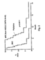
Classifications
-
- A—HUMAN NECESSITIES
- A61—MEDICAL OR VETERINARY SCIENCE; HYGIENE
- A61K—PREPARATIONS FOR MEDICAL, DENTAL OR TOILETRY PURPOSES
- A61K38/00—Medicinal preparations containing peptides
- A61K38/16—Peptides having more than 20 amino acids; Gastrins; Somatostatins; Melanotropins; Derivatives thereof
- A61K38/164—Peptides having more than 20 amino acids; Gastrins; Somatostatins; Melanotropins; Derivatives thereof from bacteria
-
- A—HUMAN NECESSITIES
- A61—MEDICAL OR VETERINARY SCIENCE; HYGIENE
- A61K—PREPARATIONS FOR MEDICAL, DENTAL OR TOILETRY PURPOSES
- A61K39/00—Medicinal preparations containing antigens or antibodies
- A61K39/02—Bacterial antigens
- A61K39/085—Staphylococcus
-
- A—HUMAN NECESSITIES
- A61—MEDICAL OR VETERINARY SCIENCE; HYGIENE
- A61K—PREPARATIONS FOR MEDICAL, DENTAL OR TOILETRY PURPOSES
- A61K40/00—Cellular immunotherapy
- A61K40/10—Cellular immunotherapy characterised by the cell type used
-
- A—HUMAN NECESSITIES
- A61—MEDICAL OR VETERINARY SCIENCE; HYGIENE
- A61K—PREPARATIONS FOR MEDICAL, DENTAL OR TOILETRY PURPOSES
- A61K40/00—Cellular immunotherapy
- A61K40/10—Cellular immunotherapy characterised by the cell type used
- A61K40/11—T-cells, e.g. tumour infiltrating lymphocytes [TIL] or regulatory T [Treg] cells; Lymphokine-activated killer [LAK] cells
-
- A—HUMAN NECESSITIES
- A61—MEDICAL OR VETERINARY SCIENCE; HYGIENE
- A61K—PREPARATIONS FOR MEDICAL, DENTAL OR TOILETRY PURPOSES
- A61K40/00—Cellular immunotherapy
- A61K40/10—Cellular immunotherapy characterised by the cell type used
- A61K40/15—Natural-killer [NK] cells; Natural-killer T [NKT] cells
-
- A—HUMAN NECESSITIES
- A61—MEDICAL OR VETERINARY SCIENCE; HYGIENE
- A61K—PREPARATIONS FOR MEDICAL, DENTAL OR TOILETRY PURPOSES
- A61K40/00—Cellular immunotherapy
- A61K40/10—Cellular immunotherapy characterised by the cell type used
- A61K40/19—Dendritic cells
-
- A—HUMAN NECESSITIES
- A61—MEDICAL OR VETERINARY SCIENCE; HYGIENE
- A61K—PREPARATIONS FOR MEDICAL, DENTAL OR TOILETRY PURPOSES
- A61K40/00—Cellular immunotherapy
- A61K40/20—Cellular immunotherapy characterised by the effect or the function of the cells
- A61K40/24—Antigen-presenting cells [APC]
-
- A—HUMAN NECESSITIES
- A61—MEDICAL OR VETERINARY SCIENCE; HYGIENE
- A61K—PREPARATIONS FOR MEDICAL, DENTAL OR TOILETRY PURPOSES
- A61K40/00—Cellular immunotherapy
- A61K40/30—Cellular immunotherapy characterised by the recombinant expression of specific molecules in the cells of the immune system
- A61K40/32—T-cell receptors [TCR]
-
- A—HUMAN NECESSITIES
- A61—MEDICAL OR VETERINARY SCIENCE; HYGIENE
- A61K—PREPARATIONS FOR MEDICAL, DENTAL OR TOILETRY PURPOSES
- A61K40/00—Cellular immunotherapy
- A61K40/40—Cellular immunotherapy characterised by antigens that are targeted or presented by cells of the immune system
- A61K40/41—Vertebrate antigens
- A61K40/42—Cancer antigens
- A61K40/4202—Receptors, cell surface antigens or cell surface determinants
- A61K40/4223—CD44 not IgG
-
- A—HUMAN NECESSITIES
- A61—MEDICAL OR VETERINARY SCIENCE; HYGIENE
- A61K—PREPARATIONS FOR MEDICAL, DENTAL OR TOILETRY PURPOSES
- A61K40/00—Cellular immunotherapy
- A61K40/40—Cellular immunotherapy characterised by antigens that are targeted or presented by cells of the immune system
- A61K40/41—Vertebrate antigens
- A61K40/42—Cancer antigens
- A61K40/4238—Regulators of development
- A61K40/424—Apoptosis related proteins, e.g. survivin or livin
- A61K40/4241—Apoptosis related proteins, e.g. survivin or livin p53
-
- A—HUMAN NECESSITIES
- A61—MEDICAL OR VETERINARY SCIENCE; HYGIENE
- A61K—PREPARATIONS FOR MEDICAL, DENTAL OR TOILETRY PURPOSES
- A61K40/00—Cellular immunotherapy
- A61K40/40—Cellular immunotherapy characterised by antigens that are targeted or presented by cells of the immune system
- A61K40/41—Vertebrate antigens
- A61K40/42—Cancer antigens
- A61K40/4256—Tumor associated carbohydrates
- A61K40/4257—Mucins, e.g. MUC-1
-
- A—HUMAN NECESSITIES
- A61—MEDICAL OR VETERINARY SCIENCE; HYGIENE
- A61K—PREPARATIONS FOR MEDICAL, DENTAL OR TOILETRY PURPOSES
- A61K40/00—Cellular immunotherapy
- A61K40/40—Cellular immunotherapy characterised by antigens that are targeted or presented by cells of the immune system
- A61K40/41—Vertebrate antigens
- A61K40/42—Cancer antigens
- A61K40/4271—Melanoma antigens
-
- A—HUMAN NECESSITIES
- A61—MEDICAL OR VETERINARY SCIENCE; HYGIENE
- A61K—PREPARATIONS FOR MEDICAL, DENTAL OR TOILETRY PURPOSES
- A61K40/00—Cellular immunotherapy
- A61K40/40—Cellular immunotherapy characterised by antigens that are targeted or presented by cells of the immune system
- A61K40/41—Vertebrate antigens
- A61K40/42—Cancer antigens
- A61K40/4271—Melanoma antigens
- A61K40/4272—Melan-A/MART
-
- A—HUMAN NECESSITIES
- A61—MEDICAL OR VETERINARY SCIENCE; HYGIENE
- A61K—PREPARATIONS FOR MEDICAL, DENTAL OR TOILETRY PURPOSES
- A61K40/00—Cellular immunotherapy
- A61K40/40—Cellular immunotherapy characterised by antigens that are targeted or presented by cells of the immune system
- A61K40/45—Bacterial antigens
-
- A—HUMAN NECESSITIES
- A61—MEDICAL OR VETERINARY SCIENCE; HYGIENE
- A61K—PREPARATIONS FOR MEDICAL, DENTAL OR TOILETRY PURPOSES
- A61K40/00—Cellular immunotherapy
- A61K40/40—Cellular immunotherapy characterised by antigens that are targeted or presented by cells of the immune system
- A61K40/46—Viral antigens
-
- A—HUMAN NECESSITIES
- A61—MEDICAL OR VETERINARY SCIENCE; HYGIENE
- A61K—PREPARATIONS FOR MEDICAL, DENTAL OR TOILETRY PURPOSES
- A61K45/00—Medicinal preparations containing active ingredients not provided for in groups A61K31/00 - A61K41/00
- A61K45/06—Mixtures of active ingredients without chemical characterisation, e.g. antiphlogistics and cardiaca
-
- A—HUMAN NECESSITIES
- A61—MEDICAL OR VETERINARY SCIENCE; HYGIENE
- A61K—PREPARATIONS FOR MEDICAL, DENTAL OR TOILETRY PURPOSES
- A61K48/00—Medicinal preparations containing genetic material which is inserted into cells of the living body to treat genetic diseases; Gene therapy
-
- A—HUMAN NECESSITIES
- A61—MEDICAL OR VETERINARY SCIENCE; HYGIENE
- A61K—PREPARATIONS FOR MEDICAL, DENTAL OR TOILETRY PURPOSES
- A61K48/00—Medicinal preparations containing genetic material which is inserted into cells of the living body to treat genetic diseases; Gene therapy
- A61K48/005—Medicinal preparations containing genetic material which is inserted into cells of the living body to treat genetic diseases; Gene therapy characterised by an aspect of the 'active' part of the composition delivered, i.e. the nucleic acid delivered
-
- A—HUMAN NECESSITIES
- A61—MEDICAL OR VETERINARY SCIENCE; HYGIENE
- A61P—SPECIFIC THERAPEUTIC ACTIVITY OF CHEMICAL COMPOUNDS OR MEDICINAL PREPARATIONS
- A61P35/00—Antineoplastic agents
-
- A—HUMAN NECESSITIES
- A61—MEDICAL OR VETERINARY SCIENCE; HYGIENE
- A61P—SPECIFIC THERAPEUTIC ACTIVITY OF CHEMICAL COMPOUNDS OR MEDICINAL PREPARATIONS
- A61P37/00—Drugs for immunological or allergic disorders
- A61P37/02—Immunomodulators
- A61P37/04—Immunostimulants
-
- A—HUMAN NECESSITIES
- A61—MEDICAL OR VETERINARY SCIENCE; HYGIENE
- A61K—PREPARATIONS FOR MEDICAL, DENTAL OR TOILETRY PURPOSES
- A61K2239/00—Indexing codes associated with cellular immunotherapy of group A61K40/00
- A61K2239/31—Indexing codes associated with cellular immunotherapy of group A61K40/00 characterized by the route of administration
-
- A—HUMAN NECESSITIES
- A61—MEDICAL OR VETERINARY SCIENCE; HYGIENE
- A61K—PREPARATIONS FOR MEDICAL, DENTAL OR TOILETRY PURPOSES
- A61K2239/00—Indexing codes associated with cellular immunotherapy of group A61K40/00
- A61K2239/38—Indexing codes associated with cellular immunotherapy of group A61K40/00 characterised by the dose, timing or administration schedule
-
- A—HUMAN NECESSITIES
- A61—MEDICAL OR VETERINARY SCIENCE; HYGIENE
- A61K—PREPARATIONS FOR MEDICAL, DENTAL OR TOILETRY PURPOSES
- A61K2239/00—Indexing codes associated with cellular immunotherapy of group A61K40/00
- A61K2239/46—Indexing codes associated with cellular immunotherapy of group A61K40/00 characterised by the cancer treated
- A61K2239/57—Skin; melanoma
-
- C—CHEMISTRY; METALLURGY
- C12—BIOCHEMISTRY; BEER; SPIRITS; WINE; VINEGAR; MICROBIOLOGY; ENZYMOLOGY; MUTATION OR GENETIC ENGINEERING
- C12N—MICROORGANISMS OR ENZYMES; COMPOSITIONS THEREOF; PROPAGATING, PRESERVING, OR MAINTAINING MICROORGANISMS; MUTATION OR GENETIC ENGINEERING; CULTURE MEDIA
- C12N15/00—Mutation or genetic engineering; DNA or RNA concerning genetic engineering, vectors, e.g. plasmids, or their isolation, preparation or purification; Use of hosts therefor
- C12N15/09—Recombinant DNA-technology
- C12N15/63—Introduction of foreign genetic material using vectors; Vectors; Use of hosts therefor; Regulation of expression
- C12N15/79—Vectors or expression systems specially adapted for eukaryotic hosts
- C12N15/85—Vectors or expression systems specially adapted for eukaryotic hosts for animal cells
- C12N15/86—Viral vectors
-
- C—CHEMISTRY; METALLURGY
- C12—BIOCHEMISTRY; BEER; SPIRITS; WINE; VINEGAR; MICROBIOLOGY; ENZYMOLOGY; MUTATION OR GENETIC ENGINEERING
- C12N—MICROORGANISMS OR ENZYMES; COMPOSITIONS THEREOF; PROPAGATING, PRESERVING, OR MAINTAINING MICROORGANISMS; MUTATION OR GENETIC ENGINEERING; CULTURE MEDIA
- C12N15/00—Mutation or genetic engineering; DNA or RNA concerning genetic engineering, vectors, e.g. plasmids, or their isolation, preparation or purification; Use of hosts therefor
- C12N15/09—Recombinant DNA-technology
- C12N15/63—Introduction of foreign genetic material using vectors; Vectors; Use of hosts therefor; Regulation of expression
- C12N15/79—Vectors or expression systems specially adapted for eukaryotic hosts
- C12N15/85—Vectors or expression systems specially adapted for eukaryotic hosts for animal cells
- C12N15/86—Viral vectors
- C12N15/863—Poxviral vectors, e.g. entomopoxvirus
- C12N15/8636—Vaccina virus vectors
Definitions
- the invention in the fields of immunology and medicine is directed to a method for treating a category of neoplastic diseases that are manifest in sheaths surrounding organs (intrathecal) by administering tumoricidal superantigens such as bacterial enterotoxins and various biologically active derivatives thereof.
- Staphylococcal enterotoxins are representative of a family of proteins known as “superantigens” (SAg) - the most powerful T lymphocyte mitogens known. They can activate between about 5 and about 30% or the total T cell population compared to the activation of 0.01% or fewer T cells by conventional antigens. Moreover, these enterotoxins elicit strong polyclonal proliferative responses at concentrations about 10 3 -fold lower than other T cell mitogens. The most potent SE on a per weight basis, Staphylococcal enterotoxin A (SEA), stimulates human T cell proliferation (measured as DNA synthesis) at concentrations of as low as 10 -13 10 -16 M .
- SAg superantigens
- SAg-activated T cells produce a variety of cytokines, including interferon-y (IFN ⁇ ), various interleukins and tumor necrosis factor- ⁇ (TNF ⁇ ) ( Dohlsten et al., Int. J. Cancer 54:482-488 (1993 ).
- IFN ⁇ interferon-y
- TNF ⁇ tumor necrosis factor- ⁇
- SAgs also stimulate other cell populations involved in innate and adaptive immunity and contribute to anti-tumor immunity. For example, SE's engage the variable (V) region of the T cell receptor (TCR) chain on the exposed face of the pleated sheet and the sides of the MHC class II molecule ( Kiting BL et al., Adv Immunol. 1993;54:99-166 ). SAgs augment T H 1 cytokine response by CD4+ cells while also activating cells of the NK, NKT and ⁇ / ⁇ T cell lineages. Cytotoxic action of NK cells is augmented by the IFN ⁇ produced by SAg activated T cells ( Morita et al., Immunity 14:331-44. (2001 ) D'Orazio JA et al., J Immunol. 154:1014-23 (1995 ).
- SAgs induce tumor killing in vivo when given alone or when conjugated to tumor-specific antibodies ( Terman U.S. Patent Ser. No. 6,221,351 ; Dohlsten U.S. Patent Ser No. 5,858,363 ). They are also effective when employed ex vivo to induce the generation of tumor sensitized T cells that are then administered in the "adoptive therapy" of (e.g., MCA 205/207) tumors ( Shu et al. J Immunol. 152: 1277-1288 (1994 ). SAg-transfected tumor cells can reduce metastatic disease in an established murine mammary carcinoma model ( Pulaski et al., Cancer Res. 60: 2710-5 (2000 ).
- SEs In addition to these biological activities, the SE's share common physicochemical properties. They are heat stable, trypsin-resistant, and soluble in water and salt solutions, have similar sedimentation coefficients, diffusion constants, partial specific volumes, isoelectric points, and extinction coefficients.
- SEB Staphylococcal enterotoxin B
- SEC Staphylococcal enterotoxin C
- SED Staphylococcal enterotoxin D
- SEE Staphylococcal enterotoxin E
- SE is a single polypeptide chain of about 30 kDa. All SEs have a characteristic disulfide loop near the middle of the chain.
- SEA is a flat monomer consisting or 233 amino acids divided into two domains: domain I comprising residues 31-116 and domain II comprising residues 117-233 together with the amino tail of residues 1-30.
- the biologically active regions of the proteins are evolutionarily conserved and show a relatively higher degree of sequence homology/similarity.
- One region of striking amino acid sequence homology between SEA, SEB, SEC, SED, and SEE is located immediately on the C-terminal side of Cys-106 (in SEA). This conserved region is thought to be responsible for T cell activation.
- a second conserved homology region at about residue 147, is believed to be responsible for emetic activity.
- This emesis-inducing region can be deleted from SE's through genetic engineering; such modified SE's are also useful therapeutics in accordance with this invention.
- SEs Sequence analysis of SEs and comparison with other bacterial toxins revealed SEA, SEB, SEC, SED, Staphylococcal toxic shock-associated toxin (TSST-1, also known as SEF), and the Streptococcal pyrogenic exotoxins (SpE's) share considerable nucleic acid and amino acid sequence similarity ( Betley et al., J. Bacteriol. 170: 34-41 (1988 )).
- SEF Staphylococcal toxic shock-associated toxin
- SpE's Streptococcal pyrogenic exotoxins
- SEs bind to MHC class II molecules and TCRs in a manner quite distinct from conventional antigens. SEs engage the V region of the TCR ⁇ chain (V ⁇ region) on an exposed face of the ⁇ pleated sheet. SEs engage the "sides" of MHC class II molecule rather than engaging the groove as do conventional antigens. In contrast to SEB and the SEC, which bind only to the MHC class II ⁇ chain, SEA, as well as SEE and SED, also interact with the MHC class II ⁇ chain in a zinc-dependent manner ( Fraser JD et al., Proc. Natl. Acad. Sci. 89:5507-11 1 (1992 )).
- T cell recognition of SAgs, such as SEs, via the TCR V ⁇ region is independent of other TCR components and diversity elements.
- Single amino acid positions and regions important for SAg-TCR interactions have been defined. These residues are located in the vicinity of the shallow cavity formed between the two SE domains. ( Lavoie PM et al., Immunol. Rev. 168: 257-269 (1999 ). Substitution of amino acid residue Asn23 in SEB by Ala has demonstrated the importance of this position in SEB/TCR interactions. This particular residue is conserved among all of the SE's and may constitute a common anchor position for SE interaction with TCR V ⁇ structures.
- Amino acid residues in positions 60-64 of SEA contribute to the TCR interaction as do the Cys residues forming the intramolecular disulfide bridge ( Kappler J et al., J Exp. Med. 175 387-96 (1992 )).
- the key points of interaction in the TCR V ⁇ region are located in the CDR1, CDR2 and HRV4 regions of the TCR V ⁇ 3 chain ( Deringer JR et a/., Mol. Microbiol. 22: 523-534 (1996 )).
- multiple and highly variable parts of the V ⁇ region contribute to the formation of the TCRs SE binding site.
- SAgs Mycoplasmal, viral, and other bacterial proteins.
- examples include Yersinia pseudotuberculosis mitogenic protein ("YPM"), and Clostridium perfringens toxin A. All SAgs activate T cells without a requirement for conventional antigen processing, and the responding T cells do not respond in a conventional MHC restricted manner. As noted, SAgs bind to and evoke responses from all T cells expressing certain TCR V ⁇ gene products independently of other TCR structures. CD4 CD8 TCR ⁇ / ⁇ T cells and ⁇ / ⁇ T cells all respond to SAgs by proliferation, production of T H 1 cytokines and generation of cytotoxic activity.
- SEs Native SEs are known to induce anti-tumor effects.
- Administration of SEB produced antitumor effects against established tumors in two animal species, rabbits and mice, with tumors of five different histologic types: rabbit VX-2 carcinoma ( Terman et al., U.S. Patent 6,126,945 ; Terman, U.S. Patent 6,340,461 ), murine CL 62 melanomas ( Penna C. et al., Cancer Res. 54: 2738-2743 (1994 )), murine A/20 lymphoma (Kalland T. Declaration in U.S. Ser. No. 07 / 689 / 799 (1992)), murine PRO4L fibrosarcoma ( Newell et al., Proc Natl. Acad.
- SEB induced objective anti-tumor effects at primary and metastatic sites.
- SEB was used ex vivo to stimulate a population of T cells pre-exposed to tumor, which, upon re-infusion into host animals with established pulmonary metastases, induced a substantial reduction of metastases.
- SEB activated T cell anti-tumor effect was specific for the immunizing tumor; the SEB stimulated T cells produced IFN ⁇ which was thought to be an important mediator of the anti-tumor effect ( Shu S et al., J. Immunol. 152: 1277-88 (1994 )).
- Fusion polypeptides comprising SEA fused to a tumor specific monoclonal antibody (mAb), designated "SEA-mAb,” induced tumoricidal responses in the murine B16 melanoma model ( Dohlsten M et al., Proc Natl Acad Sci 91:8945-9 (1994 ); Dohlsten M et a/., Proc. Natl. Acad. Sci. 88:9287-91(1991 ).
- mAb tumor specific monoclonal antibody
- the appearance of tumors in sheaths often results in production and accumulation of large volumes of fluid in the organs' sheath. Examples include (1) pleural effusion due to fluid in the pleural sheath surrounding the lung, (2) ascites originating from fluid accumulating in the peritoneal membrane and (3) cerebral edema due to metastatic carcinomatosis of the meninges. Such effusions and fluid accumulations generally develop at an advanced stage of the disease. Malignant pleural effusion (“MPE”) is the prototype of this condition. In the United States and Western Europe, 300,000 new cases of malignant pleural effusion are diagnosed annually ( Antony VB et al., Eur. Respire. J. 18:402-419 (2001 ).
- Malignant ascites is associated with 30-50% of ovarian tumors. Endometrial, breast, colonic, gastric and pancreatic carcinomas make up more than 80% or the tumors associated with intra-abdominal seeding of tumor cells and ascites formation. Ascites may be the presenting manifestation in 4-69% of cases.
- the major therapies for MPE include talc poudrage, talc slurry, doxycycline and bleomycin instillation ( Veena et al. Am J. Crit. Care Med. 162: 1987-2001 (2000 )). These therapies require 3-12 days of hospitalization with EKG and oximetry monitoring. A chest tube is inserted, and the therapeutic agent is infused and allowed to distribute over the pleural membranes. The chest tube is then connected to closed negative-pressure water seal drainage until pleural fluid volume drops below 100ml/24 hours. Respiratory therapy is usually given at least once daily.
- Talc poudrage requires the use of operating room and general anesthesia for thoracostomy and talc insufflation, followed by recovery room observation. Talc induces respiratory complications in up to 33% of patients and acute respiratory distress and hypoxemia in 10 % of patients. Response rates to bleomycin and doxycycline range between 50% and 70%, respectively and both require continuous chest tube drainage until the output is below 100 ml/24 hours. Indwelling pleural catheters for drainage and/or injection of a pleurodesis agent are an additional option (7,8); however, the catheter requires surgical placement followed by intermittent drainage of effusion fluid at home by the patient or a caregiver.
- Intrapleurally administered agents or modalities that include (a) chemotherapeutic agents such as Cisplatin, Cytarabine, Doxorubicin fluorouracil, etoposide, and mitomycin C, (b) radiation and (c) biotherapeutic agents such as IL-2, various interferons, and bacterially derived immunostimulatory agents such as Corynebacterium parvum have been ineffecftive against MPEs. Thoracentesis or chest tube drainage alone results in recurrence rates of 98% and 85% respectively within 30 days. Intraperitoneal cisplatin and etoposide has produced a complete response rate of 30% of malignant ascites.
- chemotherapeutic agents such as Cisplatin, Cytarabine, Doxorubicin fluorouracil, etoposide, and mitomycin C
- biotherapeutic agents such as IL-2, various interferons, and bacterially derived immunostimulatory agents such as Cor
- the present invention overcomes these deficiencies in the treatment of MPE by providing a new therapeutic approach to this manifestation of cancer. Unlike existing therapies, The present invention is carried out entirely in an outpatient setting and requires no hospitalization at a cost several hundred percent below that of existing therapy. Major costs of the other therapies originating from hospitalization, chest tube insertion, operating and recovery room expense, respiratory therapy and in-hospital chest tube drainage, are eliminated
- intratumoral delivery of a SAg would not require alteration of the native molecule and, as conceived by the present inventor, the presence of natural antibodies throughout the body can actually assist intratumorally-administered SAgs in evoking a tumoricidal response.
- the present invention provides a method for treating malignant pleural effusion, ascites, pericardial effusion and meningeal carcinomatosis by "intrathecal” (defined below) administration of an effective amount of a SAg into the pleural space, peritoneum, pericardium, and subarachnoid space.
- the present invention contemplates the use of any SAg, including but not limited to staphylococcal enterotoxins ("SE") A, B, C, D, E, F, G H, I, J, K, L, M, streptococcal pyrogenic exotoxins (SpE's), Yersinia pseudotuberculosis SAg, Mycoplasma arthritides SAg, and C. perfringens exotoxin, administered by injection, infusion or instillation directly into a cavity or space (thecum) surrounding an organ or body region in which a tumor is present or is causing fluid accumulation.
- SE staphylococcal enterotoxins
- Such spaces include the pleural space, peritoneum, subarachnoid space or dural space, or pericardial space.
- the generic term for administration into a sheath encasing an organ is termed "intrathecal, " defined in Dorland's Medical Dictionary 29th Edition, WB Saunders (2000 ) and Stedman's Medical Dictionary, 27th Edition, Lippincott, Williams & Wilkins (2000 ) as meaning "within a sheath.” As used herein, this term is intended to be broader than a more commonly used definition which is limited to intracranial spaces.
- SAgs in native form or conjugated to a tumor targeting agent such as monoclonal antibodies or their fragments
- mAbs monoclonal antibodies or their fragments
- SAgs or SAg conjugated to tumor specific antibodies do not reach their targets in effective concentrations for two reasons.
- the SAgs are neutralized rapidly by "natural" neutralizing SAg-specific antibodies.
- Second, SAg-mAb fusion proteins bind to cells present in the circulation that express MHC class II proteins.
- One approach to overcoming these obstacles was to mutate the SAg to reduce its affinity for MHC class II. This has met with only partial success (Hansson et al supra).
- the present invention obviates this obstacle to a large extent by administering a SAg (including a fragment, homologue or fusion partner), intrathecally, into sheaths encasing the organs which themselves are seeded with tumor or directly into tumor site(s).
- a SAg including a fragment, homologue or fusion partner
- any pre-existing or induced anti-SAg antibodies may actually assist the intrathecally or intratumorally administered SAg in promoting tumor killing by binding to the SAg after it has localized to tumor cell surfaces.
- compositions of SAg or SAg homologues consisting of amino acid substitution and deletion variants (mutants), additions (e.g., fusion proteins) and fragments with Z >10 when the sequence is compared to a native SAg using the FASTA/FASTP programs and Monte Carlo analysis.
- These composition are injected, instilled or infused intrapleurally or intraperitoneally into a patient with malignant pleural effusions and/or ascites, respectively or intratumorally into tumor site(s) and induce a tumoricidal response.
- the SAg composition is preferably administered after partial or complete drainage of the fluid from the sheath as for example in pleural effusions via thoracentesis and ascites via paracentesis.
- the SAg composition may also be administered directly into an undrained space containing the effusion, ascites and/or carcinomatosis.
- the invention also contemplates the use of the nucleic acid counterparts of the native SAgs and homologues as useful for the same indications as the polypeptide form of the molecule.
- a biologically active fragment or homologue may be fused to another protein such as (I) a tumor specific antibody, or an antigen binding fragment of such an antibody, such as an F(ab') 2 , Fv or Fd fragment, which antibody is specific for an epitope expressed on the tumor or (b) a receptor ligand specific for any receptors selectively or preferentially expressed on tumor cells.
- the fusion partner can also be a powerful costimulant such as OX-40 or 4-1BB1 which enhances the T cells proliferative response to the SAg or a "Coaguligand" which promotes coagulation in the tumor vasculature.
- the SAg composition is administered once every 3 to 10 days, preferably once weekly, and this schedule is continued until there is no re-accumulation of the effusion or ascites or reduction in the size of the tumor mass being injected. Three such treatments may suffice for intrathecal administration although this is an average; the number of treatments may varying from 1-6 or even higher.
- intrathecal administration the SAg composition is preferentially administered immediately after removal of pleural fluid via thoracentesis. Unlike the other therapies for malignant pleural effusions, the present method is carried out entirely in an outpatient setting and requires no hospitalization, chest tube insertion, use of the operating room or recovery room, respiratory therapy or in-hospital chest tube drainage.
- the SAgs disclosed herein are prepared by either biochemical isolation, or, preferably by recombinant methods.
- the following SAgs, including their sequences and biological activities have been known for a number of years. Studies of these SAgs are found throughout the biomedical literature.
- biochemical and recombinant preparation of these SAgs see the following references: Borst, DW et al., Infect. Immun. 61: 5421-5425 (1993 ); Couch, JL et al., J. Bacteriol. 170: 2954-2960 (1988 ); Jones, CL et al., J. Bacteriol. 166: 29-33 (1986 ); Bayles KW et al., J. Bacteriol.
- SAgs Staphylococcal enterotoxin A (SEA), Staphylococcal enterotoxin B (SEB), Staphylococcal enterotoxin C (SEC - actually three different proteins, SEC1, SEC2 and SEC3) ), Staphylococcal enterotoxin D (SED), Staphylococcal enterotoxin E (SEE) and toxic shock syndrome toxin-1 (TSST-1) ( U.S. Pat 6,126,945 and U.S. provisional patent application 60/389,366 filed June 15, 2002 , and the references cited therein).
- the amino acids sequences of the above group of native (wild-type) SAgs is provided below:
- Staphylococcal Enterotoxins SEG, SEH .
- SEG-SEM New Staphylococcal enterotoxins G, H, I, J, K, L and M
- SEG and SEH of this group and other enterotoxins including SPEA, SPEC, SPEG, SPEH, SME-Z, SME-Z 2 utilize zinc as part of high affinity MHC class II receptor. Amino acid substitution(s) at the high-affinity, zinc-dependent class II binding site are created to reduce their affinity for MHC class II molecules.
- Jarraud S et a / ., 2001, supra discloses methods used to identify and characterize SEG-SEM, and for cloning and recombinant expression of these proteins ; These investigators have used a number of TCR-specific mAbs (V ⁇ specificity indicated in brackets) for flow cytometric analysis: E2.2E7.2 (V ⁇ 2), LE89 (V ⁇ 3), IMMU157 (V ⁇ 5.1), 3D11 (V ⁇ 5.3), CRI304.3 (V ⁇ 6.2), 3G5D15 (V ⁇ 7), 56C5.2 (V ⁇ 8.1/8.2), FIN9 (V ⁇ 9), C21 (V ⁇ 1), S511 (V ⁇ 12), IMM1222 (V ⁇ 13.1), JIJ74 (V ⁇ 13.6), CAS1.1.13 (V ⁇ 14), Tamayal.2 (V ⁇ 16), E17.5F3 (V ⁇ 17), ⁇ A62.6 (V ⁇ 18), ELL1.4 (V ⁇ 20), IG125 (V ⁇ 21.3), IMMU546
- Jarraud S et al., 2001, supra indicates that the seven genes and pseudogenes composing the egc (enterotoxin gene cluster) operon are co-transcribed. The association of related co-transcribed genes suggested that the resulting peptides might have complementary effects on the host's immune response.
- gene recombination created new SE variants differing by their SAg activity profiles. Purified recombinant SEL, SEM, SEI, SEK, and SEGL29P (a mutant of SEL) were expressed in E . coli and analyzed. Recombinant SEL SEM, SEI, and SEK consistently induced selective expansion of distinct subpopulations ofT cells expressing particular V ⁇ genes. By contrast.
- SEGL29P failed to trigger expansion of any of 23 V ⁇ subsets, and the L29P mutation accounted for the complete loss of SAg activity (although this mutation did not induce a major conformational change). It is believed that this substitution mutation located at a position crucial for proper SAg/MHC II interaction.
- amino acid sequences of SEG-SEM are shown below
- the SpE's SPEA, SPEB, SPEC, SPEG, SPEH, SME-Z, SNE-Z 2 and SSA are SAgs induce tumoricidal effects.
- SPEA, SPEB, SPEC have been known for some time and their structures and biological activities described in numerous publications.
- SPEG, SPEH, and SPEJ genes were identified from the Streptococcus pyogenes M1 genomic database and described in detail in Proft, T et al., J. Exp. Med. 189: 89-101 (1999 ) which also describes SMEZ, SMEZ-2. This document also describes the cloning and expression of the genes encoding these proteins.
- the srnez-2 gene was isolated from the S. pyogenes strain 2035, based on sequence homology to the streptococcal mitogenic exotoxin z (smez) gene.
- SMEZ-2, SPE-G, and SPE-J are most closely related to SMEZ and SPEC, whereas SPEH is most similar to the SEs than to any other streptococcal toxin.
- SMEZ-2 appears to be the most potent SAg discovered thus far.
- Binding to a human B-lymphoblastoid line was shown to be zinc dependent with high binding affinity of 15-65 nM.
- Analysis of competition for binding between toxins of this group revealed overlapping but discrete binding to subsets of class II molecules in the hierarchical order (SMEZ, SPE-C) SMEZ-2 SPE-H> SPE-G.
- the most common targets for these SAgs were human V ⁇ 2.1- and V ⁇ 4-expressing T cells.
- SPEA can be purified from cultures of S. pyogenes as described by Kline et al., Infect. Immun. 64:861-869 (1996 ). Plasmids that include the speal gene which encode SPEA, and the expression and purification of recombinant SPEA ("rSPEA") are described by Kline et al., supra. The native SPEA sequence is shown below:
- SPFB Streptococcus Pyrogenic Exotoxin B
- SSA is an ⁇ 28-kDa SAg protein isolated from culture supernatants as described by Mollick J et al., J. Clin. Invest. 92: 710-719 (1993 ) and Reda K et al., Infect. Immun. 62: 1867-1874 (1994 ).
- SSA stimulates proliferation of human T cells bearing V ⁇ 1, V ⁇ 3, V ⁇ 5.2, and V ⁇ 15 in an MHC class II-dependent manner.
- the first 24 amino acid residues of SSA are be 62.5% identical to SEB, SBC1, and SEC3. Purification and cloning of SSA is described in Reda K et al., Infect. Immun. 62: 1867-1874 (1994 ).
- the native sequence of SSA Reda, K.B. et al., Infect. Immun. 64: 1161-1165 (1996 ) is shown below:
- Yersinia pseudotuberculosis Mitogen (Superantigen) (YPM)
- the set1 nucleotide sequences are deposited in the GenBank database under accession numbers AF094826 (set gene cluster fragment), AF188835 (NCTC 6571 set1 gene), AF188836 (FRI326 set1 gene), and AF188837 (NCTC 8325-4 set1 gene).
- Recombinant SET-1 protein stimulates production of the proinflammatory cytokines IL-1 ⁇ , IL-6, and TNF ⁇
- a preferred construct for intrathecal and intrapleural use comprises a SAg in native form.
- the preferred SAg is one to which humans do not make or make only marginal amounts neutralizing antibody fused recombinantly or biochemically to a high affinity tumor specific antibody, Fab or single chain Fv.
- SAg epitopes in the conjugate which bind endogenous (to include preexisting) SAg specific antibodies are deleted and/or substituted by alanine or amino acid sequences to which the host does not have preexisting antibodies.
- a dominant epitope on SEB recognized by anti-SEB antibodies is the sequence 225-234 ( Nishi et al., J. Immunol.
- SEA epitope on SEA recognized by anti-SEA antibodies is the sequence 121-149 ( Hobieka et al., Biochem. Biophys. Res. Comm. 223: 565-571 (1996 ).
- SAgs such as Y pseudotuberculosis or C. perfringens toxin A or to which humans do not have preexisting antibodies are used.
- Y. pseudotuberculosis SAg has, in addition, a natural RGD domain which has tumor-localizing properties.
- the present invention contemplates, in addition to native SAgs, the use of homologues of native SAgs that have the requisite biological activity to be useful in accordance with the invention.
- the present invention encompasses functional derivatives, among which homologues are preferred.
- Y. pseudotuberculosis SAg YPM, C. perfringens toxin A, M arthritides SAgs are included herein.
- a functional derivative is meant a “fragment,” “variant,” “mutant, “ “homologue,” “analogue,” or “chemical derivative.
- Homologues include fusion proteins, chimeric proteins and conjugates that include a SAg portion fused to or conjugated to a fusion partner polypeptide or peptide.
- a functional derivative retains at least a portion of the biological activity of the native protein which permits its utility in accordance with the present invention.
- Such biological activity includes stimulation of T cell proliferation and/or cytokine secretion, stimulation of T cell-mediated cytotoxic activity, as a result of interactions of the SAg composition with T cells preferably via the TCR V ⁇ region.
- a “fragment” refers to any shorter peptide.
- a “variant” of refers to a molecule substantially similar to either the entire protein or a peptide fragment thereof. Variant peptides may be conveniently prepared by direct chemical synthesis of the variant peptide, using methods well-known in the art.
- a homologue refers to a natural protein, encoded by a DNA molecule from the same or a different species. Homologues, as used herein, typically share at least about 50% sequence similarity at the DNA level or at least about 18% sequence similarity at the amino acid level, with a native protein.
- an “analogue” refers to a non- natural molecule substantially similar to either the entire molecule or a fragment thereof
- a “chemical derivative” contains additional chemical moieties not normally a part of the peptide.
- Covalent modifications of the peptide are included within the scope of this invention. Such modifications may be introduced into the molecule by reacting targeted amino acid residues of the peptide with an organic derivatizing agent that is capable of reacting with selected side chains or terminal residues.
- a fusion protein comprises a native SAg, a fragment or a homologue fused by recombinant means to another polypeptide fusion partner, optionally including a spacer between the two sequences.
- Preferred fusion partners are antibodies, Fab fragments, single chain Fv fragments.
- Other fusion partners are any peptidic receptor ligand, cytokine, extracellular domain ("ECD") of a costimulatory molecule and the like.
- sequences and sequence relationships between two or more nucleic acids or polypeptides are used in the disclosure of sequences and sequence relationships between two or more nucleic acids or polypeptides: (a) “reference sequence”, (b) “comparison window”, (c) “sequence identity”, (d) “percentage of sequence identity”, and (e) “substantial identity”
- reference sequence is a defined sequence used as a basis for sequence comparison.
- a reference sequence may be a subset or the entirety of a specified sequence; for example, as a segment of a full-length cDNA or other polynucleotide sequence, or the complete cDNA or polynucleotide sequence. The same is the case for polypeptides and their amino acid sequences.
- comparison window includes reference to a contiguous and specified segment of a polynucleotide or amino acid sequence, wherein the sequence may be compared to a reference sequence and wherein the portion of the sequence in the comparison window may comprise additions or deletions ( i.e ., gaps) compared to the reference sequence (which does not comprise additions or deletions) for optimal alignment of the two sequences.
- the comparison window is at least 20 contiguous nucleotides or amino acids in length, and optionally can be 30, 40, 50,100, or longer.
- Cys residues are aligned.
- Computerized implementations of these algorithms include, but are not limited to: CLUSTAL in the PC/Gene program by Intelligenetics, Mountain View, California, GAP, BESTFIT, BLAST, FASTA, and TFASTA in the Wisconsin Genetics Software Package, Genetics Computer Group (GCG) (Madison, WI).
- the CLUSTAL program is described by Higgins et al., Gene 73:237-244 (1988 ); Higgins et al., CABIOS 5:151-153 (1989 ); Corpet et al., Nuc Acids Res 16:881-90 (1988 ); Huang et al., CABIOS 8:155-65 (1992 ), and Pearson et al., Methods in Molecular Biology 24:307-331 (1994 ).
- the BLAST family of programs which can be used for database similarity searches includes: NBLAST for nucleotide query sequences against database nucleotide sequences; XBLAST for nucleotide query sequences against database protein sequences; BLASTP for protein query sequences against database protein sequences; TBLASTN for protein query sequences against database nucleotide sequences; and TBLASTX for nucleotide query sequences against database nucleotide sequences. See, for example, Ausubel et al., eds., Current Protocols in Molecular Biology, Chapter 19, Greene Publishing and Wiley-Interscience, New York (1995 ) or most recent edition.
- BLAST searches assume that proteins can be modeled as random sequences. However, many real proteins comprise regions of nonrandom sequence which may include homopolymeric tracts, short-period repeats, or regions rich in particular amino acids. Alignment of such regions of "low-complexity" regions between unrelated proteins may be performed even though other regions are entirely dissimilar.
- a number of low-complexity filter programs are known that reduce such low-complexity alignments. For example, the SEG ( Wooten et al., Comput. Chem. 17:149-163 (1993 )) and XNU ( Claverie et al., Comput. Chem, 17:191-201 (1993 )) low-complexity filters can be employed alone or in combination.
- sequence identity or “identity” in the context of two nucleic acid or amino acid sequences refers to the residues in the two sequences which are the same when aligned for maximum correspondence over a specified comparison window. It is recognized that when using percentages of sequence identity for proteins, a residue position which is not identical often differs by a conservative amino acid substitution, where a substituting residue has similar chemical properties (e.g., charge, hydrophobicity, etc.) and therefore does not change the functional properties of the polypeptide.
- sequence similarity or “similarity” (combination of identity and differences that are conservative substitutions).
- Means for making this adjustment are well-known in the art. Typically this involves scoring a conservative substitution as a partial rather than as a full mismatch, thereby increasing the percentage sequence identity. Thus, for example, where an identical amino acid is given a score of "1" and a non-conservative substitution is given a score of "0"zero, a conservative substitution is given a score between 0 and 1. The scoring of conservative substitutions is calculated, e.g., according to the algorithm of Meyers et al., CABIOS 4:11-17 (1988 ) as implemented in the program PC/GENE (Intelligenetics, Mountain View, Calif., USA).
- percentage of sequence identity refers to a value determined by comparing two optimally aligned sequences over a comparison window, wherein the portion of the nucleotide or amino acid sequence in the comparison window may comprise additions or deletions (i.e., gaps) as compared to the reference sequence (which lacks such additions or deletions) for optimal alignment, such as by the GAP algorithm (supra). The percentage is calculated by determining the number of positions at which the identical nucleotide or amino acid residue occurs in both sequences to yield the number of matched positions, dividing that number by the total number of positions in the window of comparison and multiplying the result by 100, thereby calculating the percentage of sequence identity.
- substantially identical of two sequences means that a polynucleotide or polypeptide comprises a sequence that has at least 60%, preferably at least 70%, more preferably at least 80%, even more preferably at least 90%, and most preferably at least 95% sequence identity to a reference sequence using one of the alignment programs described herein using standard parameters. Values can be appropriately adjusted to determine corresponding identity of the proteins encoded by two nucleotide sequences by taking into account codon degeneracy, amino acid similarity, reading frame positioning, etc.
- nucleotide sequences are substantially identical if they hybridize to one other under stringent conditions. Because of the degeneracy of the genetic code, a number of different nucleotide codons may encode the same amino acid. Hence, two given DNA sequences could encode the same polypeptide but not hybridize under stringent conditions.
- Another indication that two nucleic acid sequences are substantially identical is that the polypeptide encoded by the first nucleic acid is immunologically cross reactive with the polypeptide encoded by the second nucleic acid.
- two peptide or polypeptide sequences are substantially identical if one is immunologically reactive with antibodies raised against the other. A first peptide is substantially identical to a second peptide, if they differ only by a conservative substitution.
- Peptides which are "substantially similar" share sequences as noted above except that nonidentical residue positions may differ by conservative substitutions.
- the Lipman-Pearson FASTA or FASTP program packages (Pearson, W.R. et. al., 1988, supra; Lipman, D.J. et al, Science 227:1435-1441 (1985 )) in any of its older or newer iterations may be used to determine sequence identity or homology of a given protein, preferably using the BLOSUM 50 or PAM 250 scoring matrix, gap penalties of -12 and -2 and the PIR or SwissPROT databases for comparison and analysis purposes. The results are expressed as z values or E 0 values.
- a more widely used and preferred methodology determines the percent identity of two amino acid sequences or of two nucleic acid sequences after optimal alignment as discussed above, e.g., using BLAST.
- a polypeptide being analyzed for its homology with native SAg is at least 20%, preferably at least 40%, more preferably at least 50%, even more preferably at least 60%, and even more preferably at least 70%, 80%, or 90% as long as the reference sequence.
- the amino acid residues (or nucleotides) at corresponding positions are then compared.
- Amino acid or nucleic acid "identity" is equivalent to amino acid or nucleic acid "homology”.
- the percent identity between two amino acid sequences is determined using the Needleman and Wunsch alignment algorithm (incorporated into the GAP program in the GCG software package (available at the URL www.gcg.com), using either a Blossom 62 matrix or a PAM250 matrix, and a gap weight of 16, 14, 12, 10, 8, 6, or 4 and a length weight of 1, 2, 3, 4, 5, or 6.
- the percent identity between the encoding nucleotide sequences is determined using the GAP program in the GCG software package (also available at above URL), using a NWSgapdna.CMP matrix and a gap weight of 40, 50, 60, 70, or 80 and a length weight of 1, 2, 3, 4, 5, or 6.
- the algorithm of Meyers et al., supra (incorporated into the ALIGN program, version 2.0), is implemented using a PAM120 weight residue table, a gap length penalty of 12 and a gap penalty of 4.
- the wild-type (or native) SAg-encoding nucleic acid sequence or the SAg protein sequence can further be used as a "query sequence" to search against a public database, for example, to identify other family members or related sequences.
- search can be performed using the NBLAST and XBLAST programs, supra (see Altschul et al. (1990) J. Mol. Biol. 215:403-10 ).
- Gapped BLAST can be utilized as described in Altschul et al. (1997, supra). Default parameters of XBLAST and NBLAST can be found at the NCBI website (www.ncbi.nlm.nih.gov)
- a preferred SAg homologue is one that has a z 10.
- a preferred SAg homologue for use according the present invention has at least about 20% identity or 25% similarity to a native SAg. Preferred identity or similarity is higher. More preferably, the amino acid sequence of a homologue is substantially identical or substantially similar to a native SAg sequence as those terms are defined above.
- substitution variants are those in which at least one amino acid residue in the peptide molecule, and preferably, only one, has been removed and a different residue inserted in its place.
- substitutions which may be made in the protein or peptide molecule of the present invention may be based on analysis of the frequencies of amino acid changes between a homologous protein of different species, such as those presented in Table 1-2 of Schulz et al. (supra) and Figure 3-9 of Creighton (supra). Based on such an analysis, conservative substitutions are defined herein as exchanges within one of the following five groups:
- substitutions that are less conservative, such as between, rather than within, the above five groups which will differ more significantly in their effect on maintaining (a) the structure of the peptide backbone in the area of the substitution, for example, as a sheet or helical conformation, (b) the charge or hydrophobicity of the molecule at the target site, or (c) the bulk of the side chain.
- substitutions are (a) substitution of gly and/or pro by another amino acid or deletion or insertion of Gly or Pro; (b) substitution of a hydrophilic residue, e.g., Ser or Thr, for (or by) a hydrophobic residue, e.g., Leu, Ile, Phe, Val or Ala; (c) substitution of a Cys residue for (or by) any other residue; (d) substitution of a residue having an electropositive side chain, e.g., Lys, Arg or His, for (or by) a residue having an electronegative charge, e.g., Glu or Asp; or (e) substitution of a residue having a bulky side chain, e.g., Phe, for (or by) a residue not having such a side chain, e.g., Gly.
- a hydrophilic residue e.g., Ser or Thr
- a hydrophobic residue e.g., Leu, Ile, Phe, Val or Al
- deletions and insertions, and substitutions according to the present invention are those which do not produce radical changes in the characteristics of the protein or peptide molecule.
- substitutions, deletion, or insertion in advance of doing so, one skilled in the art will appreciate that the effect will be evaluated by routine screening assays, for example direct or competitive immunoassay or biological assay of T cell function as described herein. Modifications of such proteins or peptide properties as redox or thermal stability, hydrophobicity, susceptibility to proteolytic degradation or the tendency to aggregate with carriers or into multimers are assessed by methods well known to the ordinarily skilled artisan.
- Covalent modifications of the SAg proteins or peptide fragments thereof, preferably of SEs or peptide fragments thereof, are included herein. Such modifications may be introduced into the molecule by reacting targeted amino acid residues of the protein or peptide with an organic derivatizing agent that is capable of reacting with selected side chains or terminal residues. This may be accomplished before or after polymerization.
- Cysteinyl residues most commonly are reacted with a-haloacetates (and corresponding amines), such as 2-chloroacetic acid or chloroacetamide, to give carboxymethyl or carboxyamidomethyl derivatives. Cysteinyl residues also are derivatized by reaction with bromotrifluoroacetone, ⁇ -bromo- (5-imidozoyl)propionic acid, chloroacetyl phosphate, N-allcylmaleimides, 3-nitro-2-pyridyldisulfide, methyl 2-pyridyl disulfide, p-chloromercuribenzoate, 2-chloromercuri-4- nitrophenol, or chloro-7-nitrobenzo-2-oxa-1,3-diazole.
- Histidyl residues are derivatized by reaction with diethylprocarbonate at pH 5.5-7.0 because this agent is relatively specific for the histidyl side chain.
- Para-bromophenacyl bromide also is useful; the reaction is preferably performed in 0.1 M sodium cacodylate at pH 6.0.
- Lysinyl and amino terminal residues are reacted with succinic or other carboxylic acid anhydrides. Derivatization with these agents has the effect of reversing the charge of the lysinyl residues.
- Other suitable reagents for derivatizing a -amino-containing residues include imidoesters such as methyl picolinimidate; pyridoxal phosphate; pyridoxal; chloroborohydride; trinitrobenzenesulfonic acid; 0-methylisourea; 2,4 pentanedione; and transaminase-catalyzed reaction with glyoxylate.
- Arginyl residues are modified by reaction with one or several conventional reagents, among them phenylglyoxal, 2,3- butanedione, 1,2-cyclohexanedione, and ninhydrin. Derivatization of arginine residues requires that the reaction be performed in alkaline conditions because of the high pK of the guanidine functional group. Furthermore, these reagents may react with the groups of lysine as well as the arginine epsilon-amino group.
- Carboxyl side groups (aspartyl or glutamyl) are selectively modified by reaction with carbodiimides as noted above. Aspartyl and glutamyl residues are converted to asparaginyl and glutaminyl residues by reaction with ammonium ions.
- Glutaminyl and asparaginyl residues may be deamidated to the corresponding glutamyl and aspartyl residues. Alternatively, these residues are deamidated under mildly acidic conditions. Either form of these residues falls within the scope of this invention.
- Such derivatized moieties may improve the solubility, absorption, biological half life, and the like.
- the moieties may alternatively eliminate or attenuate any undesirable side effect of the protein and the like.
- Moieties capable of mediating such effects are disclosed, for example, in Remington's Pharmaceutical Sciences, 16th ed., Mack Publishing Co., Easton, PA (1980 ).
- variants or homologues of native SAg proteins or peptides including mutants (substitution, deletion and addition types), fusion proteins (or conjugates) with other polypeptides, are characterized by substantial sequence homology to
- SE homologues e.g., 10 or, in another embodiment, having at least about 20% sequence identity or at least about 25% sequence similarity when compared to native SEs
- T lymphocyte mitogenicity SDCC or SADCC
- the appearance of tumors in sheaths often results in production and accumulation of large volumes of fluid in the organ's sheath.
- examples include (1) pleural effusion due to fluid in the pleural sheath surrounding the lung, (2) ascites originating from fluid accumulating in the peritoneal membrane and (3) cerebral edema due to metastatic carcinomatosis of the meninges.
- effusions and fluid accumulations generally develop at an advanced stage of the disease.
- the present invention contemplates the use of any SAg or SET in any form. This includes but is not limited to staphylococcal enterotoxins A, B, C, D, E, F, G H, I, J, K, L, M, SpE's, YPM, M. arthritides SAg, C. perfringens exotoxin for direct administration into cavities or spaces, e.g., peritoneum, thecal space, pericardial and pleural space containing tumor.
- Table 1 SE-Homologues Induce T Lymphocyte Mitogenesis SE Homologue a T Lymphocyte Mitogenic Response b (Ec 50 ) c Reference (SPECIES) SEA (native) SEA (native) 1 Abmhmsen et al., EMBO J.
- SEA-SEE 200-207 1 SEE-SEA (70-71) SEA-SEE (200-207) 1 HUMAN TSST-1 (native) 1 Kum et al., J. Infect Dis. 174: 1261-1270 (1996 ) HUMAN G31R 800 SEA-C215 mAb Fab usion Protein 1 Antonnson et a/., J. Immunol.
- SEE-C215 mAb Fab Fusion protein 10 SEE/AA-C215 mAb Fab Fusion protein 1 HUMAN SEE/A-C-C215 mAb Fab Fusion protein 10 SEEIA-F-C215 mAb Fab Fusion protein 10 SEE/A-H-C215 mAb Fab Fusion protein 10 SEA/E-BDEG-mAb Fab Fusion protein 2 SEE/A-AH-215mAb Fab fusion protein 2 LEGEND FOR TABLE 1 (above) a z values for homologues range from 16-136.
- PBMC peripheral blood mononuclear cells
- PBMC peripheral blood mononuclear cells
- mutant fetal calf serum
- antibiotics in wells of 96-well microplates in 200 ⁇ l volumes.
- enriched or purified T lymphocytes from these populations were tested.
- Between 0.2 ⁇ 10 5 and 8 ⁇ 10 5 cells/well were used.
- Incubation was at 37°C in humidified air/95% CO 2 for periods of between 66 hours and 84 hours (depending on whether unfractionated or purified T lymphocytes were being used).
- T lymphocyte mitogenic responses was routinely measured as radiolabeled [ 3 H]. thymidine (“TdR”) incorporation during the final 4 -24 hrs of incubation. Cells were always harvested from the microplates onto glass fiber filters which were dried and placed in a liquid scintillation counter for evaluation of incorporated radiolabel. c Each SE or homologue was tested over a range of concentrations and the results were plotted as counts/min (cpm) of [ 3 H]TdR taken up (after subtraction of background cpm of cells incubated in medium alone, which rarely exceeded several hundred cpm) on the ordinate vs. log concentration of the SE or homologue on the abscissa.
- the concentration at which [ 3 H]TdR incorporation was 50% of maximum (the EC 50 ), which falls in the linear part of the sigmoid dose-response curve, has been provided in the publication or interpolated visually and approximated (value preceded by " ⁇ " symbol) from the published graphs.
- the EC 50 of the native SE was arbitrarily set to 1, so an EC 50 of 10 for a homologue indicates that the homologue causes half maximal mitogenic responsiveness at a 10-fold higher concentration.
- Table 2 SE Homologues Induce T Lymphocyte Mitogenesis and Anti-Tumor Effects In Vitro SE Homologue T Lymphocyte Mitogenic Response 1 (EC 50 ) SDCC 2 (EC 50 ) SADCC 3 (% of native SE) Data from: Abrahmsen et al., EMBO J.
- SEA mutant concentration resulting in half-maximum proliferation was related to the EC 50 of the native SE, arbitrarily set to 1 (see column 2).
- the SEA homologue concentration to induce half maximal response was related to the same values induced by native SEA.
- the cytotoxicity of SE (wt) and homologues against MHC class II+ Raji cells was analyzed in a standard 4 or 6 hour 51 Cr-release assay, using SE-specific T cell lines that had been stimulated in vitro (with the wild-type SE) as effector cells. Briefly, 2.5 ⁇ 10 3 51 Cr-labeled Raji cells were incubated in 0.2 ml medium (RPMI, 10% FCS) in microwells in the presence effector cells at an effector.target cell ratio of 30 and in the presence (or absence for negative controls) of the SE's or homologues. After incubation, 0.1 ml of medium was withdrawn and counted in a gamma counter to determine isotope release.
- RPMI 10% FCS
- % specific cytotoxicity was calculated as 100 x (c,p.m. experimental release - c.p.m. background release) (c.p.m. total release - c.p.m background release).
- the SE homologue concentration resulting in half-maximum cytotoxicity was related to the EC 50 of the native SE, arbitrarily set to 1.
- the SE homologue concentration needed to promote half maximal cytotoxicity was related to the same values induced by the native or wild SE.
- EC 50 values were provided by the authors, or, in the case of the Lundstrom reference, they were estimated by interpolating the curves in this publication (shown as approximate using the - symbol.
- 3 SADCC Superantigen-tumor specific antibody mediated cellular cytotoxicity.
- This is similar to SDCC but involves an antibody component in the form of a fusion protein that directs the specificity of the targeting.
- this assay measure the ability of a fusion protein comprising an SE (native or mutant) fused to an antibody Fab fragment to target activated cytotoxic T lymphocytes onto tumor cells expressing the tumor antigen (colon cancer antigen) against which the antibody (C215)is specific. This targeting leads to tumor cell lysis, as above.
- the cytotoxicity of C21 SFab-SEA(wt), C215Fab-SEA(m), SEA(wt) and SEA mutants against C215+ MHC class II neg colon carcinoma cells SW 620 was analyzed in a standard 4 hour 51 Cr 3+ release assay, using in vitro stimulated SEA specific T cell lines as effector cells. Briefly, 51 Cr 3+ -labeled SW 620 cells were incubated at 2.5 ⁇ 10 3 cells per 0.2 ml medium ⁇ RPMI, 10% FCS) in microtiter wells at effector to target cell ratio 30:1 in the presence or absence (control) of the additives. Percent specific cytotoxicity was calculated as for SDCC assays.
- the present invention contemplates the direct administration of any SAg (SEA, SEB and SEC are preferred) into a fluid space containing tumor cells or adj acent to membranes such as pleura, peritoneal, pericardial and thecal spaces containing tumor. These sites display malignant ascites, pleural and pericardial effusions or meningeal carcinomatosis.
- the SAg is preferably administered after partial or complete drainage of the fluid (e.g., ascites, pleural or pericardial effusion ) but it may also be administered directly into the undrained space containing the effusion, ascites and/or carcinomatosus.
- the SE dose may vary from 1 picogram to 10 ⁇ g and given every 3 to 10 days. It is continued until there is no reaccumulation of the ascites or effusion. Therapeutic responses are considered to be no further accumulation of four weeks after the last intrapleural administration. See Example 1 for further description of treatment.
- Fusion protein partners for the SAg include tumor specific antibodies, preferably F(ab') 2 , Fv or Fd fragments thereof, that are specific for antigens expressed on the tumor.
- a fusion partner is a polypeptide ligand f for a receptor expressed on tumor cells.
- These antibodies, fragments or receptor ligands may be in the form of synthetic polypeptides.
- the nucleic acid form of the antibody is envisioned which is useful as a fusion construct with the SAg DNA.
- One advantage of certain antibody constructs of the present fusion polypeptides is prolonged half-life and enhanced tissue penetration. Intact antibodies in which the Fc fragment of the Ig chain is present will exhibit slower blood clearance than their Fab' fragment counterparts, but a fragment-based fusion polypeptide will generally exhibit better tissue penetrating capability.
- the tumor targeting structure in the SAg conjugate e.g., tumor specific antibody, Fab or single chain Fv fragments or tumor receptor ligand
- the tumor targeting structure in the SAg conjugate has a greater affinity for the tumor than the SAg in the conjugate has for the class II molecule thus preventing the SAg from binding all MHC class II receptors and favoring binding of the conjugate to the tumor.
- the dominant epitope for neutralizing antibodies 225-234 is recombinantly or biochemically bound to the tumor targeting molecule e.g., tumor specific antibodies, Fas or Fv fragments. In so doing, it sterically interferes with the recognition of the dominant epitope by preexisting antibodies.
- tumor specific antibodies are used which are specific for more than one antigenic structures on the tumor, tumor stroma or tumor vasculature or any combination thereof.
- the tumor specific antibody or F(ab') 2 , Fab or single chain Fv fragments are mono or divalent like IgG, polyvalent for maximal affinity like IgM or chimeric with multiple tumor (tumor stroma or tumor vasculature) specificities.
- the high affinity Zn ++ dependent MHC class II binding sites in SEA, SEC2, SEC3, SED, SPEA, SPEC, SPEG, SPEH, SMEZ, SMEZ2, M. arthritides are deleted or replaced by inert sequence(s) or amino acid(s).
- SEB, SEC and SSA and other SEs or SPEs do not have a high affinity Zn++ dependent MHC class II binding site but have multiple low affinity MHC class II binding sites (K d 10 -5 - 10 -7 ). In these cases, alteration of the MHC class II binding sites is not always necessary to further reduce affinity for MHC class II receptors; at the very least mutation of one or two of the low affinity MHC class II binding sites will suffice in most instances.
- tumor specific antibodies, Fab, F(ab') 2 or single chain Fab or Fv fragments in the SAg-mAb conjugate have a higher affinity for tumor antigens ( K d 10 -11 -10 -14 or lower) than for the SAg has for MHC class II binding sites (K d 10 -5 to 10 -7 ) and its dominant epitope has for SAg specific antibodies (K d 10 -7 to 10 -11 ).
- the conjugate will bind preferentially to the tumor target in vivo rather than preexisting antibodies or MHC class II receptors.
- Antibody fragments are obtained using conventional proteolytic methods.
- a preferred procedure for preparation of F(ab') 2 fragments from IgG of rabbit and human origin is limited proteolysis by the enzyme pepsin.
- Rates of digestion of an IgG molecule may vary according to isotype; conditions are chosen to avoid significant amounts of completely degraded IgG as is known in the art.
- Fab fragments include the constant domain of the light chain (C L ) and the first constant domain (C H1 ) of the heavy chain.
- Fab' fragments differ from Fab fragments by the addition of a few residues at the C-terminus of C HI domain including one or more cysteine(s) from the antibody hinge region.
- F(ab') 2 fragments were originally produced as pairs of Fab' fragments that have hinge cysteines between them. Other chemical couplings of antibody fragments are also known.
- an "Fv” fragment is the minimum antibody fragment that contains a complete antigen-recognition and binding site. This region consists of a dimer of one heavy chain and one light chain variable domain in tight, con-covalent association. It is in this configuration that the three hypervariable regions of each variable domain interact to define an antigen-binding site on the surface of the V H -V L dimer. Collectively, the six hypervariable regions confer antigen-binding specificity to the antibody. However, even a single variable domain (or half of an Fv comprising only three hypervariable regions specific for an antigen) has the ability to recognize and bind antigen, although at a lower affinity than the entire binding site.
- Single-chain Fv or “scFv” antibody fragments comprise the V H and V L domains of antibody, wherein these domains are present in a single polypeptide chain.
- the Fv polypeptide further comprises a polypeptide linker between the V H and V L domains that enables the scFv to form the desired structure for antigen binding.
- Diabodies are small antibody fragments with two antigen-binding sites, which fragments comprise a heavy chain variable domain (VH ) connected to a light chain variable domain (V L ) in the same polypeptide chain (V H and V L ). By using a linker that is too short to allow pairing between the two domains on the same chain, the domains are forced to pair with the complementary domains of another chain and create two antigen-binding sites.
- Diabodies are described in EP 404,097 and WO 93/11161 , incorporated herein by reference.
- Linear antibodies which can be bispecific or monospecific, comprise a pair of tandem Fd segments (V H -C HI -V H -C HI ) that form a pair of antigen binding regions.
- An antibody fragment may be further modified to increase its half-life by any of a number of known techniques. Conjugation to non-protein polymers, such PEG and the like, is also contemplated
- the antibody fusion partner for use in the present invention may be specific for tumor cells, tumor stroma or tumor vasculature.
- Antigens expressed on tumor cells that are suitable targets for mAb-SAg fusion protein therapy include erb/neu, MUC1, 5T4 and many others.
- Antibodies specific for tumor vasculature bind to a molecule expressed or localized or accessible at the cell surface of blood vessels, preferably the intratumoral blood vessels, of a vascularized tumor.
- Such molecules include endoglin (TEC-4 and TEC-11 antibodies), a TGF ⁇ .
- Such antibodies may also bind to cytokine-inducible or coagulant-inducible products of intratumoral blood vessels. Certain preferred agents will bind to aminophospholipids, such as phosphatidylserine or phosphatidylethanolamine.
- a tumor cell-targeting antibody may bind to an intracellular component that is released from a necrotic or dying tumor cell.
- antibodies are mAbs or fragments thereof that bind to insoluble intracellular antigen(s) present in cells that may be induced to be permeable, or in cell ghosts of substantially all neoplastic and normal cells, but are not present or accessible on the exterior of normal living cells of a mammal.
- Anti-tumor stroma antibodies bind to a connective tissue component, a basement membrane component or an activated platelet component; as exemplified by binding to fibrin, RIBS (receptor-induced binding site) or LIBS (ligand-induced binding site).
- Fusion protein optionally include linkers or spacers.
- linkers or spacers Numerous types of disulfide-bond containing linkers are known that can be successfully employed to fuse the SAg to an antibody or fragment, certain linkers are preferred based on differing pharmacological characteristics and capabilities. For example, linkers that contain a disulfide bond that is sterically "hindered” are preferred, due to their greater stability in vivo, thus preventing release of the SAg moiety prior to binding at the site of action.
- SAgs may be conjugated to, or operatively associated with, polypeptides that are capable of directly or indirectly stimulating coagulation, thus forming a "coaguligand” ( Barinaga M et al., Science 275:482-4 (1997 ); Huang X et al., Science 275:547-50 (1997 ); Ran S et al., Cancer Res 1998 Oct 15;58(20):4646-53 ; Gottstein C et al., Biotechniques 30:190-4 (2001 )).
- the antibody may be directly linked to a direct or indirect coagulation factor, or may be linked to a second binding region that binds and then releases a direct or indirect coagulation factor.
- the 'second binding region' approach generally uses a coagulant-binding antibody as a second binding region, thus resulting in a bispecific antibody construct.
- the preparation and use of bispecific antibodies in general is well known in the art, and is further disclosed herein.
- Coaguligands are prepared by recombinant expression.
- the nucleic acid sequences encoding the SAg are linked, in-frame, to nucleic acid sequences encoding the chosen coagulant, to create an expression unit or vector.
- Recombinant expression results in translation of the new nucleic acid, to yield the desired protein product.
- any covalent linkage to the SAg should be made at a site distinct from the functional coagulating site.
- the compositions are thus "linked” in any operative manner that allows each region to perform its intended function without significant impairment.
- the SAg binds to and stimulates T cells, and the coagulation factor promotes blood clotting.
- TF Tissue Factor
- tTF truncated TF
- dimeric, multimeric and mutant TF molecules Preferred coagulation factors are Tissue Factor ("TF") compositions, such as truncated TF ("tTF”), dimeric, multimeric and mutant TF molecules.
- tTF is a truncated TF that is deficient in membrane binding due to removal of sufficient amino acids to result in this loss.
- "Sufficient” in this context refers to a number of transmembrane amino acids originally sufficient to insert the TF molecule into a cell membrane, or otherwise mediate functional membrane binding of the TF protein. The removal of a "sufficient amount of transmembrane spanning sequence” therefore creates a tTF protein or polypeptide deficient in phospholipid membrane binding capacity, such that the protein is substantially soluble and does not significantly bind to phospholipid membranes.
- tTF thus substantially fails to convert Factor VII to Factor VIIa in a standard TF assay yet retains so-
- tTF proteins Preferably, the TFs for use herein will generally lack the transmembrane and cytosolic regions (amino acids 220-263) of the protein. However, the tTF molecules are not limited to those having exactly 219 amino acids.
- any of the truncated, mutated or other TF constructs may be prepared in dimeric form. employing the standard techniques of molecular biology and recombinant expression, in which two coding regions are arranged in-frame and are expressed from an expression vector. Various chemical conjugation technologies may be employed to prepare TF dimers. Individual TF monomers may be derivatized prior to conjugation.
- the tTF constructs may be multimeric or polymeric, which means that they include 3 or more TF monomeric units.
- a "multimeric or polymeric TF construct” is a construct that comprises a first monomeric TF molecule (or derivative) linked to at least a second and a third monomeric TF molecule (or derivative).
- the multimers preferably comprise between about 3 and about 20 such monomer units.
- the constructs may be readily made using either recombinant techniques or conventional synthetic chemistry.
- TF mutants deficient in the ability to activate Factor VII are also useful.
- Such "Factor VII activation mutants” are generally defined herein as TF mutants that bind functional Factor VII/VIIa, proteolytically activate Factor X, but substantially lack the ability to proteolytically activate Factor VII.
- Factor VII activation mutants to function in promoting tumor-specific coagulation is requires their delivery to the tumor vasculature and the presence of Factor VIIa at low levels in plasma.
- the mutant Upon administration of a conjugate of a Factor VII activation mutant, the mutant will be localize within the vasculature of a vascularized tumor. Prior to localization, the TF mutant would be generally unable to promote coagulation in any other body sites, on the basis of its inability to convert Factor VII to Factor VIIa. However, upon localization and accumulation within the tumor region, the mutant will then encounter sufficient Factor VIIa from the plasma in order to initiate the extrinsic coagulation pathway, leading to tumor-specific thrombosis. Exogenous Factor VIIa could also be administered to the patient to interact with the TF mutant and tumor vasculature.
- Factor VII activation mutants Any one or more of a variety of Factor VII activation mutants may be prepared and used in connection with the present invention.
- the Factor VII activation region generally lies between about amino acid 157 and about amino acid 167 of the TF molecule. Residues outside this region may also prove to be relevant to the Factor VII activating activity. Mutations are inserted into any one or more of the residues generally located between about amino acid 106 and about amino acid 209 of the TF sequence ( WO 94/07515 ; WO 94/28017 ; each incorporated herein by reference).
- thromboin, Factor V/Va and derivatives, Factor VIII/VIIIa and derivatives, Factor IX/lXa and derivatives, Factor X/Xa and derivatives, Factor XI/XIa and derivatives, Factor XII/XIIa and derivatives, Factor XIII/XIIIaa and derivatives, Factor X activator and Factor V activator may be used in the present invention.
- conjugates are administered intrathecally in dosages of 0.01ng/kg to 100 ⁇ g/kg.
- Cytokines are an effective partner for SAgs.
- Various cytokine such as IL-2, IL-3, IL-7, IL-12, and IL-18, may be used.
- a preferred fusion polypeptide comprises a SAg fused to anti-apoptotic cytokines.
- SAg stimulation of T cells can result in activation-driven cell death.
- cytokines and bacterial lipopolysaccharide (LPS) are known to interfere with this process( Vella et al., Proc. Natl. Acad. Sci. 95: 3810-3815 (1998 )).
- LPS bacterial lipopolysaccharide
- IL-3, IL-7, IL-15 and IL-17 prevent SAg -stimulated T cells from undergoing apoptosis in vivo and in vitro.
- IL-12 and IL-18 are desirable.
- IL-18 is preferred for intratumoral injection because it induces tumor suppressive cytokines IFN ⁇ and TNF ⁇ and IL-and rescues cytotoxic T cells from apoptosis.
- SAg-mAb conjugate as described above is fused recombinantly to the extracellular domains of at least one cytokine from a group consisting of IL-2. IL-7 or IL-3 or IL-12 or IL-15 or IL-17 or IL-18.
- Other anti-T cell apoptosis agents such as LPS preparations of low virulence or a lipid A component (modified to induce less toxicity) are also effective antiapoptotic agents when conjugated biochemically to the SAg-MoAb (or F(ab') 2 , Fab, Fd or single chain Fv fragments) conjugate or if administered concomitantly with the SAg.
- Nucleic acids encoding the cytokine of choice is fused in frame with nucleic acids encoding the SAg. These conjugates are administered parenterally, intrathecally and/or intratumorally by infusion or injection in dosages of 0.01ng/kg to 100 ⁇ g/kg.
- a preferred fusion polypeptide comprises a SAg fused recombinantly to a potent costimulatory molecule, preferably the ECD of a transmembrane costimulatory protein.
- a potent costimulatory molecule preferably the ECD of a transmembrane costimulatory protein.
- costimulatory molecules are the OX-40 ligand ( Godfrey et al., J. Exp. Med. 180: 757-762 (1994 ); Gramaglia I et al., J. lmmuol 161: 6510-6517 (1998 ); Maxwell JR et al., J. Immunol. 164: 107-112 (2000 ) or 4-1BB ligand ( Kown BS et al., Proc. Natl. Acad Sci.
- any SAg epitope that binds to SAg-specific antibodies including preexisting or natural antibodies.
- Such epitopes are deleted or substituted by Ala or by amino acid sequences not recognized by preexisting host antibodies.
- a dominant epitope of SEB that is recognized by anti-SEB antibodies is the sequence at residues 225-234 ( Nishi et al., J. Immunol. 158: 247-254 (1997 ).
- An epitope of SEA that is recognized by anti-SEA antibodies is the sequence at residues 121-149 ( Hobieka et al., Biochern. Biophys. Res. Comm. 223: 565-571 (1996 ).
- SAgs such as YPM or C. perfringens toxin A to which humans do not have preexisting antibodies are selected.
- YPM in addition, a natural RGD domain which gives it tumor localizing properties.
- the SE may be modified to reduce toxicity by altering its MHC class II binding affinity (e.g., SEA D227A-high affinity Zn++ dependent binding site).
- the tumor targeting structure in SAg conjugate (e.g., tumor specific antibody or fragment, or a tumor receptor ligand) has greater affinity for the tumor than the affinity of the SAg in the conjugate for the MHC class II molecule thus preventing the SAg from binding "promiscuously" to all MHC class II molecules receptors and favoring binding to the tumor.
- the dominant epitope for neutralizing antibodies, residues 225-234 is recombinantly or biochemically conjugated to the tumor targeting molecule (e.g., tumor specific antibody, etc.) so that it can sterically interfere with the recognition of the dominant epitope by preexisting antibodies in the host.
- tumor specific antibody that is specific for more than one antigenic structures of the tumor, the tumor stroma or the tumor vasculature (or any combination).
- the tumor specific antibody or antigen-binding fragment thereof can be made mono or divalent (like IgG), polyvalent like IgM to increase avidity or chimeric with multiple tumor specificities as described above.
- IgG mono or divalent
- IgM polyvalent like IgM
- the high affinity Zn ++ dependent MHC class II binding site present in a number of SAgs (SEA, SEC2, SEC3, SED, SPEA, SPEC, SPEG, SPEH, SMEZ, SMEZ2, M. arthritides SAg) is deleted or replaced by an "inert" sequence(s) or amino acid.
- SAgs SAgs
- Such structural alterations in SE or SPEA are known to reduce the affinity for MHC class II from a K d of 10 -7 or 10 -8 to a K d of 10 -5 .
- SEB, SEC and SSA and other SAgs do not have such a high affinity Zn ++ -dependent MHC class II binding site but have multiple low affinity MHC class II binding sites (K d of 10 -5 -10 -7 ). In these cases, alteration of the MHC class II binding sites is not always necessary to further reduce affinity for MHC class II; mutation of one or two of the low affinity MHC class II binding sites will suffice in most instances.
- tumor specific antibodies or their fragments in a SAg-mAb conjugate have higher affinities for tumor antigens (K d of 10 -11 -10 -14 or lower) than (a) the affinity of the SAg for MHC class II binding sites (K d 10 -5 to 10 -7 ) or (b) the affinity a dominant SAg epitope for a SAg-specific antibody(K d 10 -7 to 10 -11 ). Because of this, the conjugate will bind preferentially to the tumor target in vivo SAg-OX-40 ligand (OX-40L) or 4-1BB ligand (4-1BBL) are fused to a tumor specific targeting structure using recombinant SAgs.
- SAg-OX-40 ligand OX-40L
- 4-1BB ligand 4-1BB ligand
- a most preferred construct combines the ECD of OX-40L or 4-1BBL with a high affinity tumor specific Fv antibody fragments.
- the nucleic acids encoding the ECD of OX-40L (Godfrey et al., supra or 4-1BBL ( Goodwin et al., Eur. J. Immunol. 23: 2631-2641 (1993 ); Melero I. et al., Eur. J. Immunol. 28: 1116-1121 (1998 )) are fused in frame with nucleic acids encoding a SAg of any type, although SEA, SEB, SEC and Y. pseudotuberculosis are preferred.
- the SAg may be modified to reduce antigenicity by modifying a dominant epitope and to reduce toxicity by altering its MHC class II binding affinity as described above.
- the tumor targeting structure may include but is not limited to a tumor receptor ligand or tumor-specific antibody or a fragment thereof.
- the affinity of the tumor targeting structure is of higher affinity than is the affinity of the modified SAg for MHC class II.
- High affinity scFv constructs specific for the OX-40 receptor and 4-1BB receptor may be used in place of the OX40L and 4-1BBL in the SAg-tumor targeting construct.
- SE-OX-40L (or 4-1BB) conjugates described above are administered parenterally, intratumorally, intrathecally (e.g., intraperitoneally, intrapleurally) by infusion or injection in conventional or sustained release vehicles in dosages of 0.0 1ng/kg to 100 ⁇ g/kg using standard protocols or those exemplified herein. Frequency of administration may be every 3-7 days.
- the SAgs may be linked directly to a fusion partner or fused/conjugated via certain preferred biochemical linker or spacer groups.
- cross-linking reagents are preferred and are used to form molecular bridges that bond together functional groups of two different molecules.
- Heterobifunctional crosslinkers can be used to link two different proteins in a step-wise manner while preventing unwanted homopolymer formation. Such cross-linkers are listed in Table 3, below.
- Hetero-bifunctional cross-linkers contain two reactive groups one (e.g., N-hydroxy succinimide) generally reacting with primary amine group and the other (e.g., pyridyl disulfide, maleimides, halogens, etc.) reacting with a thiol group.
- Compositions to be crosslinked therefore generally have, or are derivatized to have, a functional group available. This requirement is not considered to be limiting in that a wide variety of groups can be used in this manner. For example, primary or secondary amine groups, hydrazide or hydrazine groups, carboxyl, hydroxyl, phosphate, or alkylating groups may be used for binding or cross-linking.
- the spacer arm between the two reactive groups of a cross-linker may be of various length and chemical composition.
- a longer, aliphatic spacer arm allows a more flexible linkage while certain chemical groups (e.g., benzene group) lend extra stability or rigidity to the reactive groups or increased resistance of the chemical link to the action of various agents (e.g., disulfide bond resistant to reducing agents).
- Peptide spacers such as Leu-Ala-Leu-Ala, are also contemplated.
- cross-linker it is preferred that a cross-linker have reasonable stability in blood.
- Numerous known disulfide bond-containing linkers can be used to conjugate two polypeptides. Linkers that contain a disulfide bond that is sterically hindered may give greater stability in vivo, preventing release of the agent prior to binding at the desired site of action.
- a most preferred cross-linking reagents for use in with antibody chains is SMPT, a bifunctional cross-linker containing a disulfide bond that is "sterically hindered" by an adjacent benzene ring and methyl groups.
- SMPT a bifunctional cross-linker containing a disulfide bond that is "sterically hindered" by an adjacent benzene ring and methyl groups.
- Such steric hindrance of the disulfide bond may protect the bond from attack by thiolate anions (e.g., glutathione) which can be present in tissues and blood, and thereby help in preventing decoupling of the conjugate prior to the delivery to the target, preferably tumor, site.
- SMPT cross-links functional groups such as -SH or primary amines (e.g., the ⁇ -amino group of Lys).
- Hetero-bifunctional photoreactive phenylazides containing a cleavable disulfide bond for example, sulfosuccinimidyl-2-(p-azido salicylamido)-ethyl-1,3'-dithiopropionate.
- the N-hydroxy succinimidyl group reacts with primary amino groups and the phenylazide (upon photolysis) reacts non-selectively with any amino acid residue.
- cross-linkers not considered to contain or generate a protected disulfide, include SATA, SPDP and 2-iminothiolane.
- SATA SATA
- SPDP STYLE
- 2-iminothiolane 2-iminothiolane
- the conjugate is separated from unconjugated SAg and fusion partner polypeptides and from other contaminants.
- a large a number of purification techniques are available for use in providing conjugates of a sufficient degree of purity to render them clinically useful. Purification methods based upon size separation, such as gel filtration, gel permeation or high performance liquid chromatography, will generally be of most use. Other chromatographic techniques, such as Blue-Sepharose separation, may also be used.
- Chemotherapeutic agents can be used together with intrathecal or intratumoral SAg. They can be administered intrathecally, intratumorally or parenterally by infusion or injection concomitantly with SAg. Preferably they are given together with SAg after 2-7 weeks of treatment with the SAg alone.
- Anti-cancer chemotherapeutic drugs useful in this invention include but are not limited to antimetabolites, anthracycline, vinca alkaloid, anti-tubulin drugs, antibiotics and alkylating agents.
- Representative specific drugs that can be used alone or in combination include cisplatin (CDDP), adriamycin, dactinomycin, mitomycin, carminomycin, daunomycin, doxorubicin, tamoxifen, taxol, taxotere, vincristine, vinblastine, vinorelbine, etoposide (VP-16), 5-fluorouracil (5FU), cytosine arabinoside, cyclophosphamide, thiotepa, methotrexate, camptothecin, actinomycin-D, mitomycin C, aminopterin, combretastatin(s) and derivatives and prodrugs thereof.
- CDDP cisplatin
- adriamycin adriamycin
- dactinomycin mitomycin
- carminomycin daunomycin
- doxorubicin doxorubicin
- tamoxifen taxol
- chemotherapeutic and pharmacological agents may be given separately or conjugated to a therapeutic protein of the invention.
- exemplary antineoplastic agents that have been conjugated to proteins include doxorubicin, daunomycin, methotrexate and vinblastine.
- other agents such as neocarzinostatin, macromycin, trenimon and ⁇ -amanitin has been described. See U.S. Patents No. 5,660,827 ; 5,855,866 ; and 5,965,132 ; each incorporated by reference herein.
- Those of ordinary skill in the art will know how to select appropriate agents and doses, although, as disclosed, the doses of chemotherapeutic drugs are preferably reduced when used in combination with SAgs according to the present invention.
- chemotherapeutic agents comprises inducers of apoptosis. Any one or more of such drugs, including genes, vectors, antisense constructs, siRNA constructs, and ribozymes, as appropriate, may be used in conjunction with SAgs.
- agents useful herein are anti-angiogenic agents, such as angiostatin, endostatin, vasculostatin, canstatin and maspin.
- Chemotherapeutic agents are administered as single agents or multidrug combinations, in full or reduced dosage per treatment cycle. They can be administered with the intrathecal or intratumoral and optionally parenteral SAg composition although, under a preferred schedule, the chemotherapeutic agent is administered within 36 hours of the last of two to four treatments of SAg compositions administered intrathecally or intratumorally.
- chemotherapeutic drug in such combinations is determined by the nature of the underlying malignancy.
- cisplatin is preferred.
- a microtubule inhibitor such as taxotere is the preferred.
- 5-FU is preferred.
- Low dose as used with a chemotherapeutic drug refers to the dose of single agents that is 10-95% below that of the approved dosage for that agent (by the U.S. Food and Drug Administration, FDA). If the regimen consists of combination chemotherapy, then each drug dose is reduced by the same percentage. A reduction of>50% of the FDA approved dosage is preferred although therapeutic effects are seen with dosages above or below this level, with minimal side effects.
- Tumors to treat with SAgs ( ⁇ chemotherapeutics) using intratumoral injection are preferably at least 6 cm 3 and visible by x-ray, CT, ultrasound, bronchoscopy, laparoscopy, culdoscopy. Intratumoral localization of the agent being delivered is facilitated with fluoroscopic, CT or ultrasound guidance. Representative tumors that are treatable with this approach include but are not limited to hepatocellular carcinoma, lung tumors, brain tumors, head and neck tumors and unresectable breast tumors. Multiple tumors at different sites may be treated by intrathecal or intratumoral SAg.
- the chemotherapeutic agent(s) selected for therapy of a particular tumor preferably is one with the highest response rates against that type of tumor.
- NSCLC non-small cell lung cancer
- cisplatin-based drugs have been proven effective.
- Cisplatin may be given parenterally or intratumorally. When given intratumorally, Cisplatin is preferentially in small volume around 1-4 ml although larger volumes can also work. The smaller volume is designed to increase the viscosity of the Cisplatin containing solution in order to minimize or delay the clearance of the drug from the tumor site.
- NSCLC neurodegenerative cytochrometics
- paclitaxel and docetaxel include the taxanes (paclitaxel and docetaxel), vinca alkaloids (vinorelbine), antimetabolites (gemcitabine), and camptothecin (irinotecan) both as single agents and in combination with a platinum agent.
- vinca alkaloids vinca alkaloids
- gemcitabine antimetabolites
- camptothecin irinotecan
- these recommended chemotherapeutic agents are used alone or combined with other chemotherapeutics in full doses. Alternatively they may be administered parenterally by infusion or injection in doses 10-95% below the FDA recommended therapeutic dose.
- the dose of a chemotherapeutic drug or biologic agent is preferably reduced 10- to 50-fold below the FDA-recommended dose for parenteral administration.
- Cisplatin has been widely used to treat cancer, with effective doses of 20 mg/m 2 for 5 days every three weeks for a total of three courses.
- Preferred dose per treatment for intratumoral use of Cisplatin is 5-10mg whereas for intrathecal use 20-80 mg may be administered.
- Intratumoral cisplatin may be given every 7-14 days for 10-20 treatments whereas intrathecal cisplatin may be given every 2-6 weeks for 10-20 treatments.
- Cisplatin delivered in small volumes, e.g., 5-10 mg/1-5ml saline is extremely viscous and may be retained in a tumor for a sustained period, thereby acting like a controlled release drug being released from an inert surface. This is indeed the preferred mode of administration of Cisplatin when administered intratumorally with or without the SAg.
- cisplatin is administered together with the SAg in the same syringe.
- chemotherapeutic compounds include doxorubicin, etoposide, verapamil, podophyllotoxin, and the like which are administered through intravenous bolus injections at doses ranging from 25-75 mg/m 2 at 21 day intervals for adriamycin, to 35-50 mg/m 2 for etoposide intravenously.
- radiotherapeutic agents such as plant-, fungus-, or bacteria-derived toxin or coagulant
- ricin A chain deglycosylated ricin A chain
- ribosome inactivating proteins such as sarcins, gelonin, aspergillin, restricticin, a ribonuclease, a epipodophyllotoxin, diphtheria
- Additional cytotoxic, cytostatic or anti-cellular agents capable of killing or suppressing the growth or division of tumor cells include anti-angiogenic agents, apoptosis-inducing agents, coagulants, prodrugs or tumor targeted forms, tyrosine kinase inhibitors (Sieffle et al., 1998), antisense strategies, RNA aptamers, siRNA and ribozymes against VEGF or VEGF receptors (Saleh et al., 1996; Cheng et al., 1996; Ke et al., 1998; Parry et al., 1999; each incorporated herein by reference).
- tyrosine kinase inhibitors are useful when administered together with, or after, intratumoral SAg. These include, for example, the 4-aminopyrrolo[2,3-d]pyrimidines ( U.S. Pat. No. 5,639,757 ). Further examples of small organic molecules capable of modulating tyrosine kinase signal transduction via the VEGF-R2 receptor are the quinazoline compounds and compositions ( U.S. Pat. No. 5,792,771 ).
- steroids such as the angiostatic 4,9(11)-steroids and C 21 -oxygenated steroids ( U.S. Pat. No. 5,972,922 ).
- Thalidomide and related compounds, precursors, analogs, metabolites and hydrolysis products may also be used in combination with SAgs and other chemotherapeutic drugs agents to inhibit angiogenesis. These thalidomide and related compounds can be administered orally.
- CM101 bacterial polysaccharide
- LM609 the bacterial polysaccharide
- CM101 has been well characterized for its ability to induce neovascular inflammation in tumors.
- CM101 binds to and cross-links receptors expressed on dedifferentiated endothelium that stimulate the activation of the complement system. It also initiates a cytoldne-driven inflammatory response that selectively targets the tumor.
- CM101 is a uniquely antiangiogenic agent that downregulates the expression VEGF and its receptors.
- Thrombospondin (TSP-1) and platelet factor 4 (PF4) may also be used together with or after intratumoral SAg.
- TSP-1 Thrombospondin
- PF4 platelet factor 4
- Interferons and metalloproteinase inhibitors are two other classes of naturally occurring angiogenic inhibitors that can be used together with or after intratumoral SAg.
- Vascular tumors in particular are sensitive to interferon; for example, proliferating hemangiomas are successfully treated with IFN ⁇ .
- Tissue inhibitors of metalloproteinases Tissue inhibitors of metalloproteinases (TIMPs), a family of naturally occurring inhibitors of matrix metalloproteases (MMPs), can also inhibit angiogenesis and can be used in combination with SAgs.
- SAg agents are administered by injection, infusion or instillation or implanted intratumorally or subcutaneously in a controlled release formulation.
- SAg agents are most commonly administered intrathecally in patients with malignant intrathecal fluid accumulation due to primary or metastatic tumors. For example, malignant pleural effusions in patients with lung cancer or metastatic breast, gastric or ovarian cancer.
- SAg agents may also be administered intrathecally to patients with intrathecal and parenchymal tumor (e.g., involvement of pleura and lung parenchyma) but little or no fluid accumulation in the cavitary space.
- SAg agents may also be administered intrathecally to patients without malignant involvement or fluid accumulation in the cavitary space or its membranes but with primary or metastatic tumor of the organ (e.g., lung, stomach) and/or lymph nodes.
- SAg may be administered intrapleurally to patients with primary lung cancer or lung metastases from other primary tumors (eg., breast, ovary, gastric) without malignant involvement of the pleura or pleural space.
- intrathecal administration of the SAg agents may be administered simultaneously or sequentially with one or more of the SAg agents administered intratumorally, intralymphatically or intravenously.
- SAg agents are administered every 3-10 days for up to three months. Dosages of SAg agents used for intrathecal, intratumoral, intralymphatic and intravenous administration range from 0.1pg-1ng/kg.
- SAg agents are also administered intratumorally to stimulate a T cell-based inflammatory response, including release of tumoricidal cytokines and induction of cytotoxic T cells.
- the amount of SAg agent administered to a single tumor site ranges from about 0.05-1 ng/kg body weight.
- the intratumoral dose of a cytotoxic drug administered to the tumor site will generally range from about 0.1 to 500, more usually about 0.5 to 300 mg/kg body weight, depending upon the nature of the drug, size of tumor, and other considerations.
- SAg agents When used to boost the titer of SAg specific antibodies, SAg agents may be incorporated in an adjuvant vehicle such as alum or Freund's incomplete adjuvant. These compositions are administered prior to, during or after intrathecal and/or intratumoral administration of the SAg agent.
- an adjuvant vehicle such as alum or Freund's incomplete adjuvant.
- compositions of the present invention will generally comprise an effective amount of at least a SAg composition dissolved or dispersed in a pharmaceutically acceptable carrier or aqueous medium. Combined therapeutics are also contemplated, and the same type of underlying pharmaceutical compositions may be employed for both single and combined medicaments.
- the intratumoral composition can be administered to the tumor by needle or catheter via percutaneous entry or via endoscopy, bronchoscopy, culdoscopy or other modes of direct vision including directly at the time of surgery.
- the composition can be localized into the tumor with CT and/or ultrasound guidance.
- Administration may be by syringe, catheter or other convenient means allowing for introduction of a flowable composition into the tumor. Administration may be every three days, weekly, or less frequent, such as biweekly or at monthly intervals.
- phrases “pharmaceutically or pharmacologically acceptable” refer to molecular entities and compositions that do not produce an adverse, allergic or other untoward reaction when administered to an animal, or a human, as appropriate.
- Veterinary uses are equally included within the invention and "pharmaceutically acceptable” formulations include formulations for both clinical and/or veterinary use.
- pharmaceutically acceptable carrier includes any and all solvents, dispersion media, coatings, antibacterial and antifungal agents, isotonic and absorption delaying agents and the like.
- the use of such media and agents for pharmaceutical active substances is well known in the art. Except insofar as any conventional media or agent is incompatible with the active ingredient, its use in the therapeutic compositions is contemplated.
- preparations should meet sterility, pyrogenicity, general safety and purity standards as required by U.S. Food and Drug Administration. Supplementary active ingredients can also be incorporated into the compositions.
- Unit dosage formulations are those containing a dose or sub-dose of the administered ingredient adapted for a particular timed delivery.
- exemplary "unit dosage” formulations are those containing a daily dose or unit or daily sub-dose or a weekly dose or unit or weekly sub-dose and the like.
- the SAg composition of the present invention are preferably formulated for parenteral administration, e.g., introduction by injection, infusion or instillation directly into an affected organ cavity (intrathecal administration) or tumor (intratumorally).
- Means for preparing aqueous compositions that contain the SAg compositions are known to those of skill in the art in light of the present disclosure.
- such compositions can be prepared as inj ectables, either as liquid solutions or suspensions; solid forms suitable for using to prepare solutions or suspensions upon the addition of a liquid prior to injection can also be prepared; and the preparations can also be emulsified.
- the pharmaceutical forms suitable for injectable use include sterile aqueous solutions or dispersions and sterile powders for the extemporaneous preparation of sterile injectable solutions or dispersions.
- the form should be sterile and fluid to the extent that syringability exists. It should be stable under the conditions of manufacture and storage and should be preserved against the contaminating action of microorganisms, such as bacteria and fungi.
- the SAg compositions can be formulated into a sterile aqueous composition in a neutral or salt form.
- Solutions as free base or pharmacologically acceptable salts can be prepared in water.
- Pharmaceutically acceptable salts include the acid addition salts (formed with the free amino groups of the protein), and those that are formed with inorganic acids such as, for example, hydrochloric or phosphoric acids, or such organic acids as acetic, trifluoroacetic, oxalic, tartaric, mandelic, and the like.
- Salts formed with the free carboxyl groups can also be derived from inorganic bases such as, for example, sodium, potassium, ammonium, calcium, or ferric hydroxides, and such organic bases as isopropylamine, trimethylamine, histidine, procaine and the like.
- inorganic bases such as, for example, sodium, potassium, ammonium, calcium, or ferric hydroxides, and such organic bases as isopropylamine, trimethylamine, histidine, procaine and the like.
- Suitable carriers include solvents and dispersion media containing, for example, water. In many cases, it will be preferable to include isotonic agents, for example, sugars or sodium chloride.
- Sterile injectable solutions are prepared by incorporating the active agents in the required amount in the appropriate solvent with various of the other ingredients enumerated above, as desired, followed by filtered sterilization.
- dispersions are prepared by incorporating the various sterilized active ingredients into a sterile vehicle that contains the basic dispersion medium and the required other ingredients from those enumerated above.
- the preferred methods of preparation are vacuum-drying and freeze-drying techniques that yield a powder of the active ingredient, plus any additional desired ingredient from a previously sterile-filtered solution thereof.
- Suitable pharmaceutical compositions in accordance, with the invention will generally include an amount of the SAg composition admixed with an acceptable pharmaceutical diluent or excipient, such as a sterile aqueous solution, to give a range of final concentrations, depending on the intended use.
- an acceptable pharmaceutical diluent or excipient such as a sterile aqueous solution
- Endotoxin contamination should be kept minimally at a safe level, for example, less that 0.5 ng/mg protein.
- preparations should meet sterility, pyrogenicity, general safety and purity standards as required by the U.S. Food and Drug Administration.
- the therapeutic compositions are administered in a manner compatible with the dosage formulation and in such amount as is therapeutically effective.
- SAg are administered intrathecally including but not limited to intrapleurally, intraperitoneally, intra-pericardially, and/or intratumorally and optionally intra-lymph node and/or parenterally (e.g., intravenously, intramuscularly, subcutaneously) by injection or infusion.
- SAg are also delivered simultaneously or sequentially via one or more routes, e.g., intrapleurally and intravenously or intrapleurally, intratumorally and intravenously.
- SAg are also administered simultaneously or sequentially in the same or different vehicles, adjuvants and sustained release formulations.
- SAg formulations are easily administered in a variety of dosage forms, including "slow release” capsules or “sustained release” preparations or devices.
- Slow release formulations generally designed to result in a constant drug level over an extended period, are used to deliver a SAg composition as described herein.
- Such slow release formulations are implanted intrathecally or intratumorally.
- Controlled release formulations are prepared using polymers to complex or absorb the therapeutic compositions - SAgs, SAg homologues, chemotherapeutic agents or combined formulations of a SAg/homologue and a chemotherapeutic agent(s).
- the rate of release is regulated by (1) selection of appropriate macromolecules, for example polyesters, polyamino acids, polyvinyl, pyrrolidone, ethylenevinylacetate, methylcellulose, carboxymethylcellulose, and protamine sulfate, (2) the concentration of the macromolecules and (3) the method of incorporation of the active agents into the formulation.
- appropriate macromolecules for example polyesters, polyamino acids, polyvinyl, pyrrolidone, ethylenevinylacetate, methylcellulose, carboxymethylcellulose, and protamine sulfate.
- Another method to control the duration of action of the present controlled release preparations is to incorporate the SAgs, SAg homologues and/or chemotherapeutic drugs into particles of a polymeric material such as polyesters, polyamino acids, hydrogels, for example, poly(2-hydroxyethyl-methacrylate) or poly(vinylalcohol); polylactides (e.g., U.S. Pat. No.
- copolymers of L-glutamic acid and y-ethyl-L-glutamate copolymers of L-glutamic acid and y-ethyl-L-glutamate; non-degradable ethylene-vinyl acetate; degradable lactic acid-glycolic acid copolymers, such as the Lupron Depot TM (injectable microspheres of lactic acid-glycolic acid copolymer and leuprolide acetate); and poly-D-(-)-3-hydroxybutyric acid.
- Lupron Depot TM injectable microspheres of lactic acid-glycolic acid copolymer and leuprolide acetate
- poly-D-(-)-3-hydroxybutyric acid poly-D-(-)-3-hydroxybutyric acid.
- the active agents may rather be entrapped in microcapsules prepared by interfacial polymerization.
- microcapsules prepared by interfacial polymerization.
- examples include hydroxymethylcellulose or gelatin-microcapsules and poly(methylmethacrylate)-microcapsules, respectively, or in colloidal drug delivery systems, for example, liposomes, albumin microspheres, microemulsions, nanoparticles, and nanocapsules or in macroemulsions.
- colloidal drug delivery systems for example, liposomes, albumin microspheres, microemulsions, nanoparticles, and nanocapsules or in macroemulsions.
- Nanoparticles consisting of SAg, SAg homologue and/or chemotherapeutic agents are delivered intrathecally or intratumorally via insufflation using a gas or air propellant.
- a particularly attractive sustained release preparation for use herein comprises collagen and an effective amount of SAg (or homologue) and a cytotoxic drug, as described by Luck et al., RE35,748 and Roskos et al., US 6,077,545 . More detail on preparation is given in Example 2.
- the collagen composition can be used in the treatment of a wide variety of tumors including carcinomas, sarcomas and melanomas.
- tumors include such basal cell carcinoma, squamous cell carcinoma, melanoma, soft tissue sarcoma, solar keratoses, Kaposi's sarcoma, cutaneous malignant lymphoma, Bowen's disease, Wilm's tumor, hepatomas, colorectal cancer, brain tumors; mycosis fungoides, Hodgkin's lymphoma, polycythemia vera, chronic granulocytic leukemia, lymphomas, oat cell sarcoma, etc.
- the collagen and other composition will be administered to a tumor to provide a cytotoxic amount of drug at the tumor site.
- the amount of cytotoxic drug administered to the tumor site will generally range from about 0.1 to 500mg/kg body weight, more usually about 0.5 to 300 mg/kg, depending upon the nature of the drug, size of tumor, and other considerations.
- Vasoconstrictive agents will generally be present in from 1 to 50% (w/w) of the therapeutic agent. In view of the wide diversity of tumors, nature of tumors, effective concentrations of drug, relative mobility and the like, a definitive range cannot be specified. With each drug in each tumor, experience will provide an optimum level.
- One or more rounds of administration may be employed, depending upon the lifetime of the drug at the tumor site and the response of the tumor to the drug.
- Administration may be by syringe, catheter or other convenient means allowing for introduction of a flowable composition into the tumor. Administration may be every three days, weekly, or less frequent, such as biweekly or at monthly intervals.
- sustained administration would be administration of cisdiaminodichloroplatinum (CDDP).
- Drug concentrations in the sustained release preparation may vary from 0.01 to 50 mg/ml. Injection may be at one or more sites depending on the size of the lesion. Needles of about 1-2 mm diameter are convenient. For multiple injection, templates with predrilled holes may be employed. The drug dose will normally be less than 100 mg/m 2 body surface area.
- the present invention is particularly advantageous against those tumors or lesions that are clinically relevant because of high frequency.
- the compositions provide therapeutic gain with tumors greater than 100 mm 3 , more particularly, greater than 150 mm 3 , in volume.
- SAg and/or a chemotherapeutic drug may be used advantageously in conjunction with other forms of therapy.
- the tumors or lesions may be irradiated prior and/or subsequent to SAg administration by controlled release. Dose rates may vary from about 20 to 250 rad/min, usually 50 to 150 rad/min, depending on the lesion, period of exposure, and the like.
- Hyperthermia may be used as an adjunctive treatment. Treatment will usually involve heating the tumor and its surrounding tissue to a temperature of about 43° for between about 5 and 100 min.
- a SAgs and SETs or a biologically active homologue, fragment or fusion polypeptide or conjugate as described herein is used for direct intratumoral treatment of a tumor mass.
- SAgs include Staphylococcal enterotoxins A, B, C, D, E, F, G, H, I, J, K, L, M, SpE's, YPM, M arthritides, C. perfringens exotoxin for direct intratumoral treatment of tumor masses.
- Tumor mass may be those appearing in any organ, palpated or visualized on x-ray, CT scan, MRI or ultrasound.
- Intratumoral administration may be performed with fluoroscopic, CT or ultrasound guidance.
- the dose of a chemotherapeutic drug or biologic agent is preferably reduced 10- to 50-fold below the FDA-recommended dose for parenteral administration.
- a preferred dose of intratumoral Cisplatin is 5-10 mg in 1-5 ml every 7-14 days for 10-40 treatments. This regimen for intratumoral Cisplatin (with or without SAg) is preferred.
- Cisplatin is administered together with the SAg in one syringe.
- the SAg is dissolved in a conventional vehicle such as saline or it may be incorporated into a controlled release formulation (mixture or suspension) preferably biodegradable. All of the biocompatible and biodegradable and controlled release formulations described herein are useful. These formulations also include but are not limited to, ethylene-vinyl acetate (EVAc: Elvax 40W, Dupont), bioerodible polyanhydrides, polyimino carbonate, sodium alginate microspheres and hydrogels. Dosages used range from 1 ng to 10 mg. The poly-(D-, L- or DL-lactic acid/polyglycolide) copolymers are preferred.
- the SAg composition is preferably administered once weekly, and this schedule is continued until the tumor has shrunk significantly. Generally 3-10 treatments are sufficient. In some cases the tumor may appear to expand in size during such intratumoral SAg therapy. This is a result of SAg-stimulated accumulation of inflammatory cells and edema. Despite this enlargement, histological examination of such tumors performed during this phase shows evident tumoricidal effects with inflammatory cell infiltrates.
- intratumoral chemotherapy will comprise use of a selected single agent which is known in the art to be effective against a particular tumor.
- intratumoral combination chemotherapy wherein each agent is given in a reduced dose can also be used.
- Full-dose or reduced-dose systemic chemotherapy can also be used together with, or shortly after, intratumoral SAg therapy.
- intratumoral delivery may be carried out in an outpatient setting as it requires no hospitalization.
- the intratumoral therapy with a SAg and or a SAg homologue can be used to treat of a wide variety of neoplastic lesions. Indeed, an improvement in 5-year survival from 16% to 26% of small cell lung cancer was produced by increase in local control accomplished by altering the fractionation of radiation therapy ( Turisi et al., N. Eng. J. Med. 340: 265-270 (1999 )).
- Illustrative tumors amenable to intratumoral therapy with SAgs include carcinomas, sarcomas and melanomas, including such as basal cell carcinoma, squamous cell carcinoma, soft tissue sarcoma, solar keratosis, Kaposi's sarcoma, cutaneous malignant lymphoma, Bowen's disease, Wilm's tumor, neuroblastoma, gliomas astrocytomas, hepatoma, colorectal cancer, brain tumors, mycosis fungoides, Hodgkin's lymphoma, polycythemia vera, chronic granulocytic leukemia, lymphomas, oat cell sarcoma, breast carcinoma etc.
- the Intratumoral SAg has particular advantage for tumors or lesions which are among the most important clinically because of their frequency.
- the compositions and methods disclosed herein provide therapeutic gain with tumors exceeding 100 mm 3 in volume, even tumors exceeding 150 mm 3 .
- Radiotherapy may be also be given before or after the SAg therapy but in either case there should be a hiatus of no more than 30 days between the start of SAg therapy and the start or conclusion of radiotherapy.
- the median survival of patients given this type of radiotherapy alone is 5% at one year whereas the combined modality improves the median survival to more than two years.
- MST (days) is calculated according to the formula: S + AS A - 1 - B + 1 ⁇ NT S A - 1 - NT
- Day Day on which deaths are no longer considered due to drug toxicity.
- S If there are "no-takes” in the treated group, S is the sum from Day A through Day B. If there are no "no-takes" in the treated group, S is the sum of daily survivors from Day A onward.
- S(A-1) Number of survivors at the end of Day (A-1).
- NT Number of "no-takes" according to the criteria given in Protocols 7.300 and 11.103.
- T / C MST of treated group MST of control group ⁇ 100
- Positive control compounds are not considered to have "no-takes” regardless of the number of "no-takes” in the control group. Thus, all survivors on Day B are used in the calculation of T/C for the positive control. Surviving animals are evaluated and recorded on the day of evaluation as “cures" or "no-takes.”
- MedST is the median day of death for a test or control group. If deaths are arranged in chronological order of occurrence (assigning to survivors, on the final day of observation, a "day of death" equal to that day), the median day of death is a day selected so that one half of the animals died earlier and the other half died later or survived. If the total number of animals is odd, the median day of death is the day that the middle animal in the chronological arrangement died. If the total number of animals is even, the median is the arithmetical mean of the two middle values. Median survival time is computed on the basis of the entire population and there are no deletion of early deaths or survivors, with the following exception:
- the MedST in days is (X+Y)/2, where X is the earliest day when the number of survivors is N/2, and Y is the earliest day when the number of survivors (N/2)-1. IfN is odd, the MedST is X.
- the MedST in days is (X+Y)/2, where X is the earliest day when the cumulative number of deaths is N/2, and Y is the earliest day when the cumulative number of deaths is (N/2)+1. IfN is odd, the MedST is X.
- “Cures” and "no-takes” in systems evaluated by MedST are based upon the day of evaluation. On the day of evaluation any survivor not considered a “no-take” is recorded as a "cure.” Survivors on day of evaluation are recorded as “cures” or "no-takes,” but not eliminated from the calculation.
- tumor weight is estimated from tumor diameter measurements as follows.
- the resultant local tumor is considered a prolate ellipsoid with one long axis and two short axes. The two short axes are assumed to be equal.
- the effects of a drug on the local tumor diameter may be reported directly as tumor diameters without conversion to tumor weight.
- the three diameters of a tumor are averaged (the long axis and the two short axes).
- a tumor diameter T/C of 75% or less indicates activity and a T/C of 75% is approximately equivalent to a tumor weight T/C of 42%.
- the mean tumor weight is defined as the sum of the weights of individual excised tumors divided by the number of tumors. This calculation is modified according to the rules listed below regarding "no-takes.” Small tumors weighing 39 mg or less in control mice or 99 mg or less in control rats, are regarded as "no-takes" and eliminated from the computations.
- tumors are defined as "no-takes” or as true drug inhibitions according to the following rules: Percent of small tumors in treated group Percent of "no-takes" in control group Action ⁇ 17 Any percent no-take; not used in calculations 18-39 ⁇ 10 drug inhibition; use in calculations ⁇ 10 no-takes; not used in calculations ⁇ 40 ⁇ 15 drug inhibition; use in calculations ⁇ 15 Code all nontoxic tests "33"
- Positive control compounds are not considered to have "no-takes” regardless of the number of "no-takes” in the control group.
- the tumor weights of all surviving animals are used in the calculation of T/C for the positive control (T/C defined above) SDs of the mean control tumor weight are computed the factors in a table designed to estimate SD using the estimating factor for SD given the range (difference between highest and lowest observation).
- Biometrik Tables for Statisticians Pearson ES, and Hartley HG, eds.) Cambridge Press, Vol. 1, Table 22, p. 165 .
- Ascitic fluid from donor mouse is transferred into recipient BDF1 or CDF1 mice. Treatment begins 24 hours after implant. Results are expressed as a percentage of control survival time. Under normal conditions, the inoculum site for primary screening is i.p., the composition being tested is administered i.p., and the parameter is mean survival time. Origin of tumor line: induced in 1948 in spleen and lymph nodes of mice by painting skin with MCA. JNatl Cancer Inst. 13:1328, 1953. Animals One sex used for all test and control animals in one experiment. Tumor Transfer Inject ip, 0.1 ml of diluted ascitic fluid containing 10 5 cells Propagation DBA/2 mice (or BDF1 or CDF1 for one generation).
- DAY PROCEDURE 0 Implant tumor. Prepare materials. Run positive control in every odd-numbered experiment. Record survivors daily. 1 Weigh and randomize animals. Begin treatment with therapeutic composition. Typically, mice receive 1 ⁇ g of the test composition in 0.5 ml saline. Controls receive saline alone. Treatment is one dose/week. Any surviving mice are sacrificed after 4 wks of therapy. 5 Weigh animals and record. 20 If there are no survivors except those treated with positive control compound, evaluate 30 Kill all survivors and evaluate experiment.
- Quality Control Acceptable control survival time is 8-10 days.
- Positive control compound is 5-fluorouracil; single dose is 200 mg/kg/injection, intermittent dose is 60 mg/kg/injection, and chronic dose is 20 mg/kg/injection.
- Ratio of tumor to control (T/C) lower limit for positive control compound is 135%.
- Ascitic fluid from donor mouse is implanted in recipient BDF1 or CDF1 mice. Treatment begins 24 hours after implant. Results are expressed as a percentage of control survival time.
- the inoculum site for primary screening is ip
- the composition being tested is administered ip daily for 9 days, and the parameter is MedST.
- Origin of tumor line induced in 1955 in a DBA/2 mouse by painting with MCA. Scientific Proceedings, Pathologists and Bacteriologists 33:603, 1957. Animals One sex used for all test and control animals in one experiment.
- Tumor Transfer Inject ip 0.1 ml of diluted ascitic fluid containing 10 6 cells Propagation DBA/2 mice (or BDF1 or CDF1 for one generation).
- Time of Transfer Day 7 Testing BDF 1 (C57BU6 x DBA/2) or CDF 1 (BALB/c x DBA/2) Time of Transfer Day 6 or 7 Weight Within a 3-g range, minimum weight of 18 g for males and 17 g for females.
- Exp Size (n) 6/group; No. of control groups varies according to number of test groups.
- DAY PROCEDURE 0 Implant tumor. Prepare materials. Run positive control in every odd-numbered experiment. Record survivors daily. 1 Weigh and randomize animals. Begin treatment with therapeutic composition. Typically, mice receive 1 ⁇ g of the test composition in 0.5 ml saline. Controls receive saline alone. Treatment is one dose/week. Any surviving mice are sacrificed after 4 wks of therapy. 5 Weigh animals and record. 20 If there are no survivors except those treated with positive control compound, evaluate 30 Kill all survivors and evaluate experiment.
- Acceptable MedST is 9-14 days.
- Positive control compound is 5-fluorouracil: single dose is 200 mg/kg/injection, intermittent dose is 60 mg/kg/injection, and chronic dose is 20 mg/kg/injection.
- T/C lower limit for positive control compound is 135% Check control deaths, no takes, etc.
- QCl Acceptable MedST is 9-14 days.
- Positive control compound is 5-fluorouracil: single dose is 200 mg/kg/injection, intermittent dose is 60 mg/kg/injection, and chronic dose is 20 mg/kg/injection.
- T/C lower limit for positive control compound is 135%.
- Check control deaths, no takes, etc. Evaluation Compute mean animal weight on Days 1 and 5, and at the completion of testing compute T/C for all test groups with > 65% survivors on Day 5. A T/C value of 85% indicates a toxic test.
- An initial T/C of 125% is considered necessary to demonstrate activity.
- a reproduced T/C 125% is considered worthy of further study.
- a composition should have two multi-dose assays that produce a T/C 125%.
- Tumor homogenate is implanted ip or sc in BDF1 mice. Treatment begins 24 hours after either ip or sc implant or is delayed until an sc tumor of specified size (usually approximately 400 mg) can be palpated. Results expressed as a percentage of control survival time. The composition being tested is administered ip, and the parameter is mean survival time. Origin of tumor line: arose spontaneously in 1954 on the skin at the base of the ear in a C57BL/6 mouse. Handbook on Genetically Standardized Jax Mice. Jackson Memorial Laboratory, Bar Harbor, ME, 1962. See also Ann NY Acad Sci 100, Parts 1 and 2, 1963. Animals One sex used for all test and control animals in one experiment.
- Propagation Strain C57BU6 mice Tumor Transfer Implant fragment sc by trochar or 12-g needle or tumor homogenate * every 10-14 days into axillary region with puncture in inguinal region. Testing Strain BDF 1 (C57BL/6 x DBA/2) Time of Transfer Excise sc tumor on Day 10-14 from donor mice and implant as above Weight Within a 3-g range, minimum weight of 18 g for males and 17 g or females. Exp Size (n) 10/group; No. of control groups varies according to number of test groups. * Tumor homogenate: Mix 1 g or tumor with 10 ml of cold balanced salt solution, homogenize, and implant 0.5 ml of tumor homogenate ip or sc. Fragment: A 25-mg fragment may be implanted sc.
- DAY PROCEDURE 0 Implant tumor. Prepare materials. Run positive control in every odd-numbered experiment. Record survivors daily. 1 Weigh and randomize animals. Begin treatment with therapeutic composition. Typically, mice receive 1 ⁇ g of the test composition in 0.5 ml saline. Controls receive saline alone. Treatment is one dose/week. Any surviving mice are sacrificed after 8 wks of therapy. 5 Weigh animals and record. 60 Kill all survivors and evaluate experiment.
- Positive control compound is 5-fluorouracil: single dose is 200 mg/kg/injection, intermittent dose is 60 mg/kg/injection, and chronic dose is 20 mg/kg/injection.
- T/C lower limit for positive control compound is 135% Check control deaths, no takes, etc.
- mice 10 5 B16 melanoma cells in 0.3 ml saline are injected intravenously in C57BL/6 mice.
- the mice are treated intravenously with 1 g of the composition being tested in 0.5 ml saline.
- Controls receive saline alone.
- the treatment is given as one dose per week. Mice sacrificed after 4 weeks of therapy, the lungs are removed and metastases are enumerated.
- Tumor may be implanted sc as a 2-4 mm fragment, or im as a 2 x 10 6 -cell inoculum. Treatment begins 24 hours after implant or is delayed until a tumor of specified size (usually approximately 400 mg) can be palpated. The composition being tested is administered ip daily for 11 days and the results are expressed as a percentage of the control. Origin of tumor line: arose spontaneously in 1951 as carcinoma of the lung in a C57BL/6 mouse. Cancer Res 15:39, 1955 . See, also Malave, I. et al., J. Nat'l. Canc. Inst. 62:83-88 (1979 ). Animals One sex used for all test and control animals in one experiment.
- Propagation Strain C57BL/6 mice Tumor Transfer Inject cells im in hind leg or implant fragment sc in axillary region with puncture in inguinal region. Transfer on day 12-14 Testing Strain BDF 1 (C57BU6 x DBA/2) or C3H mice Time of Transfer Same as above Weight Within a 3-g range, minimum weight of 18 g for males and 17 g or females. Exp Size (n) 6/ group for sc implant, or 10/group for im implant.; No. of control groups varies according to number of test groups.
- DAY PROCEDURE 0 Implant tumor. Prepare materials. Run positive control in every odd-numbered experiment. Record survivors daily. 1 Weigh and randomize animals. Begin treatment with therapeutic composition. Typically, mice receive 1 ⁇ g of the test composition in 0.5 ml saline. Controls receive saline alone. Treatment is one dose/week. Any surviving mice are sacrificed after 4 wks of therapy. 5 Weigh animals and record. Final day Kill all survivors and evaluate experiment.
- Acceptable im tumor weight on Day 12 is 500-2500 mg.
- Acceptable im tumor MedST is 18-28 days.
- Positive control compound is cyclophosphamide: 20 mg/kg/injection, qd, Days 1-11. Check control deaths, no takes, etc.
- Tumor The 3LL Lewis Lung Carcinoma was maintained by sc transfers in C57BL/6 mice. Following sc, im or intra-footpad transplantation, this tumor produces metastases, preferentially in the lungs.
- Single-cell suspensions are prepared from solid tumors by treating minced tumor tissue with a solution of 0.3% trypsin. Cells are washed 3 times with PBS (pH 7.4) and suspended in PBS. Viability of the 3LL cells prepared in this way is generally about 95-99% (by trypan blue dye exclusion). Viable tumor cells (3 x 10 4 - 5 x 10 6 ) suspended in 0.05 ml PBS are injected into the right hind foot pads of C57BL/6 mice.
- mice receive 1 ⁇ g of the composition being tested in 0.5 ml saline. Controls receive saline alone. The treatment is given as one or two doses per week.
- mice with tumors 8-10 mm in diameter are divided into two groups. In one group, legs with tumors are amputated after ligation above the knee joints. Mice in the second group are left intact as nonamputated tumor-bearing controls. Amputation of a tumor-free leg in a tumor-bearing mouse has no known effect on subsequent metastasis, ruling out possible effects of anesthesia, stress or surgery. Surgery is performed under Nembutal anesthesia (60 mg veterinary Nembutal per kg body weight).
- mice are killed 10-14 days after amputation. Lungs are removed and weighed. Lungs are fixed in Bouin's solution and the number of visible metastases is recorded. The diameters of the metastases are also measured using a binocular stereoscope equipped with a micrometer-containing ocular under 8xmagnification. On the basis of the recorded diameters, it is possible to calculate the volume of each metastasis. To determine the total volume of metastases per lung, the mean number of visible metastases is multiplied by the mean volume of metastases. To further determine metastatic growth, it is possible to measure incorporation of 125 IdUrd into lung cells ( Thakur, M.L. et al., J. Lab. Clin. Med.
- mice Ten days following tumor amputation, 25 mg of FdUrd is inoculated into the peritoneums of tumor-bearing (and, if used, tumor-resected mice. After 30 min, mice are given 1 mCi of 125 IdUrd. One day later, lungs and spleens are removed and weighed, and a degree of 125 IdUrd incorporation is measured using a gamma counter.
- Tumor may be implanted sc in the axillary region as a 2-6 mm fragment im in the thigh as a 0.2-ml inoculum of tumor homogenate containing 10 6 viable cells, or ip as a 0.1-ml suspension containing 10 6 viable cells.
- Treatment of the composition being tested is usually ip.
- Origin of tumor line arose spontaneously in 1928 in the region of the mammary gland of a pregnant albino rat. J Natl Cancer Inst 13:1356, 1953. Animals One sex used for all test and control animals in one experiment Propagation Strain Random-bred albino Sprague-Dawley rats Tumor Transfer S.C.
- fragment implant is by trochar or 12-g needle into axillary region with puncture in inguinal area.
- l.m. implant is with 0.2 ml of tumor homogenate (containing 10 6 viable cells) into the thigh.
- l.p. implant is with 0.1 ml suspension (containing 10 viable cells) Day 7 for im or ip implant; Days 11-13 for sc implant Testing Strain Fischer 344 rats or random-bred albino rats Time of Transfer Same as above Weight 50-70 g (maximum of 10-g weight range within each experiment) Exp Size (n) 6/roup; No. of control groups varies according to number of test groups. Test system Prepare drug on day: Administer drug on days: Weigh animals on days Evaluate on days 5WA16 2 3-6 3 and 7 7 5WA12 0 1-5 1 and 5 10-14 5WA31 0 1-9 1 and 5 30
- QC Acceptable i.m. tumor weight or survival time for the above three test systems are: 5WA16: 3-12 g.; 5WA12: 3-12 g.; 5WA31 or 5WA21: 5-9 days.
- mice 10 6 murine A20 lymphoma cells in 0.3 ml saline are injected subcutaneously in Balb/c mice.
- the mice are treated intravenously with lg of the composition being tested in 0.5 ml saline. Controls receive saline alone.
- the treatment is given as one dose per week. Tumor growth is monitored daily by physical measurement of tumor size and calculation of total tumor volume. After 4 weeks of therapy the mice are sacrificed.
- hypoxemia was worse in a patient with preexisting metastatic lung tumor who also developed severe bronchospasm and a large pleural effusion requiring repeated thoracenteses. This strong reaction prompted the above authors to warn that SEB treatment should not be carried out in patients with pulmonary metastases ( Terman, DS CRC Crit. Rev. Oncol. Hematol. 4:103-24 (1985 ) ). Moreover, pathology studies of primates infused with SEB showed a tendency for the protein to localize in the pulmonary vasculature injuring endothelial cells and causing pulmonary edema ( Finegold MJ, Lab. Invest. 16:912-924 (1967 )).
- the patients received a complete physical examination, ECG, respiratory function tests, CBC, serum biochemistry tests, and urinalysis.
- Each patient had a chest radiogram and sonogram of the pleural fluid before starting treatment to document the presence of pleural fluid.
- Blood and pleural effusion samples were obtained by venipuncture and thoracentesis, respectively, before and 6 hours after selected procedures. Chest radiograms and sonograms were obtained for every case monthly until the completion of the study. Patients were also screened continuously for side effects which were graded according to the World Health Organization toxicity scale. Palliative radiotherapy was permitted as long as it was not directed at the involved pleura.
- Karnofsky performance status (KPS) was recorded for all patients but was not a criterion for eligibility in the trial.
- SEC Staphylococcal enterotoxin C 2ng/ml was used. SEC induced mitogenesis in peripheral blood mononuclear cell in doses below 10 pg/ml. Thoracentesis was performed after sonographic localization of the pleural effusion. Once MPE was demonstrated, thoracentesis was carried out with complete removal of the pleural effusion. With the patient in the sitting position, the entry site was cleaned with aseptic solution and the skin infiltrated with 2% lidocaine. The needle was directed into the intercostal space just above the rib. A small 66 skin incision was made, and an 18-gauge needle was introduced into the pleural space. Fluid was withdrawn through a three-way stopcock.
- CR Complete response
- PR partial response
- NR No response
- SEC treatments were continued every 3-7 days until there was no significant pleural fluid reaccumulation.
- the patients were then observed monthly for pleural fluid reaccumulation using ultrasound and/or chest radiography.
- Statistical Evaluation of Survival The S-Plus statistical software package (Professional Edition 6 for Windows, Insightful Corporation, Seattle, Washington) was used to evaluate survival based on measurements from first day of enterotoxin treatment. Kaplan Meir survival curves were derived with the survival analysis program developed at the Mayo Clinic and incorporated in the S-Plus package.
- Patient Characteristics Fourteen consecutive patients with NSCLC were considered for the study, 12 of which were evaluable for response and 10 for survival. Two patients were excluded from data for response and survival. The first had received intrapleural chemotherapy before this treatment and the second had a prior pneumonectomy on the affected side. Two additional patients were evaluable for response but were lost to long-term followup (therefore unevaluable for survival). Patient demographics are shown in Table 4. Of the 12 evaluable patients all were males, median age was 68 years (46-82). The NSCLC consisted of 9 adenocarcinomas and 3 squamous cell carcinomas. Pleural effusions were unilateral in 11 patients, bilateral in one and associated with ascites and pericardial effusion in two and one patient respectively. The median initial volume of the pleural effusions was 575 ml (350-1100 ml). The median pretreatment Karnofsky performance status (KPS) was 45 (30-60) (Table 4).
- SEC SEC was delivered intrapleurally immediately after thoracentesis in doses ranging from 20-40 ng. In general, the procedure was repeated every 3-7 days. A total of 45 intrapleural treatments with SSEC were administered. The mean number of IP treatments required before significant fluid reaccumulation ceased was 3.8 ⁇ 1.3. Along with intrapleural SEC, five patients also received SEC IV daily for 30, 21 14, 6 and 3 days respectively commencing at the same time as the first intrapleural administration of SEC. Tables 5 shows the SEC dosage and duration of remission in each of the 12 evaluable patients. Table 4: Patient Demographics Characteristics Numbers/values No.
- SEC was given to intrapleurally to three patients with MPEs from small cell carcinoma of the lung, uterine sarcoma and melanoma respectively according to the same protocol for NSCLC patients.
- Adverse effects associated with SSEC treatment are shown in Table 7. In general the SEC was well tolerated. The most common adverse effect was fever ranging from 37.4°-39.8°C that was unrelated to dosage. Fever reached 38°C in 5 patients and 39.8°C in 6 and lasted for 24-36 hours. However in 2 patients, it lasted more than 36 hours and was relieved by indomethacin suppository. Minimal pleuritic chest pain ipsilateral to the effusion occurred in 3 patients and abated spontaneously. There was no evidence of respiratory distress, congestive heart failure and no significant changes in hepatic or renal function during or after treatment. No stage 3 or 4 1 toxicity was observed in any case.
- Peripheral blood white blood cell, neutrophil and lymphocyte counts increased slightly but consistently six hours after treatment in all patients studied (see Table 8. Likewise, total leukocyte, neutrophil and lymphocyte counts in pleural effusions increased significantly 6 hours after treatment.
- Table 8 Hematologic Changes in Blood and Effusions of Patients Treated with Intrapleural SEC WBC (10 9 /L) Neutrophil (10 9 /L) Lymphocytes (10 9 /L) - Peripheral Blood Pre-treatment 5.200 ⁇ 0.
- Peripheral blood and pleural effusion leukocyte counts before and 6-24 hour after the initial SSEC treatment. Significant increases in leukocyte and neutrophil counts were noted in blood and pleural fluid. Lymphocyte counts were significantly elevated post-treatment only in the pleural fluid.
- initial pleural fluid volume was not predictive of improved survival. Indeed, the longest survivor had only the fifth largest initial pleural effusion.
- SEC treatment does not require thoracotomy, chest tube insertion or in-hospital tube drainage.
- the SEC treatment was performed in the office by thoracentesis followed by instillation of SEC into the pleural space.
- SEC treatment induced minimal side effects and toxicity.
- there was no dyspnea, pulmonary edema or acute respiratory distress syndrome as has been observed rarely following talc insufflation or instillation.
- SEC therapy appears to offer considerable cost reduction compared to the presently available agents which while also providing symptomatic relief and a significant survival benefit.
- Table 9 shows the comparative cost effectiveness with palliative treatments.
- SEC therapy is effective against symptomatic MPEs as given above, it is also applicable to patients with small asymptomatic malignant pleural effusions irrespective of origin or initial pleural fluid volume.
- lung cancer in particular, the presence of a malignant pleural effusion portends a prognosis of two months survival (irrespective of initial effusion volume).
- small symptomatic or asymptomatic MPEs originating from lung, breast, stomach, esophagus, colon, kidneys, ovary, uterus (or any other origin) as well as melanoma, lymphomas and mesotheliomas would be expected to benefit from this treatment which will prolong survival in these groups.
- the patient is a 75 year old man with a large adenocarcinoma in the left midlung field. He received intratumoral administration of SEC (5 ng) once weekly for 7 weeks. During weeks 8-11, the patient received weekly intratumoral injections of SEA (5 ng) together with cisplatin (10 mg). Chest x-rays were done before treatment and 1 week after the conclusion of the last dose of intratumoral SEC/cisplatin.
- Criteria for response are as set forth by the International Union against Cancer and are given in more detail below. Briefly, a complete response is defined as no measurable disease. A partial response is defined as a 50% reduction of the bidirectional diameter of measurable tumor.
- the dose of cisplatin used is more than 10-fold lower than the mean recommended dose administered systemically per cycle.
- Side effects of the SEA treatment were minimal, and cisplatin caused no toxicity.
- This patient subsequently received two cycles of systemic cisplatin and mitomycin C and remains in complete remission 7 months later.
- All patients treated have histologically confirmed malignant masses confirmed by biopsy or cytology are entered.
- Malignant diseases including carcinomas, sarcomas, melanomas, gliomas neuroblastomas, lymphomas and leukemia. The malignant disease has failed to respond or is advancing despite conventional therapy.
- Patients in all stages of malignant disease involving any organ system are included. Staging describes both tumor and host, including organ of origin of the tumor, histologic type, histologic grade, extent of tumor size, site of metastases and functional status of the patient.
- Stage 1 localized disease
- Stage 4 widespread metastases
- Patient history is obtained and physical examination performed along with conventional tests of cardiovascular and pulmonary function and appropriate radiologic procedures.
- the malignant masses are visible on x-ray or CT scan and are measurable with calipers. They have not been undergoing any other anticancer treatment for at least one month and have a clinical KPS of at least 50.
- SEA is used as the prototypical SAg (but other SAgs and homologues as described herein are used in other patients in comparable doses, yielding similar results).
- SEA is administered intratumorally in doses of0.05-lng/kg intratumorally once every 2-7 days.
- the tumors are injected under direct vision at surgery, bronchoscopy, endoscopy, peritoneoscopy, culdocopy. They are accessible to percutaneous injection with CT, ultrasound or stereotaxis used to localize and guide the injected composition into the tumor.
- Intratumoral chemotherapy preferably comprises the use of a selected single agent which is known in the art to be effective against a particular tumor. Intratumoral combination chemotherapy wherein each agent is given in a reduced dose can also be used.
- Total intratumoral dose of a chemotherapeutic agent per cycle is 3-7 fold below that of the mean recommended dose of a systemic chemotherapeutic agent per cycle.
- Recommended mean dosages for single and individual chemotherapeutic agents for human tumors are well known in the art and given in Abraham et al., supra .
- the intratumoral dose of a cytotoxic drug administered to the tumor site generally ranges from about 0.1 to 500, more usually about 0.5 to 300 mg/kg body weight, depending upon the nature of the drug, size of tumor, and other considerations.
- the intratumoral chemotherapy is given after at least 2-7 weekly of intratumoral SAg injections and within 36 hours after the previous SAg treatment.
- the SAg and chemotherapy are given at the same time and continued every 7 days for at least 3 treatments and up to 6 weekly treatments if the tumor is shrinking and the there is no dose limiting toxicity.
- Systemic chemotherapy is also used in doses 10-95% below the mean recommended therapeutic dose for a single agent alone or in combination with other chemotherapeutic agents. While a range of 10-95% reduction is useful, chemotherapeutic doses 50% below the recommended mean dose per cycle are used most often.
- Systemic or intratumoral chemotherapy is also started after the first SAg treatment but is usually given within 36 hours after 2-7 intratumoral SEA treatments.
- the SEA and chemotherapy is also given at the same time beginning with the first treatment but generally after 2-7 SEA treatments.
- the intratumoral chemotherapy is continued alone or together with intratumoral SEA for at least 3 weekly injections after at least 2-7 intratumoral treatments with SEA alone It is continued for an additional 3-6 weeks if the tumor is shrinking and there is no dose limiting toxicity.
- a typical treatment consists of percutaneous or transbronchial injection of a lung tumor nodule intratumorally with SEA 5ng every 7 days for 7 weeks followed by SEA 5ng with cisplatin 10 mg intratumorally every 7 days for three weeks.
- the chemotherapy is used alone (i.e. without the SEA) or together with SEA for the last three treatments.
- injections are given at more than one site in the tumor mass using doses that cumulatively do not exceed that of a single dose per cycle.
- additional malignant nodules or masses are treated in the same fashion as large single nodules.
- additional nodules are treated sequentially following the completion of one cycle in a single mass.
- Representative doses of single agent chemotherapeutic agents used in an average sized adult for intratumoral injection against the more common tumors are, (1) Breast carcinomas: Doxorubicin (14-30 mg/treatment ⁇ 3), Taxol (30mg/treatment ⁇ 3); (2) Colo-rectal cancer: 5-Fluorouricil (180 200mg/treatment ⁇ 3); Lung cancer: cisplatin (4-10 mg/treatment ⁇ 3).
- the drugs are administered intratumorally in 1 ml normal saline over a 1 minute period.
- Patient Evaluation Assessment of response of the tumor to the therapy is made once per week during therapy and 30 days thereafter using CT or x-ray visualization. Depending on the response to treatment, side effects, and the health status of the patient, treatment is terminated or prolonged from the standard protocol given above.
- Tumor response criteria are those established by the WHO and RECIST (Response Evaluation Criteria in Solid Tumors) summarized below (also Abraham et al., supra) RESPONSE DEFINITION Complete remission (CR) Disappearance of all evidence of disease Partial remission (PR) ⁇ 50% decrease in the product of the two greatest perpendicular tumor diameters; no new lesions Less than partial remission ( ⁇ PR) 25% - 50% decrease in tumor size, stable for at least 1 month Stable disease ⁇ 25% reduction in tumor size; no progression or new lesions Progression ⁇ 25% increase in size of any one measured lesion or appearance of new lesions despite stabilization or remission of disease in other measured sites
- the efficacy of the therapy in a patient population is evaluated using conventional statistical methods, including, for example, the Chi Square test or Fisher's exact test. Long-term changes in and short term changes in measurements are evaluated separately.
- a total of 810 patients are patients treated.
- the number of patients for each tumor type and the results of treatment are summarized in Table 10.
- Positive tumor responses are observed in as high as 80-90%% of the patients with breast, gastrointestinal, lung, prostate, renal and bladder tumors as well as melanoma and neuroblastoma as follows:
- Toxicity consists of mild short-lived fever, fatigue and anorexia not requiring treatment.
- the incidence of side effects are as follows: chills - 10; fever - 10; pain - 5; nausea - 5; respiratory - 3; headache - 3; tachycardia - 2; vomiting - 2; hypertension - 2; hypotension - 2; joint pain - 2; rash - 2; flushing - 1; diarrhea - 1; itching/hives - 1; bloody nose - 1; dizziness - ⁇ 1; cramps - ⁇ 1; fatigue - ⁇ 1; feeling faint - ⁇ 1; twitching - ⁇ 1; blurred vision - ⁇ 1; gastritis ⁇ 1; redness on hand - ⁇ 1.
- SEA in doses of 10-30 nanograms is administered intrapleurally once or twice weekly immediately after drainage of the effusion via conventional thoracentesis. This procedure is performed once or twice weekly in an outpatient or office setting. Treatment is continued once weekly until effusion does not recur. An objective response is recognized as no reaccumulation of pleural fluid 30 days after treatment ( DeCamp MM et al., Chest 112: 291S-295S (1997 ); Fenton KN et al., Am J. Surg. 170: 69-74 (1995 )).
- Toxicity in both malignant pleural effusion and ascites consists of mild short-lived fever, fatigue and anorexia not requiring treatment.
- CBC, renal and liver functions tests did not change significantly after treatments.
- the SAg has better therapeutic efficacy for malignant pleural effusions and ascites than existing agents (talc, bleomycin, doxycycline) without the discomfort and complications associated with an indwelling draining chest tube.
- existing agents talc, bleomycin, doxycycline
- pleural effusion It is also 90% more cost-effective compared to existing therapy since it is carried out in an outpatient facility and does not involve the major costs associated with hospitalization, i.e., chest tube insertion, operating and recovery room, indwelling chest tube drainage and respiratory therapy.
- the SAgs and chemotherapeutic agents are prepared in controlled release formulations.
- the preparation of the preferred biodegradable controlled release formulation for intratumoral administration of SAg and cisplatin as a preferred single agent for use in patients with NSCLC is described.
- Cisplatin is a representative chemotherapeutic agent; other chemotherapeutic agents preferred for a given type of tumor be prepared an used similarly with slight variations that are within the skill of the art.
- Cisplatin for Injection USP (Platinol®, 10 mg vial) manufactured by Bristol Laboratories or lyophilized CDDP manufactured by Faulding (David Bull Laboratories, Australia) is used.
- Aqueous collagen gel 6.5% is obtained from Collagen Corporation (Palo Alto, Calif.), 0.3 ml nominal fill in 1 ml plastic syringes.
- the gel comprises a highly purified, telopeptide-free bovine Type I collagen, 6.5% (w/w); sodium phosphates, 0.1 M; sodium chloride, 0.045 M; and has a nominal pH of 7.2.
- epinephrine is used as a solution (1 mg/ml).
- Polysorbate 80 is obtained from PPG Industries.
- Carboxymethylcellulose sodium (NaCMC) is obtained from Aqualon. 0.9% Sodium Chloride for injection, USP (10 ml vial) ("saline”) and sterile water for injection ('WFI'), USP (10 ml vial) may be obtained from common sources (e.g., Abbott Laboratories).
- Diluent contain polysorbate 80, 1.0 mg; EDTA disodium dihydrate, 0.1mg; USP carboxymethylcellulose 0.5 mg; sodium metabisulfite, 0.2 mg; glacial acetic acid USP, 0.49mg; sodium acetate, anhydrous, 0.15mg; WFI up to 1 ml. HCl and/or NaOH may be added if necessary, to adjust pH to 4.0. USP epinephrine, 0.160mg is also optional.
- a second diluent contains all of the above ingredients except for carboxymethylcellulose.
- Cisplatin The viscous form of Cisplatin is prepared by diluting 5-10 mg in 1-4 cc of diluent. The resulting solution is very viscous and can serve as a controlled release formulation upon injection into tumor. This is the preferred method of administration of this drug.
- SAg can be present in the same solution as cisplatin as a viscous mixture. In this way Both cisplatin and SAg can be injected into the tumor at the same time.
- the gel is prepared by combining the various components in a sterile environment. Upon admixture of the bovine collagen matrix and other agents, a uniform dispersion is obtained.
- the material is provided as a uniform dispersion of collagen fibrils in an aqueous medium, where the collagenous material ranges in concentration from about 5 mg/ml to not more than about 100 mg/ml.
- the drug and or SAg may then be added to the collagenous dispersion using agitation to ensure uniform dispersion of the active agents.
- Other materials, as appropriate, may be added concomitantly or sequentially. After ensuring the uniform dispersion of the various components in the mixture, the mixture is sterilized and sealed in appropriate containers.
- Vials containing either 10 mg or 25 mg of lyophilized CDDP are reconstituted by adding either 1.6 ml or 4.0 ml of diluent, respectively, to yield a suspension of CDDP.
- SEA is similarly reconstituted in sterile saline and added in desired concentration to the CDDP solution.
- Gels containing CDDP/SEA are prepared in final volumes of 2.0 ml or 5.0 ml. Final gels contained 4.0 mg/ml CDDP/5ng SEA and 0.1 mg/ml of epinephrine, in a 2% collagen matrix, ready for use.
- Intratumoral Therapy preferably comprises the use of a selected single agent ((chemo- or biotherapeutic) which is known in the art to be effective against a particular tumor, e.g., cisplatin/carboplatin for NSCLC, doxorubicin/taxotere for breast carcinoma, 5-Fluoruricil for colorectal carcinoma, etc.
- a selected single agent e.g., cisplatin/carboplatin for NSCLC, doxorubicin/taxotere for breast carcinoma, 5-Fluoruricil for colorectal carcinoma, etc.
- Intratumoral combination chemotherapy wherein each agent is given in a reduced dose are also used.
- the intratumoral injection of SEA 0.05-0.5ng/kg, is given once weekly for 2-7 weeks.
- the chemotherapy is started within 36 hours of the last dose of SEA and then every 7 days for 3 treatments.
- SEA can also be given together with the chemotherapy or beginning with the first injection or second injection of chemotherapy
- chemotherapeutic drug or biologic agent used for intratumoral administration is reduced 10- to 50-fold below the mean FDA-recommended dose for parenteral administration in a single cycle.
- Chemotherapeutic concentrations in the sustained release preparation range from 0.01 1 to 50 mg/ml.
- Chemotherapy is given within 36 hours after the 7th intratumoral SEA injection and continued once weekly for at least three weeks. It is extended to six or more weeks if the tumor is diminishing in size and there is no dose limiting toxicity. Injection of the dose is given at more than one site in tumors exceeding 40cm 2 . In this case the dose is divided into two or more portions with the cumulative dose per treatment not to exceed that for a single site full dose.
- Illustrative of the manner of sustained administration is intratumoral administration of cisdiaminodichloroplatinum (CDDP) in controlled release formulation for which the recommended intratumoral dose per weekly injection is 0.05-0.1mg/kg with a total dose range dose of 12-30mg per cycle.
- CDDP cisdiaminodichloroplatinum
- SEA is administered intratumorally in doses of 0.05-1ng/kg intratumorally once weekly for 2-7 weeks followed by CDDP (4-10mg) weekly for 3 weeks.
- the tumors are accessed via percutaneous injection using CT, ultrasound or stereotaxis to localize and guide the injected composition into the tumor.
- the tumors are injected under direct vision at surgery, or via bronchoscopy, thoracoscopy, endoscopy, peritoneoscopy, or culdocopy.
- Table 11 All Patients No. Response % of Patients Responding 657 CR 80 23 PR 3 8 ⁇ PR 1 By Tumor Type: Breast adenocarcinoma 150 CR +PR + ⁇ PR 90% Gastrointestinal carcinoma 150 CR+PR + ⁇ PR 90% Lung Carcinoma 150 CR+PR + ⁇ PR 95% Brain glioma/astrocytoma 50 CR+PR + ⁇ PR 85% Prostate Carcinoma 100 CR+PR + ⁇ PR 85% Lymphoma/Leukemia 80 CR+PR + ⁇ PR 80% Head and Neck Cancer 80 CR+PR + ⁇ PR 80% Renal and Bladder Cancer 50 CR+PR + ⁇ PR 95% Melanoma 50 CR+PR + ⁇ PR 85% Neuroblastoma 50 CR+PR + ⁇ PR 85%
- Results A total of 910 patients are patients treated. The number of patients for each tumor type and the results of treatment are summarized in Table 11. Positive tumor responses are observed in as high as 85-95% of the patients with breast, gastrointestinal, lung, prostate, renal and bladder tumors as well as melanoma and neuroblastoma as follows.
- Toxicity consists of mild short-lived fever, fatigue and anorexia not requiring treatment.
- the incidence of side effects are as follows: chills - 12; fever - 15; pain - 6; nausea - 3; respiratory - 2; headache - 2; tachycardia - 4; vomiting - 4; hypertension - 1; hypotension - 2; joint pain - 3; rash - 1; flushing - 4; diarrhea - 2; itching/hives - 1; bloody nose - 1; dizziness - ⁇ 1; cramps - ⁇ 1; fatigue - ⁇ 1; feeling faint - ⁇ 1; twitching - ⁇ 1; blurred vision - ⁇ 1; gastritis ⁇ 1; redness on hand - ⁇ 1.
- Fever and chills are the most common side effects observed. Toxic effects usually associated with systemically administered chemotherapeutic agents were not observed. For example, neurotoxicity, hematologic toxicity, and ototoxicity associated with systemically administered cisplatin were not observed.
- the bone marrow depression commonly observed with parenterally administered chemotherapy such as antimetabolites, e.g., 5-Fluorouricil, Methotrexate; alkylating agents, e.g., cyclophosphamide, Ifosamide; tumor antimicrobials, e.g., doxorubicin, mitomycin C; plant alkaloids, e.g., Taxol, Taxotere; other agents, e.g., Cisplatin, Carboplatin, Irinotecan 5-Fluorouricil, Taxol, Taxotere is not seen with intratumoral administration of the these agents in controlled release formulation.
- chemotherapy such as antimetabolites, e.g., 5-Fluorouricil, Methotrexate
- alkylating agents e.g., cyclophosphamide, Ifosamide
- tumor antimicrobials e.g., doxorubicin, mitomycin C
Landscapes
- Health & Medical Sciences (AREA)
- Life Sciences & Earth Sciences (AREA)
- Animal Behavior & Ethology (AREA)
- Veterinary Medicine (AREA)
- Public Health (AREA)
- General Health & Medical Sciences (AREA)
- Epidemiology (AREA)
- Pharmacology & Pharmacy (AREA)
- Medicinal Chemistry (AREA)
- Chemical & Material Sciences (AREA)
- Immunology (AREA)
- Engineering & Computer Science (AREA)
- Genetics & Genomics (AREA)
- Molecular Biology (AREA)
- Bioinformatics & Cheminformatics (AREA)
- Biotechnology (AREA)
- Proteomics, Peptides & Aminoacids (AREA)
- Gastroenterology & Hepatology (AREA)
- Microbiology (AREA)
- Mycology (AREA)
- Chemical Kinetics & Catalysis (AREA)
- General Chemical & Material Sciences (AREA)
- Nuclear Medicine, Radiotherapy & Molecular Imaging (AREA)
- Organic Chemistry (AREA)
- Medicines That Contain Protein Lipid Enzymes And Other Medicines (AREA)
- Peptides Or Proteins (AREA)
- Micro-Organisms Or Cultivation Processes Thereof (AREA)
- Medicines Containing Material From Animals Or Micro-Organisms (AREA)
- Oncology (AREA)
Applications Claiming Priority (8)
| Application Number | Priority Date | Filing Date | Title |
|---|---|---|---|
| US37898802P | 2002-05-08 | 2002-05-08 | |
| US38936602P | 2002-06-15 | 2002-06-15 | |
| US40669702P | 2002-08-28 | 2002-08-28 | |
| US40675002P | 2002-08-29 | 2002-08-29 | |
| US41531002P | 2002-10-01 | 2002-10-01 | |
| US41540002P | 2002-10-02 | 2002-10-02 | |
| US43868603P | 2003-01-09 | 2003-01-09 | |
| EP03750091A EP1534312B1 (de) | 2002-05-08 | 2003-05-08 | Staphylokokken-Superantigene zur Kombinationstherapie von Lungentumoren |
Related Parent Applications (2)
| Application Number | Title | Priority Date | Filing Date |
|---|---|---|---|
| EP03750091A Division-Into EP1534312B1 (de) | 2002-05-08 | 2003-05-08 | Staphylokokken-Superantigene zur Kombinationstherapie von Lungentumoren |
| EP03750091.5 Division | 2003-05-08 |
Publications (3)
| Publication Number | Publication Date |
|---|---|
| EP2386312A2 true EP2386312A2 (de) | 2011-11-16 |
| EP2386312A3 EP2386312A3 (de) | 2012-01-11 |
| EP2386312B1 EP2386312B1 (de) | 2013-07-03 |
Family
ID=29424940
Family Applications (2)
| Application Number | Title | Priority Date | Filing Date |
|---|---|---|---|
| EP10012618.4A Expired - Lifetime EP2386312B1 (de) | 2002-05-08 | 2003-05-08 | Intrathekale Superantigene zur Behandlung von malignen Erkrankungen |
| EP03750091A Revoked EP1534312B1 (de) | 2002-05-08 | 2003-05-08 | Staphylokokken-Superantigene zur Kombinationstherapie von Lungentumoren |
Family Applications After (1)
| Application Number | Title | Priority Date | Filing Date |
|---|---|---|---|
| EP03750091A Revoked EP1534312B1 (de) | 2002-05-08 | 2003-05-08 | Staphylokokken-Superantigene zur Kombinationstherapie von Lungentumoren |
Country Status (4)
| Country | Link |
|---|---|
| US (6) | US20040214783A1 (de) |
| EP (2) | EP2386312B1 (de) |
| AU (1) | AU2003232079A1 (de) |
| WO (1) | WO2003094846A2 (de) |
Families Citing this family (88)
| Publication number | Priority date | Publication date | Assignee | Title |
|---|---|---|---|---|
| US7491402B2 (en) * | 1998-12-24 | 2009-02-17 | Auckland Uniservices Limited | Superantigens SMEZ-2, SPE-G, SPE-H and SPE-J and uses thereof |
| US20040214783A1 (en) * | 2002-05-08 | 2004-10-28 | Terman David S. | Compositions and methods for treatment of neoplastic disease |
| SE0102327D0 (sv) * | 2001-06-28 | 2001-06-28 | Active Biotech Ab | A novel engineered superantigen for human therapy |
| US20060235201A1 (en) * | 2003-02-06 | 2006-10-19 | Roman Kischel | Enduring T cell response |
| SE0402025D0 (sv) * | 2004-08-13 | 2004-08-13 | Active Biotech Ab | Treatment of hyperproliferative disease with superantigens in combination with another anticancer agent |
| WO2006037059A2 (en) * | 2004-09-27 | 2006-04-06 | The Johns Hopkins Universtiy | Point-of-care mass spectrometer system |
| US7820628B2 (en) * | 2005-02-22 | 2010-10-26 | University Of Massachusetts Medical School | Tumor lesion regression and conversion in situ into autologous tumor vaccines by compositions that result in anti-Gal antibody binding |
| US20060223183A1 (en) * | 2005-03-11 | 2006-10-05 | Bates Steven E | Gamma irradiation of frozen feeder cells |
| CA2611861C (en) | 2005-06-08 | 2017-11-28 | The Brigham And Women's Hospital, Inc. | Methods and compositions for the treatment of persistent infections |
| US20150246086A1 (en) * | 2005-06-23 | 2015-09-03 | The University Of Houston System | Use of Mutant Herpes Simplex Virus-2 for Cancer Therapy |
| GB0519303D0 (en) * | 2005-09-21 | 2005-11-02 | Oxford Biomedica Ltd | Chemo-immunotherapy method |
| MX2008007292A (es) * | 2005-12-08 | 2008-10-17 | Univ Louisville Res Found | Composiciones inmunoestimuladoras y metodos. |
| WO2007067683A2 (en) * | 2005-12-08 | 2007-06-14 | University Of Louisville Research Foundation, Inc. | Methods and compositions for expanding t regulatory cells |
| US7825270B2 (en) * | 2005-12-21 | 2010-11-02 | Nutrimed Biotech | Inositolphospholipids and analogues: phosphatidylinositol products and processes |
| JP5672647B2 (ja) * | 2006-02-22 | 2015-02-18 | 独立行政法人理化学研究所 | CD1dリガンドをパルスした、標的抗原及びCD1dの共発現細胞による免疫療法 |
| DK3517151T3 (da) * | 2006-03-09 | 2021-07-12 | Aethlon Medical Inc | Ekstrakorporal fjernelse af mikrovesikulære partikler |
| WO2007117432A2 (en) * | 2006-03-30 | 2007-10-18 | The Regents Of The University Of Michigan | Methods and compositions for modulating angiogenesis and tumor growth |
| EP2059253A4 (de) * | 2006-09-14 | 2011-09-14 | Univ Pennsylvania | Modulation regulatorischer t-zellen mittels human-il-18 |
| KR101523391B1 (ko) * | 2006-12-27 | 2015-05-27 | 에모리 유니버시티 | 감염 및 종양 치료를 위한 조성물 및 방법 |
| US7980000B2 (en) | 2006-12-29 | 2011-07-19 | Applied Materials, Inc. | Vapor dryer having hydrophilic end effector |
| US8440198B2 (en) * | 2007-07-17 | 2013-05-14 | University Of Massachusetts | Compositions and methods for wound healing |
| WO2009134962A2 (en) * | 2008-04-30 | 2009-11-05 | The Regents Of The University Of California | Claudin-4 binding peptides, compositions and methods of use |
| WO2009137438A2 (en) | 2008-05-06 | 2009-11-12 | Wilson-Cook Medical Inc. | Apparatus and methods for delivering therapeutic agents |
| WO2010046889A1 (en) * | 2008-10-23 | 2010-04-29 | Quark Pharmaceuticals, Inc. | Methods for delivery of sirna to bone marrow cells and uses thereof |
| US9345662B2 (en) | 2008-11-19 | 2016-05-24 | Rutgers, The State University Of New Jersey | Degradable hydrogel compositions and methods |
| MX2011005691A (es) * | 2008-11-28 | 2011-07-20 | Univ Emory | Metodos para el tratamiento de infecciones y tumores. |
| AU2009330505A1 (en) | 2008-12-23 | 2011-06-30 | Cook Medical Technologies Llc | Apparatus and methods for containing and delivering therapeutic agents |
| JP5769698B2 (ja) * | 2009-03-30 | 2015-08-26 | モンサント テクノロジー エルエルシー | 遺伝子組換えイネ事象17314およびその使用方法 |
| US9101744B2 (en) | 2009-05-29 | 2015-08-11 | Cook Medical Technologies Llc | Systems and methods for delivering therapeutic agents |
| US8118777B2 (en) | 2009-05-29 | 2012-02-21 | Cook Medical Technologies Llc | Systems and methods for delivering therapeutic agents |
| PL2534242T3 (pl) * | 2010-02-10 | 2016-11-30 | Polepszona kompozycja do hamowania proliferacji komórek nowotworowych | |
| CN102344117B (zh) * | 2010-08-06 | 2014-01-29 | 同济大学附属上海市肺科医院 | 富集肺癌细胞复合纳米微球的制备 |
| CN102060916A (zh) * | 2010-09-08 | 2011-05-18 | 沈阳协合生物制药股份有限公司 | 肠毒素c2超抗原突变蛋白和编码基因及其制备和应用 |
| WO2012071276A1 (en) * | 2010-11-23 | 2012-05-31 | Xiaoliu Zhang | Oncolytic virus as an inducer for innate antitumor immunity |
| US9901600B2 (en) * | 2011-03-11 | 2018-02-27 | Children's Medical Center Corporation | Methods and compositions relating to mesenchymal stem cell exosomes |
| US10316332B2 (en) | 2011-07-29 | 2019-06-11 | Riken | Cell for immunotherapy, including modified nucleic acid construct encoding Wilms tumor gene product |
| EP2589388A1 (de) * | 2011-11-04 | 2013-05-08 | Lunamed AG | Verwendung einer Teichonsäure zur Behandlung von maligner Aszites |
| US20150086584A1 (en) * | 2012-03-22 | 2015-03-26 | University Of Miami | Multi-specific binding agents |
| US9328174B2 (en) | 2012-05-09 | 2016-05-03 | Novartis Ag | Chemokine receptor binding polypeptides |
| WO2014025199A2 (ko) * | 2012-08-09 | 2014-02-13 | 주식회사 한독 | 스테필로코칼 엔테로톡신 유래의 초항원 변이체 및 이에 표적 특이적 폴리펩타이드가 연결된 융합단백질 및 그 용도 |
| US10786532B2 (en) | 2012-10-02 | 2020-09-29 | Versiti Blood Research Institute Foundation, Inc. | Method of providing cellular therapy using modified natural killer cells or T lymphocytes |
| TWI448538B (zh) | 2012-10-23 | 2014-08-11 | Ind Tech Res Inst | 螢光材料與紫外光發光裝置 |
| WO2014093869A1 (en) | 2012-12-13 | 2014-06-19 | University Of Kansas | 6-substituted quinazolinone inhibitors |
| US9867931B2 (en) | 2013-10-02 | 2018-01-16 | Cook Medical Technologies Llc | Therapeutic agents for delivery using a catheter and pressure source |
| US11931227B2 (en) | 2013-03-15 | 2024-03-19 | Cook Medical Technologies Llc | Bimodal treatment methods and compositions for gastrointestinal lesions with active bleeding |
| US9682218B2 (en) | 2013-12-23 | 2017-06-20 | Carefusion 2200, Inc. | Pleurodesis device and method |
| CN103882623B (zh) * | 2014-02-25 | 2016-07-06 | 嘉兴学院 | 一种普鲁士蓝/聚偏氟乙烯复合纳米纤维膜及其制备方法 |
| DK3145493T3 (da) | 2014-05-18 | 2022-10-31 | Childrens Medical Center | Fremgangsmåder og sammensætninger vedrørende exosomer |
| JP6991857B2 (ja) * | 2014-10-10 | 2022-01-13 | イデラ ファーマシューティカルズ インコーポレイテッド | Tlr9アゴニストをチェックポイント阻害剤と共に用いるがんの治療 |
| US11318163B2 (en) | 2015-02-18 | 2022-05-03 | Enlivex Therapeutics Ltd | Combination immune therapy and cytokine control therapy for cancer treatment |
| EP3865139B1 (de) | 2015-02-18 | 2023-05-03 | Enlivex Therapeutics Rdo Ltd | Kombinationsimmuntherapie und cytokinkontrolltherapie zur krebsbehandlung |
| US11304976B2 (en) | 2015-02-18 | 2022-04-19 | Enlivex Therapeutics Ltd | Combination immune therapy and cytokine control therapy for cancer treatment |
| WO2016170541A1 (en) | 2015-04-21 | 2016-10-27 | Enlivex Therapeutics Ltd. | Therapeutic pooled blood apoptotic cell preparations and uses thereof |
| US11497767B2 (en) | 2015-02-18 | 2022-11-15 | Enlivex Therapeutics R&D Ltd | Combination immune therapy and cytokine control therapy for cancer treatment |
| US11596652B2 (en) | 2015-02-18 | 2023-03-07 | Enlivex Therapeutics R&D Ltd | Early apoptotic cells for use in treating sepsis |
| US11000548B2 (en) | 2015-02-18 | 2021-05-11 | Enlivex Therapeutics Ltd | Combination immune therapy and cytokine control therapy for cancer treatment |
| US10676723B2 (en) | 2015-05-11 | 2020-06-09 | David Gordon Bermudes | Chimeric protein toxins for expression by therapeutic bacteria |
| US20160367677A1 (en) * | 2015-06-19 | 2016-12-22 | Board Of Regents, The University Of Texas System | Lipid nanodiscs and nanorods as modulators of clotting factor function in vivo |
| CN104977412A (zh) * | 2015-07-17 | 2015-10-14 | 西南民族大学 | 一种检测金黄色葡萄球菌新型肠毒素sem的双抗夹心法 |
| KR20250071286A (ko) | 2016-01-10 | 2025-05-21 | 네오티엑스 테라퓨틱스 엘티디. | 면역강화제에 의해 증진되는 초항원 매개된 암 면역요법 |
| CN109069539A (zh) | 2016-02-18 | 2018-12-21 | 恩立夫克治疗有限责任公司 | 用于癌症治疗的联合免疫疗法和细胞因子控制疗法 |
| RS60410B1 (sr) * | 2016-04-08 | 2020-07-31 | Krystal Biotech Inc | Kompozicije za upotrebu u metodama za tretman rana, poremećaja i bolesti kože |
| US20180046771A1 (en) * | 2016-08-15 | 2018-02-15 | International Business Machines Corporation | Predicting Therapeutic Targets for Patients UNresponsive to a Targeted Therapeutic |
| MX2019002925A (es) | 2016-09-15 | 2019-09-05 | Idera Pharmaceuticals Inc | Modulacion inmune con agonistas de tlr9 para el tratamiento del cancer. |
| US20180128833A1 (en) * | 2016-11-08 | 2018-05-10 | Metaclipse Therapeutics Corporation | Methods of treating with tumor membrane vesicle-based immunotherapy and predicting therapeutic response thereto |
| US11180535B1 (en) | 2016-12-07 | 2021-11-23 | David Gordon Bermudes | Saccharide binding, tumor penetration, and cytotoxic antitumor chimeric peptides from therapeutic bacteria |
| US11129906B1 (en) | 2016-12-07 | 2021-09-28 | David Gordon Bermudes | Chimeric protein toxins for expression by therapeutic bacteria |
| ES2894106T3 (es) * | 2017-04-21 | 2022-02-11 | Safe Implant Tech Aps | Partículas de oro para su uso en la terapia para prevenir o reducir la contractura capsular |
| EP3624825A4 (de) * | 2017-05-19 | 2021-03-10 | Georgia State University Research Foundation, Inc. | Rekombinantes onkolytisches virus |
| WO2019032628A1 (en) | 2017-08-07 | 2019-02-14 | The Regents Of The University Of California | GENERATING PLATFORM FOR SAFE CELLULAR THERAPEUTIC AGENTS |
| US11612681B2 (en) * | 2017-09-06 | 2023-03-28 | The Research Foundation For The State University Of New York | Compositions and devices for removal of endotoxins and cytokines from fluids |
| EP3703723A4 (de) | 2017-10-31 | 2021-12-15 | KaliVir Immunotherapeutics, Inc. | Onkolytischer plattformvektor zur systemischen abgabe |
| JP7538044B2 (ja) | 2018-04-30 | 2024-08-21 | ザ チルドレンズ メディカル センター コーポレーション | 間葉系間質細胞エキソソームおよびそれらの使用 |
| CN108753810B (zh) * | 2018-05-22 | 2021-06-18 | 昆明理工大学 | 一种转录调节蛋白基因orf2的用途 |
| US12331320B2 (en) | 2018-10-10 | 2025-06-17 | The Research Foundation For The State University Of New York | Genome edited cancer cell vaccines |
| AU2019381526B2 (en) | 2018-11-14 | 2023-02-23 | Green Cross Lab Cell Corporation | Method for culturing cord blood-derived natural killer cells using transformed T-cells |
| EP3921334A1 (de) | 2019-02-08 | 2021-12-15 | Krystal Biotech, Inc. | Zusammensetzungen und verfahren zur verabreichung von cftr-polypeptiden |
| CA3139899A1 (en) | 2019-05-15 | 2020-11-19 | Neotx Therapeutics Ltd. | Cancer treatment |
| CN115484978A (zh) | 2020-03-05 | 2022-12-16 | 尼奥克斯医疗有限公司 | 使用免疫细胞治疗癌症的方法和组合物 |
| CN115697176A (zh) | 2020-06-05 | 2023-02-03 | 库克医学技术有限责任公司 | 用于递送治疗剂的医疗窥镜 |
| WO2022059026A1 (en) * | 2020-09-21 | 2022-03-24 | Mestastop Solutions Private Limited | Systems and methods for predicting cancer metastasis and screening of drugs |
| TW202227621A (zh) | 2020-11-19 | 2022-07-16 | 美商凱立凡爾免疫治療股份有限公司 | 重塑腫瘤微環境的溶瘤免疫療法 |
| BR112023020209A2 (pt) | 2021-04-02 | 2023-12-19 | Krystal Biotech Inc | Genoma do vírus do herpes recombinante, vírus do herpes, composição farmacêutica, uso do vírus do herpes ou da composição farmacêutica, método para expressar, realçar, aumentar, ampliar e/ou suplementar os níveis de um polipeptídeo imunomodulatório, método para prover alívio profilático, paliativo ou terapêutico, e, método para tratar câncer |
| BR112023022681A2 (pt) | 2021-04-30 | 2024-01-23 | Kalivir Immunotherapeutics Inc | Vírus oncolíticos para expressão modificada de mhc |
| US20220396787A1 (en) * | 2021-06-11 | 2022-12-15 | Acutis Diagnostics, Inc. | Method for enhanced direct detection of microbial antigens from biological fluids |
| CN113318088A (zh) * | 2021-06-16 | 2021-08-31 | 上海交通大学 | 一种复合药物及其制备方法和用途 |
| WO2023287654A1 (en) | 2021-07-14 | 2023-01-19 | Cook Medical Technologies, LLC | Systems and methods for preventing clogging of a delivery system |
| US12496429B2 (en) | 2022-10-18 | 2025-12-16 | Cook Medical Technologies Llc | Systems and methods for delivering pressurized fluid to a target site alone or in conjunction with therapeutic agents |
Citations (21)
| Publication number | Priority date | Publication date | Assignee | Title |
|---|---|---|---|---|
| US3773919A (en) | 1969-10-23 | 1973-11-20 | Du Pont | Polylactide-drug mixtures |
| EP0404097A2 (de) | 1989-06-22 | 1990-12-27 | BEHRINGWERKE Aktiengesellschaft | Bispezifische und oligospezifische, mono- und oligovalente Rezeptoren, ihre Herstellung und Verwendung |
| WO1993011161A1 (en) | 1991-11-25 | 1993-06-10 | Enzon, Inc. | Multivalent antigen-binding proteins |
| WO1994007515A1 (en) | 1992-10-06 | 1994-04-14 | The Scripps Research Institute | Mutant tissue factor lacking factor vii activation activity |
| WO1994028017A1 (en) | 1993-06-01 | 1994-12-08 | The Scripps Research Institute | Human mutant tissue factor compositions useful as tissue factor antagonists |
| US5504067A (en) | 1991-04-10 | 1996-04-02 | Oklahoma Medical Research Foundation | Treatment of bleeding with modified tissue factor in combination with FVII |
| US5593990A (en) | 1993-03-01 | 1997-01-14 | The Children's Medical Center Corporation | Methods and compositions for inhibition of angiogenesis |
| US5639757A (en) | 1995-05-23 | 1997-06-17 | Pfizer Inc. | 4-aminopyrrolo[2,3-d]pyrimidines as tyrosine kinase inhibitors |
| US5660827A (en) | 1992-03-05 | 1997-08-26 | Board Of Regents, The University Of Texas System | Antibodies that bind to endoglin |
| US5792771A (en) | 1992-11-13 | 1998-08-11 | Sugen, Inc. | Quinazoline compounds and compositions thereof for the treatment of disease |
| US5855866A (en) | 1992-03-05 | 1999-01-05 | Board Of Regenis, The University Of Texas System | Methods for treating the vasculature of solid tumors |
| US5858363A (en) | 1990-07-20 | 1999-01-12 | Pharmacia & Upjohn Ab | Target specific antibody-superantigen conjugates and their preparation |
| US5877289A (en) | 1992-03-05 | 1999-03-02 | The Scripps Research Institute | Tissue factor compositions and ligands for the specific coagulation of vasculature |
| US5965132A (en) | 1992-03-05 | 1999-10-12 | Board Of Regents, The University Of Texas System | Methods and compositions for targeting the vasculature of solid tumors |
| US5972922A (en) | 1990-06-11 | 1999-10-26 | Alcon Laboratories, Inc. | Steroids which inhibit angiogenesis |
| US6004555A (en) | 1992-03-05 | 1999-12-21 | Board Of Regents, The University Of Texas System | Methods for the specific coagulation of vasculature |
| US6077545A (en) | 1995-10-30 | 2000-06-20 | Matrix Pharmaceuticals, Inc. | Process and composition for therapeutic cisplatin (CDDP) |
| US6126945A (en) | 1989-10-03 | 2000-10-03 | Pharmacia Ab | Tumor killing effects of enterotoxins, superantigens, and related compounds |
| US6197299B1 (en) | 1990-07-20 | 2001-03-06 | Pharmacia & Upjohn Ab | Antibody conjugates |
| US6340461B1 (en) | 1996-12-17 | 2002-01-22 | David Stephen Terman | Superantigen based methods and compositions for treatment of diseases |
| US7689799B2 (en) | 2000-06-27 | 2010-03-30 | Emc Corporation | Method and apparatus for identifying logical volumes in multiple element computer storage domains |
Family Cites Families (12)
| Publication number | Priority date | Publication date | Assignee | Title |
|---|---|---|---|---|
| USRE35748E (en) | 1984-05-29 | 1998-03-17 | Matrix Pharmaceutical, Inc. | Treatments employing drug containing matrices for introduction into cellular lesion areas |
| US5194596A (en) * | 1989-07-27 | 1993-03-16 | California Biotechnology Inc. | Production of vascular endothelial cell growth factor |
| US5350836A (en) * | 1989-10-12 | 1994-09-27 | Ohio University | Growth hormone antagonists |
| WO1993024136A1 (en) * | 1991-01-17 | 1993-12-09 | Terman David S | Tumor killing effects of enterotoxins, superantigens, and related compounds |
| US7220724B2 (en) | 1993-10-22 | 2007-05-22 | University Of Southern California | Contortrostatin CN and methods for its use in preventing metastasis and other conditions |
| CA2216622A1 (en) | 1996-11-25 | 1998-05-25 | Smithkline Beecham P.L.C. | Novel compounds |
| US6713284B2 (en) * | 1997-06-25 | 2004-03-30 | The United States Of America As Represented By The Secretary Of The Army | Bacterial superantigen vaccines |
| US20040214783A1 (en) * | 2002-05-08 | 2004-10-28 | Terman David S. | Compositions and methods for treatment of neoplastic disease |
| US6706505B1 (en) | 2000-03-08 | 2004-03-16 | Amgen Inc | Human E3α ubiquitin ligase family |
| US7247501B2 (en) * | 2000-06-22 | 2007-07-24 | Henry Ford Health System | Imaging and targeting tumors using sickle cells |
| SE0102327D0 (sv) * | 2001-06-28 | 2001-06-28 | Active Biotech Ab | A novel engineered superantigen for human therapy |
| SE0402025D0 (sv) * | 2004-08-13 | 2004-08-13 | Active Biotech Ab | Treatment of hyperproliferative disease with superantigens in combination with another anticancer agent |
-
2003
- 2003-05-05 US US10/428,817 patent/US20040214783A1/en not_active Abandoned
- 2003-05-08 WO PCT/US2003/014381 patent/WO2003094846A2/en not_active Ceased
- 2003-05-08 EP EP10012618.4A patent/EP2386312B1/de not_active Expired - Lifetime
- 2003-05-08 AU AU2003232079A patent/AU2003232079A1/en not_active Abandoned
- 2003-05-08 EP EP03750091A patent/EP1534312B1/de not_active Revoked
- 2003-05-08 US US10/513,466 patent/US7776822B2/en not_active Expired - Lifetime
-
2008
- 2008-11-24 US US12/276,941 patent/US8524218B2/en not_active Expired - Fee Related
-
2010
- 2010-04-13 US US12/759,527 patent/US8128931B2/en not_active Expired - Fee Related
-
2011
- 2011-12-16 US US13/328,748 patent/US9149504B2/en not_active Expired - Fee Related
-
2013
- 2013-09-03 US US14/017,140 patent/US20140205617A1/en not_active Abandoned
Patent Citations (24)
| Publication number | Priority date | Publication date | Assignee | Title |
|---|---|---|---|---|
| US3773919A (en) | 1969-10-23 | 1973-11-20 | Du Pont | Polylactide-drug mixtures |
| EP0404097A2 (de) | 1989-06-22 | 1990-12-27 | BEHRINGWERKE Aktiengesellschaft | Bispezifische und oligospezifische, mono- und oligovalente Rezeptoren, ihre Herstellung und Verwendung |
| US6126945A (en) | 1989-10-03 | 2000-10-03 | Pharmacia Ab | Tumor killing effects of enterotoxins, superantigens, and related compounds |
| US6221351B1 (en) | 1989-10-03 | 2001-04-24 | David S. Terman | Tumor killing effects of enterotoxins, superantigens, and related compounds |
| US5972922A (en) | 1990-06-11 | 1999-10-26 | Alcon Laboratories, Inc. | Steroids which inhibit angiogenesis |
| US5858363A (en) | 1990-07-20 | 1999-01-12 | Pharmacia & Upjohn Ab | Target specific antibody-superantigen conjugates and their preparation |
| US6197299B1 (en) | 1990-07-20 | 2001-03-06 | Pharmacia & Upjohn Ab | Antibody conjugates |
| US5504067A (en) | 1991-04-10 | 1996-04-02 | Oklahoma Medical Research Foundation | Treatment of bleeding with modified tissue factor in combination with FVII |
| WO1993011161A1 (en) | 1991-11-25 | 1993-06-10 | Enzon, Inc. | Multivalent antigen-binding proteins |
| US5660827A (en) | 1992-03-05 | 1997-08-26 | Board Of Regents, The University Of Texas System | Antibodies that bind to endoglin |
| US5855866A (en) | 1992-03-05 | 1999-01-05 | Board Of Regenis, The University Of Texas System | Methods for treating the vasculature of solid tumors |
| US6051230A (en) | 1992-03-05 | 2000-04-18 | Board Of Regents, The University Of Texas System | Compositions for targeting the vasculature of solid tumors |
| US5877289A (en) | 1992-03-05 | 1999-03-02 | The Scripps Research Institute | Tissue factor compositions and ligands for the specific coagulation of vasculature |
| US5965132A (en) | 1992-03-05 | 1999-10-12 | Board Of Regents, The University Of Texas System | Methods and compositions for targeting the vasculature of solid tumors |
| US6004555A (en) | 1992-03-05 | 1999-12-21 | Board Of Regents, The University Of Texas System | Methods for the specific coagulation of vasculature |
| WO1994007515A1 (en) | 1992-10-06 | 1994-04-14 | The Scripps Research Institute | Mutant tissue factor lacking factor vii activation activity |
| US5792771A (en) | 1992-11-13 | 1998-08-11 | Sugen, Inc. | Quinazoline compounds and compositions thereof for the treatment of disease |
| US5593990A (en) | 1993-03-01 | 1997-01-14 | The Children's Medical Center Corporation | Methods and compositions for inhibition of angiogenesis |
| US5712291A (en) | 1993-03-01 | 1998-01-27 | The Children's Medical Center Corporation | Methods and compositions for inhibition of angiogenesis |
| WO1994028017A1 (en) | 1993-06-01 | 1994-12-08 | The Scripps Research Institute | Human mutant tissue factor compositions useful as tissue factor antagonists |
| US5639757A (en) | 1995-05-23 | 1997-06-17 | Pfizer Inc. | 4-aminopyrrolo[2,3-d]pyrimidines as tyrosine kinase inhibitors |
| US6077545A (en) | 1995-10-30 | 2000-06-20 | Matrix Pharmaceuticals, Inc. | Process and composition for therapeutic cisplatin (CDDP) |
| US6340461B1 (en) | 1996-12-17 | 2002-01-22 | David Stephen Terman | Superantigen based methods and compositions for treatment of diseases |
| US7689799B2 (en) | 2000-06-27 | 2010-03-30 | Emc Corporation | Method and apparatus for identifying logical volumes in multiple element computer storage domains |
Non-Patent Citations (121)
| Title |
|---|
| "Biometrik Tables for Statisticians", vol. 1, CAMBRIDGE PRESS, pages: 165 |
| "Current Protocols in Molecular Biology", 1995, GREENE PUBLISHING AND WILEY-INTERSCIENCE |
| "Dorland's Medical Dictionary", 2000, WB SAUNDERS |
| "Handbook on Genetically Standardized Jax Mice", 1962, JACKSON MEMORIAL LABORATORY |
| "Remington's Pharmaceutical Sciences", 1980 |
| "Remington's Pharmaceutical Sciences", 1980, MACK PUBLISHING CO. |
| "Remington's Pharmaceutical Sciences", 1980, MACK PUBLISHING COMPANY |
| "Stedman's Medical Dictionary", 2000, LIPPINCOTT, WILLIAMS & WILKINS |
| ABRAHAM J ET AL.: "Bethesda Handbook of Clinical Oncology", 2001, LIPPINCOTT, WILLIAMS & WILKINS |
| ABRAHAM J: "Bethesda Handbook of Clinical Oncology", 2001, LIPPINCOTT WILLIAM & WILKINS |
| ALPAUGH ET AL., CLIN. CANCER RES, vol. 4, 1998, pages 1903 - 14 |
| ALTSCHUL ET AL., J. MOL. BIOL., vol. 215, 1990, pages 403 - 10 |
| ALTSCHUL ET AL., NUC ACIDS RES., vol. 25, 1997, pages 3389 - 3402 |
| ANN NY ACAD SCI, vol. 100, 1963 |
| ANTONY VB ET AL., EUR. RESPIR. J. 18, 2001, pages 402 - 419 |
| BARINAGA M ET AL., SCIENCE, vol. 275, 1997, pages 482 - 4 |
| BAYLES KW ET AL., J. BACTERIOL., vol. 171, 1989, pages 4799 - 4806 |
| BETLEY ET AL., J. BACTERIOL., vol. 170, 1988, pages 34 - 41 |
| BLOMSTER-HAUTAMAA, DA ET AL., J. BIOL. CHEM., vol. 261, 1986, pages 15783 - 15786 |
| BOHACH GA ET AL., INFECT. IMMUN., vol. 55, 1987, pages 428 - 433 |
| BORST, DW ET AL., INFECT. IMMUN., vol. 61, 1993, pages 5421 - 5425 |
| BURROUGHS, CHEST, vol. 117, 2000, pages 73 - 78 |
| CANCER RES, vol. 15, 1955, pages 39 |
| CASCIATO, DA ET AL.: "Manual of Clinical Oncology", 2000, LIPPINCOTT WILLIAM & WILKINS |
| CLAVERIE, COMPUT. CHEM, vol. 17, 1993, pages 191 - 201 |
| CORPET, NUC ACIDS RES, vol. 16, 1988, pages 881 - 90 |
| COUCH, JL ET AL., J. BACTERIOL., vol. 170, 1988, pages 2954 - 2960 |
| CREIGHTON, T.E.: "Proteins: Structure and Molecular Properties", 1983, W.H. FREEMAN & CO. |
| DECAMP MM ET AL., CHEST, vol. 112, 1997, pages 291S - 295S |
| DERINGER JR, MOL. MICROBIOL., vol. 22, 1996, pages 523 - 534 |
| DOHLSTEN ET AL., EUR. J. IMMUNOL., vol. 21, 1991, pages 1229 - 1233 |
| DOHLSTEN ET AL., INT. J. CANCER, vol. 54, 1993, pages 482 - 488 |
| DOHLSTEN M ET AL., PROC NATL ACAD SCI USA, vol. 88, 1991, pages 9287 - 91 |
| DOHLSTEN M ET AL., PROC NATL ACAD SCI, vol. 91, 1994, pages 8945 - 9 |
| DOHLSTEN M, PROC NATL ACAD SCI USA, vol. 91, 1994, pages 8945 - 9 |
| DOHLSTEN M, PROC. NATL. ACAD. SCI., vol. 88, 1991, pages 9287 - 91 |
| D'ORAZIO JA ET AL., J IMMUNOL., vol. 154, 1995, pages 1014 - 23 |
| FENG; DOOLITTLE, JMOL EVOL, vol. 25, 1987, pages 351 - 360 |
| FENTON KN ET AL., AM J. SURG., vol. 170, 1995, pages 69 - 74 |
| FINEGOLD MJ, LAB. INVEST., vol. 16, 1967, pages 9 1 2 - 924 |
| FRASER JD ET AL., PROC. NATL. ACAD. SCI., vol. 89, 1992, pages 5507 - 11 |
| GERAN, R.I. ET AL.: "Canc. Chemother. Reports", article "Protocols for Screening Chemical Agents and Natural Products Against Animal Tumors and Other Biological Systems", pages: 1 - 112 |
| GIANTONIO ET AL., J. CLIN. ONCOL., vol. 15, 1997, pages 1994 - 2007 |
| GODFREY ET AL., J. EXP. MED, vol. 180, 1994, pages 757 - 762 |
| GODFREY ET AL., J. EXP. MED., vol. 180, 1994, pages 757 - 762 |
| GOODWIN ET AL., EUR. J IMMUNOL., vol. 23, 1993, pages 2631 - 2641 |
| GOODWIN ET AL., EUR. J. IMMUNOL., vol. 23, 1993, pages 2631 - 2641 |
| GORELIK, E. ET AL., REC. RESULTS CANC. RES., vol. 75, 1980, pages 20 - 28 |
| GORELIK, E., J. NAT'L. CANC. INST., vol. 65, 1980, pages 1257 - 1264 |
| GOTTSTEIN C ET AL., BIOTECHNIQUES, vol. 30, 2001, pages 190 - 4 |
| GRAMAGLIA I ET AL., J. IMMUNOL., vol. 161, 1998, pages 6510 - 6517 |
| GUBBA, S. ET AL., INFECT. IMMUN., vol. 66, 1998, pages 765 - 770 |
| HANSSON J ET AL., PROC. NATL ACAD. ACAD SCI. USA, vol. 94, 1997, pages 2489 - 94 |
| HANSSON, PROC. NATL. ACAD. SCI., vol. 94, 1997, pages 2489 |
| HIGGINS ET AL., CABIOS, vol. 5, 1989, pages 151 - 153 |
| HIGGINS ET AL., GENE, vol. 73, 1988, pages 237 - 244 |
| HILGARD, P. ET AL., BR. J.CANCER, vol. 35, 1977, pages 78 - 86 |
| HOBIEKA ET AL., BIOCHEM. BIOPHYS. RES. COMM., vol. 223, 1996, pages 565 - 571 |
| HUANG ET AL., CABIOS, vol. 8, 1992, pages 155 - 65 |
| HUANG X, SCIENCE, vol. 275, 1997, pages 547 - 50 |
| IANDOLO JJ ET AL., METHODS ENZYMOL, vol. 165, 1988, pages 43 - 52 |
| IANDOLO JJ, ANN. REV. MICROBIOL., vol. 43, 1989, pages 375 - 402 |
| ISAKOV, N., INVASION METAS., vol. 2, 1982, pages 12 - 32 |
| J NATL CANCER INST, vol. 13, 1953, pages 1356 |
| JACKSON DG ET AL., J IMMUNOL., vol. 144, 1990, pages 2811 - 2817 |
| JARRAUD S ET AL., J. CLIN. MICROBIOL., vol. 37, 1999, pages 2446 - 2449 |
| JARRAUD, S. ET AL., J. IMMUNOL., vol. 166, 2001, pages 669 - 677 |
| JNATL CANCER INST., vol. 13, 1953, pages 1328 |
| JOHNSON, L.P., MOL. GEN. GENET., vol. 203, 1986, pages 354 - 356 |
| JOHNSON, LP ET AL., MOL. GEN. GENET., vol. 203, 1986, pages 354 - 356 |
| JONES, CL ET AL., J. BACTERIOL., vol. 166, 1986, pages 29 - 33 |
| KAPPLER J ET AL., J EXP. MED., vol. 175, 1992, pages 387 - 96 |
| KAPUR, V. ET AL., MICROB. PATHOG., vol. 15, 1993, pages 327 - 346 |
| KAPUR,V. ET AL., INFECT. IMMUN., vol. 60, 1992, pages 3513 - 3517 |
| KITING BL ET AL., ADV IMMUNOL., vol. 54, 1993, pages 99 - 166 |
| KLINE ET AL., INFECT. IMMUN., vol. 64, 1996, pages 861 - 869 |
| KOWN BS ET AL., PROC. NATL. ACAD SCI. USA, vol. 86, 1989, pages 1963 - 67 |
| LAVOIE PM ET AL., IMMUNOL. REV., vol. 168, 1999, pages 257 - 269 |
| LI, PL ET AL., J. EXP. MED., vol. 186, 1997, pages 375 - 383 |
| LIPMAN, D.J. ET AL., SCIENCE, vol. 227, 1985, pages 1435 - 1441 |
| MALAVE, I. ET AL., J. NAT'L. CANC. INST., vol. 62, 1979, pages 83 - 88 |
| MAXWELL JR ET AL., J. IMMUNOL., vol. 164, 2000, pages 107 - 112 |
| MELERO I. ET AL., EUR. J. IMMUNOL., vol. 28, 1998, pages 1116 - 1121 |
| METHODS ENZYMOL, vol. 165, 1988, pages 37 - 43 |
| MEYERS ET AL., CABIOS, vol. 4, 1988, pages 11 - 17 |
| MIYOSHI-AKIYAMA, T. ET AL., J. IMMUNOL., vol. 154, 1995, pages 5228 - 5234 |
| MOLLICK J ET AL., J. CLIN. INVEST., vol. 92, 1993, pages 710 - 719 |
| MORITA ET AL., IMMUNITY, vol. 14, 2001, pages 331 - 44 |
| MUNSON, SH ET AL., INFECT. IMMUN., vol. 66, 1998, pages 3337 - 3345 |
| NEEDLEMAN; WUNSCH, J. MOL. BIOL., vol. 48, 1970, pages 443 |
| NEWELL ET AL., PROC NATL. ACAD. SCI., vol. 88, 1991, pages 1074 - 1079 |
| NISHI ET AL., J. IMMUNOL., vol. 158, 1997, pages 247 - 254 |
| PEARSON ET AL., METHODS IN MOLECULAR BIOLOGY, vol. 24, 1994, pages 307 - 331 |
| PEARSON; LIPMAN, PROC. NATL. ACAD. SCI., vol. 85, 1988, pages 2444 |
| PENNA C. ET AL., CANCER RES., vol. 54, 1994, pages 2738 - 2743 |
| PERSSON ET AL., ADV. DRUG DEL. RES., vol. 31, 1998, pages 143 - 152 |
| PROFT, T ET AL., J. EXP. MED., vol. 189, 1999, pages 89 - 101 |
| PULASKI ET AL., CANCER RES., vol. 60, 2000, pages 2710 - 5 |
| PUTNAM, ANN. THORAC. SURG., vol. 69, 2000, pages 369 - 375 |
| RAN S ET AL., CANCER RES, vol. 58, no. 20, 15 October 1998 (1998-10-15), pages 4646 - 53 |
| REDA K ET AL., INFECT. IMMUN., vol. 62, 1994, pages 1867 - 1874 |
| REDA, K.B. ET AL., INFECT. IMMUN., vol. 64, 1996, pages 1161 - 1165 |
| SCHULZ, G.E.: "Principles of PYOtein Structure", 1978, SPRINGER-VERLAG |
| SHU ET AL., JLMMUNOL., vol. 152, 1994, pages 1277 - 1288 |
| SHU S ET AL., J. IMMUNOL., vol. 152, 1994, pages 1277 - 88 |
| SHUFORD WW ET AL., J. EXP. MED., vol. 186, 1997, pages 47 - 55 |
| SMITH ET AL., J. IMMUNOL., vol. 149, 1992, pages 887 - 896 |
| SMITH; WATERMAN, ADV. APPL. MATH., vol. 2, 1981, pages 482 |
| SPERO L ET AL., METHODS ENZYMOL, vol. 78, 1981, pages 331 - 6 |
| T.E. CREIGHTON: "Proteins: Structure and Molecule Properties", 1983, W.H. FREEMAN & CO., pages: 79 - 86 |
| TALMADGE J.E. ET AL., J. NAT'L. CANC. INST., vol. 69, 1982, pages 975 - 980 |
| TERMAN ET AL., N. ENGL. J. MED., vol. 305, 1981, pages 1195 - 2000 |
| TERMAN, DS, CRC CRIT. REV. ONCOL. HEMATOL., vol. 4, 1985, pages 103 - 24 |
| THAKUR, M.L. ET AL., J. LAB. CLIN. MED., vol. 89, 1977, pages 217 - 228 |
| TURISI ET AL., N. ENG. J. MED., vol. 340, 1999, pages 265 - 270 |
| VEENA ET AL., AM J. CRIT. CARE MED., vol. 162, 2000, pages 1987 - 2001 |
| VELLA ET AL., PROC. NATL. ACAD. SCI., vol. 95, 1998, pages 3810 - 3815 |
| WILLIAMS, RJ. ET AL., INFECT. IMMUN., vol. 68, 2000, pages 4407 - 4414 |
| WOOTEN, CONIPUT. CHEM., vol. 17, 1993, pages 149 - 163 |
| YOUNG ET AL., AM. J. MED., vol. 75, 1983, pages 278 - 88 |
| ZILBER ET AL., PROC. NAT'L ACAD SCI. USA, vol. 97, 2000, pages 2840 - 2845 |
Also Published As
| Publication number | Publication date |
|---|---|
| AU2003232079A8 (en) | 2003-11-11 |
| US20060052295A1 (en) | 2006-03-09 |
| US8524218B2 (en) | 2013-09-03 |
| US7776822B2 (en) | 2010-08-17 |
| US20100196373A1 (en) | 2010-08-05 |
| US20040214783A1 (en) | 2004-10-28 |
| US20090123444A1 (en) | 2009-05-14 |
| WO2003094846A3 (en) | 2004-04-15 |
| AU2003232079A1 (en) | 2003-11-11 |
| US9149504B2 (en) | 2015-10-06 |
| US8128931B2 (en) | 2012-03-06 |
| EP1534312A2 (de) | 2005-06-01 |
| EP2386312A3 (de) | 2012-01-11 |
| EP2386312B1 (de) | 2013-07-03 |
| US20120148627A1 (en) | 2012-06-14 |
| WO2003094846A2 (en) | 2003-11-20 |
| EP1534312A4 (de) | 2007-02-28 |
| EP1534312B1 (de) | 2012-08-15 |
| US20140205617A1 (en) | 2014-07-24 |
Similar Documents
| Publication | Publication Date | Title |
|---|---|---|
| EP2386312B1 (de) | Intrathekale Superantigene zur Behandlung von malignen Erkrankungen | |
| Wu et al. | Combination anti-CXCR4 and anti-PD-1 immunotherapy provides survival benefit in glioblastoma through immune cell modulation of tumor microenvironment | |
| Yang et al. | STING activation reprograms tumor vasculatures and synergizes with VEGFR2 blockade | |
| Riehl et al. | Lactobacillus rhamnosus GG protects the intestinal epithelium from radiation injury through release of lipoteichoic acid, macrophage activation and the migration of mesenchymal stem cells | |
| Loeffler et al. | Immunostimulatory effects of low-dose cyclophosphamide are controlled by inducible nitric oxide synthase | |
| US20090162315A1 (en) | Enterotoxin gene cluster (egc) superantigens to treat malignant disease | |
| Hester et al. | Immunotherapy in pancreatic adenocarcinoma: beyond “copy/paste” | |
| BRPI1010307B1 (pt) | Polipeptídeos variantes de proteína a (spa), composição imunogênica compreendendo os mesmos, vacina, método de produção da referida vacina, bem como uso dos referidos polipeptídeos | |
| Bograd et al. | Immune responses and immunotherapeutic interventions in malignant pleural mesothelioma | |
| Qian et al. | MTDH/AEG-1-based DNA vaccine suppresses lung metastasis and enhances chemosensitivity to doxorubicin in breast cancer | |
| ES2806945T3 (es) | Antígenos de coagulasa estafilocócica y métodos para su uso | |
| EP0444186B2 (de) | Pharmazeutische zusammensetzung die zellen, welche mhc-klasse-ii-antigene exprimieren, zu zielzellen für cytotoxische t-zellen macht | |
| Maletzki et al. | Inflammation and immunity in the tumor environment | |
| Lou et al. | Prevention and treatment of side effects of immunotherapy for bladder cancer | |
| Fabian et al. | Alum-anchored IL-12 combined with cytotoxic chemotherapy and immune checkpoint blockade enhanced antitumor immune responses in head and neck cancer models | |
| Shao et al. | Erythromycin repurposing for hepatocellular carcinoma treatment: targeting CD24 to enhance anti-tumor immunity | |
| Jammal et al. | Antibody based therapy in relapsed acute lymphoblastic leukemia | |
| Sundstedt et al. | Immunotherapy with tumor-targeted superantigens (TTS) in combination with docetaxel results in synergistic anti-tumor effects | |
| Jie et al. | The pilot study of anti-tumor effects versus immunosuppression of staphylococcal enterotoxin C | |
| US20120148628A1 (en) | Compositions and methods for treatment of cancer | |
| US20140088020A1 (en) | Composition and Methods for Treatment of Cancer | |
| US20260000716A1 (en) | Methods and compositions for the treatment of cancer | |
| Reguera-Nuñez et al. | Variable impact of three different antiangiogenic drugs alone or in combination with chemotherapy on multiple bone marrow-derived cell populations involved in angiogenesis and immunity | |
| Shirvan et al. | T-cell-specific surface glycoprotein CD28 (TP44) antagonist, CD28 homodimer interface mimetic peptide, treatment of necrotizing soft-tissue infection | |
| US20210069314A1 (en) | Compositions and Methods for Treatment of Cancer |
Legal Events
| Date | Code | Title | Description |
|---|---|---|---|
| AC | Divisional application: reference to earlier application |
Ref document number: 1534312 Country of ref document: EP Kind code of ref document: P |
|
| AK | Designated contracting states |
Kind code of ref document: A2 Designated state(s): AT BE BG CH CY CZ DE DK EE ES FI FR GB GR HU IE IT LI LU MC NL PT RO SE SI SK TR |
|
| PUAI | Public reference made under article 153(3) epc to a published international application that has entered the european phase |
Free format text: ORIGINAL CODE: 0009012 |
|
| REG | Reference to a national code |
Ref country code: DE Ref legal event code: R079 Ref document number: 60344446 Country of ref document: DE Free format text: PREVIOUS MAIN CLASS: A61K0039020000 Ipc: A61K0038000000 |
|
| PUAL | Search report despatched |
Free format text: ORIGINAL CODE: 0009013 |
|
| RIC1 | Information provided on ipc code assigned before grant |
Ipc: C07K 16/00 20060101ALI20111130BHEP Ipc: A61K 39/085 20060101ALI20111130BHEP Ipc: A61K 38/00 20060101AFI20111130BHEP Ipc: A61M 36/14 20060101ALI20111130BHEP Ipc: A61K 38/46 20060101ALI20111130BHEP Ipc: A61K 38/16 20060101ALI20111130BHEP Ipc: C07K 14/00 20060101ALI20111130BHEP Ipc: A61P 35/00 20060101ALI20111130BHEP Ipc: A61K 48/00 20060101ALI20111130BHEP |
|
| AK | Designated contracting states |
Kind code of ref document: A3 Designated state(s): AT BE BG CH CY CZ DE DK EE ES FI FR GB GR HU IE IT LI LU MC NL PT RO SE SI SK TR |
|
| 17P | Request for examination filed |
Effective date: 20120620 |
|
| 17Q | First examination report despatched |
Effective date: 20120905 |
|
| GRAP | Despatch of communication of intention to grant a patent |
Free format text: ORIGINAL CODE: EPIDOSNIGR1 |
|
| GRAS | Grant fee paid |
Free format text: ORIGINAL CODE: EPIDOSNIGR3 |
|
| GRAA | (expected) grant |
Free format text: ORIGINAL CODE: 0009210 |
|
| AC | Divisional application: reference to earlier application |
Ref document number: 1534312 Country of ref document: EP Kind code of ref document: P |
|
| AK | Designated contracting states |
Kind code of ref document: B1 Designated state(s): AT BE BG CH CY CZ DE DK EE ES FI FR GB GR HU IE IT LI LU MC NL PT RO SE SI SK TR |
|
| REG | Reference to a national code |
Ref country code: GB Ref legal event code: FG4D |
|
| REG | Reference to a national code |
Ref country code: AT Ref legal event code: REF Ref document number: 619314 Country of ref document: AT Kind code of ref document: T Effective date: 20130715 Ref country code: CH Ref legal event code: EP |
|
| REG | Reference to a national code |
Ref country code: IE Ref legal event code: FG4D |
|
| REG | Reference to a national code |
Ref country code: DE Ref legal event code: R096 Ref document number: 60344446 Country of ref document: DE Effective date: 20130905 |
|
| PG25 | Lapsed in a contracting state [announced via postgrant information from national office to epo] |
Ref country code: SI Free format text: LAPSE BECAUSE OF FAILURE TO SUBMIT A TRANSLATION OF THE DESCRIPTION OR TO PAY THE FEE WITHIN THE PRESCRIBED TIME-LIMIT Effective date: 20130703 |
|
| REG | Reference to a national code |
Ref country code: AT Ref legal event code: MK05 Ref document number: 619314 Country of ref document: AT Kind code of ref document: T Effective date: 20130703 |
|
| REG | Reference to a national code |
Ref country code: NL Ref legal event code: VDEP Effective date: 20130703 |
|
| PG25 | Lapsed in a contracting state [announced via postgrant information from national office to epo] |
Ref country code: SE Free format text: LAPSE BECAUSE OF FAILURE TO SUBMIT A TRANSLATION OF THE DESCRIPTION OR TO PAY THE FEE WITHIN THE PRESCRIBED TIME-LIMIT Effective date: 20130703 Ref country code: CY Free format text: LAPSE BECAUSE OF FAILURE TO SUBMIT A TRANSLATION OF THE DESCRIPTION OR TO PAY THE FEE WITHIN THE PRESCRIBED TIME-LIMIT Effective date: 20130731 Ref country code: BE Free format text: LAPSE BECAUSE OF FAILURE TO SUBMIT A TRANSLATION OF THE DESCRIPTION OR TO PAY THE FEE WITHIN THE PRESCRIBED TIME-LIMIT Effective date: 20130703 Ref country code: PT Free format text: LAPSE BECAUSE OF FAILURE TO SUBMIT A TRANSLATION OF THE DESCRIPTION OR TO PAY THE FEE WITHIN THE PRESCRIBED TIME-LIMIT Effective date: 20131104 Ref country code: AT Free format text: LAPSE BECAUSE OF FAILURE TO SUBMIT A TRANSLATION OF THE DESCRIPTION OR TO PAY THE FEE WITHIN THE PRESCRIBED TIME-LIMIT Effective date: 20130703 |
|
| REG | Reference to a national code |
Ref country code: DE Ref legal event code: R082 Ref document number: 60344446 Country of ref document: DE Representative=s name: DF-MP, DE Ref country code: DE Ref legal event code: R082 Ref document number: 60344446 Country of ref document: DE Representative=s name: DF-MP DOERRIES FRANK-MOLNIA & POHLMAN PATENTAN, DE |
|
| PG25 | Lapsed in a contracting state [announced via postgrant information from national office to epo] |
Ref country code: GR Free format text: LAPSE BECAUSE OF FAILURE TO SUBMIT A TRANSLATION OF THE DESCRIPTION OR TO PAY THE FEE WITHIN THE PRESCRIBED TIME-LIMIT Effective date: 20131004 Ref country code: ES Free format text: LAPSE BECAUSE OF FAILURE TO SUBMIT A TRANSLATION OF THE DESCRIPTION OR TO PAY THE FEE WITHIN THE PRESCRIBED TIME-LIMIT Effective date: 20131014 Ref country code: FI Free format text: LAPSE BECAUSE OF FAILURE TO SUBMIT A TRANSLATION OF THE DESCRIPTION OR TO PAY THE FEE WITHIN THE PRESCRIBED TIME-LIMIT Effective date: 20130703 Ref country code: NL Free format text: LAPSE BECAUSE OF FAILURE TO SUBMIT A TRANSLATION OF THE DESCRIPTION OR TO PAY THE FEE WITHIN THE PRESCRIBED TIME-LIMIT Effective date: 20130703 |
|
| PG25 | Lapsed in a contracting state [announced via postgrant information from national office to epo] |
Ref country code: CY Free format text: LAPSE BECAUSE OF FAILURE TO SUBMIT A TRANSLATION OF THE DESCRIPTION OR TO PAY THE FEE WITHIN THE PRESCRIBED TIME-LIMIT Effective date: 20130703 |
|
| PG25 | Lapsed in a contracting state [announced via postgrant information from national office to epo] |
Ref country code: DK Free format text: LAPSE BECAUSE OF FAILURE TO SUBMIT A TRANSLATION OF THE DESCRIPTION OR TO PAY THE FEE WITHIN THE PRESCRIBED TIME-LIMIT Effective date: 20130703 Ref country code: RO Free format text: LAPSE BECAUSE OF FAILURE TO SUBMIT A TRANSLATION OF THE DESCRIPTION OR TO PAY THE FEE WITHIN THE PRESCRIBED TIME-LIMIT Effective date: 20130703 Ref country code: CZ Free format text: LAPSE BECAUSE OF FAILURE TO SUBMIT A TRANSLATION OF THE DESCRIPTION OR TO PAY THE FEE WITHIN THE PRESCRIBED TIME-LIMIT Effective date: 20130703 Ref country code: SK Free format text: LAPSE BECAUSE OF FAILURE TO SUBMIT A TRANSLATION OF THE DESCRIPTION OR TO PAY THE FEE WITHIN THE PRESCRIBED TIME-LIMIT Effective date: 20130703 Ref country code: EE Free format text: LAPSE BECAUSE OF FAILURE TO SUBMIT A TRANSLATION OF THE DESCRIPTION OR TO PAY THE FEE WITHIN THE PRESCRIBED TIME-LIMIT Effective date: 20130703 |
|
| PLBE | No opposition filed within time limit |
Free format text: ORIGINAL CODE: 0009261 |
|
| STAA | Information on the status of an ep patent application or granted ep patent |
Free format text: STATUS: NO OPPOSITION FILED WITHIN TIME LIMIT |
|
| PG25 | Lapsed in a contracting state [announced via postgrant information from national office to epo] |
Ref country code: IT Free format text: LAPSE BECAUSE OF FAILURE TO SUBMIT A TRANSLATION OF THE DESCRIPTION OR TO PAY THE FEE WITHIN THE PRESCRIBED TIME-LIMIT Effective date: 20130703 |
|
| 26N | No opposition filed |
Effective date: 20140404 |
|
| REG | Reference to a national code |
Ref country code: DE Ref legal event code: R097 Ref document number: 60344446 Country of ref document: DE Effective date: 20140404 |
|
| PG25 | Lapsed in a contracting state [announced via postgrant information from national office to epo] |
Ref country code: LU Free format text: LAPSE BECAUSE OF FAILURE TO SUBMIT A TRANSLATION OF THE DESCRIPTION OR TO PAY THE FEE WITHIN THE PRESCRIBED TIME-LIMIT Effective date: 20140508 |
|
| REG | Reference to a national code |
Ref country code: CH Ref legal event code: PL |
|
| PG25 | Lapsed in a contracting state [announced via postgrant information from national office to epo] |
Ref country code: CH Free format text: LAPSE BECAUSE OF NON-PAYMENT OF DUE FEES Effective date: 20140531 Ref country code: MC Free format text: LAPSE BECAUSE OF FAILURE TO SUBMIT A TRANSLATION OF THE DESCRIPTION OR TO PAY THE FEE WITHIN THE PRESCRIBED TIME-LIMIT Effective date: 20130703 Ref country code: LI Free format text: LAPSE BECAUSE OF NON-PAYMENT OF DUE FEES Effective date: 20140531 |
|
| REG | Reference to a national code |
Ref country code: IE Ref legal event code: MM4A |
|
| PG25 | Lapsed in a contracting state [announced via postgrant information from national office to epo] |
Ref country code: IE Free format text: LAPSE BECAUSE OF NON-PAYMENT OF DUE FEES Effective date: 20140508 |
|
| REG | Reference to a national code |
Ref country code: FR Ref legal event code: PLFP Year of fee payment: 14 |
|
| PG25 | Lapsed in a contracting state [announced via postgrant information from national office to epo] |
Ref country code: BG Free format text: LAPSE BECAUSE OF FAILURE TO SUBMIT A TRANSLATION OF THE DESCRIPTION OR TO PAY THE FEE WITHIN THE PRESCRIBED TIME-LIMIT Effective date: 20130703 |
|
| PG25 | Lapsed in a contracting state [announced via postgrant information from national office to epo] |
Ref country code: TR Free format text: LAPSE BECAUSE OF FAILURE TO SUBMIT A TRANSLATION OF THE DESCRIPTION OR TO PAY THE FEE WITHIN THE PRESCRIBED TIME-LIMIT Effective date: 20130703 Ref country code: HU Free format text: LAPSE BECAUSE OF FAILURE TO SUBMIT A TRANSLATION OF THE DESCRIPTION OR TO PAY THE FEE WITHIN THE PRESCRIBED TIME-LIMIT; INVALID AB INITIO Effective date: 20030508 |
|
| REG | Reference to a national code |
Ref country code: FR Ref legal event code: PLFP Year of fee payment: 15 |
|
| REG | Reference to a national code |
Ref country code: FR Ref legal event code: PLFP Year of fee payment: 16 |
|
| PGFP | Annual fee paid to national office [announced via postgrant information from national office to epo] |
Ref country code: DE Payment date: 20200525 Year of fee payment: 18 Ref country code: FR Payment date: 20200519 Year of fee payment: 18 |
|
| PGFP | Annual fee paid to national office [announced via postgrant information from national office to epo] |
Ref country code: GB Payment date: 20200522 Year of fee payment: 18 |
|
| REG | Reference to a national code |
Ref country code: DE Ref legal event code: R119 Ref document number: 60344446 Country of ref document: DE |
|
| GBPC | Gb: european patent ceased through non-payment of renewal fee |
Effective date: 20210508 |
|
| PG25 | Lapsed in a contracting state [announced via postgrant information from national office to epo] |
Ref country code: GB Free format text: LAPSE BECAUSE OF NON-PAYMENT OF DUE FEES Effective date: 20210508 Ref country code: DE Free format text: LAPSE BECAUSE OF NON-PAYMENT OF DUE FEES Effective date: 20211201 |
|
| PG25 | Lapsed in a contracting state [announced via postgrant information from national office to epo] |
Ref country code: FR Free format text: LAPSE BECAUSE OF NON-PAYMENT OF DUE FEES Effective date: 20210531 |