EP2388589A1 - Isolation de membrane à plasma - Google Patents
Isolation de membrane à plasma Download PDFInfo
- Publication number
- EP2388589A1 EP2388589A1 EP20100163644 EP10163644A EP2388589A1 EP 2388589 A1 EP2388589 A1 EP 2388589A1 EP 20100163644 EP20100163644 EP 20100163644 EP 10163644 A EP10163644 A EP 10163644A EP 2388589 A1 EP2388589 A1 EP 2388589A1
- Authority
- EP
- European Patent Office
- Prior art keywords
- nanoparticles
- population
- magnetic
- plasma membrane
- layer
- Prior art date
- Legal status (The legal status is an assumption and is not a legal conclusion. Google has not performed a legal analysis and makes no representation as to the accuracy of the status listed.)
- Granted
Links
- 210000000170 cell membrane Anatomy 0.000 title claims description 128
- 238000002955 isolation Methods 0.000 title claims description 45
- 239000002105 nanoparticle Substances 0.000 claims abstract description 119
- 239000002122 magnetic nanoparticle Substances 0.000 claims abstract description 47
- 229920001223 polyethylene glycol Polymers 0.000 claims abstract description 31
- 239000002202 Polyethylene glycol Substances 0.000 claims abstract description 21
- 235000014113 dietary fatty acids Nutrition 0.000 claims abstract description 13
- 239000000194 fatty acid Substances 0.000 claims abstract description 13
- 229930195729 fatty acid Natural products 0.000 claims abstract description 13
- 150000004665 fatty acids Chemical class 0.000 claims abstract description 13
- 150000003904 phospholipids Chemical class 0.000 claims abstract description 9
- 235000021281 monounsaturated fatty acids Nutrition 0.000 claims abstract description 4
- 210000004027 cell Anatomy 0.000 claims description 108
- 238000000034 method Methods 0.000 claims description 72
- 108090000623 proteins and genes Proteins 0.000 claims description 52
- 102000004169 proteins and genes Human genes 0.000 claims description 50
- 150000002632 lipids Chemical class 0.000 claims description 39
- 238000004458 analytical method Methods 0.000 claims description 38
- 230000005291 magnetic effect Effects 0.000 claims description 36
- 210000001163 endosome Anatomy 0.000 claims description 33
- 238000000576 coating method Methods 0.000 claims description 25
- 239000011248 coating agent Substances 0.000 claims description 24
- 210000003463 organelle Anatomy 0.000 claims description 22
- HEDRZPFGACZZDS-UHFFFAOYSA-N Chloroform Chemical compound ClC(Cl)Cl HEDRZPFGACZZDS-UHFFFAOYSA-N 0.000 claims description 18
- LFQSCWFLJHTTHZ-UHFFFAOYSA-N Ethanol Chemical compound CCO LFQSCWFLJHTTHZ-UHFFFAOYSA-N 0.000 claims description 18
- WGTYBPLFGIVFAS-UHFFFAOYSA-M tetramethylammonium hydroxide Chemical compound [OH-].C[N+](C)(C)C WGTYBPLFGIVFAS-UHFFFAOYSA-M 0.000 claims description 18
- 108090000765 processed proteins & peptides Proteins 0.000 claims description 12
- NLXLAEXVIDQMFP-UHFFFAOYSA-N Ammonia chloride Chemical compound [NH4+].[Cl-] NLXLAEXVIDQMFP-UHFFFAOYSA-N 0.000 claims description 10
- 230000002121 endocytic effect Effects 0.000 claims description 10
- 230000037361 pathway Effects 0.000 claims description 10
- BLRPTPMANUNPDV-UHFFFAOYSA-N Silane Chemical compound [SiH4] BLRPTPMANUNPDV-UHFFFAOYSA-N 0.000 claims description 9
- 125000003178 carboxy group Chemical group [H]OC(*)=O 0.000 claims description 9
- 239000000463 material Substances 0.000 claims description 9
- 229910000077 silane Inorganic materials 0.000 claims description 9
- 239000000696 magnetic material Substances 0.000 claims description 8
- 238000005979 thermal decomposition reaction Methods 0.000 claims description 7
- GNFTZDOKVXKIBK-UHFFFAOYSA-N 3-(2-methoxyethoxy)benzohydrazide Chemical compound COCCOC1=CC=CC(C(=O)NN)=C1 GNFTZDOKVXKIBK-UHFFFAOYSA-N 0.000 claims description 6
- 150000001412 amines Chemical class 0.000 claims description 6
- 230000001419 dependent effect Effects 0.000 claims description 6
- 238000002360 preparation method Methods 0.000 claims description 6
- 239000002904 solvent Substances 0.000 claims description 6
- 235000019270 ammonium chloride Nutrition 0.000 claims description 5
- ACTRVOBWPAIOHC-UHFFFAOYSA-N succimer Chemical compound OC(=O)C(S)C(S)C(O)=O ACTRVOBWPAIOHC-UHFFFAOYSA-N 0.000 claims description 5
- 102000005853 Clathrin Human genes 0.000 claims description 4
- 108010019874 Clathrin Proteins 0.000 claims description 4
- 239000003125 aqueous solvent Substances 0.000 claims description 4
- 230000001413 cellular effect Effects 0.000 claims description 4
- 229930193282 clathrin Natural products 0.000 claims description 4
- 230000008290 endocytic mechanism Effects 0.000 claims description 4
- 239000004593 Epoxy Substances 0.000 claims description 3
- PEEHTFAAVSWFBL-UHFFFAOYSA-N Maleimide Chemical compound O=C1NC(=O)C=C1 PEEHTFAAVSWFBL-UHFFFAOYSA-N 0.000 claims description 3
- 108010052285 Membrane Proteins Proteins 0.000 claims description 3
- 102000018697 Membrane Proteins Human genes 0.000 claims description 3
- 125000000852 azido group Chemical group *N=[N+]=[N-] 0.000 claims description 3
- 229940014144 folate Drugs 0.000 claims description 3
- 239000011724 folic acid Substances 0.000 claims description 3
- 125000000956 methoxy group Chemical group [H]C([H])([H])O* 0.000 claims description 3
- UEZVMMHDMIWARA-UHFFFAOYSA-M phosphonate Chemical compound [O-]P(=O)=O UEZVMMHDMIWARA-UHFFFAOYSA-M 0.000 claims description 3
- 102000034257 ADP-Ribosylation Factor 6 Human genes 0.000 claims description 2
- 108090000067 ADP-Ribosylation Factor 6 Proteins 0.000 claims description 2
- 230000001376 precipitating effect Effects 0.000 claims description 2
- 235000018102 proteins Nutrition 0.000 description 44
- SZVJSHCCFOBDDC-UHFFFAOYSA-N ferrosoferric oxide Chemical compound O=[Fe]O[Fe]O[Fe]=O SZVJSHCCFOBDDC-UHFFFAOYSA-N 0.000 description 18
- 102000002659 Amyloid Precursor Protein Secretases Human genes 0.000 description 16
- 108010043324 Amyloid Precursor Protein Secretases Proteins 0.000 description 16
- 239000012528 membrane Substances 0.000 description 13
- HVYWMOMLDIMFJA-DPAQBDIFSA-N cholesterol Chemical compound C1C=C2C[C@@H](O)CC[C@]2(C)[C@@H]2[C@@H]1[C@@H]1CC[C@H]([C@H](C)CCCC(C)C)[C@@]1(C)CC2 HVYWMOMLDIMFJA-DPAQBDIFSA-N 0.000 description 12
- 230000000694 effects Effects 0.000 description 12
- 239000003550 marker Substances 0.000 description 12
- 239000000203 mixture Substances 0.000 description 12
- XLYOFNOQVPJJNP-UHFFFAOYSA-N water Substances O XLYOFNOQVPJJNP-UHFFFAOYSA-N 0.000 description 12
- 239000003446 ligand Substances 0.000 description 11
- 230000008685 targeting Effects 0.000 description 11
- OKKJLVBELUTLKV-UHFFFAOYSA-N Methanol Chemical compound OC OKKJLVBELUTLKV-UHFFFAOYSA-N 0.000 description 10
- 238000001262 western blot Methods 0.000 description 10
- WRIDQFICGBMAFQ-UHFFFAOYSA-N (E)-8-Octadecenoic acid Natural products CCCCCCCCCC=CCCCCCCC(O)=O WRIDQFICGBMAFQ-UHFFFAOYSA-N 0.000 description 9
- LQJBNNIYVWPHFW-UHFFFAOYSA-N 20:1omega9c fatty acid Natural products CCCCCCCCCCC=CCCCCCCCC(O)=O LQJBNNIYVWPHFW-UHFFFAOYSA-N 0.000 description 9
- QSBYPNXLFMSGKH-UHFFFAOYSA-N 9-Heptadecensaeure Natural products CCCCCCCC=CCCCCCCCC(O)=O QSBYPNXLFMSGKH-UHFFFAOYSA-N 0.000 description 9
- 239000005642 Oleic acid Substances 0.000 description 9
- ZQPPMHVWECSIRJ-UHFFFAOYSA-N Oleic acid Natural products CCCCCCCCC=CCCCCCCCC(O)=O ZQPPMHVWECSIRJ-UHFFFAOYSA-N 0.000 description 9
- QXJSBBXBKPUZAA-UHFFFAOYSA-N isooleic acid Natural products CCCCCCCC=CCCCCCCCCC(O)=O QXJSBBXBKPUZAA-UHFFFAOYSA-N 0.000 description 9
- 230000005298 paramagnetic effect Effects 0.000 description 9
- 229910001868 water Inorganic materials 0.000 description 9
- 239000000872 buffer Substances 0.000 description 8
- 150000001720 carbohydrates Chemical class 0.000 description 8
- 235000014633 carbohydrates Nutrition 0.000 description 8
- LOKCTEFSRHRXRJ-UHFFFAOYSA-I dipotassium trisodium dihydrogen phosphate hydrogen phosphate dichloride Chemical compound P(=O)(O)(O)[O-].[K+].P(=O)(O)([O-])[O-].[Na+].[Na+].[Cl-].[K+].[Cl-].[Na+] LOKCTEFSRHRXRJ-UHFFFAOYSA-I 0.000 description 8
- 238000011534 incubation Methods 0.000 description 8
- 238000004949 mass spectrometry Methods 0.000 description 8
- 239000002609 medium Substances 0.000 description 8
- ZQPPMHVWECSIRJ-KTKRTIGZSA-N oleic acid Chemical compound CCCCCCCC\C=C/CCCCCCCC(O)=O ZQPPMHVWECSIRJ-KTKRTIGZSA-N 0.000 description 8
- 239000002953 phosphate buffered saline Substances 0.000 description 8
- 238000000926 separation method Methods 0.000 description 8
- 108010050254 Presenilins Proteins 0.000 description 7
- 102000015499 Presenilins Human genes 0.000 description 7
- VYPSYNLAJGMNEJ-UHFFFAOYSA-N Silicium dioxide Chemical compound O=[Si]=O VYPSYNLAJGMNEJ-UHFFFAOYSA-N 0.000 description 7
- 238000002296 dynamic light scattering Methods 0.000 description 7
- 102000004196 processed proteins & peptides Human genes 0.000 description 7
- 239000006228 supernatant Substances 0.000 description 7
- IAZDPXIOMUYVGZ-UHFFFAOYSA-N Dimethylsulphoxide Chemical compound CS(C)=O IAZDPXIOMUYVGZ-UHFFFAOYSA-N 0.000 description 6
- ZMXDDKWLCZADIW-UHFFFAOYSA-N N,N-Dimethylformamide Chemical compound CN(C)C=O ZMXDDKWLCZADIW-UHFFFAOYSA-N 0.000 description 6
- 102100022033 Presenilin-1 Human genes 0.000 description 6
- YXFVVABEGXRONW-UHFFFAOYSA-N Toluene Chemical compound CC1=CC=CC=C1 YXFVVABEGXRONW-UHFFFAOYSA-N 0.000 description 6
- 239000011324 bead Substances 0.000 description 6
- 230000015572 biosynthetic process Effects 0.000 description 6
- 210000004323 caveolae Anatomy 0.000 description 6
- 239000003599 detergent Substances 0.000 description 6
- 238000002372 labelling Methods 0.000 description 6
- 238000005259 measurement Methods 0.000 description 6
- VLKZOEOYAKHREP-UHFFFAOYSA-N n-Hexane Chemical compound CCCCCC VLKZOEOYAKHREP-UHFFFAOYSA-N 0.000 description 6
- 102000046701 nicastrin Human genes 0.000 description 6
- 108700022821 nicastrin Proteins 0.000 description 6
- 238000000746 purification Methods 0.000 description 6
- 150000003839 salts Chemical class 0.000 description 6
- 101000617536 Homo sapiens Presenilin-1 Proteins 0.000 description 5
- 238000003556 assay Methods 0.000 description 5
- 238000002474 experimental method Methods 0.000 description 5
- 238000007306 functionalization reaction Methods 0.000 description 5
- 239000006249 magnetic particle Substances 0.000 description 5
- -1 methoxyl silyl Chemical group 0.000 description 5
- WTJKGGKOPKCXLL-RRHRGVEJSA-N phosphatidylcholine Chemical compound CCCCCCCCCCCCCCCC(=O)OC[C@H](COP([O-])(=O)OCC[N+](C)(C)C)OC(=O)CCCCCCCC=CCCCCCCCC WTJKGGKOPKCXLL-RRHRGVEJSA-N 0.000 description 5
- 150000003905 phosphatidylinositols Chemical class 0.000 description 5
- 239000000243 solution Substances 0.000 description 5
- 238000010186 staining Methods 0.000 description 5
- 238000003786 synthesis reaction Methods 0.000 description 5
- YBJHBAHKTGYVGT-ZKWXMUAHSA-N (+)-Biotin Chemical compound N1C(=O)N[C@@H]2[C@H](CCCCC(=O)O)SC[C@@H]21 YBJHBAHKTGYVGT-ZKWXMUAHSA-N 0.000 description 4
- TZCPCKNHXULUIY-RGULYWFUSA-N 1,2-distearoyl-sn-glycero-3-phosphoserine Chemical compound CCCCCCCCCCCCCCCCCC(=O)OC[C@H](COP(O)(=O)OC[C@H](N)C(O)=O)OC(=O)CCCCCCCCCCCCCCCCC TZCPCKNHXULUIY-RGULYWFUSA-N 0.000 description 4
- PKYCWFICOKSIHZ-UHFFFAOYSA-N 1-(3,7-dihydroxyphenoxazin-10-yl)ethanone Chemical compound OC1=CC=C2N(C(=O)C)C3=CC=C(O)C=C3OC2=C1 PKYCWFICOKSIHZ-UHFFFAOYSA-N 0.000 description 4
- ZWZWYGMENQVNFU-UHFFFAOYSA-N Glycerophosphorylserin Natural products OC(=O)C(N)COP(O)(=O)OCC(O)CO ZWZWYGMENQVNFU-UHFFFAOYSA-N 0.000 description 4
- 238000013459 approach Methods 0.000 description 4
- 238000005119 centrifugation Methods 0.000 description 4
- 238000006243 chemical reaction Methods 0.000 description 4
- 235000012000 cholesterol Nutrition 0.000 description 4
- 238000009826 distribution Methods 0.000 description 4
- 238000005516 engineering process Methods 0.000 description 4
- 239000000499 gel Substances 0.000 description 4
- 230000002209 hydrophobic effect Effects 0.000 description 4
- 230000003834 intracellular effect Effects 0.000 description 4
- 238000004519 manufacturing process Methods 0.000 description 4
- 125000002924 primary amino group Chemical group [H]N([H])* 0.000 description 4
- 238000011002 quantification Methods 0.000 description 4
- 241000894007 species Species 0.000 description 4
- 108091006112 ATPases Proteins 0.000 description 3
- QTBSBXVTEAMEQO-UHFFFAOYSA-N Acetic acid Chemical compound CC(O)=O QTBSBXVTEAMEQO-UHFFFAOYSA-N 0.000 description 3
- 102000057290 Adenosine Triphosphatases Human genes 0.000 description 3
- 239000006144 Dulbecco’s modified Eagle's medium Substances 0.000 description 3
- JZNWSCPGTDBMEW-UHFFFAOYSA-N Glycerophosphorylethanolamin Natural products NCCOP(O)(=O)OCC(O)CO JZNWSCPGTDBMEW-UHFFFAOYSA-N 0.000 description 3
- 108010085220 Multiprotein Complexes Proteins 0.000 description 3
- 102000007474 Multiprotein Complexes Human genes 0.000 description 3
- 102000000447 Peptide-N4-(N-acetyl-beta-glucosaminyl) Asparagine Amidase Human genes 0.000 description 3
- 108010055817 Peptide-N4-(N-acetyl-beta-glucosaminyl) Asparagine Amidase Proteins 0.000 description 3
- 108010026552 Proteome Proteins 0.000 description 3
- GLNADSQYFUSGOU-GPTZEZBUSA-J Trypan blue Chemical compound [Na+].[Na+].[Na+].[Na+].C1=C(S([O-])(=O)=O)C=C2C=C(S([O-])(=O)=O)C(/N=N/C3=CC=C(C=C3C)C=3C=C(C(=CC=3)\N=N\C=3C(=CC4=CC(=CC(N)=C4C=3O)S([O-])(=O)=O)S([O-])(=O)=O)C)=C(O)C2=C1N GLNADSQYFUSGOU-GPTZEZBUSA-J 0.000 description 3
- 230000008901 benefit Effects 0.000 description 3
- 230000005540 biological transmission Effects 0.000 description 3
- 230000004700 cellular uptake Effects 0.000 description 3
- 238000012512 characterization method Methods 0.000 description 3
- 238000001218 confocal laser scanning microscopy Methods 0.000 description 3
- 238000011161 development Methods 0.000 description 3
- 238000002101 electrospray ionisation tandem mass spectrometry Methods 0.000 description 3
- 125000002485 formyl group Chemical group [H]C(*)=O 0.000 description 3
- 230000003993 interaction Effects 0.000 description 3
- 230000005415 magnetization Effects 0.000 description 3
- 229910052751 metal Inorganic materials 0.000 description 3
- 239000002184 metal Substances 0.000 description 3
- 230000003228 microsomal effect Effects 0.000 description 3
- 239000003068 molecular probe Substances 0.000 description 3
- 150000008104 phosphatidylethanolamines Chemical class 0.000 description 3
- 238000001556 precipitation Methods 0.000 description 3
- 238000004007 reversed phase HPLC Methods 0.000 description 3
- 239000000377 silicon dioxide Substances 0.000 description 3
- 239000000126 substance Substances 0.000 description 3
- 238000002411 thermogravimetry Methods 0.000 description 3
- 238000005199 ultracentrifugation Methods 0.000 description 3
- 238000000733 zeta-potential measurement Methods 0.000 description 3
- YWWVWXASSLXJHU-AATRIKPKSA-N (9E)-tetradecenoic acid Chemical compound CCCC\C=C\CCCCCCCC(O)=O YWWVWXASSLXJHU-AATRIKPKSA-N 0.000 description 2
- LVNGJLRDBYCPGB-LDLOPFEMSA-N (R)-1,2-distearoylphosphatidylethanolamine Chemical compound CCCCCCCCCCCCCCCCCC(=O)OC[C@H](COP([O-])(=O)OCC[NH3+])OC(=O)CCCCCCCCCCCCCCCCC LVNGJLRDBYCPGB-LDLOPFEMSA-N 0.000 description 2
- UMCMPZBLKLEWAF-BCTGSCMUSA-N 3-[(3-cholamidopropyl)dimethylammonio]propane-1-sulfonate Chemical compound C([C@H]1C[C@H]2O)[C@H](O)CC[C@]1(C)[C@@H]1[C@@H]2[C@@H]2CC[C@H]([C@@H](CCC(=O)NCCC[N+](C)(C)CCCS([O-])(=O)=O)C)[C@@]2(C)[C@@H](O)C1 UMCMPZBLKLEWAF-BCTGSCMUSA-N 0.000 description 2
- 102100023078 Early endosome antigen 1 Human genes 0.000 description 2
- 102000004190 Enzymes Human genes 0.000 description 2
- 108090000790 Enzymes Proteins 0.000 description 2
- 102000003886 Glycoproteins Human genes 0.000 description 2
- 108090000288 Glycoproteins Proteins 0.000 description 2
- 101001050162 Homo sapiens Early endosome antigen 1 Proteins 0.000 description 2
- UQSXHKLRYXJYBZ-UHFFFAOYSA-N Iron oxide Chemical compound [Fe]=O UQSXHKLRYXJYBZ-UHFFFAOYSA-N 0.000 description 2
- TWRXJAOTZQYOKJ-UHFFFAOYSA-L Magnesium chloride Chemical compound [Mg+2].[Cl-].[Cl-] TWRXJAOTZQYOKJ-UHFFFAOYSA-L 0.000 description 2
- LRHPLDYGYMQRHN-UHFFFAOYSA-N N-Butanol Chemical compound CCCCO LRHPLDYGYMQRHN-UHFFFAOYSA-N 0.000 description 2
- 230000004988 N-glycosylation Effects 0.000 description 2
- 239000007990 PIPES buffer Substances 0.000 description 2
- 108010036933 Presenilin-1 Proteins 0.000 description 2
- 102100022036 Presenilin-2 Human genes 0.000 description 2
- CDBYLPFSWZWCQE-UHFFFAOYSA-L Sodium Carbonate Chemical compound [Na+].[Na+].[O-]C([O-])=O CDBYLPFSWZWCQE-UHFFFAOYSA-L 0.000 description 2
- 102000011971 Sphingomyelin Phosphodiesterase Human genes 0.000 description 2
- 108010061312 Sphingomyelin Phosphodiesterase Proteins 0.000 description 2
- CZMRCDWAGMRECN-UGDNZRGBSA-N Sucrose Chemical compound O[C@H]1[C@H](O)[C@@H](CO)O[C@@]1(CO)O[C@@H]1[C@H](O)[C@@H](O)[C@H](O)[C@@H](CO)O1 CZMRCDWAGMRECN-UGDNZRGBSA-N 0.000 description 2
- 229930006000 Sucrose Natural products 0.000 description 2
- YRKCREAYFQTBPV-UHFFFAOYSA-N acetylacetone Chemical compound CC(=O)CC(C)=O YRKCREAYFQTBPV-UHFFFAOYSA-N 0.000 description 2
- 238000003149 assay kit Methods 0.000 description 2
- 229960002685 biotin Drugs 0.000 description 2
- 235000020958 biotin Nutrition 0.000 description 2
- 239000011616 biotin Substances 0.000 description 2
- 230000006287 biotinylation Effects 0.000 description 2
- 238000007413 biotinylation Methods 0.000 description 2
- 125000002091 cationic group Chemical group 0.000 description 2
- 230000012292 cell migration Effects 0.000 description 2
- 210000002421 cell wall Anatomy 0.000 description 2
- 230000008614 cellular interaction Effects 0.000 description 2
- 230000008859 change Effects 0.000 description 2
- 238000011109 contamination Methods 0.000 description 2
- 210000000805 cytoplasm Anatomy 0.000 description 2
- 230000002950 deficient Effects 0.000 description 2
- 238000000432 density-gradient centrifugation Methods 0.000 description 2
- POULHZVOKOAJMA-UHFFFAOYSA-N dodecanoic acid Chemical compound CCCCCCCCCCCC(O)=O POULHZVOKOAJMA-UHFFFAOYSA-N 0.000 description 2
- NLEBIOOXCVAHBD-QKMCSOCLSA-N dodecyl beta-D-maltoside Chemical compound O[C@@H]1[C@@H](O)[C@H](OCCCCCCCCCCCC)O[C@H](CO)[C@H]1O[C@@H]1[C@H](O)[C@@H](O)[C@H](O)[C@@H](CO)O1 NLEBIOOXCVAHBD-QKMCSOCLSA-N 0.000 description 2
- 238000001962 electrophoresis Methods 0.000 description 2
- 229940088598 enzyme Drugs 0.000 description 2
- 238000005530 etching Methods 0.000 description 2
- 210000003527 eukaryotic cell Anatomy 0.000 description 2
- 210000002950 fibroblast Anatomy 0.000 description 2
- 239000007850 fluorescent dye Substances 0.000 description 2
- 230000004927 fusion Effects 0.000 description 2
- IPCSVZSSVZVIGE-UHFFFAOYSA-N hexadecanoic acid Chemical compound CCCCCCCCCCCCCCCC(O)=O IPCSVZSSVZVIGE-UHFFFAOYSA-N 0.000 description 2
- AMWRITDGCCNYAT-UHFFFAOYSA-L hydroxy(oxo)manganese;manganese Chemical compound [Mn].O[Mn]=O.O[Mn]=O AMWRITDGCCNYAT-UHFFFAOYSA-L 0.000 description 2
- VKOBVWXKNCXXDE-UHFFFAOYSA-N icosanoic acid Chemical compound CCCCCCCCCCCCCCCCCCCC(O)=O VKOBVWXKNCXXDE-UHFFFAOYSA-N 0.000 description 2
- 238000001294 liquid chromatography-tandem mass spectrometry Methods 0.000 description 2
- 238000000464 low-speed centrifugation Methods 0.000 description 2
- 230000004048 modification Effects 0.000 description 2
- 238000012986 modification Methods 0.000 description 2
- 239000003960 organic solvent Substances 0.000 description 2
- SECPZKHBENQXJG-FPLPWBNLSA-N palmitoleic acid Chemical compound CCCCCC\C=C/CCCCCCCC(O)=O SECPZKHBENQXJG-FPLPWBNLSA-N 0.000 description 2
- 239000008188 pellet Substances 0.000 description 2
- 239000000137 peptide hydrolase inhibitor Substances 0.000 description 2
- 230000000704 physical effect Effects 0.000 description 2
- 230000008569 process Effects 0.000 description 2
- 238000012545 processing Methods 0.000 description 2
- 230000002035 prolonged effect Effects 0.000 description 2
- 230000002285 radioactive effect Effects 0.000 description 2
- 238000011084 recovery Methods 0.000 description 2
- 239000000523 sample Substances 0.000 description 2
- 238000010008 shearing Methods 0.000 description 2
- 230000009450 sialylation Effects 0.000 description 2
- 238000001228 spectrum Methods 0.000 description 2
- 238000003756 stirring Methods 0.000 description 2
- 239000005720 sucrose Substances 0.000 description 2
- 150000003573 thiols Chemical class 0.000 description 2
- 230000032258 transport Effects 0.000 description 2
- 238000005406 washing Methods 0.000 description 2
- 125000003837 (C1-C20) alkyl group Chemical group 0.000 description 1
- QGLWBTPVKHMVHM-KTKRTIGZSA-N (z)-octadec-9-en-1-amine Chemical compound CCCCCCCC\C=C/CCCCCCCCN QGLWBTPVKHMVHM-KTKRTIGZSA-N 0.000 description 1
- BTOOAFQCTJZDRC-UHFFFAOYSA-N 1,2-hexadecanediol Chemical compound CCCCCCCCCCCCCCC(O)CO BTOOAFQCTJZDRC-UHFFFAOYSA-N 0.000 description 1
- IHPYMWDTONKSCO-UHFFFAOYSA-N 2,2'-piperazine-1,4-diylbisethanesulfonic acid Chemical compound OS(=O)(=O)CCN1CCN(CCS(O)(=O)=O)CC1 IHPYMWDTONKSCO-UHFFFAOYSA-N 0.000 description 1
- NHJVRSWLHSJWIN-UHFFFAOYSA-N 2,4,6-trinitrobenzenesulfonic acid Chemical compound OS(=O)(=O)C1=C([N+]([O-])=O)C=C([N+]([O-])=O)C=C1[N+]([O-])=O NHJVRSWLHSJWIN-UHFFFAOYSA-N 0.000 description 1
- JKMHFZQWWAIEOD-UHFFFAOYSA-N 2-[4-(2-hydroxyethyl)piperazin-1-yl]ethanesulfonic acid Chemical compound OCC[NH+]1CCN(CCS([O-])(=O)=O)CC1 JKMHFZQWWAIEOD-UHFFFAOYSA-N 0.000 description 1
- QKNYBSVHEMOAJP-UHFFFAOYSA-N 2-amino-2-(hydroxymethyl)propane-1,3-diol;hydron;chloride Chemical compound Cl.OCC(N)(CO)CO QKNYBSVHEMOAJP-UHFFFAOYSA-N 0.000 description 1
- GNRKTORAJTTYIW-UHFFFAOYSA-N 4-(trimethylazaniumyl)butanoate;hydrochloride Chemical compound [Cl-].C[N+](C)(C)CCCC(O)=O GNRKTORAJTTYIW-UHFFFAOYSA-N 0.000 description 1
- YWWVWXASSLXJHU-UHFFFAOYSA-N 9E-tetradecenoic acid Natural products CCCCC=CCCCCCCCC(O)=O YWWVWXASSLXJHU-UHFFFAOYSA-N 0.000 description 1
- 102000002260 Alkaline Phosphatase Human genes 0.000 description 1
- 108020004774 Alkaline Phosphatase Proteins 0.000 description 1
- ATRRKUHOCOJYRX-UHFFFAOYSA-N Ammonium bicarbonate Chemical compound [NH4+].OC([O-])=O ATRRKUHOCOJYRX-UHFFFAOYSA-N 0.000 description 1
- 229910000013 Ammonium bicarbonate Inorganic materials 0.000 description 1
- VHUUQVKOLVNVRT-UHFFFAOYSA-N Ammonium hydroxide Chemical compound [NH4+].[OH-] VHUUQVKOLVNVRT-UHFFFAOYSA-N 0.000 description 1
- 102100031680 Beta-catenin-interacting protein 1 Human genes 0.000 description 1
- 238000009010 Bradford assay Methods 0.000 description 1
- DPUOLQHDNGRHBS-UHFFFAOYSA-N Brassidinsaeure Natural products CCCCCCCCC=CCCCCCCCCCCCC(O)=O DPUOLQHDNGRHBS-UHFFFAOYSA-N 0.000 description 1
- NLZUEZXRPGMBCV-UHFFFAOYSA-N Butylhydroxytoluene Chemical compound CC1=CC(C(C)(C)C)=C(O)C(C(C)(C)C)=C1 NLZUEZXRPGMBCV-UHFFFAOYSA-N 0.000 description 1
- OKTJSMMVPCPJKN-UHFFFAOYSA-N Carbon Chemical compound [C] OKTJSMMVPCPJKN-UHFFFAOYSA-N 0.000 description 1
- 238000008620 Cholesterol Assay Methods 0.000 description 1
- 108010000659 Choline oxidase Proteins 0.000 description 1
- 241000611330 Chryseobacterium Species 0.000 description 1
- 108090000317 Chymotrypsin Proteins 0.000 description 1
- RYGMFSIKBFXOCR-UHFFFAOYSA-N Copper Chemical compound [Cu] RYGMFSIKBFXOCR-UHFFFAOYSA-N 0.000 description 1
- XDTMQSROBMDMFD-UHFFFAOYSA-N Cyclohexane Chemical compound C1CCCCC1 XDTMQSROBMDMFD-UHFFFAOYSA-N 0.000 description 1
- BWGNESOTFCXPMA-UHFFFAOYSA-N Dihydrogen disulfide Chemical compound SS BWGNESOTFCXPMA-UHFFFAOYSA-N 0.000 description 1
- KCXVZYZYPLLWCC-UHFFFAOYSA-N EDTA Chemical compound OC(=O)CN(CC(O)=O)CCN(CC(O)=O)CC(O)=O KCXVZYZYPLLWCC-UHFFFAOYSA-N 0.000 description 1
- 241000196324 Embryophyta Species 0.000 description 1
- URXZXNYJPAJJOQ-UHFFFAOYSA-N Erucic acid Natural products CCCCCCC=CCCCCCCCCCCCC(O)=O URXZXNYJPAJJOQ-UHFFFAOYSA-N 0.000 description 1
- 229930183931 Filipin Natural products 0.000 description 1
- 241000589565 Flavobacterium Species 0.000 description 1
- 241000233866 Fungi Species 0.000 description 1
- 102000002702 GPI-Linked Proteins Human genes 0.000 description 1
- 108010043685 GPI-Linked Proteins Proteins 0.000 description 1
- 108010010803 Gelatin Proteins 0.000 description 1
- 108010051815 Glutamyl endopeptidase Proteins 0.000 description 1
- 101000993469 Homo sapiens Beta-catenin-interacting protein 1 Proteins 0.000 description 1
- 101000617546 Homo sapiens Presenilin-2 Proteins 0.000 description 1
- 101000584785 Homo sapiens Ras-related protein Rab-7a Proteins 0.000 description 1
- 239000005639 Lauric acid Substances 0.000 description 1
- 239000000232 Lipid Bilayer Substances 0.000 description 1
- 239000007987 MES buffer Substances 0.000 description 1
- 241000124008 Mammalia Species 0.000 description 1
- 241001465754 Metazoa Species 0.000 description 1
- 102400000108 N-terminal peptide Human genes 0.000 description 1
- 101800000597 N-terminal peptide Proteins 0.000 description 1
- 206010067482 No adverse event Diseases 0.000 description 1
- 235000021314 Palmitic acid Nutrition 0.000 description 1
- 235000021319 Palmitoleic acid Nutrition 0.000 description 1
- 229930040373 Paraformaldehyde Natural products 0.000 description 1
- 206010057249 Phagocytosis Diseases 0.000 description 1
- 108010036908 Presenilin-2 Proteins 0.000 description 1
- XBDQKXXYIPTUBI-UHFFFAOYSA-M Propionate Chemical compound CCC([O-])=O XBDQKXXYIPTUBI-UHFFFAOYSA-M 0.000 description 1
- 229940124158 Protease/peptidase inhibitor Drugs 0.000 description 1
- 101150030875 RAB7A gene Proteins 0.000 description 1
- 102100030019 Ras-related protein Rab-7a Human genes 0.000 description 1
- 240000004808 Saccharomyces cerevisiae Species 0.000 description 1
- 102100024550 Sphingomyelin phosphodiesterase 2 Human genes 0.000 description 1
- 101710201923 Sphingomyelin phosphodiesterase 2 Proteins 0.000 description 1
- 235000021355 Stearic acid Nutrition 0.000 description 1
- 108090000631 Trypsin Proteins 0.000 description 1
- 102000004142 Trypsin Human genes 0.000 description 1
- 241000700605 Viruses Species 0.000 description 1
- RZBMBMLIIRIAMH-LDLOPFEMSA-N [(2R)-2,3-dihydroxypropyl] 2-[di(octadecanoyl)amino]ethyl hydrogen phosphate Chemical compound C(CCCCCCCCCCCCCCCCC)(=O)N(CCOP(OC[C@@H](CO)O)(=O)O)C(CCCCCCCCCCCCCCCCC)=O RZBMBMLIIRIAMH-LDLOPFEMSA-N 0.000 description 1
- NPVCRCHUNAWBRA-UHFFFAOYSA-N [O-2].[Zn+2].[Ni]=O Chemical compound [O-2].[Zn+2].[Ni]=O NPVCRCHUNAWBRA-UHFFFAOYSA-N 0.000 description 1
- 230000001594 aberrant effect Effects 0.000 description 1
- 230000021736 acetylation Effects 0.000 description 1
- 238000006640 acetylation reaction Methods 0.000 description 1
- 239000002253 acid Substances 0.000 description 1
- 238000005054 agglomeration Methods 0.000 description 1
- 230000002776 aggregation Effects 0.000 description 1
- 150000001298 alcohols Chemical class 0.000 description 1
- 150000001408 amides Chemical class 0.000 description 1
- 235000001014 amino acid Nutrition 0.000 description 1
- 150000001413 amino acids Chemical class 0.000 description 1
- 235000012538 ammonium bicarbonate Nutrition 0.000 description 1
- 239000001099 ammonium carbonate Substances 0.000 description 1
- 239000003963 antioxidant agent Substances 0.000 description 1
- 230000003078 antioxidant effect Effects 0.000 description 1
- 235000006708 antioxidants Nutrition 0.000 description 1
- 239000007864 aqueous solution Substances 0.000 description 1
- 235000009582 asparagine Nutrition 0.000 description 1
- 150000001508 asparagines Chemical class 0.000 description 1
- 150000001555 benzenes Chemical class 0.000 description 1
- 238000003236 bicinchoninic acid assay Methods 0.000 description 1
- 238000012984 biological imaging Methods 0.000 description 1
- OWMVSZAMULFTJU-UHFFFAOYSA-N bis-tris Chemical compound OCCN(CCO)C(CO)(CO)CO OWMVSZAMULFTJU-UHFFFAOYSA-N 0.000 description 1
- 229920001400 block copolymer Polymers 0.000 description 1
- 235000010354 butylated hydroxytoluene Nutrition 0.000 description 1
- 229910052799 carbon Inorganic materials 0.000 description 1
- 230000001364 causal effect Effects 0.000 description 1
- 230000030833 cell death Effects 0.000 description 1
- 238000003570 cell viability assay Methods 0.000 description 1
- 238000013043 cell viability test Methods 0.000 description 1
- 229960002376 chymotrypsin Drugs 0.000 description 1
- SECPZKHBENQXJG-UHFFFAOYSA-N cis-palmitoleic acid Natural products CCCCCCC=CCCCCCCCC(O)=O SECPZKHBENQXJG-UHFFFAOYSA-N 0.000 description 1
- 238000000975 co-precipitation Methods 0.000 description 1
- 229910000428 cobalt oxide Inorganic materials 0.000 description 1
- IVMYJDGYRUAWML-UHFFFAOYSA-N cobalt(ii) oxide Chemical compound [Co]=O IVMYJDGYRUAWML-UHFFFAOYSA-N 0.000 description 1
- 239000008119 colloidal silica Substances 0.000 description 1
- 238000010835 comparative analysis Methods 0.000 description 1
- 150000001875 compounds Chemical class 0.000 description 1
- 229910052802 copper Inorganic materials 0.000 description 1
- 239000010949 copper Substances 0.000 description 1
- 210000004292 cytoskeleton Anatomy 0.000 description 1
- 239000008367 deionised water Substances 0.000 description 1
- 229910021641 deionized water Inorganic materials 0.000 description 1
- 238000013461 design Methods 0.000 description 1
- 238000001514 detection method Methods 0.000 description 1
- 238000010586 diagram Methods 0.000 description 1
- MHDVGSVTJDSBDK-UHFFFAOYSA-N dibenzyl ether Chemical compound C=1C=CC=CC=1COCC1=CC=CC=C1 MHDVGSVTJDSBDK-UHFFFAOYSA-N 0.000 description 1
- 229940042399 direct acting antivirals protease inhibitors Drugs 0.000 description 1
- 201000010099 disease Diseases 0.000 description 1
- 208000037265 diseases, disorders, signs and symptoms Diseases 0.000 description 1
- 238000000132 electrospray ionisation Methods 0.000 description 1
- 230000009881 electrostatic interaction Effects 0.000 description 1
- 230000012202 endocytosis Effects 0.000 description 1
- 210000002472 endoplasmic reticulum Anatomy 0.000 description 1
- 239000003623 enhancer Substances 0.000 description 1
- DPUOLQHDNGRHBS-KTKRTIGZSA-N erucic acid Chemical compound CCCCCCCC\C=C/CCCCCCCCCCCC(O)=O DPUOLQHDNGRHBS-KTKRTIGZSA-N 0.000 description 1
- 125000001301 ethoxy group Chemical group [H]C([H])([H])C([H])([H])O* 0.000 description 1
- DEFVIWRASFVYLL-UHFFFAOYSA-N ethylene glycol bis(2-aminoethyl)tetraacetic acid Chemical compound OC(=O)CN(CC(O)=O)CCOCCOCCN(CC(O)=O)CC(O)=O DEFVIWRASFVYLL-UHFFFAOYSA-N 0.000 description 1
- 238000001704 evaporation Methods 0.000 description 1
- 230000008020 evaporation Effects 0.000 description 1
- 230000005284 excitation Effects 0.000 description 1
- 230000007717 exclusion Effects 0.000 description 1
- 238000004880 explosion Methods 0.000 description 1
- 239000000284 extract Substances 0.000 description 1
- 238000000605 extraction Methods 0.000 description 1
- 230000005294 ferromagnetic effect Effects 0.000 description 1
- IMQSIXYSKPIGPD-NKYUYKLDSA-N filipin Chemical compound CCCCC[C@H](O)[C@@H]1[C@@H](O)C[C@@H](O)C[C@@H](O)C[C@@H](O)C[C@@H](O)C[C@@H](O)C[C@H](O)\C(C)=C\C=C\C=C\C=C\C=C\[C@H](O)[C@@H](C)OC1=O IMQSIXYSKPIGPD-NKYUYKLDSA-N 0.000 description 1
- 229950000152 filipin Drugs 0.000 description 1
- IMQSIXYSKPIGPD-UHFFFAOYSA-N filipin III Natural products CCCCCC(O)C1C(O)CC(O)CC(O)CC(O)CC(O)CC(O)CC(O)C(C)=CC=CC=CC=CC=CC(O)C(C)OC1=O IMQSIXYSKPIGPD-UHFFFAOYSA-N 0.000 description 1
- 239000000834 fixative Substances 0.000 description 1
- 238000001917 fluorescence detection Methods 0.000 description 1
- 238000005194 fractionation Methods 0.000 description 1
- 125000000524 functional group Chemical group 0.000 description 1
- 239000008273 gelatin Substances 0.000 description 1
- 229920000159 gelatin Polymers 0.000 description 1
- 235000019322 gelatine Nutrition 0.000 description 1
- 235000011852 gelatine desserts Nutrition 0.000 description 1
- 238000007429 general method Methods 0.000 description 1
- 150000004676 glycans Chemical class 0.000 description 1
- 102000035122 glycosylated proteins Human genes 0.000 description 1
- 108091005608 glycosylated proteins Proteins 0.000 description 1
- 239000011544 gradient gel Substances 0.000 description 1
- 239000001963 growth medium Substances 0.000 description 1
- 102000055060 human PSEN1 Human genes 0.000 description 1
- 229930195733 hydrocarbon Natural products 0.000 description 1
- 150000002430 hydrocarbons Chemical class 0.000 description 1
- 230000007062 hydrolysis Effects 0.000 description 1
- 238000006460 hydrolysis reaction Methods 0.000 description 1
- 150000002433 hydrophilic molecules Chemical class 0.000 description 1
- CBOIHMRHGLHBPB-UHFFFAOYSA-N hydroxymethyl Chemical compound O[CH2] CBOIHMRHGLHBPB-UHFFFAOYSA-N 0.000 description 1
- 238000005040 ion trap Methods 0.000 description 1
- 150000002500 ions Chemical class 0.000 description 1
- XEEYBQQBJWHFJM-UHFFFAOYSA-N iron Substances [Fe] XEEYBQQBJWHFJM-UHFFFAOYSA-N 0.000 description 1
- 229910052742 iron Inorganic materials 0.000 description 1
- AQBLLJNPHDIAPN-LNTINUHCSA-K iron(3+);(z)-4-oxopent-2-en-2-olate Chemical compound [Fe+3].C\C([O-])=C\C(C)=O.C\C([O-])=C\C(C)=O.C\C([O-])=C\C(C)=O AQBLLJNPHDIAPN-LNTINUHCSA-K 0.000 description 1
- 239000007788 liquid Substances 0.000 description 1
- 239000006166 lysate Substances 0.000 description 1
- 230000002132 lysosomal effect Effects 0.000 description 1
- 210000003712 lysosome Anatomy 0.000 description 1
- 230000001868 lysosomic effect Effects 0.000 description 1
- 229910001629 magnesium chloride Inorganic materials 0.000 description 1
- 238000007885 magnetic separation Methods 0.000 description 1
- 238000002826 magnetic-activated cell sorting Methods 0.000 description 1
- 230000014759 maintenance of location Effects 0.000 description 1
- 229910052748 manganese Inorganic materials 0.000 description 1
- 239000011572 manganese Substances 0.000 description 1
- 230000007246 mechanism Effects 0.000 description 1
- 238000000409 membrane extraction Methods 0.000 description 1
- 239000000693 micelle Substances 0.000 description 1
- 239000011325 microbead Substances 0.000 description 1
- 238000001000 micrograph Methods 0.000 description 1
- 238000010232 migration assay Methods 0.000 description 1
- WQEPLUUGTLDZJY-UHFFFAOYSA-N n-Pentadecanoic acid Natural products CCCCCCCCCCCCCCC(O)=O WQEPLUUGTLDZJY-UHFFFAOYSA-N 0.000 description 1
- 239000002159 nanocrystal Substances 0.000 description 1
- 239000012299 nitrogen atmosphere Substances 0.000 description 1
- 231100000252 nontoxic Toxicity 0.000 description 1
- 239000000101 novel biomarker Substances 0.000 description 1
- QIQXTHQIDYTFRH-UHFFFAOYSA-N octadecanoic acid Chemical compound CCCCCCCCCCCCCCCCCC(O)=O QIQXTHQIDYTFRH-UHFFFAOYSA-N 0.000 description 1
- OQCDKBAXFALNLD-UHFFFAOYSA-N octadecanoic acid Natural products CCCCCCCC(C)CCCCCCCCC(O)=O OQCDKBAXFALNLD-UHFFFAOYSA-N 0.000 description 1
- 238000005457 optimization Methods 0.000 description 1
- 229910052762 osmium Inorganic materials 0.000 description 1
- SYQBFIAQOQZEGI-UHFFFAOYSA-N osmium atom Chemical compound [Os] SYQBFIAQOQZEGI-UHFFFAOYSA-N 0.000 description 1
- 230000003204 osmotic effect Effects 0.000 description 1
- 229920002866 paraformaldehyde Polymers 0.000 description 1
- 230000008782 phagocytosis Effects 0.000 description 1
- 210000003800 pharynx Anatomy 0.000 description 1
- 150000008105 phosphatidylcholines Chemical class 0.000 description 1
- 238000011533 pre-incubation Methods 0.000 description 1
- 239000000047 product Substances 0.000 description 1
- BDERNNFJNOPAEC-UHFFFAOYSA-N propan-1-ol Chemical compound CCCO BDERNNFJNOPAEC-UHFFFAOYSA-N 0.000 description 1
- 238000001742 protein purification Methods 0.000 description 1
- 238000003908 quality control method Methods 0.000 description 1
- 239000002096 quantum dot Substances 0.000 description 1
- 229910052761 rare earth metal Inorganic materials 0.000 description 1
- 150000002910 rare earth metals Chemical class 0.000 description 1
- 239000011541 reaction mixture Substances 0.000 description 1
- NNNVXFKZMRGJPM-KHPPLWFESA-N sapienic acid Chemical compound CCCCCCCCC\C=C/CCCCC(O)=O NNNVXFKZMRGJPM-KHPPLWFESA-N 0.000 description 1
- 235000003441 saturated fatty acids Nutrition 0.000 description 1
- 150000004671 saturated fatty acids Chemical class 0.000 description 1
- 238000010845 search algorithm Methods 0.000 description 1
- 210000002966 serum Anatomy 0.000 description 1
- 230000035939 shock Effects 0.000 description 1
- SCPYDCQAZCOKTP-UHFFFAOYSA-N silanol Chemical compound [SiH3]O SCPYDCQAZCOKTP-UHFFFAOYSA-N 0.000 description 1
- 229910000029 sodium carbonate Inorganic materials 0.000 description 1
- 238000002415 sodium dodecyl sulfate polyacrylamide gel electrophoresis Methods 0.000 description 1
- 239000007787 solid Substances 0.000 description 1
- 239000012798 spherical particle Substances 0.000 description 1
- 239000008117 stearic acid Substances 0.000 description 1
- 238000012916 structural analysis Methods 0.000 description 1
- 230000004960 subcellular localization Effects 0.000 description 1
- 239000000725 suspension Substances 0.000 description 1
- 238000004885 tandem mass spectrometry Methods 0.000 description 1
- TUNFSRHWOTWDNC-HKGQFRNVSA-N tetradecanoic acid Chemical compound CCCCCCCCCCCCC[14C](O)=O TUNFSRHWOTWDNC-HKGQFRNVSA-N 0.000 description 1
- WROMPOXWARCANT-UHFFFAOYSA-N tfa trifluoroacetic acid Chemical compound OC(=O)C(F)(F)F.OC(=O)C(F)(F)F WROMPOXWARCANT-UHFFFAOYSA-N 0.000 description 1
- 210000001519 tissue Anatomy 0.000 description 1
- 238000004627 transmission electron microscopy Methods 0.000 description 1
- PZJJKWKADRNWSW-UHFFFAOYSA-N trimethoxysilicon Chemical group CO[Si](OC)OC PZJJKWKADRNWSW-UHFFFAOYSA-N 0.000 description 1
- 239000012588 trypsin Substances 0.000 description 1
- 238000000870 ultraviolet spectroscopy Methods 0.000 description 1
- 238000010200 validation analysis Methods 0.000 description 1
- 230000028973 vesicle-mediated transport Effects 0.000 description 1
- 230000035899 viability Effects 0.000 description 1
- 239000012130 whole-cell lysate Substances 0.000 description 1
- 210000005253 yeast cell Anatomy 0.000 description 1
- 229910052725 zinc Inorganic materials 0.000 description 1
- 239000011701 zinc Substances 0.000 description 1
Images






Classifications
-
- C—CHEMISTRY; METALLURGY
- C12—BIOCHEMISTRY; BEER; SPIRITS; WINE; VINEGAR; MICROBIOLOGY; ENZYMOLOGY; MUTATION OR GENETIC ENGINEERING
- C12N—MICROORGANISMS OR ENZYMES; COMPOSITIONS THEREOF; PROPAGATING, PRESERVING, OR MAINTAINING MICROORGANISMS; MUTATION OR GENETIC ENGINEERING; CULTURE MEDIA
- C12N13/00—Treatment of microorganisms or enzymes with electrical or wave energy, e.g. magnetism, sonic waves
-
- B—PERFORMING OPERATIONS; TRANSPORTING
- B82—NANOTECHNOLOGY
- B82Y—SPECIFIC USES OR APPLICATIONS OF NANOSTRUCTURES; MEASUREMENT OR ANALYSIS OF NANOSTRUCTURES; MANUFACTURE OR TREATMENT OF NANOSTRUCTURES
- B82Y5/00—Nanobiotechnology or nanomedicine, e.g. protein engineering or drug delivery
-
- G—PHYSICS
- G01—MEASURING; TESTING
- G01N—INVESTIGATING OR ANALYSING MATERIALS BY DETERMINING THEIR CHEMICAL OR PHYSICAL PROPERTIES
- G01N33/00—Investigating or analysing materials by specific methods not covered by groups G01N1/00 - G01N31/00
- G01N33/48—Biological material, e.g. blood, urine; Haemocytometers
- G01N33/50—Chemical analysis of biological material, e.g. blood, urine; Testing involving biospecific ligand binding methods; Immunological testing
- G01N33/5005—Chemical analysis of biological material, e.g. blood, urine; Testing involving biospecific ligand binding methods; Immunological testing involving human or animal cells
- G01N33/5008—Chemical analysis of biological material, e.g. blood, urine; Testing involving biospecific ligand binding methods; Immunological testing involving human or animal cells for testing or evaluating the effect of chemical or biological compounds, e.g. drugs, cosmetics
- G01N33/5076—Chemical analysis of biological material, e.g. blood, urine; Testing involving biospecific ligand binding methods; Immunological testing involving human or animal cells for testing or evaluating the effect of chemical or biological compounds, e.g. drugs, cosmetics involving cell organelles, e.g. Golgi complex, endoplasmic reticulum
-
- G—PHYSICS
- G01—MEASURING; TESTING
- G01N—INVESTIGATING OR ANALYSING MATERIALS BY DETERMINING THEIR CHEMICAL OR PHYSICAL PROPERTIES
- G01N33/00—Investigating or analysing materials by specific methods not covered by groups G01N1/00 - G01N31/00
- G01N33/48—Biological material, e.g. blood, urine; Haemocytometers
- G01N33/50—Chemical analysis of biological material, e.g. blood, urine; Testing involving biospecific ligand binding methods; Immunological testing
- G01N33/68—Chemical analysis of biological material, e.g. blood, urine; Testing involving biospecific ligand binding methods; Immunological testing involving proteins, peptides or amino acids
- G01N33/6803—General methods of protein analysis not limited to specific proteins or families of proteins
- G01N33/6842—Proteomic analysis of subsets of protein mixtures with reduced complexity, e.g. membrane proteins, phosphoproteins, organelle proteins
-
- H—ELECTRICITY
- H01—ELECTRIC ELEMENTS
- H01F—MAGNETS; INDUCTANCES; TRANSFORMERS; SELECTION OF MATERIALS FOR THEIR MAGNETIC PROPERTIES
- H01F1/00—Magnets or magnetic bodies characterised by the magnetic materials therefor; Selection of materials for their magnetic properties
- H01F1/01—Magnets or magnetic bodies characterised by the magnetic materials therefor; Selection of materials for their magnetic properties of inorganic materials
-
- Y—GENERAL TAGGING OF NEW TECHNOLOGICAL DEVELOPMENTS; GENERAL TAGGING OF CROSS-SECTIONAL TECHNOLOGIES SPANNING OVER SEVERAL SECTIONS OF THE IPC; TECHNICAL SUBJECTS COVERED BY FORMER USPC CROSS-REFERENCE ART COLLECTIONS [XRACs] AND DIGESTS
- Y10—TECHNICAL SUBJECTS COVERED BY FORMER USPC
- Y10S—TECHNICAL SUBJECTS COVERED BY FORMER USPC CROSS-REFERENCE ART COLLECTIONS [XRACs] AND DIGESTS
- Y10S977/00—Nanotechnology
- Y10S977/70—Nanostructure
- Y10S977/701—Integrated with dissimilar structures on a common substrate
- Y10S977/702—Integrated with dissimilar structures on a common substrate having biological material component
-
- Y—GENERAL TAGGING OF NEW TECHNOLOGICAL DEVELOPMENTS; GENERAL TAGGING OF CROSS-SECTIONAL TECHNOLOGIES SPANNING OVER SEVERAL SECTIONS OF THE IPC; TECHNICAL SUBJECTS COVERED BY FORMER USPC CROSS-REFERENCE ART COLLECTIONS [XRACs] AND DIGESTS
- Y10—TECHNICAL SUBJECTS COVERED BY FORMER USPC
- Y10S—TECHNICAL SUBJECTS COVERED BY FORMER USPC CROSS-REFERENCE ART COLLECTIONS [XRACs] AND DIGESTS
- Y10S977/00—Nanotechnology
- Y10S977/70—Nanostructure
- Y10S977/701—Integrated with dissimilar structures on a common substrate
- Y10S977/702—Integrated with dissimilar structures on a common substrate having biological material component
- Y10S977/703—Cellular
-
- Y—GENERAL TAGGING OF NEW TECHNOLOGICAL DEVELOPMENTS; GENERAL TAGGING OF CROSS-SECTIONAL TECHNOLOGIES SPANNING OVER SEVERAL SECTIONS OF THE IPC; TECHNICAL SUBJECTS COVERED BY FORMER USPC CROSS-REFERENCE ART COLLECTIONS [XRACs] AND DIGESTS
- Y10—TECHNICAL SUBJECTS COVERED BY FORMER USPC
- Y10S—TECHNICAL SUBJECTS COVERED BY FORMER USPC CROSS-REFERENCE ART COLLECTIONS [XRACs] AND DIGESTS
- Y10S977/00—Nanotechnology
- Y10S977/70—Nanostructure
- Y10S977/701—Integrated with dissimilar structures on a common substrate
- Y10S977/712—Integrated with dissimilar structures on a common substrate formed from plural layers of nanosized material, e.g. stacked structures
- Y10S977/713—Integrated with dissimilar structures on a common substrate formed from plural layers of nanosized material, e.g. stacked structures including lipid layer
-
- Y—GENERAL TAGGING OF NEW TECHNOLOGICAL DEVELOPMENTS; GENERAL TAGGING OF CROSS-SECTIONAL TECHNOLOGIES SPANNING OVER SEVERAL SECTIONS OF THE IPC; TECHNICAL SUBJECTS COVERED BY FORMER USPC CROSS-REFERENCE ART COLLECTIONS [XRACs] AND DIGESTS
- Y10—TECHNICAL SUBJECTS COVERED BY FORMER USPC
- Y10S—TECHNICAL SUBJECTS COVERED BY FORMER USPC CROSS-REFERENCE ART COLLECTIONS [XRACs] AND DIGESTS
- Y10S977/00—Nanotechnology
- Y10S977/70—Nanostructure
- Y10S977/773—Nanoparticle, i.e. structure having three dimensions of 100 nm or less
-
- Y—GENERAL TAGGING OF NEW TECHNOLOGICAL DEVELOPMENTS; GENERAL TAGGING OF CROSS-SECTIONAL TECHNOLOGIES SPANNING OVER SEVERAL SECTIONS OF THE IPC; TECHNICAL SUBJECTS COVERED BY FORMER USPC CROSS-REFERENCE ART COLLECTIONS [XRACs] AND DIGESTS
- Y10—TECHNICAL SUBJECTS COVERED BY FORMER USPC
- Y10S—TECHNICAL SUBJECTS COVERED BY FORMER USPC CROSS-REFERENCE ART COLLECTIONS [XRACs] AND DIGESTS
- Y10S977/00—Nanotechnology
- Y10S977/70—Nanostructure
- Y10S977/778—Nanostructure within specified host or matrix material, e.g. nanocomposite films
- Y10S977/779—Possessing nanosized particles, powders, flakes, or clusters other than simple atomic impurity doping
-
- Y—GENERAL TAGGING OF NEW TECHNOLOGICAL DEVELOPMENTS; GENERAL TAGGING OF CROSS-SECTIONAL TECHNOLOGIES SPANNING OVER SEVERAL SECTIONS OF THE IPC; TECHNICAL SUBJECTS COVERED BY FORMER USPC CROSS-REFERENCE ART COLLECTIONS [XRACs] AND DIGESTS
- Y10—TECHNICAL SUBJECTS COVERED BY FORMER USPC
- Y10S—TECHNICAL SUBJECTS COVERED BY FORMER USPC CROSS-REFERENCE ART COLLECTIONS [XRACs] AND DIGESTS
- Y10S977/00—Nanotechnology
- Y10S977/70—Nanostructure
- Y10S977/778—Nanostructure within specified host or matrix material, e.g. nanocomposite films
- Y10S977/783—Organic host/matrix, e.g. lipid
-
- Y—GENERAL TAGGING OF NEW TECHNOLOGICAL DEVELOPMENTS; GENERAL TAGGING OF CROSS-SECTIONAL TECHNOLOGIES SPANNING OVER SEVERAL SECTIONS OF THE IPC; TECHNICAL SUBJECTS COVERED BY FORMER USPC CROSS-REFERENCE ART COLLECTIONS [XRACs] AND DIGESTS
- Y10—TECHNICAL SUBJECTS COVERED BY FORMER USPC
- Y10S—TECHNICAL SUBJECTS COVERED BY FORMER USPC CROSS-REFERENCE ART COLLECTIONS [XRACs] AND DIGESTS
- Y10S977/00—Nanotechnology
- Y10S977/902—Specified use of nanostructure
- Y10S977/962—Specified use of nanostructure for carrying or transporting
Definitions
- the present invention relates to the manufacture and use of coated magnetic nanoparticles. More particularly the present invention relates to the use of these nanoparticles for the purification of plasma membranes and endosomes from cells. The present invention further relates to the analysis of the repertoire of proteins, lipids and carbohydrates which are present in the isolated plasma membranes and endosomes.
- Magnetic particles with targeting groups such as antibodies have been used to isolate particular proteins or cells.
- Lipid coated magnetic particles have been used to deliver substances intracellularly.
- the use of such magnetic particles for the isolation of distinct cell membranes, such as e.g. plasma membrane and endosomes/lysosomes is unexplored.
- the present inventors disclosed the advantageous properties of a magnetic particle that would remain associated with a plasma membrane but are silent on the composition of beads that would have such properties [ Nanotech Montreux meeting 17-19 Oct 2008 ].
- One aspect of the present invention relates to a population of monodisperse magnetic nanoparticles with a diameter between 1 and 100 nm which are coated with a layer with hydrophilic end groups.
- This layer with hydrophilic end groups comprises
- the population according to claim 1 a does not carry a peptide moiety such as membrane targeting peptides.
- the nanoparticles can partially labelled with a detectable marker on the coating.
- the hydrophilic end group is selected from the group consisting of phosphonate, amine, C 1 -C 20 alkane, C 1 -C 20 alkene, C 1 -C 20 alkyene, azido, epoxy, NH 2 , COOH, unsubsituted or substituted PEG, PDP, CHO and SH.
- the phospholipids in the outer layer are selected from the group consisting of Distearoyl-sn-glycero-3-phosphoethanolamine-N-[carboxy(polyethylene glycol)-2000](DSPE-PEG-COOH), Distearoyl-sn-glycero-3-phosphoethanolamine-N-[Amine (polyethylene glycol)-2000] (DSPE-PEG-Amine), Distearoyl-sn-glycero-3-phospho-ethanolamine-N-[methoxy(polyethylene glycol)-2000](DSPE-PEG), DSPE-Folate, DSPE-PEG(2000) Maleimide and DSPE-PEG(2000) Carboxyfluroscein.
- the covalently bound hydrophilic layer is selected from the group of consisting of silane, dimercaptosuccinic acid (DMSA) and ammonium chloride.
- the silane is substituted with trimethoxy silyl, methoxyl silyl, ethoxy siliyl or silanol or the end group of the silane is substituted with a group selected from phosphonate, amine, thiol, C 1 -C 20 alkyl, C 1 -C 20 alkene, C 1 -C 20 alkyne, azido and epoxy.
- the covalently bound hydrophilic layer which is bound to the nanoparticles is further modified with an endocytic pathway specific molecule wherein the pathways is selected from the group consisting of the clathrin-dependent, caveolae-dependent, ARF6-specific, clathrin-independent and caveolae-independent pathway.
- the endocytic pathway specific molecule is selected from a peptide or protein (e.g. a receptor or receptor ligand, an antibody), a carbohydrate, a biotin, a virus and optionally further comprising a chromophoric molecule, for example a fluorescent label.
- a peptide or protein e.g. a receptor or receptor ligand, an antibody
- carbohydrate e.g. a receptor or receptor ligand, an antibody
- a chromophoric molecule e.g. a fluorescent label.
- endocytic pathway specific molecules can be conjugated to the nanoparticles via e.g. an amide, a disulfide or an ester bond.
- Another aspect of the present of the invention is a method of preparing a monodisperse population of magnetic nanoparticles with a diameter between 1 and 100 nm which are coated with a layer with hydrophilic end groups, comprising the steps of:
- the layer with hydrophilic end group can be selected from the group consisting of DMSA, Silane, Tetramethylammonium hydroxide (TMAOH) and ammonium chloride.
- the non aqueous solvent can be selected from the group consisting of organic-like alcohols, hydrocarbons and benzene derivatives.
- the non aqueous solvent can be selected from the group consisting of toluene, cyclohexane, methanol, ethanol, mixtures of ethanol and toluene, chloroform, dimethylsulfoxide (DMSO) and dimethylformamide (DMF).
- a further aspect of the invention relates to a monodisperse population of magnetic nanoparticles obtainable by the method described above.
- a further aspect of the invention relates to an isolated complex of a nanoparticle with the plasma membrane or with a plasma membrane derived organelle such as an endosome.
- a further aspect of the invention relates to the use of a population of nanoparticles as described or prepared above for the isolation of cell plasma membranes or for the isolation of endosomes.
- Yet a further aspect of the invention relates to a method for isolating a plasma membrane of a cell, a fraction thereof, or a plasma membrane derived organelle, comprising the step of:
- An optional further step is the isolation of caveolae by treating the plasma membrane with mechanic shearing and detergents followed by a gradient separation.
- a further optional step is the of isolating GPI anchored protein domains wherein plasma membranes or caveolae are treated with detergents and subjected to gradient separation.
- step e) the cell organelles are removed under conditions of 0,1 to 2 M of salt concentrations and/or a pH between 10 and 12.
- a further aspect of the invention to plasma membranes obtained by the method described above, for the analysis of biological molecules comprised in the cell membranes.
- biological molecules are selected from proteins, carbohydrates and lipids.
- proteins are an enzymatically active complex of gamma secretase.
- a further aspect of the invention relates to a preparation of a plasma membrane, characterised in that at least 60 % of the proteins in the preparation are integral membrane proteins or proteins associated therewith.
- a further aspect of the invention relates to a method for isolating endosomes of a cell, comprising the step of:
- This method further comprises the step of
- the method further comprises after step b) the step of:
- a further aspect of the invention relates to the use of endosomes obtained by the method described above, for the analysis of a population of biological molecules comprised in the endosomes, such as proteins, carbohydrates or lipids.
- a particular example hereof is an enzymatically active complex of gamma secretase.
- Isolation of subcellular organelles present an attractive target for whole proteomics, lipidomics and glycomics, as their proteins, lipids and glycans complexity is reduced and lower abundant ones that are specifically enriched on subcellular organelles compared to whole cell lysates could be identified.
- subcellular approach is also advantageous in that identified proteins are linked to functional units.
- the connection to an organelle can provide the first clues as to the protein functionality.
- a global analysis on the organelle provides insights and understanding in the functional roles of the organelles.
- the present invention discloses Magnetic NanoParticle (MNP)s-based methodologies for the isolation of plasma membrane and endosomes from control and disease-related cell lines and allows to analyze the proteome, glycome and lipidome content of such plasma membrane and endosomes.
- MNP Magnetic NanoParticle
- the invention allows to perform a comparative analysis supported by bioinformatics, and allows the identification of aberrant protein expression patterns from which potentially causal gene products and/or novel biomarkers may be identified.
- the present invention relates to the development and characterization of biocompatible MNPs.
- the present invention further allows to define parameters for cellular uptake and isolation of plasma membrane and endosomal compartments.
- the present invention further provides an optimization of MNPs based plasma membrane isolation method for proteomics, glycomics and lipidomics.
- Particular embodiments relate to the analysis of proteome, glycome and lipidome content of diseased cell lines or models of diseased cell lines, such as a Presenilins (PSEN)-deficient mouse embryonic fibroblast cell lines.
- PSEN Presenilins
- the invention relates to a method for synthesis of monodisperse and surface functionalized lipid coated MNPs for plasma membrane isolation.
- an alcohol etching is performed and the lipid composition is adjusted for specific cell type plasma membrane isolation.
- the invention relates to a method for synthesis of monodisperse and surface functionalized DMSA/ Silane / TMAOH coated MNPs for endosomal isolation.
- the surface composition is adjusted for specific endosomal compartmental isolation.
- an appropriate pathway-specific and application-specific bioconjugate is selected for endosomal specific targeting and isolation.
- a first aspect of the invention relates the modification of magnetic material.
- the magnetic material which is as such hydrophilic is modified in order to obtain a hydrophilic outer layer which allows the nanoparticles to interact with and bind to the plasma membrane of a cell.
- the present invention discloses two types of nanoparticles which have these advantageous properties.
- the nanoparticles can be either magnetic paramagnetic or superparamagnetic. In a particular embodiment the nanoparticles are superparamagnetic.
- the magnetic material can be any magnetic material known to the skilled person such as iron oxide, cobalt oxide, manganese oxide, nickel oxide zinc oxide or a combination of any of these materials.
- a first type of nanoparticles relates to a population of monodisperse magnetic nanoparticles with a diameter between 1 and 100 nm which are coated with a hydrophobic layer with hydrophilic end groups. These nanoparticles contain an inner layer of monosaturated / mono-unsaturated/ a combination of fatty acids bound to the nanoparticles. These mono-unsaturated fatty acids are selected from the group of myristoleic acid, palmitoleic acid, sapienic acid, oleic acid and erucic acid. The saturated fatty acids are selected from the group of lauric acid, myristic acid, palmitic acid, stearic acid, and arachidic acid.
- lipids comprising a hydrophilic end group.
- Suitable lipids in the context of the present invention comprise Distearoyl-sn-glycero-3-phosphoethanolamine or other phospholipids conjugated to monomethoxy polyethyleneglycol (PEG).
- lipids are further substituted with a hydrophilic group such as NH 2 , COOH, unsubstituted or substituted PEG (polyethylene glycol), PDP (-(2-pyridyldithio)propionate), CHO (aldehyde) or SH (thiol).
- a hydrophilic group such as NH 2 , COOH, unsubstituted or substituted PEG (polyethylene glycol), PDP (-(2-pyridyldithio)propionate), CHO (aldehyde) or SH (thiol).
- substituted liquids comprise Distearoyl-sn-glycero-3-phosphoethanolamine-N-[carboxy(polyethyleneglycol)-2000](DSPE-PEG-COOH), Distearoyl-sn-glycero-3-phosphoethanolamine-N-[Amine(polyethylene glycol)-2000] (DSPE-PEG-Amine), Distearoyl-sn-glycero-3-phosphoethanolamine-N-[methoxy (polyethylene glycol) -2000](DSPE-PEG), DSPE-Folate, DSPE-PEG(2000) Maleimide or DSPE-PEG(2000) Carboxyfluoroscein, and DSPE-PEG(2000) Biotin.
- a second type of nanoparticles relates to a population of monodisperse magnetic nanoparticles with a diameter between 1 and 100 nm which have covalently bound thereto a hydrophilic layer.
- the same types of nanoparticles carrying a layer of fatty acids bound are used for the preparation. However instead of adding lipids to this layer, the layer is replaced by a hydrophilic layer. Suitable hydrophilic compounds for this layer are Silane, DMSA, and ammonium chloride.
- the physicochemical properties of the nanoparticles determine the fusion with the plasma membrane of a cell.
- the size of the nanoparticles is between 1 and 100 nm.
- the monodisperse population of nanoparticles has a diameter around 10 nm (between 5 and 15 nm or between 8 and 12 nm).
- nanoparticles are targeted and internalised via phagocytosis nanoparticles with a diameter between 60 and 100 nm are envisaged.
- the other properties of the nanoparticles are the hydrophilic and charge nature of the nanoparticles which can be measured by determining the zeta potential of the nanoparticles. In order to achieve a maximal uptake by a plasma membrane the zeta potential of the nanoparticles is measured and the composition of the coating is accordingly measured.
- Zeta potential has been used to detect the cellular interaction with charged nanoparticles and in general negatively charged nanoparticle will decrease the surface Zeta potential and positively charged nanoparticle will increase the surface Zeta potential.
- These interaction effects can be observed with the changes in the zeta potential values are relative to cell surface charge and nanoparticle surface charge.
- the binding of the nanoparticles to the plasma membrane will change the zeta potential value of the cells.
- cells take up external materials by invaginating a small portion of the cell surface plasma membrane to form a new intracellular vesicle around the substance to transport inside the cells.
- nanoparticle's surface composition should be optimized depending on the zeta potential of the cell surface and culture medium. This allows the skilled person to design and select for any type of cell the appropriate type of nanoparticles which can fuse with the plasma membrane or which can be internalised in the endosomes.
- the nanoparticles and methods of the present invention allow to isolate plasma membranes of any eukaryotic cell type, including fungi, yeasts, plants, and animals such as mammals.
- the properties of the plasma membrane are highly similar, allowing to application of the technology to any eukaryotic cell, when the eventual cell wall covering the plasma membrane is removed or disrupted.
- the size and physicochemical nature of the nanoparticles allow the fusion with the plasma membrane and/or the uptake by the endosomes without the additional presence of a targeting element such as an antibody, ligand, receptor, for a plasma membrane specific protein.
- nanoparticles which have a covalently bound hydrophilic layer bound to the nanoparticles can be further modified with antibodies, ligands of receptors, receptors, carbohydrates and other compounds which provide specific targeting to a subpopulation of endosomes.
- magnetic nanoparticles can be further labelled with a detectable marker, such as radioactive magnetic metal or radioactive or isotope labelling of the layer around the magnetic material.
- a detectable marker such as radioactive magnetic metal or radioactive or isotope labelling of the layer around the magnetic material.
- a part of the layer is functionalised with a fluorescent, or other chromophoric group.
- the present invention demonstrates that the efficiency of the labelling of plasma membranes and/or endosomes is influenced by the size distribution of the nanoparticles and the efficiency of coating the nanoparticles with the fatty acid layer. These advantageous properties are obtained by manufacturing of the nanoparticles using the methods as disclosed in the present invention.
- magnetic nanoparticles are manufactured by a thermal decomposition method as published in Sun and Zeng (2002) J. Am. Chem. Soc. 124(28), 8204-8205 or in US20040134565 .
- This method has the advantage that nanoparticles with a controlled size are obtained. Briefly a mixture of a metal acetylacetone, 1,2-RCH(OH)CH 2 OH + RCOOH + RNH 2 is heated at prolonged time.
- the metal can be any of Fe, Co, Mn, NI, Zn or a combination thereof and R is C 12 to C 24 .
- the reaction temperature and time can be adjusted to obtain a population of magnetic nanoparticles of desired diameter.
- the method of isolating the nanoparticles from the reaction mixture has a significant effect on further applications wherein the nanoparticles are used for binding to plasma membranes.
- the coated nanoparticles can be precipitated with an alcohol such as methanol, ethanol, propanol, butanol or mixtures thereof, whereby the unreacted magnetic material remains in solution.
- an alcohol such as methanol, ethanol, propanol, butanol or mixtures thereof.
- the precipitation step as explained above generated magnetic nanoparticles which have a homogeneous coating.
- This has the advantage that the physicochemical character of the coating and the modifications thereof will be equally of an unexpected homogeneity.
- the homogenous size and homogeneous coating of nanoparticles which are prepared as explained above provides a population of nanoparticles which will also behave more homogeneously and predictable when these nanoparticles are contacted with cells.
- the coated nanoparticles as obtained by the thermal decomposition methods are further modified by two alternative methods. In a first method the coating is further modified. In a second method the coating is replaced.
- the coated nanoparticles are mixed with one or more phospholipids in the presence of an organic solvent such as chloroform to obtain block-copolymer micelles following a procedure as published by Dubertret (2002) Science 298(5599), 1759-1762 .
- an organic solvent such as chloroform
- the nanoparticles Prior to the ligand addition step the nanoparticles are precipitated with an alcohol (less than 75 % v/v).
- a ligand exchange method is used. Briefly. nanoparticles were dispersed in an organic solvent and then DMSA, Silane, or TMAOH is added dissolved in an alcohol or solvent such as chloroform or DMSO following a procedure as published by Lee et al (2207) Nat Med 13(1), 95-99 ; Salgueirino-Maceira et al. (2004) Langmuir 20(16) 6946-6950 and Song et al. (2005) J. Am. Chem. Soc.127(28), 9992-9993 .
- complete surface etching is performed using 75% v/v) alcohol precipitation step.
- the magnetic nanoparticles of the present invention make it possible to isolate plasma membranes and endosomes with a yield and purity which is superior over prior methods.
- methods as described in the present invention require less steps than prior art methods, and result in plasma membranes and/or endosomes wherein the proteins or protein complexes have an activity which is significantly higher, or results even in isolated proteins which previously could not be isolated without loss of activity.
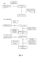
- one aspect of the present invention relates to a method for the isolation of a plasma membrane of a cell, a fraction thereof, or a plasma membrane derived organelle, comprising the following steps:
- contaminating cell organelles are washed out under high salt condition (between 0,1 and 2 M salt) and/or in the presence of alkaline buffers with a pH between 10 and 12.
- fractions of plasma membranes can be equally isolated.
- isolated plasma membranes can be further treated by mechanical shearing and the addition of detergents to release cholesterol-rich microdomains or caveolae from the plasma membrane.
- the caveolae are separated from the plasma membrane by a gradient separation.
- Another aspect of the present invention relates to the isolation of endosomes from a cell population. These methods comprise the following steps:
- the methods of the present invention provide a number of unprecedented advantages as indicated in table 1. Due to their small size ( ⁇ 100 nm) the magnetic nanoparticle, do not interfere with mass spectrometry methods and behave as a salt, and this in contrast with silica beads. The absence of detergents in the isolation method for plasma membranes preserves the structure and function of proteins, carbohydrates and lipids. Table 1.
- Fe 3 O 4 nanoparticles were synthesized using thermal decomposition method as reported by Sun (cited before).
- oleyl amine (6 mmol) and benzyl ether (20ml) were mixed and magnetically stirring the under N 2 flow conditions.
- the mixture was heated to 200°C for 2 hours and then refluxed at 300°C for 1 hour.
- the black colored mixture was cooled to room temperature by removing the heat source.
- SPMNPs with oleic acid layer as shell coating Further black material (SPMNPs with oleic acid layer as shell coating) was precipitated by adding ethanol at appropriate percentage depending on the further functionalization step and was magnetically separated using a rare earth magnet. Finally, SPMNPs were dispersed in hexane and centrifuged step (5000 rpm, 10 minutes) was performed to remove aggregates.
- Fe 3 O 4 nanoparticles were coated with DSPE (2000) phospholipids by adopting the ligand addition procedure described for water soluble quantum dots (Dubertret cited before).
- the stoichiometric ratio between SPMNPs and DSPE phospholipids can be adjusted.
- DMSA dimercaptosuccinic acid
- TMAOH Tetramethylammonium hydroxide
- COOH-TMACI ((3-carboxylpropyl) trimethylammonium chloride) coated SPMNPs were synthesized by ligand exchange methodology as cited above in [ Lee (2007) Salgueirino-Maceira, et al. (2004) and Song et al. (2005). After the reaction, SPMNPs were dissolved in water, magnetic purified and then adjusted the pH of the solution to be 7. ( Figure 1 )
- TGA Thermal gravimetric analysis
- Stability in SPMNPs Stability of SPMNPs in medium/ H 2 O was performed using UV-vis spectroscopy conducted on a Shimadzu UV-1601 PC spectrophotometer and recorded between 300 and 1000 nm with a 0.5 nm resolution.
- SPMNPs are inorganic nanocrystals characterized by superparamagnetic and size controlled physical properties which can be fine-tuned depending on the application of interest.
- nanoparticle-cell surface interaction is the critical step and mainly governed by three major physiochemical properties such as size, shape and surface coating [ Nel et al., (2009) Nat Mater 8, 543-557 ; Verma & Stellacci (2010), Small. 6(1), 12-21 ].
- the effect of surface coating dependent selection of SPMNPs for subcellular localization and magnetic isolation was investigated.
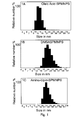
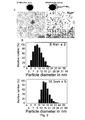
- SPMNPs with a 10 nm diameter have been synthesized by the thermal decomposition method generating monocrystalline Fe 3 O 4 with narrow size distribution and high magnetization value ( ⁇ 60emu/g).
- SPMNPs were hydrophobic due to oleic acid coating and were further functionalized using two alternative methodologies:
- HeLa and MEFs wild-type were grown to 70% confluence in 4 plates with a diameter of 10 cm dish plates. Initially cells were washed three times with PBS (37°C) and incubated with SPMNPs in DMEM/F12 medium for various time periods and at increasing nanoparticle concentration. After incubation, cells were trypsinized and harvested by centrifugation at 1000 rpm for 10 minutes. Further cell viability analysis was performed using trypan blue staining and magnetic cell isolation using SuperMACSII magnetic separation system.
- PBS ice cold phosphate buffered saline
- cells were harvested in PBS, centrifuged (1000 rpm, 10 minutes) and homogenized in homogenizing buffer HB (250 mM sucrose, 10mM Hepes and 1mM EDTA pH 7.4 supplemented with protease inhibitors) using a ball-bearing cell cracker (20 passages, clearance 10 ⁇ m, Isobiotec, Germany).
- homogenizing buffer HB 250 mM sucrose, 10mM Hepes and 1mM EDTA pH 7.4 supplemented with protease inhibitors
- the post nuclear supernatant (PNS) were loaded on to equilibrated LS column in presence of SuperMACSII magnetic system (Miltenyi Biotec), extensively washed sequentially with ice-cold HB, high salt 1M KCI and high pH 0.1M Na 2 CO 3 respectively, and the purified plasma membrane fraction was eluted by removal from the magnet.
- the plasma membrane fraction was enriched by ultracentrifugation (55,000 rpm, 1 hour) and resuspended in 200 ⁇ l HB buffer for further analysis.
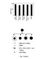
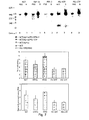
- DMSA and Lipid-SPMNPs showed medium stability and hence were used for cellular (HeLa and MEFs) viability and uptake. No detectable cell death was detected with trypan blue staining after 2 hours of SPMNPs incubation. A surface coating-dependent variation in cellular uptake of SPMNPs was observed with the following trend in magnetic cell isolation (NH 2 -lipid>DMSA>PEG-lipid>COOH-lipid-SPMNPs, whereby NH 2 -lipid coated SPMNPs have the highest cellular uptake).
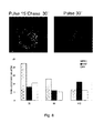
- Pulse-Chase methodology for magnetic endosome isolation was optimized based on concentration, chase time and by studying the level of subcellular compartment proteins in the magnetic fraction (MF) using western blot (WB) analysis.
- MFs from DMSA-SPMNPs were enriched either in early endosomal marker EEA1 (15-30 minutes), or the late endosomal marker Rab7 (60 minutes).
- MF from lipid-SPMNPs (NH 2 endgroup) showed enrichment in the plasma membrane marker Na + K + ATPase for all Pulse-Chase conditions. Based on these results, NH 2 -lipid-SPMNPs and DMSA-SPMNPs were selected for plasma membrane and endosomal isolation respectively.
- Example 5 Analysis of plasma membrane Proteomics Glycoproteomics and lipids.
- the nanoparticles as used in the present invention have a very high surface to volume ratio and small size. These nanoparticles do not interfere with MS analysis and provide a clear enrichment for plasma membrane (protein, lipids and glycans) compared to the total fraction. Accordingly the present invention allows to perform a novel detergent-free plasma membrane isolation method which is compatible with MS analysis compared to existing antibody/detergent based magnetic purification methods.
- N-terminal peptides For separation of N-terminal peptides by COFRADIC, cell membrane fraction were lysed, disulfide bonds were reduced and alkylated prior to acetylation of N-termini with trideutero-acetyl N-Hydroxy-Succinimide. Samples were digested with trypsin, V8 protease or chymotrypsin overnight at 25 °C or 37 °C, respectively. Purified dried peptides were reconstituted in 0.1% trifluoroacetic acid (TFA) and separated on an Ultimate 3000 LC system. During the primary run, 16 fractions of 4 minutes each were collected and dried under vacuum. N-termini of internal peptides were derivatized with TNBS.
- TFA trifluoroacetic acid
- Proteomics analysis of plasma membrane fractions showed clear enrichments for integral membrane proteins (70%) compared to the PNS fraction. Fragmented peptide spectra were identified using Mascot search algorithm and identified approximately 2000 unique proteins in the plasma membrane fraction. Similar trends were observed with respect to PSENDKO and PSEN1r samples. This degree of purity is substantially higher compared to existing technologies such as cell surface biotinylation, density gradient centrifugation and antibody based magnetic purification. Furthermore and using Gene Ontology (GO) based database analysis, 150 unique proteins were observed which are present only in the wildtype plasma membrane and 300 unique proteins which are present only in the PSENDKO plasma membrane fraction.
- GO Gene Ontology
- ICAT labeling based differential proteomics observed the absence of certain proteins in PSDKO and a, increase of certain proteins in the wildtype MEFs (about 80 proteins).
- a 100 ⁇ l sample was added to a 100 ⁇ l assay solution, which contains 100 ⁇ M Amplex Red reagent, 2 U ml ⁇ 1 HRP, 0.2 U ml ⁇ 1 choline oxidase, 8 U ml ⁇ 1 of alkaline phosphatase, 1 mU nSMase and 0.1 M Tris-HCl, 10 mM MgCl 2 , pH 7.4.
- fluorescence was measured for 30 minutes using excitation at 530 ⁇ 2.5 nm and fluorescence detection at 590 ⁇ 2.5 nm. The slope, which has to be 0, was calculated to scrutinize the completeness of the reaction. The values were corrected from the background signal that was determined by samples treated in the same way as described above but which did not contain any SMs.
- a lipid profiling on total and plasma membrane fractions in WT and PSENDKO MEFs was performed for the following lipids: Cholesterol (Chol), Phosphatidylcholine (PC), Phosphatidylserine (PS), Phosphatidylethanolamine (PE), Phosphatidylinositol (PI) and Sphingomylein (SM) lipids using ESI/MS analysis.
- SM Sphingomylein
- N-termini of internal peptides fractions were pooled as stated previously. Prior to an identical secondary RP-HPLC separation, each pooled fraction was redissolved in 85 ⁇ l of a freshly prepared 50 mM ammonium bicarbonate buffer (pH 7.8), and 0.8 units of peptide N-glycosidase (PNGase F) from Chryseobacterium (Flavobacterium) meningsepticum (proteomics grade, g95%, Sigma-Aldrich) was added. Hydrolysis of N-linked sugar chains from peptide backbone asparagines was allowed for 20 minutes at 30 °C. The reaction was stopped by adding 10 ⁇ l of 50% acetic acid.
- PNGase F peptide N-glycosidase
- Each PNGase F treated pool of primary fractions was loaded onto the same RP-HPLC column and the same solvent gradient was applied as during the primary separation.
- the altered peptides were collected in secondary fractions, dried under vacuum and prepared for LC-MS/MS analysis.
- Example 7 Development of a cell free gamma-secretase assay.
- PM and PNS fractions were extracted in CHAPS and mixed with recombinant APP-C99-FLAG affinity isolated from transiently transfected Aph1 -/-/- MEFs as described in Spasic et al. (2007) J Cell Biol 176(5), 629-640 .
- Newly produced APP intracellular domain (AICD) was separated on 10% precasted gels (NuPAGE) in MES buffer and analyzed for Western blotting.
- ⁇ -secretase complexes were extracted from microsomal membranes from PNS and plasma membrane with 0.5% dodecylmaltoside (DDM) and were run on a Native gel.
- DDM dodecylmaltoside
- the novel technology of the present invention was compared it with established methods like cell surface biotinylation (CSB) to study the levels of gamma-secretase components like Nicastrin (Nct), Presenilin 1 & 2 (PS1&2), Anterior pharynx defective-1 (APH1a) and Presenilin enhancer-2 (PEN2) at the cell surface. All methods (including confocal analysis of isolated plasma membrane sheets) confirmed the higher abundancies of PS1, NCT, PEN2 and APH1a at the cell surface while much lower levels (about 1-2%) for PSEN2 were measured.
- CAB cell surface biotinylation
- the present inventions provides an SPMNPs based plasma membrane method, which is detergent-free and does not rely on IP (Immune precipitations) for the isolation of pure and biological active PMs including active gamma-secretase complexes.
- Fluorescent modified lipid-SPMNPs were developed to confirm the WB results on the Pulse-Chase method using confocal analysis.
- We validated the method by studying the level of enrichment and purity of our plasma membrane fractions by WB analysis of an extensive number of compartment specific marker proteins.
- E glycoprotein specific Ponceau staining of PNS, UB and B fraction of MEFs Wildtype and MEFs PSDKO.
- F Gamma-Secretase Cell free activity Assay on PNS and Plasma Membrane fraction and Intensity quantification of total AICD level and enrichment with respect to PNS.
Landscapes
- Health & Medical Sciences (AREA)
- Engineering & Computer Science (AREA)
- Life Sciences & Earth Sciences (AREA)
- Chemical & Material Sciences (AREA)
- Molecular Biology (AREA)
- Biomedical Technology (AREA)
- Bioinformatics & Cheminformatics (AREA)
- Immunology (AREA)
- Physics & Mathematics (AREA)
- Hematology (AREA)
- Urology & Nephrology (AREA)
- General Health & Medical Sciences (AREA)
- Biotechnology (AREA)
- Medicinal Chemistry (AREA)
- Biophysics (AREA)
- Biochemistry (AREA)
- Microbiology (AREA)
- Cell Biology (AREA)
- Pathology (AREA)
- General Physics & Mathematics (AREA)
- Analytical Chemistry (AREA)
- Food Science & Technology (AREA)
- Nanotechnology (AREA)
- Bioinformatics & Computational Biology (AREA)
- General Engineering & Computer Science (AREA)
- Zoology (AREA)
- Wood Science & Technology (AREA)
- Genetics & Genomics (AREA)
- Organic Chemistry (AREA)
- Proteomics, Peptides & Aminoacids (AREA)
- Crystallography & Structural Chemistry (AREA)
- Pharmacology & Pharmacy (AREA)
- Medical Informatics (AREA)
- Toxicology (AREA)
- Tropical Medicine & Parasitology (AREA)
- Power Engineering (AREA)
- Peptides Or Proteins (AREA)
Priority Applications (3)
| Application Number | Priority Date | Filing Date | Title |
|---|---|---|---|
| EP10163644.7A EP2388589B1 (fr) | 2010-05-21 | 2010-05-21 | Isolation de membrane à plasma |
| US13/111,428 US8936935B2 (en) | 2010-05-21 | 2011-05-19 | Plasma membrane isolation |
| US14/563,268 US20150197742A1 (en) | 2010-05-21 | 2014-12-08 | Plasma Membrane Isolation |
Applications Claiming Priority (1)
| Application Number | Priority Date | Filing Date | Title |
|---|---|---|---|
| EP10163644.7A EP2388589B1 (fr) | 2010-05-21 | 2010-05-21 | Isolation de membrane à plasma |
Publications (2)
| Publication Number | Publication Date |
|---|---|
| EP2388589A1 true EP2388589A1 (fr) | 2011-11-23 |
| EP2388589B1 EP2388589B1 (fr) | 2015-09-30 |
Family
ID=43248879
Family Applications (1)
| Application Number | Title | Priority Date | Filing Date |
|---|---|---|---|
| EP10163644.7A Not-in-force EP2388589B1 (fr) | 2010-05-21 | 2010-05-21 | Isolation de membrane à plasma |
Country Status (2)
| Country | Link |
|---|---|
| US (2) | US8936935B2 (fr) |
| EP (1) | EP2388589B1 (fr) |
Cited By (3)
| Publication number | Priority date | Publication date | Assignee | Title |
|---|---|---|---|---|
| CN108387746A (zh) * | 2018-03-02 | 2018-08-10 | 浙江大学 | 一种捕获外泌体的超顺磁纳米微粒及其制备方法和特异性外泌体发光免疫定量检测试剂盒 |
| CN109576206A (zh) * | 2017-09-29 | 2019-04-05 | 天津大学 | 一种基于联合法分离外泌体的方法 |
| CN109576210A (zh) * | 2017-09-29 | 2019-04-05 | 天津大学 | 一种快速分离外泌体的方法 |
Families Citing this family (4)
| Publication number | Priority date | Publication date | Assignee | Title |
|---|---|---|---|---|
| CN103910794B (zh) * | 2014-03-28 | 2019-09-27 | 青岛大学 | 磁性纳米粒子对地木耳藻蓝蛋白的分离纯化 |
| WO2017030929A1 (fr) * | 2015-08-14 | 2017-02-23 | The Johns Hopkins University | Nanoparticules leurres visant à désorganiser des réseaux de cellules cancéreuses-cellules stromales |
| US20210324366A1 (en) * | 2018-07-11 | 2021-10-21 | Cornell University | Supported plant plasma membrane lipid bilayer on-a-chip |
| CN111983221B (zh) * | 2020-08-19 | 2024-04-09 | 深圳市卓润生物科技有限公司 | 表面修饰磁珠及其制备方法和应用 |
Citations (3)
| Publication number | Priority date | Publication date | Assignee | Title |
|---|---|---|---|---|
| US20040134565A1 (en) | 2003-01-13 | 2004-07-15 | International Business Machines Corporation | Process of forming magnetic nanocomposites via nanoparticle self-assembly |
| WO2009126571A2 (fr) * | 2008-04-09 | 2009-10-15 | Cornell University | Procédés de traitement aux micro-ondes utilisant des nanoparticules |
| WO2010008876A2 (fr) * | 2008-06-24 | 2010-01-21 | The Regents Of The University Of California | Procédé de préparation de nanoparticules hybrides micellaires pour applications thérapeutiques et diagnostiques et compositions associées |
Family Cites Families (1)
| Publication number | Priority date | Publication date | Assignee | Title |
|---|---|---|---|---|
| CA2487564A1 (fr) * | 2004-11-12 | 2006-05-12 | Valorisation Recherche Hscm | Nanoparticules constituees d'adn, de chitosane et d'acide folique |
-
2010
- 2010-05-21 EP EP10163644.7A patent/EP2388589B1/fr not_active Not-in-force
-
2011
- 2011-05-19 US US13/111,428 patent/US8936935B2/en not_active Expired - Fee Related
-
2014
- 2014-12-08 US US14/563,268 patent/US20150197742A1/en not_active Abandoned
Patent Citations (3)
| Publication number | Priority date | Publication date | Assignee | Title |
|---|---|---|---|---|
| US20040134565A1 (en) | 2003-01-13 | 2004-07-15 | International Business Machines Corporation | Process of forming magnetic nanocomposites via nanoparticle self-assembly |
| WO2009126571A2 (fr) * | 2008-04-09 | 2009-10-15 | Cornell University | Procédés de traitement aux micro-ondes utilisant des nanoparticules |
| WO2010008876A2 (fr) * | 2008-06-24 | 2010-01-21 | The Regents Of The University Of California | Procédé de préparation de nanoparticules hybrides micellaires pour applications thérapeutiques et diagnostiques et compositions associées |
Non-Patent Citations (22)
| Title |
|---|
| ARJUNAN, CELL BIOCHEM. BIOPHYS., vol. 53, no. 3, 2009, pages 135 - 143 |
| BLONDER J ET AL: "A detergent- and cyanogen bromide-free method for integral membrane proteomics: Application to Halobacterium purple membranes and the human epidermal membrane proteome", PROTEOMICS, vol. 4, 2004, pages 31 - 45, XP002629191 * |
| DB THIMIRI GOVINDA RAJ ET AL: "Cellular uptake of superparamagnetic nanoparticles as a strategy to isolate and characterize endosomal compartments", FRONTIERS ANNUAL MEETING LEUVEN, 23-25 OCTOBER 2007, 23 October 2007 (2007-10-23) - 23 October 2007 (2007-10-23), pages 59, XP002613505, Retrieved from the Internet <URL:http://www.frontiers-eu.org/archive/total%20Oct15%20A4%20incl%20participants%20list.pdf> [retrieved on 20101209] * |
| DREGER, MASS SPECTROM. REV., vol. 22, no. 1, 2003, pages 27 - 56 |
| DUBERTRET, SCIENCE, vol. 298, no. 5599, 2002, pages 1759 - 1762 |
| GRIMM, NAT. CELL BIOL., vol. 7, no. 11, 2005, pages 1118 - 1123 |
| HUH YS ET AL: "Rapid separation of bacteriohodopsin using a laminar-flow extraction system in a microfluidic device", BIOMICROFLUIDICS, vol. 4, 014103, 27 January 2010 (2010-01-27), pages 1 - 10, XP002629190 * |
| LAROY ET AL., NAT PROTOC., vol. 1, no. 1, 2006, pages 397 - 405 |
| LEE ET AL., NAT MED, vol. 13, no. 1, 2007, pages 95 - 99 |
| NANOTECH MONTREUX MEETING, 17 October 2008 (2008-10-17) |
| NEL ET AL., NAT MATER, vol. 8, 2009, pages 543 - 557 |
| PETER ET AL., J BIOMOL TECH., vol. 18, no. 5, 2007, pages 287 - 297 |
| RIVIERE ET AL., EU. PHYS J .E. SOFT MATTER, vol. 22, 2007, pages 1 - 10 |
| SALGUEIRINO-MACEIRA ET AL., LANGMUIR, vol. 20, no. 16, 2004, pages 6946 - 6950 |
| SIGMA-ALDRICH: "Bacteriorhodopsin from Halobacetrium halobium, product number B3636", XP002629192, Retrieved from the Internet <URL:http://www.sigmaaldrich.com> [retrieved on 20110321] * |
| SONG ET AL., J. AM. CHEM. SOC., vol. 127, no. 28, 2005, pages 9992 - 9993 |
| SPASIC ET AL., J CELL BIOL, vol. 176, no. 5, 2007, pages 629 - 640 |
| SUN; ZENG, J. AM. CHEM. SOC., vol. 124, no. 28, 2002, pages 8204 - 8205 |
| TAN F ET AL: "Enrichment of Phosphopeptides by Fe3+-Immobilized Magnetic Nanoparticles for Phosphoproteome Analysis of the Plasma Membrane of Mouse Liver", JOURNAL OF PROTEOME RESEARCH, vol. 7, 2 December 2008 (2008-12-02), pages 1078 - 1087, XP002613504 * |
| VERMA; STELLACCI, SMALL., vol. 6, no. 1, 2010, pages 12 - 21 |
| WILHELM C ET AL: "Universal cell labelling with anionic magnetic nanoparticles", BIOMATERIALS, ELSEVIER SCIENCE PUBLISHERS BV., BARKING, GB, vol. 29, no. 22, 1 August 2008 (2008-08-01), pages 3161 - 3174, XP022673739, ISSN: 0142-9612, [retrieved on 20080501], DOI: 10.1016/J.BIOMATERIALS.2008.04.016 * |
| XIAO Z & DEVREOTES PN: "Identification of Detergent-resistant Plasma Membrane Microdomains in Dictyostelium: Enrichment of Signal Transduction Proteins", MOLECULAR BIOLOGY OF THE CELL, vol. 8, May 1997 (1997-05-01), pages 855 - 869, XP002629193 * |
Cited By (3)
| Publication number | Priority date | Publication date | Assignee | Title |
|---|---|---|---|---|
| CN109576206A (zh) * | 2017-09-29 | 2019-04-05 | 天津大学 | 一种基于联合法分离外泌体的方法 |
| CN109576210A (zh) * | 2017-09-29 | 2019-04-05 | 天津大学 | 一种快速分离外泌体的方法 |
| CN108387746A (zh) * | 2018-03-02 | 2018-08-10 | 浙江大学 | 一种捕获外泌体的超顺磁纳米微粒及其制备方法和特异性外泌体发光免疫定量检测试剂盒 |
Also Published As
| Publication number | Publication date |
|---|---|
| US8936935B2 (en) | 2015-01-20 |
| EP2388589B1 (fr) | 2015-09-30 |
| US20150197742A1 (en) | 2015-07-16 |
| US20110312056A1 (en) | 2011-12-22 |
Similar Documents
| Publication | Publication Date | Title |
|---|---|---|
| US20150197742A1 (en) | Plasma Membrane Isolation | |
| Cai et al. | Immuno-modified superparamagnetic nanoparticles via host–guest interactions for high-purity capture and mild release of exosomes | |
| Sakulkhu et al. | Significance of surface charge and shell material of superparamagnetic iron oxide nanoparticle (SPION) based core/shell nanoparticles on the composition of the protein corona | |
| Wang et al. | Effect of protein corona on nanoparticle–plasma membrane and nanoparticle–biomimetic membrane interactions | |
| Yoshino et al. | Magnetic cell separation using nano‐sized bacterial magnetic particles with reconstructed magnetosome membrane | |
| CN102412046B (zh) | 富集叠氮标记生物大分子的磁性纳米材料及其制备与应用 | |
| Ma et al. | Multi-targeting magnetic hyaluronan capsules efficiently capturing circulating tumor cells | |
| WO2010097785A1 (fr) | Procédé pour la concentration sélective d'une biomolécule spécifique à faible abondance | |
| EP2904381B1 (fr) | Points de polymère semi-conducteur contenant du métal et leurs procédés de fabrication et d'utilisation | |
| Fan et al. | Biocompatible engineered erythrocytes as plasmonic sensor initiators for high-sensitive screening of non-small cell lung cancer-derived exosomal miRNA in an integrated system | |
| Thimiri Govinda Raj et al. | Surface functionalization dependent subcellular localization of superparamagnetic nanoparticle in plasma membrane and endosome | |
| Wang et al. | Effects of organic matter on uptake and intracellular trafficking of nanoparticles in Tetrahymena thermophila | |
| Jiang et al. | A post-insertion strategy for surface functionalization of bacterial and mammalian cell-derived extracellular vesicles | |
| Thimiri Govinda Raj et al. | A novel strategy for the comprehensive analysis of the biomolecular composition of isolated plasma membranes | |
| Grishaev et al. | Studying the small extracellular vesicle capture efficiency of magnetic beads coated with tannic acid | |
| Ukkola et al. | Enrichment of bovine milk-derived extracellular vesicles using surface-functionalized cellulose nanofibers | |
| Försönits et al. | Improved accessibility of extracellular vesicle surface molecules upon partial removal of the protein Corona by high ionic strength | |
| US9354237B2 (en) | Methods for isolating proteins | |
| Kankaanpää et al. | Comparative analysis of the effects of different purification methods on the yield and purity of cow milk extracellular vesicles | |
| US20180340938A1 (en) | Encapsulated functionalized diamond crystal | |
| Tran et al. | Ultrasmall gold nanoparticles for highly specific isolation/enrichment of N-linked glycosylated peptides | |
| Chakraborty et al. | Exploring the interactions between protein coronated CdSe quantum dots and nanoplastics | |
| Wang et al. | Disulfide-and terminal alkyne-functionalized magnetic silica particles for enrichment of azido glycopeptides | |
| Patil et al. | Cleavable ester-linked magnetic nanoparticles for labeling of solvent-exposed primary amine groups of peptides/proteins | |
| KR101047453B1 (ko) | 표면 막단백질의 선택적 농축분리정제용 mepset유무기복합체 및 그 표면 막단백질의 선택적농축분리정제방법 |
Legal Events
| Date | Code | Title | Description |
|---|---|---|---|
| AK | Designated contracting states |
Kind code of ref document: A1 Designated state(s): AL AT BE BG CH CY CZ DE DK EE ES FI FR GB GR HR HU IE IS IT LI LT LU LV MC MK MT NL NO PL PT RO SE SI SK SM TR |
|
| AX | Request for extension of the european patent |
Extension state: BA ME RS |
|
| PUAI | Public reference made under article 153(3) epc to a published international application that has entered the european phase |
Free format text: ORIGINAL CODE: 0009012 |
|
| 17P | Request for examination filed |
Effective date: 20120523 |
|
| 17Q | First examination report despatched |
Effective date: 20120703 |
|
| REG | Reference to a national code |
Ref country code: DE Ref legal event code: R079 Ref document number: 602010027837 Country of ref document: DE Free format text: PREVIOUS MAIN CLASS: G01N0033500000 Ipc: B82Y0005000000 |
|
| RIC1 | Information provided on ipc code assigned before grant |
Ipc: G01N 33/50 20060101ALI20150129BHEP Ipc: B82Y 5/00 20110101AFI20150129BHEP Ipc: G01N 33/68 20060101ALI20150129BHEP |
|
| GRAP | Despatch of communication of intention to grant a patent |
Free format text: ORIGINAL CODE: EPIDOSNIGR1 |
|
| INTG | Intention to grant announced |
Effective date: 20150423 |
|
| GRAS | Grant fee paid |
Free format text: ORIGINAL CODE: EPIDOSNIGR3 |
|
| GRAA | (expected) grant |
Free format text: ORIGINAL CODE: 0009210 |
|
| AK | Designated contracting states |
Kind code of ref document: B1 Designated state(s): AL AT BE BG CH CY CZ DE DK EE ES FI FR GB GR HR HU IE IS IT LI LT LU LV MC MK MT NL NO PL PT RO SE SI SK SM TR |
|
| REG | Reference to a national code |
Ref country code: CH Ref legal event code: EP Ref country code: GB Ref legal event code: FG4D |
|
| REG | Reference to a national code |
Ref country code: AT Ref legal event code: REF Ref document number: 752284 Country of ref document: AT Kind code of ref document: T Effective date: 20151015 |
|
| REG | Reference to a national code |
Ref country code: IE Ref legal event code: FG4D |
|
| REG | Reference to a national code |
Ref country code: DE Ref legal event code: R096 Ref document number: 602010027837 Country of ref document: DE |
|
| PG25 | Lapsed in a contracting state [announced via postgrant information from national office to epo] |
Ref country code: NO Free format text: LAPSE BECAUSE OF FAILURE TO SUBMIT A TRANSLATION OF THE DESCRIPTION OR TO PAY THE FEE WITHIN THE PRESCRIBED TIME-LIMIT Effective date: 20151230 Ref country code: LT Free format text: LAPSE BECAUSE OF FAILURE TO SUBMIT A TRANSLATION OF THE DESCRIPTION OR TO PAY THE FEE WITHIN THE PRESCRIBED TIME-LIMIT Effective date: 20150930 Ref country code: GR Free format text: LAPSE BECAUSE OF FAILURE TO SUBMIT A TRANSLATION OF THE DESCRIPTION OR TO PAY THE FEE WITHIN THE PRESCRIBED TIME-LIMIT Effective date: 20151231 Ref country code: FI Free format text: LAPSE BECAUSE OF FAILURE TO SUBMIT A TRANSLATION OF THE DESCRIPTION OR TO PAY THE FEE WITHIN THE PRESCRIBED TIME-LIMIT Effective date: 20150930 Ref country code: LV Free format text: LAPSE BECAUSE OF FAILURE TO SUBMIT A TRANSLATION OF THE DESCRIPTION OR TO PAY THE FEE WITHIN THE PRESCRIBED TIME-LIMIT Effective date: 20150930 |
|
| REG | Reference to a national code |
Ref country code: NL Ref legal event code: MP Effective date: 20150930 |
|
| REG | Reference to a national code |
Ref country code: LT Ref legal event code: MG4D |
|
| REG | Reference to a national code |
Ref country code: AT Ref legal event code: MK05 Ref document number: 752284 Country of ref document: AT Kind code of ref document: T Effective date: 20150930 |
|
| PG25 | Lapsed in a contracting state [announced via postgrant information from national office to epo] |
Ref country code: SE Free format text: LAPSE BECAUSE OF FAILURE TO SUBMIT A TRANSLATION OF THE DESCRIPTION OR TO PAY THE FEE WITHIN THE PRESCRIBED TIME-LIMIT Effective date: 20150930 Ref country code: HR Free format text: LAPSE BECAUSE OF FAILURE TO SUBMIT A TRANSLATION OF THE DESCRIPTION OR TO PAY THE FEE WITHIN THE PRESCRIBED TIME-LIMIT Effective date: 20150930 |
|
| REG | Reference to a national code |
Ref country code: FR Ref legal event code: PLFP Year of fee payment: 7 |
|
| PG25 | Lapsed in a contracting state [announced via postgrant information from national office to epo] |
Ref country code: ES Free format text: LAPSE BECAUSE OF FAILURE TO SUBMIT A TRANSLATION OF THE DESCRIPTION OR TO PAY THE FEE WITHIN THE PRESCRIBED TIME-LIMIT Effective date: 20150930 Ref country code: EE Free format text: LAPSE BECAUSE OF FAILURE TO SUBMIT A TRANSLATION OF THE DESCRIPTION OR TO PAY THE FEE WITHIN THE PRESCRIBED TIME-LIMIT Effective date: 20150930 Ref country code: SK Free format text: LAPSE BECAUSE OF FAILURE TO SUBMIT A TRANSLATION OF THE DESCRIPTION OR TO PAY THE FEE WITHIN THE PRESCRIBED TIME-LIMIT Effective date: 20150930 Ref country code: CZ Free format text: LAPSE BECAUSE OF FAILURE TO SUBMIT A TRANSLATION OF THE DESCRIPTION OR TO PAY THE FEE WITHIN THE PRESCRIBED TIME-LIMIT Effective date: 20150930 Ref country code: IS Free format text: LAPSE BECAUSE OF FAILURE TO SUBMIT A TRANSLATION OF THE DESCRIPTION OR TO PAY THE FEE WITHIN THE PRESCRIBED TIME-LIMIT Effective date: 20160130 Ref country code: IT Free format text: LAPSE BECAUSE OF FAILURE TO SUBMIT A TRANSLATION OF THE DESCRIPTION OR TO PAY THE FEE WITHIN THE PRESCRIBED TIME-LIMIT Effective date: 20150930 Ref country code: NL Free format text: LAPSE BECAUSE OF FAILURE TO SUBMIT A TRANSLATION OF THE DESCRIPTION OR TO PAY THE FEE WITHIN THE PRESCRIBED TIME-LIMIT Effective date: 20150930 |
|
| PG25 | Lapsed in a contracting state [announced via postgrant information from national office to epo] |
Ref country code: PL Free format text: LAPSE BECAUSE OF FAILURE TO SUBMIT A TRANSLATION OF THE DESCRIPTION OR TO PAY THE FEE WITHIN THE PRESCRIBED TIME-LIMIT Effective date: 20150930 Ref country code: AT Free format text: LAPSE BECAUSE OF FAILURE TO SUBMIT A TRANSLATION OF THE DESCRIPTION OR TO PAY THE FEE WITHIN THE PRESCRIBED TIME-LIMIT Effective date: 20150930 Ref country code: RO Free format text: LAPSE BECAUSE OF FAILURE TO SUBMIT A TRANSLATION OF THE DESCRIPTION OR TO PAY THE FEE WITHIN THE PRESCRIBED TIME-LIMIT Effective date: 20150930 Ref country code: PT Free format text: LAPSE BECAUSE OF FAILURE TO SUBMIT A TRANSLATION OF THE DESCRIPTION OR TO PAY THE FEE WITHIN THE PRESCRIBED TIME-LIMIT Effective date: 20160201 |
|
| REG | Reference to a national code |
Ref country code: DE Ref legal event code: R097 Ref document number: 602010027837 Country of ref document: DE |
|
| PLBE | No opposition filed within time limit |
Free format text: ORIGINAL CODE: 0009261 |
|
| STAA | Information on the status of an ep patent application or granted ep patent |
Free format text: STATUS: NO OPPOSITION FILED WITHIN TIME LIMIT |
|
| PG25 | Lapsed in a contracting state [announced via postgrant information from national office to epo] |
Ref country code: DK Free format text: LAPSE BECAUSE OF FAILURE TO SUBMIT A TRANSLATION OF THE DESCRIPTION OR TO PAY THE FEE WITHIN THE PRESCRIBED TIME-LIMIT Effective date: 20150930 Ref country code: BE Free format text: LAPSE BECAUSE OF NON-PAYMENT OF DUE FEES Effective date: 20160531 |
|
| 26N | No opposition filed |
Effective date: 20160701 |
|
| PG25 | Lapsed in a contracting state [announced via postgrant information from national office to epo] |
Ref country code: SI Free format text: LAPSE BECAUSE OF FAILURE TO SUBMIT A TRANSLATION OF THE DESCRIPTION OR TO PAY THE FEE WITHIN THE PRESCRIBED TIME-LIMIT Effective date: 20150930 |
|
| PG25 | Lapsed in a contracting state [announced via postgrant information from national office to epo] |
Ref country code: BE Free format text: LAPSE BECAUSE OF FAILURE TO SUBMIT A TRANSLATION OF THE DESCRIPTION OR TO PAY THE FEE WITHIN THE PRESCRIBED TIME-LIMIT Effective date: 20150930 Ref country code: LU Free format text: LAPSE BECAUSE OF FAILURE TO SUBMIT A TRANSLATION OF THE DESCRIPTION OR TO PAY THE FEE WITHIN THE PRESCRIBED TIME-LIMIT Effective date: 20160521 |
|
| REG | Reference to a national code |
Ref country code: CH Ref legal event code: PL |
|
| PG25 | Lapsed in a contracting state [announced via postgrant information from national office to epo] |
Ref country code: LI Free format text: LAPSE BECAUSE OF NON-PAYMENT OF DUE FEES Effective date: 20160531 Ref country code: CH Free format text: LAPSE BECAUSE OF NON-PAYMENT OF DUE FEES Effective date: 20160531 |
|
| REG | Reference to a national code |
Ref country code: IE Ref legal event code: MM4A |
|
| REG | Reference to a national code |
Ref country code: FR Ref legal event code: PLFP Year of fee payment: 8 |
|
| PG25 | Lapsed in a contracting state [announced via postgrant information from national office to epo] |
Ref country code: IE Free format text: LAPSE BECAUSE OF NON-PAYMENT OF DUE FEES Effective date: 20160521 |
|
| REG | Reference to a national code |
Ref country code: FR Ref legal event code: PLFP Year of fee payment: 9 |
|
| PG25 | Lapsed in a contracting state [announced via postgrant information from national office to epo] |
Ref country code: SM Free format text: LAPSE BECAUSE OF FAILURE TO SUBMIT A TRANSLATION OF THE DESCRIPTION OR TO PAY THE FEE WITHIN THE PRESCRIBED TIME-LIMIT Effective date: 20150930 Ref country code: HU Free format text: LAPSE BECAUSE OF FAILURE TO SUBMIT A TRANSLATION OF THE DESCRIPTION OR TO PAY THE FEE WITHIN THE PRESCRIBED TIME-LIMIT; INVALID AB INITIO Effective date: 20100521 Ref country code: CY Free format text: LAPSE BECAUSE OF FAILURE TO SUBMIT A TRANSLATION OF THE DESCRIPTION OR TO PAY THE FEE WITHIN THE PRESCRIBED TIME-LIMIT Effective date: 20150930 |
|
| PG25 | Lapsed in a contracting state [announced via postgrant information from national office to epo] |
Ref country code: MC Free format text: LAPSE BECAUSE OF FAILURE TO SUBMIT A TRANSLATION OF THE DESCRIPTION OR TO PAY THE FEE WITHIN THE PRESCRIBED TIME-LIMIT Effective date: 20150930 Ref country code: TR Free format text: LAPSE BECAUSE OF FAILURE TO SUBMIT A TRANSLATION OF THE DESCRIPTION OR TO PAY THE FEE WITHIN THE PRESCRIBED TIME-LIMIT Effective date: 20150930 Ref country code: MK Free format text: LAPSE BECAUSE OF FAILURE TO SUBMIT A TRANSLATION OF THE DESCRIPTION OR TO PAY THE FEE WITHIN THE PRESCRIBED TIME-LIMIT Effective date: 20150930 Ref country code: MT Free format text: LAPSE BECAUSE OF NON-PAYMENT OF DUE FEES Effective date: 20160531 |
|
| PG25 | Lapsed in a contracting state [announced via postgrant information from national office to epo] |
Ref country code: BG Free format text: LAPSE BECAUSE OF FAILURE TO SUBMIT A TRANSLATION OF THE DESCRIPTION OR TO PAY THE FEE WITHIN THE PRESCRIBED TIME-LIMIT Effective date: 20150930 |
|
| PG25 | Lapsed in a contracting state [announced via postgrant information from national office to epo] |
Ref country code: AL Free format text: LAPSE BECAUSE OF FAILURE TO SUBMIT A TRANSLATION OF THE DESCRIPTION OR TO PAY THE FEE WITHIN THE PRESCRIBED TIME-LIMIT Effective date: 20150930 |
|
| PGFP | Annual fee paid to national office [announced via postgrant information from national office to epo] |
Ref country code: DE Payment date: 20200421 Year of fee payment: 11 Ref country code: FR Payment date: 20200422 Year of fee payment: 11 |
|
| PGFP | Annual fee paid to national office [announced via postgrant information from national office to epo] |
Ref country code: GB Payment date: 20200423 Year of fee payment: 11 |
|
| REG | Reference to a national code |
Ref country code: DE Ref legal event code: R119 Ref document number: 602010027837 Country of ref document: DE |
|
| GBPC | Gb: european patent ceased through non-payment of renewal fee |
Effective date: 20210521 |
|
| PG25 | Lapsed in a contracting state [announced via postgrant information from national office to epo] |
Ref country code: GB Free format text: LAPSE BECAUSE OF NON-PAYMENT OF DUE FEES Effective date: 20210521 Ref country code: DE Free format text: LAPSE BECAUSE OF NON-PAYMENT OF DUE FEES Effective date: 20211201 |
|
| PG25 | Lapsed in a contracting state [announced via postgrant information from national office to epo] |
Ref country code: FR Free format text: LAPSE BECAUSE OF NON-PAYMENT OF DUE FEES Effective date: 20210531 |