EP2398433B1 - Phakoemulsifikator zur anwendung von ultraschall an einen augapfel - Google Patents
Phakoemulsifikator zur anwendung von ultraschall an einen augapfel Download PDFInfo
- Publication number
- EP2398433B1 EP2398433B1 EP09782230.8A EP09782230A EP2398433B1 EP 2398433 B1 EP2398433 B1 EP 2398433B1 EP 09782230 A EP09782230 A EP 09782230A EP 2398433 B1 EP2398433 B1 EP 2398433B1
- Authority
- EP
- European Patent Office
- Prior art keywords
- phacoemulsificator
- eye
- sawn
- cone element
- eye ring
- Prior art date
- Legal status (The legal status is an assumption and is not a legal conclusion. Google has not performed a legal analysis and makes no representation as to the accuracy of the status listed.)
- Not-in-force
Links
- 238000002604 ultrasonography Methods 0.000 title claims description 36
- 238000011269 treatment regimen Methods 0.000 claims description 7
- 229920002529 medical grade silicone Polymers 0.000 claims description 3
- 229920000642 polymer Polymers 0.000 claims description 3
- 210000001508 eye Anatomy 0.000 description 81
- 210000001585 trabecular meshwork Anatomy 0.000 description 29
- 238000011282 treatment Methods 0.000 description 29
- 208000010412 Glaucoma Diseases 0.000 description 28
- 238000000034 method Methods 0.000 description 23
- 238000001356 surgical procedure Methods 0.000 description 23
- 208000002177 Cataract Diseases 0.000 description 20
- 210000000695 crystalline len Anatomy 0.000 description 19
- 239000012530 fluid Substances 0.000 description 17
- 230000004410 intraocular pressure Effects 0.000 description 11
- 210000002159 anterior chamber Anatomy 0.000 description 7
- 210000001742 aqueous humor Anatomy 0.000 description 7
- 208000002352 blister Diseases 0.000 description 7
- 210000000795 conjunctiva Anatomy 0.000 description 6
- 239000007943 implant Substances 0.000 description 6
- 230000002262 irrigation Effects 0.000 description 6
- 238000003973 irrigation Methods 0.000 description 6
- 210000001328 optic nerve Anatomy 0.000 description 6
- 201000004569 Blindness Diseases 0.000 description 5
- 210000001519 tissue Anatomy 0.000 description 5
- NWIBSHFKIJFRCO-WUDYKRTCSA-N Mytomycin Chemical compound C1N2C(C(C(C)=C(N)C3=O)=O)=C3[C@@H](COC(N)=O)[C@@]2(OC)[C@@H]2[C@H]1N2 NWIBSHFKIJFRCO-WUDYKRTCSA-N 0.000 description 4
- 210000004087 cornea Anatomy 0.000 description 4
- 206010014801 endophthalmitis Diseases 0.000 description 4
- 239000000463 material Substances 0.000 description 4
- 239000002245 particle Substances 0.000 description 4
- 230000037361 pathway Effects 0.000 description 4
- 239000002504 physiological saline solution Substances 0.000 description 4
- 210000001747 pupil Anatomy 0.000 description 4
- 206010030348 Open-Angle Glaucoma Diseases 0.000 description 3
- 208000037265 diseases, disorders, signs and symptoms Diseases 0.000 description 3
- 239000007788 liquid Substances 0.000 description 3
- 230000008397 ocular pathology Effects 0.000 description 3
- 230000007170 pathology Effects 0.000 description 3
- 230000037390 scarring Effects 0.000 description 3
- 230000000007 visual effect Effects 0.000 description 3
- 201000002862 Angle-Closure Glaucoma Diseases 0.000 description 2
- XKRFYHLGVUSROY-UHFFFAOYSA-N Argon Chemical compound [Ar] XKRFYHLGVUSROY-UHFFFAOYSA-N 0.000 description 2
- 241000894006 Bacteria Species 0.000 description 2
- 208000001860 Eye Infections Diseases 0.000 description 2
- 102000009123 Fibrin Human genes 0.000 description 2
- 108010073385 Fibrin Proteins 0.000 description 2
- BWGVNKXGVNDBDI-UHFFFAOYSA-N Fibrin monomer Chemical compound CNC(=O)CNC(=O)CN BWGVNKXGVNDBDI-UHFFFAOYSA-N 0.000 description 2
- GHASVSINZRGABV-UHFFFAOYSA-N Fluorouracil Chemical compound FC1=CNC(=O)NC1=O GHASVSINZRGABV-UHFFFAOYSA-N 0.000 description 2
- 230000008901 benefit Effects 0.000 description 2
- 239000002775 capsule Substances 0.000 description 2
- 210000004027 cell Anatomy 0.000 description 2
- 239000003795 chemical substances by application Substances 0.000 description 2
- 210000004240 ciliary body Anatomy 0.000 description 2
- 201000010099 disease Diseases 0.000 description 2
- 208000011323 eye infectious disease Diseases 0.000 description 2
- 210000000744 eyelid Anatomy 0.000 description 2
- 230000004438 eyesight Effects 0.000 description 2
- 229950003499 fibrin Drugs 0.000 description 2
- 229960002949 fluorouracil Drugs 0.000 description 2
- 239000012634 fragment Substances 0.000 description 2
- 230000036541 health Effects 0.000 description 2
- 208000015181 infectious disease Diseases 0.000 description 2
- 229960004857 mitomycin Drugs 0.000 description 2
- 230000002093 peripheral effect Effects 0.000 description 2
- 239000000049 pigment Substances 0.000 description 2
- 230000009467 reduction Effects 0.000 description 2
- 231100000241 scar Toxicity 0.000 description 2
- 210000003786 sclera Anatomy 0.000 description 2
- 239000000243 solution Substances 0.000 description 2
- 230000004393 visual impairment Effects 0.000 description 2
- 206010012565 Developmental glaucoma Diseases 0.000 description 1
- 208000003164 Diplopia Diseases 0.000 description 1
- 206010016654 Fibrosis Diseases 0.000 description 1
- 206010051283 Fluid imbalance Diseases 0.000 description 1
- 201000006336 Juvenile glaucoma Diseases 0.000 description 1
- 206010061323 Optic neuropathy Diseases 0.000 description 1
- 208000035965 Postoperative Complications Diseases 0.000 description 1
- 206010047513 Vision blurred Diseases 0.000 description 1
- 230000003510 anti-fibrotic effect Effects 0.000 description 1
- 230000000340 anti-metabolite Effects 0.000 description 1
- 229940100197 antimetabolite Drugs 0.000 description 1
- 239000002256 antimetabolite Substances 0.000 description 1
- 239000012736 aqueous medium Substances 0.000 description 1
- 229910052786 argon Inorganic materials 0.000 description 1
- 210000003050 axon Anatomy 0.000 description 1
- 239000003855 balanced salt solution Substances 0.000 description 1
- 230000005540 biological transmission Effects 0.000 description 1
- 230000015572 biosynthetic process Effects 0.000 description 1
- 210000004556 brain Anatomy 0.000 description 1
- 238000001816 cooling Methods 0.000 description 1
- 230000008878 coupling Effects 0.000 description 1
- 239000007822 coupling agent Substances 0.000 description 1
- 238000010168 coupling process Methods 0.000 description 1
- 238000005859 coupling reaction Methods 0.000 description 1
- 230000007423 decrease Effects 0.000 description 1
- 230000001419 dependent effect Effects 0.000 description 1
- 208000035475 disorder Diseases 0.000 description 1
- 208000029444 double vision Diseases 0.000 description 1
- 239000003814 drug Substances 0.000 description 1
- 230000004406 elevated intraocular pressure Effects 0.000 description 1
- 230000001804 emulsifying effect Effects 0.000 description 1
- 238000005516 engineering process Methods 0.000 description 1
- 210000002950 fibroblast Anatomy 0.000 description 1
- 230000004761 fibrosis Effects 0.000 description 1
- 238000001914 filtration Methods 0.000 description 1
- 230000006870 function Effects 0.000 description 1
- 238000002347 injection Methods 0.000 description 1
- 239000007924 injection Substances 0.000 description 1
- 230000002427 irreversible effect Effects 0.000 description 1
- 230000000622 irritating effect Effects 0.000 description 1
- 208000028507 juvenile open angle glaucoma Diseases 0.000 description 1
- 210000003664 lens nucleus crystalline Anatomy 0.000 description 1
- 230000003902 lesion Effects 0.000 description 1
- 238000013160 medical therapy Methods 0.000 description 1
- 238000002324 minimally invasive surgery Methods 0.000 description 1
- 210000003205 muscle Anatomy 0.000 description 1
- 210000001087 myotubule Anatomy 0.000 description 1
- 208000020911 optic nerve disease Diseases 0.000 description 1
- 230000003287 optical effect Effects 0.000 description 1
- 230000005043 peripheral vision Effects 0.000 description 1
- 230000035699 permeability Effects 0.000 description 1
- 230000003449 preventive effect Effects 0.000 description 1
- 238000004393 prognosis Methods 0.000 description 1
- 230000005180 public health Effects 0.000 description 1
- 230000001179 pupillary effect Effects 0.000 description 1
- 210000001525 retina Anatomy 0.000 description 1
- 230000000630 rising effect Effects 0.000 description 1
- 238000007789 sealing Methods 0.000 description 1
- 230000002459 sustained effect Effects 0.000 description 1
- 230000002123 temporal effect Effects 0.000 description 1
- 230000003685 thermal hair damage Effects 0.000 description 1
- 230000002792 vascular Effects 0.000 description 1
Images

Classifications
-
- A—HUMAN NECESSITIES
- A61—MEDICAL OR VETERINARY SCIENCE; HYGIENE
- A61F—FILTERS IMPLANTABLE INTO BLOOD VESSELS; PROSTHESES; DEVICES PROVIDING PATENCY TO, OR PREVENTING COLLAPSING OF, TUBULAR STRUCTURES OF THE BODY, e.g. STENTS; ORTHOPAEDIC, NURSING OR CONTRACEPTIVE DEVICES; FOMENTATION; TREATMENT OR PROTECTION OF EYES OR EARS; BANDAGES, DRESSINGS OR ABSORBENT PADS; FIRST-AID KITS
- A61F9/00—Methods or devices for treatment of the eyes; Devices for putting in contact-lenses; Devices to correct squinting; Apparatus to guide the blind; Protective devices for the eyes, carried on the body or in the hand
- A61F9/007—Methods or devices for eye surgery
- A61F9/00736—Instruments for removal of intra-ocular material or intra-ocular injection, e.g. cataract instruments
- A61F9/00745—Instruments for removal of intra-ocular material or intra-ocular injection, e.g. cataract instruments using mechanical vibrations, e.g. ultrasonic
-
- A—HUMAN NECESSITIES
- A61—MEDICAL OR VETERINARY SCIENCE; HYGIENE
- A61N—ELECTROTHERAPY; MAGNETOTHERAPY; RADIATION THERAPY; ULTRASOUND THERAPY
- A61N7/00—Ultrasound therapy
-
- A—HUMAN NECESSITIES
- A61—MEDICAL OR VETERINARY SCIENCE; HYGIENE
- A61F—FILTERS IMPLANTABLE INTO BLOOD VESSELS; PROSTHESES; DEVICES PROVIDING PATENCY TO, OR PREVENTING COLLAPSING OF, TUBULAR STRUCTURES OF THE BODY, e.g. STENTS; ORTHOPAEDIC, NURSING OR CONTRACEPTIVE DEVICES; FOMENTATION; TREATMENT OR PROTECTION OF EYES OR EARS; BANDAGES, DRESSINGS OR ABSORBENT PADS; FIRST-AID KITS
- A61F9/00—Methods or devices for treatment of the eyes; Devices for putting in contact-lenses; Devices to correct squinting; Apparatus to guide the blind; Protective devices for the eyes, carried on the body or in the hand
- A61F9/007—Methods or devices for eye surgery
- A61F9/00781—Apparatus for modifying intraocular pressure, e.g. for glaucoma treatment
Definitions
- the present invention is generally directed to a surgical treatment for ocular pathology, and relates more particularly to a device for generating ultrasound onto at least one annular segment of the trabecular meshwork of an eye affected by glaucoma
- cataract surgery In the field of ophthalmologic diseases, one of the main surgical act that is practiced is cataract surgery.
- Cataracts cause the lens of an eye to become clouded, which interferes with proper transmission and focusing of light on the retina.
- a common practice to alleviate this condition is by surgically removing the cataractic lens and replacing it with an artificial intraocular lens.
- Phacoemulsification enables the removal of a cataractic lens through a small incision, for example between about 2.5 to about 4 mm.
- a needle is inserted through the incision into the capsular bag of the crystalline lens and the needle is ultrasonically vibrated to mechanically emulsify the lens nucleus.
- the lens material is aspirated through a lumen through the phacoemulsification needle.
- a simultaneous flow of irrigation fluid into the lens capsule is provided around the needle through an annulus established by a sleeve concentrically disposed over the needle.
- This flow of liquid into the eye is necessary to prevent collapse of the anterior chamber of the eye during aspiration.
- the irrigation fluid cools the needle in order to prevent any thermal damage of the corneal or scleral tissue.
- Phacoemulsification machines are very popular in the field of ocular medicine, and many surgeons have a phacoemulsificator for practicing cataract surgery.
- glaucoma is a significant public health problem, between 1 to 2% of population being suffering from this pathology, because glaucoma is a major cause of blindness.
- the World health organisation considers glaucoma as the third cause of blindness in the world, responsible of 15% of declared blindness occurrences, with an incidence of 2.4 millions persons per year.
- Glaucoma is an insidious health disease because at the first stage glaucoma is asymptomatic; the patient does not feel any pain or any visual problem. When the first visual troubles appear, lesions are commonly already large and despite irreversible.
- the blindness that results from glaucoma involves both central and peripheral vision and has a major impact on an individual's ability to lead an independent life.
- Glaucoma is an optic neuropathy, i.e. a disorder of the optic nerve, which usually occurs in the setting of an elevated intraocular pressure.
- the pressure within the eye increases and this is associated with changes in the appearance and function of the optic nerve. If the pressure remains high enough for a long period of time, total vision loss occurs. High pressure develops in an eye because of an internal fluid imbalance.
- the eye is a hollow structure that contains a clear fluid called "aqueous humor."
- Aqueous humor is formed in the posterior chamber of the eye by the ciliary body.
- the fluid which is made at a fairly constant rate, then passes around the lens, through the pupillary opening in the iris and into the anterior chamber of the eye. Once in the anterior chamber, the fluid drains out of the eye through two different routes.
- the fluid In the “uveoscleral" route, the fluid percolates between muscle fibers of the ciliary body. This route accounts for approximately ten percent of the aqueous outflow in humans.
- the primary pathway for aqueous outflow in humans is through the "canalicular" route that involves the trabecular meshwork and Schlemm's canal.
- the aqueous fluid builds up because it cannot exit fast enough.
- the intraocular pressure (IOP) within the eye increases.
- the increased IOP compresses the axons in the optic nerve and also may compromise the vascular supply to the optic nerve.
- the optic nerve carries vision from the eye to the brain. Some optic nerves seem more susceptible to abnormally elevated IOP than other eyes.
- the most practiced surgeries intended to improve the aqueous humor drainage are: canaloplasty, laser trabeculoplasty, laser peripheral iridotomy (in case of angle closure glaucoma), trabeculectomy, deep non perforating sclerectomy and glaucoma drainage implants.
- Canaloplasty is an advanced, nonpenetrating procedure designed to enhance and restore the eye's natural drainage system to provide sustained reduction of IOP. Canaloplasty utilizes breakthrough micro catheter technology in a simple and minimally invasive procedure. To perform a canaloplasty, a doctor will create a tiny incision to gain access to a canal in the eye. A micro catheter will circumnavigate the canal around the iris, enlarging the main drainage channel and its smaller collector channels through the injection of a sterile, gel-like material. The catheter is then removed and a suture is placed within the canal and tightened. By opening the canal, the pressure inside the eye will be relieved.
- Laser trabeculoplasty may be used to treat open angle glaucoma.
- a laser spot is aimed at the trabecular meshwork to stimulate opening of the mesh to allow more outflow of aqueous fluid.
- half of the angle is treated at a time.
- Laser peripheral iridotomy may be used in patients susceptible to or affected by angle closure glaucoma.
- laser energy is used to make a small full-thickness opening in the iris. This opening equalizes the pressure between the front and back of the iris, causing the iris to move backward.
- a partial thickness flap is made in the scleral wall of the eye, and a window opening made under the flap to remove a portion of the trabecular meshwork.
- the scleral flap is then sutured loosely back in place. This allows fluid to flow out of the eye through this opening, resulting in lowered intraocular pressure and the formation of a bleb or fluid bubble on the surface of the eye under the conjunctiva.
- Trabeculectomy is associated with many problems. Fibroblasts that are present in the episclera proliferate and migrate and can scar down the scleral flap. Failure from scarring may occur, particularly in children and young adults. Of eyes that have an initially successful trabeculectomy, eighty percent will fail from scarring within three to five years after surgery. To minimize fibrosis, surgeons now are applying antifibrotic agents such as mitomycin C (MMC) and 5-fluorouracil (5-FU) to the scleral flap at the time of surgery. The use of these agents has increased the success rate of trabeculectomy but also has increased the prevalence of hypotony. Hypotony is a problem that develops when aqueous flows out of the eye too fast.
- MMC mitomycin C
- 5-fluorouracil 5-fluorouracil
- the eye pressure drops too low (usually less than 6.0 mmHg); the structure of the eye collapses and vision decreases.
- Antimetabolites directly applied on the surgical site can be used in order to improve the surgical prognosis, especially in high risk of failure (black patients, juvenile glaucoma).
- Trabeculectomy creates a pathway for aqueous fluid to escape to the surface of the eye. At the same time, it creates a pathway for bacteria that normally live on the surface of the eye and eyelids to get into the eye. If this happens, an internal eye infection can occur called endophthalmitis. Endophthalmitis often leads to permanent and profound visual loss. Endophthalmitis can occur anytime after trabeculectomy. Another factor that contributes to infection is the placement of a bleb. Eyes that have trabeculectomy performed inferiorly have about five times the risk of eye infection than eyes that have a superior bleb. Therefore, initial trabeculectomy is performed superiorly under the eyelid, in either the nasal or temporal quadrant.
- trabeculectomy In addition to scarring, hypotony and infection, there are other complications of trabeculectomy.
- the bleb can tear and lead to profound hypotony.
- the bleb can be irritating and can disrupt the normal tear film, leading to blurred vision.
- Patients with blebs generally cannot wear contact lenses. All of the complications from trabeculectomy stem from the fact that fluid is being diverted from inside the eye to the external surface of the eye.
- Non-perforating deep sclerectomy ab externo This technique allows avoiding to open the anterior chamber of the eye and consequently reduces the risk of postoperative complications.
- the major limitation of this technique is that it is a very difficult surgical technique and only a few surgeons are able to perform it successfully.
- trabeculectomy or sclerectomy When trabeculectomy or sclerectomy doesn't successfully lower the eye pressure, the next surgical step often is an aqueous shunt device.
- glaucoma drainage implants There are several different glaucoma drainage implants. These include the original Molteno implant, the Baerveldt tube shunt, or the valved implants, such as the Ahmed glaucoma valve implant or the ExPress Mini Shunt and the later generation pressure ridge Molteno implants. These are indicated for glaucoma patients not responding to maximal medical therapy, with previous failed guarded filtering surgery (trabeculectomy).
- the flow tube is inserted into the anterior chamber of the eye and the plate is implanted underneath the conjunctiva to allow flow of aqueous fluid out of the eye into a chamber called a bleb.
- the prior art includes a number of such aqueous shunt devices, such as U.S. 4,936,825 , U.S. 5,127,901 , U.S. 5,180,362 , U.S. 5,433,701 , U.S. 4,634,418 , US 4,787,885 , U.S. 4,946,436 , U.S. 20040015140A1 and U.S. 5,360,399 .
- aqueous shunt devices such as U.S. 4,936,825 , U.S. 5,127,901 , U.S. 5,180,362 , U.S. 5,433,701 , U.S. 4,634,418 , US 4,787,885 , U.S. 4,946,436 , U.S. 20040015140A1 and U.S. 5,360,399 .
- aqueous shunt devices Many complications are associated with aqueous shunt devices. A thickened wall of scar tissue that develops around the plastic plate offers some resistance to outflow and in many eyes limits the reduction in eye pressure. In some eyes, hypotony develops because the flow through the tube is not restricted. The surgery involves operating in the posterior orbit and many patients develop an eye muscle imbalance and double vision post-operatively. Moreover, because they are open to the surface of the eye, a pathway is created for bacteria to get into the eye and endophthalmitis can potentially occur.
- An aim of the present invention is to provide a device for improving aqueous humor drainage, said device being adaptable on a phacoemulsification machine.
- This procedure could be performed systematically before cataract surgery in patients with early stage Intraocular Pressure (IOP) rising, to reduce the risk for them to develop a glaucoma.
- IOP Intraocular Pressure
- many patients with cataract have also an abnormally elevated IOP, as in both cases aged patients have higher risk to develop both pathologies. For those patients where this elevation is still not severe, it could be useful to take advantage of the cataract procedure to perform a preventive treatment to avoid further elevation of the IOP leading to a glaucoma.
- trabecular meshwork could be "cleaned" with an ultrasonic method, just before the cataract procedure so that all the debris removed from the trabecular meshwork will be washed out by the phacoemulsification hand-piece and the irrigation/aspiration circuit during cataract surgery.
- the document WO 97/07755 A1 discloses a phacoemulsificator for the removal of lens tissue according to the preamble of claim 1. The need for an improved phacoemulsificator is addressed by the features of the characterizing portion of claim 1. Various embodiments and examples are described herein in the following description of the invention which allows treating the whole circumference of the eye in only one step, without the necessity to manipulate the device during the procedure.
- a phacoemulsificator for the removal of lens tissue is disclosed.
- the phacoemulsificator comprises:
- the phacoemulsificator is remarkable in that it comprises at least one eye ring connectable to the pump wherein the proximal end of said eye ring is suitable to be applied onto an ocular globe and means to generate ultrasound beam connectable to the power source wherein said means are fixed on the distal end of the eye ring.
- the means fixed on the distal end of the eye ring composed of piezoelectric transducers are suitable to generate scattered ultrasound beam.
- the eye ring may consist in a sawn-off cone element open at both ends wherein the small base is the proximal end and the large base is the distal end.
- the proximal end of the sawn-off cone element may comprise an external annular flange suitable to be applied onto the eye globe.
- the proximal edge of the sawn-off cone element may further comprise an annular groove communicating with at least one hose formed in the sawn-off cone element and connected to the pump.
- the sawn-off cone element may be in medical grade silicon or in medical grade polymer.
- the means to generate ultrasound energy comprise a unique transducer having an annular shape, fixed on the distal end of the sawn-off cone element in such a way that said transducer extend toward the revolution axis of said sawn-off cone element.
- the transducer may have a flat segment shape.
- the means to generate ultrasound energy may include a focusing acoustic lens extending under the flat transducer.
- the means to generate ultrasound beam fixed on the distal end of the eye ring comprise a plurality of transducers arranged according to a treatment pattern.
- the means to generate ultrasound beam comprise a housing, the transducers being placed peripherally over the housing according to the treatment pattern. More particularly said transducers are placed peripherally over the whole or a part of the housing, and more preferably the transducers are circumferentially placed over the whole or a part of the circumference of the housing.
- the ultrasound energy is generated onto at least one annular segment corresponding to at least one segment of the trabecular meshwork of the eye.
- the method further comprises implementing a cataract surgery after having treated said annular segment.
- the apparatus according to the present invention allows:
- the treatment pattern corresponds to the form defined by the regions to be treated.
- the treatment pattern may be annular or semi-annular. In other cases, the treatment pattern may be elliptical, or hexagonal or octagonal.
- ultrasound can be used for their vibrating properties on small particles for the treatment of glaucoma.
- the device according to the invention can easily produce a vibration obtained with the propagation of an ultrasonic beam, transmitted to the trabecular meshwork, which unlike the apparatus described in WO 2008/024795 can concern the whole circumference of the trabeculum at the same time, more rapidly and in only one step, as will be apparent from the detailed description that follows.
- the present invention for the treatment of glaucoma is adaptable to existing devices for the treatment of cataract by phacoemulsification. This allows limiting the number of apparatus necessary to implement a cataract treatment and a glaucoma treatment during the same surgical procedure.
- a phacoemulsificator is an apparatus for the removal of lens tissue.
- a phacoemulsificator generally includes a control unit 3 comprising:
- a first needle 38 is ultrasonically vibrated by a handpiece 39.
- the handpiece 39 is interconnected:
- the first needle is designated to be introduced through the cornea and the anterior capsule in the crystalline lens nucleus 20.
- the first needle 38 fragments or emulsifies the cataractic lens which is then aspirated along with irrigation fluid through a lumen 41 in the needle 38 as indicated by the arrow 43.
- the needle 38 includes an aspiration port defined by the lumen 41.
- the aspiration port allows aspirating the fluid from the capsular bag 20.
- the device according to the present invention is adaptable on the control unit 3 of a phacoemulsificator with the need of an upgrade of the software.
- the power source 31, and the pump 32 of the control unit can be used to connect different elements that will be described in more details below.
- the connection of these elements with the control unit 3 of a phacoemulsificator allows preventing/treating glaucoma by generating ultrasound onto the trabecular meshwork of the eye in order to improve drainage efficiency of trabeculum.
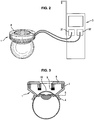
- the device according to the invention comprises an eye ring 1 wherein the proximal end of said eye ring is suitable to be applied onto the globe of the eye to be treated and (see figure 3 ) means 2 to generate ultrasound energy, said means being fixed on the distal end of the eye ring.
- Said means are connected to the control unit 3 of the phacoemulsificator. More particularly, said means 2 are connected to the power source 31 configured to provide pulsed electrical power.
- the means 2 generates ultrasounds that are not High Intensity Focussed Ultrasound.
- the aim of generating ultrasound onto the trabecular meshwork is to get the trabecular meshwork to vibrate in order to improve the drainage efficiency.
- the control unit 3 may comprise means specifying the parameters such as the frequency, the power and the duration of the ultrasound generation, etc....
- the power source 31 comprises at least a sine-wave signal generator at a determined frequency comprised between 1 kHz and 25 MHz, an amplifier and a Power meter.
- the eye ring 1 consists in a sawn-off cone element opened at both ends wherein the small base is the proximal end and the large base is the distal end.
- the proximal end of the sawn-off cone element 1 comprises an external annular flange 4 suitable to be applied onto the external surface of the eyeglobe, at approximately 2mm of the limbus, the limbus being the junction between the cornea and sclera of the eyeglobe.
- the proximal face of the annular flange 4 presents a concave profile, the radius of curvature of the concave profile being substantially equal to the radius of curvature of the eyeglobe.
- the proximal edge of the sawn-off cone element 1 comprises an annular groove 5 connected to the pump 32 of the control unit by at least one hose 7 passing through the sawn-off cone element 1 and emerging into the annular groove.
- the pump 32 is used as a suction device which is advantageously controlled by the control unit 3.
- the sawn-off cone element 1 When the sawn-off cone element 1 is applied onto the eye and the pump 32 is operated, the depression into the annular groove 5 provide a deformation of the conjunctiva of the eye, said deformation forming an o-ring in the annular groove 5.
- the sawn-off cone element 1 is then closely interlinked in such a manner that said sawn-off cone element 1 will follow the micro movements of the eye during the whole treatment time, and maintaining the quality of the centred position of the device on the visual axis.
- the sawn-off cone element 1 is advantageously obtained in medical grade silicon or other medical grade polymer which are materials compatible with the conjunctiva contact.
- sawn-off cone element 1 can be obtained in any suitable material for medical purposes well known by the skilled person, and which has been verified as biocompatible, such as biocompatible PVC, without departing with the scope of the invention.
- means 2 to generate ultrasound beam comprise a standing crown 8 holding a single transducer 9 having an annular shape.
- the transducer 9 has a flat section shape.
- Said single transducer 9 having a flat section can be associated to a focusing acoustic lens extending under said transducer 9 - i.e. held by the standing crown 8 and extending between the proximal edge of the standing crown 8 and the proximal edge of the sawn-off cone element 1.
- the focusing acoustic lens presents a cylindrical shape and a concave edge wherein the concavity is tuned towards the eyeglobe, and more particularly towards the trabecular meshwork, to focalize the ultrasound beam onto the area of interest, i.e. the trabecular meshwork of the eye.
- the transducer 9 has a concave section shape, wherein the concavity is designated to be tuned toward the eyeglobe, and more particularly toward the trabecular meshwork. In this case, this is the concave section shape of the transducer 9 which allows the focalization of the ultrasound beam onto the trabecular meshwork of the eye.
- the external radius of said standing crown 8 is sensibly equal to the internal diameter of the distal end of the sawn-off cone element 1.
- the standing crown 8 extends toward the revolution axis of said sawn-off cone element 1.
- Said transducer 9 is held in the proximal edge of the standing crown 8.
- the transducer 9 is activated by the control unit 3 to produce a vibration obtained with the propagation of an ultrasonic beam, transmitted to the trabecular meshwork over the whole or a part of its circumference.
- the means 2 to generate ultrasound beam may comprise a standing crown and a plurality of elementary transducers 9 disposed with respect to one another so as to define an annular shape.
- the standing crown 8 of transducers 9 may comprise six transducers 9.
- Each transducer 9 is a cylindrical segment able to treat 60° of the circumference of the trabecular meshwork.
- standing crown 8 can comprise two or more transducers 9 distributed among the circumference in any manner without departing with the scope of the invention.
- the surgeon must manipulate the sawn-off cone element 1 as far as the iris ring and the periphery of the cornea are centred in the distal opening of the sawn-off cone element 1. If the white ring corresponding to the visible part of the sclera trough the opening of the proximal end of the ring, has a constant thickness, the centring is correct. When the sawn-off cone element 1 is centred on the pupil, the revolution axis of said sawn-off cone element 1 and the optical axis of the eye are merging.
- the planes in which extend the distal edge and the proximal edge of the sawn-off cone element 1 are perfectly parallel to the planes of the eye such as iris plane, pupil plane or plane of the trabecular meshwork, and the transducer 9 is at the plumb of the trabecular meshwork 10. This allows a better positioning of the device according to the invention, and improves the reproducibility of the treatment.
- the device can comprise two aiming wires 14 extending crosswise and diametrally from the internal edge of the standing crown 8 or another centring system like a circular pad supposed to be centred on the pupil. This allows facilitating the centring of the sawn-off cone element with regard to the eye. To centre the sawn-off cone element 1, it is necessary to centre the intersection of the aiming wires 14 with the centre of the pupil.
- the device according to the invention can comprise other centring system known from the man skilled in the art for facilitating the centring of the sawn of cone.
- the pump 32 When the sawn-off cone element 1 is correctly centred onto the eye, the pump 32 is activated to interlink said sawn-off cone element 1 with the eye.
- the depression into the annular groove 5 provides a deformation of the conjunctiva of the eye, said deformation forming an o-ring in the annular groove 5. This insures a proper maintain in position of the device during all the treatment.
- the sawn-off cone element 1 is then filled with a physiological saline degassed solution, the o-ring formed by the deformation of the conjunctiva of the eye in the annular groove ensuring the sealing.
- the physiological saline solution provides a cooling of the eye and the device during the generation of ultrasound beam and an ultrasound coupling media that permits the propagation of ultrasound from transducer 9 to area of interest, i.e. the trabecular meshwork 10.
- the physiological saline solution moisturizes the cornea of the eye during the treatment.
- physiological saline degassed solution could be substituted by any ultrasound coupling agent such as aqueous media or lipophilic media without departing of the scope of the invention.
- the frequency and/or the power and/or the duration of each pulse are selected or already predetermined and the transducer 9 (or the plurality of transducers) is (are) activated by the control unit 3 to produce a vibration of the trabecular meshwork over the whole or a part of the circumference.
- the treatment according to the invention is advantageously a short treatment which can be performed before the phacoemulsification procedure with the same machine.
- the device according to the invention can easily produce a vibration obtained with the propagation of an ultrasonic beam, transmitted to the trabecular meshwork, which unlike the apparatus described in WO 2008/024795 can concern the whole circumference of the trabeculum at the same time, more rapidly and in only one step. Moreover, with the device according to the invention, thanks to the ring which allows centering and fixation on the eye globe, this technique can be substantially improved compared to the device described in WO 2008/024795 .
- the device according to the present invention used as a treatment of open angle glaucoma with the vibration technique applied on the trabecular meshwork, can be combined with a phacoemulsification machine has many advantages. In particular, this allows implementing both treatments (cataract and glaucoma) during a single procedure, which improves the treatment by the vibration technique.
- the particles delivered from their adherence thanks to the vibration technique according to the invention will be aspirated during the cataract surgery.
- the idea is to combine the glaucoma treatment with a phacoemulsification machine, and preferably during a cataract surgery, because during this surgery the anterior chamber and the liquid it contains, are completely washed with a balanced salt solution circulating in the irrigation / aspiration circuit, so that if the vibration technique is performed before the cataract surgery, all the debris delivered from their adherence on the trabecular meshwork, will be washed out of the anterior chamber, increasing the efficiency of the treatment.
Landscapes
- Health & Medical Sciences (AREA)
- General Health & Medical Sciences (AREA)
- Nuclear Medicine, Radiotherapy & Molecular Imaging (AREA)
- Ophthalmology & Optometry (AREA)
- Engineering & Computer Science (AREA)
- Biomedical Technology (AREA)
- Veterinary Medicine (AREA)
- Public Health (AREA)
- Life Sciences & Earth Sciences (AREA)
- Animal Behavior & Ethology (AREA)
- Surgery (AREA)
- Vascular Medicine (AREA)
- Heart & Thoracic Surgery (AREA)
- Radiology & Medical Imaging (AREA)
- Surgical Instruments (AREA)
Claims (12)
- Phakoemulsifikationsvorrichtung zum Entfernen von Linsengewebe, wobei die Phakoemulsifikationsvorrichtung Folgendes aufweist:mindestens einen Augenring (1), wobei das proximale Ende des Augenrings (1) für eine Anwendung auf einem Augapfel geeignet ist, undMittel (2) zum Erzeugen eines Ultraschallstrahls, wobei die Mittel auf dem distalen Ende des Augenrings (1) befestigt sind, dadurch gekennzeichnet, dass die Phakoemulsifikationsvorrichtung eine Energiequelle (31) aufweist, die für ein Bereitstellen von gepulster elektrischer Energie konfiguriert ist, undeine Pumpe (32), die für ein Bereitstellen von Vakuum konfiguriert ist, wobei der mindestens eine Augenring (1) mit der Pumpe verbindbar ist.
- Phakoemulsifikationsvorrichtung nach Anspruch 1, dadurch gekennzeichnet, dass die Mittel (2), die auf dem distalen Ende des Augenrings (1) befestigt sind, zum Erzeugen eines gestreuten Ultraschallstrahls geeignet sind.
- Phakoemulsifikationsvorrichtung nach einem der Ansprüche 1 bis 2, dadurch gekennzeichnet, dass der Augenring (1) aus einem an beiden Enden offenen Stumpfkegelelement (1) besteht, wobei die kleine Basis das proximale Ende und die große Basis das distale Ende bilden.
- Phakoemulsifikationsvorrichtung nach Anspruch 3, dadurch gekennzeichnet, dass das proximale Ende des Stumpfkegelelements (1) einen äußeren ringförmigen Flansch (4) aufweist, der für ein Anbringen auf dem Augapfel geeignet ist.
- Phakoemulsifikationsvorrichtung nach einem der Ansprüche 3 oder 4, dadurch gekennzeichnet, dass das proximale Ende des Stumpfkegelelements (1) eine ringförmige Nut (5) aufweist, die mit mindestens einem im Stumpfkegelelement (1) geformten Schlauch (7) kommuniziert und mit der Pumpe (32) verbunden ist.
- Phakoemulsifikationsvorrichtung nach einem der Ansprüche 1 bis 5, dadurch gekennzeichnet, dass das Stumpfkegelelement (1) aus Silizium von medizinischer Güte oder Polymer von medizinischer Güte hergestellt ist.
- Phakoemulsifikationsvorrichtung nach einem der Ansprüche 1 bis 6, dadurch gekennzeichnet, dass die Mittel (2) zum Erzeugen von Ultraschallenergie einen eindeutigen Wandler (9) von ringförmiger Form aufweisen, der am distalen Ende des Stumpfkegelelements (1) auf solche Art befestigt ist, dass sich der Wandler (9) zur Drehachse des Stumpfkegelelements (1) erstreckt.
- Phakoemulsifikationsvorrichtung nach Anspruch 7, dadurch gekennzeichnet, dass der Wandler (9) eine flache Segmentform aufweist, Mittel (2) zum Erzeugen von Ultraschallenergie eine fokussierende akustische Linse (15) aufweisen, die sich unter dem flachen Wandler (9) erstrecken.
- Phakoemulsifikationsvorrichtung nach einem der Ansprüche 1 bis 6, dadurch gekennzeichnet, dass die auf dem distalen Ende des Augenrings (1) befestigten Mittel (2) zum Erzeugen von Ultraschallstrahls mehrere Wandler (9) in einer dem Behandlungsmuster entsprechenden Anordnung aufweisen.
- Phakoemulsifikationsvorrichtung nach Anspruch 9, dadurch gekennzeichnet, dass die Mittel (2) zum Erzeugen von Ultraschallstrahls ein Gehäuse (8) aufweisen, wobei die Wandler peripher über der Gehäuse entsprechend dem Behandlungsmuster angeordnet sind.
- Phakoemulsifikationsvorrichtung nach einem der Ansprüche 9 oder 10, dadurch gekennzeichnet, dass die Wandler peripher über dem gesamten oder einem Teil des Gehäuses (8) angeordnet sind.
- Phakoemulsifikationsvorrichtung nach einem der Ansprüche 9 bis 11, dadurch gekennzeichnet, dass die Wandler (9) in Umfangsrichtung über dem gesamten oder einem Teil des Umfangs des Gehäuses (8) angeordnet sind.
Priority Applications (1)
| Application Number | Priority Date | Filing Date | Title |
|---|---|---|---|
| EP09782230.8A EP2398433B1 (de) | 2009-02-18 | 2009-08-26 | Phakoemulsifikator zur anwendung von ultraschall an einen augapfel |
Applications Claiming Priority (3)
| Application Number | Priority Date | Filing Date | Title |
|---|---|---|---|
| PCT/EP2009/051892 WO2009103721A1 (en) | 2008-02-19 | 2009-02-18 | A method of treating an ocular pathology by applying high intensity focused ultrasound and device thereof |
| EP09782230.8A EP2398433B1 (de) | 2009-02-18 | 2009-08-26 | Phakoemulsifikator zur anwendung von ultraschall an einen augapfel |
| PCT/EP2009/061014 WO2010094353A1 (en) | 2009-02-18 | 2009-08-26 | A method of treating an ocular pathology by applying ultrasound to the trabecular meshwork and device thereof |
Publications (2)
| Publication Number | Publication Date |
|---|---|
| EP2398433A1 EP2398433A1 (de) | 2011-12-28 |
| EP2398433B1 true EP2398433B1 (de) | 2017-09-06 |
Family
ID=56291127
Family Applications (1)
| Application Number | Title | Priority Date | Filing Date |
|---|---|---|---|
| EP09782230.8A Not-in-force EP2398433B1 (de) | 2009-02-18 | 2009-08-26 | Phakoemulsifikator zur anwendung von ultraschall an einen augapfel |
Country Status (1)
| Country | Link |
|---|---|
| EP (1) | EP2398433B1 (de) |
Cited By (1)
| Publication number | Priority date | Publication date | Assignee | Title |
|---|---|---|---|---|
| WO2024015299A1 (en) * | 2022-07-11 | 2024-01-18 | Clauson Creative Engineering | Pulsatile suction ring for visualizing and treating drainage pathways of the eye and methods of use |
Families Citing this family (1)
| Publication number | Priority date | Publication date | Assignee | Title |
|---|---|---|---|---|
| AU2018233971A1 (en) | 2017-03-13 | 2019-10-03 | Kejako Sa | Accommodative lens device |
-
2009
- 2009-08-26 EP EP09782230.8A patent/EP2398433B1/de not_active Not-in-force
Non-Patent Citations (1)
| Title |
|---|
| None * |
Cited By (1)
| Publication number | Priority date | Publication date | Assignee | Title |
|---|---|---|---|---|
| WO2024015299A1 (en) * | 2022-07-11 | 2024-01-18 | Clauson Creative Engineering | Pulsatile suction ring for visualizing and treating drainage pathways of the eye and methods of use |
Also Published As
| Publication number | Publication date |
|---|---|
| EP2398433A1 (de) | 2011-12-28 |
Similar Documents
| Publication | Publication Date | Title |
|---|---|---|
| US9259597B2 (en) | Method of treating an ocular pathology by applying ultrasound to the trabecular meshwork and device thereof | |
| WO2010094353A1 (en) | A method of treating an ocular pathology by applying ultrasound to the trabecular meshwork and device thereof | |
| EP1685815A1 (de) | Chirurgisches Gerät | |
| CA2538204A1 (en) | Irrigation tip | |
| EP2398432B1 (de) | Vorrichtung zur behandlung einer augenkrankheit durch verabreichung hochintensiven fokussierten ultraschalls | |
| EP2398433B1 (de) | Phakoemulsifikator zur anwendung von ultraschall an einen augapfel | |
| WO2011083358A1 (en) | Device for treating an ocular pathology by applying doses of laser energy | |
| AU2009216751B2 (en) | A method of treating an ocular pathology by applying high intensity focused ultrasound and device thereof |
Legal Events
| Date | Code | Title | Description |
|---|---|---|---|
| PUAI | Public reference made under article 153(3) epc to a published international application that has entered the european phase |
Free format text: ORIGINAL CODE: 0009012 |
|
| 17P | Request for examination filed |
Effective date: 20110824 |
|
| AK | Designated contracting states |
Kind code of ref document: A1 Designated state(s): AT BE BG CH CY CZ DE DK EE ES FI FR GB GR HR HU IE IS IT LI LT LU LV MC MK MT NL NO PL PT RO SE SI SK SM TR |
|
| DAX | Request for extension of the european patent (deleted) | ||
| GRAP | Despatch of communication of intention to grant a patent |
Free format text: ORIGINAL CODE: EPIDOSNIGR1 |
|
| RAP1 | Party data changed (applicant data changed or rights of an application transferred) |
Owner name: EYE TECH CARE Owner name: INSTITUT NATIONAL DE LA SANTE ET DE LA RECHERCHE M |
|
| INTG | Intention to grant announced |
Effective date: 20170504 |
|
| RIN1 | Information on inventor provided before grant (corrected) |
Inventor name: ROMANO, FABRIZIO Inventor name: FARCY, LAURENT Inventor name: CHAPUIS, PHILIPPE Inventor name: CHARREL, THOMAS |
|
| GRAS | Grant fee paid |
Free format text: ORIGINAL CODE: EPIDOSNIGR3 |
|
| GRAA | (expected) grant |
Free format text: ORIGINAL CODE: 0009210 |
|
| AK | Designated contracting states |
Kind code of ref document: B1 Designated state(s): AT BE BG CH CY CZ DE DK EE ES FI FR GB GR HR HU IE IS IT LI LT LU LV MC MK MT NL NO PL PT RO SE SI SK SM TR |
|
| REG | Reference to a national code |
Ref country code: GB Ref legal event code: FG4D |
|
| REG | Reference to a national code |
Ref country code: CH Ref legal event code: EP Ref country code: AT Ref legal event code: REF Ref document number: 925066 Country of ref document: AT Kind code of ref document: T Effective date: 20170915 |
|
| REG | Reference to a national code |
Ref country code: IE Ref legal event code: FG4D |
|
| REG | Reference to a national code |
Ref country code: DE Ref legal event code: R096 Ref document number: 602009048207 Country of ref document: DE |
|
| REG | Reference to a national code |
Ref country code: NL Ref legal event code: MP Effective date: 20170906 |
|
| REG | Reference to a national code |
Ref country code: LT Ref legal event code: MG4D |
|
| PG25 | Lapsed in a contracting state [announced via postgrant information from national office to epo] |
Ref country code: SE Free format text: LAPSE BECAUSE OF FAILURE TO SUBMIT A TRANSLATION OF THE DESCRIPTION OR TO PAY THE FEE WITHIN THE PRESCRIBED TIME-LIMIT Effective date: 20170906 Ref country code: NO Free format text: LAPSE BECAUSE OF FAILURE TO SUBMIT A TRANSLATION OF THE DESCRIPTION OR TO PAY THE FEE WITHIN THE PRESCRIBED TIME-LIMIT Effective date: 20171206 Ref country code: HR Free format text: LAPSE BECAUSE OF FAILURE TO SUBMIT A TRANSLATION OF THE DESCRIPTION OR TO PAY THE FEE WITHIN THE PRESCRIBED TIME-LIMIT Effective date: 20170906 Ref country code: FI Free format text: LAPSE BECAUSE OF FAILURE TO SUBMIT A TRANSLATION OF THE DESCRIPTION OR TO PAY THE FEE WITHIN THE PRESCRIBED TIME-LIMIT Effective date: 20170906 Ref country code: LT Free format text: LAPSE BECAUSE OF FAILURE TO SUBMIT A TRANSLATION OF THE DESCRIPTION OR TO PAY THE FEE WITHIN THE PRESCRIBED TIME-LIMIT Effective date: 20170906 |
|
| REG | Reference to a national code |
Ref country code: AT Ref legal event code: MK05 Ref document number: 925066 Country of ref document: AT Kind code of ref document: T Effective date: 20170906 |
|
| PG25 | Lapsed in a contracting state [announced via postgrant information from national office to epo] |
Ref country code: BG Free format text: LAPSE BECAUSE OF FAILURE TO SUBMIT A TRANSLATION OF THE DESCRIPTION OR TO PAY THE FEE WITHIN THE PRESCRIBED TIME-LIMIT Effective date: 20171206 Ref country code: LV Free format text: LAPSE BECAUSE OF FAILURE TO SUBMIT A TRANSLATION OF THE DESCRIPTION OR TO PAY THE FEE WITHIN THE PRESCRIBED TIME-LIMIT Effective date: 20170906 Ref country code: GR Free format text: LAPSE BECAUSE OF FAILURE TO SUBMIT A TRANSLATION OF THE DESCRIPTION OR TO PAY THE FEE WITHIN THE PRESCRIBED TIME-LIMIT Effective date: 20171207 Ref country code: ES Free format text: LAPSE BECAUSE OF FAILURE TO SUBMIT A TRANSLATION OF THE DESCRIPTION OR TO PAY THE FEE WITHIN THE PRESCRIBED TIME-LIMIT Effective date: 20170906 |
|
| PG25 | Lapsed in a contracting state [announced via postgrant information from national office to epo] |
Ref country code: NL Free format text: LAPSE BECAUSE OF FAILURE TO SUBMIT A TRANSLATION OF THE DESCRIPTION OR TO PAY THE FEE WITHIN THE PRESCRIBED TIME-LIMIT Effective date: 20170906 |
|
| PG25 | Lapsed in a contracting state [announced via postgrant information from national office to epo] |
Ref country code: RO Free format text: LAPSE BECAUSE OF FAILURE TO SUBMIT A TRANSLATION OF THE DESCRIPTION OR TO PAY THE FEE WITHIN THE PRESCRIBED TIME-LIMIT Effective date: 20170906 Ref country code: PL Free format text: LAPSE BECAUSE OF FAILURE TO SUBMIT A TRANSLATION OF THE DESCRIPTION OR TO PAY THE FEE WITHIN THE PRESCRIBED TIME-LIMIT Effective date: 20170906 Ref country code: CZ Free format text: LAPSE BECAUSE OF FAILURE TO SUBMIT A TRANSLATION OF THE DESCRIPTION OR TO PAY THE FEE WITHIN THE PRESCRIBED TIME-LIMIT Effective date: 20170906 |
|
| PG25 | Lapsed in a contracting state [announced via postgrant information from national office to epo] |
Ref country code: EE Free format text: LAPSE BECAUSE OF FAILURE TO SUBMIT A TRANSLATION OF THE DESCRIPTION OR TO PAY THE FEE WITHIN THE PRESCRIBED TIME-LIMIT Effective date: 20170906 Ref country code: AT Free format text: LAPSE BECAUSE OF FAILURE TO SUBMIT A TRANSLATION OF THE DESCRIPTION OR TO PAY THE FEE WITHIN THE PRESCRIBED TIME-LIMIT Effective date: 20170906 Ref country code: IT Free format text: LAPSE BECAUSE OF FAILURE TO SUBMIT A TRANSLATION OF THE DESCRIPTION OR TO PAY THE FEE WITHIN THE PRESCRIBED TIME-LIMIT Effective date: 20170906 Ref country code: SK Free format text: LAPSE BECAUSE OF FAILURE TO SUBMIT A TRANSLATION OF THE DESCRIPTION OR TO PAY THE FEE WITHIN THE PRESCRIBED TIME-LIMIT Effective date: 20170906 Ref country code: SM Free format text: LAPSE BECAUSE OF FAILURE TO SUBMIT A TRANSLATION OF THE DESCRIPTION OR TO PAY THE FEE WITHIN THE PRESCRIBED TIME-LIMIT Effective date: 20170906 Ref country code: IS Free format text: LAPSE BECAUSE OF FAILURE TO SUBMIT A TRANSLATION OF THE DESCRIPTION OR TO PAY THE FEE WITHIN THE PRESCRIBED TIME-LIMIT Effective date: 20180106 |
|
| REG | Reference to a national code |
Ref country code: DE Ref legal event code: R097 Ref document number: 602009048207 Country of ref document: DE |
|
| PLBE | No opposition filed within time limit |
Free format text: ORIGINAL CODE: 0009261 |
|
| STAA | Information on the status of an ep patent application or granted ep patent |
Free format text: STATUS: NO OPPOSITION FILED WITHIN TIME LIMIT |
|
| PG25 | Lapsed in a contracting state [announced via postgrant information from national office to epo] |
Ref country code: DK Free format text: LAPSE BECAUSE OF FAILURE TO SUBMIT A TRANSLATION OF THE DESCRIPTION OR TO PAY THE FEE WITHIN THE PRESCRIBED TIME-LIMIT Effective date: 20170906 |
|
| 26N | No opposition filed |
Effective date: 20180607 |
|
| REG | Reference to a national code |
Ref country code: FR Ref legal event code: PLFP Year of fee payment: 10 |
|
| PG25 | Lapsed in a contracting state [announced via postgrant information from national office to epo] |
Ref country code: SI Free format text: LAPSE BECAUSE OF FAILURE TO SUBMIT A TRANSLATION OF THE DESCRIPTION OR TO PAY THE FEE WITHIN THE PRESCRIBED TIME-LIMIT Effective date: 20170906 |
|
| PG25 | Lapsed in a contracting state [announced via postgrant information from national office to epo] |
Ref country code: MC Free format text: LAPSE BECAUSE OF FAILURE TO SUBMIT A TRANSLATION OF THE DESCRIPTION OR TO PAY THE FEE WITHIN THE PRESCRIBED TIME-LIMIT Effective date: 20170906 |
|
| REG | Reference to a national code |
Ref country code: CH Ref legal event code: PL |
|
| PG25 | Lapsed in a contracting state [announced via postgrant information from national office to epo] |
Ref country code: LU Free format text: LAPSE BECAUSE OF NON-PAYMENT OF DUE FEES Effective date: 20180826 Ref country code: CH Free format text: LAPSE BECAUSE OF NON-PAYMENT OF DUE FEES Effective date: 20180831 Ref country code: LI Free format text: LAPSE BECAUSE OF NON-PAYMENT OF DUE FEES Effective date: 20180831 |
|
| REG | Reference to a national code |
Ref country code: BE Ref legal event code: MM Effective date: 20180831 |
|
| PG25 | Lapsed in a contracting state [announced via postgrant information from national office to epo] |
Ref country code: BE Free format text: LAPSE BECAUSE OF NON-PAYMENT OF DUE FEES Effective date: 20180831 |
|
| PGFP | Annual fee paid to national office [announced via postgrant information from national office to epo] |
Ref country code: DE Payment date: 20190807 Year of fee payment: 11 Ref country code: FR Payment date: 20190830 Year of fee payment: 11 |
|
| PGFP | Annual fee paid to national office [announced via postgrant information from national office to epo] |
Ref country code: GB Payment date: 20190821 Year of fee payment: 11 |
|
| PG25 | Lapsed in a contracting state [announced via postgrant information from national office to epo] |
Ref country code: MT Free format text: LAPSE BECAUSE OF NON-PAYMENT OF DUE FEES Effective date: 20180826 |
|
| PG25 | Lapsed in a contracting state [announced via postgrant information from national office to epo] |
Ref country code: TR Free format text: LAPSE BECAUSE OF FAILURE TO SUBMIT A TRANSLATION OF THE DESCRIPTION OR TO PAY THE FEE WITHIN THE PRESCRIBED TIME-LIMIT Effective date: 20170906 |
|
| PG25 | Lapsed in a contracting state [announced via postgrant information from national office to epo] |
Ref country code: PT Free format text: LAPSE BECAUSE OF FAILURE TO SUBMIT A TRANSLATION OF THE DESCRIPTION OR TO PAY THE FEE WITHIN THE PRESCRIBED TIME-LIMIT Effective date: 20170906 Ref country code: HU Free format text: LAPSE BECAUSE OF FAILURE TO SUBMIT A TRANSLATION OF THE DESCRIPTION OR TO PAY THE FEE WITHIN THE PRESCRIBED TIME-LIMIT; INVALID AB INITIO Effective date: 20090826 |
|
| PG25 | Lapsed in a contracting state [announced via postgrant information from national office to epo] |
Ref country code: MK Free format text: LAPSE BECAUSE OF NON-PAYMENT OF DUE FEES Effective date: 20170906 Ref country code: IE Free format text: LAPSE BECAUSE OF NON-PAYMENT OF DUE FEES Effective date: 20180826 Ref country code: CY Free format text: LAPSE BECAUSE OF FAILURE TO SUBMIT A TRANSLATION OF THE DESCRIPTION OR TO PAY THE FEE WITHIN THE PRESCRIBED TIME-LIMIT Effective date: 20170906 |
|
| REG | Reference to a national code |
Ref country code: DE Ref legal event code: R119 Ref document number: 602009048207 Country of ref document: DE |
|
| GBPC | Gb: european patent ceased through non-payment of renewal fee |
Effective date: 20200826 |
|
| PG25 | Lapsed in a contracting state [announced via postgrant information from national office to epo] |
Ref country code: DE Free format text: LAPSE BECAUSE OF NON-PAYMENT OF DUE FEES Effective date: 20210302 Ref country code: FR Free format text: LAPSE BECAUSE OF NON-PAYMENT OF DUE FEES Effective date: 20200831 |
|
| PG25 | Lapsed in a contracting state [announced via postgrant information from national office to epo] |
Ref country code: GB Free format text: LAPSE BECAUSE OF NON-PAYMENT OF DUE FEES Effective date: 20200826 |