EP2425817A1 - Micelles polymérisées pour diagnostic - Google Patents
Micelles polymérisées pour diagnostic Download PDFInfo
- Publication number
- EP2425817A1 EP2425817A1 EP10290440A EP10290440A EP2425817A1 EP 2425817 A1 EP2425817 A1 EP 2425817A1 EP 10290440 A EP10290440 A EP 10290440A EP 10290440 A EP10290440 A EP 10290440A EP 2425817 A1 EP2425817 A1 EP 2425817A1
- Authority
- EP
- European Patent Office
- Prior art keywords
- micelles
- micelle
- polymerized
- pda
- amphiphilic
- Prior art date
- Legal status (The legal status is an assumption and is not a legal conclusion. Google has not performed a legal analysis and makes no representation as to the accuracy of the status listed.)
- Withdrawn
Links
Images


Classifications
-
- A—HUMAN NECESSITIES
- A61—MEDICAL OR VETERINARY SCIENCE; HYGIENE
- A61K—PREPARATIONS FOR MEDICAL, DENTAL OR TOILETRY PURPOSES
- A61K49/00—Preparations for testing in vivo
- A61K49/001—Preparation for luminescence or biological staining
- A61K49/0063—Preparation for luminescence or biological staining characterised by a special physical or galenical form, e.g. emulsions, microspheres
- A61K49/0069—Preparation for luminescence or biological staining characterised by a special physical or galenical form, e.g. emulsions, microspheres the agent being in a particular physical galenical form
- A61K49/0076—Preparation for luminescence or biological staining characterised by a special physical or galenical form, e.g. emulsions, microspheres the agent being in a particular physical galenical form dispersion, suspension, e.g. particles in a liquid, colloid, emulsion
- A61K49/0082—Preparation for luminescence or biological staining characterised by a special physical or galenical form, e.g. emulsions, microspheres the agent being in a particular physical galenical form dispersion, suspension, e.g. particles in a liquid, colloid, emulsion micelle, e.g. phospholipidic micelle and polymeric micelle
-
- A—HUMAN NECESSITIES
- A61—MEDICAL OR VETERINARY SCIENCE; HYGIENE
- A61K—PREPARATIONS FOR MEDICAL, DENTAL OR TOILETRY PURPOSES
- A61K49/00—Preparations for testing in vivo
- A61K49/001—Preparation for luminescence or biological staining
- A61K49/0013—Luminescence
- A61K49/0017—Fluorescence in vivo
- A61K49/0019—Fluorescence in vivo characterised by the fluorescent group, e.g. oligomeric, polymeric or dendritic molecules
- A61K49/0021—Fluorescence in vivo characterised by the fluorescent group, e.g. oligomeric, polymeric or dendritic molecules the fluorescent group being a small organic molecule
- A61K49/0032—Methine dyes, e.g. cyanine dyes
-
- A—HUMAN NECESSITIES
- A61—MEDICAL OR VETERINARY SCIENCE; HYGIENE
- A61K—PREPARATIONS FOR MEDICAL, DENTAL OR TOILETRY PURPOSES
- A61K49/00—Preparations for testing in vivo
- A61K49/001—Preparation for luminescence or biological staining
- A61K49/0013—Luminescence
- A61K49/0017—Fluorescence in vivo
- A61K49/005—Fluorescence in vivo characterised by the carrier molecule carrying the fluorescent agent
- A61K49/0054—Macromolecular compounds, i.e. oligomers, polymers, dendrimers
-
- A—HUMAN NECESSITIES
- A61—MEDICAL OR VETERINARY SCIENCE; HYGIENE
- A61K—PREPARATIONS FOR MEDICAL, DENTAL OR TOILETRY PURPOSES
- A61K9/00—Medicinal preparations characterised by special physical form
- A61K9/10—Dispersions; Emulsions
- A61K9/107—Emulsions ; Emulsion preconcentrates; Micelles
- A61K9/1075—Microemulsions or submicron emulsions; Preconcentrates or solids thereof; Micelles, e.g. made of phospholipids or block copolymers
Definitions
- the present invention relates to polymerized micelles, their method of preparation as well as their use in diagnosis, notably in the diagnosis of cancer, in particular by tumor imaging.
- NIR fluorescent light i.e. 700-1000 nm
- biological tissues low absorbance and usually negligible auto-fluorescence
- the successful use of a nanocarrier for tumor imaging and drug delivery is closely related to its biodistribution and generally hinges on the enhanced permeation and retention (EPR) effect.
- the EPR effect is the result of passive diffusion of nanometric objects through blood vessels that irrigate the tumor. These so-called neovessels have looser endothelial inter-cellular junctions, due to their inflammatory nature, and are much more permeable than ordinary vessels.
- lymphatic drainage of the tumor is poorly efficient, hence allowing objects to remain longer once they have diffused inside the tumor tissues.
- the aptitude of a nanoparticle to achieve passive targeting of tumor cells via EPR effect depends mainly on its size (to allow interstitial diffusion from the blood) [12] and surface chemistry (to permit longer blood circulation).
- micelles are good candidates for medical applications as they are easily synthesized within an appropriate size range (i.e. ⁇ 30 nm) in a reproducible manner, from non-toxic materials (as opposed to other nanoparticles like quantum dots, for instance).
- an appropriate size range i.e. ⁇ 30 nm
- two major drawbacks hinder their use: they usually have short circulation half-lives and limited cargo-shielding (unstable drug retention).
- the invention provides polymerized micelles which are non toxic and display significant tumor accumulation through enhanced permeation and retention (EPR) effect. Further, they advantageously exhibit satisfactory blood residence time, tumor uptake and imaging contrast. In addition, these polymerized micelles exhibit satisfactory drug loading capacity and cargo-shielding. Furthermore, unexpectedly, it has been shown that the polymerized micelles according to the invention exhibit improved properties, notably an enhanced EPR effect, as compared to those disclosed in the prior art, and in particular to PDA-NTA.
- the synthesis of the polymerized micelles according to the invention is easily controllable and reproducible, as well as economical.
- the invention is directed to a polymerized micelle for use in diagnosis, said polymerized micelle comprising:
- the invention provides polymerized micelles for use in cancer diagnosis, in particular by tumor imaging.
- micelle refers to a spherical aggregate of amphiphilic molecules dispersed in a liquid colloid, notably in water, and which size is inferior to 100 nm, more preferably below 30 nm.
- amphiphilic means an organic molecule, also called a surfactant, possessing both hydrophilic and lipophilic properties. Amphiphilic compounds or surfactants thus comprise or consist of two distinct hydrophobic and hydrophilic domains. Upon dispersion in aqueous media, spontaneous phase separation occurs, leading to supra-molecular core-shell micelle structure. The core is constituted by the hydrophobic region of the amphiphilic polymer and is shielded from water by the hydrophilic outer shell (hydrophilic heads). The self assembly is dependent on concentration and temperature. At lower concentrations, amphiphilic monomers are individualized species. As the concentration increases, the monomers self organize into micelles when the critical micellar concentration (CMC) is reached. The formation of the micelles is also dependent on the temperature and, more precisely, on the critical micellar temperature.
- CMC critical micellar concentration
- amphiphilic compounds or surfactants are also often classified into four primary groups: anionic, cationic, non-ionic, and zwitterionic (dual charge).
- the amphiphilic compound is cationic or non-ionic, more preferably non-ionic.
- Amphiphilic monomers described herein comprise a polymerizable unit. This reactive function allows micelle polymerization. This polymerization enables to strengthen the micellar architecture and to preserve the integrity of the structure even in a dilute medium, when the concentration is inferior to the CMC.
- amphiphilic monomer is a monomer consisting of:
- the hydrophilic head comprises or consists of a polyoxyethylene (PEG) chain, preferably having a molecular weight in the range of 300 to 10000 g. mol -1 , more preferably in the range of 1000 to 5000 g. mol -1 .
- the molecular weight ratio of the PEG relative to the lipophilic chain of the amphiphilic monomer ranges from 1 to 30.
- PEG chains are known to be biocompatible (little to no toxicity and immunogenicity) and to limit interaction of PEG-coated particles with plasma proteins and especially with opsonins often responsible for phagocytosis by macrophages, hence diminishing their recognition by cells of the reticulo-endothelial system (RES).
- RES reticulo-endothelial system
- amphiphilic monomer is a monomer of formula (I): X-L 0 -[L 1 -Y] k -L 2
- k is 1 or 2.
- Z is H or CH 2 -COOH.
- n 8 or 45.
- amphiphilic monomer of formula (I) is a monomer of formula (Ia):
- Z is H or CH 2 COOH, and n, p, q are as defined above.
- amphiphilic monomer is selected from monomers of formula (Ia), wherein:
- the amphiphilic polymer is obtained by the polymerization of a mixture containing two distinct amphiphilic monomers of formula (I), notably a first amphiphilic monomer wherein Z is H and a second amphiphilic monomer of formula (I) wherein Z is -(CH 2 ) m R 1 .
- the diagnostic agent can be a substance emitting electromagnetic rays or a substance detectable by X-ray, ultrasound or nuclear magnetic resonance.
- the polymerized micelles can thus comprise iron oxide particles, such as magnetite or maghemite, gadolinium chelates, radio-opaque materials, such as, for example, air or barium, or fluorescent compounds, such as rhodamine or nile red, or gamma emitters, such as indium or technetium, or positron emitters such as 18 F or 11 C, or any other radioactive label.
- the diagnostic agent is a fluorescent compound, such as that sold under the tradename FluoProbes® 730 (FP730) thus, allowing the cancer diagnosis to be carried out by tumor fluorescence imaging.
- FP730 FluoProbes® 730
- the diagnostic agent may be either coupled with at least one of the hydrophilic heads at the micelle surface, notably via a covalent bond, directly or via a homo- or heterobifunctional reagent, or loaded in the lipophilic core of the micelle.
- the diagnostic agent is coupled with at least one of the hydrophilic heads at the micelle surface.
- the polymerized micelles herein described may further comprise an active substance.
- the polymerized micelles may thus be useful as a carrier (vector) of an active substance.
- the active substance may be notably hydrophobic.
- hydrophobic denotes small organic compounds having a low solubility in water that is inferior to 1 g per litre in all or some pH zones. By extension, this term also includes proteins or nucleic acids having problems of solubility or of stability in an aqueous medium.
- Hydrophobic substances will be preferably included inside the hydrophobic core of the micelle, either through covalent linkage with the amphiphilic polymer or solely by hydrophobic interactions with the hydrophobic domain of the amphiphilic polymer.
- Covalent coupling of the active substance is usually achieved using a peptidic-type coupling with the hydrophobic segment of the polymer.
- An alternative to peptidic coupling could be the formation of an acetal or a hydrazone linkage. These bonds are hydrolysable in slightly acidic medium, which induces the release of the drug.
- Encapsulation of drugs by hydrophobic interaction with the hydrophobic domain of the amphiphilic polymer can be achieved by conventional methods, in four different ways: direct inclusion by stirring the drug in a polymer micelle solution, inclusion by evaporation, inclusion by dialysis or inclusion using a cosolvent followed by evaporation (nanoprecipitation).
- Inclusion of the hydrophobic active substance into the micelle may be performed either before or after the polymerization of the micelle.
- active substances which may be contained in the polymerized micelles of the invention, mention may particularly be made, without limitation, of pharmaceutical, cosmetic, veterinary, phytosanitary products, or processed foodstuffs.
- Examples of pharmaceutical products include notably, antipyretics, aspirin and derivatives, antibiotics, anti-inflammatories, antiulceratives, antihypertensives, neuroleptics, anti-depressants, analgesics, antifungics, antiviral, antitumorous agents, immunomodulators, antiparkinsonian, nucleotides, oligonucleotides, peptides, proteins, radionucleides.
- cosmetic active substances include notably self-tanning or anti-UV agents.
- processed foodstuffs are notably vitamins.
- veterinary products include notably hormones, vaccines, anti-inflammatories, antibiotics.
- phytosanitary active substances are notably herbicides, bactericides, fungicides, insecticides, acaricides or regulators of growth.
- the polymerized micelles herein described may further comprise a targeting entity, notably for enhanced efficacy.
- targeting entity refers to a specific targeting entity, generally a ligand, capable of recognizing specific antigens or receptors on the surface of the targeted cell. This targeting relies, for example, on ligand-receptor, antibody-antigen or lectin-carbohydrate interactions. Accumulation of polymerized micelles on the cell surface is thus made possible using specific targeting. For example, folate or integrin ( ⁇ v ⁇ 3 or ⁇ v ⁇ 5 ) receptors are often overexpressed on tumor cells.
- arginine-glycine-aspartic acid enables it to specifically recognize integrin ⁇ v ⁇ 3 receptors, whereas folic acid recognizes the folate receptor.
- Preferred targeting entities are notably peptides, carbohydrates, antibodies, aptamers.
- the targeting entity is preferably covalently coupled to the surface of the polymerized micelle, notably to one of the hydrophilic heads of the amphiphilic polymer.
- the targeting entity can be grafted on the amphiphilic monomer before or after the micelle has been assembled, before or after the polymerizable groups of the amphiphilic monomers have been polymerized.
- the invention provides a polymerized micelle comprising an amphiphilic polymer obtainable by the polymerization of an amphiphilic monomer of formula (I) as defined above.
- the polymerized micelle comprises an active substance, notably hydrophobic, included inside its hydrophobic core.
- At least one, notably all or some of the groups Z of the polymerized micelle are coupled to a diagnostic agent, either directly via a covalent bond, or indirectly via a homo- or heterobifunctional reagent.
- the homobifunctional reagent glutaraldehyde
- the Z groups form a covalent bond with the diagnostic agent of type ⁇ CO-NH-, ⁇ NH-CO-, -COO-, ⁇ O-CO-, more preferably of type ⁇ CO-NH- or ⁇ NH-CO-.
- the expression "all or some” means that not all the Z groups are necessarily coupled to a diagnostic agent, but that the degree of coupling is sufficient for the desired intensity of signal to be obtained.
- the invention provides an amphiphilic monomer of formula (I): X-L 0 -[L 1 -Y] k -L 2
- the invention relates to a method for preparing a polymerized micelle as defined above, said method comprising the steps of:
- the term "self-assembling" of amphiphilic monomers refers to the spontaneous organization of the amphiphilic monomers into spherical micelles in an aqueous medium at a concentration higher to the critical micelle concentration (CMC).
- CMC critical micelle concentration
- the polymerization of the self-assembled micelle may be carried out by a photopolymerization or a radical polymerization.
- the photopolymerization is a method particularly suitable for the polymerization of diacetylenic motives.
- the photopolymerization is a "clean" method, requiring a light irradiation at 254 nm and no other external chemical agent.
- the photopolymerization of diacetylenic motives involves the formation of diradical intermediates: the first step consists in forming the diradical species by a photonic excitation; the second one is the propagation reaction of the radical to a new neighboring polymerizable motive, thereby allowing the polymeric chain to grow; the last step is a termination step by coupling two radicals.
- the radical polymerization is particularly suitable for the polymerization of vinylic groups, including acrylate groups. This polymerization route is well known and commonly used.
- the initiation of radical polymerization can be performed in the presence of a radical initiator generated by a thermal dissociation, oxido-reduction reactions or by irradiations.
- the polymerization step ii) may include several types of successive polymerization, for example a photopolymerization followed by a radical polymerization.
- the obtained auto-assembled micelles are photopolymerized.
- amphiphilic monomers of the present invention may be prepared in a number of ways well known to those skilled in the art.
- the compounds can be synthesized, for example, by application or adaptation of the methods described below, or variations thereon as appreciated by the skilled artisan.
- the appropriate modifications and substitutions will be readily apparent and well known or readily obtainable from the scientific literature to those skilled in the art.
- the invention relates to a method for preparing an amphiphilic monomer as defined above, said method comprising:
- the compound Hal-[L 1 -Y] k -L 2 is prepared from the corresponding compound HO-[L 1 -Y] k -L 2 .
- This reaction may be performed by applying well-known methods, for example in the presence of CBr 4 and PPh 3 in dichloromethane.
- the compound HO-[L 1 -Y] k -L 2 is prepared by reduction of the corresponding carboxylic acid HOOC-[(CH 2 ) p-1 Y] k -L 2 .
- this reaction is carried out in the presence of a reducing agent, such as LiAlH 4 in a solvent, such as Et 2 O.
- the invention concerns a diagnostic composition
- a diagnostic composition comprising a polymerized micelle as defined above, optionally in admixture with one or more pharmaceutically acceptable excipients.
- the term "pharmaceutically acceptable” refers to those compounds, materials, compositions, and/or dosage forms which are, within the scope of sound medical judgment, suitable for contact with the tissues of human beings and animals without excessive toxicity, irritation, allergic response, or other problem complications commensurate with a reasonable benefit/risk ratio.
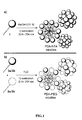
- PDA-NTA micelle Three types of micelles were synthesized and studied: the previously described PDA-NTA micelle, [16] and two new polydiacetylene-based micelles with poly(ethylene glycol) outer regions referred to as PDA-PEG350 and PDA-PEG2000 micelles, with respect to PEG length.
- PDA-NTA micelles result from the self-assembly of a single amphiphilic monomer 1 which was synthesized according to the previously reported protocol (Scheme 1) .
- PDA-PEG350 and PDA-PEG2000 micelles are composed of a mixture of two amphiphilic monomers 2 and 3.
- the general synthesis procedure, depicted in Scheme 2, starts from pentacosa-10,12-diynoic acid 4 which is first reduced into hydroxyl 5.
- An Appel halogenation reaction permits the formation of the corresponding bromide 6 which is then reacted with polyethylene glycol in presence of sodium hydride to yield either 2a or 2b, depending on PEG length.
- Deprotonation of 2a or 2b with sodium hydride and reaction with 2-bromoacetic acid 7 give rise to carboxylic acids 3a or 3b respectively.
- the degree of polymerization was controlled by UV absorption at 292 nm and more than 75% conversion was observed after 5 hours of irradiation. At that point, all three solutions had turned from colorless to yellowish.
- the obtained micelles were characterized by dynamic light scattering (DLS) which gave mean diameters of 6.4, 7.8 and 12.6 nm for PDA-NTA, PDA-PEG350 and PDA-PEG2000 micelles, respectively.
- DLS dynamic light scattering
- micelles were conjugated with NIR emitting FluoProbes ® 730 (FP730).
- the conjugation was performed via peptide coupling between the primary amine of the fluorophore and carboxylic groups at the surface of the micelles.
- PEG coatings are known to enhance blood residence time and their positive effect is clearly illustrated by these results. Early kinetics show a superior behavior of PEG-coated micelles over the NTA-coated ones, whereas long term kinetics highlight the benefit of a longer PEG chain as regards blood residence time.
- the FP730-labelled micelles were then administered to mice bearing subcutaneous tumors by injection in the caudal vein at a dose corresponding to 1 nmol of fluorescence and distribution of the objects in the whole body of the animals was monitored using planar NIR imaging.
- PDA-NTA and PDA-PEG350 micelles are quite comparable, with little tumor uptake and contrast after 24 hours.
- PDA-PEG2000 micelles showed strong and persistent tumor uptake after 24 hours with maximum contrast after 48 hours which remained constant for more than a week.
- PDA-PEG2000 micelles appeared to be the best candidates for drug delivery applications, considering their longer blood circulation residence time (which should result in a better bioavailability of the carried drug) and better overall accumulation in tumors (in terms of selectivity and quantity).
- paclitaxel a hydrophobic molecule with high anticancer activity (active compound of Taxol ® ) was chosen as a test compound.
- Encapsulation was made possible by generating the micelles in conditions similar to those described above (cf. 1 . 2 .) in the presence of an excess of suspended paclitaxel. Reproducible results obtained by this technique were measured by mass-balance, demonstrating a 10% w/w (ca. 30% mol/mol) loading of paclitaxel in PDA-PEG2000 micelles.
- This encapsulation technique is suitable for virtually any therapeutic molecule since it requires neither extended contact with water nor prolonged heating. Solutions of loaded PDA-PEG2000 micelles appeared to be very stable and no precipitate was observed after storage for more than two months at 8 °C.
- PDA-PEG2000 micelles the most promising nanoparticle presented here, have been studied further in order to determine their drug-loading ability and in vitro cytotoxicity. Obvious differences were observed between the different micelle types. It appeared that PEG350-coated micelles were superior to NTA-coated ones, but the benefit was far more significant with PEG2000-coated micelles. The latter particles demonstrated longer blood residence time, excellent tumor uptake and better imaging contrast.
- PDA-PEG2000 micelles were easily and efficiently loaded with paclitaxel, showed good in vitro cytotoxicity and behaved ideally in multiple injection conditions. Moreover, no toxicity attributable to the carrier itself was observed throughout the different experiments that were carried out.
- Preparation of polymerized micelles A solution of compound 1 (50 mg) in 0.01 M aqueous sodium hydroxide (5 mL) was sonicated with an ultrasonic probe (Output 4, DutyCycle 30%) for 10 minutes. The solution was then submitted to UV light (254 nm ⁇ low pressure mercury UV lamp - Heraeus) irradiation for 6 hours to yield PDA-NTA micelles. Hydrochloric acid was added until pH 7 was reached.
- mice All procedures were in accordance with international guidelines on the ethical use of animals. All mice that were used were female nude mice weighting approximately 23 g and housed under standard conditions with food and water ad libitum.
- Implantation of cancer cells For each mouse implanted, a syringe was prepared containing 10 6 tumor cells MDA-MB231 in a volume of 100 ⁇ L of PBS mixed with 100 ⁇ L of MatrigelTM at 0 °c. For PC 12-MEN2A cells, 3 10 6 cells were directly diluted into 200 ⁇ L of PBS.
- Biodistribution To a group of at least 3 mice having been inoculated with cancer cells (PC 12-MEN2A or MDA-MB231) a solution of fluorescent micelles (previously described) was intravenously injected in their tail under isoflurane anesthesia. Always under anesthesia, a planar fluorescent picture of each mouse's backside and frontside was taken before and after the iv injection using a 3D optical tomography apparatus developed by CEA/LETI and Cyberstar. Planar pictures were taken at various time points (1 min, 10 min, 30 min, 1 h, 3 h, 6 h, 1 day and every following day over a week) and 3D scans were done on the tumor zone before injection and every day after over a week.
- Injected nanoparticles were: PDA-PEG2000-FP730 micelles, PDA-PEG350-FP730 micelles, PDA-NTA-FP730 micelles, fluorochrome FP730 alone and a non covalent mixture of PDA-PEG2000 micelles and FP730 to be in the same concentration condition as the sample PDA-PEG2000-FP730 micelles.
- ROIs of the desired zone were selected manually by using the ImageJ software. Each ROI's value was corrected by substracting the background ROI's value (same zone but before injection) and dividing it by the exposure time. Another correction was also applied taking into account the quenching of the fluorochrome overtime: 18% in 7 days. To calculate this value a sealed capillary containing a known concentration of FP730 was put into a phantom mimicking a mouse. The phantom was put in the very same process condition and pictures were taken for the same time points.
- Blood clearance kinetics Blood samples were collected from the retro-orbital venous sinus using heparin coated capillary tubes before and after the intravenous administration of the fluorescent nanoparticles. Three mice were used for each type of micelles and samples ( ⁇ 30 ⁇ L) were collected at various time points (after 1 min, 5 min, 10 min, 30 min, 1 h, 2 h, 3 h and 24 h). Just after blood retrieval, the latter was centrifuge for 5 min at 1000 xg into an eppendorf tube in order to retrieve the plasma that contained all the fluorescence. The fluorescence was measured by analyzing the plasma into a capillary tube (so height and depth could be reproducible) with an optical tomography apparatus developed by Cyberstar and the CEA/LETI. The acquisition time was of 400 ms. For semiquantitative analysis of fluorescence images square ROIs of the capillaries were selected manually by using the ImageJ software.
- mice Plasma samples (the same as the ones used for blood clearance kinetics) were used in order to evaluate the metabolism of the injected nanoparticles. The collected samples were deposited into a 3% agarose gel (without ethidium bromide) and run over 20 minutes at 100 W. The samples were compared respectively to the same nanoparticle either in a PBS buffer and mixed with pure plasma just before the run.
- Agarose gels were analyzed with an optical apparatus (Cyberstar, CEA/LETI). The acquisition time was of 400 ms. For semiquantitative analysis of fluorescence images the ImageJ software was used.
- Drug loading of PDA-PEG2000 micelles A stock solution of compounds 2b (15 mg) and 3b (15 mg) in deionized water (3 mL) is divided in three equal parts. Paclitaxel (25 mg) is added to two of these (solution A and solution B), and the last (solution C) is left without any additive. All three solutions/suspensions are sonicated with an ultrasonic probe (Output 4, DutyCycle 30%) for 10 minutes. The solutions/suspensions are then submitted to UV light (254 nm ⁇ low pressure mercury UV lamp - Heraeus) irradiation for 6 hours and filtrated on 0.22 ⁇ m membranes to remove insoluble drug aggregates.
- UV light (254 nm ⁇ low pressure mercury UV lamp - Heraeus
- the filtrate of solution A is used for further experiments.
- In vitro cell proliferation assay In a 96 well plate were deposited 2 10 3 cells (MDA-MB231) diluted in 50 ⁇ L of culture medium. After 24 h in a cell incubator, 50 ⁇ L of a solution of Taxol ® at different paclitaxel concentrations (250, 120, 60, 40, 30, 20, 10, 5, 2, 1, and 0.5 ⁇ M) in a PBS buffer was added. Each concentration was repeated 3 times. The plate was then allowed to stand in a cell incubator for 48 h.
- MTS tetrazolium compound included in the CellTiter 96 ® AQ ueous Non-Radioactive Cell Proliferation Assay
- the data were compared to well containing only 2 10 3 cells in 50 ⁇ L of culture medium and 50 ⁇ L of PBS buffer and revealed with 20 ⁇ L of MTS.
- a background was removed consisting of 50 ⁇ L of culture medium and 50 ⁇ L of PBS buffer and revealed with 20 ⁇ L of MTS.
- the plot was expressed as a function of a percentage of living cells, 100% being the well containing only cells and MTS.
Landscapes
- Health & Medical Sciences (AREA)
- Life Sciences & Earth Sciences (AREA)
- Veterinary Medicine (AREA)
- Public Health (AREA)
- Epidemiology (AREA)
- Animal Behavior & Ethology (AREA)
- General Health & Medical Sciences (AREA)
- Chemical & Material Sciences (AREA)
- Engineering & Computer Science (AREA)
- Biomedical Technology (AREA)
- Dispersion Chemistry (AREA)
- Biophysics (AREA)
- Molecular Biology (AREA)
- Medicinal Chemistry (AREA)
- Pharmacology & Pharmacy (AREA)
- Medicines Containing Antibodies Or Antigens For Use As Internal Diagnostic Agents (AREA)
- Medicinal Preparation (AREA)
- Pharmaceuticals Containing Other Organic And Inorganic Compounds (AREA)
Priority Applications (4)
| Application Number | Priority Date | Filing Date | Title |
|---|---|---|---|
| EP10290440A EP2425817A1 (fr) | 2010-08-09 | 2010-08-09 | Micelles polymérisées pour diagnostic |
| EP11743875.4A EP2603200A1 (fr) | 2010-08-09 | 2011-07-07 | Micelles polymérisées utilisées à des fins de diagnostic |
| US13/816,414 US9839700B2 (en) | 2010-08-09 | 2011-07-07 | Polymerized micelles for diagnosis |
| PCT/IB2011/053025 WO2012020337A1 (fr) | 2010-08-09 | 2011-07-07 | Micelles polymérisées utilisées à des fins de diagnostic |
Applications Claiming Priority (1)
| Application Number | Priority Date | Filing Date | Title |
|---|---|---|---|
| EP10290440A EP2425817A1 (fr) | 2010-08-09 | 2010-08-09 | Micelles polymérisées pour diagnostic |
Publications (1)
| Publication Number | Publication Date |
|---|---|
| EP2425817A1 true EP2425817A1 (fr) | 2012-03-07 |
Family
ID=43479433
Family Applications (2)
| Application Number | Title | Priority Date | Filing Date |
|---|---|---|---|
| EP10290440A Withdrawn EP2425817A1 (fr) | 2010-08-09 | 2010-08-09 | Micelles polymérisées pour diagnostic |
| EP11743875.4A Withdrawn EP2603200A1 (fr) | 2010-08-09 | 2011-07-07 | Micelles polymérisées utilisées à des fins de diagnostic |
Family Applications After (1)
| Application Number | Title | Priority Date | Filing Date |
|---|---|---|---|
| EP11743875.4A Withdrawn EP2603200A1 (fr) | 2010-08-09 | 2011-07-07 | Micelles polymérisées utilisées à des fins de diagnostic |
Country Status (3)
| Country | Link |
|---|---|
| US (1) | US9839700B2 (fr) |
| EP (2) | EP2425817A1 (fr) |
| WO (1) | WO2012020337A1 (fr) |
Cited By (1)
| Publication number | Priority date | Publication date | Assignee | Title |
|---|---|---|---|---|
| WO2014057043A1 (fr) * | 2012-10-12 | 2014-04-17 | Georg-August-Universität Göttingen Stiftung Öffentlichen Rechts, Universitätsmedizin | Composition et véhicule de délivrance pour agents actifs et procédés associés |
Families Citing this family (1)
| Publication number | Priority date | Publication date | Assignee | Title |
|---|---|---|---|---|
| EP3225257A1 (fr) | 2016-03-30 | 2017-10-04 | Université de Strasbourg | Nanovecteurs à base de monomères amphiphiles et leur utilisation dans l'administartion d'arnsi |
Citations (8)
| Publication number | Priority date | Publication date | Assignee | Title |
|---|---|---|---|---|
| US5512294A (en) * | 1994-08-05 | 1996-04-30 | Li; King C. | Targeted polymerized liposome contrast agents |
| US6306598B1 (en) * | 1992-11-13 | 2001-10-23 | Regents Of The University Of California | Nucleic acid-coupled colorimetric analyte detectors |
| WO2002100439A1 (fr) * | 2001-06-08 | 2002-12-19 | Labopharm, Inc. | Polyelectrolytes auto-assembles stabilises solubles dans l'eau |
| WO2005058376A1 (fr) * | 2003-12-17 | 2005-06-30 | Smithkline Beecham Corporation | Complexes micellaires polymeres et vehicules d'administration de medicaments |
| WO2005120585A1 (fr) * | 2004-06-04 | 2005-12-22 | Case Western Reserve University | Micelles polymeres a fonction double |
| WO2006113666A2 (fr) * | 2005-04-19 | 2006-10-26 | Massachusetts Institute Of Technology | Polymeres amphiphiles et leurs procedes d'utilisation |
| US20070275371A1 (en) * | 2005-12-16 | 2007-11-29 | Sim Sang-Jun | Colorimetric sensor using polydiacetylene supramolecule |
| WO2009133325A2 (fr) | 2008-04-29 | 2009-11-05 | Les Laboratoires Servier | Micelles polymerisees |
Family Cites Families (3)
| Publication number | Priority date | Publication date | Assignee | Title |
|---|---|---|---|---|
| US20050003008A1 (en) * | 1997-09-23 | 2005-01-06 | Natalya Rapoport | Method of in vivo drug targeting to solid tumors via acoustically triggered drug delivery in polymeric micelles |
| CA2425508A1 (fr) * | 2000-10-11 | 2002-04-18 | John S. Pease | Agents therapeutiques cibles |
| WO2008124634A1 (fr) | 2007-04-04 | 2008-10-16 | Massachusetts Institute Of Technology | Micelles inverses encapsulées par un polymère |
-
2010
- 2010-08-09 EP EP10290440A patent/EP2425817A1/fr not_active Withdrawn
-
2011
- 2011-07-07 WO PCT/IB2011/053025 patent/WO2012020337A1/fr not_active Ceased
- 2011-07-07 US US13/816,414 patent/US9839700B2/en not_active Expired - Fee Related
- 2011-07-07 EP EP11743875.4A patent/EP2603200A1/fr not_active Withdrawn
Patent Citations (8)
| Publication number | Priority date | Publication date | Assignee | Title |
|---|---|---|---|---|
| US6306598B1 (en) * | 1992-11-13 | 2001-10-23 | Regents Of The University Of California | Nucleic acid-coupled colorimetric analyte detectors |
| US5512294A (en) * | 1994-08-05 | 1996-04-30 | Li; King C. | Targeted polymerized liposome contrast agents |
| WO2002100439A1 (fr) * | 2001-06-08 | 2002-12-19 | Labopharm, Inc. | Polyelectrolytes auto-assembles stabilises solubles dans l'eau |
| WO2005058376A1 (fr) * | 2003-12-17 | 2005-06-30 | Smithkline Beecham Corporation | Complexes micellaires polymeres et vehicules d'administration de medicaments |
| WO2005120585A1 (fr) * | 2004-06-04 | 2005-12-22 | Case Western Reserve University | Micelles polymeres a fonction double |
| WO2006113666A2 (fr) * | 2005-04-19 | 2006-10-26 | Massachusetts Institute Of Technology | Polymeres amphiphiles et leurs procedes d'utilisation |
| US20070275371A1 (en) * | 2005-12-16 | 2007-11-29 | Sim Sang-Jun | Colorimetric sensor using polydiacetylene supramolecule |
| WO2009133325A2 (fr) | 2008-04-29 | 2009-11-05 | Les Laboratoires Servier | Micelles polymerisees |
Non-Patent Citations (20)
| Title |
|---|
| A. PLUEN; Y. BOUCHER; S. RAMANUJAN; T. D. MCKEE; T. GOHONGI; E. DI TOMASO; E. B. BROWN; Y. IZUMI; R. B. CAMPBELL; D. A. BERK, PROC. NATL. ACAD. SCI. USA, vol. 98, 2001, pages 4628 |
| A.-C. FAURE; S. DUFORT; V. JOSSERAND; P. PERRIAT; J.-L. COLL; S. ROUX; O. TILLEMENT, SMALL, vol. 5, 2009, pages 2565 |
| CHOI HYE ET AL: "Synthesis of poly(ethylene glycol)-polydiacetylene conjugates and their micellar and chromic characteristics.", JOURNAL OF NANOSCIENCE AND NANOTECHNOLOGY OCT 2008 LNKD- PUBMED:19198400, vol. 8, no. 10, October 2008 (2008-10-01), pages 5104 - 5108, XP008132375, ISSN: 1533-4880 * |
| D. E.OWENS 3 RD; N. A. PEPPAS, INT. J PHARM., vol. 307, 2006, pages 93 |
| D. PEER; JEFFREY M. KARP; S. HONG; O. C. FAROKHZAD; R. MARGALIT; R. LANGER, NATURE NANOTECHNOLOGY, vol. 2, 2007, pages 751 |
| E. K. U. LARSEN; T. NIELSEN; T. WITTENBORN; H. BIRKEDAL; T. VORUP-JENSEN; M. H. JAKOBSEN; L. 0STERGAARD; M. R. HORSMAN; F. BESENBA, ACS NANO, vol. 3, 2009, pages 1947 |
| J. GAO; K. CHEN; R. XIE; J. XIE; S. LEE; Z. CHENG; X. PENG; X. CHEN, SMALL, vol. 6, 2010, pages 256 |
| J. OGIER; T. ARNAULD; G. CARROT; A. LHUMEAU; J.M. DELBOS; C. BOURSIER; O. LOREAU; F. LEFOULON; E. DORIS, ORG. BIOMOL. CHEM., 2010 |
| J.-H. LEE; K. LEE; S. H. MOON; Y. LEE; T. G. PARK; J. CHEON, ANGEW. CHEM. INT. ED., vol. 48, 2009, pages 4174 |
| K. E. ADAMS; S. KE; S. KWON; F. LIANG; Z. FAN; Y. LU; K. HIRSCHI; M. E. MAWAD; M. A. BARRY; E. M. SEVICK-MURACA, J. BIOMED. OPT., vol. 12, 2007, pages 024017 |
| L.-Y. TANG; Y.-C. WANG; Y. LI; J.-Z. DU; J. WANG, BIOCONJUGATE CHEM., vol. 20, 2009, pages 1095 |
| NURUNNABI M ET AL: "Targeted near-IR QDs-loaded micelles for cancer therapy and imaging", BIOMATERIALS 2010 ELSEVIER LTD GBR LNKD- DOI:10.1016/J.BIOMATERIALS.2010.03.057, vol. 31, no. 20, July 2010 (2010-07-01), pages 5436 - 5444, XP008132401, ISSN: 0142-9612 * |
| R.C. LAROCK: "Comprehensive Organic Transformations", 1989, VCH PUBLISHERS |
| S. DHARA; F. X. GUB; R. LANGERB; O. C. FAROKHZADC; S. J. LIPPARD, PROC. NATL. ACAD. SCI. USA, vol. 105, 2008, pages 17356 |
| V. NTZIACHRISTOS; C. BREMER; R. WEISSLEDER, EUR. RADIOL., vol. 13, 2003, pages 195 |
| V. NTZIACHRISTOS; J. RIPOLL; R. WEISSLEDER, OPT. LETT., vol. 27, 2002, pages 527 |
| V. P. TORCHILIN, PHARM. RES., vol. 24, 2007, pages 1 |
| W. WEI; G.-H. MA; G. HU; D. YU; T. MCLEISH; Z.-G. SU; Z.-Y. SHEN, J. AM. CHEM. SOC., vol. 130, 2008, pages 15808 |
| Z. YANG; S. ZHENG; W. J. HARRISON; J. HARDER; X. WEN; J. G. GELOVANI; A. QIAO; C. LI, BIOMACROMOLECULES, vol. 8, 2007, pages 3422 |
| Z. ZHANG; J. CHEN; L. DING; H. JIN; J. F. LOVELL; I. R. CORBIN; W. CAO; P.-C. LO; M. YANG; M.-S. TSAO, SMALL, vol. 6, 2010, pages 430 |
Cited By (2)
| Publication number | Priority date | Publication date | Assignee | Title |
|---|---|---|---|---|
| WO2014057043A1 (fr) * | 2012-10-12 | 2014-04-17 | Georg-August-Universität Göttingen Stiftung Öffentlichen Rechts, Universitätsmedizin | Composition et véhicule de délivrance pour agents actifs et procédés associés |
| US9950074B2 (en) | 2012-10-12 | 2018-04-24 | Georg-August-Universität Göttingen Stiftung Öffenlichen Rechts, Universitätsmedizin | Composition and delivery vehicle for active agents and methods therefor |
Also Published As
| Publication number | Publication date |
|---|---|
| US9839700B2 (en) | 2017-12-12 |
| US20130230467A1 (en) | 2013-09-05 |
| EP2603200A1 (fr) | 2013-06-19 |
| WO2012020337A1 (fr) | 2012-02-16 |
Similar Documents
| Publication | Publication Date | Title |
|---|---|---|
| Huang et al. | Mesenchymal stem cell-based cell engineering with multifunctional mesoporous silica nanoparticles for tumor delivery | |
| Luk et al. | Current advances in polymer-based nanotheranostics for cancer treatment and diagnosis | |
| Xiao et al. | Gold nanorods conjugated with doxorubicin and cRGD for combined anticancer drug delivery and PET imaging | |
| Kaul et al. | Biodistribution and targeting potential of poly (ethylene glycol)-modified gelatin nanoparticles in subcutaneous murine tumor model | |
| Fu et al. | Aspartic acid-based modified PLGA–PEG nanoparticles for bone targeting: In vitro and in vivo evaluation | |
| Park et al. | Advances in the synthesis and application of nanoparticles for drug delivery | |
| Guo et al. | Image-guided and tumor-targeted drug delivery with radiolabeled unimolecular micelles | |
| ES2828752T3 (es) | Formulaciones en gel para guiar la radioterapia | |
| Luo et al. | Thermosensitive nanocomposite gel for intra-tumoral two-photon photodynamic therapy | |
| Zhang et al. | A PEG-Fmoc conjugate as a nanocarrier for paclitaxel | |
| US20080095699A1 (en) | Imaging contrast agents using nanoparticles | |
| JP6230443B2 (ja) | 近赤外色素結合ヒアルロン酸誘導体およびそれを有する光イメージング用造影剤 | |
| US20120087859A1 (en) | Nanocarrier having enhanced skin permeability, cellular uptake and tumour delivery properties | |
| CN103096874A (zh) | 前药组合物、前药纳米粒子及其使用方法 | |
| JP2011105792A (ja) | ブロックコポリマー、ブロックコポリマー−金属錯体複合体、及びそれを用いた中空構造体キャリア | |
| Lin et al. | Ultrasound-responsive glycopolymer micelles for targeted dual drug delivery in cancer therapy | |
| Wen et al. | Near-infrared/pH dual-sensitive nanocarriers for enhanced intracellular delivery of doxorubicin | |
| Zhou et al. | Tumor-penetrating and mitochondria-targeted drug delivery overcomes doxorubicin resistance in lung cancer | |
| Li et al. | Zirconium-containing nanoscale coordination polymers for positron emission tomography and fluorescence-guided cargo delivery to triple-negative breast tumors | |
| Liu et al. | Magnetic resonance imaging-guided multi-stimulus-responsive drug delivery strategy for personalized and precise cancer treatment | |
| CN114588271A (zh) | 一种金属-药物全活性纳米药物及其制备方法与用途 | |
| Liu et al. | Microenvironment-activated nanoparticles for oxygen self-supplemented photodynamic cancer therapy | |
| Cruje et al. | Polymer assembly encapsulation of lanthanide nanoparticles as contrast agents for in vivo micro-CT | |
| Yuan et al. | Enhanced combination therapy through tumor microenvironment-activated cellular uptake and ROS-sensitive drug release using a dual-sensitive nanogel | |
| Yan et al. | Sequentially dynamic polymeric micelles with detachable PEGylation for enhanced chemotherapeutic efficacy |
Legal Events
| Date | Code | Title | Description |
|---|---|---|---|
| AK | Designated contracting states |
Kind code of ref document: A1 Designated state(s): AL AT BE BG CH CY CZ DE DK EE ES FI FR GB GR HR HU IE IS IT LI LT LU LV MC MK MT NL NO PL PT RO SE SI SK SM TR |
|
| AX | Request for extension of the european patent |
Extension state: BA ME RS |
|
| PUAI | Public reference made under article 153(3) epc to a published international application that has entered the european phase |
Free format text: ORIGINAL CODE: 0009012 |
|
| STAA | Information on the status of an ep patent application or granted ep patent |
Free format text: STATUS: THE APPLICATION IS DEEMED TO BE WITHDRAWN |
|
| 18D | Application deemed to be withdrawn |
Effective date: 20120908 |