EP2463300A2 - Molécules de liaison spécifiques à GalNAc et utilisations associées - Google Patents
Molécules de liaison spécifiques à GalNAc et utilisations associées Download PDFInfo
- Publication number
- EP2463300A2 EP2463300A2 EP12151031A EP12151031A EP2463300A2 EP 2463300 A2 EP2463300 A2 EP 2463300A2 EP 12151031 A EP12151031 A EP 12151031A EP 12151031 A EP12151031 A EP 12151031A EP 2463300 A2 EP2463300 A2 EP 2463300A2
- Authority
- EP
- European Patent Office
- Prior art keywords
- mgl
- cells
- binding
- cell
- galnac
- Prior art date
- Legal status (The legal status is an assumption and is not a legal conclusion. Google has not performed a legal analysis and makes no representation as to the accuracy of the status listed.)
- Withdrawn
Links
Images






























Classifications
-
- C—CHEMISTRY; METALLURGY
- C07—ORGANIC CHEMISTRY
- C07K—PEPTIDES
- C07K14/00—Peptides having more than 20 amino acids; Gastrins; Somatostatins; Melanotropins; Derivatives thereof
- C07K14/435—Peptides having more than 20 amino acids; Gastrins; Somatostatins; Melanotropins; Derivatives thereof from animals; from humans
- C07K14/46—Peptides having more than 20 amino acids; Gastrins; Somatostatins; Melanotropins; Derivatives thereof from animals; from humans from vertebrates
- C07K14/47—Peptides having more than 20 amino acids; Gastrins; Somatostatins; Melanotropins; Derivatives thereof from animals; from humans from vertebrates from mammals
- C07K14/4701—Peptides having more than 20 amino acids; Gastrins; Somatostatins; Melanotropins; Derivatives thereof from animals; from humans from vertebrates from mammals not used
- C07K14/4726—Lectins
-
- A—HUMAN NECESSITIES
- A61—MEDICAL OR VETERINARY SCIENCE; HYGIENE
- A61P—SPECIFIC THERAPEUTIC ACTIVITY OF CHEMICAL COMPOUNDS OR MEDICINAL PREPARATIONS
- A61P29/00—Non-central analgesic, antipyretic or antiinflammatory agents, e.g. antirheumatic agents; Non-steroidal antiinflammatory drugs [NSAID]
-
- A—HUMAN NECESSITIES
- A61—MEDICAL OR VETERINARY SCIENCE; HYGIENE
- A61P—SPECIFIC THERAPEUTIC ACTIVITY OF CHEMICAL COMPOUNDS OR MEDICINAL PREPARATIONS
- A61P35/00—Antineoplastic agents
-
- A—HUMAN NECESSITIES
- A61—MEDICAL OR VETERINARY SCIENCE; HYGIENE
- A61P—SPECIFIC THERAPEUTIC ACTIVITY OF CHEMICAL COMPOUNDS OR MEDICINAL PREPARATIONS
- A61P37/00—Drugs for immunological or allergic disorders
-
- C—CHEMISTRY; METALLURGY
- C07—ORGANIC CHEMISTRY
- C07K—PEPTIDES
- C07K14/00—Peptides having more than 20 amino acids; Gastrins; Somatostatins; Melanotropins; Derivatives thereof
- C07K14/435—Peptides having more than 20 amino acids; Gastrins; Somatostatins; Melanotropins; Derivatives thereof from animals; from humans
- C07K14/46—Peptides having more than 20 amino acids; Gastrins; Somatostatins; Melanotropins; Derivatives thereof from animals; from humans from vertebrates
- C07K14/47—Peptides having more than 20 amino acids; Gastrins; Somatostatins; Melanotropins; Derivatives thereof from animals; from humans from vertebrates from mammals
- C07K14/4701—Peptides having more than 20 amino acids; Gastrins; Somatostatins; Melanotropins; Derivatives thereof from animals; from humans from vertebrates from mammals not used
- C07K14/4727—Mucins, e.g. human intestinal mucin
-
- C—CHEMISTRY; METALLURGY
- C07—ORGANIC CHEMISTRY
- C07K—PEPTIDES
- C07K14/00—Peptides having more than 20 amino acids; Gastrins; Somatostatins; Melanotropins; Derivatives thereof
- C07K14/435—Peptides having more than 20 amino acids; Gastrins; Somatostatins; Melanotropins; Derivatives thereof from animals; from humans
- C07K14/705—Receptors; Cell surface antigens; Cell surface determinants
- C07K14/7056—Lectin superfamily, e.g. CD23, CD72
-
- C—CHEMISTRY; METALLURGY
- C07—ORGANIC CHEMISTRY
- C07K—PEPTIDES
- C07K16/00—Immunoglobulins [IG], e.g. monoclonal or polyclonal antibodies
- C07K16/18—Immunoglobulins [IG], e.g. monoclonal or polyclonal antibodies against material from animals or humans
-
- C—CHEMISTRY; METALLURGY
- C07—ORGANIC CHEMISTRY
- C07K—PEPTIDES
- C07K16/00—Immunoglobulins [IG], e.g. monoclonal or polyclonal antibodies
- C07K16/18—Immunoglobulins [IG], e.g. monoclonal or polyclonal antibodies against material from animals or humans
- C07K16/28—Immunoglobulins [IG], e.g. monoclonal or polyclonal antibodies against material from animals or humans against receptors, cell surface antigens or cell surface determinants
- C07K16/2851—Immunoglobulins [IG], e.g. monoclonal or polyclonal antibodies against material from animals or humans against receptors, cell surface antigens or cell surface determinants against the lectin superfamily, e.g. CD23, CD72
-
- C—CHEMISTRY; METALLURGY
- C07—ORGANIC CHEMISTRY
- C07K—PEPTIDES
- C07K16/00—Immunoglobulins [IG], e.g. monoclonal or polyclonal antibodies
- C07K16/18—Immunoglobulins [IG], e.g. monoclonal or polyclonal antibodies against material from animals or humans
- C07K16/28—Immunoglobulins [IG], e.g. monoclonal or polyclonal antibodies against material from animals or humans against receptors, cell surface antigens or cell surface determinants
- C07K16/30—Immunoglobulins [IG], e.g. monoclonal or polyclonal antibodies against material from animals or humans against receptors, cell surface antigens or cell surface determinants from tumour cells
- C07K16/3076—Immunoglobulins [IG], e.g. monoclonal or polyclonal antibodies against material from animals or humans against receptors, cell surface antigens or cell surface determinants from tumour cells against structure-related tumour-associated moieties
- C07K16/3092—Immunoglobulins [IG], e.g. monoclonal or polyclonal antibodies against material from animals or humans against receptors, cell surface antigens or cell surface determinants from tumour cells against structure-related tumour-associated moieties against tumour-associated mucins
-
- G—PHYSICS
- G01—MEASURING; TESTING
- G01N—INVESTIGATING OR ANALYSING MATERIALS BY DETERMINING THEIR CHEMICAL OR PHYSICAL PROPERTIES
- G01N33/00—Investigating or analysing materials by specific methods not covered by groups G01N1/00 - G01N31/00
- G01N33/48—Biological material, e.g. blood, urine; Haemocytometers
- G01N33/50—Chemical analysis of biological material, e.g. blood, urine; Testing involving biospecific ligand binding methods; Immunological testing
- G01N33/53—Immunoassay; Biospecific binding assay; Materials therefor
- G01N33/575—Immunoassay; Biospecific binding assay; Materials therefor for cancer
-
- G—PHYSICS
- G01—MEASURING; TESTING
- G01N—INVESTIGATING OR ANALYSING MATERIALS BY DETERMINING THEIR CHEMICAL OR PHYSICAL PROPERTIES
- G01N33/00—Investigating or analysing materials by specific methods not covered by groups G01N1/00 - G01N31/00
- G01N33/48—Biological material, e.g. blood, urine; Haemocytometers
- G01N33/50—Chemical analysis of biological material, e.g. blood, urine; Testing involving biospecific ligand binding methods; Immunological testing
- G01N33/53—Immunoassay; Biospecific binding assay; Materials therefor
- G01N33/575—Immunoassay; Biospecific binding assay; Materials therefor for cancer
- G01N33/57505—Immunoassay; Biospecific binding assay; Materials therefor for cancer of the blood, e.g. leukaemia
-
- A—HUMAN NECESSITIES
- A61—MEDICAL OR VETERINARY SCIENCE; HYGIENE
- A61K—PREPARATIONS FOR MEDICAL, DENTAL OR TOILETRY PURPOSES
- A61K39/00—Medicinal preparations containing antigens or antibodies
- A61K2039/505—Medicinal preparations containing antigens or antibodies comprising antibodies
-
- A—HUMAN NECESSITIES
- A61—MEDICAL OR VETERINARY SCIENCE; HYGIENE
- A61K—PREPARATIONS FOR MEDICAL, DENTAL OR TOILETRY PURPOSES
- A61K38/00—Medicinal preparations containing peptides
-
- C—CHEMISTRY; METALLURGY
- C07—ORGANIC CHEMISTRY
- C07K—PEPTIDES
- C07K2317/00—Immunoglobulins specific features
- C07K2317/70—Immunoglobulins specific features characterized by effect upon binding to a cell or to an antigen
- C07K2317/73—Inducing cell death, e.g. apoptosis, necrosis or inhibition of cell proliferation
-
- C—CHEMISTRY; METALLURGY
- C07—ORGANIC CHEMISTRY
- C07K—PEPTIDES
- C07K2319/00—Fusion polypeptide
- C07K2319/30—Non-immunoglobulin-derived peptide or protein having an immunoglobulin constant or Fc region, or a fragment thereof, attached thereto
-
- G—PHYSICS
- G01—MEASURING; TESTING
- G01N—INVESTIGATING OR ANALYSING MATERIALS BY DETERMINING THEIR CHEMICAL OR PHYSICAL PROPERTIES
- G01N2333/00—Assays involving biological materials from specific organisms or of a specific nature
- G01N2333/435—Assays involving biological materials from specific organisms or of a specific nature from animals; from humans
- G01N2333/705—Assays involving receptors, cell surface antigens or cell surface determinants
- G01N2333/7056—Selectin superfamily, e.g. LAM-1, GlyCAM, ELAM-1, PADGEM
-
- G—PHYSICS
- G01—MEASURING; TESTING
- G01N—INVESTIGATING OR ANALYSING MATERIALS BY DETERMINING THEIR CHEMICAL OR PHYSICAL PROPERTIES
- G01N2333/00—Assays involving biological materials from specific organisms or of a specific nature
- G01N2333/435—Assays involving biological materials from specific organisms or of a specific nature from animals; from humans
- G01N2333/705—Assays involving receptors, cell surface antigens or cell surface determinants
- G01N2333/70589—CD45
Definitions
- the present invention relates to GalNAc specific binding molecules, uses thereof and means and methods for manipulating macrophage galactose-type lectin (MGL) binding mediated signalling in cells that bind or have MGL.
- the invention further relates to uses of means of the invention as a diagnostic tool and/or as a medicament, in particular for the prophylaxis or treatment of chronic inflammatory conditions, autoimmune diseases, unwanted inflammatory responses in transplantation settings and/or tumours. It also relates to an in vitro method for diagnosis and/or prognosis of chronic inflammatory conditions, autoimmune diseases, and/or unwanted inflammatory responses in transplantation settings, a tumour and T cell leukaemia.
- Dendritic cells (DC) and macrophages (M ⁇ ) are professional antigen presenting cells (APC), which line all peripheral tissues where they screen their environment for incoming pathogens and for local changes in immune homeostasis. Both types of APC display a unique plasticity; thay are capable of adopting several functional phenotypes depending on environmental changes and external stimuli they receive. APC are instrumental in initiating adaptive immunity and pathogen clearance, however they also actively control several processes involved in tissue repair, clearance of apoptotic cells and maintenance of tolerance to non-harmful (self) antigens. Microbial products, like lipopolysaccharide (LPS), trigger DC maturation, thereby licensing DC for naive T cell activation.
- LPS lipopolysaccharide
- M ⁇ exposure to LPS and IFN ⁇ drives the differentiation of classically activated M ⁇ with an enhanced ability to produce nitric oxide and to kill intracellular pathogens.
- glucocorticoids instruct the differentiation of tolerogenic DC or alternatively activated M ⁇ (aaM ⁇ ). Both these glucocorticoid-elicited APC have a strongly reduced capacity to stimulate naive T cell proliferation, implicating these APC in steady state control.
- aaM ⁇ can provide negative regulatory signals to protect the host form excessive inflammatory responses.
- CLR C-type lectin receptors
- the CLR Macrophage Galactose-type lectin is expressed by human immature DC and is considered to be a marker for aaM ⁇ .
- MGL Macrophage Galactose-type lectin
- the present inventors set out to identify counterreceptors for MGL and study the functional consequences of such interaction.
- CD45 is the prototype tyrosine phosphatase expressed on the T cell surface.
- CD45 can dephosphorylate the negative regulatory tyrosine on the protein tyrosine kinase p56 lck . This results in an active pool of Lck that can phopshorylate TCR ITAMs leading to propagation of TCR signalling.
- CD45 is alternatively spliced into five different isoforms with differential glycosylation. Considering that glycosylation of CD45 changes during T cell differentiation and activation, cell-specific glycosylation could provide a means to influence various aspects of the immune system, including TCR and cytokine receptor signalling.
- autoimmune diseases In the situation of autoimmune diseases, however, they are not only mediators of the immune response, but also key players of the autoimmune response, and they are found in high numbers in chronic inflammatory diseases. Directly or indirectly (via cytokines) they are, through action of CD8+ T cells, responsible for the characteristic tissue destruction, triggered by the recognition of autoantigens. Suppression of activated T cells is particularly important for the attenuation of overshooting immune reactions.
- MGL on tolerogenic APC binds specifically to GalNAc structures present on CD45 on effector T cells.
- MGL is involved in suppression of effector CD4 + T cell proliferation by tolerogenic DC or aaM ⁇ . Binding of MGL reduces CD45 phosphatase activity and inhibits calcium mobilization after CD3 triggering.
- the MGL-CD45 interaction inhibits TCR-mediated T cell activation characterized by reduced production of inflammatory cytokine and even the induction of T cell death.
- activated T cells can be suppressed by the interaction between the carbohydrate recognition domain of MGL and the carbohydrate structures, in particular the terminal GalNAc residues, on CD45.
- MGL is potently involved in regulating the effector T cell function.
- chronic inflammatory diseases such as Multiple Sclerosis, Rheumatoid arthritis and osteoarthritis
- MGL expression is upregulated.
- the present inventors have found that the upregulation of MGL expression is a self-protecting mechanism initiated to prevent excessive inflammation and extensive tissue damage.
- Via the MGL-CD45 interaction tolerogenic APC are able to enhance the TCR threshold for activation, resulting in reduced cytokine production and proliferatio, providing another mechanism for APC to control unwanted effector responses.
- the present invention in one embodiment provides a method for providing a CD45 positive cell with a binding molecule comprising providing said cell with a binding molecule specific for a terminal GalNAc residue and that comprises a carbohydrate binding part of a macrophage galactose-type lectin (MGL).
- MML macrophage galactose-type lectin
- CD45 positive cells Several isoforms of CD45 are present on CD45 positive cells. It was found that MGL specific GalNAc residues are present on a subset of CD45 isoforms. As the different isoforms are expressed at different levels in CD45 expressing cells the invention further provides a method for differentially marking CD45 positive cells, comprising contacting a (sample containing a) subpopulation of CD45 positive cells with a binding molecule specific for a terminal GalNAc residue.
- said CD45 isoform that binds said GalNAc specific binding molecule comprises at least the RA, RB or RC part of CD45 or a combination thereof.
- said binding molecule comprises a carbohydrate binding part of an MGL. Said method for marking can be used, for example, for detecting chronic inflammation in an individual.
- target cells for binding of said GalNAc specific binding molecule are CD45 positive T-cells.
- target cells are primate cells, preferably human cells.
- the present invention it was found that binding of a GalNAc specific binding molecule of the invention results in alteration of the response in a collection of T-cells.
- the present invention therefore further provides the use of a binding molecule specific for GalNAc for altering a T-cell response of a collection of T cells.
- T-cell receptor mediated signalling is inhibited in the cell population.
- a method or a binding molecule of the invention for inhibiting a T-cell receptor (TCR)-mediated signalling pathway.
- the invention provides the use of a method or a binding molecule of the invention for inducing apoptosis in a T-cell wherein a TCR-mediated signalling pathway is activated.
- the invention provides the use of a method or a binding molecule of the invention for raising the activation threshold of a CD45 positive effector type T-cell. In yet another preferred embodiment the invention provides the use of a method or a binding molecule of the invention for inhibiting proliferation of a CD45 positive effector type T-cell.
- the invention further provides a method for providing an MGL positive cell with a binding molecule comprising providing said cell with a terminal GalNAc containing part of a MUC-1 molecule.
- a molecule that was artificially provided with one or more C-terminal GalNAc residues (a neo-glycoconjugated molecule) binds MGL.
- Means and methods for producing a neo-glycoconjugated molecule are known in the art. Another, novel method therefore is provided in example 5.
- the invention provides a method for generating an activated carbohydrate with a free maleimide moiety, the method comprising providing a bi-functional crosslinker comprising a hydrazide moiety and a maleimide moiety, coupling a carbohydrate to the hydrazide moiety of said crosslinker, and stabilizing the resulting imine group with a mild reductant.
- the hydrazide moeity of the crosslinker is covalently coupled to the reducing terminus of the carbohydrate since the carbohydrate has a free reducing terminus which is in equilibrium between the ring closed (cyclic) and ring open (acylic) forms.
- the hydrazide group of the crosslinker performs a nucleophilic attack on the carbonyl carbon of the acyclic reducing terminal residue to form a partially stable Schiff's base.
- the Schiff's base imine group is subsequently chemically reduced with a mild reductant to give a stable carbohydrate-derivate
- the resulting stabilized activated carbohydrate can be purified with any method known in the art, such as gel filtration. After said coupling and purifying, the activated carbohydrate structure can be stored indefinitely at minus -20.
- a preferred bi-functional crosslinker is (4-N-maleimidophenyl)butyric acid hydrazide.
- the invention further provides a method for modifying a cysteine residue or a lysine residue of a polypeptide with a carbohydrate, comprising providing the activated carbohydrate of the invention, and coupling said activated carbohydrate with a cystein-residue or lysine residue in said polypeptide.
- Modification of a cysteine residue can be performed by reaction of said activated carbohydrate at neutral pH with the side chain of cysteines (thiols).
- Modification of a lysine residue can be performed by changing the pH of the reaction.
- said molecule provided with said terminal GalNAc residues is a peptide.
- a preferred peptide is a polypeptide.
- a preferred polypeptide has a molecular weight of less than 100 kiloDalton (kD), more preferred less than 10 kD, more preferred less than 5 kD, more preferred less that 1 kD. It is furthermore preferred that said polypeptide is soluble in an aquous solution and has a pharmaceutically acceptable half life.
- Said polypeptide can be modified by for example PEGylation, the addition of polyethyleneglycol, to improve pharmaceutically relevant parameters such as shielding of antigenic and immunogenic epitopes, and preventing recognition and degradation by proteolytic enzymes, as is known to a skilled artisan.
- an MGL positive cell with said binding molecule comprising a carbohydrate containing part of a CD45 molecule, MUC-1 or a terminal GalNAc containing part of a MUC-1 molecule, or a terminal GalNAc containing molecule such as a neo-glycoconjugated molecule allows modulation of APC signalling and thereby modulation of various aspects of the immune system, including alteration of a T-cell response of a subset of T-cells, inhibition of T-cell receptor-mediated signalling, and induction of apoptosis in a T-cell wherein a TCR_mediated signalling pathway is activated.
- an MGL positive cell with said binding molecule comprising a carbohydrate containing part of a CD45 molecule, MUC-1 or a terminal GalNAc containing part of a MUC-1 molecule, or a terminal GalNAc-containing molecule such as a neo-glycoconjugated molecule, and/or an antibody specific for MGL or a functional part, derivative and/or analogue thereof allows modulation of APC signalling and thereby modulation of various aspects of the immune system, including but not limited to alteration of a T-cell response of a subset of T-cells, inhibition of T-cell DC maturation, antigen presentation and cytokine release -mediated, and/orinduction of apoptosis in a T-cell wherein a TCR_mediated signalling pathway is activated.
- said modulation results in altered cytokine production, such as for example the production of interleukins, such as IL6, IL-10, IL-12p70, IL-27, IL-23p19 and signalling molecules such as interferon alpha, interferon gamma, TGF ⁇ and TNF ⁇ .
- Said modulation also comprises enhanced antigen presentation of a MGL-positive cell to specific T cells, resulting in, for example, enhanced proliferation of said T cells and thus making MGL a suitable target in immunization and vaccination protocols.
- Said modulation furthermore can lead to DC maturation, macrophage activation, and/or functional modification of the DC or macrophage phenotype resulting in an alteration of a Th2-inducing capacity.
- Said alteration of a Th2-inducing capacity provides an alternative strategy for treatment of autoimmune diseases, pro-inflammatory responses, anti-inflammatory responses, and allergic responses such as asthma.
- antigen is directed to the MGL positive antigen presenting cells, thereby enabling and/or enhancing presentation of said antigen by said antigen presenting cell. It has been found that antigen presentation of antigens that are provided to the antigen presenting cell via MGL is enhanced in matured or upon maturation of the antigen presenting cell. Maturation can be stimulated in various ways. In a preferred embodiment of the invention maturation of antigen presenting cells is stimulated through administering to said subject a composition for stimulating DC-maturation and/or activation. Various agents and maturation stimulating compositions and compounds are known to the person skilled in the art.
- said compound and/or composition for stimulating DC maturation comprises an anti CD40 molecule, a Toll-like receptor ligand, a pro-inflammatory cytokine such as Interleukin-1, IL-6, tumor necrosis factor alpha, and/or prostaglandin E-2, an general adjuvant such as Montanide adjuvant or Freund adjuvant, or in situ maturation through the injection of immature DCs into adjuvant-treated skin.
- a pro-inflammatory cytokine such as Interleukin-1, IL-6, tumor necrosis factor alpha, and/or prostaglandin E-2
- an general adjuvant such as Montanide adjuvant or Freund adjuvant
- the invention further provides a vaccine comprising a binding molecule comprising a terminal GalNAc residue and/or a binding molecule specific for MGL, and an antigen against which the immune response is to be directed.
- Said vaccine can be used to immunize of tolerize for said antigen, depending on the presence or absence of DC maturation or an agent that modulates the maturation of DC.
- the invention further provides a method for modulating an immune response in a subject wherein said modulation comprises dampening said immune response. It has been found that immature DC efficiently take up antigen via MGL and that this presentation leads to a dampening of an immune response in said subject. This dampening effect can lead to the induction of tolerance in said subject. The dampening effect is more pronounced when no further composition for stimulating and/or inducing an immune response is provided to said subject.
- said method comprises administering to said subject said binding molecule in the absence of a composition for stimulating maturation of DC.
- the dampening effect of said binding molecule or complex comprising said binding molecule is enhanced when said subject is also administered afurther composition for dampening an immune response in said subject.
- the invention comprises a method for modulating an immune response comprising stimulating the Th2-inducing capacity in said subject.
- modulation of said immune response comprises altering cytokine release by MGL positive antigen presenting cells.
- said modulation is preferably achieved with a binding molecule comprising a terminal GalNAc, an anti-MGL antibody, or an MGL-binding part of a CD45 molecule, MUC-1 or a terminal GalNAc containing part of a MUC-1 molecule, or a terminal GalNAc-containing molecule such as a neo-glycoconjugated molecule.
- said MGL specific binding molecule comprises an antibody or a fragment or analogue thereof.
- the invention comprises a method for modulating an immune response wherein said molecule and/or said complex further comprises a toxic moiety and/or immune suppresive agent.
- said molecule and/or said complex further comprises a toxic moiety and/or immune suppresive agent.
- MGL positive cells are provided with said toxic moiety thereby counteracting the effect of said MGL positive cell in a subject.
- said method for modulation of said immune response comprises altering and preferably stimulating MHC-I and/or MHC-II presentation by antigen presenting cells.
- antigen presenting cells Preferably of an antigen also provided.
- said modulation results in altered cytokine release, altered antigen presentation, and/or altered DC maturation and macrophage activation.
- Altered cytokine release results, for example, in the production of interleukins, such as IL6, IL-10, IL-12p70, IL-27, IL-23p19 and signalling molecules such as interferon alpha, interferon gamma, TGF ⁇ and TNF ⁇ .
- Altered antigen presentation, preferably enhanced antigen presentation, of a MGL-positive cell to specific T cells results in, for example, enhanced proliferation of said T cells and thus making MGL a suitable target in immunization and vaccination protocols.
- the invention provides a method for providing an MGL positive cell with a binding molecule of the invention for enhancing antigen presentation in an immunization and/or vaccination protocol.
- Said modulation furthermore can lead to DC maturation, macrophage activation, and/or functional modification of the DC or macrophage phenotype resulting in an alteration of the Th2-inducing capacity.
- Said alteration of a Th2-inducing capacity provides an alternative strategy for treatment of autoimmune diseases, pro-inflammatory responses, anti-inflammatory responses, and allergic responses such as asthma.
- the invention further provides a method for preferentially marking immature and/or tolerogenic professional antigen presenting cells (APC) in a sample comprising marking said cells with a binding molecule specific for MGL.
- said binding molecule comprises an anti-MGL antibody, or an MGL-binding part thereof.
- a preferred antibody is a Tn-specific antibody, that recognizes the blood cell epitope T-nouvelle (Tn).
- said binding molecule comprises one or more terminal GalNAc residues. In one aspect this embodiment is used to purify said immature and/or tolerogenic professional antigen presenting cells (APC) from a collection of cells.
- the invention provides a protein selected from a fusion protein comprising CD45, the RA, RB, RC part of CD45, MUC-1, or a terminal GalNAc containing part of a MUC-1 molecule.
- the invention furthermore provides the use of said fusion protein or of a neo-glycoconjugated protein for elucidating an immune response against said fusion protein or neo-glycoconjugated protein.
- Peptides derived from said fusion protein or neo-glycoconjugated protein will be presented by an MGL-positive APC upon contact of an APC with the fusion protein or neo-glycoconjugated protein.
- Said presentation can be more effective than presentation of peptides derived from a protein without GalNAc. Furthermore, said presentation is specific for said protein.
- said fusion protein or neo-glycoconjugated protein comprises an antigen.
- An antigen is any substance that causes the production of antibodies or the induction of effector cells by the immune system.
- a preferred antigen is provided by a bacterial or viral protein or an antigenic part thereof.
- Another preferred antigen is provided by a tumor protein, or part of a tumor protein.
- a tumor protein is a protein that is expressed in a tumor cell, such as, for example, MUC-1, EpCAM and ERBB2 (HER2/Neu).
- Another preferred antigen is a tumor-specific antigen, resulting from a tumor specific mutation, or a tumor-associated antigen. Yet a further preferred antigen is provided by a string-of-beads comprising multiple tumor-associated cytotoxic T lymphocyte epitopes ( Toes et al., 1997 PNAS 94: 14660-65 ).
- Stimulating an immune response is preferably performed on mature DC.
- DC can be matured, for instance, by adding TLR ligand or anti-CD40 in combination with said fusion protein or neo-glycoconjugated protein.
- the invention further provides a method for altering cell-cell mediated signalling in a culture of T-cells and APC, comprising altering MGL-CD45 mediated signalling in said collection of cells.
- Said MGL-CD45 mediated signalling is preferably altered by providing said culture with a molecule capable of blocking said interaction.
- a preferred molecule capable of blocking said interaction is GalNAc or a molecule comprising a terminal GalNAc residue.
- Another preferred example is a carbohydrate binding part of MGL, HPA, a terminal GalNAc specific antibody and/or EGTA.
- said signalling is blocked for stimulating an immune response against an antigen.
- tumour cells of epithelial origin contain MUC-1 molecules that have terminal GalNAc residues that are recognized by MGL.
- the invention thus further provides a method for detecting tumour cells of epithelial origin comprising contacting a sample comprising said cells with a carbohydrate binding part of a macrophage galactose-type lectin (MGL) and detecting the presence of cells that have said carbohydrate binding part. Said part, when linked to a toxin or an otherwise cell death promoting compound said MGL can also be used to selective kill said tumour cells of epithelial origin.
- MML macrophage galactose-type lectin
- the mentioned binding molecule specific for a terminal GalNAc residue is preferably used for the preparation of a composition for binding CD45 positive T-cells in an animal body.
- Such compositions are preferably used for at least inhibiting a T-cell mediated immune response in said animal body.
- said animal is a human.
- the invention provides a protein comprising a carbohydrate recognition domain of MGL.
- MGL is human MGL or mouse MGL2.
- said protein is not MGL-Fc.
- MGL is normally associated with the cellular membrane
- the extracellular part containing said carbohydrate recognition domain is preferably modified such that it rendered soluble.
- membrane associated proteins can be rendered soluble. Often this is done by linking said extracellular part to the constant part of an immunoglobulin.
- said carbohydrate recognition domain of MGL is connected to a multimerisation domain. Such a configuration closely mimics the cellular situation where MGL is at least in part present in multimers.
- said multimerisation domain is a dimerisation, trimerisation or tetramerisation domain.
- the resultant protein is a chimeric molecule.
- Preferred immunoglobulin constant regions or multimerization domains are mouse IgG2A Fc and human IgG1-Fc.
- said protein is MGL-Fc.
- the invention further provides protein comprising a carbohydrate recognition domain of MGL and multimerisation domain for use as a medicament.
- the invention further provides use of a binding molecule specific for a terminal GalNAc residue in the manufacture of a medicament for the prophylaxis or treatment of chronic inflammatory conditions, autoimmune diseases, and/or unwanted inflammatory responses in transplantation settings.
- a binding molecule specific for a terminal GalNAc residue in the manufacture of a medicament for the prophylaxis or treatment of T cell leukaemia.
- said binding molecule comprises a carbohydrate binding part of MGL.
- said MGL is human MGL.
- said binding molecule specific for a terminal GalNAc residue comprises an antibody specific a terminal GalNAc residue.
- an antibody specific for Tn-antigen Preferably an antibody specific for Tn-antigen.
- the invention provides an in vitro method for diagnosis and/or prognosis of chronic inflammatory conditions, autoimmune diseases, and/or unwanted inflammatory responses in transplantation settings, said method comprising the step of allowing a binding molecule specific for a terminal GalNAc residue, preferably comprising a carbohydrate recognition domain of MGL, to bind to effector T cells.
- an in vitro method for diagnosis and/or prognosis of a tumour said method comprising the step of allowing a binding molecule specific for a terminal GalNAc residue, preferably comprising a carbohydrate recognition domain of MGL, to bind to tumour cells.
- an in vitro method for diagnosis and/or prognosis of T cell leukaemia comprising the step of allowing a binding molecule specific for a terminal GalNAc residue, preferably comprising a carbohydrate recognition domain of MGL, to bind to T cell leukaemia cells.
- said terminal GalNAc specific binding molecule comprises an antibody.
- said antibody is a Tn-specific antibody.
- MGL detects elevated levels of CD45 positive cells in blood and synovial fluid of patients suffering from chronic inflammation. Further provided is thus a method for the detecting CD45 positive cells in an individual suffering from chronic inflammation comprising contacting a carbohydrate binding part of MGL with a blood sample or a synovial fluid sample of said individual and detecting binding of said carbohydrate part to cells in said sample.
- said chronic inflammation comprises multiple sclerosis, arthritis, preferably rheumatoid arthritis or osteoarthritis.
- MGL is among others expressed as a trimeric protein, it is preferred that the carbohydrate recognition domain of MGL is coupled to a multimerisation domain as to create multimers.
- MGL is preferably a human MGL.
- the present invention further relates to a chimeric protein comprising a carbohydrate recognition domain of MGL and a multimerisation domain, preferably separated by means of a spacer.
- MGL-Fc is excluded.
- the carbohydrate recognition domain of MGL is located in the 141 C-terminal amino acids, i.e. amino acids 152-292. Therefore, preferably the C-terminal 141 amino acids of MGL are present in the chimeric protein.
- Carbohydrate recognition domain refers to the protein domain that is responsible for recognition of and binding to terminal GalNAc structures on a carbohydrate structure.
- a “carbohydrate” as used herein refers to a sequence of one or more sugars linked in any way possible.
- Multimerisation domain refers to an amino acid sequence that induces multimerisation. Such multimerisation domain may be a dimerisation domain, a trimerisation domain, a tetramerisation domain, and the like. Any multimerisation domain may be used. In an embodiment, the multimerisation domain is derivable from a soluble protein. Examples of a multimerisation domain that can be used in the present invention are the Fc chain, His -tag, or Avi-tag.
- the carbohydrate recognition domain of MGL may be separated from the multimerisation by means of a spacer.
- a spacer may be any amino acid sequence allowing the carbohydrate recognition domain and the multimerisation domain to perform their function, i.e. recognition of and binding to a carbohydrate region comprising terminal GalNAc structures and multimerisation, respectively.
- the multimerisation domain is a dimerisation, trimerisation or tetramerisation domain.
- MGL is expressed as a trimer in its natural environment.
- MGL-Fc dimeric form of MGL
- the multimerisation domain is mouse IgG2A Fc.
- the multimerisation domain is IgG1-Fc, preferably human IgG1-Fc.
- IgG1-Fc is a dimerisation domain that when used in the present invention yields an active chimeric protein.
- said Fc is a human Fc.
- said Fc is an Fc that is deficient in complement activation.
- said deficient Fc is an IgG4 Fc or a mutant IgG1.
- the present invention also relates to a DNA molecule encoding such chimeric protein, a gene construct comprising such DNA molecule, and a host cell comprising such gene construct.
- the present invention relates to a chimeric protein comprising the carbohydrate recognition domain of MGL and a multimerisation domain for use as a medicament. Hitherto, no therapeutical implication for such chimeric protein, including MGL-Fc, has been envisaged and described.
- the present invention relates to the use of a chimeric protein comprising the carbohydrate recognition domain of MGL and a multimerisation domain in the manufacture of a medicament for the prophylaxis or treatment of chronic inflammatory conditions, autoimmune diseases, and/or unwanted inflammatory responses in transplantation settings.
- the present invention is directed to the use of a chimeric protein comprising the carbohydrate recognition domain of MGL and a multimerisation domain in the manufacture of a medicament for the prophylaxis or treatment of T cell leukaemia.
- a chimeric protein comprising the carbohydrate recognition domain of MGL and a multimerisation domain in the manufacture of a medicament for the prophylaxis or treatment of T cell leukaemia.
- the use of such chimeric protein such as MGL-Fc may induce apoptosis of the leukaemic T cells, thereby providing a system to manage the disease.
- said chimeric protein can be combined with any suitable carrier, diluent, adjuvant, excipient, etc. in order to obtain the medicament in the desired administration form.
- said medicament is administered intravenously.
- the chimeric protein according to the invention may be administered alone or in admixture with a pharmaceutically acceptable carrier, in suitable pharmaceutical formulations that are a further object of the invention.
- formulations which may be prepared using well known methods and excipients, such as those described in "Remington's Pharmaceutical Sciences Handbook", Mack Pub. Co., N.Y. U.S.A., are tablets, capsules, syrups, and the like for oral administration, whereas for the parental administration suitable forms are sterile solutions or suspensions in acceptable liquids, implants, etc.
- the posology will depend on several factors such as type and seriousness of the conditions to be treated, patient's weight and sex, etc. and will be easily determined by the skilled practitioner.
- the chimeric protein is MGL-Fc, since it has been shown to have the activity desired for the present invention.
- the immune system protects the body from potentially harmful substances (antigens). Harmful antigens are destroyed by the immune response. Immune system disorders occur when the immune response is inappropriate, excessive, or lacking.
- Chronic inflammatory diseases are diseases that represent an inflammatory response of prolonged duration whose extended time course is provoked by persistence of the causative stimulus to inflammation in the tissue.
- Non-limiting examples of chronic inflammatory diseases are tuberculosis, chronic cholecystitis, bronchiectasis, rheumatoid arthritis, (Hashimoto's) thyroiditis, inflammatory bowel disease (ulcerative colitis and Chrohn's disease), silicosis and other pneumoconioses, and an implanted foreign body in a wound.
- autoimmune diseases develop when the immune system destroys normal body tissues. Normally, the immune system is capable of differentiating "self” from “non-self” antigens. In autoimmune diseases the normal control process is disrupted and the immune system cannot differentiate anymore between "self” and “non-self” antigens.
- Non-limiting examples of autoimmune diseases are Graves' disease, Hashimoto's disease, multiple sclerosis, rheumatoid arthritis, systemic lupus erythematosus, pernicious anemia, Addison's disease, type I diabetes, dermatomyositis, Sjogren's syndrome, lupus erythematosus, myasthenia gravis, and Reiter's syndrome.
- the present invention relates to an in vitro method for diagnosis and/or prognosis of chronic inflammatory conditions, autoimmune diseases, and/or unwanted inflammatory responses in transplantation settings, said method comprising the step of allowing a chimeric protein, said chimeric protein comprising the carbohydrate recognition domain of MGL and a multimerisation domain, to bind to effector T cells.
- the present invention relates to an in vitro method for diagnosis and/or prognosis of a tumour, said method comprising the step of allowing a chimeric protein, said chimeric protein comprising the carbohydrate recognition domain of MGL and a multimerisation domain, to bind to tumour cells.
- the present invention relates to an in vitro method for diagnosis and/or prognosis of T cell leukaemia, said method comprising the step of allowing a chimeric protein, said chimeric protein comprising the carbohydrate recognition domain of MGL and a multimerisation domain, to bind to T cell leukaemia cells.
- Suitable diagnostic systems include ELISA-based tests
- the CLR MGL as the receptor expressed by professional APC for the tyrosine phosphatase CD45.
- the mucin-type protein CD45 contains high amounts of O-linked carbohydrates, mainly localized in the differentially expressed A-, B-, and C-domains15. MGL clearly recognized all A-, B- and C-containing isoforms, whereas CD45RO was not bound.
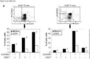
- CD45RAneg memory and effector T cells retain expression of the CD45B and CD45RO isoforms. As CD45RO is not recognized, MGL-mediated T cell binding is most likely mediated via the B-isoform.
- Glycosylation is a cell-specific process, depending not only on the cellular composition of glycosyltransferases and glycosidases, but also on the activation and differentiation status of the cell 19 . Glycosylation can thus influence diverse aspects of the immune system, although single glycans can have a highly specific functions.
- CD45 glycosylation changes during T cell development, pheripheral activation and aging 18,30 . Furthermore, different CD45 isoforms were shown to be differentially glycosylated 16 . Therefore differential glycosylation of CD45 provides a scaffold for CLR-based modulation of TCR-mediated responses.
- CD45 on Effector T cells (T-eff) exhibits the necessary glycan structures, due to cell-specific glycosylation, to ensure MGL recognition.
- CD45 activity can be controlled via receptor-mediated mechanisms 15 ; we clearly demonstrate here that MGL on APC is capable of modulating CD45 phosphatase activity and function.
- CLR 6 Every professional APC expresses a wide variety but unique repertoire of CLR 6 .
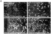
- CLR MGL and DC-SIGN are localized on different APC in skin and LN, whereas in lamina basement of the jejunum they are co-expressed on the same APC.
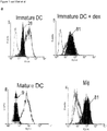
- MGL is expressed on in vitro monocyte-derived DC and Macrophages (Mf).
- APC cultured with dexamethasone display an enhanced and prolonged MGL expression. Mature DC have lost all MGL surface expression.
- CLR clear and internalize self-antigens for presentation under steady state conditions without causing overt DC maturation, thereby actively induce tolerance towards sequestered self-antigens 7 .
- crosslinking of the CLR Dectin-1 induces intracellular signaling pathways leading to production of inflammatory mediators such as reactive oxygen species and TNF ⁇ 10 .
- CLR-mediated signaling modulates APC function
- MGL is involved in regulation of the responder T cell.
- MGL preferentially recognized antigen-experienced CD27neg T-eff, which are characterized by high levels of pro-inflammatory cytokine production and tissue homing properties 27 . T-eff no longer require extensive co-stimulation, antigen recognition is sufficient for these cells to be activated 33 .
- MGL expression is equally upregulated 11 . Upregulation of MGL expression during RA disease course could therefore be a self-protecting mechanism initiated to prevent excessive inflammation and tissue damage. Via the MGL-CD45 interaction tolerogenic APC, expressing low levels of co-stimulatory molecules are able to suppress TCR-mediated T cell activation, resulting in reduced cytokine production and proliferation, providing another mechanism for APC to control unwanted effector responses. Likewise, LPS-matured DC that initiate adaptive T cell immunity, have no need for receptors with a downregulatory function, explaining why mature DC are essentially MGLneg. The overall negative regulatory effect of MGL on effector T cell activation, together with its expression profile, suggests that MGL controls unwanted effector T cell activation in the steady state or during chronic inflammation. Therefore MGL contributes to the maintenance of tolerance by controlling excessive immune responses.
- CD45 splice variants have similar phosphatase activities; still the different isoforms show very restricted expression patterns 34 . How can changes in extracellular CD45 isoforms modulate CD45 function?
- One model proposes that homodimerization inhibits phosphatase activity through interactions between an inhibitory structural wedge and the catalytic site within the cytoplasmic CD45 phosphatase domains 35 .
- the recently published crystal structure of CD45 seems incompatible with the wedge model 36 .
- Another straightforward explanation is receptor-mediated regulation of CD45 phosphatase activity. MGL specifically recognizes CD45 depending on the isoform and the cellular context in which this isoform is expressed.
- glycosylation patterns of single cells determine whether MGL is able to interact with and modulate CD45 function.
- alternations in human CD45 splice variants are associated with autoimmune diseases, such as multiple sclerosis and systemic lupus erythromatosis 37,34 . It would be interesting to investigate whether these mutations lead to altered glycosylation patterns, thus affecting MGL recognition and CD45 activity.
- CD45 can also negatively influence signaling pathways initiated by surface receptors 38 .
- Several reports show that engagement of cell surface CD45 by monoclonal antibodies induced a programmed cell death pathway in lymphocytes 39-41 .
- CD45 regulates apoptosis in peripheral T cells of transgenic mice expressing one single CD45 isoform 42 . Similar CD45-dependent apoptosis could be detected in Jurkat after MGL-Fc binding; however cell death was equally dependent on CD3-triggering, hinting to antigen-specific induction of cell death.
- CD45 The unique activating and suppressive features of CD45 make it an attractive therapeutical target molecule.
- anti-CD45RB antibodies potently inhibit T cell responses.
- T-eff the CD45RBbrightCD4+ T cell population harbors T-eff, some of which are capable of inducing autoimmunity or inflammatory bowel disease 44,45 .
- Treatment with anti-CD45RB monoclonal antibodies prolongs long-term allograft survival and donor-specific tolerance in several murine renal and islet transplantation models 46 .
- An anti-human CD45RO/RB specific antibody effectively inhibited antigen-specific and polyclonal T cell responses, mediated apoptosis of CD4+CD45RO/RBbright T cells and induced type 1 T-regs in vitro 47 .
- These IL-10 producing T-regs can likewise be generated by differentiating naive T cells in the presence of dexamethasone treated APC 48 .
- MGL-Fc Compared to the anti-CD45 antibodies, MGL-Fc exhibits an enhanced specificity, targeting only those T-eff cells that have properly glycosylated CD45. In contrast anti-CD45RB antibodies bind and potentially affect all leukocytes that express the isoform, independent of cellular glycosylation patterns.
- the invention further provides a method for providing cells with a specific binding molecule comprising providing said cells with a binding molecule specific for a terminal GalNAc residue.
- said cells are human lymphatic endothelial cells.
- said cells are human mammary epithelial cells.
- the invention further provides a method for providing human CD34 positive cells with a specific binding molecule said method comprising providing said cells with a binding molecule specific for MGL.
- said human CD34 positive cells are AML cells.
- said MGL binding molecule is an MGL specific antibody.
- said binding molecule comprises a carbohydrate containing part of a CD45 or a MUC1 molecule as described herein above.
- said MGL binding molecule is a binding molecule or complex comprising a terminal GalNAc residue as described herein above.
- said cells are provided with said binding molecule for marking said cells.
- MUC1 glycosylated MUC1.
- MUC1 from healthy or allergic mothers exhibited differences in glycosylation that correlated with the disease status.
- the total MUC1 content did not vary significantly.
- MUC1 from allergic mothers displays a higher MGL binding, which can be explained by the higher exposure of ⁇ -GalNAc epitopes as measured by the ⁇ -GalNAc-specific lectin HPA.
- MUC1 protein is also present in various infant formulas, particularly in those recommended for infants that are suffering from or at risk of suffering from an allergy.
- the invention now provides a method for preparing an infant food, preferably an infant formula or a protein containing component thereof comprising testing said food and/or said component for the presence of MUC 1 comprising a terminal GalNAc residue (preferably an ⁇ -GalNAc epitope).
- the invention thus further provides an infant formula that is tested with a method of the invention.
- the number of ⁇ -GalNAc epitopes in an infant formula, particularly for the mentioned infants is preferably reduced.
- MUC1 is particularly prevalent in milk derived protein sources.
- the current processes for preparing milk derived protein source for infant formula favor the generation of MUC 1 exhibiting ⁇ -GalNAc epitopes. These can be adapted to reduce the amount of ⁇ -GalNAc epitope on MUC1.
- the invention further provides an infant formula comprising a protein source containing MUC1, preferably from milk, wherein the amount of ⁇ -GalNAc epitopes is reduced.
- said infant formula is essentially free of ⁇ -GalNAc epitopes on MUC1.
- the invention further provides the use of such infant formula for feeding infants suffering from or at risk of suffering from an allergy.
- said ⁇ -GalNAc epitope comprises a terminal GalNAc residue.
- said MGL, CD45 and/or MUC 1 protein and/or cells comprising said protein are human proteins and/or human cells.
- the invention further provides the use of a terminal GalNAc specific antibody to bind to CD45 positive cells or MUC1 positive cells, preferably MUC 1 positive tumor cells, preferably colon carcinoma cells.
- a terminal GalNAc specific antibody to bind to CD45 positive cells or MUC1 positive cells, preferably MUC 1 positive tumor cells, preferably colon carcinoma cells.
- said antibody is used in a method for modulating an immune response in a subject.
- said antibody is a Tn-specific antibody.
- An antibody is preferably a human, humanized or deimmunised antibody.
- An antibody may be produced by a cell or generated synthetically.
- an antibody is said to be human when it is derived from a human cell, or the equivalent thereof.
- Suitable equivalents are for instance, transgenic mice comprising the human antibody locus or in vitro antibody (fragment) selection systems expressing (parts of) human antibodies.
- Non-limiting examples are phage libraries expression human single chain Fv fragments.
- a human antibody thus preferably comprises a sequence that has an equivalent sequence in a human.
- CDR regions are typically generated in response to an antigen the equivalence typically does not extend to the CDR regions.
- a humanized antibody typically comprises the CDR regions of a non-human antibody grafted on a human constant region.
- the framework region is typically also equivalent to a human antibody.
- a deimmunized antibody comprises a variable region derived from an animal other than a human from which human T-cell epitopes in at least the
- the invention further provides a method for providing a (poly)peptide with a carbohydrate residue, said method comprising producing a stable reactive carbohydrate intermediate.
- CD45 is the main counterreceptor for MGL on lymphocytes.
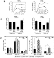
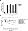
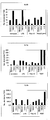
- Capture of ligands from PBL or Jurkat lysates on MGL-Fc coated plates was detected by specific antibodies to CD43, CD45 or LFA-1 (isotype control) in an ELISA-binding assay. MGL-Fc binding was blocked by the addition of 10 mM EGTA. One representative experiment out of three is shown. Standard deviation ⁇ 0.05 OD 450 nm.
- MGL only recognizes CD45 isolated from PBL.
- MGL binding proteins were captured from lysates of PBL, monocytes, immature DC or PMN by MGL-Fc coated plates was detected by specific antibodies to CD45 in an ELISA-binding assay.MGL-Fc binding was blocked by the addition of 10 mM EGTA. One representative experiment out is shown. Standard deviation ⁇ 0.05 OD 450 nm.
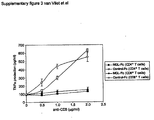
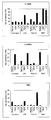
- Purified CD4+ and CD8+ T cells (>95 pure) were seeded onto MGL-Fc/anti-CD3- or control-Fc/anti-CD3-coated plates After 24 hours supernatants were harvested and TNF ⁇ production was measured by ELISA.
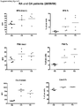
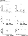
- MGL binds to PBLs from chronic infected patients with RA and OA.
- HPA which has a similar glycan specificity as MGL showed enhanced staining of PBL from RA and OA patients.
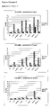
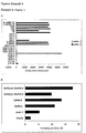
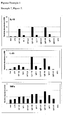
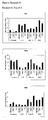
- Example 1 Plant lectin staining on PBLs from healthy donors and MS patients (I).
- Example 1 Plant lectin staining on PBLs from healthy donors and MS patients (II).
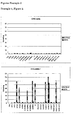
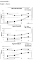
- Example 1 MGL-Fc binding to PBLs from healthy donors and MS patients (I).
- Example 1 MGL-Fc binding to PBLs from healthy donors and MS patients (II).
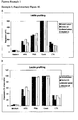
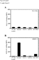
- Example 1 Supplementary figure 10 . Lectin profiling of surface glycans on mouse lymphocytes. Mouse lymphocytes were isolated from lymph node and spleen. Cell surface glycans were determined by staining with plant lectins and measured by flow cytometry. In Figure 10A the mean fluorescent intensity (MFI) is shown and in Figure 10B the percentage of positive cells.
- MFI mean fluorescent intensity
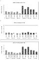
- Example 1 MGL recognition of mouse T cells (I). Beads binding was determined by flow cytometry. Binding is denoted as the percentage of cells, which have bound the coated beads.
- Example 1 MGL recognition of mouse T cells (II). Binding of human MGL-Fc (A and B), mMGL1-Fc (C and D) and mMGL2-Fc (E and F) was determined by flow cytometry.
- MUC1-Fc fusion proteins contain Tn-antigens
- MUC1-Fc was serially diluted and coated on ELISA plate.
- Glycan structures analysed with Tn-specific antibodies (5E5), HPA (specific for Tn-antigens) and TF-specific antibodies (HH8).
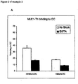
- Example 2 MUC1-Tn interacts with dendritic cells
- MUC1-Fc proteins were coupled to fluorescent beads and incubated with day 5 immature or mature monocyte derived DC in the presence or absence of 10 mM EGTA (A).
- Beads coated with polyacrylamide coupled Lewis X structures (ligand for DC-SIGN on DC) served as a positive control for beads binding (B).
- the figure shows the percentage of cells binding beads as determined by flow-cytometry.
- Mature DC were obtained by incubating DC in the presence of 100 ng/ml lipopolysaccharide overnight. Samples were analyzed in triplicates (error bars represent standard deviation of the mean) and 1 representative experiment is shown out of 3.
- Example 2 The Macrophage Galactose-type C-type lectin (MGL) binds to MUC1
- A To determine the specificity of MUC1 interaction with DC, cells were pre-incubated with different carbohydrate structures before adding MUC1-Fc coated beads. Only GalNAc structures reduced binding of MUC1 to DC and mannan blocked binding of Lewis x.
- B MGL expression on day 5 immature and mature DC was determined by mAb 18E4
- C DC were pre-incubated with different anti-CLR blocking antibodies before incubation with MUC1-Fc coated beads. The figure shows the percentage of cells binding beads as determined by flow-cytometry. Samples were analyzed in triplicates (error bars represent standard deviation of the mean) and 1 representative experiment is shown out of 3.
- Example 2 Figure 4: MUC1 binds to MGL transfected CHO cells
- the mucins were detected by the same antibody and binding by MGL-Fc, HPA, mAb HH8 and MAA investigated.
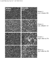
- Cryosections were stained with anti-MGL (18E4), anti-CD68 (EBM11), anti-DC-SIGN (AZN-D1) or anti-MUC1 antibodies (mAb 214D4) for 2 h at 37°C. After washing the sections were incubated with Alexa-594 labeled goat anti-mouse IgG2a (18E4) and Alexa-488 labeled goat anti-mouse IgG1.
- MUC1 staining was performed on both normal tissue (C) and colon carcinoma (D).
- Cryosections were stained with MGL-murine Fc for 2 h at 37°C, in the absence (A and B) or presence (C and D) of 100 mM GalNAc monosaccharides. After washing the sections were incubated with Alexa-488 labeled goat anti-mouse IgG. Both normal colon epithelium (A and C) and primary colon carcinoma (B and D) were stained from the same patient. Results from one representative patient out of 4 is shown.
- Example 2 Figure 8. MGL-Fc detects secreted MUC1 from tumor cell line ZR75
- Example 2 Figure 9. MGL is expressed on subsets of AML.
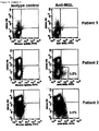
- Bone marrow samples were of patients with AML were stained with MGL antibodies. Clearly a subset of these CD34 positive cells were MGL positive. The expression of MGL on these leukemic cells may inhibit good recognition and removal of the leukemic cells by effector T cells, by the induction of apoptosis or by reducing the effectiveness of the effector T cells.
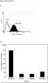
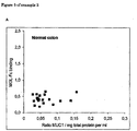
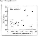
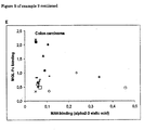
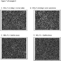
- Example 2 Figure 10 . MUC1 from allergic mothers displays more GalNAc epitopes and MGL binding sites compared to MUC1 from healthy mothers.
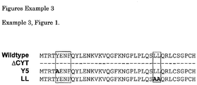
- Example 3 Figure 2 . Ligand binding induces endocytosis of MGL.
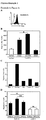
- A Expression of MGL and DC-SIGN on human monocyte-derived DCs as measured by flowcytometry. Open histogram, isotype control; filled histogram, MGL or DC-SIGN expression.
- B Internalization of cell surface bound antibodies to MGL, DC-SIGN and MHC II as measured by flowcytometry on immature DCs. Fixed cells and cells on ice were included to control for the antibody off-rate. Values represent mean of duplicates.
- C Internalization of cell surface bound ligands to MGL (GalNAc-PAA) and DC-SIGN (PAA-Mannose and PAA-Lewis X) as measured by flowcytometry on immature DCs.
- A Expression of wildtype and mutant MGL on CHO-transfectants as measured by flowcytometry. Open histogram, isotype control; filled histogram, MGL expression.
- B Binding of GalNAc-coated beads to CHO-transfectants of wildtype and mutant MGL. Standard deviation ⁇ 5%
- C Internalization of cell surface bound antibodies to MGL as measured by flowcytometry on CHO-MGL transfectants. Values represent mean of duplicates. Results are representative for three independent experiments.
- Example 3 Figure 4. MGL functions as an antigen receptor on human DCs.
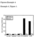
- Example 4 MGL-mFc recognizes ⁇ -GalNAc and LacdiNAc structures. MGL-mFc binding was determined by an ELISA-based assay. Standard deviation ⁇ 0.05 OD 450 nm. One representative experiment out of three is shown.
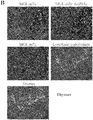
- Example 4 MGL ligands can be detected in on the sinusoidal and lymphatic endothelium human lymph node and thymus.
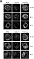
- MGL-mFc red specifically interacted with the sinusoidal endothelium (LYVE-1, green) of lymph node (A) and with the lymphatic endothelium (LYVE-1, green) of the thymus (B) as determined by immunohistochemistry. No staining was observed in the presence of free GalNAc monosaccharides, showing the specificity of the MGL binding. Nuclei were visualized in blue. One out of two donors is shown. Original magnifications 200x and 400x.
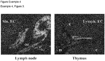
- Example 4 Figure 3 . Distribution of MGLpos cells and sinusoidal or lymphatic endothelial cells in lymph node and thymus. Human tissue sections of lymph node and thymus were stained with anti-MGL (red) and LYVE-1 (green). Nuclei are visualized in blue. Sin. EC, sinusoidal endothelial cells, OZ, outer zone of the paracortex, Lymph. EC, lymphatic endothelial cells, C, cortex, IS, interlobular space. One out of two donors is shown. Original magnification 400x.
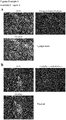
- Example 4 Endothelial structures express GalNAc epitopes as visualized by HPA staining. HPA (green) specifically interacted with the sinusoidal endothelium (LYVE-1, red) of LN (A) and lymphatic endothelium (LYVE-1, red) in thymus (B). Original magnification 400x.
- Example 4 MGL specifically recognizes HMEC-1 endothelial cells. Binding of MGL-Fc to primary HUVEC (A) or HMEC-1 (B) was determined by flow cytometry. MGL-Fc binding to HMEC-1 can be blocked by EGTA, free GalNAc monosaccharides and anti-MGL antibodies. C, HMEC-1 cells express ⁇ -GalNAc, ⁇ 1-2 linked fucose, ⁇ 2-6 linked sialic acid and ⁇ 2-3 linked sialic acid, as stained by the lectins HPA, UEA-1, SNA and MAA respectively. D, MGL-Fc binding can be partially blocked by the ⁇ -GalNAc-specific lectin HPA. *indicates a significant difference between the MGL-Fc binding and the MGL-Fc binding in the presence of HPA (p ⁇ 0.05). All results are representative for three independent experiments.
- Example 4 MGL mediates retention of immature DCs.
- A MGL expression on immature DCs was determined by flow cytometry. Open histogram, isotype control; filled histogram MGL staining.
- B Transendothelial migration of immature DCs across an HMEC-1 monolayer in response to a RANTES gradient in the presence of antibodies to MGL, ⁇ 2-integrin or isotype control antibody. * indicates a significant difference between the isotype control and the anti-MGL (p ⁇ 0.05).
- C and D DC migration on glycoconjugate-coated plates was measured using time-lapse video microscopy. Anti-MGL antibodies increase DC migration on GalNAc, whereas they have no effect on the Lewis X coating. ** indicates a significant difference between isotype control and the anti-MGL antibodies (p ⁇ 0.01). NS, not significant. All results are representative for three independent experiments.
- Example 5 Figure 1. Reaction scheme for synthesis of activated carbohydrate.
- Example 6 Figure 1 . Carbohydrate specificity of mMGL1 and mMGL2. mMGL1-Fc and mMGL2-Fc binding to coated PAA-glycoconjugates was determined by ELISA.
- Example 6 Figure 2 . Binding of murine MGL1 expressed by CHO cells to carbohydrates. Binding of PAA-glycoconjugates (10 ⁇ g/ml) to CHO parental and CHO-mMGL transfectants was determined by flow cytometry.
- Lewis X is the high affinity ligand for mMGL1.
- Example 6 Figure 4 . mMGL1 and mMGL2 bind Lewis X and GalNAc structures respectively. A) MGL-Fc binding was determined by glycan array analysis. B) Binding of bone marrow derived dendritic cells (BM-DC) to PAA-biotin conjugated glycans was analyzed by flow cytometry.
- BM-DC bone marrow derived dendritic cells
- Example 6, Figure 5 Presentation of neo-glycoconjugates by BM-DC results in enhanced CD4 (A) and CD8 (B) T cell proliferation.
- Day 6 BM-DC were harvested and cultured with OVA-GalNAc and OVA-Lewis X for 5-6 hours. Cells were washed and cultured with OT-I or OT-II T cells for 48 hrs. Culture was pulsed with 3 H-thymidine for another 18 hrs. T cell proliferation was determined by measuring 3 H-thymidine incorporation.
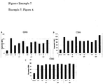
- Example 7, Figure 1 MGL specifically interacts with N. gonorrhoeae.
- Example 7 Binding of N. gonorrhoeae to DCs. Binding of day 4 DCs to FITC-labeled N. gonorrhoeae (MOI 100) was determined by flow cytometry.
- Example 7, Figure 3 Internalization of N. gonorrhoeae by DCs. DCs were incubated for various time points with FITC-labeled N. gonorrhoeae (MOI 100). Binding was determined in the absence (detecting extracellular and intracellular bacteria) or presence of 0.4% Tryphanblue (detecting only intracellular bacteria).
- N. gonorrhoeae induces DC maturation.
- DCs were incubated overnight at different MOI of N. gonorrhoeae.
- DC maturation was measured by flow cytometry using the maturation markers CD83, CD80 and CD86.
- N. gonorrhoeae induces macrophage activation. Macrophages were incubated overnight at different MOI of N. gonorrhoeae. Macrophage activation was measured by flow cytometry using the markers CD 14, CD80 and CD86.
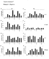
- Example 7 Cytokine production of DCs and macrophages incubated with N. gonorrhoeae. DCs and macrophages were incubated overnight at different MOI of N. gonorrhoeae. Cytokine production was determined by ELISA.
- Example 7, Figure 7 Cytokine mRNA levels in DCs incubated with N. gonorrhoeae. DCs were incubated overnight at different MOI of N. gonorrhoeae. Cytokine mRNA levels were determined by RT-PCR.
- N. gonorrhoeae wildtype induces a Th2-inducing phenotype in DCs.
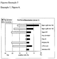
- DCs were incubated with TLR ligands or N. gonorrhoeae (MOI 100). After 48 hours mature DCs were cocultured with naive CD4+ T cells for an additional 14 days. T cells were restimulated with PMA and ionomycin and intracellular levels of IFN ⁇ and IL-4 were determined by flow cytometry. Controls include: E . coli LPS (mixed Th1/Th2 response), LPS/PGE2 (Th2 response) and Poly I:C or Poly I:C/R837 (Th1 response).
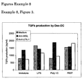
- Example 8 Figure 1 . MGL-mediated immunomodulation via the MUC1-MGL interaction.
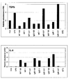
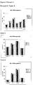
- Example 8 Altered cytokine production in DCs incubated with anti-MGL and TLR-ligands. Cytokine production was measured by RT-PCR and displayed as relative abundance compared to the reference gene GAPDH.
- Example 8 Figure 3. TGF ⁇ secretion by dexamethasone treated DCs.
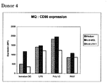
- Example 8 Figure 4 . CD86 expression by macrophages. Macrophages were stimulated for 20 hours with TLR-ligands in the presence or absence of anti-MGL. Subsequently the expression of maturation markers was determined by flow cytometry.
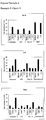
- Example 8 Altered cytokine production in macrophages incubated with anti-MGL and TLR-ligands. Cytokine production was measured by RT-PCR and displayed as relative abundance compared to the reference gene GAPDH
- Immature DC were cultured for 4-7 days from monocytes obtained from buffy coats of healthy donors (Sanquin, Amsterdam) in the presence of IL-4 (500 U/ml) and GM-CSF (800 U/ml). DC were matured by addition of 1 ⁇ g/ml LPS (Salmonella typhosa, Sigma-Aldrich, St. Louis, MO). Mf were generated from monocytes by culturing in the presence of 1000 U/ml GM-CSF. Dexamethasone was added at 1 ⁇ M.
- Antibodies and reagents The following monoclonal antibodies were used: AZN-D1 (IgG1, anti-DC-SIGN20, SPV-L7 and NKI-L15 (anti-CD11a), MEM-59 (anti-CD43), MEM-28 (anti-CD45, kindly provided by dr. V. Horesji), D3/9 (anti-CD45), RP1/11 (anti-CD45RA), RP2/21 (anti-CD45RB), RP1/12 (anti-CD45RC, all kindly provided by dr. F. Sanchez-Madrid, 17, GF4bio (anti-CD27, kindly provided by prof. dr. R.
- T3b (stimulating anti-CD3,50, anti-CD45RO (UCHL-1, BD Biosciences, San Diego, CA), anti-CD4PE (clone 13B8.2, Beckman Coulter, Fullerton, CA), anti-CD8PE (clone B9.11, Beckman Coulter), 28-14-8 (mouse anti-mouse H2 Db), anti-CD1a (OKT6), anti-CD68 (EMB 11, Dako, Glostrup Denmark), anti-CD 163 (Edhu1), anti-Mannose receptor (3.29.B1), anti-CD11b (Bear-1) anti-CD11c (SHCL-3), anti-CD80 (BD Biosciences), anti-CD86 (BD Biosciences) and CD83 (Beckman Coulter).
- the anti-MGL antibodies 18E4 and 1G6.6 were generated by immunizing Balb/c mice with purified MGL-Fc. Hybridoma supernatants were screened for the presence of anti-MGL antibodies on CHO-MGL transfectants.
- MGL-Fc and control Fc-proteins (ICAM-3-Fc or CAMEL-Fc) were generated as previously described 13 .
- MGL-ligand expression To analyze MGL-ligand expression, cells were incubated with MGL-Fc (10 ⁇ g/ml) in TSA (20 mM Tris-HCl pH 7.4, 150 mM NaCl, 2 mM MgCl2, 1 mM CaCl2, 0.5% BSA) for 30 min at 37°C, followed by staining with a secondary FITC-labeled anti-human Fc antibody (Jackson, West grove, PA) and analyzed on FACScalibur. T cells were identified by co-staining for 10 min at RT with subset-specific markers.
- CHO and CHO-MGL were seeded 35.10 3 cells/well in 96-wells plates.
- Calcein-AM Molecular probes, Eugene, OR
- Non-adherent cells were removed by gentle washing.
- Adherent cells were lysed and fluorescence was quantified on a Fluorstar spectrofluorimeter (BMG Labtech, Offenburg, Germany).
- Cells were surface-biotinylated with 0.5 mg/ml sulfo-NHS-biotin (Pierce, Rockford, IL) in PBS for 30 min at 4°C and subsequently lysed in lysisbuffer (10 mM TEA pH 8.2, 150 mM NaCl, 1 mM MgCl2, 1mM CaCl2, 1% Triton X-100, containing EDTA-free protease inhibitors (Roche Diagnotics, Penzberg Germany)) for 1 hour at 4°C.
- lysisbuffer 10 mM TEA pH 8.2, 150 mM NaCl, 1 mM MgCl2, 1mM CaCl2, 1% Triton X-100, containing EDTA-free protease inhibitors (Roche Diagnotics, Penzberg Germany)
- MGL ligands were immunoprecipitated using Fc-protein-coupled protein A-sepharose beads (CL-4B, Pharmacia, Uppsala, Sweden). The immunoprecipitations were seperated by 8% SDS-PAGE electrophoresis and transferred to nitrocellulose membranes. Blots were blocked with BSA and immunoblotted with streptavidin-coupled peroxidase (Vector Laboratories, Burlingame, CA) or specific antibodies followed by a secondary goat anti-mouse-Peroxidase (Jackson). Blots were developed by enhanced chemiluminescence.
- MGL-Fc ligand ELISA MGL-Fc ligand ELISA.
- NUNC maxisorb plates (Roskilde, Denmark) were coated with goat anti-human-Fc antibody (4 ⁇ g/ml for 1 hour at 37°C, Jackson), followed by a 1% BSA blocking step (30 min at 37°C) and MGL-Fc (1 ⁇ g/ml for 1 hour at 37°C).
- MGL-Fc coated plates were incubated overnight at 4°C with cell lysates (10.10 6 cells/ml for cell lines, 500.10 6 /ml for PBL). After extensive washing 1 ⁇ g/ml monoclonal antibodies were added for 2 hours at room temperature. Binding was detected using a peroxidase-labeled goat anti-mouse antibody (Jackson).
- CD45 phosphastase activity was quantified by incubating the beads with 2 mM 4-Nitrophenyl phosphate (Roche, Basel, Switzerland) in CD45 Ph assay buffer (100 mM HEPES pH 7.2, 2 mM EDTA, 2 mM DTT) for 4 hours at 37°C. The resulting color change was measured at 410 nm. Specificity was determined by adding a specific CD45 phosphatase inhibitor (Calbiochem, Darmstadt, Germany).
- MGL is expressed on APC subsets in vivo and in vitro
- APC subsets were cultured from human monocytes and analyzed for MGL expression.
- the CLR DC-SIGN was used as a well known general marker for DC and alternative active Macrophages (aaMf) 20,21 .
- MGL could be detected on immature DC and Mf, whereas its expression was enhanced both on the protein and mRNA level on immature DC and aaMf, generated in the presence of dexamethasone ( Fig. 1a and van Vliet Immunobiology in press).
- LPS-induced DC maturation completely abolished MGL expression.
- MGL expression could not detected on other mature DC or activated Mf (Supplementary table 1).
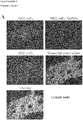
- MGLpos APC As in vitro cultured MGLpos APC, except immature Mf, co-expressed DC-SIGN, we examined whether in vivo MGL and DC-SIGN are found on identical APC subsets as well. Strikingly, in healthy lymph node and skin, MGL and DC-SIGN are expressed on separate APC subsets in the outer zones of the paracortex and dermis respectively, whereas in jejunum a complete cellular overlap between MGL and DC-SIGN expression was found ( Fig. 1b ). Further characterization of the MGLpos cells in skin and lymph node identifies them as myeloid-type APC (Supplementary table 2).
- MGL is preferentially expressed on dexamethasone-treated DC and Mf subets, as well as on APC in several human tissues. MGL levels appear to coincide with chronic inflammatory conditions.
- MGL-Fc specifically recognized PBL, as shown by the complete block using EGTA ( Fig. 2b ).
- T cells also some NK cells and B cells were stained by MGL-Fc, whereas monocytes were not recognized (data not shown).
- NK cells and B cells were stained by MGL-Fc, whereas monocytes were not recognized (data not shown).
- cell-cell adhesion experiments of PBL and Jurkat to MGL-transfectants were performed. PBL specifically adhered to CHO-MGL, expressing high levels of MGL, as compared to the parental CHO cells. Also Jurkat cells firmly interacted with CHO-MGL, whereas only background adhesion was observed to CHO cells. ( Fig. 2c ).
- MGL counterreceptors To investigate the MGL counterreceptors we performed immunoprecipitations on lysates of cell-surface biotinylated Jurkat lysates with MGL-Fc. Immunoblotting for precipitated proteins identified MGL recognition of three membrane proteins of approximately 220, 190 and 110 kD ( Fig. 2d ). Based on the observed molecular weights and the heavy O-glycosylation we considered the mucin-type proteins CD43 (95-115 kD) and CD45 (180-220 kD), as primary candidates for the MGL counterreceptors.
- MGL-Fc specifically interacted with both CD43 and CD45 from Jurkat as shown by the complete inhibition with EGTA, whereas on PBL only CD45 was recognized and not CD43 (Supplementary fig. 2a ).
- CD45 constituted as the main counterreceptor for MGL (Supplementary table 3).
- MGL exclusively recognized CD45 from lymphocytes but not CD45 from other leukocytes, such as monocytes, immature DC and PMN (Supplementary fig. 2b ).
- the CD45 mRNA is subject to alternative splicing in the A-, B- and C-domains, giving rise to five different isoforms on human leukocytes (ABC, AB, BC, B and RO) 15,23 .
- the different CD45 isoforms can be distinguished on the basis of their reactivity with CD45RA, RB or RC-specific moabs. None of these antibodies directed against the protein backbone, were capable of blocking the MGL interaction with specific glycan structures on CD45 (data not shown).
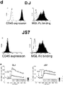
- MGL recognition of different CD45 isoforms we utilized single human CD45 isoform transfectants of the murine cell line 3001917. Flowcytometric analysis confirmed the expression of the correct CD45 isoform in the mouse transfectants (Table 1).
- CD45 on human lymphocytes is the counterreceptor for MGL; whereby MGL is capable of interacting with all CD45 isoforms, except CD45RO.
- expression of GalNAc epitopes on these CD45 isoforms constitutes a prerequisite for binding, a process probably regulated by T cell differentiation.
- MGL modulates CD45 phosphatase activity and TCR mediated signalling.
- CD45 affects cellular responses by controlling the threshold of sensitivity to external stimuli, especially those received by the TCR. Therefore we investigated whether the MGL-CD45 interaction would influence CD45 phosphatase activity and function, using the Jurkat E6.1 cell line, which has proven to be an unsurpassed model for studying TCR-signaling pathways 24 .
- CD45 phosphatase activity can be regulated via receptor dimerization, whereby increased dimerization results in a decreased activity 25 .
- To achieve maximal receptor dimerization Jurkat cells were seeded on CHO or CHO-MGL, after which cells were lysed and the CD45 phosphatase activity was measured. MGL binding to CD45 resulted in a time-dependent decrease in CD45 phosphatase activity ( Fig. 3a ).
- CD45-specifc phosphatase inhibitors No residual phosphatase activity in the presence of CD45-specifc phosphatase inhibitors was observed, confirming that the observed phosphatase activity was completely CD45-mediated.
- the first downstream target of CD45 in T cells is Lck.
- CD45 dephosphorylates the negative regulatory tyrosine Y505, resulting in an active pool of Lck.
- the reduced CD45 phosphatase activity after MGL-CD45 interaction could lead to decreased Lck activity by enhanced phosphorylation on Y505.
- an increase in the phosphorylation status of Lck Y505 was observed 10-20 minutes after the addition of MGL-Fc, which acts as an agonistic agent mimicking binding to cellular MGL ( Fig. 3b ).
- MGL function with free GalNAc lowered Y505 phosphorylation to control and unstimulated levels. Engagement of the TCR leads to activation of downstream signaling pathways, including intracellular calcium mobilization, which plays a critical role in early T cell signaling transduction.
- Jurkat E6.1 cells were labeled with a calcium sensitive dye, Fluo-3/AM and stimulated with MGL-Fc coated beads and an anti-CD3 antibody. Within one sample the fluorescent beads were used to distinguish between cells that are subjected to MGL-mediated crosslinking or not. Binding of MGL-Fc beads alone did not trigger calcium mobilization, although the subsequent calcium flux triggered by the anti-CD3 antibody was strongly downregulated.
- MGL-Fc induces cell death in a TCR- and CD45-dependent manner
- MGL-Fc/anti-CD3 induced cell death resulted in exposure of phosphatidylserine as measured by staining with annexin V ( Fig. 4b ) and nuclear fragmentation (data not shown).
- Cell death was completely dependent on both CD3 triggering and MGL binding, as incubations with anti-CD3 or MGL-Fc alone had no effect. Occasionally some cell death was induced by MGL-Fc alone; however it was always markedly increased by TCR stimulation (data not shown).
- apoptosis was MGL-specific as addition of free GalNAc could block all cell death and control Fc-proteins did not induce apoptosis ( Fig. 4b ).
- cell death could not be triggered by anti-CD3/MGL-Fc in JS7, validating the complete CD45-dependence in the MGL-Fc-induced apoptosis ( Fig. 4c ).
- naive T cells express both CD27 and CD45RA
- memory T cells are characterized as CD27posCD45RAneg.
- Memory T cells are characterized by their expression of CD45RO, although expression of the CD45B-isoform is retained ( 17 and data not shown).
- Teff generally lose expression of CD27; however a number of Teff regain CD45RA.
- MGL displayed a strong preference for both CD27neg CD4+ and CD8+ Teff ( Fig. 5a ). MGL-Fc binding was specific as shown by the EGTA block.
- MGL indeed has a downregulatory effect on T cell activation and thus on proliferation
- blocking anti-MGL antibodies should revert this effect and enhance proliferation.
- Monocyte-derived APC expressing high or low levels of MGL ( Fig. 1a ) were cultured with highly purified allogeneic CD4+ T cells in the presence or absence of blocking MGL antibodies and after 4 days proliferation was measured by [methyl-3H]Thymidine incorporation.
- the different APC subsets could clearly be divided in immunogenic MGLlow/neg APC (immature DC, mature DC and Mf), capable of inducing high T cell proliferation and dexamethasone-treated MGLhigh APC, which lack the ability to stimulate strong T cell division, even at high APC:T cell ratios ( Fig.
- Anti-MGL antibodies had no effect on proliferation and cytokine production in the immunogenic APC-T cell co-cultures ( Fig. 5d and data not shown). In contrast anti-MGL antibodies could enhance proliferation about 3-fold in the dexamethasone-APC-T cell co-cultures ( Fig. 5d ). This effect was not observed for two isotype-matched control antibodies, a blocking anti-LFA-1 and a non-binding isotype control (mouse anti-mouse H2 Db). Strikingly T cell proliferation by immature DC is not enhanced by antibodies against MGL in contrast to the dexDC.
- the CLR MGL on immature APC with a tolerogenic phenotype specifically recognizes CD45 on Teff, thereby suppressing T cell activation.
- MGL knock-out mice are made lacking the gene for MGL. Subsequently, it is determined whether the induction of EAE (Multiple Sclerosis (MS)-like disease in mice) is enhanced when the mice are vaccinated with MOG (myeline) peptides. The same experimental set-up can be followed for Rheumatoid Arthritis, and when vaccination takes place with collagen.
- EAE Multiple Sclerosis
- PBL of auto-immune patients such as RA PBL or MS patients, were screened for upregulated expression of GalNAc on CD45. It is expected that addition of MGL-Fc will induce apoptosis.
- MGL binds to PBLs from chronic infected patients with RA ,OA and multiple sclerosis.
- MGL preferentially recognizes effector-type T cells as measured by 4-color analysis of MGL-Fc binding and co-staining with anti-CD4 or CD8 -specific antibodies. Standard deviation ⁇ 5%. Frozen total PBLs were thawed and stained with MGL-Fc as earlier mentioned. Also plant lectins such as HPA, with specificity for GalNAc, Con A, high mannose and diantennary, and PNA, where used to determine glycan epitopes on subsets of cells.
- HPA which has a similar glycan specificity as MGL showed enhanced staining of PBL from RA and OA patients.
- Supplementary figures 6-9 show the results of these experiments. Frozen PBL were thawed and stained with MGL-Fc ( Figures 8 and 9 ). Also plant lectins such as HPA, with specificity for GalNAc, Con A, high mannose and diantennary glycans and PNA, TF antigen, were used to determine glycan epitopes present on subset of cells ( Figures 6 and 7 ). Significant differences were observed between the MGL-Fc binding between SPMS and PPMS patients. In RRMS patients that were treated with IFN ⁇ MGL binding increased. HD, healthy donors, SPMS, secondary progressive MS, PPMS, primary progressive MS, RRMS, relapsing-remitting MS.
- MGL recognizes a counter-receptor on mouse lymphocytes.
- mouse MGL1 or 2 could share ligands as well. Therefore the recognition of mouse T cells and mouse CD45 was investigated to determine whether mMGL1 or 2 might interact with mouse T cells and mouse CD45, similar to human MGL.
- the mMGL1 KO mouse has an elevated level of antigen experienced T cells, suggesting that the MGL-mediated control of effector T cells could be present in the mouse as well.
- plant lectins such as HPA, with specificity for GalNAc, Con A, high mannose and diantennary glycans, PNA, TF antigen and LTA, ⁇ 3-4 linked fucose, were used to determine glycan epitopes present on mouse lymphocytes.
- control spleen and lymph node were used of uninfected control C57/B16 mice and MGL KO mice infected with several pathogens.
- mMGL1-Fc shows low binding to T cells from LN and to splenic T cells from uninfected mice. mMGL1-Fc binding to splenic T cells from infected mice is increased. mMGL2-Fc does not bind mouse lymphocytes with the exception of splenic T cells from infected mice, which are recognized by mMGL2-Fc.
- mMGL can recognize mouse effector T cells, as present during infection or inflammation, similar to the human situation.
- CHO Chinese hamster ovary cells
- FBS fetal bovine serum
- MoDC Monocyte derived dendritic cells
- human blood monocytes were isolated from buffy coats by a Ficoll gradient step, followed by MACS beads isolation of CD 14+ cells (Miltenyi Biotec, Bergisch Gladbach, Germany).
- the monocytes were cultured in the presence of IL-4 and GM-CSF (500 and 800 U/mL, respectively; ScheringPlough, Brussels, Belgium) for 4 to 6 days.
- IL-4 and GM-CSF 500 and 800 U/mL, respectively; ScheringPlough, Brussels, Belgium
- the phenotype of immature DC was confirmed by flow cytometry (CD11bhigh, CD11chigh, ICAM-lhigh, CD80low, CD83low, CD86low).
- the DC were matured by incubation with lipopolysaccharide (LPS, Salmonella typhosa, 1 ⁇ g/mL, Sigma Aldrich, St. Louis, Mo).
- LPS lipopolysaccharide
- CD80, CD83 and CD86 expression was >90%.
- MUC1-Fc containing 32 tandem repeats of MUC1 fused with the Fc part of murine IgG2a was produced in CHO-K1 cells as described before 30.
- Purified MUC1-Fc glycoprotein was treated sequentially with V. cholerae neuraminidase (Roche) and ⁇ -galactosidase from bovine testis (Sigma) to remove sialic acid and galactose residues.
- the resulting glycoform was determined by ELISA using glycan specific antibodies and lectins (see “Binding assay (ELISA)").
- Tissue samples from colon carcinoma were collected after surgical removal of the tumor with informed consent of the patients.
- Primary colon carcinoma tissue and the corresponding normal tissue were obtained from patients from resection specimens, following national and institutional ethical guidelines regarding the use of human tissues.
- Tissue samples were gently homogenized and digested in lysis buffer (TEA buffer containing 1 % Triton-X-100, 2 mM CaCl2, 2 mM MgCl2) for 2 days at 4°C under rotation. The lysates were centrifuged at 14000 rpm for 15 min, supernatant collected and stored in aliquots at -80°C.
- lysis buffer TAA buffer containing 1 % Triton-X-100, 2 mM CaCl2, 2 mM MgCl2
- MUC1 specific antibodies (clone 214D4 provided by John Hilkens, Netherlands Cancer Institute, Amsterdam); this antibody was biotinylated using a biotinylation kit (Pierce, Rockford, IL, USA); DC-SIGN specific antibody AZN-D1 31; CD68 specific antibody EBM-11; FITC-labeled goat anti-mouse IgG (Jackson Immunoresearch, West Grove, PA); Peroxidase-labeled goat anti-human Fc (Jackson Immunoresearch); Alexa-488 labeled goat anti-mouse IgG1 and Alexa-594 labeled goat anti-mouse IgG2a (Molecular Probes).
- Anti-glycan antibodies 5E5 (MUC1-Tn) and HH8 (MUC1-TF) were provided by Henrik Clausen, University of Copenhagen, Denmark.
- the MGL-human Fc fusion protein was made in the following way: the extracellular part of MGL (amino acids 61-289) was amplified on pRc/CMV-MGL by PCR, confirmed by sequence analysis and fused at the C-terminus to human IgG1-Fc in the Sig-pIG1-vector 28.
- MGL-Fc was produced by transfection of CHO cells and concentration determined by ELISA.
- An MGL-murine Fc fusion protein was generated by cloning the extracellular part of MGL (including the CD38 signal sequence from the Sig-pIG1 vector) into an pcDNA3 expression vector containing exon 1-3 of murine IgG2a-Fc 30.
- the 18E4 monoclonal antibody to MGL was generated by immunizing Balb/c mice with purified MGL-murine Fc. Hybridoma supernatants were screened for the presence of anti-MGL antibodies on CHO-MGL transfected cells (S. van Vliet, submitted).
- Carboxylate-modified TransFluorSpheres (488/645 nm, 1.0 ⁇ m; Molecular Probes, Eugene, OR) were coated with streptavidin as previously described 32. Streptavidin coated beads were incubated with biotinylated F(ab')2 fragments of goat anti-mouse IgG2a (6 ⁇ g/ml, Jackson Immunoresearch) in 0.5 mL PBS 0.5% BSA for 2 hours at 37°C, washed and further incubated with MUC1-Fc fusion proteins at 4°C overnight. Finally, beads were washed, stored in 100 ⁇ l PBS 0.5% BSA, and used within one week. The beads adhesion assay was performed as previously described 32.
- DC were incubated with coated beads in Tris-sodium buffer (20 mM Tris-HCl, pH 7, 150 mM NaCl, 2 mM CaCl2, 2 mM MgCl2) with 0.5% BSA and adhesion was determined in the absence or presence of blocking agents (10 mM EGTA, 25 mM monosaccharides (Sigma) or 5 mM mannan) at 37°C for 45 min. Cells were washed and analysed by flow cytometry.
- the solid phase adhesion assay was performed by coating ELISA plates with purified anti-MUC1 antibodies (clone 214D4) in 0.2 M Na2CO3, followed by blocking with 1% BSA in Tris-sodium buffer. Negative controls were included for each sample, where no anti-MUC1 antibodies were added. After washing with Tris-sodium buffer, tissue lysates were added and incubated at 4°C overnight. Plates were washed and further incubated with MGL-human Fc (0.5 ⁇ g/mL) or biotinylated HPA (Sigma), dioxygene labeled MAA (Boehringer Mannheim) or anti-TF-antibodies (clone HH8) for 1 h at room temperature (RT).
- MGL-human Fc 0.5 ⁇ g/mL
- biotinylated HPA Sigma
- dioxygene labeled MAA Boehringer Mannheim
- anti-TF-antibodies clone HH8
- Cryosections (7 ⁇ ) of normal colon epithelium or primary colon carcinoma were fixed in 100% aceton (10 min), washed with PBS, and incubated with first antibody (10 ⁇ g/ml) for 1h at 37°C. After washing, the final staining was performed with Alexa-594 labeled goat-anti mouse IgG2a or Alexa-488 labeled goat-anti mouse IgG1 and nuclear staining was performed with Hoechst.
- cryosections were fixed in 2% paraformaldehyde and all incubation steps were performed in Tris-sodium buffer. The final staining was performed with Alexa-488 labeled goat-anti mouse IgG.
- Helix pomatia agglutinin HPA, Helix pomatia
- MGL amino acid residues 61-289
- Fc domain of human IgG1 The following antibodies were used: MUC1 antibodies 214D4 (IgG1 isotype, provided by John Hilkens, Netherlands Cancer Institute, Amsterdam); Peroxidase-labeled goat anti-human Fc (Jackson Immunoresearch, West Grove, PA); Peroxidase-labeled streptavidin.
- the solid phase adhesion assay was performed by coating ELISA plates (Maxisorp, Nunc) directly with milk samples, diluted in 0.2 M Na 2 CO 3 and incubated overnight at 4°C. The plates were washed and incubated with Tris-sodium buffer (20 mM Tris-HCl, pH 7, 150 mM NaCl, 2 mM CaCl 2 , 2 mM MgCl 2 ) containing 1% BSA.
- Tris-sodium buffer (20 mM Tris-HCl, pH 7, 150 mM NaCl, 2 mM CaCl 2 , 2 mM MgCl 2
- anti-MUC1 antibodies were coated in 0.2 M Na 2 CO 3 , followed by blocking with BSA and incubation with milk (1:100) overnight at 4°C.
- the protein was desialylated and treated with ⁇ -galactosidase, leaving only the core GalNAcs on the protein.
- the resulting MUC1-Tn reacted with antibodies against Tn, but not against TF in ELISA ( Figure 1 ). These results indicate that the glycosylation mainly consisted of GalNAcs.
- MUC1-Fc containing Tn-antigens interacts with monocyte derived dendritic cells
- MUC1-Fc containing Tn-antigens (GalNAc ⁇ Ser/Thr) was coupled to fluorescent beads and validated for interaction with monocyte derived DC (moDC). Both immature DC and DC matured with lipopolysaccharide were analysed for binding. MUC1-Tn bound efficiently to immature DC and binding was calcium dependent, suggesting a role for CLR ( Figure 2A ). Mature DC showed strongly reduced interaction with MUC1-Tn, concomitant with the fact that mature DC down-regulate expression of CLR on their cell surface ( Figure 2A ).
- Lewis-X bound strongly to immature DC that express high levels of the CLR DC-SIGN and binding to mature DC, that express low levels of DC-SIGN, was significantly reduced ( Figure 2B ).
- MGL expression is down-modulated upon DC maturation ( Figure 3B ), coinciding with loss of MUC1 binding.
- Pre-incubation of moDC with anti-MGL antibodies reduced binding of MUC1-Tn, indicating that MGL is a receptor for this MUC 1 glycoform, although the block was not complete.
- EGTA which inhibits the lectin domain of CLR to recognize glycans also inhibited the interaction to the same level as anti-MGL antibodies.
- MGL binds MUC1 derived from primary colon carcinoma
- MGL expressing cells were present in colon tissue and whether these could interact with MUC 1 in tumors.
- tissue sections were analysed for cells expressing MGL and MUC1.
- MGL expressing cells were detected within the normal colonic mucosa of 4 individuals analysed as well as in primary tumor sections of all these individuals.
- MGL-expressing cells did not appear to express the macrophage marker CD68 in the tumor sections ( Figure 6A ), although some co-localization was observed with the CLR DC-SIGN, suggesting that MGL may be expressed on certain DC populations ( Figure 6B ).
- MUC1 positive cells were detected at the epithelial layer and some MUC1 positive cells were scattered throughout the mucosa ( Figures 6C and 6D ).
- MUC1 staining was strong in the tumor tissue.
- the finding that the MGL expressing cells are located in the vicinity of MUC 1 expressing cells demonstrate that MGL positive antigen presenting cells are present in close contact with epithelial tumor cells.
- MGL-Fc showed strong staining of colon carcinoma cells and not with normal colon epithelium ( Figure 7 ).
- the binding of MGL-Fc was blocked by GalNAc monosaccharides, demonstrates the requirement of the carbohydrate recognition domain for staining.
- MGL-Fc detects secreted MUC1 from tumor cellline ZR75
- FIG 8 A MUC1 was harvested out of the supernatant of cultured ZR75 tumor cells and coated on ELISA plates. Both MGLFc and DC-SIGNFc were used to look for carbohydrate binding epitopes expressed on tumor associated MUC1. As illustrated MGLFc binds strongly the secreted tumor associated MUC1 whereas DC-SIGN does not bind. Furthermore EGTA which inhibits lectin binding to carbohydrates illustrates that the recognition of MGL to MUC1 is mediated by the expression of carbohydrates.
- Breast milk is a rich source of glycosylated MUC1.
- MUC1 was captured in ELISA from milk of 10 healthy mothers and 10 allergic mothers.
- Figure 10A and B show the MUC1 concentrations in milk, which is not significantly different between healthy and allergic mothers.
- MUC1 from allergic mothers displays a higher MGL binding ( Figure 10C ), which can be explained by the higher exposure of ⁇ -GalNAc epitopes as measured by the ⁇ -GalNAc-specific lectin HPA ( Figure 10D ).
- the epithelial mucin MUC1 is a tumor antigen where post translational modification is altered during malignant transformation 14 .
- the protein backbone is retained, but O-linked glycan structures are modified. This creates epitopes that may be recognized by the immune system, including the protein backbone and carbohydrate structures generally not present in non-malignant tissues.
- Adenocarcinoma patients may develop humoral and cellular immune responses to the MUC1 protein. These responses are, however, weak and not sufficient to eradicate the tumor 35,36 . Nonetheless, anti-MUC1 antibody levels have been shown to correlate with survival 37 indicating the importance of this tumor antigen for protection against cancer.
- MUC1-Tn bound efficiently to DC.
- the interaction was calcium dependent and was significantly contributed by the CLR MGL, that is expressed on immature DC, a receptor for GalNAc containing carbohydrate structures 28,38 .
- DC maturation resulted in abrogation of MUC1-Tn binding coinciding with loss of MGL expression.
- mannose receptor that has previously been described to be a receptor for glycosylated MUC1 39 . The discrepancy between the two studies is likely due to differences in MUC1 glycoforms.
- the MUC1 glycoforms studied by Hiltbold et al. may have contained additional glycan epitopes that target the mannose receptor.
- MGL positive cells both within normal colon mucosa and within primary carcinomas, demonstrating that these cells may be in close contact with the MUC-Tn expressing tumor cells. Interestingly, these cells seemed to exhibit DC-like phenotype, and they did not express the macrophage marker CD68. MGL-Fc bound strongly to colon carcinoma cells, supporting the hypothesis that MGL positive cells may interact with tumor cells.
- Tumor cells may shed MUC1 into the micromilieu and it may be detected in serum of cancer patients 4 .
- These forms of MUC 1 lack a cytoplasmic tail and can (in advanced colon cancer patients) reach such high levels that they can interfere with cell interactions, including selectin interactions 43 .
- These shed forms of MUC1 thus have the possibility to interfere with the immune system and help the tumor cells escape immune surveillance 43,44 .
- Interaction with MGL on antigen presenting cells may similarily influence immune responses. It is interesting to mention that MGL is expressed on human immature DC and in particular highly expressed on tolerogenic DC and/or macrophages that are cultured in the presence of dexamethazone (S. van Vliet, submitted).
- Different APC subsets were cultured from freshly isolated human monocytes in the presence of indicated cytokines. The following concentrations were used: for Mf culture: GM-CSF 1000 U/ml, dexamethasone 10 -6 M, LPS 1 ⁇ g/ml, IFN ⁇ 1000 U/ml and for DC culture: GM-CSF 500 U/ml, IL-4 800 U/ml, dexamethasone 10 -6 M, LPS 1 ⁇ g/ml, Poly I:C 10 ⁇ g/ml and Flagellin 5 ⁇ g/ml. MGL expression was determined by flowcytometry using the 18E4 antibody. One representative experiment out of two is shown.
- anti-CD1a OKT6
- anti-CD68 EMB 11, Dako
- anti-CD 163 Edhu1
- anti-DC-SIGN AZN-D1
- anti-Mannose receptor 3.29.B1
- anti-CD11b Bear-1
- anti-CD11c SHCL-3
- T3b anti-CD3
- MGL thus represents a marker for the skin APC described in Angel et al JI 2006 .
- APC subsets were cultured from freshly isolated human monocytes in the presence of GM-CSF 500 U/ml/IL-4 800 U/ml (DC) or GM-CSF 1000 U/ml (Macrophages). Dexamethasone was added at 10 -6 M and LPS at 1 ⁇ g/ml. Expression of co-stimulatory molecules was determined by flowcytometry using the specific antibodies to CD80, CD86 (both from BD Biosciences) and CD83 (Beckman Coulter).
- anti-MGL 18E4 (IgG2a), 1G6.6 (IgG2a) [1] and MLD-1 (IgG1), kindly provided by dr. T. Irimura [2], anti-DC-SIGN (AZN-D1, IgG1) [3], anti-MHC II (Q5/13, IgG2a), anti-LAMP-1 (H4A3, IgG1, BD Pharmingen), EEA-1 (IgG1, Abcam, Cambridge, UK) and anti-ICAM-2 (12A2, IgG1 isotype control [4]).
- Biotinylated polyacrylamide (PAA)-coupled glycoconjugates were purchased from Lectinity (Lappeenranta, Finland).
- Immature monocyte derived DCs were cultured for 4-7 days from monocytes obtained from buffy coats of healthy donors (Sanquin, Amsterdam) in the presence of IL-4 (500 U/ml) and GM-CSF (800 U/ml). The phenotype of the cultured DC was confirmed by flowcytometric analysis. DC consistently expressed high levels of MHC class I and II and low levels of CD80 and CD86. CHO and CHO-MGL cells were maintained in RPMI 1640 medium (Invitrogen, Carlsbad, CA) containing 10% Fetal Calf Serum.
- Mutations in the cDNA encoding MGL were generated using the QuikChange site-directed mutagenesis kit (Stratagene, La Jolla, CA) and pRc/CMV-MGL plasmid according to the manufacturer's protocol.
- the cytoplasmic tail deletion mutant was generated by replacing the entire cytoplasmic region by a new protein initiation codon. Plasmid sequencing confirmed the introduction of the mutations in the MGL cytoplasmic region ( Fig. 1 ).
- Stable CHO-MGL transfectants were generated using lipofectamin (Invitrogen) and selected by the addition of 2 mg/ml Geneticin (Invitrogen). MGL positive cells were sorted using the MoFlo (DAKOcytomation, Glostrup, Denmark).
- Immature DCs were incubated with antibodies (10 ⁇ g/ml) or biotinylated PAA-glycoconjugates (5 ⁇ g/ml) in TSA (20 mM Tris-HCl (pH 8.0), 150 mM NaCl, 1 mM CaCl2, 2 mM MgCl2, and 0.5% BSA) for 1 h on ice and subsequently washed.
- TSA Tris-HCl (pH 8.0), 150 mM NaCl, 1 mM CaCl2, 2 mM MgCl2, and 0.5% BSA
- Cells were incubated at 37°C for various time points, placed on ice, and incubated with FITC-conjugated secondary antibodies or streptavidin-Alexa488 (Molecular Probes).
- FITC-conjugated secondary antibodies or streptavidin-Alexa488 Molecular Probes
- Anti-MGL 1G6.6 was labeled with Alexa Fluor 488 according to the manufacturer's protocol (Molecular Probes, Eugene, OR). Dendritic cells were incubated for 4 hours on ice, 2 hrs on ice followed by 2 hours at 37°C or 4 hours at 37°C with Alexa Fluor 488-conjugated anti-MGL (10 ⁇ g/ml). Labeled cells were fixed and permeabilized for 20 min at 4°C (BD cytofix/cytopermTM, BD).
- Cells were stained in phosphate-buffered saline containing 0.5 % bovine serum albumin and 0.1% saponin, with antibodies against LAMP-1 (lysosomes) or EEA-1 (early endosomes) and subsequently with Alexa Fluor 594-conjugated anti-mouse IgG1. Cells were allowed to adhere to poly-L-lysine-coated glass slides, mounted with anti-bleach reagent and analyzed by confocal microscopy.
- Leica AOBS SP2 confocal laser scanning microscope (CLSM) system was used, containing a DM-IRE2 microscope with glycerol objective lens (PL APO 63x/NA1.30) and images were acquired using Leica confocal software (version 2.61).
- CLSM Leica AOBS SP2 confocal laser scanning microscope
- HD7 a CD4+ T cell that recognizes a peptide derived from mouse IgG1 antibodies in HLA-DR0101/DQw1, was used [6].
- Immature DCs (20.10 3 /well) from a typed donor were preincubated with serial dilutions of antibodies and cocultured with 80.10 3 T cells. After 48 hours, IFN ⁇ production by the T cells was measured by ELISA according to the manufacturer's protocol (Biosource international,.Camarillo, CA).
- the C-type lectin DC-SIGN known to rapidly internalize after ligand binding [4], was used as a positive control for endocytic uptake by DCs.
- Human monocyte-derived DCs expressed moderate levels of MGL and high levels of DC-SIGN ( Fig. 2A ).
- Mature DCs do not express the MGL molecule (data not shown).
- antibodies directed against the MGL carbohydrate recognition domain that mimic ligand binding and induce receptor crosslinking essential for endocytosis. Antibodies to MGL were internalized from the DC surface similar to anti-DC-SIGN antibodies ( Fig. 2B ).
- the YENF motif is essential for MGL-mediated endocytosis.
- MGL cytoplasmic domain mutants by site-directed mutagenesis and a complete cytoplasmic deletion mutant ( Fig. 1 ).
- Stable cell lines were generated by transfecting the constructs encoding wildtype and mutant MGL into CHO cells.
- MGL mutants were expressed at the cell surface at comparable levels to the wildtype MGL ( Fig. 3A ).
- mutating or deleting the cytoplasmic tail did not affect ligand recognition, as all MGL-transfectants displayed equal binding to GalNAc-coated beads ( Fig. 3B ), demonstrating that the cytoplasmic region is not involved in ligand binding. MGL binding could be inhibited by addition of the Ca 2+ -chelator EGTA, confirming the specificity of this interaction.
- Antibodies to MGL are internalized with similar kinetics on immature DCs and CHO-MGL (compare fig. 1B and fig. 3C ).
- deletion of the entire MGL cytoplasmic tail (MGL ⁇ CYT) abrogated MGL-mediated endocytosis ( Fig. 3C ).
- Disruption of the YENF motif (MGL Y5) completely blocked internalization, whereas mutating the dileucine motif (MGL LL) reduces internalization by 45%.
- the YENF motif directly facilitates and is therefore essential to MGL-mediated internalization.
- C-type lectin receptors such as the MR
- MGL binding to its ligand GalNAc at different pH to investigate at which pH lectins dissociate form their antigens.
- the pH used mimics the pH of the different intracellular compartments.
- MGL functions as a genuine antigen uptake receptor on DC. Similar to DC-SIGN, MR and Dectin-1, MGL is capable of targeting soluble ligands for degradation and presentation on MHC class II molecules, resulting in activation of responder T cells [4, 14, 15]. It will be interesting to pursue whether endocytosis via MGL leads to crosspresentation of antigens in MHC class I as well.
- YENF motif in the MGL cytoplasmic tail is crucial for this process. Disruption of the tyrosine-5 residue in this YXX ⁇ consensus motif completely abrogated MGL-mediated endocytosis in CHO cells, whereas mutating the dileucine motif that only partially matches the consensus motif, only slightly reduced internalization. It is likely that this YENF motif is also functionally relevant in DC.
- the YXX ⁇ motif facilitates chlathrin-mediated endocytosis, through the direct interaction of the adaptor protein AP-2 [16, 17]. Subsequently, clathrin is recruited and endocytosis is triggered.
- Clathrin-coated vesicles are uncoated after entry and then fuse with the early endosomes [18].
- the YXX ⁇ motif has also been implicated in lysosomal targeting, whereby acidic amino acids at the X positions favour lysosomal sorting [16].
- the presence of an acidic glutamic acid (E) at position two of the MGL YENF motif indicates that MGL might direct proteins for lysosomal destruction.
- E acidic glutamic acid
- MGL releases its cargo at higher pH, thus at an earlier stage in the endocytic pathway, likely in early/late endosomes.
- MGL travels together with its antigen to the lysosomes. Since the internalization route is also partly dependent on the antigen [19], ligand-induced transfer to the lysosomes cannot be excluded, as we observed clear targeting of MGL-endocytosed cargo to the lysosomes.
- a similar YXXF motif is present in the cytoplasmic tail of the human transferring receptor [20], which continuously recycles between the plasma membrane and the endosomal compartments. Since MGL mostly resides intracellular [10] and already dissociates from its ligands at pH ⁇ 6, it might similarly recycle to the cell surface. However, we have not been able to demonstrate any steady state or ligand-induced recycling of MGL (data not shown). Although we observed no major effect of disruption of the dileucine motif on MGL-mediated internalization, we cannot rule out that this motif is involved in the regulation of subsequent intracellular cargo transport.
- Tn-antigen a high affinity ligand for MGL
- ⁇ -GalNAc-Ser/Thr a high affinity ligand for MGL
- Tn-expression positively correlates with tumor aggressiveness and early death of patients [21].
- Tn-antigens can be presented in the context of MHC class I, resulting in the generation of functionally competent effector T cells [22, 23].
- anti-Tn antibodies are raised in carcinoma patients, proving that Tn-epitopes elicit humoral responses and it is therefore an interesting tumor vaccine candidate. The efficacy of such a vaccine is currently being tested in several clinical trials [24, 25].
- MGL functions as an efficient antigen receptor on DC, capable of delivering soluble antigens for processing and presentation to T cells.
- the high affinity binding of MGL to Tn-epitopes on tumor antigens suggests that in vivo MGL may actively participate in stimulating anti-tumor immune responses.
- This hypothesis is supported by the data obtained in a murine model of ovarian cancer. In this model mMGL+ cells specifically home to the metastatic tumor site and injection of blocking mMGL-antibodies increases metastatic tumor loads, suggesting that MGL positively contributes to anti-tumor immunity [26, 27].
- MGL levels are increased on human DC with a tolerogenic phenotype [1], indicating that presentation of tumor antigens by these subsets might result in the induction of regulatory T cells or T cell unresponsiveness, thereby promoting tumor growth instead of preventing growth.
- HMEC-1 cells were cultured for 4-7 days from monocytes obtained from buffy coats of healthy donors (Sanquin, Amsterdam) in the presence of IL-4 and GM-CSF (500 U/ml and 800 U/ml respectively, Biosource, Camarillo, CA).
- HMEC-1 cells were cultured in MCDB 131 medium (Invitrogen, Carlsbad, CA) supplemented with 10% FCS, 10 ng/ml epidermal growth factor and 1 ⁇ g/ml hydrocortisone.
- Human umbilical vein endothelial cells were isolated as previously described and cultured in M199 medium (Cambrex, East Ruhterford, NJ) supplemented with 10% human serum, 10% newborn calf serum, 5 U/ml heparin and 5 ng/ml basic fibroblast growth factor.
- Polyacrylamide (PAA)-coupled glycoconjugates were purchased from Lectinity (Lappeenranta, Finland).
- Biotinylated Helix pomatia agglutinin (HPA) was purchased form Sigma Aldrich (St. Louis, Mo).
- Biotinylated Maackia amurensis agglutinin (MAA), Sambucus nigra agglutinin (SNA) and Ulex europaeus agglutinin-1 (UEA-1) were purchased from Vector Laboratories (Burlingame, CA).
- An MGL-murine Fc fusion protein was generated by cloning the extracellular part of MGL into an pcDNA3 expression vector containing exon 1-3 of murine IgG2a-Fc.
- MGL-mFc was produced by transient transfection in CHO cells and MGL-mFc concentrations were determined by ELISA.
- PAA-glycoconjugates were coated at 5 ⁇ g/ml on NUNC maxisorb plates (Roskilde, Denmark) overnight at room temperature. Plates were blocked with 1% BSA and MGL-mFc was added (0.5 ⁇ g/ml) for 2 hours at room temperature in the presence or absence of 10 mM EGTA. Binding was detected using a peroxidase-labeled anti-mouse IgG Fc antibody (Jackson, West grove, PA).
- Cryosections of healthy tissues (7 ⁇ m) were fixed with 2% paraformaldehyde.
- MGL-mFc (25 ⁇ g/ml) or anti-MGL (18E4, 10 ⁇ g/ml) were added in TSM buffer (20 mM Tris-HCl, pH 7.4, 150 mM NaCl, 2 mM CaCl 2 , 2 mM MgCl 2 ) and incubated for 2 hours at 37°C. Binding was detected with an Alexa 594-conjugated goat anti-mouse IgG2A-specific antibody (Molecular Probes, Eugene, OR).
- ⁇ -GalNAc epitopes were incubated with HPA (5 ⁇ g/ml) for 2 hours at 37°C. HPA binding was detected using an Alexa 488-conjugated streptavidin (Molecular Probes). Where indicated, sections were co-stained using primary mouse antibodies to LYVE-1 (lymphatic/sinusoidal endothelium) for 1 hour at room temperature, followed by a secondary Alexa 488- or Alexa 594-conjugated goat anti-mouse IgG1-specific antibody (Molecular Probes). Sections were counterstained using Hoechst.
- MGL protein expression was determined by incubating sells with primary antibody (5 ⁇ g/ml), followed by staining with a secondary FITC-labeled goat anti-mouse antibody (Zymed, San Francisco, CA) and analyzed on FACScalibur (BD Biosciences, San Diego, CA).
- a secondary FITC-labeled goat anti-mouse antibody Zymed, San Francisco, CA
- FACScalibur FACScalibur
- MGL-Fc MGL-Fc
- TSM TSM supplemented with 0.5% BSA
- a secondary FITC-labeled anti-human IgG Fc antibody Jackson, West grove, PA
- FACScalibur FACScalibur
- MGL-Fc was preincubated for 15 minutes at room temperature with 10 mM EGTA, 100 mM free GalNAc (Sigma Aldrich, St. Louis, MO), 20 ⁇ g/ml of the lectins or 20 ug/ml anti-MGL antibodies.
- Transwell 24-well plates (8 ⁇ m pore, Greiner Bio-one, Frickenhausen, Germany) were coated with 1% gelatine for 1 hour at 37°C.
- HMEC-1 cells (70.000) were seeded on the inserts and after 24 hours 200.000 DCs were added to the monolayer of endothelial HMEC-1 cells.
- the lower chamber contained human RANTES (100 ng/ml, Biosource). After 2 hours at 37°C, the number of transmigrated DCs (lower chamber) was determined by flow cytometry. Transendothelial migration was measured in the presence or absence of blocking or isotype-matched antibodies (20 ⁇ g/ml).
- DC migration on coated PAA-glycoconjugates (5 ⁇ g/ml) was studied using time-lapse video microscopy. 45.000 DCs were added to the plates, allowed to settle for 30 minutes and the number of migrating DC was assessed for a 30-minute period.
- the migration assay was conducted in the absence and presence blocking antibodies (20 ⁇ g/ml). Migration was scored as the percentage of cells displaying spatial movement on the coating, accompanied with changes in cell shape.
- MGL ligands can be detected in healthy human lymph node and thymus
- Example 4 figure 1 shows MGL-mFc displays an identical carbohydrate recognition profile of terminal GalNAc residues as MGL expressed by human immature DCs.
- cryosections of healthy human tissues were labeled with MGL-mFc to investigate the expression of potential MGL ligands in situ.
- MGL-mFc MGL-mFc
- MGL + APCs are located just below the lymph node sinusoidal endothelium (Example 4, fig. 3 ).
- thymus MGL + APCs are found in the interlobular space, where also the lymphatic vessels are situated.
- MGL specifically interacts with endothelial cells in an ⁇ -GalNAc-dependent manner
- Human umbilical vein endothelial cells (HUVECs), a model system for endothelial cells lining blood vessels, were not bound by MGL-Fc (Example 4, fig. 4A ).
- HMEC-1 cells a human endothelial cell line that expresses several lymphatic endothelial markers, strongly interacted with MGL-Fc (Example 4, fig. 4B ).
- MGL binding could be blocked by the addition of the Ca 2+ -chelator EGTA, free GalNAc monosaccharides or anti-MGL antibodies, demonstrating the specificity of this interaction.
- ⁇ 1-2 linked fucose, ⁇ 2-3 and ⁇ 2-6 linked sialic acid structures are present on HMEC-1 cells, as visualized by the reactivity of Ulex europaeus agglutinin-1 (UEA-1), Sambucus nigra agglutinin (SNA) and Maackia amurensis agglutinin (MAA) respectively (Example 4, fig. 4C ).
- UAA-1 Ulex europaeus agglutinin-1
- SNA Sambucus nigra agglutinin
- MAA Maackia amurensis agglutinin
- Immature DCs expressed moderate levels of MGL (Example4, fig. 6A ).
- RANTES chemokine RANTES
- transmigration of immature DCs was enhanced about 3-fold and largely dependent on the ⁇ 2-integrin, as shown by the block using anti- ⁇ 2 antibodies.
- anti-MGL significantly increased transmigration compared to isotype control antibodies (Example 4, fig. 6B ).
- anti-MGL antibodies significantly increased the percentage of migrating DCs on the GalNAc-coating, as well as the speed by which the DCs moved (Example 4, fig. 6D and data not shown).
- the increased migration was not due to a nonspecific effect on the immature DCs, as isotype control antibodies did not induce enhanced mobility.
- the anti-MGL antibodies did not have any enhancing effect on DC migration on Lewis X (Example 4, fig. 6D ).
- DCs incubated with anti-MGL antibodies still interacted with the GalNAc-coated surface, suggesting that immature DCs express another GalNAc receptor in addition to MGL.
- Coupling of carbohydrates to ovalbumin was achieved via a 2 step coupling reaction with the use of a bifunctional crosslinker (4-N-Maleimidophenyl)butyric acid hydrazide; Pierce).
- This crosslinker contains a hydrazide moiety and a maleimide moiety.
- the first step is coupling of carbohydrate structure to the bifunctional crosslinker.
- Via a one pot reductive amination reaction the hydrazide moeity of the crosslinker is covalently coupled to the reducing terminus of the carbohydrate since the carbohydrate has a free reducing terminus which is in equilibrium between the ring closed (cyclic) and ring open (acylic) forms.
- the hydrazide group of the crosslinker performs a nucleophilic attack on the carbonyl carbon of the acyclic reducing terminal residue to form a partially stable Schiff's base.
- the Schiff's base imine group is subsequently chemically reduced with a mild reductant to give a stable carbohydrate-MPBH derivate (see Example 5, Figure 1 ).
- the activated carbohydrate structure can be stored indefenitely at minus -20 (purified carbohydrate derivative results in a dry powder).
- This activated carbohydrate can be coupled to any protein of interest even if the protein doesn't contain any cysteines on its surface.
- the covalent linkage between primary amines (lysines) can also be achieved.
- Stable activated carbohydrate is ready-to-use and has an universal applicability, namely it can be used to glycomize any protein of interest.
- CHO Chinese hamster ovary cells
- FBS fetal bovine serum
- mice C57BL/6 mice were purchased from Charles River Laboratories and used 8-12 weeks of age.
- OT-I mice and OT-II mice bred in our animal facility under pathogen free conditions, express a transgenic V ⁇ 2 V ⁇ 5.1/5.2 T cell receptor (TCR) specific for the OVA peptides presented on H2-Kb (amino acids 257-264; SIINFEKL) or on I-Ab (amino acids 323-339; ISQAVHAAHAEINEAGR), respectively.
- TCR T cell receptor
- BM-DC Bone marrow derived DCs
- mice The femeur and tibia of mice were removed, both ends were cut and marrow was flushed with IMDM medium using a syringe with 0.45mm diameter needle.
- the bone marrow suspension was vigorously resuspended and passed over a 100 ⁇ m gauge to obtain single cell suspension.
- CD4 + and CD8 + T cells were prepared from spleen and lymph nodes cell suspensions from OT-II and OT-I mice respectively. Lymph nodes and spleen were crushed and cell suspensions were pelleted. RBCs were lysed in spleen cell suspensions and the cells were passed through cell strainer. CD4 and CD8 T cells were isolated from the suspensions using Dynal mouse CD4/CD8 negative isolation kit according to the manufacturer's protocol.
- PAA-glycoconjugates were coated at 5 ⁇ g/ml on NUNC maxisorb plates (Roskilde, Denmark) overnight at room temperature. Plates were blocked with 1% BSA and mMGL1-FC, mMGL2-Fc or human MGL-Fc was added (0.5 ⁇ g/ml) for 2 hours at room temperature in the presence or absence of 10 mM EGTA. Binding was detected using a peroxidase-labeled anti-mouse IgG Fc antibody (Jackson, West grove, PA).
- Glycan array (Consortium for Functional Glycomics.
- Biotinylated synthetic or natural glycan structures were coated at saturating densities to streptavidin coated high binding capacity black plates (Pierce, Rockford, IL) and probed with mMGL1-Fc and mMGL2-Fc (2.5 ⁇ g/ml). Bound MGL-Fc was detected using a FITC-labeled anti-human IgG-Fc antibody. Plates were read at 485-535 nm on a Wallac Victor 2 1420 multi-label counter (Perkin Elmer, Wellesley, MA). Standard procedures for glycan array testing are available at (www.functionalglycomics.org).
- Irradiated BM-DC were pulsed with neo-glycoconjugates (see example 5) in round bottom 96-wells plate and washed three times.
- Purified T cells either CD4+ Tcells from OT-II mice or CD8+ve T cells from OT-I mice were added to each well and co-cultured with antigen-pulsed BM-DC for 48 hours. After 48 h, [ 3 H]thymidine (1 ⁇ Ci; Amersham Biosciences) was added for 16 h to detect incorporation into DNA of proliferating T cells. Cells were harvested onto filters and [ 3 H]thymidine incorporation was assessed using beta counter.
- the mouse MGL homologues mMGL1 and mMGL2 were cloned. Also Fc-chimeric proteins of these homologues were generated. Next the carbohydrate recognition profiles of mMGL1 and mMGL2 were determined.Firstly, mMGL1-Fc and mMGL2-Fc were tested on coated PAA-glycoconjugates in an ELISA based assay. In this assay, mMGL1 and mMGL2 have distinct carbohydrate recognition profiles.
- mMGL2-Fc recognized strucutres containing both terminal GalNAc and galactose
- mMGL1-Fc displayed an exclusive specificity for Lewis X structures.
- Mouse bonemarrow-derived dendritic cells (BM-DC) express both mMGL1 and mMGL2. Therefore the binding of glycoconjugates to BM-DC was investigated.
- BM-DC bind Lewis X and terminal GalNAc sugars, via mMGL1 and mMGL2 respectively (Example 6, figure 4B ).
- OVA-neoglycoconjugates (see example 5) containing GalNAc or Lewis X were incubated with BM-DC, washed and subsequently co-cultured with OVA-specific CD4+ and CD8+ T cells, OT-I and OT-II respectively.
- OVA-GalNAc as wells as OVA-Lewis X induced more substantial proliferation of the responder CD4+ and CD8+ T cells than native OVA.
- OVA-GalNAc was superieur in these assays compared to OVA-Lewis X.
- Our results show that modification of model antigens with GalNAc or Lewis X carbohydrates leads to enhanced binding to mouse MGL molecules and superior antigen presentation to specific T cells.
- Immature DC were cultured for 4-7 days from monocytes obtained from buffy coats of healthy donors (Sanquin, Amsterdam) in the presence of IL-4 (500 U/ml) and GM-CSF (800 U/ml). DC were matured by addition of 1 ⁇ g/ml LPS (Salmonella typhosa, Sigma-Aldrich, St. Louis, MO). Macrophages were generated from monocytes by culturing in the presence of 1000 U/ml GM-CSF. Dexamethasone was added at 1 ⁇ M.
- Neisseria gonorrhoeae were coated at 2.5 10 6 /well on NUNC maxisorb plates (Roskilde, Denmark) overnight at room temperature. Plates were blocked with 1% BSA and human MGL-Fc was added (0.5 ⁇ g/ml) for 2 hours at room temperature in the presence or absence of 10 mM EGTA, blocking anti-MGL antibodies (1G6.6, 20 ⁇ g/ml) or free GalNAc monosaccharides (100 mM). Binding was detected using a peroxidase-labeled anti-mouse IgG Fc antibody (Jackson, West grove, PA).
- DC were incubated with FITC-labeled bacteria at DC:bacteria ratio of 1:100 in RPMI containing 10% fetal calf's serum for 15 min to 4 h. DC were washed and analyzed twice by flowcytometry in the presence or absence of 0,4% Tryphan blue, detecting intracellalur and both intracellular and extracellularly bound bacteria, respectively.
- Immature monocyte derived DC or macrophages were stimulated with paraformaldehyde fixed N. gonorrhoeae at indicated MOI for 20 hrs at 37°C.
- Cell-surface expression of CD 14 (Macrophages only), CD80, CD83 (DC only), and CD86 using PE-conjugated antibodies was used to determine maturation.
- cytokines For the detection of cytokines, culture supernatants were harvested 20 hrs after DC or macrophage activation and frozen at -80°C until analysis. Cytokines were measured by enzyme-linked immunosorbant assay (ELISA) with CytoSetsTM ELISA kits (Biosource) according to the manufacturer's instructions.
- ELISA enzyme-linked immunosorbant assay
- mRNA was isolated by poly (A+) RNA capture in streptavidin-coated tubes with an mRNA Capture kit (Roche, Switzerland) and cDNA was synthesized with the Reverse Transcription System kit (Promega, USA) following manufacturer's guidelines.
- cells were washed twice with ice-cold PBS and harvested with 100 ⁇ l lysis-buffer. Lysates were incubated with biotin-labeled oligo(dT) 20 for 5 min at 37°C. The mix was transferred to streptavidin-coated tubes and incubated for 5 min at 37°C.
- samples were heated at 99°C for 5 min, transferred to microcentrifuge tubes and incubated on ice for 5 min, diluted 1:2 in nuclease-free water, and stored at -20°C.
- Oligonucleotides were designed by using computer software Primer Express 2.0 (Applied Biosystems, USA) and synthesized by Isogen lifescience (IJsselstein, the Netherlands). Primer specificity was computer tested (BLAST, National center for Biotechnology Information) by homology search with the human genome and later confirmed by dissociation curve analysis. PCR reactions were performed with SYBR green method in an ABI 7900HT sequence detection system (Applied Biosystems, USA). The reactions were set on a 96 well-plate by mixing 4 ⁇ l of the 2 times concentrated Power SYBR Green Master Mix (Applied Biosystems) with 2 ⁇ l of the primer solution containing 5 nmol/ ⁇ l of both primers and 2 ⁇ l of a cDNA solution.
- the thermal profile for all the reactions was 2 min at 50°C, followed by 10 min at 95°C and then 40 cycles of 15 sec at 95°C and 1 min 60°C.
- the fluorescence monitoring occurred at the end of each cycle.
- the Ct value is defined as the number of PCR cycles where the fluorescence signal exceeds the threshold value, which is fixed above 10 times the standard deviation of the fluorescence during the first 15 cycles and typically corresponds to 0.2 relative fluorescence units. This threshold is set constant throughout the study and corresponds to the log linear range of the amplification curve.
- the normalized amount of target, or relative abundance reflects the relative amount of target transcripts with respect to the expression of the endogenous reference gene. GAPDH served as an endogenous reference gene.
- coli LPS (mixed Th1/ Th2 response); (2) 10 ⁇ g/ml PGE2 and 10 ng/ml LPS (Th2); (3) 10 ⁇ g/ml poly I:C (Th1) and (4) 20 ⁇ g/ml poly I:C and 5 ⁇ g /ml R837 (Th1).
- DCs were washed a At day 5, rIL-2 (10 U/ml) was added, and the cultures were expanded for the next 7 days.
- rIL-2 (10 U/ml) was added, and the cultures were expanded for the next 7 days.
- To determine cytokine production by Th cells at day 12-15 quiescent T cells were re-stimulated with 10 ng/ml PMA and 1 ⁇ g/ml ionomycin (both Sigma-Aldrich) for 6 h.
- IL-4 and IFN ⁇ were determined by intracellular flow cytometric analysis. Cells were fixed in 2% PFA, permeabilized with 0.5% saponin (Sigma-Aldrich) and stained with anti-human IFN ⁇ -FITC and anti-human IL-4-PE (BD Pharmingen).
- the Neisseria gonorrhoeae lipooligosaccharide (LOS) undergoes substantial phase variation in vivo. This can lead to the exposure of different terminal sugars on the LOS core structure (See Example 7, Figure 9 ).
- the F62 wildtype strain has a terminal GalNAc sugar and is thus a potential ligand for the MGL molecule.
- WT terminal GalNAc
- N. gonorrhoeae IgtD mutant terminal galactose
- N. gonorrhoeae IgtB mutant terminal GlcNAc
- MGL-Fc specifically interacted with N.gonorrhoeae WT and not with the IgtD or IgtB mutants.
- the MGL interaction could be blocked by the addition of EGTA, anti-MGL antibodies or an excess of amount of free ligand (Example 7, Figure 1A ).
- binding of cellularly expressed MGL was tested.
- CHO and CHO-MGL were incubated with FITC-labeled N. gonorrhoeae and binding was analzyed by flow cytometry. Only background binding of the bacteria to parental CHO cells was observed, strong binding was observed of CHO-MGL to N.
- gonorrhoeae WT (Example 7, Figure 1B ).) Binding of the wildtype strain could be blocked by Ca2+-depletion (EGTA), blocking anti-MGL antibodies (1G6.6) and the addition of excess amount of free ligand (GalNAc), demonstrating MGL-dependent binding.
- EGTA Ca2+-depletion
- GalNAc free ligand
- N. gonorrhoeae Binding of N. gonorrhoeae to DC. Human immature DC naturally express the MGL receptor. So binding of N. gonorrhoeae to DC was investigated. Binding of the wildtype strain could be blocked by Ca2+-depletion (EGTA) or blocking anti-MGL antibodies (1G6.6) and not with an isotype control antibody (anti-LFA-1), demonstrating MGL-dependent binding (Example 7, figure 2 ). N. gonorrhoeae IgtD displayed only low binding to DCs. Binding of N.
- EGTA Ca2+-depletion
- anti-LFA-1 isotype control antibody
- gonorrhoeae IgtB to DCs was higher compared to the wildtype strain, however binding could only be blocked with EGTA and not with anti-MGL antibodies, suggesting that the observed binding is C-type lectin-mediated but not MGL-dependent.
- N. gonorrhoeae strains are internalized by DCs, although uptake of the wildtype stain seems to be delayed compared to N. gonorrhoeae IgtD and IgtB (Example 7, figure 3 ).
- cytokine production of DC and macrophages incubated with N. gonorrhoeae was investigated by measuring cytokine secretion in the supernatants of the stimulated cells.
- DC and macrophages stimulated with N. gonorrhoeae wildtype secrete reduced amounts of IL-10 compared to DC and macrophages incubated with N. gonorrhoeae IgtD or IgtB.
- the lower IL-10 levels correlate with an increased production of IL-12p70 in the DC (Example 7, figure 6 ).
- DCs incubated with N. gonorrhoeae wildtype displayed strongly reduced levels of IL-23p19 and TGF ⁇ and slightly lower mRNA levels of TNF ⁇ , IL-6 and IL-10 (Example 7, figure 7 ).
- MGL specifically interacts with N. gonorrhoeae wildtype that contains a terminal GalNAc sugar. This interaction leads to functional modification of the DC or macrophage phenotype, indicated by the increased IL-12p70 production, the lower IL-10 secretion and the Th2-inducing capacity.
- CHO cells and CHO cells transfected with MUC1 were incubated with MGL-Fc (0.5 ⁇ g/ml) in Tris-sodium buffer for 30 min at room temperature. The cells were washed and further incubated with FITC-labeled goat anti-human IgG (Jackson laboratories) for 30 min. Binding was analyzed by flow cytometry.
- Immature monocyte derived DC or macrophages were stimulated with anti-MGL (20 ⁇ g/ml) alone or anti-MGL in combination with LPS (10 ng/ml), Poly I:C (10 ⁇ g/ml), Pam3CysK4 (1 ⁇ g/ml) or R837 (5 ⁇ g/ml) for 20 hrs at 37°C.
- LPS 10 ng/ml
- Poly I:C (10 ⁇ g/ml)
- Pam3CysK4 (1 ⁇ g/ml)
- R837 5 ⁇ g/ml
- Monocyte derived DC (day 4) were generated in the presence of dexamethasone. 200.10 3 DC were incubated in the presence of MUC1 transfected CHO cells and CHO cells overnight, before the addition of LPS (10 ng/ml) or poly I:C (20 ⁇ g/ml). The cells were further incubated for 6 hours, collected and lysed. Cytokines were quantified by real time PCR.
- cytokines For the detection of cytokines, culture supernatants were harvested 20 hrs after DC or macrophage activation and frozen at -80°C until analysis. Cytokines were measured by enzyme-linked immunosorbant assay (ELISA) with CytoSetsTM ELISA kits (Biosource) according to the manufacturer's instructions.
- ELISA enzyme-linked immunosorbant assay
- mRNA was isolated by poly (A+) RNA capture in streptavidin-coated tubes with an mRNA Capture kit (Roche, Switzerland) and cDNA was synthesized with the Reverse Transcription System kit (Promega, USA) following manufacturer's guidelines.
- cells were washed twice with ice-cold PBS and harvested with 100 ⁇ l lysis-buffer. Lysates were incubated with biotin-labeled oligo(dT) 20 for 5 min at 37°C. The mix was transferred to streptavidin-coated tubes and incubated for 5 min at 37°C.
- samples were heated at 99°C for 5 min, transferred to microcentrifuge tubes and incubated on ice for 5 min, diluted 1:2 in nuclease-free water, and stored at -20°C.
- Oligonucleotides were designed by using computer software Primer Express 2.0 (Applied Biosystems, USA) and synthesized by Isogen lifescience (IJsselstein, the Netherlands). Primer specificity was computer tested (BLAST, National center for Biotechnology Information) by homology search with the human genome and later confirmed by dissociation curve analysis. PCR reactions were performed with SYBR green method in an ABI 7900HT sequence detection system (Applied Biosystems, USA). The reactions were set on a 96 well-plate by mixing 4 ⁇ l of the 2 times concentrated Power SYBR Green Master Mix (Applied Biosystems) with 2 ⁇ l of the primer solution containing 5 nmol/ ⁇ l of both primers and 2 ⁇ l of a cDNA solution.
- the thermal profile for all the reactions was 2 min at 50°C, followed by 10 min at 95°C and then 40 cycles of 15 sec at 95°C and 1 min 60°C.
- the fluorescence monitoring occurred at the end of each cycle.
- the Ct value is defined as the number of PCR cycles where the fluorescence signal exceeds the threshold value, which is fixed above 10 times the standard deviation of the fluorescence during the first 15 cycles and typically corresponds to 0.2 relative fluorescence units. This threshold is set constant throughout the study and corresponds to the log linear range of the amplification curve.
- the normalized amount of target, or relative abundance reflects the relative amount of target transcripts with respect to the expression of the endogenous reference gene. GAPDH served as an endogenous reference gene.
- MGL ligands here MUC1, or specific anti-MGL antibodies (which mimick ligand binding) and evaluated for maturation markers and cytokine responses.
- CHO cells were transfected with the MUC1 protein and binding of MGL-Fc to these cells to MGL was measured by flow cytometry. MGL-Fc specifically interacted with MUC1 transfected into CHO cells, whereas parental cells are not bound (Example 8, figure 1A ).
- dexamethasone treated DC were incubated in the presence of CHO cells or CHO cells expressing MUC1. Cytokine secretion of the dexamethasone DC was determined by RT-PCR and is displayed as relative abundance compared to the housekeeping gene GAPDH, which served as an endogenous reference gene. Dexamethasone DC incubated on CHO-MUC1 displayed increased mRNA levels of IL-10 (Example 8, figure 1B )and IL-27 (Example 8, figure 1C ).
- Antigen presenting cells APCs
- immature DCs immature DCs
- dexamethasone DCs and macrophages
- TLR-ligands TLR-ligands
- anti-MGL as a surrogate ligand
- Anti-LFA-1 was included as an isotype control. After 20 hours incubation cells were evaluated for maturation markers and cytokine production. Incubation of APCs with TLR-ligands in the presence or absence of anti-MGL does not lead to altered expression of maturation markers (data not shown).
- Immature DC were treated with TLR ligands in the presence or absence of anti-MGL and evaluated for maturation markers or cytokine mRNA levels. No alteration in the expression of maturation markers was observed (data not shown). However, incubation of anti-MGL without any TLR-ligands increased mRNA levels or IL-18.
- Poly I:C treated DC displayed reduced levels of IL-27p28, IL-23 and TNF ⁇ , and higher levels of TGF ⁇ .
- R837 treated DC displayed lower levels of TGF ⁇ and higher levels of IL-23 and IL-6 (Example 8, figure 2 ).
- cytokine profiles of macrophages incubated with anti-MGL showed remarkable differences. Cytokine production was measured by RT-PCR and displayed as relative abundance compared to the reference gene GAPDH (Example 8, figure 5 ). The following differences were observed: anti-MGL alone: Upregulation of IL-10, (IL-6), IL-23, IL-1 ⁇ , IL-1 ⁇ and IL-27EBI3. Downregulation of TGF ⁇ ; anti-MGL+LPS: Upregulation of TGF ⁇ , IL-23, IL-27p28 and TNF ⁇ . Downregulation of IL-10, IL-1 ⁇ and IL-27EBI3; anti-MGL+Poly I:C: Upregulation of IL-10. Downregulation of TGF ⁇ and IL-23; anti-MGL+R837 or Pam3CysK4: Upregulation of IL-6 and TGF ⁇ . Downregulation of IL-10 and IL-18.
Landscapes
- Health & Medical Sciences (AREA)
- Chemical & Material Sciences (AREA)
- Life Sciences & Earth Sciences (AREA)
- Organic Chemistry (AREA)
- Immunology (AREA)
- Molecular Biology (AREA)
- General Health & Medical Sciences (AREA)
- Medicinal Chemistry (AREA)
- Biochemistry (AREA)
- Engineering & Computer Science (AREA)
- Biophysics (AREA)
- Genetics & Genomics (AREA)
- Proteomics, Peptides & Aminoacids (AREA)
- Hematology (AREA)
- Cell Biology (AREA)
- Urology & Nephrology (AREA)
- Biomedical Technology (AREA)
- Toxicology (AREA)
- Zoology (AREA)
- Gastroenterology & Hepatology (AREA)
- Microbiology (AREA)
- Biotechnology (AREA)
- Analytical Chemistry (AREA)
- Pathology (AREA)
- General Physics & Mathematics (AREA)
- Food Science & Technology (AREA)
- Physics & Mathematics (AREA)
- Chemical Kinetics & Catalysis (AREA)
- General Chemical & Material Sciences (AREA)
- Veterinary Medicine (AREA)
- Pharmacology & Pharmacy (AREA)
- Public Health (AREA)
- Animal Behavior & Ethology (AREA)
- Nuclear Medicine, Radiotherapy & Molecular Imaging (AREA)
- Bioinformatics & Cheminformatics (AREA)
- Rheumatology (AREA)
- Pain & Pain Management (AREA)
- Medicines That Contain Protein Lipid Enzymes And Other Medicines (AREA)
- Peptides Or Proteins (AREA)
- Polysaccharides And Polysaccharide Derivatives (AREA)
Applications Claiming Priority (2)
| Application Number | Priority Date | Filing Date | Title |
|---|---|---|---|
| PCT/NL2006/000348 WO2008007941A1 (fr) | 2006-07-10 | 2006-07-10 | Molécules deliaison spécifiques à gainac et leurs utilisations |
| EP07768926A EP2061806A2 (fr) | 2006-07-10 | 2007-07-10 | Molécules se liant spécifiquement à gainac et leurs utilisations |
Related Parent Applications (1)
| Application Number | Title | Priority Date | Filing Date |
|---|---|---|---|
| EP07768926.3 Division | 2007-07-10 |
Publications (2)
| Publication Number | Publication Date |
|---|---|
| EP2463300A2 true EP2463300A2 (fr) | 2012-06-13 |
| EP2463300A3 EP2463300A3 (fr) | 2012-09-12 |
Family
ID=37056572
Family Applications (3)
| Application Number | Title | Priority Date | Filing Date |
|---|---|---|---|
| EP09157130A Withdrawn EP2113515A3 (fr) | 2006-07-10 | 2007-07-10 | Molécules de liaison spécifiques à GalNAc et utilisations associées |
| EP12151031A Withdrawn EP2463300A3 (fr) | 2006-07-10 | 2007-07-10 | Molécules de liaison spécifiques à GalNAc et utilisations associées |
| EP07768926A Withdrawn EP2061806A2 (fr) | 2006-07-10 | 2007-07-10 | Molécules se liant spécifiquement à gainac et leurs utilisations |
Family Applications Before (1)
| Application Number | Title | Priority Date | Filing Date |
|---|---|---|---|
| EP09157130A Withdrawn EP2113515A3 (fr) | 2006-07-10 | 2007-07-10 | Molécules de liaison spécifiques à GalNAc et utilisations associées |
Family Applications After (1)
| Application Number | Title | Priority Date | Filing Date |
|---|---|---|---|
| EP07768926A Withdrawn EP2061806A2 (fr) | 2006-07-10 | 2007-07-10 | Molécules se liant spécifiquement à gainac et leurs utilisations |
Country Status (3)
| Country | Link |
|---|---|
| US (2) | US20100028418A1 (fr) |
| EP (3) | EP2113515A3 (fr) |
| WO (2) | WO2008007941A1 (fr) |
Cited By (2)
| Publication number | Priority date | Publication date | Assignee | Title |
|---|---|---|---|---|
| WO2020046835A1 (fr) * | 2018-08-27 | 2020-03-05 | Nantbio, Inc. | Compositions de rp182 et procédés |
| US10684285B2 (en) | 2015-07-03 | 2020-06-16 | Kaivogen Oy | Diagnostics of gyneacological diseases, especially epithelial ovarian cancer |
Families Citing this family (6)
| Publication number | Priority date | Publication date | Assignee | Title |
|---|---|---|---|---|
| CN101893572A (zh) * | 2010-06-22 | 2010-11-24 | 广西医科大学 | 细胞早期凋亡的检测方法 |
| US20140227719A1 (en) * | 2011-09-23 | 2014-08-14 | Merck Sharp & Dohme Corp | Mdl-1 ligand |
| EP2669679A1 (fr) * | 2012-05-28 | 2013-12-04 | Universitätsklinikum Hamburg-Eppendorf | Procédé de détection de glycanes utilisant des polypeptides comprenant le domaine de reconnaissance d'hydrates de carbone de CD301 |
| JP7042815B2 (ja) | 2016-07-14 | 2022-03-28 | カイヴォゲン オサケ ユキチュア | レクチンベースの癌の診断 |
| BR112022021104A2 (pt) * | 2020-04-20 | 2022-11-29 | Duogenic Stemcells Corp | Macrófagos modificados, suas composições e seus usos |
| AU2021295358B2 (en) * | 2020-06-24 | 2026-01-22 | Sapreme Technologies B.V. | Conjugate of galnac and saponin, therapeutic composition comprising said conjugate and a galnac-oligonucleotide conjugate |
Family Cites Families (5)
| Publication number | Priority date | Publication date | Assignee | Title |
|---|---|---|---|---|
| US6060054A (en) * | 1996-04-10 | 2000-05-09 | National Jewish Medical And Research Center | Product for T lymphocyte immunosuppression |
| EP1417965A1 (fr) * | 2002-11-07 | 2004-05-12 | Vereniging Voor Christelijk Wetenschappelijk Onderwijs | Molécules capables de se lier à des lectines de type-C, leur identification et usages |
| DE10303664A1 (de) * | 2003-01-23 | 2004-08-12 | Nemod Immuntherapie Ag | Erkennungsmoleküle zur Behandlung und Detektion von Tumoren |
| CA2538189A1 (fr) * | 2003-09-09 | 2005-03-17 | Vib Vzw | Nouveaux marqueurs destines a des macrophages actives en alternance |
| ATE509027T1 (de) * | 2006-01-13 | 2011-05-15 | Pasteur Institut | Enzymatische synthese von grossen mengen von mucin glyconjugaten und deren immunogenische anwendungen |
-
2006
- 2006-07-10 WO PCT/NL2006/000348 patent/WO2008007941A1/fr not_active Ceased
-
2007
- 2007-07-10 EP EP09157130A patent/EP2113515A3/fr not_active Withdrawn
- 2007-07-10 EP EP12151031A patent/EP2463300A3/fr not_active Withdrawn
- 2007-07-10 EP EP07768926A patent/EP2061806A2/fr not_active Withdrawn
- 2007-07-10 WO PCT/NL2007/050344 patent/WO2008007960A2/fr not_active Ceased
- 2007-07-10 US US12/373,256 patent/US20100028418A1/en not_active Abandoned
-
2012
- 2012-04-10 US US13/443,670 patent/US20120251557A1/en not_active Abandoned
Non-Patent Citations (125)
Cited By (3)
| Publication number | Priority date | Publication date | Assignee | Title |
|---|---|---|---|---|
| US10684285B2 (en) | 2015-07-03 | 2020-06-16 | Kaivogen Oy | Diagnostics of gyneacological diseases, especially epithelial ovarian cancer |
| WO2020046835A1 (fr) * | 2018-08-27 | 2020-03-05 | Nantbio, Inc. | Compositions de rp182 et procédés |
| US12385903B2 (en) | 2018-08-27 | 2025-08-12 | Nantbio, Inc. | RP182 compositions and methods |
Also Published As
| Publication number | Publication date |
|---|---|
| WO2008007941A1 (fr) | 2008-01-17 |
| WO2008007960A2 (fr) | 2008-01-17 |
| EP2463300A3 (fr) | 2012-09-12 |
| EP2113515A2 (fr) | 2009-11-04 |
| EP2061806A2 (fr) | 2009-05-27 |
| US20100028418A1 (en) | 2010-02-04 |
| EP2113515A3 (fr) | 2010-01-20 |
| US20120251557A1 (en) | 2012-10-04 |
| WO2008007960A3 (fr) | 2008-03-13 |
Similar Documents
| Publication | Publication Date | Title |
|---|---|---|
| Saeland et al. | The C-type lectin MGL expressed by dendritic cells detects glycan changes on MUC1 in colon carcinoma | |
| US20120251557A1 (en) | GalNAc-SPECIFIC BINDING MOLECULES AND USES THEREOF | |
| van Gisbergen et al. | Dendritic cells recognize tumor-specific glycosylation of carcinoembryonic antigen on colorectal cancer cells through dendritic cell–specific intercellular adhesion molecule-3–grabbing nonintegrin | |
| Baldus et al. | MUC1 and the MUCs: a family of human mucins with impact in cancer biology | |
| AU2007318483B2 (en) | Anti-human Dlk-1 antibody showing anti-tumor activity in vivo | |
| Hubert et al. | Antibody-dependent cell cytotoxicity synapses form in mice during tumor-specific antibody immunotherapy | |
| JP2024073610A (ja) | Muc16に対する抗体およびその使用方法 | |
| EP2417984B1 (fr) | Procédé pour le traitement d'une tumeur sanguine utilisant un anticorps anti-tim-3 | |
| Ohta et al. | Immunomodulation of monocyte-derived dendritic cells through ligation of tumor-produced mucins to Siglec-9 | |
| US20050042209A1 (en) | MUC1 extracellular domain and cancer treatment compositions and methods derived therefrom | |
| Correa et al. | Form and pattern of MUC1 expression on T cells activated in vivo or in vitro suggests a function in T‐cell migration | |
| Mattie et al. | The discovery and preclinical development of ASG-5ME, an antibody–drug conjugate targeting SLC44A4-positive epithelial tumors including pancreatic and prostate cancer | |
| CN101778867A (zh) | 用于治疗胰腺肿瘤的组合物和方法 | |
| US20100178292A1 (en) | Cryptic glycan markers and applications thereof | |
| Naito et al. | Generation of novel anti-MUC1 monoclonal antibodies with designed carbohydrate specificities using MUC1 glycopeptide library | |
| US20230263890A1 (en) | Treatment of cd30-positive cancer | |
| An et al. | A novel anti-sTn monoclonal antibody 3P9 inhibits human xenografted colorectal carcinomas | |
| CN101553259A (zh) | 显示cd63表面表达的细胞的细胞毒性介导 | |
| de Laurentiis et al. | Mass spectrometry-based identification of the tumor antigen UN1 as the transmembrane CD43 sialoglycoprotein | |
| Neumann et al. | Targeting a cancer-specific LYPD3 glycoform for tumor therapy | |
| JP4344237B2 (ja) | 受容体、その使用、およびそのマウス抗体 | |
| Hollander | Complex N-Glycosylation in Anti-Tumor Immunity | |
| Bellis | Role of receptor sialylation in the ovarian tumor cell phenotype | |
| Swindall | The role of ST6Gal-I sialylation in Fas (CD95) death receptor function and tumorigenesis | |
| Ahmad et al. | Impact of the Siglec-Sialoglycan Axis on the Cancer Immune Response |
Legal Events
| Date | Code | Title | Description |
|---|---|---|---|
| PUAI | Public reference made under article 153(3) epc to a published international application that has entered the european phase |
Free format text: ORIGINAL CODE: 0009012 |
|
| AC | Divisional application: reference to earlier application |
Ref document number: 2061806 Country of ref document: EP Kind code of ref document: P |
|
| AK | Designated contracting states |
Kind code of ref document: A2 Designated state(s): AT BE BG CH CY CZ DE DK EE ES FI FR GB GR HU IE IS IT LI LT LU LV MC MT NL PL PT RO SE SI SK TR |
|
| PUAL | Search report despatched |
Free format text: ORIGINAL CODE: 0009013 |
|
| AK | Designated contracting states |
Kind code of ref document: A3 Designated state(s): AT BE BG CH CY CZ DE DK EE ES FI FR GB GR HU IE IS IT LI LT LU LV MC MT NL PL PT RO SE SI SK TR |
|
| RIC1 | Information provided on ipc code assigned before grant |
Ipc: C07K 14/705 20060101AFI20120807BHEP Ipc: C07K 14/47 20060101ALI20120807BHEP |
|
| STAA | Information on the status of an ep patent application or granted ep patent |
Free format text: STATUS: THE APPLICATION IS DEEMED TO BE WITHDRAWN |
|
| 18D | Application deemed to be withdrawn |
Effective date: 20130313 |