EP2471575A1 - Implantierbare aktive medizinische Vorrichtung zur Aurikelstimulation für die Behandlung von diastolischer Herzinsuffizienz - Google Patents
Implantierbare aktive medizinische Vorrichtung zur Aurikelstimulation für die Behandlung von diastolischer Herzinsuffizienz Download PDFInfo
- Publication number
- EP2471575A1 EP2471575A1 EP11185520A EP11185520A EP2471575A1 EP 2471575 A1 EP2471575 A1 EP 2471575A1 EP 11185520 A EP11185520 A EP 11185520A EP 11185520 A EP11185520 A EP 11185520A EP 2471575 A1 EP2471575 A1 EP 2471575A1
- Authority
- EP
- European Patent Office
- Prior art keywords
- contraction
- left atrial
- atrial
- delay
- sensor
- Prior art date
- Legal status (The legal status is an assumption and is not a legal conclusion. Google has not performed a legal analysis and makes no representation as to the accuracy of the status listed.)
- Granted
Links
Images

Classifications
-
- A—HUMAN NECESSITIES
- A61—MEDICAL OR VETERINARY SCIENCE; HYGIENE
- A61N—ELECTROTHERAPY; MAGNETOTHERAPY; RADIATION THERAPY; ULTRASOUND THERAPY
- A61N1/00—Electrotherapy; Circuits therefor
- A61N1/18—Applying electric currents by contact electrodes
- A61N1/32—Applying electric currents by contact electrodes alternating or intermittent currents
- A61N1/36—Applying electric currents by contact electrodes alternating or intermittent currents for stimulation
- A61N1/362—Heart stimulators
- A61N1/3627—Heart stimulators for treating a mechanical deficiency of the heart, e.g. congestive heart failure or cardiomyopathy
-
- A—HUMAN NECESSITIES
- A61—MEDICAL OR VETERINARY SCIENCE; HYGIENE
- A61N—ELECTROTHERAPY; MAGNETOTHERAPY; RADIATION THERAPY; ULTRASOUND THERAPY
- A61N1/00—Electrotherapy; Circuits therefor
- A61N1/18—Applying electric currents by contact electrodes
- A61N1/32—Applying electric currents by contact electrodes alternating or intermittent currents
- A61N1/36—Applying electric currents by contact electrodes alternating or intermittent currents for stimulation
- A61N1/362—Heart stimulators
- A61N1/365—Heart stimulators controlled by a physiological parameter, e.g. heart potential
- A61N1/36514—Heart stimulators controlled by a physiological parameter, e.g. heart potential controlled by a physiological quantity other than heart potential, e.g. blood pressure
- A61N1/36578—Heart stimulators controlled by a physiological parameter, e.g. heart potential controlled by a physiological quantity other than heart potential, e.g. blood pressure controlled by mechanical motion of the heart wall, e.g. measured by an accelerometer or microphone
-
- A—HUMAN NECESSITIES
- A61—MEDICAL OR VETERINARY SCIENCE; HYGIENE
- A61N—ELECTROTHERAPY; MAGNETOTHERAPY; RADIATION THERAPY; ULTRASOUND THERAPY
- A61N1/00—Electrotherapy; Circuits therefor
- A61N1/18—Applying electric currents by contact electrodes
- A61N1/32—Applying electric currents by contact electrodes alternating or intermittent currents
- A61N1/36—Applying electric currents by contact electrodes alternating or intermittent currents for stimulation
- A61N1/362—Heart stimulators
- A61N1/365—Heart stimulators controlled by a physiological parameter, e.g. heart potential
- A61N1/368—Heart stimulators controlled by a physiological parameter, e.g. heart potential comprising more than one electrode co-operating with different heart regions
Definitions
- the invention relates to "active implantable medical devices" as defined by the Council of European Communities Directive 90/385 / EEC of 20 June 1990, more specifically implants for continuously monitoring the heart rate and delivering if necessary to the heart. electrical pacing, resynchronization and / or defibrillation pulses in case of rhythm disturbance detected by the device.
- It relates more particularly to implants intended to treat heart failure, as an alternative or in addition to the treatment of cardiac rhythm disorders.
- This therapy aims to resynchronize the contraction of the cardiac cavities (atrium and ventricle, and the two ventricles with each other) so as to improve the patient's state by optimizing the different phases of the hemodynamic cycle, this cycle comprising: pre-ejection, isovolumic contraction , systolic ejection, isovolumic relaxation, and finally filling of the cavity.
- CTR Cardiac Resynchronization Therapy
- BVP Bi-Ventricular Pacing
- the patient is implanted with an apparatus provided with electrodes for stimulating the one and the other ventricle, the device applying between the respective instants of stimulation of the left and right ventricles a so-called "interventricular delay” (VV delay).
- VV delay interventricular delay
- This biventricular resynchronization technique only addresses one of the forms of heart failure, called “systolic insufficiency".
- the heart muscle is unable to provide the effort necessary to ensure sufficient cardiac output, and the patient exhibits signs of dilatation resulting in a widening of the QRS complex, which is the expression of depolarization delay of the left ventricle, resulting in desynchronization.
- the biventricular CRT stimulation then makes it possible to resynchronize the ventricles and to make the cardiac contraction more homogeneous.
- diastolic insufficiency or "preserved systolic function” does not have the desynchronization character of the ventricles, but comes from a filling defect of the left ventricle.
- a biventricular CRT stimulation will therefore have no effect in this case.
- the interauricular block introduces a bad synchronization between the contraction of the left atrium and the left ventricle: the delay of the contraction of the left atrium causes it to contract at almost the same time as the left ventricle, and therefore can not properly fulfill its function, which is to complete the filling of the left ventricle.
- the US 2005/0102002 A1 proposes a device equipped with a sensor measuring a hemodynamic parameter (intraventricular pressure sensor, pulmonary venous flow sensor, flow sensor through the mitral valve, acoustic sensor, accelerometer), whose signal is used to calculate a representative index the diastolic function of the patient. Stimulation parameters are modified as needed in order to maximize this performance index, for example by appropriate selection of stimulation sites and / or control of atrioventricular delay.
- a hemodynamic parameter intraventricular pressure sensor, pulmonary venous flow sensor, flow sensor through the mitral valve, acoustic sensor, accelerometer
- the US 2007/0179542 A1 proposes another device of the same type, comprising a sensor delivering an LV acceleration signal representative of contractions of the left ventricle.
- the device also includes means for varying the atrioventricular (AV) and interauricular (AA) delays according to various parameters, including the LV left ventricular acceleration signal, in an attempt to improve the cardiac function of the patient.
- AV atrioventricular
- AA interauricular
- One of the aims of the invention is to propose a new technique for treating diastolic heart failure by interauricular block, which overcomes the disadvantages of the methods proposed so far.
- Another object of the invention is to propose such a technique that makes it possible to dispense with the measurement and the calculation of a diastolic performance index, so as to be able to ensure a restoration of the diastolic function in a manner that is both simple (in terms of computing resources) and very reactive (efficiency obtained cycle to cycle).
- the invention proposes for this purpose to operate by exclusive atrial stimulation (biauricular stimulation), with a coordinated stimulation of the two atria making it possible to restore a satisfactory sequencing of the contraction of the left atrium with respect to that of the ventricle, so that the atrium can properly fulfill its function of completing the filling of the left ventricle.
- exclusive atrial stimulation biauricular stimulation
- ventricular pacing means and / or collection of ventricular depolarizations.
- These can certainly exist, for example in the case of a multisite stimulator ensuring, in addition to the stimulation of the atria, that of the right ventricle or even that of the two ventricles (quadruple chamber stimulator).
- these ventricular means are not involved in the treatment of diastolic heart failure, which aims to overcome a pre-existing conduction disorder between the atria, namely an interauricular block resulting in an excessive delay (or even an absence) of conduction between the right atrium and the left atrium.
- the invention proposes to use a sensor delivering a signal representative of the mechanical movements produced by the cyclic contractions of the heart, for example a pressure sensor or, advantageously, an endocardial acceleration sensor (EA) incorporated into an implanted probe.
- EA endocardial acceleration sensor
- EA signal an EA signal delivered by a sensor placed on an endocavity probe
- the invention is also applicable to an analysis performed on the basis of a signal EA delivered by other types of implanted sensors, such as a motion sensor of a myocardial wall, an epicardial sensor or an accelerometer placed in the housing of an implant, as soon as it allows to collect a signal endocardial acceleration (EA signal) representative of the contractions of the atrium.
- EA signal signal endocardial acceleration
- such a sensor delivers a functional signal (the EA signal) representative of the heart mechanics, and not a signal resulting from the electrical propagation of the depolarization wave as that delivered from the sensor acquisition electrodes.
- the atrial contraction (systole) is expressed by the presence in the EA signal of a specific component, called EA4, that can be distinguished from the contraction of the ventricles.
- EA4 component of the EA signal essentially corresponds to the activity of the left atrium.
- Atrial systole One of the effects of atrial systole is to terminate the activation of the ventricular muscle and to induce the onset of contraction of the left ventricle.
- delayed contraction of the left atrium will result in the EA4 component associated with atrial contraction (mainly, contraction of the atrial left atrium) will be embedded in the ventricular contraction, and can not be detected and isolated on the collected EA signal.
- the invention proposes, essentially, to stimulate the left atrium with a gradually reduced interauricular delay, until finding on the EA signal an atrial contraction presenting prematurity sufficient to distinguish it from the contraction of the left ventricle.
- This configuration corresponds to a correct sequencing of the contraction of the two left cavities, and therefore to a conformal diastolic function.
- the invention proposes a device comprising, in a manner known in itself and disclosed by the US 2007/0179542 A1 above: means for collecting right atrial depolarizations; of the means for collecting left atrial depolarizations; means for delivering left atrial pacing pulses; a contraction sensor, able to deliver a contraction signal representative of the movements produced by the cyclic contractions of the myocardium; means for analyzing the contraction signal, able to reveal the occurrence of an effective left atrial contraction, temporally discernible from the ventricular contraction of the same cardiac cycle; and control means, capable of determining and applying to the left atrial stimulation pulse delivery means an interauricular delay between the collection of a right atrial depolarization and the delivery of a left atrial stimulation pulse, this interauricular delay being modifiable according to the contraction signal.
- control means are suitable means: in case of absence of left atrial contraction revealed by the contraction signal analysis means, to iteratively reduce the interauricular delay during successive cardiac cycles, from a value initial up to an adjustment value ensuring the occurrence of a left atrial contraction revealed by the contraction signal analysis means; and then maintaining the interauricular delay at this adjustment value as long as the presence of left atrial contraction is demonstrated by the contraction signal analyzing means, so as to restore the patient's diastolic function.
- the invention can be implemented by appropriate programming of the control software of a known pacemaker, for example of pacemaker, resynchronizer and / or defibrillator type, comprising means of acquisition of a signal provided by endocardial probes and / or one or more implanted sensors.
- the invention can in particular be applied to implantable devices such as those of the Ovatio and Paradym families produced and marketed by Sorin CRM, Clamart, France.
- the invention is more particularly concerned with the EA4 component, which is directly related to the presence of atrial contraction.
- this component is immediately before the EA1 component.
- EA0 rhythmologists, since, from an electrical point of view, the contraction of the atrium precedes the contraction of the ventricle; on the other hand, if we take the point of view of the blood flow pumped by the myocardium, the contraction of the atrium (corresponding to the EA4 component) completes the filling of the ventricle at the end of the diastole (component EA2) and is situated therefore, from the point of view of cardiac hemodynamics, after this last-where the designation "EA4".
- EA4 component can be sought by an EA signal analysis technique such as that described, for example, in the EP 2 189 180 A1 or EP 2 189 182 A1 (ELA Medical), in particular to find and confirm the presence of an EA4 component, and to precisely define the start and end times thereof in the cardiac cycle.
- EA signal analysis technique such as that described, for example, in the EP 2 189 180 A1 or EP 2 189 182 A1 (ELA Medical), in particular to find and confirm the presence of an EA4 component, and to precisely define the start and end times thereof in the cardiac cycle.
- the Figure 2 illustrates the various signals characterizing the activity of the heart during a cardiac cycle, with: the profile of intracardiac pressures, a surface electrocardiogram (ECG) record, and variations in the endocardial acceleration signal (EA).
- ECG surface electrocardiogram
- EA endocardial acceleration signal
- the characteristic P A illustrates the variations of the aortic pressure, P VG those of the left ventricle and P OG those in the left atrium.
- Points A to E correspond to the following phases: A, contraction of the left atrium; B, closure of the mitral valve; C, opening of the aortic valve; D, closure of the aortic valve; and E, opening of the mitral valve.
- the ECG signal comprises successively: the P wave corresponding to the depolarization of the atrium, the QRS complex corresponding to the depolarization of the ventricle and the Ventricular repolarization T wave.
- the endocardial acceleration signal EA for its part, can be broken down as follows: the component EA4 corresponds to the contraction of the atrium (P wave), and is followed by the component EA1, which begins as a result of the QRS complex and is caused by a combination of atrioventricular valve closure, opening of the semilunar valves and contraction of the left ventricle.
- the EA2 component that follows it accompanies the end of the ventricular systole and is generated by the closure of the semilunar valves.
- the signal EA is therefore a functional signal representative of cardiac mechanics, distinct from the electrical depolarization signal collected after occurrence of a spontaneous or stimulated event - in this case, an atrial, right or left event.
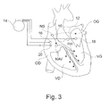
- the Figure 3 schematically illustrates the heart with its four cavities: right atrium OD, right ventricle VD, left atrium OG and left ventricle VG.
- the coordinated contraction of the different cavities originates at the sinus node NS, then the depolarization wave is conducted at the atrio-ventricular node NAV (conduction schematized by the arrow 10), then from this node to the bundle of His FH, and finally to the tissues of the right and left ventricles VD and VG, causing the contraction of these.
- the depolarization wave originating from the sinus node NS causes the contraction of the right atrium OD, then, after interauricular conduction (conduction schematized by the arrow 12) to the left atrium OG, causes the contraction of the latter. .
- atrioventricular conduction (arrow 10, from the NS sinus node to the atrio-ventricular node NAV) is preserved, as well as the conduction pathways to ensure contraction. synchronous of the two ventricles right and left VD and VG.
- the interauricular conduction (arrow 12) can be altered, causing a delay of the depolarization and therefore of the contraction of the left atrium OG with respect to the ventricles.
- This induces a poor sequencing of the contraction of the left atrium OG compared to that of the left ventricle LV, with a more or less concomitant contraction of these two cavities.
- the left atrium OG can more properly fulfill its function, which is to complete the filling of the LV left ventricle.
- the invention proposes a technique of diagnosis and therapy of this pathology, by means of a stimulator acting solely on the two atria.
- This stimulator comprises a generator 14 connected to an electrode 16 for stimulating / collecting depolarizations at the level of the right atrium OD, and an electrode 18 for stimulating / collecting depolarizations at the level of the left atrium OG.
- the electrode 18 may in particular be positioned in the coronary sinus, or in the interatrial septum, or directly in the left atrium after puncture of the interatrial septum.
- the probe placed at the level of the right atrium is also equipped with a sensor 20 capable of delivering a functional signal representative of the contractions (and not depolarizations) of the cardiac cavities, typically an endocardial acceleration signal EA of the type exhibited more top with reference to Figures 1 and 2 .
- the electrode 16 and the sensor 20 are arranged on a conventional endocavity probe implanted at the level of the right atrium via the venous network.
- the sensor 20 will be sensitive to accelerations of the cardiac walls, with a signal amplitude corresponding mainly to the activity of the left atrium and the left ventricle, inasmuch as the most important cardiac mass is that of the left cavities (and this although the sensor is implanted on the right side).
- the contraction sensor 20 is disposed at the atria, on an atrial probe, because the device is of the "double atrial chamber” type, devoid of ventricular sensing / pacing means. It is also arranged on the right atrial lead because it is of a type that lends itself more easily to the integration of a sensor (endocavity probe) than in the case of a left atrial probe (very fine probe , introduced into the coronary network or through the interauricular septum). But other placements of the acceleration sensor are possible, for example on a ventricular probe in the case of a stimulator of the "triple-chamber / double-ear" type, since the signal collected makes it possible to characterize the contraction. of the atrium (component EA4 sufficiently discernible on the collected signal).
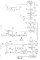
- the Figure 4 is a detailed flowchart showing the manner in which the processing of the signals collected by the electrodes 16, 18 and the contraction sensor 20 is carried out, for the implementation of the invention.
- the first phase (step 100) consists in performing an initial spontaneous rhythm analysis with: measurement of the interauricular delay (designated "OD-sti OG") separating the spontaneous depolarizations of the right atrium and the left atrium, measurement of the atrial coupling interval PP (delay separating two atrial depolarizations of the same cavity, representative of the duration of a cycle in spontaneous rhythm), and measurement of the filling time FT.
- the counter of cycles also initialized has a predetermined value N.
- the contraction sensor 20 then analyzes the signal EA to determine whether an EA4 component is present or not (step 102).
- the absence of a detectable EA4 component means that the contraction of the left atrium OG is embedded in that of contraction of the left ventricle LV, and therefore that the atrium contracts late and does not fulfill its function of completing the filling of the left atrium. left ventricle at the end of the diastole of the latter.
- a detectable EA4 component means that the contraction of the left atrium OG occurs before the contraction of the left ventricle, thus at the end of diastole, with a correct sequencing allowing the contraction of the atrium to complete the filling of the ventricle before this last does not begin to contract.
- step 104 the process waits for the flow of the N cycles (steps 104, 106) before repeating the previous phase of initialization of the step.
- step 102 the absence of EA4 component is confirmed, after detection of the spontaneous contraction of the right atrium OD stimulation is delivered to the left atrium OG, with application of a delay (designated "OD -sti OG ”) corresponding to spontaneous interauricular delay measured in step 100, decreased by a fixed step, for example a step of 10 ms (step 108).
- the cycle counter is also reset to N.
- step 114 After the lapse of N cycles (steps 110, 112), the presence of an EA4 component is again tested (step 114).
- the stimulation is maintained with the same delay (steps 128 to 146, described below).
- step 116 If the component EA4 is absent, and if the atrial stimulation delay OD-sti OG is not at its minimum (step 116), then this delay is further reduced by one step, and the stimulation is delivered under these conditions during N cycles (steps 118, 120 and 122, homologs of steps 108, 110 and 112 above).
- step 116 (i) the atrial pacing delay is minimized (10 ms or less), (ii) the EA4 component is still absent, and (iii) the AA atrial pacing interval is greater at the PP coupling interval in a spontaneous rhythm, to a limiting value, then left atrial pacing is delivered with an interval AA equal to the decreased sinus cycle time of 10 ms (step 126), and so on during N cycles (steps 120, 122) until an EA4 component appears (step 114 above).
- the process performs an additional optimization by comparative measurement of the filling time FT.
- EP 2 206 531 A1 (ELA Medical), which describes how to analyze a number of hemodynamic parameters, including the FT filling time, to optimize the adjustment of stimulation parameters.
- the FT filling time is the time interval between closure of the aortic valve and closure of the mitral valve; it is generally expressed in relative terms, as a percentage of the complete cycle time (RR duration), with an ideal value typically FT> 40%.
- the instants of the different hemodynamic phases of the same cycle, allowing in particular to determine the filling time FT, may in particular be determined by a technique such as that described in FIG. EP 2 092 885 A1 (ELA Medical), where the different temporal markers of the characteristic instants of the cardiac cycle are determined by the analysis of an EA signal.
- the data provided by the signal EA reflect in fact very precisely and in real time, as explained above, the phenomena contributing to the operation mechanical heart and allow, after filtering and analysis, to provide temporal markers of systole and other indices of haemodynamic performance of the myocardium. These parameters can be determined in real time, beat after beat, which allows to optimize without delay the therapy applied to the patient.
- a comparative measurement is performed between the filling time in spontaneous rhythm (parameter FT measured in the initial step 100) and that measured after reappearance of the component EA4 following a controlled stimulation.
Landscapes
- Health & Medical Sciences (AREA)
- Cardiology (AREA)
- Heart & Thoracic Surgery (AREA)
- Life Sciences & Earth Sciences (AREA)
- Public Health (AREA)
- Engineering & Computer Science (AREA)
- Biomedical Technology (AREA)
- Nuclear Medicine, Radiotherapy & Molecular Imaging (AREA)
- Radiology & Medical Imaging (AREA)
- Animal Behavior & Ethology (AREA)
- General Health & Medical Sciences (AREA)
- Veterinary Medicine (AREA)
- Physiology (AREA)
- Biophysics (AREA)
- Hematology (AREA)
- Hospice & Palliative Care (AREA)
- Electrotherapy Devices (AREA)
Applications Claiming Priority (1)
| Application Number | Priority Date | Filing Date | Title |
|---|---|---|---|
| FR1061335 | 2010-12-29 |
Publications (2)
| Publication Number | Publication Date |
|---|---|
| EP2471575A1 true EP2471575A1 (de) | 2012-07-04 |
| EP2471575B1 EP2471575B1 (de) | 2018-11-21 |
Family
ID=43828116
Family Applications (1)
| Application Number | Title | Priority Date | Filing Date |
|---|---|---|---|
| EP11185520.1A Active EP2471575B1 (de) | 2010-12-29 | 2011-10-17 | Implantierbare aktive medizinische Vorrichtung zur Aurikelstimulation für die Behandlung von diastolischer Herzinsuffizienz |
Country Status (2)
| Country | Link |
|---|---|
| US (1) | US8428714B2 (de) |
| EP (1) | EP2471575B1 (de) |
Cited By (4)
| Publication number | Priority date | Publication date | Assignee | Title |
|---|---|---|---|---|
| EP2737925A1 (de) * | 2012-11-29 | 2014-06-04 | Sorin CRM SAS | Aktive implantierbare Herzprothese mit Erfassung der aurikularen mechanischen Aktivität |
| EP2873436A1 (de) | 2013-11-15 | 2015-05-20 | Sorin CRM SAS | Implantierbare aktive medizinische Vorrichtung zur biaurikulären Stimulation für die Behandlung von Herzinsuffizienz mit erhaltener Auswurffraktion (HFpEF) |
| EP2881140A1 (de) | 2013-12-04 | 2015-06-10 | Sorin CRM SAS | Implantierbare aktive medizinische Vorrichtung zur Doppelkammer-Stimulation für die Behandlung von Herzinsuffizienz mit erhaltener Auswurffraktion (HFpEF) |
| CN109789309A (zh) * | 2016-09-29 | 2019-05-21 | 美敦力公司 | 心内心室起搏器中的心房跟踪 |
Families Citing this family (3)
| Publication number | Priority date | Publication date | Assignee | Title |
|---|---|---|---|---|
| US9399140B2 (en) * | 2014-07-25 | 2016-07-26 | Medtronic, Inc. | Atrial contraction detection by a ventricular leadless pacing device for atrio-synchronous ventricular pacing |
| US11207527B2 (en) | 2016-07-06 | 2021-12-28 | Cardiac Pacemakers, Inc. | Method and system for determining an atrial contraction timing fiducial in a leadless cardiac pacemaker system |
| AU2017308071B2 (en) * | 2016-08-11 | 2020-02-27 | Cardiac Pacemakers, Inc. | Diastolic endocardial accelerations for heart failure monitoring |
Citations (11)
| Publication number | Priority date | Publication date | Assignee | Title |
|---|---|---|---|---|
| EP0515319A2 (de) | 1991-05-21 | 1992-11-25 | SORIN BIOMEDICA CARDIO S.p.A. | Taktempfindlicher Herzschrittmacher |
| EP1108446A1 (de) | 1999-12-17 | 2001-06-20 | ELA MEDICAL (Société anonyme) | Implantierbare aktive Vorrichtung des Typs multisite mit Mitteln zum Resynchronisieren der Ventrikeln |
| US20020151934A1 (en) * | 2001-04-12 | 2002-10-17 | Levine Paul A. | System and method for programmably controlling electrode activation sequence in a multi-site cardiac stimulation device |
| WO2005011803A2 (en) * | 2003-07-30 | 2005-02-10 | Medtronic, Inc. | Method of optimizing cardiac resynchronization therapy using sensor signals of septal wall motion |
| US20050102002A1 (en) | 2003-11-07 | 2005-05-12 | Salo Rodney W. | Electrical therapy for diastolic dysfunction |
| US20070179542A1 (en) | 2006-01-31 | 2007-08-02 | Rajan Prakash | Method and apparatus for determining optimal pacing therapy timing intervals |
| US20070179541A1 (en) * | 2006-01-31 | 2007-08-02 | Rajan Prakash | Method and apparatus for determining excitation-physiologic response delays |
| EP2092885A1 (de) | 2008-02-20 | 2009-08-26 | Ela Medical | Analysevorrichtung eines endokardialen Beschleunigungssignals |
| EP2189180A1 (de) | 2008-11-19 | 2010-05-26 | Ela Medical | Aktives medizinisches Implantat, das mit Berechnungsmitteln der atrioventrikulären Verzögerung ausgestattet ist |
| EP2189182A1 (de) | 2008-11-19 | 2010-05-26 | Ela Medical | Aktives medizinisches Implantat, das mit Testmitteln zur aurikulären Datenerfassung ausgestattet ist |
| EP2206531A1 (de) | 2009-01-09 | 2010-07-14 | Ela Medical | Aktive medizinische Vorrichtung vom Typ implantierbare Herzprothese für die Behandlung von Herzinsuffizienz durch kontrollierte Anpassung der atrioventrikulären und intraventrikulären Durchblutungszeiten |
Family Cites Families (1)
| Publication number | Priority date | Publication date | Assignee | Title |
|---|---|---|---|---|
| US6339723B1 (en) * | 2000-01-20 | 2002-01-15 | Pacesetter, Inc. | Method and apparatus for preventing atrial fusion during automatic pacing threshold determination in a cardiac stimulation device |
-
2011
- 2011-10-17 EP EP11185520.1A patent/EP2471575B1/de active Active
- 2011-12-29 US US13/340,535 patent/US8428714B2/en not_active Expired - Fee Related
Patent Citations (11)
| Publication number | Priority date | Publication date | Assignee | Title |
|---|---|---|---|---|
| EP0515319A2 (de) | 1991-05-21 | 1992-11-25 | SORIN BIOMEDICA CARDIO S.p.A. | Taktempfindlicher Herzschrittmacher |
| EP1108446A1 (de) | 1999-12-17 | 2001-06-20 | ELA MEDICAL (Société anonyme) | Implantierbare aktive Vorrichtung des Typs multisite mit Mitteln zum Resynchronisieren der Ventrikeln |
| US20020151934A1 (en) * | 2001-04-12 | 2002-10-17 | Levine Paul A. | System and method for programmably controlling electrode activation sequence in a multi-site cardiac stimulation device |
| WO2005011803A2 (en) * | 2003-07-30 | 2005-02-10 | Medtronic, Inc. | Method of optimizing cardiac resynchronization therapy using sensor signals of septal wall motion |
| US20050102002A1 (en) | 2003-11-07 | 2005-05-12 | Salo Rodney W. | Electrical therapy for diastolic dysfunction |
| US20070179542A1 (en) | 2006-01-31 | 2007-08-02 | Rajan Prakash | Method and apparatus for determining optimal pacing therapy timing intervals |
| US20070179541A1 (en) * | 2006-01-31 | 2007-08-02 | Rajan Prakash | Method and apparatus for determining excitation-physiologic response delays |
| EP2092885A1 (de) | 2008-02-20 | 2009-08-26 | Ela Medical | Analysevorrichtung eines endokardialen Beschleunigungssignals |
| EP2189180A1 (de) | 2008-11-19 | 2010-05-26 | Ela Medical | Aktives medizinisches Implantat, das mit Berechnungsmitteln der atrioventrikulären Verzögerung ausgestattet ist |
| EP2189182A1 (de) | 2008-11-19 | 2010-05-26 | Ela Medical | Aktives medizinisches Implantat, das mit Testmitteln zur aurikulären Datenerfassung ausgestattet ist |
| EP2206531A1 (de) | 2009-01-09 | 2010-07-14 | Ela Medical | Aktive medizinische Vorrichtung vom Typ implantierbare Herzprothese für die Behandlung von Herzinsuffizienz durch kontrollierte Anpassung der atrioventrikulären und intraventrikulären Durchblutungszeiten |
Cited By (10)
| Publication number | Priority date | Publication date | Assignee | Title |
|---|---|---|---|---|
| EP2737925A1 (de) * | 2012-11-29 | 2014-06-04 | Sorin CRM SAS | Aktive implantierbare Herzprothese mit Erfassung der aurikularen mechanischen Aktivität |
| US9272146B2 (en) | 2012-11-29 | 2016-03-01 | Sorin Crm S.A.S. | Implantable medical device with active detection of atrial mechanical activity |
| US10016168B2 (en) | 2012-11-29 | 2018-07-10 | Sorin Crm Sas | Implantable medical device with active detection of atrial mechanical activity |
| EP2873436A1 (de) | 2013-11-15 | 2015-05-20 | Sorin CRM SAS | Implantierbare aktive medizinische Vorrichtung zur biaurikulären Stimulation für die Behandlung von Herzinsuffizienz mit erhaltener Auswurffraktion (HFpEF) |
| US9283393B2 (en) | 2013-11-15 | 2016-03-15 | Sorin Crm Sas | Active implantable medical device for atrial stimulation for the treatment of heart failure with preserved ejection fraction |
| US9526901B2 (en) | 2013-11-15 | 2016-12-27 | Sorin Crm Sas | Active implantable medical device for atrial stimulation for the treatment of heart failure with preserved ejection fraction |
| EP2881140A1 (de) | 2013-12-04 | 2015-06-10 | Sorin CRM SAS | Implantierbare aktive medizinische Vorrichtung zur Doppelkammer-Stimulation für die Behandlung von Herzinsuffizienz mit erhaltener Auswurffraktion (HFpEF) |
| US9259581B2 (en) | 2013-12-04 | 2016-02-16 | Sorin Crm Sas | Active implantable medical device with dual chamber pacing for the treatment of heart failure with preserved ejection fraction |
| CN109789309A (zh) * | 2016-09-29 | 2019-05-21 | 美敦力公司 | 心内心室起搏器中的心房跟踪 |
| US12186569B2 (en) | 2016-09-29 | 2025-01-07 | Medtronic, Inc. | Atrial tracking in an intracardiac ventricular pacemaker |
Also Published As
| Publication number | Publication date |
|---|---|
| US8428714B2 (en) | 2013-04-23 |
| US20120172943A1 (en) | 2012-07-05 |
| EP2471575B1 (de) | 2018-11-21 |
Similar Documents
| Publication | Publication Date | Title |
|---|---|---|
| EP2189182B1 (de) | Aktives medizinisches Implantat, das mit Testmitteln zur aurikulären Datenerfassung ausgestattet ist | |
| EP2070562B1 (de) | Medizinische Vorrichtung zur Bestimmung des Herzzustands eines Patienten, dem ein aktives Implantat zur biventrikulären Stimulation eingesetzt wurde | |
| EP2263747B1 (de) | Implantierbare Herzprothese mit Mittel zur Analyse der Patiententoleranz bezüglich der Stimulationsart die spontane Atrio-Ventrukuläre Leitung bevorzugend | |
| EP2092885B1 (de) | Analysevorrichtung eines endokardialen Beschleunigungssignals | |
| EP2471575B1 (de) | Implantierbare aktive medizinische Vorrichtung zur Aurikelstimulation für die Behandlung von diastolischer Herzinsuffizienz | |
| EP3015132B1 (de) | Aktiver implantierbarer stimulator, insbesondere autonome intrakardiale kapsel, zum erfassen der vorhof-aktivität ohne erfassung der atrialen elektrischen aktivität | |
| EP2189180B1 (de) | Aktives medizinisches Implantat, das mit Berechnungsmitteln der atrioventrikulären Verzögerung ausgestattet ist | |
| EP2737925B1 (de) | Aktive implantierbare Herzprothese mit Erfassung der aurikularen mechanischen Aktivität | |
| EP0862927B1 (de) | Multi-Ort Herzschrittmacher zur Behandlung von Herzinsuffizienzen mit Reizimpulsen | |
| EP2311524B1 (de) | Implantierbarer medizinischer Herzschrittmacher und Verfahren zur Optimierung des Implantatortes für interventrikuläre und atrioventrikuläre Intervalle | |
| FR2901146A1 (fr) | Dispositif medical implantable actif de stimulation cardiaque, resynchronisation, cardioversion et/ou defibrillation, comportant des moyens de detection d'artefacts de bruit ventriculaire | |
| EP1736203B1 (de) | Implantierbares medizinisches Gerät zur biventrikulären Stimulation und zur automatischen Optimierung der Anregungskonfiguration | |
| EP2357020B1 (de) | Aktive medizinische Vorrichtung vom Typ Herzschrittmacher, Defibrillator und/oder Resynchronisator mit automatischer Optimierung der atrioventrikulären Verzögerung | |
| EP2206531B1 (de) | Aktive medizinische Vorrichtung vom Typ implantierbare Herzprothese für die Behandlung von Herzinsuffizienz durch kontrollierte Anpassung der atrioventrikulären und intraventrikulären Durchblutungszeiten | |
| EP2499971A1 (de) | Adaptative Datenverarbeitungsvorrichtung eines endokardialen Beschleunigungssignals | |
| EP3187223A1 (de) | Aktive medizinische vorrichtung zur elektrischen neurostimulation mit automatischer kontrolle der ladungskompensation | |
| EP2374503B1 (de) | Aktive implantierbare medizinische Vorrichtung für Vagusnervstimulation mit Optimisierung der Ventrikelfüllung | |
| EP2712549B1 (de) | Vorrichtung zur Bewertung der zeitlichen ventrikulären Desynchronisation | |
| EP2508227B1 (de) | Implantierbare Herzprothese zur Resynchronisation des Herzens durch biventrikuläre Stimulation, die Mittel zur inversen Herzrekonfiguration umfasst | |
| EP2495013B1 (de) | Anordnung für die Suche nach einer optimalen Konfiguration eines Implantats zur bi-, tri- oder multiventrikulären Resynchronisation des Herzens | |
| EP2803385B1 (de) | Implantierbares CRT-Gerät zur Stimulation beider Ventrikel und zur Erkennung von Erfassungsverlusten und anodischen Stimulationen | |
| EP3081257A1 (de) | Aktive implantierbare medizinische vorrichtung zur herzstimulation, die detektionsmittel für remodellierungserscheinungen oder inversen remodellierungserscheinungen des patienten umfasst | |
| EP3075413B1 (de) | Implantierbares medizinisches gerät für die biventrikuläre stimulation, umfassend mittel zur messung intraventrikulärer leitungszeit für die diagnose von herzdekompensation | |
| EP2873436B1 (de) | Implantierbare aktive medizinische Vorrichtung zur biaurikulären Stimulation für die Behandlung von Herzinsuffizienz mit erhaltener Auswurffraktion (HFpEF) | |
| EP2881140B1 (de) | Implantierbare aktive medizinische Vorrichtung zur Doppelkammer-Stimulation für die Behandlung von Herzinsuffizienz mit erhaltener Auswurffraktion (HFpEF) |
Legal Events
| Date | Code | Title | Description |
|---|---|---|---|
| AK | Designated contracting states |
Kind code of ref document: A1 Designated state(s): AL AT BE BG CH CY CZ DE DK EE ES FI FR GB GR HR HU IE IS IT LI LT LU LV MC MK MT NL NO PL PT RO RS SE SI SK SM TR |
|
| AX | Request for extension of the european patent |
Extension state: BA ME |
|
| PUAI | Public reference made under article 153(3) epc to a published international application that has entered the european phase |
Free format text: ORIGINAL CODE: 0009012 |
|
| 17P | Request for examination filed |
Effective date: 20121010 |
|
| STAA | Information on the status of an ep patent application or granted ep patent |
Free format text: STATUS: EXAMINATION IS IN PROGRESS |
|
| 17Q | First examination report despatched |
Effective date: 20161214 |
|
| GRAP | Despatch of communication of intention to grant a patent |
Free format text: ORIGINAL CODE: EPIDOSNIGR1 |
|
| STAA | Information on the status of an ep patent application or granted ep patent |
Free format text: STATUS: GRANT OF PATENT IS INTENDED |
|
| INTG | Intention to grant announced |
Effective date: 20180605 |
|
| GRAS | Grant fee paid |
Free format text: ORIGINAL CODE: EPIDOSNIGR3 |
|
| GRAA | (expected) grant |
Free format text: ORIGINAL CODE: 0009210 |
|
| STAA | Information on the status of an ep patent application or granted ep patent |
Free format text: STATUS: THE PATENT HAS BEEN GRANTED |
|
| AK | Designated contracting states |
Kind code of ref document: B1 Designated state(s): AL AT BE BG CH CY CZ DE DK EE ES FI FR GB GR HR HU IE IS IT LI LT LU LV MC MK MT NL NO PL PT RO RS SE SI SK SM TR |
|
| REG | Reference to a national code |
Ref country code: CH Ref legal event code: EP |
|
| REG | Reference to a national code |
Ref country code: IE Ref legal event code: FG4D Free format text: LANGUAGE OF EP DOCUMENT: FRENCH |
|
| REG | Reference to a national code |
Ref country code: DE Ref legal event code: R096 Ref document number: 602011054030 Country of ref document: DE |
|
| REG | Reference to a national code |
Ref country code: AT Ref legal event code: REF Ref document number: 1066856 Country of ref document: AT Kind code of ref document: T Effective date: 20181215 |
|
| REG | Reference to a national code |
Ref country code: NL Ref legal event code: MP Effective date: 20181121 |
|
| REG | Reference to a national code |
Ref country code: AT Ref legal event code: MK05 Ref document number: 1066856 Country of ref document: AT Kind code of ref document: T Effective date: 20181121 |
|
| PG25 | Lapsed in a contracting state [announced via postgrant information from national office to epo] |
Ref country code: IS Free format text: LAPSE BECAUSE OF FAILURE TO SUBMIT A TRANSLATION OF THE DESCRIPTION OR TO PAY THE FEE WITHIN THE PRESCRIBED TIME-LIMIT Effective date: 20190321 Ref country code: FI Free format text: LAPSE BECAUSE OF FAILURE TO SUBMIT A TRANSLATION OF THE DESCRIPTION OR TO PAY THE FEE WITHIN THE PRESCRIBED TIME-LIMIT Effective date: 20181121 Ref country code: BG Free format text: LAPSE BECAUSE OF FAILURE TO SUBMIT A TRANSLATION OF THE DESCRIPTION OR TO PAY THE FEE WITHIN THE PRESCRIBED TIME-LIMIT Effective date: 20190221 Ref country code: LT Free format text: LAPSE BECAUSE OF FAILURE TO SUBMIT A TRANSLATION OF THE DESCRIPTION OR TO PAY THE FEE WITHIN THE PRESCRIBED TIME-LIMIT Effective date: 20181121 Ref country code: HR Free format text: LAPSE BECAUSE OF FAILURE TO SUBMIT A TRANSLATION OF THE DESCRIPTION OR TO PAY THE FEE WITHIN THE PRESCRIBED TIME-LIMIT Effective date: 20181121 Ref country code: LV Free format text: LAPSE BECAUSE OF FAILURE TO SUBMIT A TRANSLATION OF THE DESCRIPTION OR TO PAY THE FEE WITHIN THE PRESCRIBED TIME-LIMIT Effective date: 20181121 Ref country code: ES Free format text: LAPSE BECAUSE OF FAILURE TO SUBMIT A TRANSLATION OF THE DESCRIPTION OR TO PAY THE FEE WITHIN THE PRESCRIBED TIME-LIMIT Effective date: 20181121 Ref country code: NO Free format text: LAPSE BECAUSE OF FAILURE TO SUBMIT A TRANSLATION OF THE DESCRIPTION OR TO PAY THE FEE WITHIN THE PRESCRIBED TIME-LIMIT Effective date: 20190221 Ref country code: AT Free format text: LAPSE BECAUSE OF FAILURE TO SUBMIT A TRANSLATION OF THE DESCRIPTION OR TO PAY THE FEE WITHIN THE PRESCRIBED TIME-LIMIT Effective date: 20181121 |
|
| PG25 | Lapsed in a contracting state [announced via postgrant information from national office to epo] |
Ref country code: PT Free format text: LAPSE BECAUSE OF FAILURE TO SUBMIT A TRANSLATION OF THE DESCRIPTION OR TO PAY THE FEE WITHIN THE PRESCRIBED TIME-LIMIT Effective date: 20190321 Ref country code: AL Free format text: LAPSE BECAUSE OF FAILURE TO SUBMIT A TRANSLATION OF THE DESCRIPTION OR TO PAY THE FEE WITHIN THE PRESCRIBED TIME-LIMIT Effective date: 20181121 Ref country code: RS Free format text: LAPSE BECAUSE OF FAILURE TO SUBMIT A TRANSLATION OF THE DESCRIPTION OR TO PAY THE FEE WITHIN THE PRESCRIBED TIME-LIMIT Effective date: 20181121 Ref country code: GR Free format text: LAPSE BECAUSE OF FAILURE TO SUBMIT A TRANSLATION OF THE DESCRIPTION OR TO PAY THE FEE WITHIN THE PRESCRIBED TIME-LIMIT Effective date: 20190222 Ref country code: NL Free format text: LAPSE BECAUSE OF FAILURE TO SUBMIT A TRANSLATION OF THE DESCRIPTION OR TO PAY THE FEE WITHIN THE PRESCRIBED TIME-LIMIT Effective date: 20181121 Ref country code: SE Free format text: LAPSE BECAUSE OF FAILURE TO SUBMIT A TRANSLATION OF THE DESCRIPTION OR TO PAY THE FEE WITHIN THE PRESCRIBED TIME-LIMIT Effective date: 20181121 |
|
| PG25 | Lapsed in a contracting state [announced via postgrant information from national office to epo] |
Ref country code: CZ Free format text: LAPSE BECAUSE OF FAILURE TO SUBMIT A TRANSLATION OF THE DESCRIPTION OR TO PAY THE FEE WITHIN THE PRESCRIBED TIME-LIMIT Effective date: 20181121 Ref country code: DK Free format text: LAPSE BECAUSE OF FAILURE TO SUBMIT A TRANSLATION OF THE DESCRIPTION OR TO PAY THE FEE WITHIN THE PRESCRIBED TIME-LIMIT Effective date: 20181121 Ref country code: PL Free format text: LAPSE BECAUSE OF FAILURE TO SUBMIT A TRANSLATION OF THE DESCRIPTION OR TO PAY THE FEE WITHIN THE PRESCRIBED TIME-LIMIT Effective date: 20181121 |
|
| REG | Reference to a national code |
Ref country code: DE Ref legal event code: R097 Ref document number: 602011054030 Country of ref document: DE |
|
| PG25 | Lapsed in a contracting state [announced via postgrant information from national office to epo] |
Ref country code: SK Free format text: LAPSE BECAUSE OF FAILURE TO SUBMIT A TRANSLATION OF THE DESCRIPTION OR TO PAY THE FEE WITHIN THE PRESCRIBED TIME-LIMIT Effective date: 20181121 Ref country code: RO Free format text: LAPSE BECAUSE OF FAILURE TO SUBMIT A TRANSLATION OF THE DESCRIPTION OR TO PAY THE FEE WITHIN THE PRESCRIBED TIME-LIMIT Effective date: 20181121 Ref country code: SM Free format text: LAPSE BECAUSE OF FAILURE TO SUBMIT A TRANSLATION OF THE DESCRIPTION OR TO PAY THE FEE WITHIN THE PRESCRIBED TIME-LIMIT Effective date: 20181121 Ref country code: EE Free format text: LAPSE BECAUSE OF FAILURE TO SUBMIT A TRANSLATION OF THE DESCRIPTION OR TO PAY THE FEE WITHIN THE PRESCRIBED TIME-LIMIT Effective date: 20181121 |
|
| PLBE | No opposition filed within time limit |
Free format text: ORIGINAL CODE: 0009261 |
|
| STAA | Information on the status of an ep patent application or granted ep patent |
Free format text: STATUS: NO OPPOSITION FILED WITHIN TIME LIMIT |
|
| 26N | No opposition filed |
Effective date: 20190822 |
|
| PG25 | Lapsed in a contracting state [announced via postgrant information from national office to epo] |
Ref country code: SI Free format text: LAPSE BECAUSE OF FAILURE TO SUBMIT A TRANSLATION OF THE DESCRIPTION OR TO PAY THE FEE WITHIN THE PRESCRIBED TIME-LIMIT Effective date: 20181121 |
|
| PG25 | Lapsed in a contracting state [announced via postgrant information from national office to epo] |
Ref country code: TR Free format text: LAPSE BECAUSE OF FAILURE TO SUBMIT A TRANSLATION OF THE DESCRIPTION OR TO PAY THE FEE WITHIN THE PRESCRIBED TIME-LIMIT Effective date: 20181121 |
|
| PG25 | Lapsed in a contracting state [announced via postgrant information from national office to epo] |
Ref country code: MC Free format text: LAPSE BECAUSE OF FAILURE TO SUBMIT A TRANSLATION OF THE DESCRIPTION OR TO PAY THE FEE WITHIN THE PRESCRIBED TIME-LIMIT Effective date: 20181121 |
|
| REG | Reference to a national code |
Ref country code: CH Ref legal event code: PL |
|
| PG25 | Lapsed in a contracting state [announced via postgrant information from national office to epo] |
Ref country code: CH Free format text: LAPSE BECAUSE OF NON-PAYMENT OF DUE FEES Effective date: 20191031 Ref country code: LU Free format text: LAPSE BECAUSE OF NON-PAYMENT OF DUE FEES Effective date: 20191017 Ref country code: LI Free format text: LAPSE BECAUSE OF NON-PAYMENT OF DUE FEES Effective date: 20191031 |
|
| REG | Reference to a national code |
Ref country code: BE Ref legal event code: MM Effective date: 20191031 |
|
| PG25 | Lapsed in a contracting state [announced via postgrant information from national office to epo] |
Ref country code: BE Free format text: LAPSE BECAUSE OF NON-PAYMENT OF DUE FEES Effective date: 20191031 |
|
| PG25 | Lapsed in a contracting state [announced via postgrant information from national office to epo] |
Ref country code: IE Free format text: LAPSE BECAUSE OF NON-PAYMENT OF DUE FEES Effective date: 20191017 |
|
| PG25 | Lapsed in a contracting state [announced via postgrant information from national office to epo] |
Ref country code: CY Free format text: LAPSE BECAUSE OF FAILURE TO SUBMIT A TRANSLATION OF THE DESCRIPTION OR TO PAY THE FEE WITHIN THE PRESCRIBED TIME-LIMIT Effective date: 20181121 |
|
| PG25 | Lapsed in a contracting state [announced via postgrant information from national office to epo] |
Ref country code: MT Free format text: LAPSE BECAUSE OF FAILURE TO SUBMIT A TRANSLATION OF THE DESCRIPTION OR TO PAY THE FEE WITHIN THE PRESCRIBED TIME-LIMIT Effective date: 20181121 Ref country code: HU Free format text: LAPSE BECAUSE OF FAILURE TO SUBMIT A TRANSLATION OF THE DESCRIPTION OR TO PAY THE FEE WITHIN THE PRESCRIBED TIME-LIMIT; INVALID AB INITIO Effective date: 20111017 |
|
| REG | Reference to a national code |
Ref country code: DE Ref legal event code: R082 Ref document number: 602011054030 Country of ref document: DE Representative=s name: PAGE, WHITE & FARRER GERMANY LLP, DE Ref country code: DE Ref legal event code: R082 Ref document number: 602011054030 Country of ref document: DE Representative=s name: GULDE & PARTNER PATENT- UND RECHTSANWALTSKANZL, DE |
|
| PG25 | Lapsed in a contracting state [announced via postgrant information from national office to epo] |
Ref country code: MK Free format text: LAPSE BECAUSE OF FAILURE TO SUBMIT A TRANSLATION OF THE DESCRIPTION OR TO PAY THE FEE WITHIN THE PRESCRIBED TIME-LIMIT Effective date: 20181121 |
|
| P01 | Opt-out of the competence of the unified patent court (upc) registered |
Effective date: 20230714 |
|
| PGFP | Annual fee paid to national office [announced via postgrant information from national office to epo] |
Ref country code: DE Payment date: 20241010 Year of fee payment: 14 |
|
| PGFP | Annual fee paid to national office [announced via postgrant information from national office to epo] |
Ref country code: GB Payment date: 20241028 Year of fee payment: 14 |
|
| PGFP | Annual fee paid to national office [announced via postgrant information from national office to epo] |
Ref country code: FR Payment date: 20241024 Year of fee payment: 14 |
|
| PGFP | Annual fee paid to national office [announced via postgrant information from national office to epo] |
Ref country code: IT Payment date: 20241010 Year of fee payment: 14 |
|
| REG | Reference to a national code |
Ref country code: DE Ref legal event code: R082 Ref document number: 602011054030 Country of ref document: DE Representative=s name: GULDE & PARTNER PATENT- UND RECHTSANWALTSKANZL, DE |