EP2473852B1 - Biomarqueurs pour la prédiction d'événements indésirables cardiaques majeurs et utilisations de ceux-ci - Google Patents
Biomarqueurs pour la prédiction d'événements indésirables cardiaques majeurs et utilisations de ceux-ci Download PDFInfo
- Publication number
- EP2473852B1 EP2473852B1 EP10752669.1A EP10752669A EP2473852B1 EP 2473852 B1 EP2473852 B1 EP 2473852B1 EP 10752669 A EP10752669 A EP 10752669A EP 2473852 B1 EP2473852 B1 EP 2473852B1
- Authority
- EP
- European Patent Office
- Prior art keywords
- patient
- level
- symptom
- presentation
- year
- Prior art date
- Legal status (The legal status is an assumption and is not a legal conclusion. Google has not performed a legal analysis and makes no representation as to the accuracy of the status listed.)
- Not-in-force
Links
- 230000000747 cardiac effect Effects 0.000 title claims description 139
- 230000002411 adverse Effects 0.000 title claims description 128
- 239000000090 biomarker Substances 0.000 title claims description 74
- 208000004476 Acute Coronary Syndrome Diseases 0.000 claims description 96
- 208000024891 symptom Diseases 0.000 claims description 85
- 238000000034 method Methods 0.000 claims description 80
- 102100035194 Placenta growth factor Human genes 0.000 claims description 63
- 238000012360 testing method Methods 0.000 claims description 63
- 101710128251 Troponin I, cardiac muscle Proteins 0.000 claims description 45
- 102100036859 Troponin I, cardiac muscle Human genes 0.000 claims description 45
- FFFHZYDWPBMWHY-VKHMYHEASA-N L-homocysteine Chemical compound OC(=O)[C@@H](N)CCS FFFHZYDWPBMWHY-VKHMYHEASA-N 0.000 claims description 39
- 208000010125 myocardial infarction Diseases 0.000 claims description 31
- OEYIOHPDSNJKLS-UHFFFAOYSA-N choline Chemical compound C[N+](C)(C)CCO OEYIOHPDSNJKLS-UHFFFAOYSA-N 0.000 claims description 23
- 229960001231 choline Drugs 0.000 claims description 23
- 230000000250 revascularization Effects 0.000 claims description 13
- 230000034994 death Effects 0.000 claims description 12
- 231100000517 death Toxicity 0.000 claims description 12
- 206010019280 Heart failures Diseases 0.000 claims description 10
- 208000020832 chronic kidney disease Diseases 0.000 claims description 2
- 238000009662 stress testing Methods 0.000 claims description 2
- 108060006698 EGF receptor Proteins 0.000 claims 19
- 101100537532 Rattus norvegicus Tnni3 gene Proteins 0.000 claims 3
- 108010082093 Placenta Growth Factor Proteins 0.000 claims 1
- 102000052116 epidermal growth factor receptor activity proteins Human genes 0.000 claims 1
- 108700015053 epidermal growth factor receptor activity proteins Proteins 0.000 claims 1
- YOHYSYJDKVYCJI-UHFFFAOYSA-N n-[3-[[6-[3-(trifluoromethyl)anilino]pyrimidin-4-yl]amino]phenyl]cyclopropanecarboxamide Chemical compound FC(F)(F)C1=CC=CC(NC=2N=CN=C(NC=3C=C(NC(=O)C4CC4)C=CC=3)C=2)=C1 YOHYSYJDKVYCJI-UHFFFAOYSA-N 0.000 claims 1
- 239000012491 analyte Substances 0.000 description 115
- 101800000407 Brain natriuretic peptide 32 Proteins 0.000 description 94
- 239000000523 sample Substances 0.000 description 82
- 239000012634 fragment Substances 0.000 description 69
- 101000595923 Homo sapiens Placenta growth factor Proteins 0.000 description 62
- 238000003556 assay Methods 0.000 description 41
- 230000027455 binding Effects 0.000 description 38
- 208000037265 diseases, disorders, signs and symptoms Diseases 0.000 description 35
- 108090000765 processed proteins & peptides Proteins 0.000 description 33
- 108090000623 proteins and genes Proteins 0.000 description 33
- 239000000427 antigen Substances 0.000 description 30
- 102000036639 antigens Human genes 0.000 description 30
- 108091007433 antigens Proteins 0.000 description 30
- 238000003018 immunoassay Methods 0.000 description 30
- 102000004169 proteins and genes Human genes 0.000 description 30
- 235000018102 proteins Nutrition 0.000 description 29
- 210000004027 cell Anatomy 0.000 description 28
- 102000004196 processed proteins & peptides Human genes 0.000 description 28
- 108010074051 C-Reactive Protein Proteins 0.000 description 26
- 229920001184 polypeptide Polymers 0.000 description 25
- 238000001514 detection method Methods 0.000 description 21
- 102000003896 Myeloperoxidases Human genes 0.000 description 20
- 108090000235 Myeloperoxidases Proteins 0.000 description 20
- 201000010099 disease Diseases 0.000 description 20
- 210000002381 plasma Anatomy 0.000 description 19
- -1 serum and plasma Substances 0.000 description 19
- 108010023015 ischemia-modified albumin Proteins 0.000 description 16
- 150000001413 amino acids Chemical group 0.000 description 15
- 239000003153 chemical reaction reagent Substances 0.000 description 15
- 208000035475 disorder Diseases 0.000 description 15
- 102000014914 Carrier Proteins Human genes 0.000 description 14
- 108091008324 binding proteins Proteins 0.000 description 14
- 210000004369 blood Anatomy 0.000 description 14
- 239000008280 blood Substances 0.000 description 14
- 102100032752 C-reactive protein Human genes 0.000 description 13
- 230000000694 effects Effects 0.000 description 13
- 239000000047 product Substances 0.000 description 13
- HTTJABKRGRZYRN-UHFFFAOYSA-N Heparin Chemical compound OC1C(NC(=O)C)C(O)OC(COS(O)(=O)=O)C1OC1C(OS(O)(=O)=O)C(O)C(OC2C(C(OS(O)(=O)=O)C(OC3C(C(O)C(O)C(O3)C(O)=O)OS(O)(=O)=O)C(CO)O2)NS(O)(=O)=O)C(C(O)=O)O1 HTTJABKRGRZYRN-UHFFFAOYSA-N 0.000 description 12
- 235000001014 amino acid Nutrition 0.000 description 12
- DDRJAANPRJIHGJ-UHFFFAOYSA-N creatinine Chemical compound CN1CC(=O)NC1=N DDRJAANPRJIHGJ-UHFFFAOYSA-N 0.000 description 12
- 229960002897 heparin Drugs 0.000 description 12
- 229920000669 heparin Polymers 0.000 description 12
- 239000013598 vector Substances 0.000 description 12
- 238000003776 cleavage reaction Methods 0.000 description 11
- 230000006870 function Effects 0.000 description 11
- 230000007017 scission Effects 0.000 description 11
- 108010047041 Complementarity Determining Regions Proteins 0.000 description 10
- 238000004458 analytical method Methods 0.000 description 10
- 239000000872 buffer Substances 0.000 description 10
- 239000011859 microparticle Substances 0.000 description 10
- 108010029697 CD40 Ligand Proteins 0.000 description 9
- 102100032937 CD40 ligand Human genes 0.000 description 9
- 101800001904 NT-proBNP Proteins 0.000 description 9
- 102400001263 NT-proBNP Human genes 0.000 description 9
- 239000003085 diluting agent Substances 0.000 description 9
- 108010008064 pro-brain natriuretic peptide (1-76) Proteins 0.000 description 9
- 230000009870 specific binding Effects 0.000 description 9
- 102000016752 1-Alkyl-2-acetylglycerophosphocholine Esterase Human genes 0.000 description 8
- NFGXHKASABOEEW-UHFFFAOYSA-N 1-methylethyl 11-methoxy-3,7,11-trimethyl-2,4-dodecadienoate Chemical compound COC(C)(C)CCCC(C)CC=CC(C)=CC(=O)OC(C)C NFGXHKASABOEEW-UHFFFAOYSA-N 0.000 description 8
- 108010024976 Asparaginase Proteins 0.000 description 8
- 102000004190 Enzymes Human genes 0.000 description 8
- 108090000790 Enzymes Proteins 0.000 description 8
- 239000003637 basic solution Substances 0.000 description 8
- 108091033319 polynucleotide Proteins 0.000 description 8
- 102000040430 polynucleotide Human genes 0.000 description 8
- 239000002157 polynucleotide Substances 0.000 description 8
- 210000002966 serum Anatomy 0.000 description 8
- 108020004414 DNA Proteins 0.000 description 7
- KCXVZYZYPLLWCC-UHFFFAOYSA-N EDTA Chemical compound OC(=O)CN(CC(O)=O)CCN(CC(O)=O)CC(O)=O KCXVZYZYPLLWCC-UHFFFAOYSA-N 0.000 description 7
- 238000002965 ELISA Methods 0.000 description 7
- 102000013394 Troponin I Human genes 0.000 description 7
- 108010065729 Troponin I Proteins 0.000 description 7
- 230000015572 biosynthetic process Effects 0.000 description 7
- 239000013078 crystal Substances 0.000 description 7
- 230000002255 enzymatic effect Effects 0.000 description 7
- 230000024924 glomerular filtration Effects 0.000 description 7
- 239000007787 solid Substances 0.000 description 7
- 230000001154 acute effect Effects 0.000 description 6
- 229940109239 creatinine Drugs 0.000 description 6
- 208000028867 ischemia Diseases 0.000 description 6
- 230000000302 ischemic effect Effects 0.000 description 6
- 150000007523 nucleic acids Chemical group 0.000 description 6
- 230000008569 process Effects 0.000 description 6
- 238000004393 prognosis Methods 0.000 description 6
- 230000035945 sensitivity Effects 0.000 description 6
- 238000013517 stratification Methods 0.000 description 6
- 239000000126 substance Substances 0.000 description 6
- 238000002560 therapeutic procedure Methods 0.000 description 6
- 206010007558 Cardiac failure chronic Diseases 0.000 description 5
- 206010008479 Chest Pain Diseases 0.000 description 5
- 101001001487 Homo sapiens Phosphatidylinositol-glycan biosynthesis class F protein Proteins 0.000 description 5
- 108010001336 Horseradish Peroxidase Proteins 0.000 description 5
- 108060003951 Immunoglobulin Proteins 0.000 description 5
- WHXSMMKQMYFTQS-UHFFFAOYSA-N Lithium Chemical compound [Li] WHXSMMKQMYFTQS-UHFFFAOYSA-N 0.000 description 5
- 239000011324 bead Substances 0.000 description 5
- 239000011248 coating agent Substances 0.000 description 5
- 238000000576 coating method Methods 0.000 description 5
- 230000006957 competitive inhibition Effects 0.000 description 5
- 150000001875 compounds Chemical class 0.000 description 5
- 239000003814 drug Substances 0.000 description 5
- 239000012530 fluid Substances 0.000 description 5
- 102000018358 immunoglobulin Human genes 0.000 description 5
- 238000011534 incubation Methods 0.000 description 5
- 229910052744 lithium Inorganic materials 0.000 description 5
- 239000013612 plasmid Substances 0.000 description 5
- 239000000243 solution Substances 0.000 description 5
- 239000000758 substrate Substances 0.000 description 5
- 238000005406 washing Methods 0.000 description 5
- YBJHBAHKTGYVGT-ZKWXMUAHSA-N (+)-Biotin Chemical compound N1C(=O)N[C@@H]2[C@H](CCCCC(=O)O)SC[C@@H]21 YBJHBAHKTGYVGT-ZKWXMUAHSA-N 0.000 description 4
- FXZCCINCKDATPA-UHFFFAOYSA-N (4-aminophenyl) dihydrogen phosphate Chemical compound NC1=CC=C(OP(O)(O)=O)C=C1 FXZCCINCKDATPA-UHFFFAOYSA-N 0.000 description 4
- PLIKAWJENQZMHA-UHFFFAOYSA-N 4-aminophenol Chemical compound NC1=CC=C(O)C=C1 PLIKAWJENQZMHA-UHFFFAOYSA-N 0.000 description 4
- 108010088751 Albumins Proteins 0.000 description 4
- 102000009027 Albumins Human genes 0.000 description 4
- 206010007559 Cardiac failure congestive Diseases 0.000 description 4
- 206010008469 Chest discomfort Diseases 0.000 description 4
- 208000000059 Dyspnea Diseases 0.000 description 4
- 206010013975 Dyspnoeas Diseases 0.000 description 4
- MHAJPDPJQMAIIY-UHFFFAOYSA-N Hydrogen peroxide Chemical compound OO MHAJPDPJQMAIIY-UHFFFAOYSA-N 0.000 description 4
- 206010061218 Inflammation Diseases 0.000 description 4
- 102000004903 Troponin Human genes 0.000 description 4
- 108090001027 Troponin Proteins 0.000 description 4
- 238000002820 assay format Methods 0.000 description 4
- 238000011161 development Methods 0.000 description 4
- 239000013604 expression vector Substances 0.000 description 4
- 230000004054 inflammatory process Effects 0.000 description 4
- 239000003550 marker Substances 0.000 description 4
- 230000004048 modification Effects 0.000 description 4
- 238000012986 modification Methods 0.000 description 4
- 102000039446 nucleic acids Human genes 0.000 description 4
- 108020004707 nucleic acids Proteins 0.000 description 4
- 229920000642 polymer Polymers 0.000 description 4
- 238000012545 processing Methods 0.000 description 4
- 230000010076 replication Effects 0.000 description 4
- 238000012502 risk assessment Methods 0.000 description 4
- 230000001225 therapeutic effect Effects 0.000 description 4
- 210000001519 tissue Anatomy 0.000 description 4
- 230000009466 transformation Effects 0.000 description 4
- 238000011282 treatment Methods 0.000 description 4
- 210000002700 urine Anatomy 0.000 description 4
- ZIIUUSVHCHPIQD-UHFFFAOYSA-N 2,4,6-trimethyl-N-[3-(trifluoromethyl)phenyl]benzenesulfonamide Chemical compound CC1=CC(C)=CC(C)=C1S(=O)(=O)NC1=CC=CC(C(F)(F)F)=C1 ZIIUUSVHCHPIQD-UHFFFAOYSA-N 0.000 description 3
- BZTDTCNHAFUJOG-UHFFFAOYSA-N 6-carboxyfluorescein Chemical compound C12=CC=C(O)C=C2OC2=CC(O)=CC=C2C11OC(=O)C2=CC=C(C(=O)O)C=C21 BZTDTCNHAFUJOG-UHFFFAOYSA-N 0.000 description 3
- 102000002260 Alkaline Phosphatase Human genes 0.000 description 3
- 108020004774 Alkaline Phosphatase Proteins 0.000 description 3
- 206010002388 Angina unstable Diseases 0.000 description 3
- 102000004895 Lipoproteins Human genes 0.000 description 3
- 108090001030 Lipoproteins Proteins 0.000 description 3
- 208000000770 Non-ST Elevated Myocardial Infarction Diseases 0.000 description 3
- 102000015439 Phospholipases Human genes 0.000 description 3
- 108010064785 Phospholipases Proteins 0.000 description 3
- KWYUFKZDYYNOTN-UHFFFAOYSA-M Potassium hydroxide Chemical compound [OH-].[K+] KWYUFKZDYYNOTN-UHFFFAOYSA-M 0.000 description 3
- HEMHJVSKTPXQMS-UHFFFAOYSA-M Sodium hydroxide Chemical compound [OH-].[Na+] HEMHJVSKTPXQMS-UHFFFAOYSA-M 0.000 description 3
- 208000007814 Unstable Angina Diseases 0.000 description 3
- QBFNAVMTMIICEI-UHFFFAOYSA-N acridine-9-carboxamide Chemical compound C1=CC=C2C(C(=O)N)=C(C=CC=C3)C3=NC2=C1 QBFNAVMTMIICEI-UHFFFAOYSA-N 0.000 description 3
- 230000004913 activation Effects 0.000 description 3
- 230000006978 adaptation Effects 0.000 description 3
- 239000013626 chemical specie Substances 0.000 description 3
- 239000005081 chemiluminescent agent Substances 0.000 description 3
- 208000029078 coronary artery disease Diseases 0.000 description 3
- 230000007423 decrease Effects 0.000 description 3
- 239000003599 detergent Substances 0.000 description 3
- 238000003745 diagnosis Methods 0.000 description 3
- 235000005911 diet Nutrition 0.000 description 3
- 230000037213 diet Effects 0.000 description 3
- 229940079593 drug Drugs 0.000 description 3
- 230000002349 favourable effect Effects 0.000 description 3
- 239000007850 fluorescent dye Substances 0.000 description 3
- 230000002757 inflammatory effect Effects 0.000 description 3
- 201000004332 intermediate coronary syndrome Diseases 0.000 description 3
- 238000002955 isolation Methods 0.000 description 3
- 238000001294 liquid chromatography-tandem mass spectrometry Methods 0.000 description 3
- 238000004020 luminiscence type Methods 0.000 description 3
- 238000004519 manufacturing process Methods 0.000 description 3
- 239000000203 mixture Substances 0.000 description 3
- 238000012544 monitoring process Methods 0.000 description 3
- 238000003908 quality control method Methods 0.000 description 3
- 238000003127 radioimmunoassay Methods 0.000 description 3
- 230000000306 recurrent effect Effects 0.000 description 3
- 230000004044 response Effects 0.000 description 3
- 150000003839 salts Chemical class 0.000 description 3
- 238000013207 serial dilution Methods 0.000 description 3
- 239000007790 solid phase Substances 0.000 description 3
- 238000003786 synthesis reaction Methods 0.000 description 3
- 108091032973 (ribonucleotides)n+m Proteins 0.000 description 2
- 206010002383 Angina Pectoris Diseases 0.000 description 2
- 208000037260 Atherosclerotic Plaque Diseases 0.000 description 2
- 108090001008 Avidin Proteins 0.000 description 2
- 102400000667 Brain natriuretic peptide 32 Human genes 0.000 description 2
- 101800002247 Brain natriuretic peptide 45 Proteins 0.000 description 2
- VTYYLEPIZMXCLO-UHFFFAOYSA-L Calcium carbonate Chemical compound [Ca+2].[O-]C([O-])=O VTYYLEPIZMXCLO-UHFFFAOYSA-L 0.000 description 2
- 208000020446 Cardiac disease Diseases 0.000 description 2
- 108091026890 Coding region Proteins 0.000 description 2
- 102000053602 DNA Human genes 0.000 description 2
- 108091029865 Exogenous DNA Proteins 0.000 description 2
- FFFHZYDWPBMWHY-UHFFFAOYSA-N HOMOCYSTEINE Chemical compound OC(=O)C(N)CCS FFFHZYDWPBMWHY-UHFFFAOYSA-N 0.000 description 2
- 108010054477 Immunoglobulin Fab Fragments Proteins 0.000 description 2
- 102000001706 Immunoglobulin Fab Fragments Human genes 0.000 description 2
- 108010067060 Immunoglobulin Variable Region Proteins 0.000 description 2
- 102000017727 Immunoglobulin Variable Region Human genes 0.000 description 2
- 101000709586 Neurospora crassa (strain ATCC 24698 / 74-OR23-1A / CBS 708.71 / DSM 1257 / FGSC 987) Phosphatidylethanolamine N-methyltransferase Proteins 0.000 description 2
- 239000004793 Polystyrene Substances 0.000 description 2
- 239000004372 Polyvinyl alcohol Substances 0.000 description 2
- 108020004511 Recombinant DNA Proteins 0.000 description 2
- XUIMIQQOPSSXEZ-UHFFFAOYSA-N Silicon Chemical compound [Si] XUIMIQQOPSSXEZ-UHFFFAOYSA-N 0.000 description 2
- CDBYLPFSWZWCQE-UHFFFAOYSA-L Sodium Carbonate Chemical compound [Na+].[Na+].[O-]C([O-])=O CDBYLPFSWZWCQE-UHFFFAOYSA-L 0.000 description 2
- 208000006011 Stroke Diseases 0.000 description 2
- 108060008682 Tumor Necrosis Factor Proteins 0.000 description 2
- 229910052770 Uranium Inorganic materials 0.000 description 2
- OIPILFWXSMYKGL-UHFFFAOYSA-N acetylcholine Chemical compound CC(=O)OCC[N+](C)(C)C OIPILFWXSMYKGL-UHFFFAOYSA-N 0.000 description 2
- 229960004373 acetylcholine Drugs 0.000 description 2
- DZBUGLKDJFMEHC-UHFFFAOYSA-O acridine;hydron Chemical compound C1=CC=CC2=CC3=CC=CC=C3[NH+]=C21 DZBUGLKDJFMEHC-UHFFFAOYSA-O 0.000 description 2
- 230000004075 alteration Effects 0.000 description 2
- 230000000890 antigenic effect Effects 0.000 description 2
- 239000004599 antimicrobial Substances 0.000 description 2
- 238000013459 approach Methods 0.000 description 2
- 238000003491 array Methods 0.000 description 2
- 230000001580 bacterial effect Effects 0.000 description 2
- 230000004071 biological effect Effects 0.000 description 2
- 238000000225 bioluminescence resonance energy transfer Methods 0.000 description 2
- 229960002685 biotin Drugs 0.000 description 2
- 235000020958 biotin Nutrition 0.000 description 2
- 239000011616 biotin Substances 0.000 description 2
- 210000004899 c-terminal region Anatomy 0.000 description 2
- AXCZMVOFGPJBDE-UHFFFAOYSA-L calcium dihydroxide Chemical compound [OH-].[OH-].[Ca+2] AXCZMVOFGPJBDE-UHFFFAOYSA-L 0.000 description 2
- 239000000920 calcium hydroxide Substances 0.000 description 2
- 229910001861 calcium hydroxide Inorganic materials 0.000 description 2
- 238000004422 calculation algorithm Methods 0.000 description 2
- 238000011088 calibration curve Methods 0.000 description 2
- 210000000170 cell membrane Anatomy 0.000 description 2
- 230000008859 change Effects 0.000 description 2
- 238000006243 chemical reaction Methods 0.000 description 2
- 238000000546 chi-square test Methods 0.000 description 2
- GTKRFUAGOKINCA-UHFFFAOYSA-M chlorosilver;silver Chemical compound [Ag].[Ag]Cl GTKRFUAGOKINCA-UHFFFAOYSA-M 0.000 description 2
- 229910017052 cobalt Inorganic materials 0.000 description 2
- 239000010941 cobalt Substances 0.000 description 2
- GUTLYIVDDKVIGB-UHFFFAOYSA-N cobalt atom Chemical compound [Co] GUTLYIVDDKVIGB-UHFFFAOYSA-N 0.000 description 2
- 238000007398 colorimetric assay Methods 0.000 description 2
- 230000009137 competitive binding Effects 0.000 description 2
- 239000002131 composite material Substances 0.000 description 2
- 238000007796 conventional method Methods 0.000 description 2
- 210000004351 coronary vessel Anatomy 0.000 description 2
- 238000009795 derivation Methods 0.000 description 2
- 230000009977 dual effect Effects 0.000 description 2
- 201000006549 dyspepsia Diseases 0.000 description 2
- 238000004520 electroporation Methods 0.000 description 2
- 238000005516 engineering process Methods 0.000 description 2
- 238000006911 enzymatic reaction Methods 0.000 description 2
- 238000003094 enzyme-multiplied immunoassay technique Methods 0.000 description 2
- 210000003527 eukaryotic cell Anatomy 0.000 description 2
- 238000011156 evaluation Methods 0.000 description 2
- MHMNJMPURVTYEJ-UHFFFAOYSA-N fluorescein-5-isothiocyanate Chemical compound O1C(=O)C2=CC(N=C=S)=CC=C2C21C1=CC=C(O)C=C1OC1=CC(O)=CC=C21 MHMNJMPURVTYEJ-UHFFFAOYSA-N 0.000 description 2
- 238000009472 formulation Methods 0.000 description 2
- 230000002538 fungal effect Effects 0.000 description 2
- PCHJSUWPFVWCPO-UHFFFAOYSA-N gold Chemical compound [Au] PCHJSUWPFVWCPO-UHFFFAOYSA-N 0.000 description 2
- 229910052737 gold Inorganic materials 0.000 description 2
- 239000010931 gold Substances 0.000 description 2
- 208000019622 heart disease Diseases 0.000 description 2
- 229940127121 immunoconjugate Drugs 0.000 description 2
- 239000003112 inhibitor Substances 0.000 description 2
- 230000002401 inhibitory effect Effects 0.000 description 2
- 230000000977 initiatory effect Effects 0.000 description 2
- 230000003993 interaction Effects 0.000 description 2
- 208000017169 kidney disease Diseases 0.000 description 2
- 238000002372 labelling Methods 0.000 description 2
- 230000003902 lesion Effects 0.000 description 2
- 210000000265 leukocyte Anatomy 0.000 description 2
- 239000003446 ligand Substances 0.000 description 2
- 238000001638 lipofection Methods 0.000 description 2
- 239000006249 magnetic particle Substances 0.000 description 2
- 238000004949 mass spectrometry Methods 0.000 description 2
- 238000005259 measurement Methods 0.000 description 2
- 239000012528 membrane Substances 0.000 description 2
- 239000002207 metabolite Substances 0.000 description 2
- 125000002496 methyl group Chemical group [H]C([H])([H])* 0.000 description 2
- 208000031225 myocardial ischemia Diseases 0.000 description 2
- HPNRHPKXQZSDFX-OAQDCNSJSA-N nesiritide Chemical compound C([C@H]1C(=O)NCC(=O)N[C@@H](CCCNC(N)=N)C(=O)N[C@@H](CCCCN)C(=O)N[C@@H](CCSC)C(=O)N[C@@H](CC(O)=O)C(=O)N[C@@H](CCCNC(N)=N)C(=O)N[C@H](C(N[C@@H](CO)C(=O)N[C@@H](CO)C(=O)N[C@@H](CO)C(=O)N[C@@H](CO)C(=O)NCC(=O)N[C@@H](CC(C)C)C(=O)NCC(=O)N[C@@H](CSSC[C@@H](C(=O)N1)NC(=O)CNC(=O)[C@H](CO)NC(=O)CNC(=O)[C@H](CCC(N)=O)NC(=O)[C@@H](NC(=O)[C@H](CCSC)NC(=O)[C@H](CCCCN)NC(=O)[C@H]1N(CCC1)C(=O)[C@@H](N)CO)C(C)C)C(=O)N[C@@H](CCCCN)C(=O)N[C@@H](C(C)C)C(=O)N[C@@H](CC(C)C)C(=O)N[C@@H](CCCNC(N)=N)C(=O)N[C@@H](CCCNC(N)=N)C(=O)N[C@@H](CC=1N=CNC=1)C(O)=O)=O)[C@@H](C)CC)C1=CC=CC=C1 HPNRHPKXQZSDFX-OAQDCNSJSA-N 0.000 description 2
- 239000002773 nucleotide Substances 0.000 description 2
- 125000003729 nucleotide group Chemical group 0.000 description 2
- 230000001575 pathological effect Effects 0.000 description 2
- 230000037361 pathway Effects 0.000 description 2
- 239000008194 pharmaceutical composition Substances 0.000 description 2
- 125000002467 phosphate group Chemical group [H]OP(=O)(O[H])O[*] 0.000 description 2
- 229920002223 polystyrene Polymers 0.000 description 2
- 229920002451 polyvinyl alcohol Polymers 0.000 description 2
- 238000002360 preparation method Methods 0.000 description 2
- 210000001236 prokaryotic cell Anatomy 0.000 description 2
- 230000000069 prophylactic effect Effects 0.000 description 2
- 238000001742 protein purification Methods 0.000 description 2
- 238000000746 purification Methods 0.000 description 2
- 230000002285 radioactive effect Effects 0.000 description 2
- 239000000700 radioactive tracer Substances 0.000 description 2
- 108020003175 receptors Proteins 0.000 description 2
- 102000005962 receptors Human genes 0.000 description 2
- 230000002829 reductive effect Effects 0.000 description 2
- PYWVYCXTNDRMGF-UHFFFAOYSA-N rhodamine B Chemical compound [Cl-].C=12C=CC(=[N+](CC)CC)C=C2OC2=CC(N(CC)CC)=CC=C2C=1C1=CC=CC=C1C(O)=O PYWVYCXTNDRMGF-UHFFFAOYSA-N 0.000 description 2
- 230000028327 secretion Effects 0.000 description 2
- 229910052710 silicon Inorganic materials 0.000 description 2
- 239000010703 silicon Substances 0.000 description 2
- 241000894007 species Species 0.000 description 2
- 238000010561 standard procedure Methods 0.000 description 2
- 238000001356 surgical procedure Methods 0.000 description 2
- 208000011580 syndromic disease Diseases 0.000 description 2
- 229940124597 therapeutic agent Drugs 0.000 description 2
- 230000002537 thrombolytic effect Effects 0.000 description 2
- 230000005030 transcription termination Effects 0.000 description 2
- 238000013519 translation Methods 0.000 description 2
- 230000014616 translation Effects 0.000 description 2
- 102000003390 tumor necrosis factor Human genes 0.000 description 2
- 239000013603 viral vector Substances 0.000 description 2
- 239000002699 waste material Substances 0.000 description 2
- RYCNUMLMNKHWPZ-SNVBAGLBSA-N 1-acetyl-sn-glycero-3-phosphocholine Chemical compound CC(=O)OC[C@@H](O)COP([O-])(=O)OCC[N+](C)(C)C RYCNUMLMNKHWPZ-SNVBAGLBSA-N 0.000 description 1
- DMGQABMILTYHNY-UHFFFAOYSA-M 10-methyl-9-(phenoxycarbonyl)acridinium fluorosulfonate Chemical group [O-]S(F)(=O)=O.C12=CC=CC=C2[N+](C)=C2C=CC=CC2=C1C(=O)OC1=CC=CC=C1 DMGQABMILTYHNY-UHFFFAOYSA-M 0.000 description 1
- SXGZJKUKBWWHRA-UHFFFAOYSA-N 2-(N-morpholiniumyl)ethanesulfonate Chemical compound [O-]S(=O)(=O)CC[NH+]1CCOCC1 SXGZJKUKBWWHRA-UHFFFAOYSA-N 0.000 description 1
- UAIUNKRWKOVEES-UHFFFAOYSA-N 3,3',5,5'-tetramethylbenzidine Chemical group CC1=C(N)C(C)=CC(C=2C=C(C)C(N)=C(C)C=2)=C1 UAIUNKRWKOVEES-UHFFFAOYSA-N 0.000 description 1
- HUDPLKWXRLNSPC-UHFFFAOYSA-N 4-aminophthalhydrazide Chemical compound O=C1NNC(=O)C=2C1=CC(N)=CC=2 HUDPLKWXRLNSPC-UHFFFAOYSA-N 0.000 description 1
- 102100031126 6-phosphogluconolactonase Human genes 0.000 description 1
- 108010029731 6-phosphogluconolactonase Proteins 0.000 description 1
- 239000005541 ACE inhibitor Substances 0.000 description 1
- 108091006112 ATPases Proteins 0.000 description 1
- 206010000087 Abdominal pain upper Diseases 0.000 description 1
- 108010085238 Actins Proteins 0.000 description 1
- 102000007469 Actins Human genes 0.000 description 1
- 108010043137 Actomyosin Proteins 0.000 description 1
- 108010062271 Acute-Phase Proteins Proteins 0.000 description 1
- 102000011767 Acute-Phase Proteins Human genes 0.000 description 1
- 102000057290 Adenosine Triphosphatases Human genes 0.000 description 1
- VHUUQVKOLVNVRT-UHFFFAOYSA-N Ammonium hydroxide Chemical compound [NH4+].[OH-] VHUUQVKOLVNVRT-UHFFFAOYSA-N 0.000 description 1
- BSYNRYMUTXBXSQ-UHFFFAOYSA-N Aspirin Chemical compound CC(=O)OC1=CC=CC=C1C(O)=O BSYNRYMUTXBXSQ-UHFFFAOYSA-N 0.000 description 1
- 201000001320 Atherosclerosis Diseases 0.000 description 1
- 241000894006 Bacteria Species 0.000 description 1
- 108010039206 Biotinidase Proteins 0.000 description 1
- 241000282472 Canis lupus familiaris Species 0.000 description 1
- 206010049993 Cardiac death Diseases 0.000 description 1
- 208000024172 Cardiovascular disease Diseases 0.000 description 1
- 108010001857 Cell Surface Receptors Proteins 0.000 description 1
- 102000000844 Cell Surface Receptors Human genes 0.000 description 1
- 241000282693 Cercopithecidae Species 0.000 description 1
- 108091035707 Consensus sequence Proteins 0.000 description 1
- 206010011906 Death Diseases 0.000 description 1
- 241000702421 Dependoparvovirus Species 0.000 description 1
- 206010061818 Disease progression Diseases 0.000 description 1
- 208000032928 Dyslipidaemia Diseases 0.000 description 1
- 238000012286 ELISA Assay Methods 0.000 description 1
- 241000196324 Embryophyta Species 0.000 description 1
- 206010048554 Endothelial dysfunction Diseases 0.000 description 1
- 206010015137 Eructation Diseases 0.000 description 1
- 241000588724 Escherichia coli Species 0.000 description 1
- 241000206602 Eukaryota Species 0.000 description 1
- 108010049003 Fibrinogen Proteins 0.000 description 1
- 102000008946 Fibrinogen Human genes 0.000 description 1
- 229910052688 Gadolinium Inorganic materials 0.000 description 1
- 108700028146 Genetic Enhancer Elements Proteins 0.000 description 1
- 108010018962 Glucosephosphate Dehydrogenase Proteins 0.000 description 1
- 108090000288 Glycoproteins Proteins 0.000 description 1
- 102000003886 Glycoproteins Human genes 0.000 description 1
- 229940121710 HMGCoA reductase inhibitor Drugs 0.000 description 1
- 241000238631 Hexapoda Species 0.000 description 1
- 241000282412 Homo Species 0.000 description 1
- 208000008454 Hyperhidrosis Diseases 0.000 description 1
- 206010020772 Hypertension Diseases 0.000 description 1
- 206010061216 Infarction Diseases 0.000 description 1
- 208000032382 Ischaemic stroke Diseases 0.000 description 1
- ROHFNLRQFUQHCH-YFKPBYRVSA-N L-leucine Chemical compound CC(C)C[C@H](N)C(O)=O ROHFNLRQFUQHCH-YFKPBYRVSA-N 0.000 description 1
- 208000007177 Left Ventricular Hypertrophy Diseases 0.000 description 1
- ROHFNLRQFUQHCH-UHFFFAOYSA-N Leucine Natural products CC(C)CC(N)C(O)=O ROHFNLRQFUQHCH-UHFFFAOYSA-N 0.000 description 1
- 208000017170 Lipid metabolism disease Diseases 0.000 description 1
- 108060001084 Luciferase Proteins 0.000 description 1
- 239000005089 Luciferase Substances 0.000 description 1
- 208000019693 Lung disease Diseases 0.000 description 1
- 241001465754 Metazoa Species 0.000 description 1
- 241001421711 Mithras Species 0.000 description 1
- 241000699670 Mus sp. Species 0.000 description 1
- 108020001621 Natriuretic Peptide Proteins 0.000 description 1
- 102000004571 Natriuretic peptide Human genes 0.000 description 1
- 206010028813 Nausea Diseases 0.000 description 1
- 244000061176 Nicotiana tabacum Species 0.000 description 1
- 235000002637 Nicotiana tabacum Nutrition 0.000 description 1
- 108091028043 Nucleic acid sequence Proteins 0.000 description 1
- 108091005461 Nucleic proteins Proteins 0.000 description 1
- 230000004989 O-glycosylation Effects 0.000 description 1
- 108091034117 Oligonucleotide Proteins 0.000 description 1
- 241000283973 Oryctolagus cuniculus Species 0.000 description 1
- 208000002193 Pain Diseases 0.000 description 1
- 102000003992 Peroxidases Human genes 0.000 description 1
- 102000011420 Phospholipase D Human genes 0.000 description 1
- 108090000553 Phospholipase D Proteins 0.000 description 1
- 102000001708 Protein Isoforms Human genes 0.000 description 1
- 108010029485 Protein Isoforms Proteins 0.000 description 1
- 238000001604 Rao's score test Methods 0.000 description 1
- 241000700159 Rattus Species 0.000 description 1
- 108091028664 Ribonucleotide Proteins 0.000 description 1
- MEFKEPWMEQBLKI-AIRLBKTGSA-N S-adenosyl-L-methioninate Chemical compound O[C@@H]1[C@H](O)[C@@H](C[S+](CC[C@H](N)C([O-])=O)C)O[C@H]1N1C2=NC=NC(N)=C2N=C1 MEFKEPWMEQBLKI-AIRLBKTGSA-N 0.000 description 1
- 208000006117 ST-elevation myocardial infarction Diseases 0.000 description 1
- 240000004808 Saccharomyces cerevisiae Species 0.000 description 1
- 235000014680 Saccharomyces cerevisiae Nutrition 0.000 description 1
- UIIMBOGNXHQVGW-DEQYMQKBSA-M Sodium bicarbonate-14C Chemical compound [Na+].O[14C]([O-])=O UIIMBOGNXHQVGW-DEQYMQKBSA-M 0.000 description 1
- 108010090804 Streptavidin Proteins 0.000 description 1
- 108091008874 T cell receptors Proteins 0.000 description 1
- 102000016266 T-Cell Antigen Receptors Human genes 0.000 description 1
- 108010073923 Vascular Endothelial Growth Factor C Proteins 0.000 description 1
- 102100038232 Vascular endothelial growth factor C Human genes 0.000 description 1
- 208000036142 Viral infection Diseases 0.000 description 1
- 206010047700 Vomiting Diseases 0.000 description 1
- HCHKCACWOHOZIP-UHFFFAOYSA-N Zinc Chemical compound [Zn] HCHKCACWOHOZIP-UHFFFAOYSA-N 0.000 description 1
- JLCPHMBAVCMARE-UHFFFAOYSA-N [3-[[3-[[3-[[3-[[3-[[3-[[3-[[3-[[3-[[3-[[3-[[5-(2-amino-6-oxo-1H-purin-9-yl)-3-[[3-[[3-[[3-[[3-[[3-[[5-(2-amino-6-oxo-1H-purin-9-yl)-3-[[5-(2-amino-6-oxo-1H-purin-9-yl)-3-hydroxyoxolan-2-yl]methoxy-hydroxyphosphoryl]oxyoxolan-2-yl]methoxy-hydroxyphosphoryl]oxy-5-(5-methyl-2,4-dioxopyrimidin-1-yl)oxolan-2-yl]methoxy-hydroxyphosphoryl]oxy-5-(6-aminopurin-9-yl)oxolan-2-yl]methoxy-hydroxyphosphoryl]oxy-5-(6-aminopurin-9-yl)oxolan-2-yl]methoxy-hydroxyphosphoryl]oxy-5-(6-aminopurin-9-yl)oxolan-2-yl]methoxy-hydroxyphosphoryl]oxy-5-(6-aminopurin-9-yl)oxolan-2-yl]methoxy-hydroxyphosphoryl]oxyoxolan-2-yl]methoxy-hydroxyphosphoryl]oxy-5-(5-methyl-2,4-dioxopyrimidin-1-yl)oxolan-2-yl]methoxy-hydroxyphosphoryl]oxy-5-(4-amino-2-oxopyrimidin-1-yl)oxolan-2-yl]methoxy-hydroxyphosphoryl]oxy-5-(5-methyl-2,4-dioxopyrimidin-1-yl)oxolan-2-yl]methoxy-hydroxyphosphoryl]oxy-5-(5-methyl-2,4-dioxopyrimidin-1-yl)oxolan-2-yl]methoxy-hydroxyphosphoryl]oxy-5-(6-aminopurin-9-yl)oxolan-2-yl]methoxy-hydroxyphosphoryl]oxy-5-(6-aminopurin-9-yl)oxolan-2-yl]methoxy-hydroxyphosphoryl]oxy-5-(4-amino-2-oxopyrimidin-1-yl)oxolan-2-yl]methoxy-hydroxyphosphoryl]oxy-5-(4-amino-2-oxopyrimidin-1-yl)oxolan-2-yl]methoxy-hydroxyphosphoryl]oxy-5-(4-amino-2-oxopyrimidin-1-yl)oxolan-2-yl]methoxy-hydroxyphosphoryl]oxy-5-(6-aminopurin-9-yl)oxolan-2-yl]methoxy-hydroxyphosphoryl]oxy-5-(4-amino-2-oxopyrimidin-1-yl)oxolan-2-yl]methyl [5-(6-aminopurin-9-yl)-2-(hydroxymethyl)oxolan-3-yl] hydrogen phosphate Polymers Cc1cn(C2CC(OP(O)(=O)OCC3OC(CC3OP(O)(=O)OCC3OC(CC3O)n3cnc4c3nc(N)[nH]c4=O)n3cnc4c3nc(N)[nH]c4=O)C(COP(O)(=O)OC3CC(OC3COP(O)(=O)OC3CC(OC3COP(O)(=O)OC3CC(OC3COP(O)(=O)OC3CC(OC3COP(O)(=O)OC3CC(OC3COP(O)(=O)OC3CC(OC3COP(O)(=O)OC3CC(OC3COP(O)(=O)OC3CC(OC3COP(O)(=O)OC3CC(OC3COP(O)(=O)OC3CC(OC3COP(O)(=O)OC3CC(OC3COP(O)(=O)OC3CC(OC3COP(O)(=O)OC3CC(OC3COP(O)(=O)OC3CC(OC3COP(O)(=O)OC3CC(OC3COP(O)(=O)OC3CC(OC3COP(O)(=O)OC3CC(OC3CO)n3cnc4c(N)ncnc34)n3ccc(N)nc3=O)n3cnc4c(N)ncnc34)n3ccc(N)nc3=O)n3ccc(N)nc3=O)n3ccc(N)nc3=O)n3cnc4c(N)ncnc34)n3cnc4c(N)ncnc34)n3cc(C)c(=O)[nH]c3=O)n3cc(C)c(=O)[nH]c3=O)n3ccc(N)nc3=O)n3cc(C)c(=O)[nH]c3=O)n3cnc4c3nc(N)[nH]c4=O)n3cnc4c(N)ncnc34)n3cnc4c(N)ncnc34)n3cnc4c(N)ncnc34)n3cnc4c(N)ncnc34)O2)c(=O)[nH]c1=O JLCPHMBAVCMARE-UHFFFAOYSA-N 0.000 description 1
- PNNCWTXUWKENPE-UHFFFAOYSA-N [N].NC(N)=O Chemical compound [N].NC(N)=O PNNCWTXUWKENPE-UHFFFAOYSA-N 0.000 description 1
- 229960001138 acetylsalicylic acid Drugs 0.000 description 1
- DZBUGLKDJFMEHC-UHFFFAOYSA-N acridine Chemical class C1=CC=CC2=CC3=CC=CC=C3N=C21 DZBUGLKDJFMEHC-UHFFFAOYSA-N 0.000 description 1
- 230000009471 action Effects 0.000 description 1
- 206010000891 acute myocardial infarction Diseases 0.000 description 1
- 229960001570 ademetionine Drugs 0.000 description 1
- 229940000279 aggrastat Drugs 0.000 description 1
- 150000001412 amines Chemical class 0.000 description 1
- 125000000539 amino acid group Chemical group 0.000 description 1
- 239000000908 ammonium hydroxide Substances 0.000 description 1
- 229940044094 angiotensin-converting-enzyme inhibitor Drugs 0.000 description 1
- 210000004102 animal cell Anatomy 0.000 description 1
- 238000013176 antiplatelet therapy Methods 0.000 description 1
- 206010003119 arrhythmia Diseases 0.000 description 1
- 239000012131 assay buffer Substances 0.000 description 1
- 238000003149 assay kit Methods 0.000 description 1
- 230000000712 assembly Effects 0.000 description 1
- 238000000429 assembly Methods 0.000 description 1
- 230000003143 atherosclerotic effect Effects 0.000 description 1
- 230000001746 atrial effect Effects 0.000 description 1
- 239000002585 base Substances 0.000 description 1
- 208000027687 belching Diseases 0.000 description 1
- 230000009286 beneficial effect Effects 0.000 description 1
- 239000012472 biological sample Substances 0.000 description 1
- 125000004057 biotinyl group Chemical group [H]N1C(=O)N([H])[C@]2([H])[C@@]([H])(SC([H])([H])[C@]12[H])C([H])([H])C([H])([H])C([H])([H])C([H])([H])C(*)=O 0.000 description 1
- 230000036772 blood pressure Effects 0.000 description 1
- 230000036770 blood supply Effects 0.000 description 1
- 210000001124 body fluid Anatomy 0.000 description 1
- 239000010839 body fluid Substances 0.000 description 1
- 210000001185 bone marrow Anatomy 0.000 description 1
- 210000004556 brain Anatomy 0.000 description 1
- UHYPYGJEEGLRJD-UHFFFAOYSA-N cadmium(2+);selenium(2-) Chemical compound [Se-2].[Cd+2] UHYPYGJEEGLRJD-UHFFFAOYSA-N 0.000 description 1
- NKWPZUCBCARRDP-UHFFFAOYSA-L calcium bicarbonate Chemical compound [Ca+2].OC([O-])=O.OC([O-])=O NKWPZUCBCARRDP-UHFFFAOYSA-L 0.000 description 1
- 229910000020 calcium bicarbonate Inorganic materials 0.000 description 1
- 229910000019 calcium carbonate Inorganic materials 0.000 description 1
- 235000014633 carbohydrates Nutrition 0.000 description 1
- 150000001720 carbohydrates Chemical class 0.000 description 1
- 125000003178 carboxy group Chemical group [H]OC(*)=O 0.000 description 1
- 230000003683 cardiac damage Effects 0.000 description 1
- 230000007211 cardiovascular event Effects 0.000 description 1
- 230000001413 cellular effect Effects 0.000 description 1
- 210000001175 cerebrospinal fluid Anatomy 0.000 description 1
- 239000003795 chemical substances by application Substances 0.000 description 1
- 238000002038 chemiluminescence detection Methods 0.000 description 1
- 210000000349 chromosome Anatomy 0.000 description 1
- 238000004737 colorimetric analysis Methods 0.000 description 1
- 230000000295 complement effect Effects 0.000 description 1
- 239000002299 complementary DNA Substances 0.000 description 1
- 238000007887 coronary angioplasty Methods 0.000 description 1
- 238000002425 crystallisation Methods 0.000 description 1
- 230000008025 crystallization Effects 0.000 description 1
- 235000018417 cysteine Nutrition 0.000 description 1
- XUJNEKJLAYXESH-UHFFFAOYSA-N cysteine Natural products SCC(N)C(O)=O XUJNEKJLAYXESH-UHFFFAOYSA-N 0.000 description 1
- 230000001086 cytosolic effect Effects 0.000 description 1
- 239000002254 cytotoxic agent Substances 0.000 description 1
- 229940127089 cytotoxic agent Drugs 0.000 description 1
- 231100000599 cytotoxic agent Toxicity 0.000 description 1
- 230000006378 damage Effects 0.000 description 1
- 230000003247 decreasing effect Effects 0.000 description 1
- 230000007123 defense Effects 0.000 description 1
- 230000002950 deficient Effects 0.000 description 1
- 239000007857 degradation product Substances 0.000 description 1
- 230000001419 dependent effect Effects 0.000 description 1
- 230000001687 destabilization Effects 0.000 description 1
- 206010012601 diabetes mellitus Diseases 0.000 description 1
- 208000013219 diaphoresis Diseases 0.000 description 1
- 230000003205 diastolic effect Effects 0.000 description 1
- 235000014113 dietary fatty acids Nutrition 0.000 description 1
- 230000005750 disease progression Effects 0.000 description 1
- 230000002526 effect on cardiovascular system Effects 0.000 description 1
- 230000008694 endothelial dysfunction Effects 0.000 description 1
- 230000007613 environmental effect Effects 0.000 description 1
- 229940088598 enzyme Drugs 0.000 description 1
- 235000020774 essential nutrients Nutrition 0.000 description 1
- 150000002148 esters Chemical class 0.000 description 1
- VJDOPFARMOLELX-ZDUSSCGKSA-N ethyl 3-[[(3s)-1-(4-carbamimidoylphenyl)-2-oxopyrrolidin-3-yl]carbamoylamino]propanoate Chemical compound O=C1[C@@H](NC(=O)NCCC(=O)OCC)CCN1C1=CC=C(C(N)=N)C=C1 VJDOPFARMOLELX-ZDUSSCGKSA-N 0.000 description 1
- 230000007717 exclusion Effects 0.000 description 1
- 239000000194 fatty acid Substances 0.000 description 1
- 229930195729 fatty acid Natural products 0.000 description 1
- 150000004665 fatty acids Chemical class 0.000 description 1
- 229940012952 fibrinogen Drugs 0.000 description 1
- GNBHRKFJIUUOQI-UHFFFAOYSA-N fluorescein Chemical compound O1C(=O)C2=CC=CC=C2C21C1=CC=C(O)C=C1OC1=CC(O)=CC=C21 GNBHRKFJIUUOQI-UHFFFAOYSA-N 0.000 description 1
- 238000002875 fluorescence polarization Methods 0.000 description 1
- 229940014144 folate Drugs 0.000 description 1
- OVBPIULPVIDEAO-LBPRGKRZSA-N folic acid Chemical compound C=1N=C2NC(N)=NC(=O)C2=NC=1CNC1=CC=C(C(=O)N[C@@H](CCC(O)=O)C(O)=O)C=C1 OVBPIULPVIDEAO-LBPRGKRZSA-N 0.000 description 1
- 235000019152 folic acid Nutrition 0.000 description 1
- 239000011724 folic acid Substances 0.000 description 1
- 230000004927 fusion Effects 0.000 description 1
- UIWYJDYFSGRHKR-UHFFFAOYSA-N gadolinium atom Chemical compound [Gd] UIWYJDYFSGRHKR-UHFFFAOYSA-N 0.000 description 1
- 230000007274 generation of a signal involved in cell-cell signaling Effects 0.000 description 1
- KWIUHFFTVRNATP-UHFFFAOYSA-N glycine betaine Chemical compound C[N+](C)(C)CC([O-])=O KWIUHFFTVRNATP-UHFFFAOYSA-N 0.000 description 1
- 230000013595 glycosylation Effects 0.000 description 1
- 238000006206 glycosylation reaction Methods 0.000 description 1
- 230000012010 growth Effects 0.000 description 1
- 230000036541 health Effects 0.000 description 1
- 210000005003 heart tissue Anatomy 0.000 description 1
- 208000024798 heartburn Diseases 0.000 description 1
- 239000005556 hormone Substances 0.000 description 1
- 229940088597 hormone Drugs 0.000 description 1
- 230000009215 host defense mechanism Effects 0.000 description 1
- 238000009396 hybridization Methods 0.000 description 1
- 230000001900 immune effect Effects 0.000 description 1
- 238000003365 immunocytochemistry Methods 0.000 description 1
- 238000010348 incorporation Methods 0.000 description 1
- 230000007574 infarction Effects 0.000 description 1
- 238000003780 insertion Methods 0.000 description 1
- 230000037431 insertion Effects 0.000 description 1
- 150000002500 ions Chemical class 0.000 description 1
- 230000002427 irreversible effect Effects 0.000 description 1
- 208000037906 ischaemic injury Diseases 0.000 description 1
- 229910052747 lanthanoid Inorganic materials 0.000 description 1
- 150000002602 lanthanoids Chemical class 0.000 description 1
- 230000000670 limiting effect Effects 0.000 description 1
- 150000002632 lipids Chemical class 0.000 description 1
- 239000007788 liquid Substances 0.000 description 1
- HWYHZTIRURJOHG-UHFFFAOYSA-N luminol Chemical compound O=C1NNC(=O)C2=C1C(N)=CC=C2 HWYHZTIRURJOHG-UHFFFAOYSA-N 0.000 description 1
- 210000001165 lymph node Anatomy 0.000 description 1
- 229920002521 macromolecule Polymers 0.000 description 1
- VTHJTEIRLNZDEV-UHFFFAOYSA-L magnesium dihydroxide Chemical compound [OH-].[OH-].[Mg+2] VTHJTEIRLNZDEV-UHFFFAOYSA-L 0.000 description 1
- 239000000347 magnesium hydroxide Substances 0.000 description 1
- 229910001862 magnesium hydroxide Inorganic materials 0.000 description 1
- 210000004962 mammalian cell Anatomy 0.000 description 1
- 230000001404 mediated effect Effects 0.000 description 1
- 238000002483 medication Methods 0.000 description 1
- 108020004999 messenger RNA Proteins 0.000 description 1
- 229910052751 metal Inorganic materials 0.000 description 1
- 239000002184 metal Substances 0.000 description 1
- 238000002156 mixing Methods 0.000 description 1
- 238000010369 molecular cloning Methods 0.000 description 1
- 239000003068 molecular probe Substances 0.000 description 1
- 230000035772 mutation Effects 0.000 description 1
- 239000000692 natriuretic peptide Substances 0.000 description 1
- 230000008693 nausea Effects 0.000 description 1
- 239000002858 neurotransmitter agent Substances 0.000 description 1
- 238000002515 oligonucleotide synthesis Methods 0.000 description 1
- 230000003287 optical effect Effects 0.000 description 1
- 229950002383 orbofiban Drugs 0.000 description 1
- 210000000056 organ Anatomy 0.000 description 1
- 150000002894 organic compounds Chemical class 0.000 description 1
- 230000004792 oxidative damage Effects 0.000 description 1
- 239000002245 particle Substances 0.000 description 1
- 230000001717 pathogenic effect Effects 0.000 description 1
- 230000001991 pathophysiological effect Effects 0.000 description 1
- 230000007310 pathophysiology Effects 0.000 description 1
- 230000000737 periodic effect Effects 0.000 description 1
- 108040007629 peroxidase activity proteins Proteins 0.000 description 1
- 238000005502 peroxidation Methods 0.000 description 1
- 230000002085 persistent effect Effects 0.000 description 1
- 239000000825 pharmaceutical preparation Substances 0.000 description 1
- 239000002831 pharmacologic agent Substances 0.000 description 1
- 230000000144 pharmacologic effect Effects 0.000 description 1
- 239000012071 phase Substances 0.000 description 1
- 150000003904 phospholipids Chemical class 0.000 description 1
- LFGREXWGYUGZLY-UHFFFAOYSA-N phosphoryl Chemical group [P]=O LFGREXWGYUGZLY-UHFFFAOYSA-N 0.000 description 1
- 108060006184 phycobiliprotein Proteins 0.000 description 1
- 230000007505 plaque formation Effects 0.000 description 1
- 230000010287 polarization Effects 0.000 description 1
- 230000008488 polyadenylation Effects 0.000 description 1
- 238000003752 polymerase chain reaction Methods 0.000 description 1
- 239000013641 positive control Substances 0.000 description 1
- 230000012495 positive regulation of renal sodium excretion Effects 0.000 description 1
- 239000002243 precursor Substances 0.000 description 1
- 125000002924 primary amino group Chemical group [H]N([H])* 0.000 description 1
- 239000000092 prognostic biomarker Substances 0.000 description 1
- 230000002035 prolonged effect Effects 0.000 description 1
- 230000001681 protective effect Effects 0.000 description 1
- 238000000159 protein binding assay Methods 0.000 description 1
- 230000002797 proteolythic effect Effects 0.000 description 1
- 230000002685 pulmonary effect Effects 0.000 description 1
- 239000002096 quantum dot Substances 0.000 description 1
- 239000000941 radioactive substance Substances 0.000 description 1
- 230000009257 reactivity Effects 0.000 description 1
- 238000003259 recombinant expression Methods 0.000 description 1
- 238000010188 recombinant method Methods 0.000 description 1
- 230000009467 reduction Effects 0.000 description 1
- 230000029865 regulation of blood pressure Effects 0.000 description 1
- 238000009877 rendering Methods 0.000 description 1
- 230000003362 replicative effect Effects 0.000 description 1
- 238000011160 research Methods 0.000 description 1
- 230000029058 respiratory gaseous exchange Effects 0.000 description 1
- 230000002441 reversible effect Effects 0.000 description 1
- 239000002336 ribonucleotide Substances 0.000 description 1
- 125000002652 ribonucleotide group Chemical group 0.000 description 1
- 210000003705 ribosome Anatomy 0.000 description 1
- 238000005070 sampling Methods 0.000 description 1
- 229920006395 saturated elastomer Polymers 0.000 description 1
- 238000012216 screening Methods 0.000 description 1
- 230000009863 secondary prevention Effects 0.000 description 1
- 230000035807 sensation Effects 0.000 description 1
- 238000010206 sensitivity analysis Methods 0.000 description 1
- 208000013220 shortness of breath Diseases 0.000 description 1
- 230000019491 signal transduction Effects 0.000 description 1
- 230000011664 signaling Effects 0.000 description 1
- 150000003384 small molecules Chemical class 0.000 description 1
- 229910000029 sodium carbonate Inorganic materials 0.000 description 1
- 238000001228 spectrum Methods 0.000 description 1
- 210000000952 spleen Anatomy 0.000 description 1
- 238000007619 statistical method Methods 0.000 description 1
- 230000000638 stimulation Effects 0.000 description 1
- 239000012089 stop solution Substances 0.000 description 1
- 210000003699 striated muscle Anatomy 0.000 description 1
- 229940124530 sulfonamide Drugs 0.000 description 1
- 150000003456 sulfonamides Chemical class 0.000 description 1
- 125000000472 sulfonyl group Chemical group *S(*)(=O)=O 0.000 description 1
- 238000000672 surface-enhanced laser desorption--ionisation Methods 0.000 description 1
- 238000003239 susceptibility assay Methods 0.000 description 1
- 230000016978 synaptic transmission, cholinergic Effects 0.000 description 1
- 210000001179 synovial fluid Anatomy 0.000 description 1
- 150000007970 thio esters Chemical class 0.000 description 1
- COKMIXFXJJXBQG-NRFANRHFSA-N tirofiban Chemical compound C1=CC(C[C@H](NS(=O)(=O)CCCC)C(O)=O)=CC=C1OCCCCC1CCNCC1 COKMIXFXJJXBQG-NRFANRHFSA-N 0.000 description 1
- 230000005026 transcription initiation Effects 0.000 description 1
- 230000001052 transient effect Effects 0.000 description 1
- 238000012384 transportation and delivery Methods 0.000 description 1
- 241000701161 unidentified adenovirus Species 0.000 description 1
- 241001430294 unidentified retrovirus Species 0.000 description 1
- 230000002792 vascular Effects 0.000 description 1
- 230000002861 ventricular Effects 0.000 description 1
- 230000009385 viral infection Effects 0.000 description 1
- 230000003612 virological effect Effects 0.000 description 1
- 235000019156 vitamin B Nutrition 0.000 description 1
- 239000011720 vitamin B Substances 0.000 description 1
- 229940046001 vitamin b complex Drugs 0.000 description 1
- 230000008673 vomiting Effects 0.000 description 1
- 239000011534 wash buffer Substances 0.000 description 1
- 229910052725 zinc Inorganic materials 0.000 description 1
- 239000011701 zinc Substances 0.000 description 1
Images


Classifications
-
- G—PHYSICS
- G01—MEASURING; TESTING
- G01N—INVESTIGATING OR ANALYSING MATERIALS BY DETERMINING THEIR CHEMICAL OR PHYSICAL PROPERTIES
- G01N33/00—Investigating or analysing materials by specific methods not covered by groups G01N1/00 - G01N31/00
- G01N33/48—Biological material, e.g. blood, urine; Haemocytometers
- G01N33/50—Chemical analysis of biological material, e.g. blood, urine; Testing involving biospecific ligand binding methods; Immunological testing
- G01N33/68—Chemical analysis of biological material, e.g. blood, urine; Testing involving biospecific ligand binding methods; Immunological testing involving proteins, peptides or amino acids
- G01N33/6893—Chemical analysis of biological material, e.g. blood, urine; Testing involving biospecific ligand binding methods; Immunological testing involving proteins, peptides or amino acids related to diseases not provided for elsewhere
-
- G—PHYSICS
- G01—MEASURING; TESTING
- G01N—INVESTIGATING OR ANALYSING MATERIALS BY DETERMINING THEIR CHEMICAL OR PHYSICAL PROPERTIES
- G01N33/00—Investigating or analysing materials by specific methods not covered by groups G01N1/00 - G01N31/00
- G01N33/48—Biological material, e.g. blood, urine; Haemocytometers
- G01N33/50—Chemical analysis of biological material, e.g. blood, urine; Testing involving biospecific ligand binding methods; Immunological testing
- G01N33/68—Chemical analysis of biological material, e.g. blood, urine; Testing involving biospecific ligand binding methods; Immunological testing involving proteins, peptides or amino acids
- G01N33/6887—Chemical analysis of biological material, e.g. blood, urine; Testing involving biospecific ligand binding methods; Immunological testing involving proteins, peptides or amino acids from muscle, cartilage or connective tissue
-
- G—PHYSICS
- G01—MEASURING; TESTING
- G01N—INVESTIGATING OR ANALYSING MATERIALS BY DETERMINING THEIR CHEMICAL OR PHYSICAL PROPERTIES
- G01N2800/00—Detection or diagnosis of diseases
- G01N2800/32—Cardiovascular disorders
- G01N2800/324—Coronary artery diseases, e.g. angina pectoris, myocardial infarction
-
- G—PHYSICS
- G01—MEASURING; TESTING
- G01N—INVESTIGATING OR ANALYSING MATERIALS BY DETERMINING THEIR CHEMICAL OR PHYSICAL PROPERTIES
- G01N2800/00—Detection or diagnosis of diseases
- G01N2800/50—Determining the risk of developing a disease
-
- G—PHYSICS
- G01—MEASURING; TESTING
- G01N—INVESTIGATING OR ANALYSING MATERIALS BY DETERMINING THEIR CHEMICAL OR PHYSICAL PROPERTIES
- G01N2800/00—Detection or diagnosis of diseases
- G01N2800/60—Complex ways of combining multiple protein biomarkers for diagnosis
Definitions
- the present invention relates to combinations of biomarkers that may be used, for example, in the prognosis or determination of future risk associated with the occurrence of a major adverse cardiac event (MACE) in a patient over a particular period of time.
- MACE major adverse cardiac event
- Major adverse cardiac event typically refers to the endpoints of death (either all cause or cardiac related), myocardial infarction (MI) and revascularization (either percutaneous coronary revascularization or coronary artery bypass surgery).
- MI myocardial infarction
- revascularization either percutaneous coronary revascularization or coronary artery bypass surgery.
- Secondary endpoints often include congestive/chronic heart failure (CHF), stroke, re-revascularization and hospitalization for ACS or life threatening dysrythmias.
- CHF congestive/chronic heart failure
- patients having a significant risk of such events can lower this risk by obtaining appropriate therapeutic intervention whether through surgery, administration of medications, changes in diet and exercise and/or periodic cardiac monitoring.
- Acute coronary syndrome covers a group of clinical symptoms compatible with acute myocardial ischemia which is chest pain due to insufficient blood supply to the heart. In particular, it covers a spectrum of clinical conditions ranging from unstable angina to non-ST elevation myocardial infarction and ST-elevation myocardial infarction.
- the Redefinition of myocardial infarction in 2000 J Am Coll Cardiol. 2000;36:959-969
- the new Universal Definition of myocardial infarction in 2007 Thygesen et al., J Am Coll Cardiol. immunoassays. Immunosensors and their methods of manufacture and operation in single-use test devices are described, for example in, U.S. Patent No. 5,063,081 , U.S.
- a microfabricated silicon chip is manufactured with a pair of gold amperometric working electrodes and a silver-silver chloride reference electrode.
- polystyrene beads (0.2 mm diameter) with immobilized anti-analyte, monoclonal/polyclonal antibody (or a fragment thereof, a variant thereof, or a fragment of a variant thereof) or anti-analyte DVD-Ig (or a fragment thereof, a variant thereof, or a fragment of a variant thereof), are adhered to a polymer coating of patterned polyvinyl alcohol over the electrode.
- This chip is assembled into an I-STAT® cartridge with a fluidics format suitable for immunoassay.
- a layer comprising a specific binding partner for an analyte, such as an anti-analyte, monoclonal/polyclonal antibody (or a fragment thereof, a variant thereof, or a fragment of a variant thereof that can bind the analyte) or an anti-analyte DVD-Ig (or a fragment thereof, a variant thereof, or a fragment of a variant thereof that can bind the analyte), either of which can be detectably labeled.
- an aqueous reagent that includes p-aminophenol phosphate.
- a sample suspected of containing an analyte is added to the holding chamber of the test cartridge, and the cartridge is inserted into the I-STAT® reader.
- a pump element within the cartridge forces the sample into a conduit containing the chip. Here it is oscillated to promote formation of the sandwich.
- fluid is forced out of the pouch and into the conduit to wash the sample off the chip and into a waste chamber.
- the alkaline phosphatase label reacts with p-aminophenol phosphate to cleave the phosphate group and permit the liberated p-aminophenol to be electrochemically oxidized at the working electrode.
- the reader is able to calculate the amount of analyte in the sample by means of an embedded algorithm and factory-determined calibration curve.
- kits as described herein necessarily encompass other reagents and methods for carrying out the assays.
- IIIB trial samples from patients with Acute Coronary Syndrome were analyzed for cardiac Troponin I (cTnI) and the relation between the level of cTnI and mortality at 42 days was determined. There was a statistically significant increase in mortality with increasing levels of cTnI.
- BNP B-Type Natriuretic peptide
- biomarkers and the use thereof, as described herein offer the ability to predict such events and thus allow physicians and patients the opportunity to implement the appropriate steps in order to save lives which would otherwise be lost without such prognostic information.
- the present disclosure encompasses a method of determining risk of experiencing a major adverse cardiac event (MACE), in a patient, within one year from presentation of at least one symptom of acute coronary syndrome (ACS).
- This method comprises the steps of: a) obtaining a test sample from the patient; b) determining the amount of at least three biomarkers selected from the group consisting of Troponin I (TnI), pro-B-type natriuretic peptide (proBNP) or a cleavage product thereof, high sensitivity C reactive protein (hsCRP), myeloperoxidase (MPO), placental growth factor (PlGF), estimated glomerular filtration rate (eGFR), homocysteine (HCY), choline, ischemia modified albumin (IMA), soluble CD40 ligand (sCD40L) and lipoprotein-associated phospholipase A2 (LpPLA2) in the test sample; and c) comparing the amount of the at least three biomarkers to bio
- these at least three biomarkers may be selected from the group consisting of, for example, proBNP or a cleavage product thereof, PlGF, eGRF and homocysteine.
- the at least three biomarkers are selected from the group consisting of, for example, proBNP or a cleavage product thereof (e.g., BNP or NT-proBNP), PlGF and eGFR.
- the at least three biomarkers are selected from the group consisting of, for example, cTnI, proBNP or a cleavage product thereof and PlGF.
- the at least three biomarkers are selected from, for example, the group consisting of cTnI, proBNP or a cleavage product thereof, choline and eGFR.
- the test sample may be selected from the group consisting of blood, serum, and plasma.
- the present invention provides a method of determining risk of experiencing a major adverse cardiac event (MACE), in a patient, within one year from presentation of at least one symptom of acute coronary syndrome (ACS) comprising the steps of:
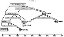
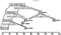
- the present invention includes a method of determining incidence of experiencing a major adverse cardiac event, in a patient, within one year from presentation of at least one symptom of ACS comprising the step of: assessing the level of BNP in a test sample from the patient, wherein a BNP level less than or equal to approximately 150 pg/mL indicates that the level of PlGF in said test sample must be assessed and a BNP level greater than approximately 150 pg/mL indicates that the eGRF of said patient must be assessed, wherein if the PlGF level must be assessed, a PlGF level less than or equal to approximately 19.5 pg/mL indicates that the level of HCY in the test sample must be assessed and a PlGF level greater than approximately 19.5 indicates the patient has a moderate risk of experiencing a major adverse cardiac event within one year from presentation of at least one symptom of ACS, wherein if the eGFR must be assessed, an eGFR greater than approximately 68 mL/min/1.73 m 2 indicates the patient has a moderate risk
- a patient having a BNP level greater than approximately 150 pg/mL has an approximately 27.6% incidence of having a major adverse cardiac event within one year from presentation of at least one symptom of ACS. Further, a patient having a BNP level of less than or equal to approximately 150 pg/mL has an approximately 6.4% incidence of having a major adverse cardiac event within one year from presentation of at least one symptom of ACS. Also, a patient having a PlGF value of greater than approximately 19.5 pg/mL has an approximately 14.5% incidence of having a major adverse cardiac event within one year from presentation of at least one symptom of ACS.
- a patient having a PlGF level less than or equal to approximately 19.5 pg/mL has an approximately 2.4% incidence of having a major adverse cardiac event within one year from presentation of at least one symptom of ACS.
- a patient having an eGFR of greater than approximately 68 mL/min/1.73 m 2 has an approximately 14.3% incidence of having a major adverse cardiac event within one year from presentation of at least one symptom of ACS.
- a patient having an eGFR level of less than or equal to approximately 68 mL/min/1.73 m 2 has an approximately 36.5% incidence of having a major adverse cardiac event within one year from presentation of at least one symptom of ACS.
- a patient having a HCY level of less than or equal to 12.5 ⁇ mol/L has a less than approximately 1% incidence of having a major adverse cardiac event within one year from presentation of at least one symptom of ACS.
- a patient having a HCY level of greater than approximately 12.5 ⁇ mol/L has an approximately 10% incidence of having a major adverse cardiac event within one year from presentation of at least one symptom of ACS.
- the present invention encompasses a method of determining risk of experiencing a major adverse cardiac event, in a patient with previously established or diagnosed chronic kidney disease, within one year from presentation of at least one symptom of ACS comprising the steps of: determining whether said patient is cardiac Troponin I negative by comparison with a established reference standard (e.g., cTnI ⁇ approximately 0.015 ng/mL); and assessing the level of BNP in a test sample from said patient, if said patient is cardiac Troponin I negative, wherein a BNP level less than or equal to approximately 150 pg/mL indicates that the level of PlGF in said test sample must be assessed and a BNP level greater than approximately 150 pg/mL indicates that the eGRF of said patient must be assessed, wherein if said PlGF level must be assessed, a PlGF level less than or equal to approximately 19.5 pg/mL indicates that the level of BNP in said test sample must be reassessed, wherein if said level of BNP must be a
- the present disclosure relates to the detection and level of combinations of biomarkers (or absence thereof) for the determination of risk (i.e., formulation of a prognosis and risk stratification) associated with the potential occurrence of a major adverse cardiac event (MACE) including, for example, myocardial infarction, death and/or cardiac revascularization. More specifically, the present disclosure encompasses use of the presence (or absence) and level of at least three biomarkers present in a test sample from a patient.
- MACE major adverse cardiac event
- markers are selected from the group consisting of, for example, cardiac Troponin I (cTnI), B-type natriuretic peptide (BNP), pro-B-type natriuretic peptide (proBNP), NT-proBNP (N-terminal pro-B-type natriuretic peptide), placental growth factor (PlGF), homocysteine (HCY), choline, ischemia modified albumin (IMA), soluble CD40 ligand (sCD40L), lipoprotein-associated phospholipase A 2 (LpPLA 2 ), myeloperoxidase (MPO) and high sensitivity C reactive protein (hsCRP) and may be used, along with estimated glomerular filtration rate (eGFR), in predicting the future occurrence of such a cardiac event, as described above, in an at-risk patient or a patient presenting with clinical symptoms of acute coronary syndrome, or even in an otherwise healthy patient undergoing a complete cardiac assessment.
- Detection of the presence or absence of such markers, as well as level thereof, may be accomplished by a variety of assay formats well known to those of ordinary skill in the art (see, e.g., The Immunoassay Handbook, 2nd Edition, edited by David Wild, Nature Publishing Group, London 2001 ).
- the presence, amount or concentration of the biomarkers described herein (or a fragment thereof) in a test sample can be achieved using an immunoassay such as 1) a sandwich immunoassay (e.g., monoclonal, polyclonal and/or DVD-Ig sandwich immunoassays or any variation thereof (e.g., monoclonal/DVD-Ig or DVD-Ig/polyclonal), including chemiluminescence detection, radioisotope detection (e.g., radioimmunoassay (RIA)) and enzyme detection (e.g., enzyme immunoassay (EIA) or enzyme-linked immunosorbent assay (ELISA) (e.g., Quantikine ELISA assays, R&D Systems, Minneapolis, MN))), 2) a competitive inhibition immunoassay (e.g., forward and reverse), 3) a fluorescence polarization immunoassay (FPIA), 4) an enzyme multiplied immunoassay technique (EMIT), 5-
- test sample can comprise further moieties in addition to the analyte of interest, such as antibodies, antigens, haptens, hormones, drugs, enzymes, receptors, proteins, peptides, polypeptides, oligonucleotides and/or polynucleotides.
- the sample may be a whole blood sample obtained from a subject. It can be necessary or desired that a test sample, particularly whole blood, be treated prior to being subjected to an immunoassay or other assay, as described herein, for example, with a pretreatment reagent. Even in cases where pretreatment is not necessary (e.g., most urine samples), pretreatment optionally can be done (e.g., as part of a regimen on a commercial platform).
- Solid supports or phases which may be utilized in the assays described herein are well-known in the art and included, but are not limited to, a magnetic particle, a bead, a test tube, a microtiter plate, a cuvette, a membrane, a scaffolding molecule, a film, a filter paper, a disc and a chip.
- the detectable label can be a radioactive label (such as 3 H, 125 I, 35 S, 14 C, 32 P, and 33 P), an enzymatic label (such as horseradish peroxidase, alkaline peroxidase, glucose 6-phosphate dehydrogenase, and the like), a chemiluminescent label (such as acridinium esters, thioesters, or sulfonamides; luminol, isoluminol, phenanthridinium esters, and the like), a fluorescent label (such as fluorescein (e.g., 5-fluorescein, 6-carboxyfluorescein, 3'6-carboxyfluorescein, 5(6)-carboxyfluorescein, 6-hexachloro-fluorescein, 6-tetrachlorofluorescein, fluorescein isothio
- fluorescein e.g., 5-fluorescein, 6-carboxyfluorescein, 3'6
- An acridinium compound can be used as a detectable label in a homogeneous or heterogeneous chemiluminescent assay (see, e.g., Adamczyk et al., Bioorg. Med. Chem. Lett. 16: 1324-1328 (2006 ); Adamczyk et al., Bioorg. Med. Chem. Lett. 4: 2313-2317 (2004 ); Adamczyk et al., Biorg. Med. Chem. Lett. 14: 3917-3921 (2004 ); and Adamczyk et al., Org. Lett. 5: 3779-3782 (2003 )).
- a preferred acridinium compound is an acridinium-9-carboxamide.
- Methods for preparing acridinium 9-carboxamides are described in Mattingly, J. Biolumin. Chemilumin. 6: 107-114 (1991 ); Adamczyk et al., J. Org. Chem. 63: 5636-5639 (1998 ); Adamczyk et al., Tetrahedron 55: 10899-10914 (1999 ); Adamczyk et al., Org. Lett. 1: 779-781 (1999 ); Adamczyk et al., Bioconjugate Chem.
- Another preferred acridinium compound is an acridinium-9-carboxylate aryl ester.
- an acridinium-9-carboxylate aryl ester is 10-methyl-9-(phenoxycarbonyl)acridinium fluorosulfonate (available from Cayman Chemical, Ann Arbor, MI).
- Methods for preparing acridinium 9-carboxylate aryl esters are described in McCapra et al., Photochem. Photobiol. 4: 1111-21 (1965 ); Razavi et al., Luminescence 15: 245-249 (2000 ); Razavi et al., Luminescence 15: 239-244 (2000 ); and U.S. Patent No. 5,241,070 . Further details regarding acridinium-9-carboxylate aryl ester and its use are set forth in U.S.
- Chemiluminescent assays can be performed in accordance with the methods described in Adamczyk et al., Anal. Chim. Acta 579(1): 61-67 (2006 ). While any suitable assay format can be used, a microplate chemiluminometer (Mithras LB-940, Berthold Technologies U.S.A., LLC, Oak Ridge, TN) enables the assay of multiple samples of small volumes rapidly.
- a detectable signal namely, a chemiluminescent signal
- the basic solution contains at least one base and has a pH greater than or equal to 10, preferably, greater than or equal to 12.
- Examples of basic solutions include, but are not limited to, sodium hydroxide, potassium hydroxide, calcium hydroxide, ammonium hydroxide, magnesium hydroxide, sodium carbonate, sodium bicarbonate, calcium hydroxide, calcium carbonate, and calcium bicarbonate.
- the amount of basic solution added to the sample depends on the concentration of the basic solution. Based on the concentration of the basic solution used, one skilled in the art can easily determine the amount of basic solution to add to the sample.
- the chemiluminescent signal that is generated can be detected using routine techniques known to those skilled in the art. Based on the intensity of the signal generated, the amount of analyte in the sample can be quantified. Specifically, the amount of analyte in the sample is proportional to the intensity of the signal generated. The amount of analyte present can be quantified by comparing the amount of light generated to a standard curve for the analyte or by comparison to a reference standard. The standard curve can be generated using serial dilutions or solutions of known concentrations of analyte by mass spectroscopy, gravimetric methods, and other techniques known in the art. While the above is described with emphasis on use of an acridinium compound as the chemiluminescent agent, one of ordinary skill in the art can readily adapt this description for use of other chemiluminescent agents.
- analyte immunoassays generally can be conducted using any format known in the art, such as, but not limited to, a sandwich format. Specifically, in one immunoassay format, at least two antibodies are employed to separate and quantify analyte such as human analyte or a fragment thereof in a sample.
- the at least two antibodies bind to different epitopes on an analyte (or a fragment thereof) forming an immune complex, which is referred to as a "sandwich.”
- one or more antibodies can be used to capture the analyte (or a fragment thereof) in the test sample (i.e., these antibodies are frequently referred to as a “capture” antibody or “capture” antibodies) and one or more antibodies can be used to bind a detectable (namely, quantifiable) label to the sandwich (i.e., these antibodies are frequently referred to as the "detection antibody,” the “detection antibodies,” the “conjugate,” or the “conjugates").
- an antibody in the context of a sandwich immunoassay format, an antibody (or a fragment, a variant, or a fragment of a variant thereof) can be used as a capture antibody, a detection antibody, or both.
- one DVD-Ig having a domain that can bind a first epitope on an analyte (or a fragment thereof) can be used as a capture antibody and/or another DVD-Ig having a domain that can bind a second epitope on an analyte (or a fragment thereof) can be used as a detection antibody.
- a DVD-Ig having a first domain that can bind a first epitope on an analyte (or a fragment thereof) and a second domain that can bind a second epitope on an analyte (or a fragment thereof) can be used as a capture antibody and/or a detection antibody.
- one DVD-Ig having a first domain that can bind an epitope on a first analyte (or a fragment thereof) and a second domain that can bind an epitope on a second analyte (or a fragment thereof) can be used as a capture antibody and/or a detection antibody to detect, and optionally quantify, two or more analytes.
- a sample being tested for can be contacted with at least one capture antibody (or antibodies) and at least one detection antibody (which can be a second detection antibody or a third detection antibody or even a successively numbered antibody, e.g., as where the capture and/or detection antibody comprise multiple antibodies) either simultaneously or sequentially and in any order.
- the test sample can be first contacted with at least one capture antibody and then (sequentially) with at least one detection antibody.
- the test sample can be first contacted with at least one detection antibody and then (sequentially) with at least one capture antibody.
- the test sample can be contacted simultaneously with a capture antibody and a detection antibody.
- a sample suspected of containing the analyte (or a fragment thereof) is first brought into contact with at least one first capture antibody under conditions that allow the formation of a first antibody/analyte complex. If more than one capture antibody is used, a first capture antibody/analyte complex comprising two or more capture antibodies is formed.
- the antibodies i.e., preferably, the at least one capture antibody, are used in molar excess amounts of the maximum amount of analyte (or a fragment thereof) expected in the test sample. For example, from about 5 ⁇ g to about 1 mg of antibody per mL of buffer (e.g., microparticle coating buffer) can be used.
- competitive inhibition immunoassays which are often used to measure small analytes because binding by only one antibody is required, comprise sequential and classic formats.
- a sequential competitive inhibition immunoassay a capture antibody to an analyte of interest is coated onto a well of a microtiter plate or other solid support.
- the analyte of interest binds to the capture antibody.
- a known amount of labeled analyte e.g., acridinium, biotin or horseradish peroxidase (HRP)
- HRP horseradish peroxidase
- An example of a suitable substrate for HRP is 3,3',5,5'-tetramethylbenzidine (TMB).
- TMB 3,3',5,5'-tetramethylbenzidine
- the signal generated by the labeled analyte is measured and is inversely proportional to the amount of analyte in the sample.
- a classic competitive inhibition immunoassay an antibody to an analyte of interest is coated onto a solid support (e.g., a well of a microtiter plate).
- the sample and the labeled analyte are added to the well at the same time. Any analyte in the sample competes with labeled analyte for binding to the capture antibody.
- the signal generated by the labeled analyte is measured and is inversely proportional to the amount of analyte in the sample.
- the concentration of analyte (e.g., biomarker) or a fragment thereof in the test sample is determined by appropriate means, such as by use of a standard curve that has been generated using serial dilutions of analyte or a fragment thereof of known concentration.
- the standard curve can be generated gravimetrically, by mass spectroscopy and by other techniques known in the art.
- the conjugate diluent pH may be about 6.0 +/- 0.2
- the microparticle coating buffer may be maintained at about room temperature (i.e., at from about 17 to about 27 °C)
- the microparticle coating buffer pH may be about 6.5 +/- 0.2
- the microparticle diluent pH may be about 7.8 +/- 0.2.
- Solids preferably are less than about 0.2%, such as less than about 0.15%, less than about 0.14%, less than about 0.13%, less than about 0.12%, or less than about 0.11%, such as about 0.10%.
- these ranges or numbers may be altered in order to enhance such properties of the assay including, for example, reduction in background interference, increased sensitivity, increased specificity, etc.
- FPIAs are based on competitive binding immunoassay principles.
- a fluorescently labeled compound when excited by a linearly polarized light, will emit fluorescence having a degree of polarization inversely proportional to its rate of rotation.
- the emitted light remains highly polarized because the fluorophore is constrained from rotating between the time light is absorbed and the time light is emitted.
- a "free" tracer compound i.e., a compound that is not bound to an antibody
- FPIAs are advantageous over RIAs inasmuch as there are no radioactive substances requiring special handling and disposal.
- FPIAs are homogeneous assays that can be easily and rapidly performed.
- anti-analyte antibodies Assay (and kit therefor), it may be possible to employ commercially available anti-analyte antibodies or methods for production of anti-analyte as described in the literature.
- Commercial supplies of various antibodies include, but are not limited to, Santa Cruz Biotechnology Inc. (Santa Cruz, CA), GenWay Biotech, Inc. (San Diego, CA), and R&D Systems (RDS; Minneapolis, MN).
- a predetermined level can be employed as a benchmark against which to assess results obtained upon assaying a test sample for an analyte or a fragment thereof, e.g., for detecting disease or determining risk of disease.
- the predetermined level is obtained by running a particular assay a sufficient number of times and under appropriate conditions such that a linkage or association of analyte presence, amount or concentration with a particular stage or endpoint of a disease, disorder or condition or with particular clinical indicia can be made.
- the predetermined level is obtained with assays of reference subjects (or populations of subjects).
- the analyte measured can include fragments thereof, degradation products thereof, and/or enzymatic cleavage products thereof.
- the amount or concentration of analyte or a fragment thereof may be “unchanged,” “favorable” (or “favorably altered”), or “unfavorable” (or “unfavorably altered”).
- “Elevated” or “increased” refers to an amount or a concentration in a test sample that is higher than a typical or normal level or range (e.g., predetermined level), or is higher than another reference level or range (e.g., earlier or baseline sample).
- lowered or reduced refers to an amount or a concentration in a test sample that is lower than a typical or normal level or range (e.g., predetermined level), or is lower than another reference level or range (e.g., earlier or baseline sample).
- altered refers to an amount or a concentration in a sample that is altered (increased or decreased) over a typical or normal level or range (e.g., predetermined level), or over another reference level or range (e.g., earlier or baseline sample).
- the typical or normal level or range for analyte is defined in accordance with standard practice. Because the levels of analyte in some instances will be very low, a so-called altered level or alteration can be considered to have occurred when there is any net change as compared to the typical or normal level or range, or reference level or range, which cannot be explained by experimental error or sample variation. Thus, the level measured in a particular sample will be compared with the level or range of levels determined in similar samples from a so-called normal subject.
- a "normal subject” is an individual with no detectable disease, for example, and a "normal” (sometimes termed "control") patient or population is/are one(s) that exhibit(s) no detectable disease, respectively, for example.
- a "normal subject” can be considered an individual with no substantial detectable increased or elevated amount or concentration of analyte, and a "normal” (sometimes termed “control") patient or population is/are one(s) that exhibit(s) no substantial detectable increased or elevated amount or concentration of analyte.
- An "apparently normal subject” is one in which analyte has not yet been or currently is being assessed.
- the level of an analyte is said to be "elevated" when the analyte is normally undetectable (e.g., the normal level is zero, or within a range of from about 25 to about 75 percentiles of normal populations), but is detected in a test sample, as well as when the analyte is present in the test sample at a higher than normal level.
- the disclosure provides a method of screening for a subject having, or at risk of having, a particular disease, disorder, or condition.
- the method of assay can also involve the assay of other markers and the like.
- the methods described herein also can be used to determine whether or not a subject has or is at risk of developing a given disease, disorder or condition.
- a method can comprise the steps of: (a) determining the concentration or amount of an analyte (or fragment thereof) in a test sample from a subject using the methods described herein, or methods known in the art; and (b) comparing the concentration or amount of the analyte (or a fragment thereof) determined in step (a) with a predetermined level, wherein, if the concentration or amount of analyte determined in step (a) is favorable with respect to a predetermined level, then the subject is determined not to have or be at risk for a given disease, disorder or condition. However, if the concentration or amount of analyte determined in step (a) is unfavorable with respect to the predetermined level, then the subject is determined to have or be at risk for a given disease, disorder or condition.
- a method of monitoring the progression of disease in a subject comprising the steps of: (a) determining the concentration or amount of an analyte (e.g., biomarker) in a test sample from a subject; (b) determining the concentration or amount of the analyte in a later test sample from the subject; and (c) comparing the concentration or amount of analyte as determined in step (b) with the concentration or amount of analyte determined in step (a), wherein if the concentration or amount determined in step (b) is unchanged or is unfavorable when compared to the concentration or amount of analyte determined in step (a), then the disease in the subject is determined to have continued, progressed or worsened.
- an analyte e.g., biomarker
- step (b) By comparison, if the concentration or amount of analyte as determined in step (b) is favorable when compared to the concentration or amount of analyte as determined in step (a), then the disease in the subject is determined to have discontinued, regressed or improved.
- the method further comprises comparing the concentration or amount of analyte as determined in step (b), for example, with a predetermined level. Further, optionally the method comprises treating the subject with one or more pharmaceutical compositions for a period of time if the comparison shows that the concentration or amount of analyte as determined in step (b), for example, is unfavorably altered with respect to the predetermined level.
- the disclosure also provides a method of determining whether a subject having, or at risk for, a given disease, disorder or condition is a candidate for therapy.
- the subject is one who has experienced some symptom of a given disease, disorder or condition or who has actually been diagnosed as having, or being at risk for, a given disease, disorder or condition, and/or who demonstrates an unfavorable concentration or amount of analyte or a fragment thereof, as described herein.
- the assays and kits can be employed to assess analyte in other diseases, disorders and conditions.
- the method of assay can also involve the assay of other markers and the like.
- the kit comprises at least one component for assaying the test sample for the analyte (or a fragment thereof) and instructions for assaying the test sample for the analyte (or a fragment thereof).
- the at least one component for assaying the test sample for the analyte (or a fragment thereof) can include a composition comprising, for example, an antibody or antibodies against the biomarker or biomarkers (or a fragment, a variant, or a fragment of a variant thereof), which is optionally immobilized on a solid phase.
- the kit can comprise at least one component for assaying the test sample for an analyte by assay, e.g., chemiluminescent microparticle immunoassay, and instructions for assaying the test sample for an analyte by immunoassay, e.g., chemiluminescent microparticle immunoassay.
- assay e.g., chemiluminescent microparticle immunoassay
- immunoassay e.g., chemiluminescent microparticle immunoassay
- the kit can comprise at least one specific binding partner for an analyte such as an anti-analyte, monoclonal/polyclonal antibody (or a fragment thereof that can bind to the analyte, a variant thereof that can bind to the analyte, or a fragment of a variant that can bind to the analyte), either of which can be detectably labeled.
- an analyte such as an anti-analyte, monoclonal/polyclonal antibody (or a fragment thereof that can bind to the analyte, a variant thereof that can bind to the analyte, or a fragment of a variant that can bind to the analyte), either of which can be detectably labeled.
- the kit can comprise detectably labeled analyte (or a fragment thereof that can bind to an anti-analyte, monoclonal/polyclonal antibody or an anti-analyte DVD-Ig (or a fragment, a variant, or a fragment of a variant thereof)), which can compete with any analyte in a test sample for binding to an anti-analyte, monoclonal/polyclonal antibody (or a fragment thereof that can bind to the analyte, a variant thereof that can bind to the analyte, or a fragment of a variant that can bind to the analyte) or an anti-analyte DVD-Ig (or a fragment, a variant, or a fragment of a variant thereof), either of which can be immobilized on a solid support.
- analyte or a fragment thereof that can bind to an anti-analyte, monoclonal/polyclonal
- the kit can comprise one or more calibrators or controls, e.g., isolated or purified analyte.
- the kit can comprise at least one container (e.g., tube, microtiter plates or strips, which can be already coated with a first specific binding partner, for example) for conducting the assay, and/or a buffer, such as an assay buffer or a wash buffer, either one of which can be provided as a concentrated solution, a substrate solution for the detectable label (e.g., an enzymatic label) or a stop solution.
- a buffer such as an assay buffer or a wash buffer, either one of which can be provided as a concentrated solution, a substrate solution for the detectable label (e.g., an enzymatic label) or a stop solution.
- the kit to perform the assay.
- the instructions can be in paper form or computer-readable form such as a disk, CD, DVD or the like.
- any antibodies such as an anti-biomarker antibody or an anti-analyte DVD-Ig, or tracer can incorporate a detectable label as described herein such as a fluorophore, a radioactive moiety, an enzyme, a biotin/avidin label, a chromophore, a chemiluminescent label or the like, or the kit can include reagents for carrying out detectable labeling.
- a detectable label as described herein such as a fluorophore, a radioactive moiety, an enzyme, a biotin/avidin label, a chromophore, a chemiluminescent label or the like
- the kit can include reagents for carrying out detectable labeling.
- the antibodies, calibrators and/or controls can be provided in separate containers or pre-dispensed into an appropriate assay format, for example, into microtiter plates.
- the kit includes quality control components (for example, sensitivity panels, calibrators, and positive controls).
- quality control components for example, sensitivity panels, calibrators, and positive controls.
- Preparation of quality control reagents is well-known in the art and is described on insert sheets for a variety of immunodiagnostic products.
- Sensitivity panel members optionally are used to establish assay performance characteristics, and further optionally are useful indicators of the integrity of the assay kit reagents, and the standardization of assays.
- the kit can also optionally include other reagents required to conduct a diagnostic assay or facilitate quality control evaluations such as buffers, salts, enzymes, enzyme cofactors, enzyme substrates, detection reagents, and the like.
- Other components such as buffers and solutions for the isolation and/or treatment of a test sample (e.g., pretreatment reagents) also can be included in the kit.
- the kit can additionally include one or more other controls.
- One or more of the components of the kit can be lyophilized, in which case the kit can further comprise reagents suitable for the reconstitution of the lyophilized components.
- kits for holding or storing a sample (e.g., a container or cartridge for a urine sample).
- a sample e.g., a container or cartridge for a urine sample
- the kit optionally also can contain reaction vessels, mixing vessels, and other components that facilitate the preparation of reagents or the test sample.
- the kit can also include one or more instruments for assisting with obtaining a test sample, such as a syringe, pipette, forceps, measured spoon, or the like.
- the kit can comprise at least one acridinium-9-carboxamide, at least one acridinium-9-carboxylate aryl ester, or any combination thereof. Further, if the detectable label is at least one acridinium compound, the kit also can comprise a source of hydrogen peroxide, such as a buffer, a solution, and/or at least one basic solution. If desired, the kit can contain a solid phase, such as a magnetic particle, bead, test tube, microtiter plate, cuvette, membrane, scaffolding molecule, film, filter paper, disc or chip.
- the kit (or components thereof), as well as the method of determining the presence, amount or concentration of an analyte in a test sample by an assay, such as the assays described herein, can be adapted for use in a variety of automated and semi-automated systems (including those wherein the solid phase comprises a microparticle), as described, e.g., in U.S. Patent Nos. 5,089,424 and 5,006,309 , and as commercially marketed, e.g., by Abbott Laboratories (Abbott Park, IL) as ARCHITECT®.
- Some of the differences between an automated or semi-automated system as compared to a non-automated system include the substrate to which the first specific binding partner (e.g., an anti-analyte, monoclonal/polyclonal antibody (or a fragment thereof, a variant thereof, or a fragment of a variant thereof) or an anti-analyte DVD-Ig (or a fragment thereof, a variant thereof, or a fragment of a variant thereof) is attached; either way, sandwich formation and analyte reactivity can be impacted), and the length and timing of the capture, detection and/or any optional wash steps.
- the first specific binding partner e.g., an anti-analyte, monoclonal/polyclonal antibody (or a fragment thereof, a variant thereof, or a fragment of a variant thereof) or an anti-analyte DVD-Ig (or a fragment thereof, a variant thereof, or a fragment of a variant thereof) is attached; either way
- an automated or semi-automated format e.g., ARCHITECT®, Abbott Laboratories
- a relatively shorter incubation time e.g., approximately 18 minutes for ARCHITECT®
- an automated or semi-automated format may have a relatively shorter incubation time (e.g., approximately 4 minutes for the ARCHITECT®).
- kits and kit components can be employed in other formats, for example, on electrochemical or other hand-held or point-of-care assay systems.
- the present disclosure is, for example, applicable to the commercial Abbott Point of Care (i-STAT®, Abbott Laboratories) electrochemical immunoassay system that performs sandwich immunoassays. Immunosensors and their methods of manufacture and operation in single-use test devices are described, for example in, U.S. Patent No.
- a microfabricated silicon chip is manufactured with a pair of gold amperometric working electrodes and a silver-silver chloride reference electrode.
- polystyrene beads (0.2 mm diameter) with immobilized anti-analyte, monoclonal/polyclonal antibody (or a fragment thereof, a variant thereof, or a fragment of a variant thereof) or anti-analyte DVD-Ig (or a fragment thereof, a variant thereof, or a fragment of a variant thereof), are adhered to a polymer coating of patterned polyvinyl alcohol over the electrode.
- This chip is assembled into an I-STAT® cartridge with a fluidics format suitable for immunoassay.
- a layer comprising a specific binding partner for an analyte, such as an anti-analyte, monoclonal/polyclonal antibody (or a fragment thereof, a variant thereof, or a fragment of a variant thereof that can bind the analyte) or an anti-analyte DVD-Ig (or a fragment thereof, a variant thereof, or a fragment of a variant thereof that can bind the analyte), either of which can be detectably labeled.
- an aqueous reagent that includes p-aminophenol phosphate.
- a sample suspected of containing an analyte is added to the holding chamber of the test cartridge, and the cartridge is inserted into the I-STAT® reader.
- a pump element within the cartridge forces the sample into a conduit containing the chip. Here it is oscillated to promote formation of the sandwich.
- fluid is forced out of the pouch and into the conduit to wash the sample off the chip and into a waste chamber.
- the alkaline phosphatase label reacts with p-aminophenol phosphate to cleave the phosphate group and permit the liberated p-aminophenol to be electrochemically oxidized at the working electrode.
- the reader is able to calculate the amount of analyte in the sample by means of an embedded algorithm and factory-determined calibration curve.
- kits as described herein necessarily encompass other reagents and methods for carrying out the assays.
- various buffers such as are known in the art and/or which can be readily prepared or optimized to be employed, e.g., for washing, as a conjugate diluent, microparticle diluent, and/or as a calibrator diluent.
- An exemplary conjugate diluent is ARCHITECT® conjugate diluent employed in certain kits (Abbott Laboratories, Abbott Park, IL) and containing 2-(N-morpholino)ethanesulfonic acid (MES), a salt, a protein blocker, an antimicrobial agent, and a detergent.
- MES 2-(N-morpholino)ethanesulfonic acid
- An exemplary calibrator diluent is ARCHITECT® human calibrator diluent employed in certain kits (Abbott Laboratories, Abbott Park, IL), which comprises a buffer containing MES, other salt, a protein blocker, and an antimicrobial agent. Additionally, as described in U.S. Patent Application No. 61/142,048 filed December 31, 2008 , improved signal generation may be obtained, e.g., in an I-Stat cartridge format, using a nucleic acid sequence linked to the signal antibody as a signal amplifier.
- Biomarker is a characteristic or entity that is objectively measured and evaluated as an indicator of normal biologic processes, pathogenic processes or pharmacologic responses to a therapeutic intervention. This can include physiological indicators such as blood pressure or heart rate and can also refer to compounds or biological entities such as proteins, peptides or small molecules that are produced by the body, released into the bloodstream and are measurable and quantifiable. Included in this definition are also parameters that are based on biomarkers, such as estimated glomerular filtration rate (eGFR) that may be based on creatinine, albumin, blood urea nitrogen, age, gender, ethnic group and body mass.
- eGFR estimated glomerular filtration rate
- ProBNP or "pro-B-type natriuretic peptide” or “pro-brain natriuretic peptide”, as used herein, refers to the 108 amino acid sequence that is derived from the pre-pro BNP molecule. ProBNP is enzymatically processed to form NT-proBNP (amino acids 1-76) and BNP (amino acids 77-108). ProBNP has been shown to circulate in blood ( Seferian et al., Clin Chem, 2007;53:866-873 ) and may have a role in determining endogenous natriuretic peptide activity in heart failure patients ( Lam et al., J Am Coll Cardiol, 2007;49:1193-1202 ).
- BNP B-type natriuretic peptide which contains 32 amino acids and is 4 kDa. It is involved in the natriuresis system for blood pressure regulation and fluid balance ( Bonow, R.O., Circulation 93:1946-1950, 1996 ). It is the carboxyl terminal (C terminal) 32 amino acids from the precursor proBNP molecule (amino acid numbers 77 to 108). Increased BNP levels are associated with raised atrial and pulmonary wedge pressures as well as reduced ventricular systolic and diastolic function, left ventricular hypertrophy and mycocardial infarction ( Sagnella, G.A., Clinical Science 95:519-5219, 1998 ).
- BNP is used clinically to aid in the diagnosis of heart failure in patients presenting to the emergency department with dyspnea (i.e., shortness of breath) based on data from the "Breathing Not Properly” trial ( Maisel et al., N Engl J Med. 2003;347:161-7 ) and in the risk stratification in ACS patients ( Morrow et al., J Am Coll Cardiol. 2003;41:1264-72 ).
- N-terminal pro-B-type natriuretic peptide or "NT-proBNP”, as used herein, refers to the inactive amino terminal (N terminal) polypeptide that is cleaved from proBNP. It includes amino acid numbers 1-76 of proBNP. NT-proBNP's clinical utility is very similar to that of BNP with studies supporting its use as an aid in the diagnosis of heart failure in patients presenting with dyspnea ( Januzzi el al., Am J Cardiology 2005;95:948-954 ).
- Troponin I or “TnI” (briefly described above) is a 25 kDa inhibitory element of the troponin complex which is found in all striated muscle tissue.
- the protein binds to actin in the absence of Ca 2+ thereby inhibiting the ATPase activity of actomyosin.
- the one found in cardiac tissue i.e., cTnI
- cTnI may be present in elevated levels in patients experiencing an acute myocardial infarction or other adverse cardiac events ( Khan et al., Am. J. Emerg. Med. 17:225-229, 1999 ). Free and complexed cardiac troponin I also exist.
- Homocysteine is an amino acid which is also referred to as 2-amino-4-mercaptobutanoic acid. It is a homologue of the amino acid cysteine.
- a high level of blood serum homocysteine has been determined to be a significant risk factor for cardiovascular disease and stroke ( Boushey CJ et al., JAMA. 1995;274:1049-57 ).
- homocysteine-lowering therapies have failed to reduce cardiovascular event frequency in secondary prevention ( Loscalzo, J., N Engl J Med 354(15):1629-1632, 2006 ). Elevated homocysteine levels have also been associated with increased ischemic injury in patients with ACS ( Al-Obaidi MK et al., J Am Coll Cardiol. 2000;36:1217-22 ).
- Choline is an organic compound which is a water-soluble essential nutrient and is grouped within the Vitamin B complex. It is a natural, quarternary, saturated amine found in lipids that make up cell membranes and is also found in the neurotransmitter acetylcholine. In particular, choline and its metabolites are required for structural integrity and signaling roles for cell membranes, cholinergic neurotransmission (i.e., acetylcholine synthesis) and as a major source for methyl groups (via its metabolite trimethylglycine) that participate in the S-adenosylmethionine synthesis pathways.
- choline is released after the stimulation of phospholipase D and the activation of cell surface receptors from various cell types related to plaque destabilization.
- An increased concentration of choline at hospital admission is a predictor of adverse cardiac events in patients with suspected ACS particularly in troponin negative patients ( Danne et al., Am J Cardiol. 2003;91:1060-67 ).
- eGFR or "estimated glomerular filtration rate” is based on creatinine concentration and calculated based on numerous equations, typically the “Modification in Diet and Renal Disease” equation (MDRD)( Levey et al., Clin Chem, 53(4):766, 2007 ) or the Cockcroft-Gault equation.
- MDRD Modification in Diet and Renal Disease
- Ischemia modified albumin or "IMA ® ,” as used herein refers to a modification of albumin that occurs in the ischemic state. It is assessed using the albumin cobalt binding test (ACB ® ) manufactured by Inverness Medical (Waltham, MA.). This is a clinical chemistry, colorimetric assay that can be performed on numerous clinical chemistry analyzers. The current use of this marker is for "rule-out" of myocardial infarction.
- C-reactive protein is a homopentameric Ca2+-binding acute phase protein which is involved in defense of the host, often in response to inflammation.
- CRP C-reactive protein
- CRP high sensitivity CRP
- hsCRP hsCRP
- PlGF or "placental growth factor” or “vascular endothelial growth factor-related protein,” as used herein, is a protein whose expression, within human atherosclerotic lesions, is associated with plaque inflammation and neovascular growth.
- PlGF vascular endothelial growth factor-related protein
- Plasma PlGF level may be an independent biomarker of adverse outcome in patients suspected with ACS, and the single initial measurement appears to extend the predictive and prognostic information gained from traditional inflammatory markers.
- a further analysis after 4 years ( Lenderink er al., J Am Coll Cardiol. 2006;47:307-11 ) continued to support the prognostic utility in patients with ACS.)
- Myeloperoxidase or "MPO” as used herein is a hemoprotein stored in leukocytes and functions in the host-defense mechanisms against a broad range of organisms. Activation of leukocytes results in the secretion of MPO, which catalyzes the hydrogen peroxide mediated peroxidation of halide ions. These products promote the oxidative damage at sites of inflammation. MPO has been linked to inflammation and events involved in early endothelial dysfunction, plaque initiation and progression, development of vulnerable plaque and subsequent complications of atherosclerotic plaque.
- MPO has been shown to identify troponin negative patients at risk for cardiac events ( Baldus et al., Circulation.2003;108:1440-1445 ) and add more predictive value than other conventional markers ( Brennan et al. NEJM. 2003;349:1595-604 ).
- LpPLA2 or “lipoprotein-associated phospholipase A 2 ", as used herein, is a vascular-specific inflammatory enzyme implicated in the formation of rupture prone plaque.
- LpPLA 2 cleaves phospholipids into two inflammatory molecules: lysophosphatidylcholine as well as oxidized fatty acids, resulting in a cascade of events leading to atherosclerotic plaque formation.
- LpPLA 2 as measured by the PLACTM assay DiaDexus, South San Franciso, CA) is used in conjunction with clinical evaluation and patient risk assessment as an aid in predicting risk for coronary heart disease and ischemic stroke associated with atherosclerosis.
- sCD40L or "soluble CD40 ligand”, as used herein, is a member of the tumor necrosis factor (TNF) superfamily and is a multi-functional ligand.
- CD40L is a 39 kDA glycoprotein that can undergo proteolytic cleave to produce soluble forms of CD40L (15-18 kDa).
- sCD40L lacks the transmembrane region and a portion of the extracellular domain, but contains the entire TNF- ⁇ homology region.
- Prognosis refers to an increased likelihood that a particular course or outcome will occur. In other words, it is a course or outcome that is more likely to occur in a patient appearing to have a particular disease or condition, when compared to patients not having the disease or condition.
- One of ordinary skill in the art understands that correlating a prognostic biomarker with the risk of having an adverse event or outcome is based upon a statistical analysis using predefined cutoffs or ranges of the biomarker(s) which serve as reference standards.
- Bio activity refers to all inherent biological properties of the markers described herein (e.g., Troponin I, PIGF and BNP). Such properties include, for example, the ability to bind to the antibodies described herein.
- MACE myeloma sarcoma sarcoma sarcoma sarcoma sarcoma sarcoma sarcoma sarcoma sarcoma sarcoma sarcoma sarcoma sarcoma sarcoma sarcoma sarcoma sarcoma sarcoma sarcoma sarcoma sarcoma sarcoma sarcoma sarcoma sarcoma sarcoma sarcoma sarcoma sarcoma sarcoma sarcoma sarcoma sarcoma sarcoma sarcoma sarcoma sarcoma sarcoma sarcoma sarcoma sarcoma sarcoma sarcoma sarcoma sarcoma sarcoma sarcoma sarcoma sarcoma
- Low risk is defined as less than or equal to a ten percent chance, preferably less than a five percent chance and more preferably less than a two percent chance of a patient having a MACE within one year of presentation of at least one symptom of an acute cardiac syndrome (ACS).
- ACS acute cardiac syndrome
- Moderate risk is defined as greater than a ten percent and less than a thirty percent risk of a patient having a MACE within one year of presentation of at least one symptom of an ACS.
- High risk is defined as greater than a twenty-five percent, preferably greater than or equal to a thirty percent and more preferably greater than a thirty-five percent risk of a patient having a MACE within one year of presentation of at least one symptom of an ACS.
- ranges and cutoff points recited herein in connection with the terms “low risk”, “moderate risk” and “high risk” may vary depending upon the specific study utilized in order to gather the relevant data in connection with risk assessment. Further, it should be noted that these cutoff points relate to event risk and not relative risk.
- multivalent binding protein is used in this specification to denote a binding protein comprising two or more antigen binding sites.
- the multivalent binding protein is preferably engineered to have the three or more antigen binding sites, and is generally not a naturally occurring antibody.
- multispecific binding protein refers to a binding protein capable of binding two or more related or unrelated targets.
- Dual variable domain (DVD) binding proteins as used herein, are binding proteins that comprise two or more antigen binding sites and are tetravalent or multivalent binding proteins. Such DVDs may be monospecific, i.e., capable of binding one antigen or multispecific, i.e., capable of binding two or more antigens.
- DVD binding proteins comprising two heavy chain DVD polypeptides and two light chain DVD polypeptides are referred to a DVD Ig. Each half of a DVD Ig comprises a heavy chain DVD polypeptide, and a light chain DVD polypeptide, and two antigen binding sites.
- Each binding site comprises a heavy chain variable domain and a light chain variable domain with a total of 6 CDRs involved in antigen binding per antigen binding site.
- DVD binding proteins and methods of making DVD binding proteins are disclosed in U.S. Patent Application US2007-0071675 .
- “Functional equivalent” as used herein refers to a protein (e.g., an antibody) having the same characteristics (e.g., binding affinity) of the antibodies to the biomarkers, described herein.
- polypeptide refers to any polymeric chain of amino acids.
- peptide and protein are used interchangeably with the term polypeptide and also refer to a polymeric chain of amino acids.
- polypeptide encompasses native or artificial proteins, protein fragments and polypeptide analogs of a protein sequence.
- a polypeptide may be monomeric or polymeric.
- isolated protein or "isolated polypeptide” is a protein or polypeptide that by virtue of its origin or source of derivation is not associated with naturally associated components that accompany it in its native state; is substantially free of other proteins from the same species; is expressed by a cell from a different species; or does not occur in nature.
- a polypeptide that is chemically synthesized or synthesized in a cellular system different from the cell from which it naturally originates will be “isolated” from its naturally associated components.
- a protein may also be rendered substantially free of naturally associated components by isolation, using protein purification techniques well known in the art.
- recovering refers to the process of rendering a chemical species such as a polypeptide substantially free of naturally associated components by isolation, e.g., using protein purification techniques well known in the art.
- telomere binding in reference to the interaction of an antibody, a protein, or a peptide with a second chemical species, mean that the interaction is dependent upon the presence of a particular structure (e.g., an antigenic determinant or epitope) on the chemical species; for example, an antibody recognizes and binds to a specific protein structure rather than to proteins generally. If an antibody is specific for epitope "A”, the presence of a molecule containing epitope A (or free, unlabeled A), in a reaction containing labeled "A” and the antibody, will reduce the amount of labeled A bound to the antibody.
- a particular structure e.g., an antigenic determinant or epitope
- antibody broadly refers to any immunoglobulin (Ig) molecule comprised of four polypeptide chains, two heavy (H) chains and two light (L) chains, or any functional fragment, mutant, variant, or derivation thereof, which retains the essential epitope binding features of an Ig molecule.
- Ig immunoglobulin
- Such mutant, variant, or derivative anitbody formats are known in the art. Nonlimiting embodiments of which are discussed below.
- each heavy chain is comprised of a heavy chain variable region (abbreviated herein as HCVR or VH) and a heavy chain constant region.
- the heavy chain constant region is comprised of three domains, CH1, CH2 and CH3.
- Each light chain is comprised of a light chain variable region (abbreviated herein as LCVR or VL) and a light chain constant region.
- the light chain constant region is comprised of one domain, CL.
- the VH and VL regions can be further subdivided into regions of hypemariability, termed complementarity determining regions (CDR), interspersed with regions that are more conserved, termed framework regions (FR).
- CDR complementarity determining regions
- Each VH and VL is composed of three CDRs and four FRs, arranged from amino-terminus to carboxy-terminus in the following order: FR1, CDR1, FR2, CDR2, FR3, CDR3, FR4.
- Immunoglobulin molecules can be of any type (e.g., IgG, IgE, IgM, IgD, IgA and IgY), class (e.g., IgG 1, IgG2, IgG3, IgG4, IgA1 and IgA2) or subclass.
- antibody portion refers to one or more fragments of an antibody that retain the ability to specifically bind to an antigen (e.g., one or more epitopes of the biomarker of interest). It has been shown that the antigen-binding function of an antibody can be performed by one or more fragments of a full-length antibody. Such antibody embodiments may also be bispecific, dual specific, or multi-specific, specifically binding to two or more different antigens.
- binding fragments encompassed within the term "antigen-binding portion" of an antibody include (i) a Fab fragment, a monovalent fragment consisting of the VL, VH, CL and CH1 domains; (ii) a F(ab') 2 fragment, a bivalent fragment comprising two Fab fragments linked by a disulfide bridge at the hinge region; (iii) a Fd fragment consisting of the VH and CH1 domains; (iv) a Fv fragment consisting of the VL and VH domains of a single arm of an antibody, (v) a dAb fragment ( Ward et al., (1989) Nature 341:544-546 , Winter et al., International Appln. Publication No.
- WO 90/05144 A1 which comprises a single variable domain; and (vi) an isolated complementarity determining region (CDR).
- CDR complementarity determining region
- the two domains of the Fv fragment, VL and VH are coded for by separate genes, they can be joined, using recombinant methods, by a synthetic linker that enables them to be made as a single protein chain in which the VL and VH regions pair to form monovalent molecules (known as single chain Fv (scFv); see e.g., Bird et al. (1988) Science 242:423-426 ; and Huston et al. (1988) Proc. Natl. Acad. Sci. USA 85:5879-5883 ).
- scFv single chain Fv
- single chain antibodies are also encompassed within the term "antigen-binding portion" of an antibody.
- Other forms of single chain antibodies, such as diabodies, are also encompassed.
- Diabodies are bivalent, bispecific antibodies in which VH and VL domains are expressed on a single polypeptide chain, but using a linker that is too short to allow for pairing between the two domains on the same chain, thereby forcing the domains to pair with complementary domains of another chain and creating two antigen binding sites (see e.g., Holliger, P., et al. (1993) Proc. Natl. Acad. Sci. USA 90:6444-6448 ; Poljak, R.J., et al. (1994) Structure 2:1121-1123 ).
- antibody binding portions are known in the art ( Kontermann and Dubel eds., Antibody Engineering (2001) Springer-Verlag. New York. 790 pp. (ISBN 3-540-41354-5 ).
- the term "antibody construct" as used herein refers to a polypeptide comprising one or more the antigen binding portions of the invention linked to a linker polypeptide or an immunoglobulin constant domain.
- Linker polypeptides comprise two or more amino acid residues joined by peptide bonds and are used to link one or more antigen binding portions.
- Such linker polypeptides are well known in the art (see e.g., Holliger, P., et al. (1993) Proc. Natl. Acad. Sci.
- An immunoglobulin constant domain refers to a heavy or light chain constant domain. Human IgG heavy chain and light chain constant domain amino acid sequences are known in the art.
- an “isolated antibody”, as used herein, is intended to refer to an antibody that is substantially free of other antibodies having different antigenic specificities (e.g., anisolated antibody that specifically binds at least one epitope of the biomarker of interest and is substantially free of antibodies that specifically bind antigens or epitopes other than those present within the biomarker).
- CDR refers to the complementarity determining region within antibody variable sequences. There are three CDRs in each of the variable regions of the heavy chain and the light chain, which are designated CDR1, CDR2 and CDR3, for each of the variable regions.
- CDR set refers to a group of three CDRs that occur in a single variable region capable of binding the antigen. The exact boundaries of these CDRs have been defined differently according to different systems.
- Kabat Kabat et al., Sequences of Proteins of Immunological Interest (National Institutes of Health, Bethesda, MD (1987) and (1991 )) not only provides an unambiguous residue numbering system applicable to any variable region of an antibody, but also provides precise residue boundaries defining the three CDRs. These CDRs may be referred to as Kabat CDRs. Chothia and coworkers ( Chothia & Lesk, J. Mol. Biol. 196:901-917 (1987 ) and Chothia et al., Nature 342:877-883 (1989 )) found that certain sub- portions within Kabat CDRs adopt nearly identical peptide backbone conformations, despite having great diversity at the level of amino acid sequence.
- CDR boundary definitions may not strictly follow one of the above systems, but will nonetheless overlap with the Kabat CDRs, although they may be shortened or lengthened in light of prediction or experimental findings that particular residues or groups of residues or even entire CDRs do not significantly impact antigen binding.
- the methods used herein may utilize CDRs defined according to any of these systems, although preferred embodiments use Kabat or Chothia defined CDRs.
- key residues refer to certain residues within the variable region that have more impact on the binding specificity and/or affinity of an antibody, in particular a humanized antibody.
- a key residue includes, but is not limited to, one or more of the following: a residue that is adjacent to a CDR, a potential glycosylation site (can be either N- or O-glycosylation site), a rare residue, a residue capable of interacting with the antigen, a residue capable of interacting with a CDR, a canonical residue, a contact residue between heavy chain variable region and light chain variable region, a residue within the Vernier zone, and a residue in the region that overlaps between the Chothia definition of a variable heavy chain CDR1 and the Kabat definition of the first heavy chain framework.
- activity includes activities such as the binding specificity/affinity of an antibody for an antigen, for example, the antigen or antigens with which antibodies are reactive.
- epitope includes any polypeptide determinant capable of specific binding to an immunoglobulin or T-cell receptor.
- epitope determinants include chemically active surface groupings of molecules such as amino acids, sugar side chains, phosphoryl, or sulfonyl and, in certain embodiments, may have specific three-dimensional structural characteristics, and/or specific charge characteristics.
- An epitope is a region of an antigen that is bound by an antibody.
- an antibody is said to specifically bind an antigen when it preferentially recognizes its target antigen in a complex mixture of proteins and/or macromolecules.
- label binding protein refers to a protein with a label incorporated that provides for the identification of the binding protein.
- the label is a detectable marker, e.g., incorporation of a radiolabeled amino acid or attachment to a polypeptide of biotinylated moieties that can be detected by marked avidin (e.g., streptavidin containing a fluorescent marker or enzymatic activity that can be detected by optical or colorimetric methods).
- labels for polypeptides include, but are not limited to, the following: radioisotopes or radionuclides (e.g., 3 H, 14 C, 35 S, 90 Y, 99 Tc, 111 In, 125 I, 131 I, 177 Lu, 166 Ho, or 153 Sm); fluorescent labels (e.g., FITC, rhodamine, lanthanide phosphors), enzymatic labels (e.g., horseradish peroxidase, luciferase, alkaline phosphatase); chemiluminescent markers; biotinyl groups; predetermined polypeptide epitopes recognized by a secondary reporter (e.g., leucine zipper pair sequences, binding sites for secondary antibodies, metal binding domains, epitope tags); and magnetic agents, such as gadolinium chelates.
- radioisotopes or radionuclides e.g., 3 H, 14 C, 35 S, 90 Y, 99 Tc,
- antibody conjugate refers to a binding protein, such as an antibody, chemically linked to a second chemical moiety, such as a therapeutic or cytotoxic agent.
- crystal refers to an antibody, or antigen-binding portion thereof, that exists in the form of a crystal.
- Crystals are one form of the solid state of matter, which is distinct from other forms such as the amorphous solid state or the liquid crystalline state.
- Crystals are composed of regular, repeating, three-dimensional arrays of atoms, ions, molecules (e.g., proteins such as antibodies), or molecular assemblies (e.g., antigen/antibody complexes). These three-dimensional arrays are arranged according to specific mathematical relationships that are well-understood in the field.
- the fundamental unit, or building block, that is repeated in a crystal is called the asymmetric unit.
- Repetition of the asymmetric unit in an arrangement that conforms to a given, well-defined crystallographic symmetry provides the "unit cell" of the crystal. Repetition of the unit cell by regular translations in all three dimensions provides the crystal. See Giege, R. and Ducruix, A. Barrett, Crystallization of Nucleic Acids and Proteins, a Practical Approach, 2nd ed., pp. 20 1-16, Oxford University Press, New York, New York, (1999 ).
- polynucleotide as referred to herein is a polymeric form of two or more nucleotides, either ribonucleotides or deoxvnucleotides or a modified form of either type of nucleotide.
- the term includes single-stranded and double-stranded forms of DNA but preferably is double-stranded DNA.
- isolated polynucleotide shall mean a polynucleotide (e.g., of genomic, cDNA, or synthetic origin, or some combination thereof) that, by virtue of its origin, is not associated with all or a portion of a polynucleotide with which the "isolated polynucleotide” is found in nature; is operably linked to a polynucleotide that it is not linked to in nature; or does not occur in nature as part of a larger sequence.
- a polynucleotide e.g., of genomic, cDNA, or synthetic origin, or some combination thereof
- vector is intended to refer to a nucleic acid molecule capable of transporting another nucleic acid to which it has been linked.
- plasmid refers to a circular double stranded DNA loop into which additional DNA segments may be ligated.
- viral vector Another type of vector is a viral vector, wherein additional DNA segments may be ligated into the viral genome.
- Certain vectors are capable of autonomous replication in a host cell into which they are introduced (e.g., bacterial vectors having a bacterial origin of replication and episomal mammalian vectors).
- vectors e.g., non-episomal mammalian vectors
- vectors can be integrated into the genome of a host cell upon introduction into the host cell, and thereby are replicated along with the host genome.
- certain vectors are capable of directing the expression of genes to which they are operatively linked.
- Such vectors are referred to herein as "recombinant expression vectors" (or simply, "expression vectors”).
- expression vectors of utility in recombinant DNA techniques are often in the form of plasmids.
- plasmid and vector may be used interchangeably as the plasmid is the most commonly used form of vector.
- the invention is intended to include such other forms of expression vectors, such as viral vectors (e.g., replication defective retroviruses, adenoviruses and adeno-associated viruses), which serve equivalent functions.
- operably linked refers to a juxtaposition wherein the components described are in a relationship permitting them to function in their intended manner.
- a control sequence "operably linked" to a coding sequence is ligated in such a way that expression of the coding sequence is achieved under conditions compatible with the control sequences.
- "Operably linked” sequences include both expression control sequences that are contiguous with the gene of interest and expression control sequences that act in trans or at a distance to control the gene of interest.
- expression control sequence refers to polynucleotide sequences that are necessary to effect the expression and processing of coding sequences to which they are ligated.
- Expression control sequences include appropriate transcription initiation, termination, promoter and enhancer sequences; efficient RNA processing signals such as splicing and polyadenylation signals; sequences that stabilize cytoplasmic mRNA; sequences that enhance translation efficiency (i.e., Kozak consensus sequence); sequences that enhance protein stability; and when desired, sequences that enhance protein secretion.
- the nature of such control sequences differs depending upon the host organism; in prokaryotes, such control sequences generally include promoter, ribosomal binding site, and transcription termination sequence; in eukaryotes, generally, such control sequences include promoters and transcription termination sequence.
- control sequences is intended to include components whose presence is essential for expression and processing, and can also include additional components whose presence is advantageous, for example, leader sequences and fusion partner sequences.
- Transformation refers to any process by which exogenous DNA enters a host cell. Transformation may occur under natural or artificial conditions using various methods well known in the art. Transformation may rely on any known method for the insertion of foreign nucleic acid sequences into a prokaryotic or eukaryotic host cell. The method is selected based on the host cell being transformed and may include, but is not limited to, viral infection, electroporation, lipofection, and particle bombardment. Such "transformed” cells include stably transformed cells in which the inserted DNA is capable of replication either as an autonomously replicating plasmid or as part of the host chromosome. They also include cells that transiently express the inserted DNA or RNA for limited periods of time.
- host cell is intended to refer to a cell into which exogenous DNA has been introduced. It should be understood that such terms are intended to refer not only to the particular subject cell but also to the progeny of such a cell. Because certain modifications may occur in succeeding generations due to either mutation or environmental influences, such progeny may not, in fact, be identical to the parent cell, but are still included within the scope of the term "host cell” as used herein.
- host cells include prokaryotic and eukaryotic cells selected from any of the Kingdoms of life. Preferred eukaryotic cells include protist, fungal, plant and animal cells.
- host cells include but are not limited to the prokaryotic cell line E. coli; mammalian cell lines CHO, HEK 293 and COS; the insect cell line Sf9; and the fungal cell Saccharomyces cerevisiae.
- Standard techniques may be used for recombinant DNA, oligonucleotide synthesis, and tissue culture and transformation (e.g., electroporation, lipofection).
- Enzymatic reactions and purification techniques may be performed according to manufacturer's specifications or as commonly accomplished in the art or as described herein.
- the foregoing techniques and procedures may be generally performed according to conventional methods well known in the art and as described in various general and more specific references that are cited and discussed throughout the present specification. See e.g., Sambrook et al. Molecular Cloning: A Laboratory Manual (2d ed., Cold Spring Harbor Laboratory Press, Cold Spring Harbor, N.Y. (1989 )).
- the term “regulate”and “modulate” are used interchangeably, and, as used herein, refers to a change or an alteration in the activity of a molecule of interest Modulation may be an increase or a decrease in the magnitude of a certain activity or function of the molecule of interest.
- exemplary activities and functions of a molecule include, but are not limited to, binding characteristics, enzymatic activity, cell receptor activation, and signal transduction.
- a modulator is a compound capable of changing or altering an activity or function of a molecule of interest.
- a modulator may cause an increase or decrease in the magnitude of a certain activity or function of a molecule compared to the magnitude of the activity or function observed in the absence of the modulator.
- a modulator is an inhibitor, which decreases the magnitude of at least one activity or function of a molecule.
- Exemplary inhibitors include, but are not limited to, proteins, peptides, antibodies, peptibodies, carbohydrates or small organic molecules. Peptibodies are described, e.g., in International Application Publication No. WO 01/83525 .
- the term "effective amount” refers to the amount of a therapy which is sufficient to reduce or ameliorate the severity and/or duration of a disorder or one or more symptoms thereof, prevent the advancement of a disorder, cause regression of a disorder, prevent the recurrence, development, onset or progression of one or more symptoms associated with a disorder, detect a disorder, or enhance or improve the prophylactic or therapeutic effect(s) of another therapy (e.g., prophylactic or therapeutic agent).
- sample includes, but is not limited to, any quantity of a substance from a living thing or formerly living thing.
- living things include, but are not limited to, humans, mice, rats, monkeys, dogs, rabbits and other mammalian or non-mammalian animals.
- substances include, but are not limited to, blood, serum, urine, synovial fluid, cells, organs, tissues (e.g., brain), bone marrow, lymph nodes, cerebrospinal fluid, and spleen.
- a process may be used as described in International Application Publication No. WO 2004/067561 .
- Said process comprises unfolding a natural, recombinant or synthetic peptide or a derivative thereof; exposing the at least partially unfolded peptide or derivative thereof to a detergent, reducing the detergent action and continuing incubation.
- a prospective, blinded, multi-center cohort clinical study was performed.
- the objective of the study was to determine whether biomarker concentrations would provide prognostic value, either independently or in various combinations, to predict (positive predictive value) or rule out (negative predictive value) death, nonfatal MI, or the need for revascularization in patients presenting with ischemic symptoms suggestive of ACS.
- the study sought to determine whether the biomarkers could be used to distinguish those patients who presented with chest pain due to cardiac ischemia from noncardiac causes.
- the study also sought to determine the utility of these biomarkers in the prediction of secondary events including heart failure, need for repeat revascularization, re-hospitalization for ACS or life threatening dysrhythmias.
- MI Five-hundred-ninety eight (598) patients were enrolled in the study. Eligible patients with ischemic symptoms suggestive of a new NSTEMI/ACS were recruited at presentation to the emergency department. Data regarding clinical characteristics, cardiac procedures and cardiac events during hospitalization were collected. Telephone follow up occurred at 30 days and approximately one year after enrollment to assess for primary and secondary endpoints. MI was adjudicated by a local committee utilizing the joint European Society of Cardiology/American College of Cardiology Committee definition of myocardial infarction ( Alpert JS, Thygesen K, et al. Eur Heart J, 2000;21:1502-13 .). Accordingly, either one of the following criteria satisfies the diagnosis for an acute, evolving or recent MI:
- Patient blood samples were obtained at enrollment, 4 to 8 hours later, and if still hospitalized, 12-16 hours after enrollment. Blood was collected and processed to provide five different specimen types (Lithium Heparin Plasma, Lithium Heparin Whole Blood, EDTA Plasma, EDTA Whole Blood, and Serum). Samples were stored centrally at -70°C prior to analysis.
- Biomarkers evaluated with the clinical samples as shown in Table 1 include cardiac Troponin-I (cTnI), B-type natriuretic peptide (BNP), N-terminal proBNP (NT-proBNP), C-reactive protein (hsCRP), myeloperoxidase (MPO), placental growth factor (P1GF), choline, lipoprotein-associated phospholipase A 2 (LpPLA 2 ), ischemia modified albumin (IMA), soluble CD40 ligand (sCD40L), and Homocysteine (HCY).
- cTnI cardiac Troponin-I
- BNP B-type natriuretic peptide
- NT-proBNP N-terminal proBNP
- hsCRP C-reactive protein
- MPO myeloperoxidase
- P1GF placental growth factor
- choline lipoprotein-associated phospholipase A 2
- LpPLA 2 lipoprotein
- biomarkers were tested to determine whether increased concentrations would predict death, nonfatal MI, or the need for revasularization in this patient population (positive predictive value). The ability of these biomarkers to rule-out future death, nonfatal MI, or the need for revascularization (negative predictive value) was also determined.
- Soluble CD40L was measured by R&D Systems ELISA.
- IMA was measured with the Albumin Cobalt Binding assay (ACB, Ischemia Technologies Inc.) on a Hitachi instrument.
- LpPLA 2 was measured by ELISA (DiaDexus).
- NTproBNP was determined on an Ortho Vitros instrument.
- the maximum biomarker value from all available timepoints was used for the subsequent analysis.
- Each biomarker and laboratory parameter were independently categorized into several dichotomous variables (as defined by 10% intervals) to identify the dichotomy that reveals the strongest association with the incidence of major adverse cardiac event at one year.
- the chi-square test was used to determine the optimal cutoff. The relative risk for each biomarker at the optimal cutoff was then determined.
- the present disclosure encompasses a method of determining risk of experiencing a major adverse cardiac event (MACE), in a patient, within one year from presentation of at least one symptom of acute coronary syndrome (ACS).
- This method comprises the steps of: a) obtaining a test sample from the patient; b) determining the amount of at least three biomarkers selected from the group consisting of Troponin I (Tnl), pro-B-type natriuretic peptide (ProBNP) or a cleavage product thereof, high sensitivity C reactive protein (hsCRP), myeloperoxidase (MPO), placental growth factor (PIGF), estimated glomerular filtration rate (eGFR), homocysteine (HCY), choline, ischemia modified albumin (IMA), soluble CD40 ligand (sCD40L) and lipoprotein-associated phospholipase A2 (LpPLA2) in the test sample; and c) comparing the amount of the at least three biomarkers to biomark
- these at least three biomarkers may be selected from the group consisting of, for example, proBNP or a cleavage product thereof, PIGF, eGRF and homocysfeine.
- the at least three biomarkers are selected from the group consisting of, for example, proBNP or a cleavage product thereof (e.g., BNP or NT-proBNP), PIGF and eGFR.
- the at least three biomarkers are selected from the group consisting of, for example, cTnl, proBNP or a cleavage product thereof and PIGF.
- the at least three biomarkers are selected from, for example, the group consisting of cTnI, ProBNP or a cleavage product thereof, and therefore the most significant association with prognosis or risk stratification of patients.
- a BNP cutoff of 150 pg/mL was used in the first split. There are 362 patients that have a BNP value less than or equal to 150 pg/mL with a 6.4% incidence of having a major adverse cardiac event and 87 patients that have a BNP value greater than 150 pg/mL with a 27.6% incidence of having a major adverse cardiac event.
- eGFR Estimated GFR
- P1GF was used to differentiate the 362 patients with BNP value less than or equal to 150 pg/mL (see Figure 1 ).
- Using a P1GF cutoff of 19.5 pg/mL there are 245 patients with P1GF less than or equal to 19.5 pg/mL that have a 2.4% incidence of having a major adverse cardiac event and 117 patients with P1GF greater than 19.5 pg/mL that have a 14.5% incidence of having a major adverse cardiac event.
- the 245 patients with P1GF less than or equal to 19.5 pg/mL can be further split based on a BNP value of 65 pg/mL.
- the combination of BNP, P1GF, and eGFR is able to stratify the patient population into low, moderate, and high risk groups.
- the low risk group contains 212 patients with a less than 1% incidence of having a major adverse cardiac event while the high risk group contains 52 patients with a 36.5% incidence of having a major adverse cardiac event.
- BNP was evaluated from 40 to 180 pg/mL, as shown in Table 4, and NTproBNP was evaluated from 125 to 1,200 pg/mL as shown in Table 5.
- HCY can be substituted for BNP to further differentiate the patients with P1GF less than or equal to 19.5 pg/mL.
- FIG 2 there are 205 patients with HCY less than or equal to 12.5 ⁇ mol/L that have a 1.0% incidence of a major adverse cardiac event and 40 patients with HCY greater than 12.5 ⁇ mol/L that have a 10.0% incidence of having a major adverse cardiac event.
- the combination of BNP, P1GF, HCY, and eGFR is also able to stratify the patient population into low, moderate, and high risk groups.
- the low risk group contains 205 patients with a 1% incidence (99% negative predictive value) of having a major adverse cardiac event while the high risk group contains 52 patients with a 36.5% incidence of having a major adverse cardiac event.
- cTnI was used in the first split in place of BNP as shown in Figures 3 and 4 .
- Figure 3 a cTnI cutoff of less than or equal to 0.015 ng/mL was used in the first split.
- BNP was then used to further differentiate the 87 patients with a cTnI value greater than 0.015 ng/mL. There are 50 patients with BNP greater than 150 pg/mL with a 36.0% incidence of having a major adverse cardiac event and 37 patients with BNP less than or equal to 150 pg/mL with a 13.5% incidence of having a major adverse cardiac event.
- P1GF was used to differentiate the 362 patients with a cTnI value less than or equal to 0.015 ng/mL (see Figure 3 ).
- a PlGF cutoff of 21.5 pg/mL was used; there are 275 patients with P1GF less than or equal to 21.5 pg/mL that have a 3.6% incidence of having a major adverse cardiac event and 87 patients with P1GF greater than 21.5 pg/mL that have a 16.1% incidence of having a major adverse cardiac event.
- the 275 patients can be further split based on a BNP value of 90 pg/mL.
- the combination of cTnI, BNP, and P1GF is also able to stratify the patient population into low, moderate, and high risk groups.
- the low risk group contains 237 patients with less than 2% incidence of having a major adverse cardiac event, while the high risk group contains 50 patients with a 36% incidence of having a major adverse cardiac event.
- FIG. 4 An alternative analysis to that described in Figure 3 is shown in Figure 4 where the plasma choline result was substituted for P1GF.
- a cutoff of 15.3 mol/L was used for choline, and there are 283 patients with choline less than or equal to 15.3 ⁇ mol/L that have a 4.2 % incidence of having a major adverse cardiac event.
- An eGFR cutoff of 68 mL/min/1.73 m 2 was then used to differentiate the 283 patients with choline less than or equal to 15.3 ⁇ mol/L.
- the combination of cTnI, plasma choline, BNP, and eGFR is also able to stratify the patient population into low, moderate, and high risk groups.
- the low risk group contains 225 patients with a 2.2% incidence of having a major adverse cardiac event while the high risk group contains 50 patients with a 36% incidence of having a major adverse cardiac event.
- biomarkers that are statistically significant in this study are: NTproBNP, BNP, cTnI, HCY, eGFR, P1GF, Choline, IMA, and hsCRP.
- the biomarkers (listed in Table 3) fall within different pathophysiologic pathways ( Vasan RS, Circulation, 2006;113:2335-2362 and Apple FS, Clin Chem. 2005; 51:810-824 ). Based on the different pathophysiology of biomarkers, a combination or combinations of biomarkers would be beneficial to assist in the risk stratification of patients. Biomarker combinations would include at least three biomarkers from the markers discussed above.
Landscapes
- Life Sciences & Earth Sciences (AREA)
- Health & Medical Sciences (AREA)
- Engineering & Computer Science (AREA)
- Biomedical Technology (AREA)
- Molecular Biology (AREA)
- Chemical & Material Sciences (AREA)
- Urology & Nephrology (AREA)
- Hematology (AREA)
- Immunology (AREA)
- Biotechnology (AREA)
- Microbiology (AREA)
- Cell Biology (AREA)
- Proteomics, Peptides & Aminoacids (AREA)
- Food Science & Technology (AREA)
- Medicinal Chemistry (AREA)
- Physics & Mathematics (AREA)
- Analytical Chemistry (AREA)
- Biochemistry (AREA)
- General Health & Medical Sciences (AREA)
- General Physics & Mathematics (AREA)
- Pathology (AREA)
- Investigating Or Analysing Biological Materials (AREA)
Claims (14)
- Procédé pour la détermination du risque d'événement cardiaque indésirable majeur (MACE), chez un patient, dans l'année suivant la manifestation d'au moins un symptôme du syndrome coronarien aigu (SCA), comprenant les étapes consistant à :a) déterminer, dans un échantillon de test prélevé sur ledit patient, la quantité d'au moins trois biomarqueurs choisis dans le groupe constitué par le BNP, le P1GF, l'eGFR et l'homocystéine ;
ou dans le groupe constitué par le BNP, le P1GF, et l'eGFR ;
ou dans le groupe constitué par le cTnI, le BNP et le P1GF ;
ou dans le groupe constitué par le cTnI, le BNP, la choline et l'eGFR dans ledit échantillon de test ; etb) comparer la quantité desdits au moins trois biomarqueurs aux standards de référence des biomarqueurs, ledit risque étant déterminé par les résultats de ladite comparaison, le standard de référence étant déterminé par les résultats de ladite comparaison, le standard de référence du biomarqueur étant de 150 pg/ml pour le BNP, 20 pg/ml pour le P1GF, 68 ml/min/1,73 m2 pour l'eGFR, 12,5 µmol/litre pour l'homocystéine, 49 µmol/litre pour la choline, et 0,015 ng/ml pour la cTnI. - Procédé selon la revendication 1, dans lequel lesdits résultats sont utilisés pour déterminer le risque d'événement cardiaque indésirable majeur dans l'année suivant la manifestation d'au moins un symptôme du syndrome coronarien aigu consécutivement à un test d'endurance subi par ledit patient.
- Procédé selon la revendication 1, dans lequel ledit événement cardiaque indésirable majeur comprend au moins un état choisi dans le groupe constitué par l'infarctus du myocarde, la mort, la revascularisation, la revascularisation répétée, l'attaque, l'insuffisance cardiaque et les dysrythmies.
- Procédé pour la détermination du risque d'événement cardiaque indésirable majeur, chez un patient, dans l'année suivant la manifestation d'au moins un symptôme du SCA, comprenant l'étape consistant à : évaluer le taux de BNP dans un échantillon de test prélevé sur ledit patient, un taux de BNP inférieur ou égal à 150 pg/ml indiquant que le taux de P1GF dans ledit échantillon de test doit être évalué et un taux de BNP supérieur à 150 pg/ml indiquant que l'eGFR dudit patient doit être évalué, au cas où ledit taux de PlGF devrait être évalué, un taux de P1GF inférieur ou égal à 19,5 pg/ml indiquant que le taux de BNP dans ledit échantillon de test doit être réévalué, au cas où ledit taux de BNP devrait être évalué, un taux de BNP inférieur ou égal à 65 pg/ml indiquant que ledit patient présente un risque faible de connaître un événement cardiaque indésirable majeur dans l'année suivant la manifestation d'au moins un symptôme du SCA et un taux de BNP supérieur à 65 pg/ml indiquant que ledit patient présente un risque modéré de connaître un événement cardiaque indésirable majeur dans l'année suivant la manifestation d'au moins un symptôme du SCA, et au cas où l'eGFR devrait être évalué, un taux d'eGFR supérieur à 68 ml/min/1,73 m2 indiquant que ledit patient présente un risque modéré de connaître un événement cardiaque indésirable majeur dans l'année suivant la manifestation d'au moins un symptôme du SCA, et un taux d'eGFR inférieur ou égal à 68 ml/min/1,73 m2 indiquant que ledit patient présente un risque élevé de connaître un événement cardiaque indésirable majeur dans l'année suivant la manifestation d'au moins un symptôme du SCA.
- Procédé selon la revendication 4, dans lequel un patient ayant un taux de BNP inférieur ou égal à 150 pg/ml présente une incidence de 6,4 % d'avoir un événement cardiaque indésirable majeur dans l'année suivant la manifestation d'au moins un symptôme du ou dans lequel un patient ayant un taux de BNP supérieur à 150 pg/ml présente une incidence de 27,6 % d'avoir un évènement cardiaque indésirable majeur dans l'année suivant la manifestation d'au moins un symptôme du SCA.
- Procédé selon la revendication 4, dans lequel un patient ayant un taux de P1GF inférieur ou égal à 19,5 pg/ml présente une incidence de 2,4 % d'avoir un événement cardiaque indésirable majeur dans l'année suivant la manifestation d'au moins un symptôme du SCA ou dans lequel un patient ayant un taux de P1GF supérieur à 19,5 pg/ml présente une incidence de 14,5 % d'avoir un événement cardiaque indésirable majeur dans l'année suivant la manifestation d'au moins un symptôme du SCA.
- Procédé selon la revendication 4, dans lequel un patient ayant un taux d'eGFR supérieur à 68 ml/min/1,73 m2 présente une incidence de 14,3 % d'avoir un événement cardiaque indésirable majeur dans l'année suivant la manifestation d'au moins un symptôme du ou dans lequel un patient ayant un taux d'eGFR inférieur ou égal à 68 ml/min/1,73 m2 présente une incidence de 36,5 % d'avoir un évènement cardiaque indésirable majeur dans l'année suivant la manifestation d'au moins un symptôme du SCA.
- Procédé selon la revendication 4, dans lequel un patient ayant un taux de BNP inférieur ou égal à 65 pg/ml présente une incidence de 1 % d'avoir un évènement cardiaque indésirable majeur dans l'année suivant la manifestation d'au moins un symptôme du SCA ou dans lequel un patient ayant un taux de BNP supérieur à 65 pg/ml présente une incidence de 12,1 % d'avoir un évènement cardiaque indésirable majeur dans l'année suivant la manifestation d'au.moins un symptôme du SCA.
- Procédé pour la détermination de l'incidence d'un évènement cardiaque indésirable majeur, chez un patient, dans l'année suivant la manifestation d'au moins un symptôme du SCA, comprenant l'étape consistant à : évaluer le taux de BNP dans un échantillon de test prélevé sur ledit patient, un taux de BNP inférieur ou égal à 150 pg/ml indiquant que le taux de P1GF dans ledit échantillon de test doit être évalué et un taux de BNP supérieur à 150 pg/ml indiquant que l'eGFR dudit patient doit être évalué, au cas où ledit taux de P1GF devrait être évalué, un taux de P1GF inférieur ou égal à 19,5 pg/ml indiquant que le taux de HCY dans ledit échantillon de test doit être évalué et un taux de P1GF supérieur à 19,5 pg/ml indiquant que ledit patient présente un risque modéré de connaître un événement cardiaque indésirable majeur dans l'année suivant la manifestation d'au moins un symptôme du SCA, au cas où ledit eGFR devrait être évalué, un taux d'eGFR supérieur à 68 ml/min/1,73 m2 indiquant que ledit patient présente un risque modéré de connaître un évènement cardiaque indésirable majeur dans l'année suivant la manifestation d'au moins un symptôme du SCA, et un taux d'eGFR inférieur ou égal à 68 ml/min/1,73 m2 indiquant que ledit patient présente un risque élevé de connaître un événement cardiaque indésirable majeur dans l'année suivant la manifestation d'au moins un symptôme du SCA, et au cas où ledit taux de HCY devrait être évalué, un taux de HCY inférieur ou égal à 12,5 µmol/litre indiquant que ledit patient présente un risque faible de connaître un évènement cardiaque indésirable majeur dans l'année suivant la manifestation d'au moins un symptôme du SCA et un taux de HCY supérieur à 12,5 µmol/litre indiquant que ledit patient présente un risque modéré de connaître un événement cardiaque indésirable majeur dans l'année suivant la manifestation d'au moins un symptôme du SCA.
- Procédé selon la revendication 9, dans lequel un patient ayant un taux de BNP supérieur à 150 pg/ml présente une incidence de 27,6 % d'avoir un évènement cardiaque indésirable majeur dans l'année suivant la manifestation d'au moins un symptôme du SCA ou dans lequel un patient ayant un taux de BNP inférieur ou égal à 150 pg/ml présente une incidence de 6,4% d'avoir un évènement cardiaque indésirable majeur dans l'année suivant la manifestation d'au moins un symptôme du SCA.
- Procédé selon la revendication 9, dans lequel un patient ayant un taux de PlGF supérieur à 19,5 pg/ml présente une incidence de 14,5 % d'avoir un événement cardiaque indésirable majeur dans l'année suivant la manifestation d'au moins un symptôme du SCA ou dans lequel un patient ayant un taux de P1GF inférieur ou égal à 19,5 pg/ml présente une incidence de 2,4 % d'avoir un évènement cardiaque indésirable majeur dans l'année suivant la manifestation d'au moins un symptôme du SCA.
- Procédé selon la revendication 9, dans lequel un patient ayant un taux d'eGFR supérieur à 68 ml/min/1,73 m2 présente une incidence de 14,3 % d'avoir un évènement cardiaque indésirable majeur dans l'année suivant la manifestation d'au moins un symptôme du SCA ou dans lequel un patient ayant un taux d'eGFR inférieur ou égal à 68 ml/min/1,73 m2 présente une incidence de 36,5 % d'avoir un évènement cardiaque indésirable majeur dans l'année suivant la manifestation d'au moins un symptôme du SCA.
- Procédé selon la revendication 9, dans lequel un patient ayant un taux de HCY inférieur ou égal à 12,5 µmol/litre présente une incidence de 1 % d'avoir un évènement cardiaque indésirable majeur dans l'année suivant la manifestation d'au moins un symptôme du SCA ou dans lequel un patient ayant un taux de HCY supérieur à 12,5 µmol/litre présente une incidence de 10 % d'avoir un évènement cardiaque indésirable majeur dans l'année suivant la manifestation d'au moins un symptôme du SCA.
- Procédé pour la détermination du risque d'évènement cardiaque indésirable majeur, chez un patient, qui a été diagnostiqué atteint d'une maladie rénale chronique, dans l'année suivant la manifestation d'au moins un symptôme du SCA, comprenant l'étape consistant à : déterminer si ledit patient possède un taux de troponine I cardiaque inférieur ou égal à 0,015 ng/ml ; et évaluer le taux de BNP dans un échantillon de test prélevé sur ledit patient, au cas où ledit patient aurait un taux de troponine I cardiaque inférieur ou égal à 0,015 ng/ml, un taux de BNP inférieur ou égal à 150 pg/ml indiquant que le taux de P1GF dans ledit échantillon de test doit être évalué et un taux de BNP supérieur à 150 pg/ml indiquant que l'eGFR dudit patient doit être évalué, au cas où ledit taux de PlGF devrait être évalué, un taux de P1GF inférieur ou égal à 19,5 pg/ml indiquant que le taux de BNP dans ledit échantillon de test doit être réévalué, au cas où ledit taux de BNP devrait être réévalué, un taux de BNP inférieur ou égal à 65 pg/ml indiquant que ledit patient présente un risque faible de connaître un évènement cardiaque indésirable majeur dans l'année suivant la manifestation d'au moins un symptôme du SCA et un taux de BNP supérieur à 65 pg/ml indiquant que ledit patient présente un risque modéré de connaître un évènement cardiaque indésirable majeur dans l'année suivant la manifestation d'au moins un symptôme du SCA, et au cas où l'eGFR devrait être évalué, un taux d'eGFR supérieur à 68 ml/min/1,73 m2 indiquant que ledit patient présente un risque modéré de connaître un évènement cardiaque indésirable majeur dans l'année suivant la manifestation d'au moins un symptôme du SCA, et un taux d'eGFR inférieur ou égal à 68 ml/min/1,73 m2 indiquant que ledit patient présente un risque élevé de connaître un évènement cardiaque indésirable majeur dans l'année suivant la manifestation d'au moins un symptôme du SCA.
Applications Claiming Priority (2)
| Application Number | Priority Date | Filing Date | Title |
|---|---|---|---|
| US23854709P | 2009-08-31 | 2009-08-31 | |
| PCT/US2010/047111 WO2011026017A1 (fr) | 2009-08-31 | 2010-08-30 | Biomarqueurs pour la prédiction d'événements indésirables cardiaques majeurs et utilisations de ceux-ci |
Publications (2)
| Publication Number | Publication Date |
|---|---|
| EP2473852A1 EP2473852A1 (fr) | 2012-07-11 |
| EP2473852B1 true EP2473852B1 (fr) | 2016-03-30 |
Family
ID=42968402
Family Applications (1)
| Application Number | Title | Priority Date | Filing Date |
|---|---|---|---|
| EP10752669.1A Not-in-force EP2473852B1 (fr) | 2009-08-31 | 2010-08-30 | Biomarqueurs pour la prédiction d'événements indésirables cardiaques majeurs et utilisations de ceux-ci |
Country Status (5)
| Country | Link |
|---|---|
| US (2) | US8501420B2 (fr) |
| EP (1) | EP2473852B1 (fr) |
| JP (1) | JP5715137B2 (fr) |
| CA (1) | CA2772014A1 (fr) |
| WO (1) | WO2011026017A1 (fr) |
Cited By (1)
| Publication number | Priority date | Publication date | Assignee | Title |
|---|---|---|---|---|
| WO2024170698A1 (fr) * | 2023-02-16 | 2024-08-22 | Universität Heidelberg | Algorithmes générés par apprentissage automatique pour patients suspectés d'être atteints d'un syndrome coronarien aigu dans les services d'urgences |
Families Citing this family (10)
| Publication number | Priority date | Publication date | Assignee | Title |
|---|---|---|---|---|
| CA2772014A1 (fr) * | 2009-08-31 | 2011-03-03 | Abbott Laboratories | Biomarqueurs pour la prediction d'evenements indesirables cardiaques majeurs et utilisations de ceux-ci |
| CN103163295B (zh) * | 2011-12-08 | 2015-11-18 | 吴宗贵 | 一种用于急性冠脉综合征的液相芯片试剂盒以其制备方法 |
| WO2016048388A1 (fr) | 2014-09-26 | 2016-03-31 | Somalogic, Inc. | Prédiction d'évènement de risque cardio-vasculaire et leurs utilisations |
| CN107912057A (zh) * | 2014-10-22 | 2018-04-13 | 豪夫迈·罗氏有限公司 | 生物标记和预测方法 |
| WO2016140958A1 (fr) * | 2015-03-02 | 2016-09-09 | Estes Edward Harvey | Procédé et dispositif pour prédire des événements cardiovasculaires négatifs et la mortalité à partir d'un score de risque validé sur base d'un électrocardiogramme |
| CA2996903C (fr) | 2015-08-28 | 2024-01-02 | Mcmaster University | Procede de determination du risque d'un evenement cardiovasculaire indesirable |
| US20200264196A1 (en) * | 2017-09-30 | 2020-08-20 | Alfred Health | Method of prognosis |
| EP3931573A1 (fr) * | 2019-03-01 | 2022-01-05 | Abbott Laboratories | Procédés de prédiction d'événements cardiovasculaires indésirables majeurs chez des sujets atteints d'une coronaropathie |
| GB202007047D0 (en) * | 2020-05-13 | 2020-06-24 | Binding Site Group Ltd | Mass spectrometry controls |
| WO2023086746A1 (fr) * | 2021-11-11 | 2023-05-19 | Beckman Coulter, Inc. | Évaluation d'un risque d'événement cardiaque indésirable majeur |
Family Cites Families (23)
| Publication number | Priority date | Publication date | Assignee | Title |
|---|---|---|---|---|
| DE3750503T2 (de) | 1986-10-22 | 1995-02-09 | Abbott Lab | Chemilumineszierende Acridinium- und Phenantridiniumsalze. |
| US5006309A (en) | 1988-04-22 | 1991-04-09 | Abbott Laboratories | Immunoassay device with liquid transfer between wells by washing |
| US5089424A (en) | 1988-06-14 | 1992-02-18 | Abbott Laboratories | Method and apparatus for heterogeneous chemiluminescence assay |
| US5241070A (en) | 1988-09-26 | 1993-08-31 | Ciba Corning Diagnostics Corp. | Nucleophilic polysubstituted aryl acridinium esters and uses thereof |
| EP0368684B2 (fr) | 1988-11-11 | 2004-09-29 | Medical Research Council | Clonage de séquences d'immunoglobulines de domaines variables. |
| US5063081A (en) * | 1988-11-14 | 1991-11-05 | I-Stat Corporation | Method of manufacturing a plurality of uniform microfabricated sensing devices having an immobilized ligand receptor |
| CA2069530A1 (fr) * | 1991-06-03 | 1992-12-04 | Cass J. Grandone | Trousse de reactifs pour dosages immunologiques |
| JP3327551B2 (ja) * | 1992-03-30 | 2002-09-24 | アボツト・ラボラトリーズ | 流体試料中のチロキシンを検出・定量するための試薬および方法 |
| US5352803A (en) * | 1992-03-30 | 1994-10-04 | Abbott Laboratories | 5(6)-methyl substituted fluorescein derivatives |
| AU5943201A (en) | 2000-05-03 | 2001-11-12 | Amgen Inc | Modified peptides as therapeutic agents |
| US7419821B2 (en) | 2002-03-05 | 2008-09-02 | I-Stat Corporation | Apparatus and methods for analyte measurement and immunoassay |
| US20040018577A1 (en) * | 2002-07-29 | 2004-01-29 | Emerson Campbell John Lewis | Multiple hybrid immunoassay |
| DE10303974A1 (de) | 2003-01-31 | 2004-08-05 | Abbott Gmbh & Co. Kg | Amyloid-β(1-42)-Oligomere, Verfahren zu deren Herstellung und deren Verwendung |
| US7723099B2 (en) * | 2003-09-10 | 2010-05-25 | Abbott Point Of Care Inc. | Immunoassay device with immuno-reference electrode |
| US7682833B2 (en) | 2003-09-10 | 2010-03-23 | Abbott Point Of Care Inc. | Immunoassay device with improved sample closure |
| EP1530047A1 (fr) * | 2003-11-07 | 2005-05-11 | Roche Diagnostics GmbH | Marqueurs proximaux de la thrombose arterielle et de l'inflammation pour la stratification de risque d'une maladie cardiaque coronaire |
| JP5776078B2 (ja) * | 2004-07-16 | 2015-09-09 | アンスティチュ ナショナル ドゥ ラ サンテ エ ドゥ ラ ルシェルシュ メディカル | 心血管疾患のための予知および診断マーカーとしての分泌性ホスホリパーゼa2(spla2) |
| DE102004051847B4 (de) * | 2004-10-25 | 2008-09-18 | Dade Behring Marburg Gmbh | Verhältnis von PIGF und Flt-1 als prognostischer Parameter bei kardio-vaskulären Erkrankungen |
| US7612181B2 (en) * | 2005-08-19 | 2009-11-03 | Abbott Laboratories | Dual variable domain immunoglobulin and uses thereof |
| JP4820192B2 (ja) * | 2006-03-17 | 2011-11-24 | 一般財団法人化学及血清療法研究所 | 急性冠症候群でのadamts13の測定と利用 |
| US7906293B2 (en) | 2007-04-09 | 2011-03-15 | Abbott Laboratories | Acridinium phenyl esters useful in the analysis of biological |
| EP2015075A1 (fr) * | 2007-07-10 | 2009-01-14 | CellTrend GmbH | Procédé de prédiction de la vraisemblance d'un événement coronaire important indésirable (MACE) chez un patient avec des syndromes coronaires aigus |
| CA2772014A1 (fr) * | 2009-08-31 | 2011-03-03 | Abbott Laboratories | Biomarqueurs pour la prediction d'evenements indesirables cardiaques majeurs et utilisations de ceux-ci |
-
2010
- 2010-08-30 CA CA2772014A patent/CA2772014A1/fr not_active Abandoned
- 2010-08-30 JP JP2012527062A patent/JP5715137B2/ja not_active Expired - Fee Related
- 2010-08-30 WO PCT/US2010/047111 patent/WO2011026017A1/fr not_active Ceased
- 2010-08-30 US US12/871,056 patent/US8501420B2/en not_active Expired - Fee Related
- 2010-08-30 EP EP10752669.1A patent/EP2473852B1/fr not_active Not-in-force
-
2013
- 2013-06-25 US US13/926,647 patent/US8658384B2/en not_active Expired - Fee Related
Cited By (1)
| Publication number | Priority date | Publication date | Assignee | Title |
|---|---|---|---|---|
| WO2024170698A1 (fr) * | 2023-02-16 | 2024-08-22 | Universität Heidelberg | Algorithmes générés par apprentissage automatique pour patients suspectés d'être atteints d'un syndrome coronarien aigu dans les services d'urgences |
Also Published As
| Publication number | Publication date |
|---|---|
| JP2013503349A (ja) | 2013-01-31 |
| US20110053179A1 (en) | 2011-03-03 |
| US8658384B2 (en) | 2014-02-25 |
| US20130288269A1 (en) | 2013-10-31 |
| WO2011026017A1 (fr) | 2011-03-03 |
| JP5715137B2 (ja) | 2015-05-07 |
| EP2473852A1 (fr) | 2012-07-11 |
| US8501420B2 (en) | 2013-08-06 |
| CA2772014A1 (fr) | 2011-03-03 |
Similar Documents
| Publication | Publication Date | Title |
|---|---|---|
| EP2473852B1 (fr) | Biomarqueurs pour la prédiction d'événements indésirables cardiaques majeurs et utilisations de ceux-ci | |
| US20070218498A1 (en) | Use of soluble FLT-1 and its fragments in cardiovascular conditions | |
| JP4944185B2 (ja) | 症状のある患者における急性および慢性の心筋壊死の区別のための手段と方法 | |
| CN104755935B (zh) | 癌症的预后和诊断方法 | |
| JP6934508B2 (ja) | 心不全におけるスタチン治療階層化のためのマーカー | |
| CN105785053B (zh) | 风险患者和中风原因的基于肌钙蛋白和bnp的诊断 | |
| JP2011501113A (ja) | 急性冠症候群を有する患者での既存の心筋機能不全の評価のためのナトリウム利尿ペプチド/トロポニン比 | |
| EP2376919A1 (fr) | Dosages de peptides natriurétiques combinés | |
| US11079395B2 (en) | Methods for predicting major adverse cardiovascular events in subjects with coronary artery disease | |
| Sacks | Acute coronary ischemia: troponin I and T | |
| EP2916134B1 (fr) | Utilisation de la séprase pour le diagnostic différentiel de la dyspnée aiguë | |
| US20130330745A1 (en) | Methods of prognosis and diagnosis in sepsis | |
| JP2011523072A (ja) | 1型糖尿病を有する患者の合併症の評価 | |
| Sodi | ANALYTICAL ASPECTS | |
| Class et al. | Patent application title: METHODS OF PROGNOSIS AND DIAGNOSIS OF RHEUMATOID ARTHRITIS Inventors: Kaori Morota (Chiba-Ken, JP) Ryotaro Chiba (Chiba-Ken, JP) Rika Sekiya (Chiba-Ken, JP) Assignees: ABBOTT JAPAN CO., LTD. | |
| WO2015031614A1 (fr) | Dosage de la cmet soluble | |
| HK1124391A (en) | The use of bnp-type peptides for predicting the need for dialysis | |
| HK1211345B (en) | Methods of prognosis and diagnosis of cancer |
Legal Events
| Date | Code | Title | Description |
|---|---|---|---|
| PUAI | Public reference made under article 153(3) epc to a published international application that has entered the european phase |
Free format text: ORIGINAL CODE: 0009012 |
|
| 17P | Request for examination filed |
Effective date: 20120307 |
|
| AK | Designated contracting states |
Kind code of ref document: A1 Designated state(s): AL AT BE BG CH CY CZ DE DK EE ES FI FR GB GR HR HU IE IS IT LI LT LU LV MC MK MT NL NO PL PT RO SE SI SK SM TR |
|
| DAX | Request for extension of the european patent (deleted) | ||
| 17Q | First examination report despatched |
Effective date: 20130724 |
|
| GRAP | Despatch of communication of intention to grant a patent |
Free format text: ORIGINAL CODE: EPIDOSNIGR1 |
|
| INTG | Intention to grant announced |
Effective date: 20150930 |
|
| GRAS | Grant fee paid |
Free format text: ORIGINAL CODE: EPIDOSNIGR3 |
|
| GRAA | (expected) grant |
Free format text: ORIGINAL CODE: 0009210 |
|
| AK | Designated contracting states |
Kind code of ref document: B1 Designated state(s): AL AT BE BG CH CY CZ DE DK EE ES FI FR GB GR HR HU IE IS IT LI LT LU LV MC MK MT NL NO PL PT RO SE SI SK SM TR |
|
| REG | Reference to a national code |
Ref country code: GB Ref legal event code: FG4D |
|
| REG | Reference to a national code |
Ref country code: CH Ref legal event code: EP |
|
| REG | Reference to a national code |
Ref country code: AT Ref legal event code: REF Ref document number: 785937 Country of ref document: AT Kind code of ref document: T Effective date: 20160415 |
|
| REG | Reference to a national code |
Ref country code: IE Ref legal event code: FG4D |
|
| REG | Reference to a national code |
Ref country code: DE Ref legal event code: R096 Ref document number: 602010031763 Country of ref document: DE |
|
| REG | Reference to a national code |
Ref country code: LT Ref legal event code: MG4D |
|
| PG25 | Lapsed in a contracting state [announced via postgrant information from national office to epo] |
Ref country code: GR Free format text: LAPSE BECAUSE OF FAILURE TO SUBMIT A TRANSLATION OF THE DESCRIPTION OR TO PAY THE FEE WITHIN THE PRESCRIBED TIME-LIMIT Effective date: 20160701 Ref country code: NO Free format text: LAPSE BECAUSE OF FAILURE TO SUBMIT A TRANSLATION OF THE DESCRIPTION OR TO PAY THE FEE WITHIN THE PRESCRIBED TIME-LIMIT Effective date: 20160630 Ref country code: FI Free format text: LAPSE BECAUSE OF FAILURE TO SUBMIT A TRANSLATION OF THE DESCRIPTION OR TO PAY THE FEE WITHIN THE PRESCRIBED TIME-LIMIT Effective date: 20160330 Ref country code: HR Free format text: LAPSE BECAUSE OF FAILURE TO SUBMIT A TRANSLATION OF THE DESCRIPTION OR TO PAY THE FEE WITHIN THE PRESCRIBED TIME-LIMIT Effective date: 20160330 |
|
| REG | Reference to a national code |
Ref country code: NL Ref legal event code: MP Effective date: 20160330 |
|
| REG | Reference to a national code |
Ref country code: AT Ref legal event code: MK05 Ref document number: 785937 Country of ref document: AT Kind code of ref document: T Effective date: 20160330 |
|
| PG25 | Lapsed in a contracting state [announced via postgrant information from national office to epo] |
Ref country code: SE Free format text: LAPSE BECAUSE OF FAILURE TO SUBMIT A TRANSLATION OF THE DESCRIPTION OR TO PAY THE FEE WITHIN THE PRESCRIBED TIME-LIMIT Effective date: 20160330 Ref country code: LV Free format text: LAPSE BECAUSE OF FAILURE TO SUBMIT A TRANSLATION OF THE DESCRIPTION OR TO PAY THE FEE WITHIN THE PRESCRIBED TIME-LIMIT Effective date: 20160330 Ref country code: LT Free format text: LAPSE BECAUSE OF FAILURE TO SUBMIT A TRANSLATION OF THE DESCRIPTION OR TO PAY THE FEE WITHIN THE PRESCRIBED TIME-LIMIT Effective date: 20160330 |
|
| PG25 | Lapsed in a contracting state [announced via postgrant information from national office to epo] |
Ref country code: NL Free format text: LAPSE BECAUSE OF FAILURE TO SUBMIT A TRANSLATION OF THE DESCRIPTION OR TO PAY THE FEE WITHIN THE PRESCRIBED TIME-LIMIT Effective date: 20160330 |
|
| PG25 | Lapsed in a contracting state [announced via postgrant information from national office to epo] |
Ref country code: PL Free format text: LAPSE BECAUSE OF FAILURE TO SUBMIT A TRANSLATION OF THE DESCRIPTION OR TO PAY THE FEE WITHIN THE PRESCRIBED TIME-LIMIT Effective date: 20160330 Ref country code: IS Free format text: LAPSE BECAUSE OF FAILURE TO SUBMIT A TRANSLATION OF THE DESCRIPTION OR TO PAY THE FEE WITHIN THE PRESCRIBED TIME-LIMIT Effective date: 20160730 Ref country code: EE Free format text: LAPSE BECAUSE OF FAILURE TO SUBMIT A TRANSLATION OF THE DESCRIPTION OR TO PAY THE FEE WITHIN THE PRESCRIBED TIME-LIMIT Effective date: 20160330 |
|
| PG25 | Lapsed in a contracting state [announced via postgrant information from national office to epo] |
Ref country code: SK Free format text: LAPSE BECAUSE OF FAILURE TO SUBMIT A TRANSLATION OF THE DESCRIPTION OR TO PAY THE FEE WITHIN THE PRESCRIBED TIME-LIMIT Effective date: 20160330 Ref country code: ES Free format text: LAPSE BECAUSE OF FAILURE TO SUBMIT A TRANSLATION OF THE DESCRIPTION OR TO PAY THE FEE WITHIN THE PRESCRIBED TIME-LIMIT Effective date: 20160330 Ref country code: PT Free format text: LAPSE BECAUSE OF FAILURE TO SUBMIT A TRANSLATION OF THE DESCRIPTION OR TO PAY THE FEE WITHIN THE PRESCRIBED TIME-LIMIT Effective date: 20160801 Ref country code: SM Free format text: LAPSE BECAUSE OF FAILURE TO SUBMIT A TRANSLATION OF THE DESCRIPTION OR TO PAY THE FEE WITHIN THE PRESCRIBED TIME-LIMIT Effective date: 20160330 Ref country code: CZ Free format text: LAPSE BECAUSE OF FAILURE TO SUBMIT A TRANSLATION OF THE DESCRIPTION OR TO PAY THE FEE WITHIN THE PRESCRIBED TIME-LIMIT Effective date: 20160330 Ref country code: RO Free format text: LAPSE BECAUSE OF FAILURE TO SUBMIT A TRANSLATION OF THE DESCRIPTION OR TO PAY THE FEE WITHIN THE PRESCRIBED TIME-LIMIT Effective date: 20160330 Ref country code: AT Free format text: LAPSE BECAUSE OF FAILURE TO SUBMIT A TRANSLATION OF THE DESCRIPTION OR TO PAY THE FEE WITHIN THE PRESCRIBED TIME-LIMIT Effective date: 20160330 |
|
| PG25 | Lapsed in a contracting state [announced via postgrant information from national office to epo] |
Ref country code: IT Free format text: LAPSE BECAUSE OF FAILURE TO SUBMIT A TRANSLATION OF THE DESCRIPTION OR TO PAY THE FEE WITHIN THE PRESCRIBED TIME-LIMIT Effective date: 20160330 Ref country code: BE Free format text: LAPSE BECAUSE OF FAILURE TO SUBMIT A TRANSLATION OF THE DESCRIPTION OR TO PAY THE FEE WITHIN THE PRESCRIBED TIME-LIMIT Effective date: 20160330 |
|
| REG | Reference to a national code |
Ref country code: DE Ref legal event code: R097 Ref document number: 602010031763 Country of ref document: DE |
|
| PG25 | Lapsed in a contracting state [announced via postgrant information from national office to epo] |
Ref country code: DK Free format text: LAPSE BECAUSE OF FAILURE TO SUBMIT A TRANSLATION OF THE DESCRIPTION OR TO PAY THE FEE WITHIN THE PRESCRIBED TIME-LIMIT Effective date: 20160330 |
|
| PLBE | No opposition filed within time limit |
Free format text: ORIGINAL CODE: 0009261 |
|
| STAA | Information on the status of an ep patent application or granted ep patent |
Free format text: STATUS: NO OPPOSITION FILED WITHIN TIME LIMIT |
|
| REG | Reference to a national code |
Ref country code: DE Ref legal event code: R119 Ref document number: 602010031763 Country of ref document: DE |
|
| 26N | No opposition filed |
Effective date: 20170103 |
|
| PG25 | Lapsed in a contracting state [announced via postgrant information from national office to epo] |
Ref country code: MC Free format text: LAPSE BECAUSE OF FAILURE TO SUBMIT A TRANSLATION OF THE DESCRIPTION OR TO PAY THE FEE WITHIN THE PRESCRIBED TIME-LIMIT Effective date: 20160330 |
|
| REG | Reference to a national code |
Ref country code: CH Ref legal event code: PL |
|
| GBPC | Gb: european patent ceased through non-payment of renewal fee |
Effective date: 20160830 |
|
| PG25 | Lapsed in a contracting state [announced via postgrant information from national office to epo] |
Ref country code: LI Free format text: LAPSE BECAUSE OF NON-PAYMENT OF DUE FEES Effective date: 20160831 Ref country code: CH Free format text: LAPSE BECAUSE OF NON-PAYMENT OF DUE FEES Effective date: 20160831 |
|
| REG | Reference to a national code |
Ref country code: FR Ref legal event code: ST Effective date: 20170428 |
|
| PG25 | Lapsed in a contracting state [announced via postgrant information from national office to epo] |
Ref country code: SI Free format text: LAPSE BECAUSE OF FAILURE TO SUBMIT A TRANSLATION OF THE DESCRIPTION OR TO PAY THE FEE WITHIN THE PRESCRIBED TIME-LIMIT Effective date: 20160330 |
|
| REG | Reference to a national code |
Ref country code: IE Ref legal event code: MM4A |
|
| PG25 | Lapsed in a contracting state [announced via postgrant information from national office to epo] |
Ref country code: FR Free format text: LAPSE BECAUSE OF NON-PAYMENT OF DUE FEES Effective date: 20160831 Ref country code: IE Free format text: LAPSE BECAUSE OF NON-PAYMENT OF DUE FEES Effective date: 20160830 Ref country code: GB Free format text: LAPSE BECAUSE OF NON-PAYMENT OF DUE FEES Effective date: 20160830 Ref country code: DE Free format text: LAPSE BECAUSE OF NON-PAYMENT OF DUE FEES Effective date: 20170301 |
|
| PG25 | Lapsed in a contracting state [announced via postgrant information from national office to epo] |
Ref country code: LU Free format text: LAPSE BECAUSE OF NON-PAYMENT OF DUE FEES Effective date: 20160830 |
|
| PG25 | Lapsed in a contracting state [announced via postgrant information from national office to epo] |
Ref country code: HU Free format text: LAPSE BECAUSE OF FAILURE TO SUBMIT A TRANSLATION OF THE DESCRIPTION OR TO PAY THE FEE WITHIN THE PRESCRIBED TIME-LIMIT; INVALID AB INITIO Effective date: 20100830 Ref country code: CY Free format text: LAPSE BECAUSE OF FAILURE TO SUBMIT A TRANSLATION OF THE DESCRIPTION OR TO PAY THE FEE WITHIN THE PRESCRIBED TIME-LIMIT Effective date: 20160330 |
|
| PG25 | Lapsed in a contracting state [announced via postgrant information from national office to epo] |
Ref country code: MK Free format text: LAPSE BECAUSE OF FAILURE TO SUBMIT A TRANSLATION OF THE DESCRIPTION OR TO PAY THE FEE WITHIN THE PRESCRIBED TIME-LIMIT Effective date: 20160330 Ref country code: MT Free format text: LAPSE BECAUSE OF NON-PAYMENT OF DUE FEES Effective date: 20160831 Ref country code: TR Free format text: LAPSE BECAUSE OF FAILURE TO SUBMIT A TRANSLATION OF THE DESCRIPTION OR TO PAY THE FEE WITHIN THE PRESCRIBED TIME-LIMIT Effective date: 20160330 |
|
| PG25 | Lapsed in a contracting state [announced via postgrant information from national office to epo] |
Ref country code: BG Free format text: LAPSE BECAUSE OF FAILURE TO SUBMIT A TRANSLATION OF THE DESCRIPTION OR TO PAY THE FEE WITHIN THE PRESCRIBED TIME-LIMIT Effective date: 20160330 |
|
| PG25 | Lapsed in a contracting state [announced via postgrant information from national office to epo] |
Ref country code: AL Free format text: LAPSE BECAUSE OF FAILURE TO SUBMIT A TRANSLATION OF THE DESCRIPTION OR TO PAY THE FEE WITHIN THE PRESCRIBED TIME-LIMIT Effective date: 20160330 |