EP2478833A1 - Dispositif d'affichage d'images médicales, procédé et programme associés - Google Patents
Dispositif d'affichage d'images médicales, procédé et programme associés Download PDFInfo
- Publication number
- EP2478833A1 EP2478833A1 EP10816883A EP10816883A EP2478833A1 EP 2478833 A1 EP2478833 A1 EP 2478833A1 EP 10816883 A EP10816883 A EP 10816883A EP 10816883 A EP10816883 A EP 10816883A EP 2478833 A1 EP2478833 A1 EP 2478833A1
- Authority
- EP
- European Patent Office
- Prior art keywords
- medical image
- lesion
- character
- link
- radiology report
- Prior art date
- Legal status (The legal status is an assumption and is not a legal conclusion. Google has not performed a legal analysis and makes no representation as to the accuracy of the status listed.)
- Ceased
Links
Images








Classifications
-
- G—PHYSICS
- G06—COMPUTING OR CALCULATING; COUNTING
- G06T—IMAGE DATA PROCESSING OR GENERATION, IN GENERAL
- G06T19/00—Manipulating three-dimensional [3D] models or images for computer graphics
-
- G—PHYSICS
- G16—INFORMATION AND COMMUNICATION TECHNOLOGY [ICT] SPECIALLY ADAPTED FOR SPECIFIC APPLICATION FIELDS
- G16H—HEALTHCARE INFORMATICS, i.e. INFORMATION AND COMMUNICATION TECHNOLOGY [ICT] SPECIALLY ADAPTED FOR THE HANDLING OR PROCESSING OF MEDICAL OR HEALTHCARE DATA
- G16H15/00—ICT specially adapted for medical reports, e.g. generation or transmission thereof
-
- G—PHYSICS
- G16—INFORMATION AND COMMUNICATION TECHNOLOGY [ICT] SPECIALLY ADAPTED FOR SPECIFIC APPLICATION FIELDS
- G16H—HEALTHCARE INFORMATICS, i.e. INFORMATION AND COMMUNICATION TECHNOLOGY [ICT] SPECIALLY ADAPTED FOR THE HANDLING OR PROCESSING OF MEDICAL OR HEALTHCARE DATA
- G16H30/00—ICT specially adapted for the handling or processing of medical images
- G16H30/20—ICT specially adapted for the handling or processing of medical images for handling medical images, e.g. DICOM, HL7 or PACS
-
- G—PHYSICS
- G16—INFORMATION AND COMMUNICATION TECHNOLOGY [ICT] SPECIALLY ADAPTED FOR SPECIFIC APPLICATION FIELDS
- G16H—HEALTHCARE INFORMATICS, i.e. INFORMATION AND COMMUNICATION TECHNOLOGY [ICT] SPECIALLY ADAPTED FOR THE HANDLING OR PROCESSING OF MEDICAL OR HEALTHCARE DATA
- G16H30/00—ICT specially adapted for the handling or processing of medical images
- G16H30/40—ICT specially adapted for the handling or processing of medical images for processing medical images, e.g. editing
-
- A—HUMAN NECESSITIES
- A61—MEDICAL OR VETERINARY SCIENCE; HYGIENE
- A61B—DIAGNOSIS; SURGERY; IDENTIFICATION
- A61B6/00—Apparatus or devices for radiation diagnosis; Apparatus or devices for radiation diagnosis combined with radiation therapy equipment
- A61B6/46—Arrangements for interfacing with the operator or the patient
- A61B6/461—Displaying means of special interest
- A61B6/463—Displaying means of special interest characterised by displaying multiple images or images and diagnostic data on one display
-
- A—HUMAN NECESSITIES
- A61—MEDICAL OR VETERINARY SCIENCE; HYGIENE
- A61B—DIAGNOSIS; SURGERY; IDENTIFICATION
- A61B6/00—Apparatus or devices for radiation diagnosis; Apparatus or devices for radiation diagnosis combined with radiation therapy equipment
- A61B6/52—Devices using data or image processing specially adapted for radiation diagnosis
- A61B6/5211—Devices using data or image processing specially adapted for radiation diagnosis involving processing of medical diagnostic data
- A61B6/5217—Devices using data or image processing specially adapted for radiation diagnosis involving processing of medical diagnostic data extracting a diagnostic or physiological parameter from medical diagnostic data
-
- A—HUMAN NECESSITIES
- A61—MEDICAL OR VETERINARY SCIENCE; HYGIENE
- A61B—DIAGNOSIS; SURGERY; IDENTIFICATION
- A61B6/00—Apparatus or devices for radiation diagnosis; Apparatus or devices for radiation diagnosis combined with radiation therapy equipment
- A61B6/52—Devices using data or image processing specially adapted for radiation diagnosis
- A61B6/5211—Devices using data or image processing specially adapted for radiation diagnosis involving processing of medical diagnostic data
- A61B6/5229—Devices using data or image processing specially adapted for radiation diagnosis involving processing of medical diagnostic data combining image data of a patient, e.g. combining a functional image with an anatomical image
- A61B6/5247—Devices using data or image processing specially adapted for radiation diagnosis involving processing of medical diagnostic data combining image data of a patient, e.g. combining a functional image with an anatomical image combining images from an ionising-radiation diagnostic technique and a non-ionising radiation diagnostic technique, e.g. X-ray and ultrasound
-
- A—HUMAN NECESSITIES
- A61—MEDICAL OR VETERINARY SCIENCE; HYGIENE
- A61B—DIAGNOSIS; SURGERY; IDENTIFICATION
- A61B8/00—Diagnosis using ultrasonic, sonic or infrasonic waves
- A61B8/46—Ultrasonic, sonic or infrasonic diagnostic devices with special arrangements for interfacing with the operator or the patient
- A61B8/461—Displaying means of special interest
- A61B8/463—Displaying means of special interest characterised by displaying multiple images or images and diagnostic data on one display
-
- A—HUMAN NECESSITIES
- A61—MEDICAL OR VETERINARY SCIENCE; HYGIENE
- A61B—DIAGNOSIS; SURGERY; IDENTIFICATION
- A61B8/00—Diagnosis using ultrasonic, sonic or infrasonic waves
- A61B8/52—Devices using data or image processing specially adapted for diagnosis using ultrasonic, sonic or infrasonic waves
- A61B8/5215—Devices using data or image processing specially adapted for diagnosis using ultrasonic, sonic or infrasonic waves involving processing of medical diagnostic data
- A61B8/5223—Devices using data or image processing specially adapted for diagnosis using ultrasonic, sonic or infrasonic waves involving processing of medical diagnostic data for extracting a diagnostic or physiological parameter from medical diagnostic data
-
- A—HUMAN NECESSITIES
- A61—MEDICAL OR VETERINARY SCIENCE; HYGIENE
- A61B—DIAGNOSIS; SURGERY; IDENTIFICATION
- A61B8/00—Diagnosis using ultrasonic, sonic or infrasonic waves
- A61B8/52—Devices using data or image processing specially adapted for diagnosis using ultrasonic, sonic or infrasonic waves
- A61B8/5215—Devices using data or image processing specially adapted for diagnosis using ultrasonic, sonic or infrasonic waves involving processing of medical diagnostic data
- A61B8/5238—Devices using data or image processing specially adapted for diagnosis using ultrasonic, sonic or infrasonic waves involving processing of medical diagnostic data for combining image data of patient, e.g. merging several images from different acquisition modes into one image
- A61B8/5261—Devices using data or image processing specially adapted for diagnosis using ultrasonic, sonic or infrasonic waves involving processing of medical diagnostic data for combining image data of patient, e.g. merging several images from different acquisition modes into one image combining images from different diagnostic modalities, e.g. ultrasound and X-ray
Definitions
- the present invention relates to a medical image display apparatus, method, and program, and more particularly to a medical image display apparatus, method, and program for displaying, when a lesion character in a radiology report is specified, an image of the lesion area.
- radiology reports In image diagnoses using medical images, in general, diagnosticians make diagnoses with reference to radiology reports generated by radiologists. Some radiology reports allow a reference image to be linked to a character string in the reports as described, for example, in Japanese Unexamined Patent Publication No. 2005-301453 . Use of such radiology reports allows the radiological image reader to display a corresponding image by simply specifying a character described in the report.
- Japanese Unexamined Patent Publication No. 2005-301453 allows reference to the medical image corresponding to the character string in the radiology report, the position where the lesion area described in the radiology report is present or the character string representing the corresponding lesion cannot be known from the medical image itself.
- a medical image display apparatus of the present invention is an apparatus, including:
- a medical image display method of the present invention is a method for displaying a medical image with reference to a medical image storage means for storing a first medical image and a second medical image reconstructed from a plurality of medical images, including the first medical image, a radiology report storage means for storing a radiology report that includes a lesion character representing lesion contents of a lesion area in the first medical image, a lesion storage means for storing the lesion character and a position of the lesion area in association with each other, and a link position storage means for calculating a position in the second medical image corresponding to the position of the lesion area and storing the calculated position as a link position, the method including the steps of:
- a medical image program of the present invention is a program for causing a computer to function as:
- the "first medical image” as used herein may be any medical image as long as it includes a lesion area, and various types of medical images may be used.
- typical medical images that may be used as the first medical image may include but not limited to two-dimensional medical images obtained by CT, MRI, PET, ultrasonic diagnostic equipment, and the like.
- the term "reconstructed second medical image” as used herein refers to three-dimensional medical images reconstructed from multiple slice images obtained by CT, MRI, PET, ultrasonic diagnostic equipment, or the like, or two-dimensional medical images.
- Typical reconstructed images include those represented by volume rendering, ray summation, maximum intensity projection (MIP), and multi planar reformat (MPR).
- lesion character refers to a character that includes a disease name, keyword or symbol representing a disease name, or abbreviation thereof. Further, the lesion name may include an organ name.
- the "position of the lesion area” as used herein may be a point indicating an arbitrary position in the lesion area or the position indicating the lesion area itself.
- the area may be indicated by various methods, including a circle, rectangle, arrow, closed surface, and the like.
- the gravity center of the area, center of the area, or the like may be selected as appropriate.
- the term "displaying a link character constituted by the lesion character and a position indicator indicating the link position corresponding to the position of the lesion area represented by the link character in the second medical image in association with each other" as used herein may be achieved by any method as long as it may display the link character and corresponding position indicator in a distinguishable manner.
- an association index such as a leading line that associates the link character with the corresponding position indicator may be used, or the link character may be displayed adjacent to the corresponding position indicator.
- the term "displaying in association with each other" may include the case in which the link character and the corresponding position indicator can be identified, such as the case in which only one link character and only one position indicator are displayed, even if the association indicator is not used or the link character is not displayed adjacent to the position indicator.
- the second medical image may be a three-dimensional image.
- the link position storage means may be a means that stores the lesion character and a word or phrase before and after the lesion character from the radiology report stored in the radiology report storage means
- the link information display means may be a means that displays the word or phrase before and after the lesion character in addition to the link character.
- the apparatus may further include a link character specifying means for specifying the link character in the second medical image and a radiology report display means for displaying the radiology report
- the link character may be a character linked to the lesion character in the radiology report by a hyperlink
- the radiology report display means is preferable to be a means that, in response to the specification of the link character in the second medical image, additionally displays the entirety of the radiology report that includes the lesion character corresponding to the specified link character with the lesion character corresponding to the specified link character in the additionally displayed radiology report being highlighted.
- the "link character" linked by a hyperlink is displayed in a manner distinguishable from a non-linked character using any known method, such as coloring of the lesion character, coloring of the background, or use of an underline, blinking, boldface, different font type or size, or frame for the lesion character.
- the character of the "link character” may include a character string, symbol, or numerical character.
- lesion character with the lesion character being highlighted” as used herein refers to that the lesion character is displayed in an emphasized manner so as to be distinguishable from the other lesion characters using any known method such as coloring of the lesion character, coloring of the background, or use of an underline, blinking, boldface, different font type or size, or frame for the lesion character.
- link character refers to that the link character is specified by a mouse, keyboard, or other input devices.
- the lesion character may be a character linked to the first medical image by a hyperlink
- the medical image display means may be a means that displays the first medical image, other than the second medical image, in response to the specification of the lesion character in the additionally displayed radiology report
- the link information display means may be a means that further displays an indicator indicating the position of the lesion area in the first medical image.
- the radiology report display means may be a means that displays, in conjunction with the radiology report, an attached image which is a reduced image of the medical image that includes the lesion area corresponding to the specified link character.
- the radiology report may include a plurality of radiology reports with respect to past medical images or with respect to a plurality of different lesion positions.
- a radiology report in which a position of a lesion area in a first medical image and a lesion character representing the lesion are associated is stored, the lesion character and the position of the lesion area are stored in association with each other, and a link character constituted by the lesion character and a position indicator indicating the link position corresponding to the position of the lesion area represented by the link character are displayed in a second medical image reconstructed from a plurality of medical images, including the first medical image, in association with each other.
- This allows the position of the lesion area and a character string representing the lesion to be referenced in the second medical image, whereby the position of the lesion area may be accurately recognized.
- the position of the lesion area may be associated with the three-dimensional image and observed, so that the position of the lesion area may be recognized more accurately.
- the link position storage means is a means that stores the lesion character and a word or phrase before and after the lesion character from the radiology report stored in the radiology report storage means
- the link information display means is a means that displays the word or phrase before and after the lesion character in addition to the link character, not only the lesion character representing a lesion but also a word or phrase before and after the lesion character are displayed. This allows more information described in the radiology report to be obtained from the medical image, resulting in accurate image reading.
- the apparatus further includes a link character specifying means for specifying the link character in the second medical image and a radiology report display means for displaying the radiology report
- the link character is a character linked to the lesion character in the radiology report by a hyperlink
- the radiology report display means is a means that, in response to the specification of the link character in the second medical image, additionally displays the entirety of the radiology report that includes the lesion character corresponding to the specified link character with the lesion character corresponding to the specified link character in the additionally displayed radiology report being highlighted, the entirety of the radiology report may be displayed in response to the specification of the link character in the medical image, so that the entire radiology report may be referenced as required, whereby image reading may be performed easily.
- the medical image display means is a means that displays the first medical image, other than the second medical image, in response to the specification of the lesion character in the additionally displayed radiology report
- the link information display means is a means that further displays an indicator indicating the position of the lesion area in the first medical image, in addition to the position of the lesion area in the second medical image, the corresponding position of the lesion area in the medical image may be recognized accurately, resulting in accurate medical image reading.
- the radiology report display means is a means that displays, in conjunction with the radiology report, an attached image which is a reduced image of the medical image that includes the lesion area corresponding to the specified link character, the attached image that includes the position of the lesion area may be referenced easily, whereby image reading may be performed easily.
- the radiology report includes a plurality of radiology reports with respect to past medical images or with respect to a plurality of different lesion positions
- information of a plurality of radiology reports may be referenced and image reading may be performed accurately.
- Figure 1 is a hardware configuration diagram of a medical image processing system, illustrating an overview thereof. As shown in Figure 1 , the system includes modality 1, image storage server 2, and image processing workstation 3 communicatively linked via network 9.
- Modality 1 is equipment for obtaining a medical image V representing a subject, and more specifically it is CT, MRI, PET, ultrasonic diagnostic equipment, or the like.
- Image storage server 2 is a computer for storing/managing a medical image V obtained by modality 1 and a medical image V generated in workstation 3 by image processing, and includes an external large capacity recording medium and database management software (for example, ORDB (object relational database) management software).
- database management software for example, ORDB (object relational database) management software.
- Image processing workstation 3 is a computer for performing image processing on a medical image V obtained from modality 1 or image storage server 2 in response to a request from a radiological image reader and displaying an image generated by the image processing.
- the image data storage format and communication between each component of the system via network 9 is based on a protocol, such as DICOM (digital imaging and communications in medicine) or the like.
- DICOM digital imaging and communications in medicine
- FIG. 2 is a schematic block diagram of workstation 3, illustrating the configuration thereof.
- the medical image display apparatus according to an embodiment of the present embodiment is made up of image processing workstation 3 that includes display section 301, such as a liquid crystal monitor, for performing various types of display operations, input section 303 that includes a keyboard and mouse for performing various types of input operations, hard disk 305 for storing various types of programs for controlling the medical image display apparatus according to the present embodiment, including the medical image processing program of the present invention, and various types of data such as image data and the like, CPU 307 for controlling the medical image display apparatus according to the present embodiment through execution of the various types of programs, memory 309 serving as a work area during execution of a program, and communication interface 311 for connection to a network via bus 313.
- display section 301 such as a liquid crystal monitor
- input section 303 that includes a keyboard and mouse for performing various types of input operations
- hard disk 305 for storing various types of programs for controlling the medical image display apparatus according to the present embodiment, including the medical image processing program of
- the function of the present invention is implemented by a computer with a program installed thereon from outside.
- the program including a group of other information, may be those provided by a recording medium, such as a CD-ROM, a flashmemory, a FD, or the like, or supplied from an external recording medium through a network, and installed on the computer.
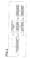
- Figure 3 is a functional block diagram illustrating a portion relevant to the medical image display apparatus of the first embodiment of the present invention.
- the medical image display apparatus of the present invention includes medical image storage means 50 for obtaining, via network 9, a first medical image and a second medical image reconstructed from a plurality of medical images, including the first medical image, in response to a request from modality 1 or image storage server 2, radiology report storage means 10 for storing a radiology report that includes a lesion character representing contents of a lesion area in the first medical image, lesion storage means 20 for storing a position of the lesion area and a lesion character in association with each other, link position storage means 30 for calculating a position in the second medical image corresponding to the position of the lesion area and storing the calculated position as a link position, medical image display means 60 for obtaining the second medical image from medical image storage means 50 and displaying the obtained image, and link information display means 40 for displaying a link character constituted by the lesion character and a position indicator indicating
- Radiology report storage means 10 is mainly constituted by hard disk 305 and stores a radiology report that includes a lesion character representing lesion contents of a lesion area in a medical image V.
- Lesion storage means 20 mainly constituted by hard disk 305 and stores the lesion character of the radiology report stored in radiology report storage means 10 and the position of the lesion area corresponding to the lesion character in association with each other.
- Link position storage means 30 is mainly constituted by hard disk 305 and CPU 307, and calculates a lesion position in reconstructed second medical image 133 corresponding to the lesion position in the first medical image stored in lesion storage means 20.
- Link information display means 40 is mainly constituted by display section 301, and displays a link character and a position indicator indicating the position of the associated lesion area in reconstructed medical image 133 in association with each other.
- the position indicator may be a circular mark like indicator 135C shown in Figure 8 , or any other known indicator capable of indicating a position, such as a point, crisscross, arrow, or the like.
- the position indicator may be used to indicate a particular point in the lesion area. The particular point may be selected from the gravity center, center, and point representing the characteristic of the lesion area as appropriate.
- the "displaying in association with each other” may be achieved by any method as long as it may display the link character and corresponding position indicator in association with each other in distinguishable manner.
- the link character and the corresponding position indicator may be displayed by connecting them with a line, arrow, or association indicator of graphic, such as a speech balloon extending from the position indicator.
- the link character may be displayed adjacent to the corresponding position indicator.
- the term "displaying in association with each other” may include the case in which the link character and the corresponding position indicator can be identified, such as the case in which only one link character and only one position indicator are displayed, even if the association indicator is not used or the link character is not displayed adjacent to the position indicator.
- Medical image storage means 50 is mainly constituted by hard disk and stores slice images from CT, MRI, PET, ultrasonic diagnostic equipment, or the like and three-dimensional medical images reconstructed from the slice images, or two-dimensional medical images.
- Typical reconstructed images include those represented by volume rendering, ray summation, maximum intensity projection (MIP), and multi planar reformat (MPR).
- Medical image display means 60 is mainly constituted by communication interface 311 for obtaining a medical image V from network 9 and display section 301 for displaying the obtained medical image V, and display a medical image obtained from medical image storage means 50 on display section 301.
- a method of generating a radiology report, a method of generating link information, and a method of displaying a medical image with the generated link information according to a first embodiment will be described briefly.
- Figure 5 is a conceptual diagram of a radiology report generation screen of the first embodiment.
- Figure 5 shows radiology report generation screen 100 by way of example.
- Radiology report generation screen 100 shown in Figure 5 includes patient information area 101 for entering patient information. For example, a patient name, patient ID, gender, age, and the like, which can be obtained from header information of DICOM, are displayed in the patient information area.
- medical image information area 103 for indicating information of a medical image V, the date of the image, modality used, requesting department, and the like are entered from input section 303, such as the mouse or keyboard, or from header information of DICOM and displayed.
- Radiology report area 104 includes radiologist name field 106, finding field 105 which is a radiology report, and diagnostic field 107. A doctor or the like, as the operator, may enter or edit information in radiologist name field 106, finding field 105, and diagnostic field 107, using the mouse or keyboard.
- radiology report generation screen 100 may include radiology report editing buttons 109 for storing or cancelling an edited radiology report and various other function buttons as appropriate.
- radiology report editing buttons 109 for storing or cancelling an edited radiology report and various other function buttons as appropriate.
- a temporary storage button, finalize button, cancel button are shown, by way of example.
- Radiology report generation screen 100 may include necessary buttons for referencing information, such as past report selection button 119A, reference image selection button 119B, and the like, as appropriate, and includes link input selection button 111 for enabling link input of an embodiment of the present invention.
- link input selection button 111 may by provided in various known forms within a range in which the purpose thereof is achieved.
- past report selection button 119A When past report selection button 119A is selected, past reports are selectably displayed in reference information area 117. If reference image selection button 119B is selected, thumbnails of medical images V are selectably displayed in reference information area 117, as shown in Figure 5 .
- Radiology report generation screen 100 further includes detailed image display area 113 and reference information such as thumbnails of medical images V, past reports, or the like, are displayed in reference information selection area 117 as appropriate.
- thumbnail 121A is selected from a plurality of thumbnails representing medical images V in reference information area 117 and medical image 115 corresponding to thumbnail 121A is displayed in detailed image display area 113.
- the plurality of thumbnails displayed in reference information area 117 may be those of medical images representing lesions included in one series of medical images V or those of medical images representing lesions included in a plurality of series of medical images V.
- Detailed image display area 113 include, as appropriate, editing buttons 113A, 113B for processing and editing the medical image displayed therein.
- FIG. 4 is a flowchart representing a method of generating a radiology report of the present embodiment.
- link input button 111 is selected by input section 303 to set the link input mode to ON (ST101).
- crosshair pointer 113C is selected from editing buttons 113A using input section 303.
- a crosshair pointer is displayed and when the crosshair pointer is placed on the position of a lesion and clicked with the mouse, lesion position 115A is inputted (ST102 is YES).
- lesion position 115A When lesion position 115A is inputted, information including information indicating the storage location of medical image 115 that includes lesion position 115A and position information indicating the position of the lesion area in medical image 115, such as coordinates of the lesion area, is obtained.
- the position information may further include information obtained, for example, from header information of DICOM, such as image ID or the like.
- the position information may be obtained as coordinate values in medical image 115, while if it is a circle, the position information may be obtained as coordinate values of the center of the circle and a length of the radius in medical image 115.
- the position of the lesion is a diagram encircled by a straight line or curved line
- information representing the diagram such as an end point of the straight line in medical image 115 or a function representing the curved line, may be obtained, as appropriate, as the position information.
- a character string not yet used in finding field 105 is selected from predetermined character strings, and a character string to be inserted as lesion character 105A is determined (ST103).
- the predetermined character string may be a character string, symbol, or numeric character, and (1) a fixed character string (2) if medical image 115 is a medical image constituting one series formed of a series of tomographic images, a combination of the medical image series number with a slice number or the like as information to identify a medical image that includes a lesion positions (3) a pixel value of a lesion position (4) an anatomical region name of a lesion position such as left upper lung lobe, left lingular segment, or the like (5) coordinate values of a lesion position and (6) a combination thereof may be used.
- the character string may be extracted and inserted in the following manner.
- the organ to which the inputted lesion position belongs is extracted by computer aided diagnosis (CAD).
- CAD computer aided diagnosis
- any other organ recognition technique may be used as long as it is capable of automatically extracting the organ to which the specified lesion position belongs.
- the organ extraction described may be performed on a medical image that includes a lesion position and if the extracted organ includes the lesion position, the organ may be determined as the organ to which the lesion position belongs.
- the anatomical region name representing the organ may be inserted as the lesion character.
- the symptom or position of the lesion area may be assumed from the lesion character, as well as a linking operation may be implemented without inputting a lesion character from input section 303, so that the radiology report may be generated efficiently.
- each organ or a region name representing a region of each organ may be used as the region name.
- left lung or right lung may be used as the region name or otherwise further classified names of right upper lung lobe, right middle lung lobe, right lower lung lobe, left upper lung lobe, or left lower lung lobe may be used as the region name.
- a region name classified by the lung segment such as the superior lingular segment (S4) of left upper lung lobe or abbreviated names thereof may be used.
- the character string to be inserted may be determined in the following manner.
- an association table of position of lesion area and lesion character candidate associating organ names, characteristic information 203 of positions of lesion areas, and lesion character candidates with each other is provided.
- luminance values 203 of CT images are associated as the characteristic information 203 of the positions of lesion areas.
- the organ to which the inputted position of lesion area belongs is extracted using the specific extraction method described above, such as the computer aided diagnosis or the like. Then, the luminance value of the image at the inputted position of lesion area is obtained as the position information.
- the luminance value at the position of lesion area inputted as characteristic information 203 of the position of lesion area is -50 which falls with in the range from -2000 to 0.
- four character strings of bracket, pneumothorax, lung emphysema, and bronchodilation are determined from among lesion character candidates 205 as the corresponding lesion character candidates 205A.
- the luminance value is 100 which falls in the range from 50 to 100 and also in the range from 100 to 1000, so that nodule GGO, infiltrative shadow, and calcification are determined as the link character candidates.
- lesion character candidates 20A are displayed on radiology report generation screen 100 as lesion character candidate list 210 when the position of lesion area is specified by the user.
- lesion character candidate 210A is selected from the lesion character candidate list 210 by the user, the selected lesion character candidate 210A is inserted in the radiology report as the lesion character.
- "other" shown in Figure 14 may be selected, and the character string may be inputted from the input section.
- the display of the lesion character candidate list may be in the form of a pull-down menu or any other method which allows selection of a region name.
- an average, dispersion, or pattern of luminance values adjacent to the position of lesion area may be obtained as information to be obtained from the lesion position, and characteristic information 203 may be provided according to the average, dispersion, or pattern of luminance values adjacent to the position of lesion area, thereby allowing the corresponding link character candidate to be selected.
- lesion storage means 20 stores lesion information at lesion position 115A (ST104).
- the lesion information may be, for example, (1) lesion ID (2) position information of lesion area constituted by series name, slice name, and coordinates on the slice and (3) lesion character.
- the lesion ID and position information of lesion area constituted by series name, slice name, and coordinates on the slice may be obtained from the position information inputted from input section 303.
- a medical image that includes the position 115A of lesion area obtained from input section 303 and the lesion character 105A linked by a hyperlink are inserted in finding field 105 (ST105).
- the lesion character 105A is inserted in a manner so as to be distinguishable from the other characters, such as coloring of the lesion character, coloring of the background, or use of underline, blinking, boldface, different font type or size, or frame for the lesion character, thereby indicating that the lesion character is linked by a hyperlink.
- step ST106 if a position of lesion area is inputted in the same manner as in step ST102 (ST106 is YES), the steps from ST102 to ST105 are repeated. That is, if a plurality of lesion positions is specified in the medical image V, the corresponding number of lesion characters is inserted.
- lesion character insertion is completed.
- medical image display means 60 obtains position information of a medical image 115 corresponding to the lesion character 105A from lesion storage means 20 and displays the medical image 115 in detailed image display area 113. Further, link information display means 40 obtains a position 115A of lesion area from lesion storage means 20 and displays the position 115A of lesion area corresponding to the lesion character 105A in the medical image in the detailed image display area.
- the indicator may be displayed as the representative point in the area. The display form of the indicator may be made selectable from arrow, area, pointer for pointing a representative point in the lesion area, and the like.
- Figure 6 is a flow diagram illustrating a process flow for changing a link character according to the first embodiment.
- Figure 7 is a conceptual diagram of a radiological report generated by changing a portion of the link character according to the first embodiment.
- Figure 7 shows, by way of example, finding field 105 of radiology report generation screen 100.
- Figure 7 shows that three lesion characters of "lesion 1", “lesion 2" (105A), and “lesion 3" (105C) are inserted in the finding field, in which lesion 1 has been edited and changed to a new lesion character of "irregular nodule" (105A).
- one of the lesion characters is selected by input section 303 (ST201) .
- the selected lesion character is changed to a desired character string, symbol, or numeric character by operating the mouse or keyboard. More specifically, if a selection tool such as arrow 113 is pointed on the lesion character and the right mouse button is clicked, lesion character change options are displayed in a pull-down menu format.
- the lesion character is changed by selecting one of the lesion character change options (ST202) .
- the lesion character may be changed using a different method, as appropriate, as long as it allows only the character string of the lesion character to be changed with the hyperlinked state being maintained. Then, the position of the lesion area corresponding to the original lesion character is linked to the new lesion character by a hyperlink and corresponding lesion information is updated (ST203).
- the radiology report used in the first embodiment is generated.
- the medical image that includes the position of lesion area associated with the lesion character is a first medical image used in the first embodiment.
- Figure 9 a flow diagram illustrating a process flow for generating link information in the medical image display method of the present embodiment.
- link position storage means 30 obtains a lesion position in a slice image 115 and a corresponding lesion character from lesion storage means 20 (ST301) . Then coordinates in a medical image 133 reconstructed by stacking a plurality of slice images, including the slice image 115 (ST302) .
- Typical reconstructed images include those represented by volume rendering, ray summation, maximum intensity projection (MIP), and multi planar reformat (MPR).
- a three-dimensional image is reconstructed from multiple slice images, i.e., multiple two-dimensional images sliced at a predetermined thickness
- coordinates of the position of a point specified in any of the two-dimensional images in the three-dimensional image may be calculated from the slice position and coordinates of the point, and the coordinate position in the three-dimensional image corresponding to the coordinates of the lesion position in the slice image is identified.
- the lesion position specified in the slice image can be represented in the three-dimensional image.
- link position storage means 30 stores the coordinate values and corresponding lesion character in association with each other as link information (ST303).
- the link information may be constituted by, for example, (1) link ID (2) link position which is the position in the reconstructed image 133 corresponding to the lesion position in the slice image (3) link character and (4) corresponding lesion ID.
- the lesion ID may be obtained from the lesion information inputted from lesion storage means 20.
- Figure 8 is a conceptual diagram of a medical image display screen displayed according to the first embodiment.
- the layout of medical image display screen 110 shown in Figure 8 will be described.
- reference image selection button 119B is selected and a thumbnail 117B of a reference image is displayed in reference image area 117.
- the thumbnail 117B of a reference image in reference image area is selected and an image 133, which is a reconstructed second medical image corresponding to the selected thumbnail image 117B, is displayed in detailed information area 113.
- Selection tool 113D is provided in detailed information display area 113 in which the reconstructed second medical image 133 is displayed to allow the reconstructed image 133 to be observed by changing the size and angle thereof.
- the medical image display apparatus includes link display button 131 for displaying link information.
- link display button 131 is selected, and link information, i.e., link characters 135A, 137A, link positions 135C, 137C, and association indicators 135B, 137C are displayed.
- the link position 135C obtained from link position storage means 30 and the link character 135A constituted by lesion character 105D corresponding to the link position 135C is inserted in the reconstructed medical image 133 (ST307) .
- link characters and link positions corresponding to all of the positions of the plurality of lesion areas are displayed.
- positions of two lesion areas are stored in link position storage means 30 so that link characters 135A, 137A and link positions 135C, 137C are displayed.
- the link character and the corresponding link position are displayed in association with each other.
- the link character 135A and the corresponding link position 135C are associated by the association indicator 135B while the link character 137A and the corresponding link position 137C are associated by the association indicator 137B.
- the association between the link character and corresponding link position is not necessarily implemented by the association indicator, and they may be displayed in any manner as long as it allows identification of which link character corresponds to which position indicator.
- the link character 135A may be displayed adjacent to the corresponding position indicator 135C.
- the association indicator is not required or the link character is not required to be displayed adjacent to the position indicator.
- the display of link positions 135C, 137C, and link characters 135A, 137A in the medical image allows the position of the lesion area and character string representing the lesion area described in the radiology report to be referenced simply by observing the medical image. Then, from the link positions 135C, 137C, the positions of the lesion areas may be accurately recognized, and from the link characters 135A, 137A, the lesions may be assessed easily.
- the present embodiment displays a link position using a position indicator so that the position of the lesion area may be accurately recognized.
- link positions 135C, 137C and link characters 135A, 137A are respectively associated with each other in a medical image, so that even when a plurality of link characters and a plurality of link positions are displayed at the same time, the correspondence relationship between each link position and each link character may be recognized easily.
- the positions of a plurality of lesion areas and character strings representing a plurality of corresponding lesions may be recognized at a time by simply observing the medical image, whereby the diagnosis may be made more easily.
- the relative position between link positions 135C and 137C may be recognized easily.
- the display of the link position in the reconstructed medical image allows the position of lesion area in a slice image to be recognized by a different medical image, so that the position of the lesion area may be recognized more accurately.
- the position of the lesion area may be recognized stereoscopically, and accurate radiological image reading may be performed base on the lesion area.
- the reconstructed three-dimensional image may be made rotatable and scalable, and the link position and link character may be displayed in an interlocking manner with the rotation and scaling. In this case, the position of the lesion area may be viewed from a different angle with a different size, thereby allowing more accurate radiological image reading.
- lesion storage means 20 may be a means that further stores a word or phrase before and after the lesion character in the radiology report as the lesion information
- link position storage means 30 may be a means that further obtains the word or phrase before and after the lesion character in the radiology report from lesion storage means 20
- link information display means 40 may be a means that displays the word or phrase before and after the link character in addition to the link character 135A as shown, by way of example, in 135D in Figure 11 .
- ten characters before the link character and ten characters after the link character may be extracted and displayed or one sentence, including the link character, may be extracted and displayed.
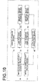
- Figure 10 is a functional block diagram of a radiology report generation function of the second embodiment.
- the configuration shown in Figure 10 is identical to that shown in Figure 3 except that the configuration in Figure 10 further includes radiology report display means 70 and link character specifying means 80. Therefore, identical portions will not be elaborated upon further here.
- Radiology report display means 70 is mainly constituted by display section 301 for displaying a radiology report and displays a radiology report stored in radiology report storage means 10 on display section 301 in response to the specification by link character specifying means 80.
- Link character specifying means 80 is mainly constituted by input section 303 for inputting a lesion area and in response to the specification of a link character or a lesion character by the operator via input section 303, inputs the specified link character to either one of link information display means 40, medical image display means 60, and radiology report display means 70.
- link information display means 40 displays the link character 137A in the medical image by linking the character to the corresponding lesion character 141 described in the radiology report by a hyperlink.
- Figure 11 shows, by way of example, medical image display screen 110 according to the second embodiment.
- a phrase before and after the lesion character 141 described in the radiology report is displayed in addition to the link character 135A, as shown in a comment 135D.
- a phrase before and after the lesion character corresponding to the link character 137A described in the radiology report is displayed, as shown in a comment 137D.
- Components given the same reference numerals as those in Figure 8 are identical and will not be elaborated upon further here.
- radiology report display means 70 displays the radiology report, that includes the lesion character 141, in finding field 105 in response to the specification of the link character 137A with selection tool 123 of link character specifying means 80.
- finding field 105 by way of example, only a portion of the report adjacent to the lesion character 141 is displayed and it is indicated that the entire radiology report may be referenced by moving cursor 139. Note that, however, the display method of the radiology report is not limited to this, and the radiology report may be displayed in a separate window or in its entirety in response to the specification of the link character 137A.
- the link character 137A and image that includes lesion character 141 are linked by a hyperlink, so that by simply specifying the link character 137A in the image, the corresponding detailed information in the radiology report may be referenced easily, thereby allowing an efficient diagnosis.
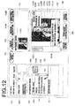
- FIG. 12 shows, by way of example, medical image display screen 110 according to the modification of the second embodiment.
- Components given the same reference numerals as those in Figure 11 are identical and will not be elaborated upon further here.
- the lesion character 141 described in the radiology report and a slice image 121A which is the first medical image corresponding to the lesion character 141, are linked by a hyperlink.
- the display method of the medical image that includes the position of lesion area is not limited to this, and the slice image 121A may be displayed in a separate window or as a detailed medical image instead of the thumbnail in response to the specification of the lesion character 141.
- the lesion character 141 and slice image 121A are linked by a hyperlink, so that by simply specifying the lesion character 141 in the radiology report, the corresponding slice image 121A may be referenced easily, thereby allowing an efficient diagnosis.
- the corresponding lesion character 141 in the radiology report may be referenced, and from the lesion character 141, the corresponding slice image 121A may be referenced, the corresponding detailed information may be obtained easily, thereby allowing an efficient radiological image reading.
- the specified link character 137A and the position 137C of lesion area are displayed in a distinguishable manner from the other link character 137A and link position 137C.
- This allows clear understanding as to which link character 137A is specified, whereby it is easy to understand that the radiology report corresponding to the link character 137A is displayed.
- the link character and the position of the corresponding lesion area may be highlighted by various methods for displaying in a distinguishable manner, such as blinking of the mark, character, or both, coloring of the link character or background, or use of underline, blinking, boldface, different font type or size, or frame for the link character.
- various methods for displaying in a distinguishable manner such as blinking of the mark, character, or both, coloring of the link character or background, or use of underline, blinking, boldface, different font type or size, or frame for the link character.
- Radiology report storage means 10 stores a plurality of radiology reports, including past radiology reports with respect to past medical images of the same patient.
- Lesion storage means 20 converts a lesion position in a past medical image to the corresponding lesion position in a current image representing the same region and same position and stores as lesion information. More specifically, with respect to a slice image 143 representing a lesion position in a past medical image, a medical image constituted by a group of tomographic images, including the past slice image 143, is aligned with a medical image constituted by a group of current tomographic images using the technique described in Japanese Unexamined Patent Publication No.
- lesion storage means 20 further associates radiology report identification information for identifying the radiology report that includes the lesion character with the lesion information and stores the resultant information.
- the radiology report identification information is constituted by information capable of identifying the radiology report, such as the file name of the report, data of the report, and the like.
- link position storage means 30 obtains the lesion information from lesion storage means 20, associates the radiology report identification information with the link information, and stores the resultant information.
- Link information display means 40 displays the past lesion character and current lesion character in a distinguishable manner based on the radiology report identification information. For example, the date of radiology report may be displayed in the reconstructed medical image as the radiology report identification information, in addition to the link character. Further, when displaying the corresponding radiology report in response to the specification of the link character 137A, the radiology report identification information, such as the date of radiology report or the like may be displayed in addition to the radiology report by applying the second embodiment to the third embodiment.
- link information display means 40 to display a past lesion character representing a lesion described in a past radiology report in a reconstructed second medical image, whereby a lesion position corresponding to the past lesion position may be referenced easily in the current medical image.
- a diagnosis with reference to the progress of the past lesion may be made easily and image reading may be performed efficiently.
- radiology report identification information is displayed in a reconstructed medical image
- a diagnosis may be made in consideration of the passage of time from the date of the report, resulting in an accurate image reading.
- the second medical image reconstructed from a plurality of medical images, including the first medical image may be more than one.
- a fourth embodiment in which a plurality of second medical image is provided will be described.
- each second medical image is a medical image reconstructed by a different reconstruction method or a medical image different in display position and size from each other.
- Images reconstructed by various methods such as volume rendering, surface rendering, ray summation, maximum intensity projection (MIP), multi planar reformat (MPR), minimum intensity projection (MNIP), curved planar reformation (CPR), and the like, may be applied to the second medical image.
- MIP maximum intensity projection
- MPR multi planar reformat
- MNIP minimum intensity projection
- CPR curved planar reformation
- Figure 15 illustrates a medical image display screen displayed according to a modification of the first embodiment.
- Figure 15 shows only detailed information area 113 shown in Figure 8 .
- the thumbnail 117B of a reference image in reference image area 117 is selected, and an image represented by volume rendering (VR), which is a reconstructed second medical image 133 corresponding to the selected thumbnail 117B, and an image represented by MPR, which is a second medical image 151 corresponding to the selected thumbnail image, are displayed in the detailed information area 113 as shown in Figure 15 .
- VR volume rendering
- MPR which is a second medical image 151 corresponding to the selected thumbnail image
- link information of link character 135A, link position 135C, and association indicator 135B is displayed in both images, one of which is represented by VR and the other of which is represented by MPR.
- link character 135A in addition to the link character 135A, a phrase before and after the lesion character 141 described in the radiology report is further displayed in comment 135D.
- the same link information is displayed in each of a plurality of second medical images, but the display method is not limited to this, and the link information may be selectively displayed according to the type of each of the second medical images, such as VR, MIP, or the like.
- a selective display option may be provided with respect to each link character and if VR is set to the selective display option, the link information may be displayed only in an image represented by VR method, while if MIP is set to the selective display option, the link information may be displayed only in an image represented by MIP method.
- the selective display option may be set with respect to each radiology report. More specifically, radiology report identification information for identifying each radiology report, such as the date and time of each radiology report or the like, maybe set as the link information, and the same selective display option is set to the same link information.
- an image diagnosis may be made using a plurality of reconstructed medical images with respect to the same lesion, whereby the image reading may be made more efficiently and accurately.
- the link information is selectively displayed with respect to the type of second medical image, only necessary information may be displayed on the second medical image according to the type of second medical image, resulting in a more efficient and accurate image reading.
- a configuration may be adopted in which, in one of the second medical images, display of another second medical image is selectable from GUI, and the another second image is displayed instead of or in addition to the one of the second medical image on medical image display means 60 in response to the selection to display the another second image.
- a configuration may be adopted in which when the right button of the mouse is clicked on a second medical image represented by VR method, display methods, including MIP, MPR, and the like, are selectably displayed in a pull-down menu, and if MIP is selected by the user from the pull-down menu using the mouse or the like, another second medical image represented by MIP method is displayed instead of the second medical image represented by VR method.
- the second medical image in the case where the second medical image has time series image data, the second medical image may be displayed four-dimensionally with the link information being displayed at the link position of the second medical image.
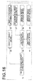
- Figure 16 is a functional block diagram of the fifth embodiment.
- the fifth embodiment further includes display condition determination means 90 for obtaining a lesion character described in a radiology report from lesion storage means 20, extracting a keyword corresponding to the lesion character, and determining the display condition from the extracted keyword, and second medical image generation means 91 for generating a second medical image based on the determined display condition and storing the generated second medical image in medical image storage means 50.
- medical image display means 60 displays the second medical image based on the determined display condition.
- FIG 17 illustrates display condition determination means 90 broken into functional blocks.
- display condition determination means 90 includes keyword table 93, keyword extraction section 92, display condition table 94, and display condition determination section 95.
- Keyword extraction section 92 obtains a lesion character described in a radiology report from lesion storage means 20 and extracts keywords K1, K2 serving as a display condition determinant factor using keyword table 93.
- Figure 18 shows, by way of example, keyword table 93.
- keyword table 93 is a table in which keywords and lesion characters are associated and information of anatomical structures and lesions are registered as lesion characters and extraction target words.
- Keyword extraction section 92 obtains a lesion character registered in the lesion information from lesion storage means 20. In this example, it is assumed that "left lung" and "nodule” are obtained as the lesion characters in the lesion information.
- keyword extraction section 92 converts a lesion character hit by searching to a keyword. That is, "left lung” is converted to "lung” (keyword K1) which is associated with various words, while the "nodule” becomes a keyword “nodule”(keyword K2) as-is.
- the keyword extraction method is not limited to that described above, and it is possible to employ any known natural language processing techniques such as those used in search engines. But, the searching using the keyword table described above is sufficiently effective if the names of anatomical structures and lesions are standardized by providing a user interface for selectively inputting anatomical structures and lesions when generating a radiology report.
- keyword extraction section 92 may directly use a lesion character as a keyword without providing the keyword table. Provision of the keyword table, however, allows one keyword to be associated with a plurality of lesion characters, so that the display condition for various lesion characters may be set efficiently without unnecessarily increasing the number of keywords registered in the display condition table.
- display condition determination section 95 identifies the display condition associated with these keywords K1, k2 by reference to display condition table 94 for determining image processing method for generating and displaying a second medical image from a plurality of medical images, including a first medical image, and/or determining the display mode of the second medical image.
- Such display conditions can be maintained by the users of doctors of the diagnosis and treatment department, or the like by performing registration, modification, and deletion.
- the maintenance may be directly performed on display condition table 94, but it is preferable that the maintenance may be implemented through a GUI for interactively performing input or selection operations for the contents of the display condition table. Further, it is also desirable that the display condition for the currently displayed image may be confirmed from the GUI. The reason is that this will facilitate understanding of the users at the time of image diagnosis and operability for editing display condition table may be improved.
- display condition table 94 includes display conditions associated with each keyword, such as VR color template, parameters of gradation and emphasis processing, window width of medical image, window level, image recognition processing using CAD and the like.
- VR color template is a template for determining the VR color display mode in which RGB or opacity is set in association with the density value of the image data.
- Figure 19 shows, by way of example, display condition table 94a.
- display condition determination means 90 extracts "blood vessel” as the keyword from the keyword table, and determines the display condition "101" corresponding to the keyword "blood vessel” from display condition table 94a. Then, display condition determination means 90 outputs the determined display condition "101" and lesion character "blood vessel” to second medical image generation means 91.
- Second medical image generation means 91 obtains the display condition "101" and lesion character "blood vessel” from display condition determination means 90 and obtains image data that include a first medical image associated with the lesion character "blood vessel” by reference to lesion storage means 20. Then, based on the image type "VR" associated with the display condition "101", second medical image generation means 91 generates a second medical image by the VR method from the obtained image data and stores the generated second medical image in medical image storage means 50.
- the second medical image is stored in medical image storage means 50 with information identifying the display condition of the second medical image, for example, display condition ID "101", being associated by tag information or the like.
- the second medical image may be generated by the reconstruction method appropriate for the lesion or organ and displayed, resulting in more efficient and accurate image reading.
- medical image display means 60 obtains the second medical image and information identifying the display condition of the second medical image, such as display condition ID "101", from medical image storage means 50, obtains display condition "101" from display condition determination means 90, and displays the second medical image based on the display condition "101".
- a color template that best displays each organ may be provided. For example, if the link character is "liver", color template Cr2 provided for livers is selected and the liver is displayed by the VR method based on the selected color template Cr2. As the second medical image is displayed according to the lesion character, more efficient and accurate image reading may be performed.
- medical image display means 60 may be a means that displays the second medical image centered on the link position in the image.
- medical image display means 60 may be a means that obtains the link position from link position storage means 30 and, based on the display condition obtained from display condition determination means 90, displays the second medical image in a display window of a specific size disposed at a specific position such that the link position comes to the center of the display window.
- the second medical image By displaying the second medical image such that the link position comes to the center of the display window, the second medical image may be displayed such that the position of lesion area is visually recognized easily, so that more efficient and accurate image reading may be performed. Note that the second medical image is not necessarily displayed such that the position of lesion area comes to the center of the display window as long as the position of lesion area is visually recognized easily.
- second medical image generation means 91 is not required since it is not necessary to generate the second medical image. More specifically, display condition determination means 90 obtains a lesion character described in a radiology report from lesion storage means 20, then extracts the keyword corresponding to the lesion character, and determines the display condition for the second medical image from the extracted keyword. Then, medical image display means 60 may display the second medical image already generated and stored in medical image storage means 50 based on the determined display condition.
- Figure 20 shows, by way of example, a display condition table 94b of the further modification of the fifth embodiment.
- the medical image display apparatus further includes a plurality of CADs of different types and the display condition table 94b is a table in which image processing using automatic recognition processing by CAD is set in association with each keyword.
- various known automatic recognition techniques including the examples described above, may be used.
- display condition determination means 90 extracts a keyword "heart”, and determines a display condition "201" corresponding to the keyword "heart” from the display condition table 94b.
- the display condition "201" includes cardiac extraction and coronary artery extraction, and distinguishable display of the extracted heart and coronary artery in association with the keyword "heart”.
- Medical image display means 60 performs cardiac extraction and coronary artery extraction on a second medical image obtained from medical image storage means 50 based on the display condition "201" and displays the second medical image with the extracted heart being displayed in a distinguishable manner and the extracted coronary artery of the heart being displayed in a further distinguishable manner.
- an option for hiding an anatomical structure, such as the extracted organ or the like, by masking the structure may be set in association with the keyword.
- liver extraction and "non-display other than the extracted anatomical structure” may be set in association with the keyword “liver”, or spleen extraction and “non-display other than the extracted anatomical structure” may be set in association with the keyword “spleen”.
- display condition determination means 90 extracts keywords "liver” and “spleen” from keyword table 93, and determines display conditions "202" and "203" from the display condition table 94b.
- Medical image display means 60 obtains the determined display conditions "202" and "203" from display condition determination means 90, obtains a plurality of medical images, including a first medical image or a second medical image from medical image storage means 50, and extracts the liver and spleen from the obtained medical image by CAD. Then, medical image display means 60 hides an organ other than the extracted liver and spleen by masking.
- an option for extracting a bone by CAD and hiding the extracted anatomical structure may be set in association with the keyword "blood vessel".
- medical image display means 60 obtains the display condition "204", extracts a bone by CAD from the second medical image obtained from medical image storage means 50, and hides the extracted bone by masking when displaying the second medical image.
- the second image may be displayed with the lesion or organ being displayed in an easily distinguishable manner according to the keyword extracted in association with the lesion character of a lesion, organ, or the like, whereby more efficient and accurate image reading may be performed.
- Embodiments described above may be applicable to other embodiments without the nature thereof being changed.
Landscapes
- Health & Medical Sciences (AREA)
- Engineering & Computer Science (AREA)
- General Health & Medical Sciences (AREA)
- Public Health (AREA)
- Primary Health Care (AREA)
- Medical Informatics (AREA)
- Epidemiology (AREA)
- Nuclear Medicine, Radiotherapy & Molecular Imaging (AREA)
- Radiology & Medical Imaging (AREA)
- General Physics & Mathematics (AREA)
- Computer Hardware Design (AREA)
- Theoretical Computer Science (AREA)
- Computer Graphics (AREA)
- General Engineering & Computer Science (AREA)
- Physics & Mathematics (AREA)
- Software Systems (AREA)
- Measuring And Recording Apparatus For Diagnosis (AREA)
- Medical Treatment And Welfare Office Work (AREA)
Applications Claiming Priority (3)
| Application Number | Priority Date | Filing Date | Title |
|---|---|---|---|
| JP2009215100 | 2009-09-17 | ||
| JP2010067799A JP5349384B2 (ja) | 2009-09-17 | 2010-03-24 | 医用画像表示装置および方法並びにプログラム |
| PCT/JP2010/005639 WO2011033769A1 (fr) | 2009-09-17 | 2010-09-15 | Dispositif d'affichage d'images médicales, procédé et programme associés |
Publications (2)
| Publication Number | Publication Date |
|---|---|
| EP2478833A1 true EP2478833A1 (fr) | 2012-07-25 |
| EP2478833A4 EP2478833A4 (fr) | 2017-06-21 |
Family
ID=43758383
Family Applications (1)
| Application Number | Title | Priority Date | Filing Date |
|---|---|---|---|
| EP10816883.2A Ceased EP2478833A4 (fr) | 2009-09-17 | 2010-09-15 | Dispositif d'affichage d'images médicales, procédé et programme associés |
Country Status (5)
| Country | Link |
|---|---|
| US (1) | US8625867B2 (fr) |
| EP (1) | EP2478833A4 (fr) |
| JP (1) | JP5349384B2 (fr) |
| CN (1) | CN102497805B (fr) |
| WO (1) | WO2011033769A1 (fr) |
Cited By (4)
| Publication number | Priority date | Publication date | Assignee | Title |
|---|---|---|---|---|
| EP2804147A3 (fr) * | 2013-04-25 | 2015-03-18 | Samsung Medison Co., Ltd. | Procédé et appareil pour un enregistrement d'image |
| WO2015044259A1 (fr) * | 2013-09-27 | 2015-04-02 | Koninklijke Philips N.V. | Activation de révision d'une image médicale |
| EP2915486A1 (fr) * | 2014-03-05 | 2015-09-09 | Samsung Medison Co., Ltd. | Procédé et appareil pour afficher une image en 3D |
| EP2984630A1 (fr) * | 2013-04-10 | 2016-02-17 | Koninklijke Philips N.V. | Visualisation de données image reconstruites |
Families Citing this family (53)
| Publication number | Priority date | Publication date | Assignee | Title |
|---|---|---|---|---|
| JP5746908B2 (ja) * | 2011-05-06 | 2015-07-08 | 株式会社東芝 | 医用画像処理装置 |
| CN102846338A (zh) * | 2011-07-01 | 2013-01-02 | 深圳迈瑞生物医疗电子股份有限公司 | 输出目标信息的方法及其超声成像装置、工作站及其系统 |
| JP6025311B2 (ja) * | 2011-08-01 | 2016-11-16 | キヤノン株式会社 | 眼科診断支援装置および方法 |
| JP5794305B2 (ja) * | 2011-09-01 | 2015-10-14 | 株式会社島津製作所 | データ表示装置およびそれを備えた断層撮影装置 |
| US9922026B2 (en) * | 2011-11-14 | 2018-03-20 | Koninklijke Philips N.V. | System and method for processing a natural language textual report |
| KR20130053587A (ko) * | 2011-11-15 | 2013-05-24 | 삼성전자주식회사 | 의료기기 및 이를 이용한 의료영상 디스플레이 방법 |
| JP5238894B1 (ja) * | 2012-02-22 | 2013-07-17 | 株式会社東芝 | X線ct装置 |
| JP6106259B2 (ja) * | 2012-03-21 | 2017-03-29 | コーニンクレッカ フィリップス エヌ ヴェKoninklijke Philips N.V. | 医用イメージングと生検データとを統合する臨床ワークステーション及びこれを使用する方法 |
| US10667790B2 (en) * | 2012-03-26 | 2020-06-02 | Teratech Corporation | Tablet ultrasound system |
| US10354049B2 (en) | 2012-08-22 | 2019-07-16 | Koninklijke Philips N.V. | Automatic detection and retrieval of prior annotations relevant for an imaging study for efficient viewing and reporting |
| USD742396S1 (en) * | 2012-08-28 | 2015-11-03 | General Electric Company | Display screen with graphical user interface |
| US20150213725A1 (en) * | 2012-09-07 | 2015-07-30 | Tractus Corporation | Method, apparatus, and system for viewing multiple-slice medical images |
| US20140100861A1 (en) * | 2012-10-09 | 2014-04-10 | David Gerard Ledet | Medical analysis application and response system |
| JP6143433B2 (ja) * | 2012-10-31 | 2017-06-07 | キヤノン株式会社 | 医用画像撮影装置、医用画像の表示方法 |
| US20140140598A1 (en) * | 2012-11-21 | 2014-05-22 | General Electric Company | Systems and methods for 2d and 3d image integration and synchronization |
| JP6387003B2 (ja) * | 2013-06-18 | 2018-09-05 | キヤノン株式会社 | トモシンセシス撮影の制御装置、撮影装置、撮影システム、制御方法および当該制御方法をコンピュータに実行させるためのプログラム |
| WO2014210430A1 (fr) | 2013-06-27 | 2014-12-31 | Tractus Corporation | Systèmes et procédés pour la cartographie de tissu |
| JP6026982B2 (ja) * | 2013-09-30 | 2016-11-16 | 富士フイルム株式会社 | 画像表示制御装置およびその作動方法並びに画像表示制御プログラム |
| JP6629729B2 (ja) * | 2013-11-26 | 2020-01-15 | コーニンクレッカ フィリップス エヌ ヴェKoninklijke Philips N.V. | 放射線レポートにおける参照画像コンテキストに基づくウィンドウ幅/レベルの自動設定 |
| JP6183177B2 (ja) * | 2013-11-28 | 2017-08-23 | コニカミノルタ株式会社 | 医用画像システム及びプログラム |
| JP6261301B2 (ja) * | 2013-11-28 | 2018-01-17 | キヤノン株式会社 | 医用画像表示装置およびその制御方法 |
| JP2015215740A (ja) | 2014-05-09 | 2015-12-03 | キヤノン株式会社 | 情報処理装置、情報処理方法及びプログラム |
| EP2980753A1 (fr) * | 2014-08-01 | 2016-02-03 | Centre Hospitalier Universitaire d'Angers | Procédé d'affichage d'images médicales facilement compréhensibles |
| JP2016087279A (ja) * | 2014-11-08 | 2016-05-23 | キヤノン株式会社 | 医用情報処理装置、x線撮影装置、医用情報処理システム、情報処理方法及びプログラム |
| KR101728044B1 (ko) * | 2015-02-02 | 2017-04-18 | 삼성전자주식회사 | 의료 영상을 디스플레이 하기 위한 방법 및 장치 |
| CN107257977B (zh) * | 2015-02-25 | 2021-08-24 | 皇家飞利浦有限公司 | 检测缺失的发现用于自动创建纵向发现视图 |
| US9858385B2 (en) | 2015-07-23 | 2018-01-02 | International Business Machines Corporation | Identifying errors in medical data |
| US10181187B2 (en) | 2015-09-09 | 2019-01-15 | Canon Kabushiki Kaisha | Information processing apparatus, method thereof, information processing system, and computer-readable storage medium that display a medical image with comment information |
| JP6827706B2 (ja) * | 2015-09-09 | 2021-02-10 | キヤノン株式会社 | 情報処理装置及びその方法、情報処理システム、コンピュータプログラム |
| JP6646395B2 (ja) | 2015-09-28 | 2020-02-14 | キヤノン株式会社 | 情報処理装置及びその方法、情報処理システム、コンピュータプログラム |
| JP6711678B2 (ja) * | 2016-04-13 | 2020-06-17 | キヤノン株式会社 | 情報処理システム、情報処理方法及びプログラム |
| JP6711676B2 (ja) * | 2016-04-13 | 2020-06-17 | キヤノン株式会社 | 医用レポート作成装置及びその制御方法、医用レポート作成システム、並びに、プログラム |
| WO2018192841A1 (fr) * | 2017-04-18 | 2018-10-25 | Koninklijke Philips N.V. | Dispositif de visualisation global de patient en radiologie |
| CN107945169B (zh) * | 2017-12-01 | 2022-02-15 | 中国人民解放军第三军医大学 | 一种冠状动脉影像分析方法 |
| JP7000206B2 (ja) * | 2018-03-06 | 2022-01-19 | 富士フイルム株式会社 | 医用画像処理装置、医用画像処理方法、及び医用画像処理プログラム |
| CN108766510A (zh) * | 2018-05-30 | 2018-11-06 | 中南大学湘雅医院 | 智能病史采集方法、装置及移动采集设备 |
| CN108922601A (zh) * | 2018-07-09 | 2018-11-30 | 成都数浪信息科技有限公司 | 一种医学影像处理系统 |
| CN109256193A (zh) * | 2018-09-04 | 2019-01-22 | 北京慧影明图科技有限公司 | 图像显示方法及装置、存储介质、电子终端 |
| US10593041B1 (en) | 2019-02-21 | 2020-03-17 | Westside Veterinary Innovation, Llc | Methods and apparatus for the application of machine learning to radiographic images of animals |
| KR102761629B1 (ko) | 2019-03-12 | 2025-02-03 | 삼성메디슨 주식회사 | 초음파 영상 표시 방법, 초음파 진단 장치 및 컴퓨터 프로그램 제품 |
| CN110459297A (zh) * | 2019-08-14 | 2019-11-15 | 上海联影医疗科技有限公司 | 一种图像存储方法、系统及存储介质 |
| WO2021059659A1 (fr) | 2019-09-27 | 2021-04-01 | 富士フイルム株式会社 | Dispositif d'aide à des soins médicaux |
| CN110853739B (zh) * | 2019-10-16 | 2024-05-03 | 平安科技(深圳)有限公司 | 图像管理显示方法、装置、计算机设备及存储介质 |
| WO2021187483A1 (fr) | 2020-03-17 | 2021-09-23 | 富士フイルム株式会社 | Dispositif, procédé et programme d'aide à la création de documents |
| WO2021210676A1 (fr) * | 2020-04-16 | 2021-10-21 | 富士フイルム株式会社 | Dispositif de traitement d'image médicale, système d'endoscope, procédé de fonctionnement pour appareil de traitement d'image médicale et programme pour dispositif de traitement d'image médicale |
| US11545267B2 (en) | 2020-08-04 | 2023-01-03 | Signalpet, Llc | Methods and apparatus for the application of reinforcement learning to animal medical diagnostics |
| US20240057970A1 (en) * | 2020-12-30 | 2024-02-22 | Koninklijke Philips N.V. | Ultrasound image acquisition, tracking and review |
| KR20220147957A (ko) * | 2021-04-28 | 2022-11-04 | 자이메드 주식회사 | 생체 이미지 조직 표시 방법 및 장치 |
| JP7830461B2 (ja) * | 2021-06-14 | 2026-03-16 | 富士フイルム株式会社 | 超音波システムおよび超音波システムの制御方法 |
| US20220409172A1 (en) * | 2021-06-24 | 2022-12-29 | Biosense Webster (Israel) Ltd. | Reconstructing a 4d shell of a volume of an organ using a 4d ultrasound catheter |
| US12308108B2 (en) * | 2021-08-20 | 2025-05-20 | GE Precision Healthcare LLC | Automatically detecting characteristics of a medical image series |
| CN113763345A (zh) * | 2021-08-31 | 2021-12-07 | 苏州复颖医疗科技有限公司 | 医学影像病灶位置查看方法、系统、设备及存储介质 |
| JP2024072431A (ja) * | 2022-11-16 | 2024-05-28 | 富士フイルムヘルスケア株式会社 | 超音波診断装置及びデータ処理方法 |
Family Cites Families (29)
| Publication number | Priority date | Publication date | Assignee | Title |
|---|---|---|---|---|
| US5644689A (en) | 1992-01-13 | 1997-07-01 | Hitachi, Ltd. | Arbitrary viewpoint three-dimensional imaging method using compressed voxel data constructed by a directed search of voxel data representing an image of an object and an arbitrary viewpoint |
| JPH05189541A (ja) | 1992-01-13 | 1993-07-30 | Hitachi Ltd | 画像操作方法 |
| JP2627607B2 (ja) | 1993-06-16 | 1997-07-09 | 日本アイ・ビー・エム株式会社 | ボリュームレンダリング方法 |
| WO2001011548A1 (fr) * | 1999-08-09 | 2001-02-15 | Wake Forest University | Procede et procedure informatiques de creation de rapports electroniques multimedia |
| JP4702971B2 (ja) | 1999-11-10 | 2011-06-15 | 株式会社東芝 | コンピュータ支援診断システム |
| JP4748843B2 (ja) | 2000-01-25 | 2011-08-17 | 株式会社日立メディコ | 臓器の特定領域抽出表示装置及び臓器の特定領域抽出表示方法をコンピュータに実行させるプログラムを記録した記録媒体 |
| US6747561B1 (en) * | 2000-06-20 | 2004-06-08 | Med-Datanet, Llc | Bodily worn device for digital storage and retrieval of medical records and personal identification |
| JP3486615B2 (ja) | 2001-05-22 | 2004-01-13 | 畦元 将吾 | 医療用画像の領域抽出方法 |
| JP2003296451A (ja) * | 2002-03-29 | 2003-10-17 | Osaka Industrial Promotion Organization | 診断レポート出力方法、診断レポート出力装置、診断レポート作成方法、診断レポート作成装置、コンピュータプログラム、及び記録媒体 |
| JP4352449B2 (ja) | 2002-08-30 | 2009-10-28 | 株式会社日立メディコ | 画像表示装置 |
| US7965907B2 (en) * | 2002-11-21 | 2011-06-21 | Canon Kabushiki Kaisha | Medical image handling system and method |
| US7187790B2 (en) * | 2002-12-18 | 2007-03-06 | Ge Medical Systems Global Technology Company, Llc | Data processing and feedback method and system |
| US20050065424A1 (en) * | 2003-06-06 | 2005-03-24 | Ge Medical Systems Information Technologies, Inc. | Method and system for volumemetric navigation supporting radiological reading in medical imaging systems |
| US7319781B2 (en) * | 2003-10-06 | 2008-01-15 | Carestream Health, Inc. | Method and system for multiple passes diagnostic alignment for in vivo images |
| JP4389011B2 (ja) | 2004-04-07 | 2009-12-24 | 国立大学法人名古屋大学 | 医用レポート作成装置、医用レポート作成方法及びそのプログラム |
| US7301535B2 (en) * | 2004-09-02 | 2007-11-27 | Siemens Medical Solutions Usa, Inc. | 3D summary display for reporting of organ tumors |
| US8064663B2 (en) * | 2004-12-02 | 2011-11-22 | Lieven Van Hoe | Image evaluation system, methods and database |
| US7873526B2 (en) | 2005-05-30 | 2011-01-18 | Kabushiki Kaisha Toshiba | Medical report producing apparatus, medical report referencing apparatus, and method for producing medical report |
| JP5242022B2 (ja) * | 2005-05-30 | 2013-07-24 | 株式会社東芝 | 医用レポート作成装置、医用レポート参照装置及びそのプログラム |
| US7607079B2 (en) * | 2005-07-08 | 2009-10-20 | Bruce Reiner | Multi-input reporting and editing tool |
| JP2007275558A (ja) * | 2006-03-15 | 2007-10-25 | Toshiba Corp | 医用画像読影装置、及びカーソル移動プログラム |
| US8188971B2 (en) | 2006-03-15 | 2012-05-29 | Kabushiki Kaisha Toshiva | Medical image interpreting apparatus and cursor-moving method |
| DE102006026575A1 (de) * | 2006-03-23 | 2007-09-27 | Hydro Aluminium Deutschland Gmbh | Funktionale Direktbeschichtung einer Aluminiumfolie |
| JP2007264773A (ja) * | 2006-03-27 | 2007-10-11 | Toshiba Corp | 読影レポート作成システム及び読影レポート作成装置 |
| JP5279995B2 (ja) | 2006-08-17 | 2013-09-04 | 富士フイルム株式会社 | 領域確定装置 |
| JP5188693B2 (ja) * | 2006-10-13 | 2013-04-24 | 富士フイルム株式会社 | 画像処理装置 |
| JP2008253293A (ja) | 2007-03-30 | 2008-10-23 | Fujifilm Corp | Ct画像からの肺野領域抽出方法 |
| JP4616872B2 (ja) | 2007-09-21 | 2011-01-19 | 富士フイルム株式会社 | 画像表示装置および画像表示プログラム |
| JP5491700B2 (ja) * | 2008-02-14 | 2014-05-14 | 株式会社東芝 | データ処理装置及びx線装置 |
-
2010
- 2010-03-24 JP JP2010067799A patent/JP5349384B2/ja active Active
- 2010-09-15 CN CN201080041062.7A patent/CN102497805B/zh active Active
- 2010-09-15 WO PCT/JP2010/005639 patent/WO2011033769A1/fr not_active Ceased
- 2010-09-15 EP EP10816883.2A patent/EP2478833A4/fr not_active Ceased
- 2010-09-15 US US13/496,752 patent/US8625867B2/en active Active
Non-Patent Citations (1)
| Title |
|---|
| See references of WO2011033769A1 * |
Cited By (6)
| Publication number | Priority date | Publication date | Assignee | Title |
|---|---|---|---|---|
| EP2984630A1 (fr) * | 2013-04-10 | 2016-02-17 | Koninklijke Philips N.V. | Visualisation de données image reconstruites |
| EP2804147A3 (fr) * | 2013-04-25 | 2015-03-18 | Samsung Medison Co., Ltd. | Procédé et appareil pour un enregistrement d'image |
| US9355448B2 (en) | 2013-04-25 | 2016-05-31 | Samsung Medison Co., Ltd. | Method and apparatus for image registration |
| WO2015044259A1 (fr) * | 2013-09-27 | 2015-04-02 | Koninklijke Philips N.V. | Activation de révision d'une image médicale |
| EP2915486A1 (fr) * | 2014-03-05 | 2015-09-09 | Samsung Medison Co., Ltd. | Procédé et appareil pour afficher une image en 3D |
| US9959261B2 (en) | 2014-03-05 | 2018-05-01 | Samsung Medison Co., Ltd. | Method and apparatus for displaying 3D image |
Also Published As
| Publication number | Publication date |
|---|---|
| CN102497805B (zh) | 2014-06-11 |
| JP2011083590A (ja) | 2011-04-28 |
| US8625867B2 (en) | 2014-01-07 |
| WO2011033769A1 (fr) | 2011-03-24 |
| US20120183188A1 (en) | 2012-07-19 |
| JP5349384B2 (ja) | 2013-11-20 |
| CN102497805A (zh) | 2012-06-13 |
| EP2478833A4 (fr) | 2017-06-21 |
Similar Documents
| Publication | Publication Date | Title |
|---|---|---|
| US8625867B2 (en) | Medical image display apparatus, method, and program | |
| US20220199230A1 (en) | Context driven summary view of radiology findings | |
| US7835560B2 (en) | Image reporting method and system | |
| CN102573614B (zh) | 医学图像显示设备、医学图像显示方法和医学图像显示程序 | |
| US8744149B2 (en) | Medical image processing apparatus and method and computer-readable recording medium for image data from multiple viewpoints | |
| US20060277073A1 (en) | Atlas reporting | |
| AU6900000A (en) | A method and computer-implemented procedure for creating electronic, multimedia reports | |
| JP2011036383A (ja) | 医療診断支援装置、医療診断支援装置の制御方法およびプログラム | |
| US20100082365A1 (en) | Navigation and Visualization of Multi-Dimensional Image Data | |
| CN113329684A (zh) | 注释支持装置、注释支持方法及注释支持程序 | |
| KR102821475B1 (ko) | 의료데이터 컴퓨팅 시스템 및 방법 | |
| JP5816321B2 (ja) | 情報処理装置、情報処理システム、情報処理方法およびプログラム | |
| EP4014240B1 (fr) | Système et procédé de rapport concernant des images médicales | |
| JP5784082B2 (ja) | 診断支援装置及び診断支援方法 | |
| JP7671042B2 (ja) | 診断支援装置、診断支援方法および診断支援プログラム | |
| CN115881261A (zh) | 医学报告生成方法、医学报告生成系统以及记录介质 |
Legal Events
| Date | Code | Title | Description |
|---|---|---|---|
| PUAI | Public reference made under article 153(3) epc to a published international application that has entered the european phase |
Free format text: ORIGINAL CODE: 0009012 |
|
| 17P | Request for examination filed |
Effective date: 20120329 |
|
| AK | Designated contracting states |
Kind code of ref document: A1 Designated state(s): AL AT BE BG CH CY CZ DE DK EE ES FI FR GB GR HR HU IE IS IT LI LT LU LV MC MK MT NL NO PL PT RO SE SI SK SM TR |
|
| DAX | Request for extension of the european patent (deleted) | ||
| RA4 | Supplementary search report drawn up and despatched (corrected) |
Effective date: 20170518 |
|
| RIC1 | Information provided on ipc code assigned before grant |
Ipc: G06T 1/00 20060101ALI20170512BHEP Ipc: G06T 11/60 20060101ALI20170512BHEP Ipc: A61B 6/03 20060101ALI20170512BHEP Ipc: A61B 5/00 20060101AFI20170512BHEP Ipc: G06F 19/00 20110101ALI20170512BHEP Ipc: A61B 5/055 20060101ALI20170512BHEP |
|
| STAA | Information on the status of an ep patent application or granted ep patent |
Free format text: STATUS: EXAMINATION IS IN PROGRESS |
|
| 17Q | First examination report despatched |
Effective date: 20180518 |
|
| REG | Reference to a national code |
Ref country code: DE Ref legal event code: R003 |
|
| STAA | Information on the status of an ep patent application or granted ep patent |
Free format text: STATUS: THE APPLICATION HAS BEEN REFUSED |
|
| 18R | Application refused |
Effective date: 20200622 |