EP2495597A1 - Microscope d'opération doté de deux systèmes d'agrandissement - Google Patents
Microscope d'opération doté de deux systèmes d'agrandissement Download PDFInfo
- Publication number
- EP2495597A1 EP2495597A1 EP11156350A EP11156350A EP2495597A1 EP 2495597 A1 EP2495597 A1 EP 2495597A1 EP 11156350 A EP11156350 A EP 11156350A EP 11156350 A EP11156350 A EP 11156350A EP 2495597 A1 EP2495597 A1 EP 2495597A1
- Authority
- EP
- European Patent Office
- Prior art keywords
- magnification
- surgical microscope
- factor
- magnification system
- front lens
- Prior art date
- Legal status (The legal status is an assumption and is not a legal conclusion. Google has not performed a legal analysis and makes no representation as to the accuracy of the status listed.)
- Withdrawn
Links
- 230000007246 mechanism Effects 0.000 claims description 3
- 230000003287 optical effect Effects 0.000 description 6
- 102000002508 Peptide Elongation Factors Human genes 0.000 description 1
- 108010068204 Peptide Elongation Factors Proteins 0.000 description 1
- 230000008878 coupling Effects 0.000 description 1
- 238000010168 coupling process Methods 0.000 description 1
- 238000005859 coupling reaction Methods 0.000 description 1
- 230000007423 decrease Effects 0.000 description 1
- 230000004313 glare Effects 0.000 description 1
- 238000003780 insertion Methods 0.000 description 1
- 230000037431 insertion Effects 0.000 description 1
- 230000000717 retained effect Effects 0.000 description 1
- 238000001356 surgical procedure Methods 0.000 description 1
Images

Classifications
-
- G—PHYSICS
- G02—OPTICS
- G02B—OPTICAL ELEMENTS, SYSTEMS OR APPARATUS
- G02B21/00—Microscopes
- G02B21/0004—Microscopes specially adapted for specific applications
- G02B21/0012—Surgical microscopes
-
- A—HUMAN NECESSITIES
- A61—MEDICAL OR VETERINARY SCIENCE; HYGIENE
- A61B—DIAGNOSIS; SURGERY; IDENTIFICATION
- A61B90/00—Instruments, implements or accessories specially adapted for surgery or diagnosis and not covered by any of the groups A61B1/00 - A61B50/00, e.g. for luxation treatment or for protecting wound edges
- A61B90/20—Surgical microscopes characterised by non-optical aspects
-
- G—PHYSICS
- G02—OPTICS
- G02B—OPTICAL ELEMENTS, SYSTEMS OR APPARATUS
- G02B21/00—Microscopes
- G02B21/18—Arrangements with more than one light path, e.g. for comparing two specimens
- G02B21/20—Binocular arrangements
- G02B21/22—Stereoscopic arrangements
Definitions
- the invention relates to a surgical microscope comprising a front lens, a glimpse and a first afocal magnification system arranged between the front lens and the glimpse.
- the front lens, the first magnification system and the insight define an observation beam path.
- the first magnification system has a variable magnification factor.
- magnification system has a variable magnification factor. At high magnification, details in the surgical field can be viewed in high resolution. At a low magnification factor, the surgeon gets an overview of the surgical field.
- the invention is based on the object to present a surgical microscope, which can be used flexibly. Based on the above-mentioned prior art, the object is achieved with the features of claim 1. According to the invention, a second afocal magnification system is arranged between the front lens and the view. Advantageous embodiments can be found in the subclaims.
- An afocal magnification system designates a unit of optical elements arranged so that two light rays entering the magnification system parallel to each other and parallel to the optical axis of the magnification system also emerge in parallel, the distance between the light rays at the entrance being different as at the exit. In the magnification system no intermediate image is generated regularly. Even a picture reversal does not take place in the magnification system regularly.
- a magnification system may include both the case that the distance of the incident light beams is larger than the distance of the exiting light beams, and the case that the distance of the incident light beams is smaller than that Distance of the exiting light rays.
- a variable magnification factor it can be included as a special case that the distance of the incoming light beams and the exiting light beams coincides, ie no enlargement or reduction takes place.
- expansion factor refers to the quotient of the largest possible and the smallest possible magnification factor.
- An insight is a unit of optical elements that are arranged so that an image can be generated from light rays entering parallel, so that the image can be viewed through an eyepiece.
- the front lens, the magnification systems and the insight are arranged so that an enlarged image of an object arranged in front of the front lens can be seen in the eyepiece. It does not matter in which order the first magnification system and the second magnification system follow each other.
- the surgical microscope is usually stereoscopic, so it is provided for each eye a separate observation beam path.
- the invention has recognized that the insertion of a second magnification system between the front lens and the view is possible, without the operating microscope otherwise having to be adapted. Since further adjustments do not have to be made, this solution is cost effective despite the additional required second magnification system. In addition, the high optical quality is retained. This is especially true when compared to a single magnification system designed for large strain factors. Experiments have shown that the optical quality decreases when one tries to increase the elongation factor compared to today's magnification systems.
- magnification range is extended compared to a surgical microscope that includes only the first magnification system.
- the second magnification system can assume different states in the observation beam path.
- the second magnification system has a fixed magnification factor.
- the larger overall expansion factor may result in this case from the fact that the second magnification system is arranged in a first state in the observation beam path and is arranged in a second state outside the observation beam path.
- the second magnification system may be movably suspended from the surgical microscope.
- the second magnification system may be formed as a module that can be separated from the surgical microscope.
- the second magnification system has a fixed magnification factor, it is preferably greater than 1, where 1 denotes the case that no magnification takes place.
- the magnification factor is preferably smaller than the expansion factor of the first magnification system.
- the magnification range which the surgical microscope has when the two magnification systems are connected in series then closes seamlessly with the magnification range which is given without the second magnification system.
- the variable magnification of the first magnification system is located at one stop and the second magnification system is arranged in the observation beam path.
- the variable magnification of the first magnification system is located at the other stop and the second magnification system is located outside the observation beam path. From these two limits of the magnification range, the total expansion factor can be determined.
- the second magnification system may alternatively have a variable magnification factor.
- the magnification factor of the second magnification system may be adjustable separately from the magnification factor of the first magnification system. It is more convenient for the user if the setting of the magnification factors of the two magnification systems is coupled with one another.
- the coupling is preferably such that the entire magnification range can be continuously traversed from the state where both magnification systems have the smallest magnification factor to the state where both magnification systems have the largest magnification factor.
- the total expansion factor can be determined from these two limits.
- the surgical microscope can be set up so that only a single operating element is provided for setting both magnification systems.
- the magnification systems can be provided for this purpose with electric motors which are actuated via the operating element.
- variable magnification factor of a magnification system can be based on different mechanisms.
- the magnification system may comprise a plurality of sub-magnification systems which have fixed but different magnification factors and which are selectively introduced into the beam path (Galilean changer). Also possible is a magnification system, in which the magnification factor is continuously adjustable (zoom system).
- the overall expansion factor offered by the combination of the first and second magnification systems is preferably greater than 10, more preferably greater than 20, more preferably greater than 30. If the overall expansion factor of the magnification systems is large, a large magnification range can be covered with the surgical microscope ,
- the front lens may be a fixed focal length lens.
- the front lens then defines a fixed working distance to be maintained between the front lens and the object.
- the surgical microscope can be used more flexibly if a front lens with variable focal length is used (vario front lens).
- the working distance between the front lens and the object can then be set differently depending on requirements.
- vario-front lenses surgical microscopes generally do not achieve the magnification factor that is achieved with fixed-lens front lenses. The use of the invention in combination with a Vario front lens is therefore of particular interest.
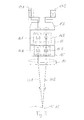
- a stereoscopic surgical microscope in Fig. 1 comprises a front lens 14, a first magnification system 15, a second magnification system 16 and an eyepiece 17 with eyepieces 19A and 19B. These elements are arranged relative to one another in such a way that when viewed through the eyepieces 19A, 19B an enlarged image of an object 18 arranged in front of the front lens 14 is seen. After passing through the common fron lens 14, the observation beam path splits into a beam path 20A for one eye and a beam path 20B for the other eye. The surgical microscope is therefore stereoscopic.
- the front lens 14 has a fixed focal length.
- the working distance between the front lens 14 and the object 18 corresponds to the focal length of the front lens 14 and is thus fixed.
- a beam path 20A, 20B emanating from a point of the object 18 is converted by the front lens 14 into a parallel beam, which then enters the first magnification system 15. The operation of such an afocal magnification system will be explained below.
- the first magnification system 15 includes a left magnification system 15A for the beam path 20A and a right one Magnification system 15B for the beam path 20B.
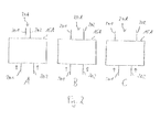
- the operation of a magnification system is based on Fig. 2 explained on the example of the left magnification system 15A.
- a parallel beam 20A entering the magnification system 15A emerges again as a parallel beam 20A.
- Two parallel light beams 201, 202 of the beam 20A have a different distance from each other depending on the set magnification factor of the magnification system 15A when entering the magnification system 15A than at the exit.
- the magnification of the magnification system 15A is set to a value less than 1.
- the distance between the two light beams 201, 202, whose direction is indicated by arrows, is greater on entry into the magnification system 15A than at the exit.
- the magnification factor is 1. In this special case, no magnification takes place, but the light beams 201, 202 have the same distance to each other at the entrance and exit.
- the magnification factor is greater than 1 and the distance between the light beams 201, 202 is greater at the exit than at the entrance.
- the right magnification system 15B works analogously.
- the magnification systems 15A, 15B are coupled together so that the magnification factor is always set in common. It's a one in Fig. 1 not shown control element is provided via which is acted upon mechanically on the optical elements of the magnification system 15 to change their distance from one another.
- the magnification factor of the magnification system 15 can be varied continuously between 1/6 and 6. The as a quotient
- the expansion factor of the magnification system 15 determined by these two values is six.
- the second magnification system 16 adjoining the first magnification system 15 is also stereoscopic (16A, 16B), but has a fixed magnification factor in the range between 1.5 and 3.
- the second magnification system 16 is designed as a module which is optionally introduced into the beam path of the surgical microscope be or can be removed from this.
- the module can be connected for this purpose, for example, pivotally connected to the housing of the surgical microscope. Also possible are embodiments in which the module is completely separated from the surgical microscope. Since the fixed magnification factor of the second magnification system 16 is smaller than the expansion factor of the first magnification system 15, the magnification can be adjusted continuously.
- the magnification area accessible with the second magnification system 16 seamlessly joins the magnification area that is accessible without the second magnification system 16.
- the glimpse 17, which adjoins the second magnification system 16 has a self-magnification of 8.
- the magnification factor of the surgical microscope as a whole is the product of the magnification factors of the magnification systems 15, 16 and the glare 17 and the quotient of 250 mm / focal length front lens.
- the value 250 mm indicates the reference distance.
- the embodiment according to Fig. 3 differs from Fig. 1 in two ways.
- the front lens 14 is a zoom front lens whose focal length varies as needed can be.
- the second magnification system 16 has a variable magnification factor.
- Each of the magnification systems 15, 16 is equipped with an electric motor, not shown, with which the magnification factor can be adjusted.
- the electric motors are coupled with each other so that they can be operated via a common operating element. With the control both electric motors are operated simultaneously, so that the entire magnification range can be continuously passed through from the smallest possible magnification factor to the largest possible magnification factor.
Landscapes
- Health & Medical Sciences (AREA)
- Physics & Mathematics (AREA)
- Surgery (AREA)
- Life Sciences & Earth Sciences (AREA)
- Analytical Chemistry (AREA)
- General Physics & Mathematics (AREA)
- General Health & Medical Sciences (AREA)
- Optics & Photonics (AREA)
- Chemical & Material Sciences (AREA)
- Biomedical Technology (AREA)
- Pathology (AREA)
- Engineering & Computer Science (AREA)
- Oral & Maxillofacial Surgery (AREA)
- Heart & Thoracic Surgery (AREA)
- Medical Informatics (AREA)
- Molecular Biology (AREA)
- Animal Behavior & Ethology (AREA)
- Nuclear Medicine, Radiotherapy & Molecular Imaging (AREA)
- Public Health (AREA)
- Veterinary Medicine (AREA)
- Microscoopes, Condenser (AREA)
Priority Applications (1)
| Application Number | Priority Date | Filing Date | Title |
|---|---|---|---|
| EP11156350A EP2495597A1 (fr) | 2011-03-01 | 2011-03-01 | Microscope d'opération doté de deux systèmes d'agrandissement |
Applications Claiming Priority (1)
| Application Number | Priority Date | Filing Date | Title |
|---|---|---|---|
| EP11156350A EP2495597A1 (fr) | 2011-03-01 | 2011-03-01 | Microscope d'opération doté de deux systèmes d'agrandissement |
Publications (1)
| Publication Number | Publication Date |
|---|---|
| EP2495597A1 true EP2495597A1 (fr) | 2012-09-05 |
Family
ID=44123230
Family Applications (1)
| Application Number | Title | Priority Date | Filing Date |
|---|---|---|---|
| EP11156350A Withdrawn EP2495597A1 (fr) | 2011-03-01 | 2011-03-01 | Microscope d'opération doté de deux systèmes d'agrandissement |
Country Status (1)
| Country | Link |
|---|---|
| EP (1) | EP2495597A1 (fr) |
Cited By (1)
| Publication number | Priority date | Publication date | Assignee | Title |
|---|---|---|---|---|
| EP4561056A3 (fr) * | 2017-04-24 | 2025-08-13 | Alcon Inc. | Caméra et plate-forme de visualisation stéréoscopique |
Citations (4)
| Publication number | Priority date | Publication date | Assignee | Title |
|---|---|---|---|---|
| US5612816A (en) * | 1992-04-28 | 1997-03-18 | Carl-Zeiss-Stiftung | Endoscopic attachment for a stereoscopic viewing system |
| WO2005040866A2 (fr) * | 2003-10-23 | 2005-05-06 | Zeiss Carl Ag | Optique de representation au pouvoir refringent reglable et procede de reglage du pouvoir refringent d'une optique |
| EP1731941A1 (fr) * | 2004-03-31 | 2006-12-13 | Olympus Corporation | Dispositif d"observation et dispositif d"observation a lumiere fluorescente |
| DE102005050171A1 (de) * | 2005-10-19 | 2007-04-26 | Carl Zeiss Surgical Gmbh | Optisches Vergrösserungsänderungssystem zur Bereitstellung von optischen Abbildungsvergrösserungen und Mikroskop mit einem solchen |
-
2011
- 2011-03-01 EP EP11156350A patent/EP2495597A1/fr not_active Withdrawn
Patent Citations (4)
| Publication number | Priority date | Publication date | Assignee | Title |
|---|---|---|---|---|
| US5612816A (en) * | 1992-04-28 | 1997-03-18 | Carl-Zeiss-Stiftung | Endoscopic attachment for a stereoscopic viewing system |
| WO2005040866A2 (fr) * | 2003-10-23 | 2005-05-06 | Zeiss Carl Ag | Optique de representation au pouvoir refringent reglable et procede de reglage du pouvoir refringent d'une optique |
| EP1731941A1 (fr) * | 2004-03-31 | 2006-12-13 | Olympus Corporation | Dispositif d"observation et dispositif d"observation a lumiere fluorescente |
| DE102005050171A1 (de) * | 2005-10-19 | 2007-04-26 | Carl Zeiss Surgical Gmbh | Optisches Vergrösserungsänderungssystem zur Bereitstellung von optischen Abbildungsvergrösserungen und Mikroskop mit einem solchen |
Cited By (1)
| Publication number | Priority date | Publication date | Assignee | Title |
|---|---|---|---|---|
| EP4561056A3 (fr) * | 2017-04-24 | 2025-08-13 | Alcon Inc. | Caméra et plate-forme de visualisation stéréoscopique |
Similar Documents
| Publication | Publication Date | Title |
|---|---|---|
| EP3017334B1 (fr) | Procédé d'acquisition d'images pour un système de microscope et système de microscope associé | |
| DE102006036300B4 (de) | Hochleistungs-Stereomikroskop | |
| EP1326117B2 (fr) | Adaptateur ophtalmoscopique et microscope chirurgical | |
| DE19541420B4 (de) | Stereomikroskop-Anordnung | |
| DE102012223712A1 (de) | Variables abbildungssystem mit objektiv fester brennweite | |
| DE102009012707A1 (de) | Mikroskop mit mehreren optischen Systemen im Abbildungsstrahlengang | |
| WO2006008025A1 (fr) | Dispositif pour usiner un objet au moyen de rayons laser | |
| DE102010002722B4 (de) | Afokales Zoomsystem für ein Mikroskop, Mikroskop mit einem solchen Zoomsystem und Verfahren zum Betreiben eines solchen Zoomsystems | |
| DE102005036486A1 (de) | Optisches Gerät mit erhöhter Schärfentiefe | |
| WO2002027379A2 (fr) | Systeme de redressement de l'image, module d'ophtalmoscopie additionnel et microscope d'operation | |
| EP1424582B1 (fr) | Stéréomicroscope | |
| DE102007029893A1 (de) | Mikroskop mit zentrierter Beleuchtung | |
| DE102010003640A1 (de) | Video-Stereomikroskop | |
| DE102008037074A1 (de) | Verfahren und Einrichtung zur Steuerung von Aperturblenden | |
| DE10255960A1 (de) | Stereomikroskop | |
| DE102007029896B3 (de) | Mikroskop mit zentrierter Beleuchtung | |
| EP1938138A1 (fr) | Systeme de microscopie | |
| CH697533B1 (de) | Beleuchtungs- und Beobachtungseinrichtung. | |
| DE10316242A1 (de) | Umlenksystem für eine Beobachtungseinrichtung sowie Beobachtungseinrichtung | |
| DE102006050846B4 (de) | Stereomikroskop mit Strahlteilereinrichtung | |
| DE202013011877U1 (de) | Mikroskopsystem | |
| DE102012006749B4 (de) | Stereomikroskop | |
| DE102013201632B4 (de) | Mikroskop mit Zoom-Blenden-Kopplung und Zoomsystem für ein Mikroskop | |
| DE102006022592B4 (de) | Mikroskop mit Beleuchtungseinheit | |
| EP2495597A1 (fr) | Microscope d'opération doté de deux systèmes d'agrandissement |
Legal Events
| Date | Code | Title | Description |
|---|---|---|---|
| PUAI | Public reference made under article 153(3) epc to a published international application that has entered the european phase |
Free format text: ORIGINAL CODE: 0009012 |
|
| AK | Designated contracting states |
Kind code of ref document: A1 Designated state(s): AL AT BE BG CH CY CZ DE DK EE ES FI FR GB GR HR HU IE IS IT LI LT LU LV MC MK MT NL NO PL PT RO RS SE SI SK SM TR |
|
| AX | Request for extension of the european patent |
Extension state: BA ME |
|
| STAA | Information on the status of an ep patent application or granted ep patent |
Free format text: STATUS: THE APPLICATION IS DEEMED TO BE WITHDRAWN |
|
| 18D | Application deemed to be withdrawn |
Effective date: 20130306 |