EP2532736A1 - Substitut de tissu vivant mis au point - Google Patents
Substitut de tissu vivant mis au point Download PDFInfo
- Publication number
- EP2532736A1 EP2532736A1 EP11169176A EP11169176A EP2532736A1 EP 2532736 A1 EP2532736 A1 EP 2532736A1 EP 11169176 A EP11169176 A EP 11169176A EP 11169176 A EP11169176 A EP 11169176A EP 2532736 A1 EP2532736 A1 EP 2532736A1
- Authority
- EP
- European Patent Office
- Prior art keywords
- cells
- collagen
- tissue
- cell
- macromolecules
- Prior art date
- Legal status (The legal status is an assumption and is not a legal conclusion. Google has not performed a legal analysis and makes no representation as to the accuracy of the status listed.)
- Withdrawn
Links
- 238000004519 manufacturing process Methods 0.000 claims abstract description 19
- 210000001519 tissue Anatomy 0.000 claims description 61
- 210000004027 cell Anatomy 0.000 claims description 58
- 210000002966 serum Anatomy 0.000 claims description 31
- 229920002521 macromolecule Polymers 0.000 claims description 28
- 210000002950 fibroblast Anatomy 0.000 claims description 26
- 108091003079 Bovine Serum Albumin Proteins 0.000 claims description 21
- 229920001525 carrageenan Polymers 0.000 claims description 21
- 235000010418 carrageenan Nutrition 0.000 claims description 21
- 229940113118 carrageenan Drugs 0.000 claims description 21
- 239000000679 carrageenan Substances 0.000 claims description 21
- 238000000034 method Methods 0.000 claims description 21
- UHVMMEOXYDMDKI-JKYCWFKZSA-L zinc;1-(5-cyanopyridin-2-yl)-3-[(1s,2s)-2-(6-fluoro-2-hydroxy-3-propanoylphenyl)cyclopropyl]urea;diacetate Chemical compound [Zn+2].CC([O-])=O.CC([O-])=O.CCC(=O)C1=CC=C(F)C([C@H]2[C@H](C2)NC(=O)NC=2N=CC(=CC=2)C#N)=C1O UHVMMEOXYDMDKI-JKYCWFKZSA-L 0.000 claims description 21
- 229920002307 Dextran Polymers 0.000 claims description 19
- 239000012091 fetal bovine serum Substances 0.000 claims description 19
- CIWBSHSKHKDKBQ-JLAZNSOCSA-N Ascorbic acid Chemical compound OC[C@H](O)[C@H]1OC(=O)C(O)=C1O CIWBSHSKHKDKBQ-JLAZNSOCSA-N 0.000 claims description 16
- QAOWNCQODCNURD-UHFFFAOYSA-L Sulfate Chemical compound [O-]S([O-])(=O)=O QAOWNCQODCNURD-UHFFFAOYSA-L 0.000 claims description 15
- 229910021653 sulphate ion Inorganic materials 0.000 claims description 15
- 210000000329 smooth muscle myocyte Anatomy 0.000 claims description 12
- 239000011159 matrix material Substances 0.000 claims description 11
- 235000010323 ascorbic acid Nutrition 0.000 claims description 8
- 239000011668 ascorbic acid Substances 0.000 claims description 8
- 229960005070 ascorbic acid Drugs 0.000 claims description 8
- 239000001963 growth medium Substances 0.000 claims description 8
- 210000004072 lung Anatomy 0.000 claims description 8
- 230000002500 effect on skin Effects 0.000 claims description 3
- 210000001644 umbilical artery Anatomy 0.000 claims description 3
- 229910019142 PO4 Inorganic materials 0.000 claims description 2
- JXLHNMVSKXFWAO-UHFFFAOYSA-N azane;7-fluoro-2,1,3-benzoxadiazole-4-sulfonic acid Chemical compound N.OS(=O)(=O)C1=CC=C(F)C2=NON=C12 JXLHNMVSKXFWAO-UHFFFAOYSA-N 0.000 claims description 2
- 238000012258 culturing Methods 0.000 claims description 2
- NBIIXXVUZAFLBC-UHFFFAOYSA-K phosphate Chemical compound [O-]P([O-])([O-])=O NBIIXXVUZAFLBC-UHFFFAOYSA-K 0.000 claims description 2
- 239000010452 phosphate Substances 0.000 claims description 2
- 230000017423 tissue regeneration Effects 0.000 abstract description 4
- 102000008186 Collagen Human genes 0.000 description 43
- 108010035532 Collagen Proteins 0.000 description 43
- 229920001436 collagen Polymers 0.000 description 43
- 230000008021 deposition Effects 0.000 description 38
- 102000010834 Extracellular Matrix Proteins Human genes 0.000 description 27
- 108010037362 Extracellular Matrix Proteins Proteins 0.000 description 27
- 210000002744 extracellular matrix Anatomy 0.000 description 27
- 102000012422 Collagen Type I Human genes 0.000 description 18
- 108010022452 Collagen Type I Proteins 0.000 description 18
- 229940096422 collagen type i Drugs 0.000 description 14
- 102000016359 Fibronectins Human genes 0.000 description 10
- 108010067306 Fibronectins Proteins 0.000 description 10
- 241001465754 Metazoa Species 0.000 description 9
- 230000037319 collagen production Effects 0.000 description 9
- 239000010410 layer Substances 0.000 description 9
- 239000000047 product Substances 0.000 description 8
- 238000002415 sodium dodecyl sulfate polyacrylamide gel electrophoresis Methods 0.000 description 8
- 238000010217 densitometric analysis Methods 0.000 description 7
- 238000002296 dynamic light scattering Methods 0.000 description 7
- 238000011156 evaluation Methods 0.000 description 7
- 238000003365 immunocytochemistry Methods 0.000 description 7
- PLXBWHJQWKZRKG-UHFFFAOYSA-N Resazurin Chemical compound C1=CC(=O)C=C2OC3=CC(O)=CC=C3[N+]([O-])=C21 PLXBWHJQWKZRKG-UHFFFAOYSA-N 0.000 description 6
- 238000000338 in vitro Methods 0.000 description 6
- 238000001727 in vivo Methods 0.000 description 6
- 230000002503 metabolic effect Effects 0.000 description 6
- 239000012981 Hank's balanced salt solution Substances 0.000 description 5
- 108010050808 Procollagen Proteins 0.000 description 5
- 238000004458 analytical method Methods 0.000 description 5
- 238000004113 cell culture Methods 0.000 description 5
- 230000000694 effects Effects 0.000 description 5
- 230000006870 function Effects 0.000 description 5
- 238000005259 measurement Methods 0.000 description 5
- 239000002609 medium Substances 0.000 description 5
- 230000007935 neutral effect Effects 0.000 description 5
- 108090000623 proteins and genes Proteins 0.000 description 5
- 102000004169 proteins and genes Human genes 0.000 description 5
- 210000003491 skin Anatomy 0.000 description 5
- 238000006243 chemical reaction Methods 0.000 description 4
- 230000000295 complement effect Effects 0.000 description 4
- 210000004087 cornea Anatomy 0.000 description 4
- LOKCTEFSRHRXRJ-UHFFFAOYSA-I dipotassium trisodium dihydrogen phosphate hydrogen phosphate dichloride Chemical compound P(=O)(O)(O)[O-].[K+].P(=O)(O)([O-])[O-].[Na+].[Na+].[Cl-].[K+].[Cl-].[Na+] LOKCTEFSRHRXRJ-UHFFFAOYSA-I 0.000 description 4
- 201000010099 disease Diseases 0.000 description 4
- 208000037265 diseases, disorders, signs and symptoms Diseases 0.000 description 4
- 210000001723 extracellular space Anatomy 0.000 description 4
- 210000005260 human cell Anatomy 0.000 description 4
- 238000002135 phase contrast microscopy Methods 0.000 description 4
- 239000002953 phosphate buffered saline Substances 0.000 description 4
- 238000011069 regeneration method Methods 0.000 description 4
- 230000008439 repair process Effects 0.000 description 4
- 230000009469 supplementation Effects 0.000 description 4
- QTBSBXVTEAMEQO-UHFFFAOYSA-N Acetic acid Chemical compound CC(O)=O QTBSBXVTEAMEQO-UHFFFAOYSA-N 0.000 description 3
- 239000006144 Dulbecco’s modified Eagle's medium Substances 0.000 description 3
- HEMHJVSKTPXQMS-UHFFFAOYSA-M Sodium hydroxide Chemical compound [OH-].[Na+] HEMHJVSKTPXQMS-UHFFFAOYSA-M 0.000 description 3
- 230000015572 biosynthetic process Effects 0.000 description 3
- 230000005574 cross-species transmission Effects 0.000 description 3
- 239000003814 drug Substances 0.000 description 3
- 238000005516 engineering process Methods 0.000 description 3
- 239000003102 growth factor Substances 0.000 description 3
- 230000028993 immune response Effects 0.000 description 3
- 239000000463 material Substances 0.000 description 3
- 230000008569 process Effects 0.000 description 3
- 230000002797 proteolythic effect Effects 0.000 description 3
- 239000002994 raw material Substances 0.000 description 3
- 230000001172 regenerating effect Effects 0.000 description 3
- 230000008929 regeneration Effects 0.000 description 3
- 239000007787 solid Substances 0.000 description 3
- 229920002994 synthetic fiber Polymers 0.000 description 3
- FWBHETKCLVMNFS-UHFFFAOYSA-N 4',6-Diamino-2-phenylindol Chemical compound C1=CC(C(=N)N)=CC=C1C1=CC2=CC=C(C(N)=N)C=C2N1 FWBHETKCLVMNFS-UHFFFAOYSA-N 0.000 description 2
- 239000012099 Alexa Fluor family Substances 0.000 description 2
- BWGVNKXGVNDBDI-UHFFFAOYSA-N Fibrin monomer Chemical compound CNC(=O)CNC(=O)CN BWGVNKXGVNDBDI-UHFFFAOYSA-N 0.000 description 2
- WQZGKKKJIJFFOK-GASJEMHNSA-N Glucose Natural products OC[C@H]1OC(O)[C@H](O)[C@@H](O)[C@@H]1O WQZGKKKJIJFFOK-GASJEMHNSA-N 0.000 description 2
- 102000003886 Glycoproteins Human genes 0.000 description 2
- 108090000288 Glycoproteins Proteins 0.000 description 2
- 102000035195 Peptidases Human genes 0.000 description 2
- 108091005804 Peptidases Proteins 0.000 description 2
- 238000013459 approach Methods 0.000 description 2
- 238000003556 assay Methods 0.000 description 2
- 210000002469 basement membrane Anatomy 0.000 description 2
- 230000031018 biological processes and functions Effects 0.000 description 2
- 210000004204 blood vessel Anatomy 0.000 description 2
- 229940096423 bovine collagen type i Drugs 0.000 description 2
- 150000001875 compounds Chemical class 0.000 description 2
- 238000010790 dilution Methods 0.000 description 2
- 239000012895 dilution Substances 0.000 description 2
- 239000006185 dispersion Substances 0.000 description 2
- 230000002255 enzymatic effect Effects 0.000 description 2
- 239000000499 gel Substances 0.000 description 2
- 239000008103 glucose Substances 0.000 description 2
- 238000003306 harvesting Methods 0.000 description 2
- 238000002513 implantation Methods 0.000 description 2
- 230000005541 medical transmission Effects 0.000 description 2
- 239000000203 mixture Substances 0.000 description 2
- 238000010899 nucleation Methods 0.000 description 2
- 108090000765 processed proteins & peptides Proteins 0.000 description 2
- 235000019833 protease Nutrition 0.000 description 2
- 230000012846 protein folding Effects 0.000 description 2
- 230000009467 reduction Effects 0.000 description 2
- 210000001626 skin fibroblast Anatomy 0.000 description 2
- 238000012916 structural analysis Methods 0.000 description 2
- 239000013589 supplement Substances 0.000 description 2
- 210000002435 tendon Anatomy 0.000 description 2
- DBSABEYSGXPBTA-RXSVEWSESA-N (2r)-2-[(1s)-1,2-dihydroxyethyl]-3,4-dihydroxy-2h-furan-5-one;phosphoric acid Chemical compound OP(O)(O)=O.OC[C@H](O)[C@H]1OC(=O)C(O)=C1O DBSABEYSGXPBTA-RXSVEWSESA-N 0.000 description 1
- BUOYTFVLNZIELF-UHFFFAOYSA-N 2-phenyl-1h-indole-4,6-dicarboximidamide Chemical compound N1C2=CC(C(=N)N)=CC(C(N)=N)=C2C=C1C1=CC=CC=C1 BUOYTFVLNZIELF-UHFFFAOYSA-N 0.000 description 1
- 102100028728 Bone morphogenetic protein 1 Human genes 0.000 description 1
- 108090000654 Bone morphogenetic protein 1 Proteins 0.000 description 1
- 241000283690 Bos taurus Species 0.000 description 1
- 241000283707 Capra Species 0.000 description 1
- 208000020406 Creutzfeldt Jacob disease Diseases 0.000 description 1
- 208000003407 Creutzfeldt-Jakob Syndrome Diseases 0.000 description 1
- 208000010859 Creutzfeldt-Jakob disease Diseases 0.000 description 1
- 101100136092 Drosophila melanogaster peng gene Proteins 0.000 description 1
- 108090000790 Enzymes Proteins 0.000 description 1
- 102000004190 Enzymes Human genes 0.000 description 1
- 239000007755 F10 Nutrient Mixture Substances 0.000 description 1
- 102000009123 Fibrin Human genes 0.000 description 1
- 108010073385 Fibrin Proteins 0.000 description 1
- 229920001917 Ficoll Polymers 0.000 description 1
- 241000287828 Gallus gallus Species 0.000 description 1
- 108010010803 Gelatin Proteins 0.000 description 1
- 101000891579 Homo sapiens Microtubule-associated protein tau Proteins 0.000 description 1
- ZDXPYRJPNDTMRX-VKHMYHEASA-N L-glutamine Chemical compound OC(=O)[C@@H](N)CCC(N)=O ZDXPYRJPNDTMRX-VKHMYHEASA-N 0.000 description 1
- 229930182816 L-glutamine Natural products 0.000 description 1
- 241000283973 Oryctolagus cuniculus Species 0.000 description 1
- 229930040373 Paraformaldehyde Natural products 0.000 description 1
- 102000057297 Pepsin A Human genes 0.000 description 1
- 108090000284 Pepsin A Proteins 0.000 description 1
- 102000029797 Prion Human genes 0.000 description 1
- 108091000054 Prion Proteins 0.000 description 1
- 108010003894 Protein-Lysine 6-Oxidase Proteins 0.000 description 1
- 102100026858 Protein-lysine 6-oxidase Human genes 0.000 description 1
- 102000016611 Proteoglycans Human genes 0.000 description 1
- 108010067787 Proteoglycans Proteins 0.000 description 1
- 239000012980 RPMI-1640 medium Substances 0.000 description 1
- 240000004808 Saccharomyces cerevisiae Species 0.000 description 1
- 101710172711 Structural protein Proteins 0.000 description 1
- CZMRCDWAGMRECN-UGDNZRGBSA-N Sucrose Chemical compound O[C@H]1[C@H](O)[C@@H](CO)O[C@@]1(CO)O[C@@H]1[C@H](O)[C@@H](O)[C@H](O)[C@@H](CO)O1 CZMRCDWAGMRECN-UGDNZRGBSA-N 0.000 description 1
- 229930006000 Sucrose Natural products 0.000 description 1
- 101150057615 Syn gene Proteins 0.000 description 1
- 102000004142 Trypsin Human genes 0.000 description 1
- 108090000631 Trypsin Proteins 0.000 description 1
- 208000018756 Variant Creutzfeldt-Jakob disease Diseases 0.000 description 1
- 238000002835 absorbance Methods 0.000 description 1
- 239000000853 adhesive Substances 0.000 description 1
- 230000001070 adhesive effect Effects 0.000 description 1
- 230000003281 allosteric effect Effects 0.000 description 1
- 238000012801 analytical assay Methods 0.000 description 1
- 238000000429 assembly Methods 0.000 description 1
- 230000000712 assembly Effects 0.000 description 1
- 238000004630 atomic force microscopy Methods 0.000 description 1
- 230000008901 benefit Effects 0.000 description 1
- WQZGKKKJIJFFOK-VFUOTHLCSA-N beta-D-glucose Chemical compound OC[C@H]1O[C@@H](O)[C@H](O)[C@@H](O)[C@@H]1O WQZGKKKJIJFFOK-VFUOTHLCSA-N 0.000 description 1
- 230000008827 biological function Effects 0.000 description 1
- 239000012620 biological material Substances 0.000 description 1
- 230000005540 biological transmission Effects 0.000 description 1
- 229920001222 biopolymer Polymers 0.000 description 1
- 210000004369 blood Anatomy 0.000 description 1
- 239000008280 blood Substances 0.000 description 1
- 210000001124 body fluid Anatomy 0.000 description 1
- 239000010839 body fluid Substances 0.000 description 1
- 230000010478 bone regeneration Effects 0.000 description 1
- 229940098773 bovine serum albumin Drugs 0.000 description 1
- 208000005881 bovine spongiform encephalopathy Diseases 0.000 description 1
- 239000006227 byproduct Substances 0.000 description 1
- 239000002775 capsule Substances 0.000 description 1
- 230000000747 cardiac effect Effects 0.000 description 1
- 206010061592 cardiac fibrillation Diseases 0.000 description 1
- 210000000845 cartilage Anatomy 0.000 description 1
- 230000015556 catabolic process Effects 0.000 description 1
- 230000010261 cell growth Effects 0.000 description 1
- 230000012292 cell migration Effects 0.000 description 1
- 210000003855 cell nucleus Anatomy 0.000 description 1
- 230000003833 cell viability Effects 0.000 description 1
- 230000019522 cellular metabolic process Effects 0.000 description 1
- 239000003795 chemical substances by application Substances 0.000 description 1
- 239000011248 coating agent Substances 0.000 description 1
- 238000000576 coating method Methods 0.000 description 1
- 230000036570 collagen biosynthesis Effects 0.000 description 1
- 239000000501 collagen implant Substances 0.000 description 1
- 210000002808 connective tissue Anatomy 0.000 description 1
- 239000000470 constituent Substances 0.000 description 1
- 238000004132 cross linking Methods 0.000 description 1
- 210000004748 cultured cell Anatomy 0.000 description 1
- 239000007857 degradation product Substances 0.000 description 1
- 238000006731 degradation reaction Methods 0.000 description 1
- 230000002939 deleterious effect Effects 0.000 description 1
- 238000009826 distribution Methods 0.000 description 1
- 238000012377 drug delivery Methods 0.000 description 1
- 229940088598 enzyme Drugs 0.000 description 1
- 238000002474 experimental method Methods 0.000 description 1
- 238000000605 extraction Methods 0.000 description 1
- 102000013373 fibrillar collagen Human genes 0.000 description 1
- 108060002894 fibrillar collagen Proteins 0.000 description 1
- 230000002600 fibrillogenic effect Effects 0.000 description 1
- 229950003499 fibrin Drugs 0.000 description 1
- 239000012634 fragment Substances 0.000 description 1
- 210000001156 gastric mucosa Anatomy 0.000 description 1
- 239000008273 gelatin Substances 0.000 description 1
- 229920000159 gelatin Polymers 0.000 description 1
- 235000019322 gelatine Nutrition 0.000 description 1
- 235000011852 gelatine desserts Nutrition 0.000 description 1
- 238000001476 gene delivery Methods 0.000 description 1
- 239000003292 glue Substances 0.000 description 1
- 102000057063 human MAPT Human genes 0.000 description 1
- 230000001969 hypertrophic effect Effects 0.000 description 1
- 230000005847 immunogenicity Effects 0.000 description 1
- 238000011534 incubation Methods 0.000 description 1
- 208000015181 infectious disease Diseases 0.000 description 1
- 230000003993 interaction Effects 0.000 description 1
- 239000003446 ligand Substances 0.000 description 1
- 230000004807 localization Effects 0.000 description 1
- 230000014759 maintenance of location Effects 0.000 description 1
- 210000004962 mammalian cell Anatomy 0.000 description 1
- 210000003864 metabolising cell Anatomy 0.000 description 1
- 238000000386 microscopy Methods 0.000 description 1
- 239000007758 minimum essential medium Substances 0.000 description 1
- 230000004048 modification Effects 0.000 description 1
- 238000012986 modification Methods 0.000 description 1
- 238000004264 monolayer culture Methods 0.000 description 1
- 239000000178 monomer Substances 0.000 description 1
- 239000012120 mounting media Substances 0.000 description 1
- 210000002200 mouth mucosa Anatomy 0.000 description 1
- 210000005036 nerve Anatomy 0.000 description 1
- 238000006386 neutralization reaction Methods 0.000 description 1
- 230000006911 nucleation Effects 0.000 description 1
- 235000015097 nutrients Nutrition 0.000 description 1
- 210000000056 organ Anatomy 0.000 description 1
- 229920002866 paraformaldehyde Polymers 0.000 description 1
- 230000007170 pathology Effects 0.000 description 1
- 230000007310 pathophysiology Effects 0.000 description 1
- 230000037361 pathway Effects 0.000 description 1
- 229940111202 pepsin Drugs 0.000 description 1
- 230000007030 peptide scission Effects 0.000 description 1
- 230000003239 periodontal effect Effects 0.000 description 1
- 239000002861 polymer material Substances 0.000 description 1
- 230000004481 post-translational protein modification Effects 0.000 description 1
- 238000000746 purification Methods 0.000 description 1
- 238000009790 rate-determining step (RDS) Methods 0.000 description 1
- 231100000241 scar Toxicity 0.000 description 1
- 230000028327 secretion Effects 0.000 description 1
- 238000001338 self-assembly Methods 0.000 description 1
- 230000008477 smooth muscle tissue growth Effects 0.000 description 1
- 230000002269 spontaneous effect Effects 0.000 description 1
- 210000000130 stem cell Anatomy 0.000 description 1
- 229960005322 streptomycin Drugs 0.000 description 1
- 239000005720 sucrose Substances 0.000 description 1
- 238000003786 synthesis reaction Methods 0.000 description 1
- 229920000208 temperature-responsive polymer Polymers 0.000 description 1
- 238000002560 therapeutic procedure Methods 0.000 description 1
- 238000002054 transplantation Methods 0.000 description 1
- 239000012588 trypsin Substances 0.000 description 1
- 108020005087 unfolded proteins Proteins 0.000 description 1
- 241000701447 unidentified baculovirus Species 0.000 description 1
- 210000002700 urine Anatomy 0.000 description 1
- 230000035899 viability Effects 0.000 description 1
- XLYOFNOQVPJJNP-UHFFFAOYSA-N water Substances O XLYOFNOQVPJJNP-UHFFFAOYSA-N 0.000 description 1
Images


Classifications
-
- C—CHEMISTRY; METALLURGY
- C12—BIOCHEMISTRY; BEER; SPIRITS; WINE; VINEGAR; MICROBIOLOGY; ENZYMOLOGY; MUTATION OR GENETIC ENGINEERING
- C12N—MICROORGANISMS OR ENZYMES; COMPOSITIONS THEREOF; PROPAGATING, PRESERVING, OR MAINTAINING MICROORGANISMS; MUTATION OR GENETIC ENGINEERING; CULTURE MEDIA
- C12N5/00—Undifferentiated human, animal or plant cells, e.g. cell lines; Tissues; Cultivation or maintenance thereof; Culture media therefor
- C12N5/06—Animal cells or tissues; Human cells or tissues
- C12N5/0602—Vertebrate cells
- C12N5/0603—Embryonic cells ; Embryoid bodies
- C12N5/0605—Cells from extra-embryonic tissues, e.g. placenta, amnion, yolk sac, Wharton's jelly
-
- C—CHEMISTRY; METALLURGY
- C12—BIOCHEMISTRY; BEER; SPIRITS; WINE; VINEGAR; MICROBIOLOGY; ENZYMOLOGY; MUTATION OR GENETIC ENGINEERING
- C12N—MICROORGANISMS OR ENZYMES; COMPOSITIONS THEREOF; PROPAGATING, PRESERVING, OR MAINTAINING MICROORGANISMS; MUTATION OR GENETIC ENGINEERING; CULTURE MEDIA
- C12N5/00—Undifferentiated human, animal or plant cells, e.g. cell lines; Tissues; Cultivation or maintenance thereof; Culture media therefor
- C12N5/0018—Culture media for cell or tissue culture
-
- C—CHEMISTRY; METALLURGY
- C12—BIOCHEMISTRY; BEER; SPIRITS; WINE; VINEGAR; MICROBIOLOGY; ENZYMOLOGY; MUTATION OR GENETIC ENGINEERING
- C12N—MICROORGANISMS OR ENZYMES; COMPOSITIONS THEREOF; PROPAGATING, PRESERVING, OR MAINTAINING MICROORGANISMS; MUTATION OR GENETIC ENGINEERING; CULTURE MEDIA
- C12N5/00—Undifferentiated human, animal or plant cells, e.g. cell lines; Tissues; Cultivation or maintenance thereof; Culture media therefor
- C12N5/06—Animal cells or tissues; Human cells or tissues
- C12N5/0602—Vertebrate cells
- C12N5/0652—Cells of skeletal and connective tissues; Mesenchyme
- C12N5/0656—Adult fibroblasts
-
- C—CHEMISTRY; METALLURGY
- C12—BIOCHEMISTRY; BEER; SPIRITS; WINE; VINEGAR; MICROBIOLOGY; ENZYMOLOGY; MUTATION OR GENETIC ENGINEERING
- C12N—MICROORGANISMS OR ENZYMES; COMPOSITIONS THEREOF; PROPAGATING, PRESERVING, OR MAINTAINING MICROORGANISMS; MUTATION OR GENETIC ENGINEERING; CULTURE MEDIA
- C12N5/00—Undifferentiated human, animal or plant cells, e.g. cell lines; Tissues; Cultivation or maintenance thereof; Culture media therefor
- C12N5/06—Animal cells or tissues; Human cells or tissues
- C12N5/0602—Vertebrate cells
- C12N5/0688—Cells from the lungs or the respiratory tract
-
- C—CHEMISTRY; METALLURGY
- C12—BIOCHEMISTRY; BEER; SPIRITS; WINE; VINEGAR; MICROBIOLOGY; ENZYMOLOGY; MUTATION OR GENETIC ENGINEERING
- C12N—MICROORGANISMS OR ENZYMES; COMPOSITIONS THEREOF; PROPAGATING, PRESERVING, OR MAINTAINING MICROORGANISMS; MUTATION OR GENETIC ENGINEERING; CULTURE MEDIA
- C12N2500/00—Specific components of cell culture medium
- C12N2500/30—Organic components
- C12N2500/34—Sugars
-
- C—CHEMISTRY; METALLURGY
- C12—BIOCHEMISTRY; BEER; SPIRITS; WINE; VINEGAR; MICROBIOLOGY; ENZYMOLOGY; MUTATION OR GENETIC ENGINEERING
- C12N—MICROORGANISMS OR ENZYMES; COMPOSITIONS THEREOF; PROPAGATING, PRESERVING, OR MAINTAINING MICROORGANISMS; MUTATION OR GENETIC ENGINEERING; CULTURE MEDIA
- C12N2500/00—Specific components of cell culture medium
- C12N2500/30—Organic components
- C12N2500/38—Vitamins
-
- C—CHEMISTRY; METALLURGY
- C12—BIOCHEMISTRY; BEER; SPIRITS; WINE; VINEGAR; MICROBIOLOGY; ENZYMOLOGY; MUTATION OR GENETIC ENGINEERING
- C12N—MICROORGANISMS OR ENZYMES; COMPOSITIONS THEREOF; PROPAGATING, PRESERVING, OR MAINTAINING MICROORGANISMS; MUTATION OR GENETIC ENGINEERING; CULTURE MEDIA
- C12N2500/00—Specific components of cell culture medium
- C12N2500/50—Soluble polymers, e.g. polyethyleneglycol [PEG]
Definitions
- the present invention relates generally to the field of tissue engineering and in particular to the production of tissue films, sheets or cell matrices, which can be used as a living tissue substitute or an artificial tissue construct in tissue repair or replacement.
- tissue engineering is to repair or replace tissues and organs with artificial tissue constructs. Scaffolds are typically used for this process, giving mechanical support, and assisting in cell migration and attachment, cell retention and delivery at the site of repair.
- the scaffold thus mimics the natural matrix found in the body.
- films or layers of tissue itself may be suitable for the repair or transplant. The cells in such films should thus be as similar as possible to those produced naturally by the body. It is thus important that cells grown into these tissue layers or films are properly aligned, and metabolise to produce factors that the tissue they are destined to replace would produce.
- the extracellular matrix is a complex variety of glycoproteins and proteoglycans, which provides tissue integrity, acts as a native scaffold for cell attachment and interaction and acts as a reservoir for growth factors.
- collagen makes up the bulk of the extracellular matrix, where it functions as a structural protein as well as a binding partner for glycans that store growth factors.
- Collagen is a family of glycoproteins, the most abundant being type I found in skin tendon and capsules, type IV found in all basement membranes and type VII found in the basement membrane of skin, oral mucosa and cornea. These collagen assemblies differ depending on the tissue location and function.
- the deposition of a collagen matrix depends on the conversion of denovo synthesised pro-collagen to collagen in the extracellular space immediately before its release into the space. This limiting step for collagen matrix deposition is very slow in vitro, both in monolayer cultures and in three dimensional scaffolds.
- tissue grafts autografts, allografts or xenografts
- tissue grafts are considered to be the 'gold standards'.
- autografts autografts, allografts or xenografts
- tissue engineering was pioneered as the only viable alternative to the transplantation crisis.
- degradable and non-degradable synthetic materials have been evaluated over the years [2-10].
- non-degradable synthetic materials may become harmful due to mechanical impingement or infection and require a second operation, whilst the degradation products of biodegradable synthetic materials could be deleterious to the surrounding cells and tissues.
- Natural biomaterials such as collagen [15-18], gelatin [19, 20] and fibrin [21] have been used as raw materials for scaffold fabrication with promising early results.
- the use of collagen as a raw material for scaffold fabrication has been advocated because, as a natural occurring biopolymer that constitutes approximately one third of the total body proteins, it is perceived by the body as a normal constituent rather than foreign matter.
- collagen remains an animal derived by-product and its use in clinical applications can be limited due to concerns of inter-species transmission of disease, especially for collagen extracted from bovine tissues (e.g. Bovine Spongiform Encephalopathy and Creutzfeldt Jakob Disease). In fact, 2-3% of patients have an immune response to collagen implants using collagen derived from land-based animals. For this reason, human recombinant collagen has been investigated for scaffold fabrication.
- bovine tissues e.g. Bovine Spongiform Encephalopathy and Creutzfeldt Jakob Disease
- procollagen expression levels have been low (15mg/ml for mammalian cell culture; 15mg/ml in yeast; and 60mg/ml in baculovirus) which was prohibited commercialisation and clinical applications.
- procollagen expression levels have been low (15mg/ml for mammalian cell culture; 15mg/ml in yeast; and 60mg/ml in baculovirus) which was prohibited commercialisation and clinical applications.
- recombinant collagens can be expressed in a thermo-stable triple helical form, they lack specific domains otherwise present in native fibrillar collagens, which can compromise their biological function and further reduce their use. In light of this, it has been predicted that companies developing new implantable products are more likely to focus on human collagen products, rather than on products utilising animal-sourced collagen.
- Cell-based injectable systems and cell-sheets derived from autologous primary cell isolates; from established cell lines; and from a variety of stem cells have been used for numerous clinical targets, including cornea, skin, blood vessel, cartilage, lung, cardiac patch, oesophagus and periodontal applications.
- ECM extracellular matrix
- typical proteolytic harvest by trypsin digests both deposited ECM and cell-to-cell junctions.
- culture dishes covered with a temperature-responsive polymer allow harvesting of intact cell sheets along with their deposited ECM, by simple temperature reduction.
- this technology has still not taken off primarily due to the substantial long period of time required to culture the cells and develop an implantable cell-sheet.
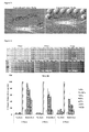
- Table 1 Concentration of solids in conventional cell culture system MEDIA COMPANY SOLID CONCENTRATION Minimum Essential Medium Invitrogen, Cat.No: 10370021, 10370039,10370047,10370054,10370070 11.52g/l F 12 Nutrient Mixture Invitrogen, Cat. No: 21765029 11.87g/l RPMI 1640 Medium Invitrogen, Cat. No. 11835030, 11835055,11835063, 11835071 12.39g/l Ham F10 Nutrient Mixture Invitrogen, Cat. No. 22390017, 22390025 16.55g/l DMEM: F12 Medium ATCC, Cat. No. 30-2006 16.78g/l DMEM High Glucose (4.5g/) and L-glutamine Invitrogen, Cat. No.41965039, 41965047 17.22g/l
- Table 2 Macromolecules that have been used to-date as crowding agents CROWDER USED RESULTS REFERENCE Sucrose and glucose (monomers for dextran and Ineffective Zimmermann and Harrison, 1987, PNAS, FicollTM), PEG 0.2 Kda FicollTM 70Kda, Dextran T70Kda, PEG 8Kda, PEG 35KDa MMC significantly increased enzymatic activity 84: 1871-1875 FicollTM 70KDa, Dextran 70KDa Faster protein folding rates van den Berg et al., 1999, EMBO Journal, 18(24): 6927-6933 PEG 3.5Kda, FicollTM 70KDa MMC dramatically increase fibrillation of unfolded proteins Munishkina et al., 2008 Biochemistry, 47(34): 8993-9006 FicollTM 70Kda, Dextran 70KDa MMC dramatically accelerated the nucleation step of fibril formation of human Tau fragment & prion
- the present inventors have surprisingly found that by using poly-dispersed macromolecular crowders, that cell metabolism and extracellular matrix production can be enhanced to such a level that significant quantities of tissue substitute films are produced after as little as 48 hours.
- tissue substitutes or artificial tissue constructs which is rapid.
- These tissue substitutes or constructs can take the form of tissue layers or sheets of cells.
- the object of the invention is to provide a method of producing commercially viable quantities of tissue substitute films within a period of 2-5 days.
- the substitute can be produced within in about 48 hours.
- Another object is to address the shortfalls of animal-extracted and recombinant molecules and to provide a new method for producing tissue substitutes without the addition of any animal compounds.
- Such a product would harbour no risks for interspecies transmission of disease (as compared to animal extracted collagen) and also is fully biologically active (as opposed to recombinant collagen) since it is produced naturally from cells.
- An object is to employ the principles of macromolecular crowding on human cells and produce live human cell sheets for tissue engineering and regenerative medicine applications.
- This system will facilitate production of host-specific cell-based scaffolds from the patient's own cells and as such will avoid immune rejection problems from implantation of materials from another subject.
- this system can also be used to produce host specific proteins (e.g. collagen) that can be used for tissue engineering applications.
- this system can be used to accelerate in vitro biological processes (e.g. enzyme activity; degradation of proteins, etc).
- the tissues so produced may be used for tissue engineering applications.
- Such applications include: Tendon regeneration, Bone regeneration, Nerve regeneration, Cornea regeneration, Drug delivery, Gene delivery, coating of medical devices to avoid immune response, and tissue glues/adhesives.
- Such products will replace products that are based on animal extracted molecules and products that are based on human recombinant molecules.
- a method for the production of a tissue substitute comprising culturing cells in the presence of macromolecular crowders, wherein the macromolecular crowders are large poly-dispersed macromolecules.
- the macro molecules may be negatively charged macromolecules.
- the large poly-dispersed macromolecules may be selected from the group comprising carrageenan, dextran sulphate and polysodium-1-styrene sulphonate. Particularly preferred is carrageenan.
- the cells may be selected from lung fibroblasts, dermal fibroblasts and human umbilical arteries smooth muscle cells.
- the cells may be cultured in the presence of culture medium supplemented with fetal bovine serum, human serum, ascorbic acid phosphate, or a combination thereof.
- the human serum may be used at 0.5% to 1% volume to volume.
- the invention also provides a cell matrix film, tissue sheet or tissue substitute whenever produced by a method as described above.
- poly-dispersed means that the molecules have a broad range of size, shape and mass characteristics, as opposed to molecules which have a uniform size, shape and mass distribution which are mono-dispersed molecules.
- Polymer materials are poly-dispersed if their chain length varies over a wide range of molecular masses. This poly-dispersity can be seen in Figure 11A-E .
- a number of negatively charged macromolecules are poly-dispersed. It is however apparent from the figure that carrageenan is the most poly-dispersed of the molecules tested in this invention, and the inventors hypothesis that this is the reason for the superiority of carrageenan in the method of the present invention.
- This invention enables the production of substantial amounts of cell tissue within only 48h, whilst the current in vitro systems take in excess of 6 weeks to produce the same amount of tissue.
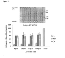
- human serum was used and it surprisingly increased further the tissue and extracellular matrix production as typified by collagen production.
- a number of cell types were screened (e.g. lung fibroblasts, skin fibroblasts, smooth muscle cells) and surprisingly, it was found that smooth muscle cells can be very rapidly produced by the method of the invention. It was also found that poly-dispersed macromolecules occupy more space and facilitate higher collagen production in comparison to mono-dispersed molecules, indicating higher tissue layer production. Low serum concentration is sufficient for high collagen production which makes this invention even more financially viable.
- herein we describe for very first time the rapid production of human tissue substitutes that can be used for any tissue engineering applications.
- Cells were seeded at 50,000 cells/well in 24-well plates and were allowed to attach for 24 hours. After 24 hours the medium was changed with medium containing macromolecular crowders (100 ⁇ g/ml dextran sulphate 500 kDa; 37.5mg/ml FicollTM70 and 25mg/ml FicollTM400; 100 ⁇ g/ml Polysodium-4-Styrenesulfonate, 75 ⁇ g/ml carrageenan and 100 ⁇ l/ml sepharose-CL) and with various percentages of FBS (0%, 0.5%, 1%, 2%, 5%, and 10%). To induce collagen synthesis, fibroblasts were supplemented with 100 ⁇ M L-ascorbic acid phosphate. The HUASMC was supplemented with various concentration of ascorbic acid (0 ⁇ M, 25 ⁇ M, 50 ⁇ M and 100 ⁇ M). In some of the experiment commercially available human serum (Lonza, Belgium) was also used as supplement in place of FBS.
- culture media were collected into separate vials, whereas cell layers were washed twice with Hank's balanced salt solution (HBSS). Both culture medium and washed cell layer were digested with porcine gastric mucosa pepsin in a final concentration of 1mg/ml in 0.5M acetic acid. Samples were incubated at 37°C for 2 hours with gentle shaking followed by neutralization with 0.1 N NaOH.
- HBSS Hank's balanced salt solution
- Fibroblasts were seeded on 4-well Lab-TekTM II chamber slides at 50,000 cells/chamber and after 24 hours of seeding cells were treated with crowders. After 2 days of culture, medium was removed and cell layers were washed with HBSS and fixed with 2% paraformaldehyde at room temparature for 15 min. After several washes in phosphate-buffered saline (PBS), nonspecific sites were blocked with 3% bovine serum albumin in PBS for 30 min. The cells were incubated for 90 min at room temperature simultaneously with Collagen I (Rabbit anti-human): dilution 1:100 and Fibronectin (Mouse anti-human) dilution 1:200 for 90 min.
- Collagen I Rabbit anti-human
- Fibronectin Mae anti-human
- Bound antibodies were visualized using AlexaFluor ® 488 chicken anti-rabbit and AlexaFluor ® 555 goat anti-mouse 1:400 in PBS for 30min. Post-fixation was with 2% PFA for 15 min. Cell nuclei were counterstained with 4,6-diamidino- 2-phenylindole (DAPI) and slides were mounted with Vectashield ® mounting media. Images were captured with an Olympus IX-81 inverted fluorescence microscope (Olympus Corporation, Tokyo, Japan).
- Dynamic light scattering (DLS) measurements of macromolecules were done using Zetasizer Nano ZS90 (Malvern Instruments) at 25°C. Molecules were dissolved in HBSS, pH 7.4 for size (Z-Ave; d.nm) measurements, and for zeta ( ⁇ )- potential measurements, macromolecules were dissolved in water. The measurement of zeta ( ⁇ )-potential and size (Z-Ave. diameter in nano meter) were analysed by the help of Zetasizer software 6.12 (Malvern Instruments).
- alamarBlue ® assay was performed to quantify the influence of various crowders and serum on metabolic activity of the fibroblasts.
- effect of ascorbic acid supplementations (0 ⁇ M, 25 ⁇ M, 50 ⁇ M and 100 ⁇ M) on cell's metabolic activity was also analysed. Briefly, at the end of culture time points, cells were washed with HBSS and then diluted alamarBlue ® was added. After 4 hours of incubation at 37°C, absorbance was measured at 550 and 595nm. Cell viability was expressed in terms of reduction percentage of alamarblue ® .
- Human lung fibroblasts WI-38; American tissue culture collection), human dermal fibroblasts (WS-1; American tissue culture collection), and human umbilical arterial smooth muscle cells (huasmc; clonetics, UASMC-human umbilical arterial smooth muscle cells; Lonza cc-2579) were evaluated. Cells from pathophysiologies (eg hypertrophic scar) have not been evaluated since disease transmission may occur. Although we would have expected fibroblasts to produce more collagen, our data indicated that HUASMC deposited higher amounts of collagen-1 when crowded with 100 ⁇ g/ml of DxS and 75 ⁇ g/ml carrageenan.
- the fibronectin deposition was also analysed immunocytochemically to evaluate its pattern of deposition with collagen type I. Immunocytochemistry results further confirmed the enhanced deposition of collagen I and its co-localisation with fibronectin in the presence of macromolecular crowders as shown in the immunocytochemistry figures.
- Phase contrast microscopy revealed that the fibroblasts maintained their spindle-shaped morphology independent of the macromolecular crowder present or the serum concentration up to 6-days in culture.
- AlamarBlue® analysis demonstrated that cell metabolic activity was not affected, independent of the macromolecular crowder present or the serum concentration even up to 6- days in culture (p>0.05).
- the size dispersion by intensity confirms that the negatively charged macromolecules (carrageenan, dextran sulphate and PSS) are highly polydispersed and among them, carrageenan is the most polydispersed.
- FIG 20 shows a preliminary structural analysis by atomic microscopy of the human tissue substitute of the invention.
- Tissue substitute shows a quartered staggered arrangement in which collagen fibres are aligned and similar to those found in native tissue.
- the tissue matrix or substitute of the invention imitates native tissue.
- the invention has shown that the growth of cells in a macro molecular crowded environment using highly polydispersed molecules results in an appropriately aligned and functionally metabolising cell layer.
Landscapes
- Health & Medical Sciences (AREA)
- Engineering & Computer Science (AREA)
- Life Sciences & Earth Sciences (AREA)
- Biomedical Technology (AREA)
- Biotechnology (AREA)
- Zoology (AREA)
- Organic Chemistry (AREA)
- Bioinformatics & Cheminformatics (AREA)
- Genetics & Genomics (AREA)
- Chemical & Material Sciences (AREA)
- Wood Science & Technology (AREA)
- Cell Biology (AREA)
- Microbiology (AREA)
- Biochemistry (AREA)
- General Engineering & Computer Science (AREA)
- General Health & Medical Sciences (AREA)
- Developmental Biology & Embryology (AREA)
- Gynecology & Obstetrics (AREA)
- Reproductive Health (AREA)
- Rheumatology (AREA)
- Pregnancy & Childbirth (AREA)
- Pulmonology (AREA)
- Micro-Organisms Or Cultivation Processes Thereof (AREA)
- Materials For Medical Uses (AREA)
Priority Applications (5)
| Application Number | Priority Date | Filing Date | Title |
|---|---|---|---|
| EP11169176A EP2532736A1 (fr) | 2011-06-08 | 2011-06-08 | Substitut de tissu vivant mis au point |
| PCT/EP2012/060945 WO2012168465A1 (fr) | 2011-06-08 | 2012-06-08 | Substitut de tissu vivant manipulé |
| EP12729432.0A EP2718421B1 (fr) | 2011-06-08 | 2012-06-08 | Substitut de tissu vivant manipulé |
| US14/124,568 US10619133B2 (en) | 2011-06-08 | 2012-06-08 | Engineered living tissue substitute |
| EP18189623.4A EP3453752B1 (fr) | 2011-06-08 | 2012-06-08 | Substitut de tissu vivant manipulé |
Applications Claiming Priority (1)
| Application Number | Priority Date | Filing Date | Title |
|---|---|---|---|
| EP11169176A EP2532736A1 (fr) | 2011-06-08 | 2011-06-08 | Substitut de tissu vivant mis au point |
Publications (1)
| Publication Number | Publication Date |
|---|---|
| EP2532736A1 true EP2532736A1 (fr) | 2012-12-12 |
Family
ID=46354201
Family Applications (3)
| Application Number | Title | Priority Date | Filing Date |
|---|---|---|---|
| EP11169176A Withdrawn EP2532736A1 (fr) | 2011-06-08 | 2011-06-08 | Substitut de tissu vivant mis au point |
| EP12729432.0A Not-in-force EP2718421B1 (fr) | 2011-06-08 | 2012-06-08 | Substitut de tissu vivant manipulé |
| EP18189623.4A Active EP3453752B1 (fr) | 2011-06-08 | 2012-06-08 | Substitut de tissu vivant manipulé |
Family Applications After (2)
| Application Number | Title | Priority Date | Filing Date |
|---|---|---|---|
| EP12729432.0A Not-in-force EP2718421B1 (fr) | 2011-06-08 | 2012-06-08 | Substitut de tissu vivant manipulé |
| EP18189623.4A Active EP3453752B1 (fr) | 2011-06-08 | 2012-06-08 | Substitut de tissu vivant manipulé |
Country Status (3)
| Country | Link |
|---|---|
| US (1) | US10619133B2 (fr) |
| EP (3) | EP2532736A1 (fr) |
| WO (1) | WO2012168465A1 (fr) |
Cited By (2)
| Publication number | Priority date | Publication date | Assignee | Title |
|---|---|---|---|---|
| WO2023285813A1 (fr) * | 2021-07-12 | 2023-01-19 | 3D Bio-Tissues Limited | Milieu de culture cellulaire et compléments pour la production de viande cellulaire |
| WO2023285816A1 (fr) * | 2021-07-12 | 2023-01-19 | 3D Bio-Tissues Limited | Milieu de culture cellulaire et compléments pour la culture de cellules cornéennes et cutanées |
Families Citing this family (5)
| Publication number | Priority date | Publication date | Assignee | Title |
|---|---|---|---|---|
| EP2532736A1 (fr) * | 2011-06-08 | 2012-12-12 | National University of Ireland, Galway | Substitut de tissu vivant mis au point |
| GB2516826B (en) * | 2013-07-23 | 2016-06-22 | Canon Kk | Method, device and computer program for encapsulating partitioned timed media data by creating tracks to be independently encapsulated in at least one media f |
| US20170182221A1 (en) * | 2014-05-05 | 2017-06-29 | National University Of Singapore | Methods Of Producing Tissue-Mimetic Constructs And Uses Thereof |
| WO2020228733A1 (fr) * | 2019-05-16 | 2020-11-19 | The Chinese University Of Hong Kong | Substance de matrice extracellulaire et utilisations associées |
| EP4366794A1 (fr) | 2021-07-09 | 2024-05-15 | National University of Ireland Galway | Développement accéléré de modules tissulaires tridimensionnels fonctionnels |
Citations (3)
| Publication number | Priority date | Publication date | Assignee | Title |
|---|---|---|---|---|
| US20020028192A1 (en) * | 1998-03-24 | 2002-03-07 | S. Dan Dimitrijevich | Non-contracting tissue equivalent |
| US20050089512A1 (en) * | 2001-12-19 | 2005-04-28 | Kordula Schlotmann | Skin/hair equivalent with reconstructed papillae |
| WO2006097701A2 (fr) * | 2005-03-14 | 2006-09-21 | Intercytex Limited | Culture d'equivalents cutanes |
Family Cites Families (2)
| Publication number | Priority date | Publication date | Assignee | Title |
|---|---|---|---|---|
| WO2010027789A2 (fr) * | 2008-08-25 | 2010-03-11 | University Of South Florida | Agent d’exclusion de volume pour renforcer la formation d’une matrice extracellulaire |
| EP2532736A1 (fr) * | 2011-06-08 | 2012-12-12 | National University of Ireland, Galway | Substitut de tissu vivant mis au point |
-
2011
- 2011-06-08 EP EP11169176A patent/EP2532736A1/fr not_active Withdrawn
-
2012
- 2012-06-08 EP EP12729432.0A patent/EP2718421B1/fr not_active Not-in-force
- 2012-06-08 WO PCT/EP2012/060945 patent/WO2012168465A1/fr not_active Ceased
- 2012-06-08 EP EP18189623.4A patent/EP3453752B1/fr active Active
- 2012-06-08 US US14/124,568 patent/US10619133B2/en active Active
Patent Citations (3)
| Publication number | Priority date | Publication date | Assignee | Title |
|---|---|---|---|---|
| US20020028192A1 (en) * | 1998-03-24 | 2002-03-07 | S. Dan Dimitrijevich | Non-contracting tissue equivalent |
| US20050089512A1 (en) * | 2001-12-19 | 2005-04-28 | Kordula Schlotmann | Skin/hair equivalent with reconstructed papillae |
| WO2006097701A2 (fr) * | 2005-03-14 | 2006-09-21 | Intercytex Limited | Culture d'equivalents cutanes |
Non-Patent Citations (26)
| Title |
|---|
| "Dimensional Salmon Fibrin Gels", BIOMATERIALS, vol. 28, no. 12, 2007, pages 2097 - 2108 |
| AHMED, M.R. ET AL.: "Microwave irradiated collagen tubes as a better matrix for peripheral nerve regeneration", BRAIN RESEARCH, vol. 1046, no. 1-2, 2005, pages 55 - 67, XP004914211, DOI: doi:10.1016/j.brainres.2005.03.022 |
| ARCHIBALD, S. ET AL.: "Monkey median nerve repaired by nerve graft or collagen nerve guide tube", J NEUROSCI: PART 2, vol. 15, no. 5, 1995, pages 4109 - 4123 |
| CHEN HAI-MIN ET AL: "lambda-Carrageenan oligosaccharides elicit reactive oxygen species production resulting in mitochondrial-dependent apoptosis in human umbilical vein endothelial cells", INTERNATIONAL JOURNAL OF MOLECULAR MEDICINE, vol. 24, no. 6, December 2009 (2009-12-01), pages 801 - 806, XP002665126, ISSN: 1107-3756 * |
| CHEN, P.-R. ET AL.: "Biocompatibility ofNGF-grafted GTG membranes for peripheral nerve repair using cultured Schwann cells", BIOMATERIALS, vol. 25, no. 25, 2004, pages 5667 - 5673, XP004509950, DOI: doi:10.1016/j.biomaterials.2004.01.052 |
| CHEN, Y.-S. ET AL.: "An in vivo evaluation of a biodegradable genipin- cross-linked gelatin peripheral nerve guide conduit material", BIOMATERIALS, vol. 26, no. 18, 2005, pages 3911 - 3918, XP025280533, DOI: doi:10.1016/j.biomaterials.2004.09.060 |
| CHEW, S.Y. ET AL.: "Aligned Protein-Polymer Composite Fibers Enhance Nerve Regeneration: A Potential Tissue-Engineering Platform", ADV FUNCT MATER, vol. 17, no. 8, 2007, pages 1288 - 1296, XP055068419, DOI: doi:10.1002/adfm.200600441 |
| CLARICE CHEN ET AL: "Applying macromolecular crowding to enhance extracellular matrix deposition and its remodeling in vitro for tissue engineering and cell-based therapies", ADVANCED DRUG DELIVERY REVIEWS, vol. 63, no. 4-5, 1 April 2011 (2011-04-01), pages 277 - 290, XP055013967, ISSN: 0169-409X, DOI: 10.1016/j.addr.2011.03.003 * |
| COLIN, W., R. DONOFF: "Nerve regeneration through collagen tubes", J DENT RES, vol. 63, no. 7, 1984, pages 987 - 993 |
| DEN DUNNEN, W. ET AL.: "A new PLLAIPCL copolymer for nerve regeneration", J MATER SCI: MATER MED, vol. 4, 1993, pages 521 - 525 |
| EVANS, G. ET AL.: "In vivo evaluation of poly(l-lactic acid) porous conduits for peripheral nerve regeneration", BIOMATERIALS, vol. 20, no. 12, 1999, pages 1109 - 1115, XP002977355, DOI: doi:10.1016/S0142-9612(99)00010-1 |
| EVANS, G.R. ET AL.: "Bioactive poly(L-lactic acid) conduits seeded with Schwann cells for peripheral nerve regeneration", BIOMATERIALS, vol. 23, no. 3, 2002, pages 841 - 8, XP004348096, DOI: doi:10.1016/S0142-9612(01)00190-9 |
| EVANS, P. ET AL.: "Cold preserved nerve allografts: changes in basement membrane, viability, immunogenicity, and regeneration", MUSCLE NERVE, vol. 21, no. 11, 1998, pages 1507 - 1522 |
| HARLEY, B.A. ET AL.: "Fabricating tubular scaffolds with a radial pore size gradient by a spinning technique", BIOMATERIALS, vol. 27, no. 6, 2006, pages 866 - 874 |
| HARVE K, VIGNESHWAR R, RAJAGOPALAN R, RAGHUNATH M: "Macromolecular crowding in vitro as means of emulating cellular interiors: when less might be more", PNAS, vol. 105, no. 51, 2008, pages 119 |
| ITOH, S. ET AL.: "Evaluation of cross-linking procedures of collagen tubes used in peripheral nerve repair", BIOMATERIALS, vol. 23, no. 23, 2002, pages 4475 - 4481, XP004377518, DOI: doi:10.1016/S0142-9612(02)00166-7 |
| KIM, Y.-T. ET AL.: "The role of aligned polymer fiber-based constructs in the bridging of long peripheral nerve gaps", BIOMATERIALS, vol. 29, 2008, pages 3117 - 3127 |
| LAREU ET AL: "Collagen matrix deposition is dramatically enhanced in vitro when crowded with charged macromolecules: The biological relevance of the excluded volume effect", FEBS LETTERS, ELSEVIER, AMSTERDAM, NL, vol. 581, no. 14, 31 May 2007 (2007-05-31), pages 2709 - 2714, XP022100064, ISSN: 0014-5793, DOI: 10.1016/J.FEBSLET.2007.05.020 * |
| LAREU R, HARVE K, RAGHUNATH M.: "Emulating a crowded intracellular environment in vitro dramatically improves RT-PCR performance", BIOCHEM BIOPHYS RES COMMUN, vol. 363, no. 1, 2007, pages 171 - 177, XP022265406, DOI: doi:10.1016/j.bbrc.2007.08.156 |
| LAREU RR, ARSIANTI I, SUBRAMHANYA HK, YANXIAN P, RAGHUNATH M.: "In Vitro Enhancement of Collagen Matrix Formation and Crosslinking for Applications in Tissue Engineering: A Preliminary Study", TISSUE ENGINEERING, vol. 13, no. 2, 2007, pages 385 - 391, XP055013970, DOI: doi:10.1089/ten.2006.0224 |
| LAREU RR, SUBRAMHANYA KH, PENG Y, BENNY P, CHEN C, WANG Z, RAJAGOPALAN R, RAGHUNATH M.: "Collagen matrix deposition is dramatically enhanced in vitro when crowded with charged macromolecules: The biological relevance of the excluded volume effect", FEBS LETTERS, vol. 581, 2007, pages 2709 - 2714, XP022100064, DOI: doi:10.1016/j.febslet.2007.05.020 |
| LI, X.-K. ET AL.: "Characteristics of PLGA-gelatin complex as potential artificial nerve scaffold", COLLOIDS AND SURFACES B: BIOINTERFACES, vol. 57, no. 2, 2007, pages 198 - 203 |
| MADISON, R. ET AL.: "Increased rate ofperipheral nerve regeneration using bioresorbable nerve guides and a laminin-containing gel", EXP NEUROL, vol. 88, no. 3, 1985, pages 767 - 772 |
| R. C. PEREIRA ET AL: "Novel injectable gel (system) as a vehicle for human articular chondrocytes in cartilage tissue regeneration", JOURNAL OF TISSUE ENGINEERING AND REGENERATIVE MEDICINE, vol. 3, no. 2, 1 February 2009 (2009-02-01), pages 97 - 106, XP055013974, ISSN: 1932-6254, DOI: 10.1002/term.145 * |
| RICKY R. LAREU ET AL: "In Vitro Enhancement of Collagen Matrix Formation and Crosslinking for Applications in Tissue Engineering: A Preliminary Study", TISSUE ENGINEERING, vol. 13, no. 2, 1 February 2007 (2007-02-01), pages 385 - 391, XP055013970, ISSN: 1076-3279, DOI: 10.1089/ten.2006.0224 * |
| WILLERTH, S.M., S.E. SAKIYAMA-ELBERT: "Approaches to Neural Tissue Engineering Using Scaffolds for Drug Delivery", ADV DRUG DELIV REV, vol. 59, no. 4-5, 2007, pages 325 - 338, XP022110912, DOI: doi:10.1016/j.addr.2007.03.014 |
Cited By (2)
| Publication number | Priority date | Publication date | Assignee | Title |
|---|---|---|---|---|
| WO2023285813A1 (fr) * | 2021-07-12 | 2023-01-19 | 3D Bio-Tissues Limited | Milieu de culture cellulaire et compléments pour la production de viande cellulaire |
| WO2023285816A1 (fr) * | 2021-07-12 | 2023-01-19 | 3D Bio-Tissues Limited | Milieu de culture cellulaire et compléments pour la culture de cellules cornéennes et cutanées |
Also Published As
| Publication number | Publication date |
|---|---|
| WO2012168465A1 (fr) | 2012-12-13 |
| EP3453752B1 (fr) | 2022-10-26 |
| EP3453752A3 (fr) | 2019-04-24 |
| EP2718421B1 (fr) | 2018-08-22 |
| US10619133B2 (en) | 2020-04-14 |
| US20150024424A1 (en) | 2015-01-22 |
| EP3453752A2 (fr) | 2019-03-13 |
| EP2718421A1 (fr) | 2014-04-16 |
Similar Documents
| Publication | Publication Date | Title |
|---|---|---|
| EP2718421B1 (fr) | Substitut de tissu vivant manipulé | |
| AU2013375655B2 (en) | Preparation of extracellular matrix-modified tissue engineered nerve grafts for peripheral nerve injury repair | |
| Giovannini et al. | Micromass co-culture of human articular chondrocytes and human bone marrow mesenchymal stem cells to investigate stable neocartilage tissue formation in vitro | |
| US9464271B2 (en) | Cell-matrix microspheres, methods for preparation and applications | |
| US8764828B2 (en) | System and method for forming bone, ligament, and bone-ligament constructs | |
| Bichara et al. | Porous poly (vinyl alcohol)-alginate gel hybrid construct for neocartilage formation using human nasoseptal cells | |
| CN101589139A (zh) | 包含获得自肋软骨的软骨细胞的人工软骨及其制备方法 | |
| CN108339155A (zh) | 用于修复软骨缺损的试剂盒和可植入的基质的制备方法 | |
| WO2021254296A1 (fr) | Composition de substance bioactive, milieu de culture exempt de sérum comprenant la composition et ses utilisations | |
| US20040030404A1 (en) | Method for cultivating a cartilage replacement and a biomatrix produced according to this method | |
| EP2780048B1 (fr) | Petit tissu à base de dextrane contenant un lysat à plasma riche en plaquettes pour la réparation des cartilages | |
| CN115305233B (zh) | 大豆苷元与芹菜素复合琼脂糖-胶原蛋白水凝胶三维培养干细胞及细胞外囊泡的制备与用途 | |
| Altunbek et al. | Development of human-derived photocrosslinkable gelatin hydrogels for tissue engineering | |
| DE102006012162A1 (de) | Knochen Reparaturmaterial unter Verwendung einer Knorpelzelle mit dem Potenzial für Hypertrophie und ein Gerüst | |
| KR20230051077A (ko) | 세포 배양을 통한 세포외기질 제조방법 및 제조된 세포외기질의 용도 | |
| EP1727893A2 (fr) | Structures de support vivantes autogenes et matrices tissulaires vivantes: methodes et utilisations associees | |
| Li et al. | In vivo bioreactor using cellulose membrane benefit engineering cartilage by improving the chondrogenesis and modulating the immune response | |
| WO2008082829A2 (fr) | Procédés permettant de générer un tissu cartilagineux | |
| KR20220074545A (ko) | 인공 구강점막모델을 위한 광경화성 GelMA 하이드로젤 | |
| JP2006314759A (ja) | 移植用軟骨組成物 | |
| CN111450321A (zh) | 人工皮肤替代物及其无支架自组装构建方法和用途 | |
| CA2682453C (fr) | Procede d'obtention de structures tridimensionnelles destinees au genie tissulaire | |
| Qassim | Bone tissue engineering for correcting of cleft palate during oral and maxillofacial surgery | |
| Tchoukalova et al. | Optimization of seeding cell density for differentiation of adipose-derived stem cells into epithelial-like cells on bioengineered composite scaffolds | |
| WO2025154238A1 (fr) | Procédé d'induction de la différenciation en cellules nerveuses |
Legal Events
| Date | Code | Title | Description |
|---|---|---|---|
| PUAI | Public reference made under article 153(3) epc to a published international application that has entered the european phase |
Free format text: ORIGINAL CODE: 0009012 |
|
| AK | Designated contracting states |
Kind code of ref document: A1 Designated state(s): AL AT BE BG CH CY CZ DE DK EE ES FI FR GB GR HR HU IE IS IT LI LT LU LV MC MK MT NL NO PL PT RO RS SE SI SK SM TR |
|
| AX | Request for extension of the european patent |
Extension state: BA ME |
|
| STAA | Information on the status of an ep patent application or granted ep patent |
Free format text: STATUS: THE APPLICATION IS DEEMED TO BE WITHDRAWN |
|
| 18D | Application deemed to be withdrawn |
Effective date: 20130613 |