EP2540222A2 - Verfahren und vorrichtung zur diagnose von herzrhythmusstörungen - Google Patents
Verfahren und vorrichtung zur diagnose von herzrhythmusstörungen Download PDFInfo
- Publication number
- EP2540222A2 EP2540222A2 EP11747749A EP11747749A EP2540222A2 EP 2540222 A2 EP2540222 A2 EP 2540222A2 EP 11747749 A EP11747749 A EP 11747749A EP 11747749 A EP11747749 A EP 11747749A EP 2540222 A2 EP2540222 A2 EP 2540222A2
- Authority
- EP
- European Patent Office
- Prior art keywords
- electric wave
- heart
- cardiac electric
- arrhythmia
- electrocardiogram signal
- Prior art date
- Legal status (The legal status is an assumption and is not a legal conclusion. Google has not performed a legal analysis and makes no representation as to the accuracy of the status listed.)
- Withdrawn
Links
Images


Classifications
-
- A—HUMAN NECESSITIES
- A61—MEDICAL OR VETERINARY SCIENCE; HYGIENE
- A61B—DIAGNOSIS; SURGERY; IDENTIFICATION
- A61B5/00—Measuring for diagnostic purposes; Identification of persons
- A61B5/103—Measuring devices for testing the shape, pattern, colour, size or movement of the body or parts thereof, for diagnostic purposes
- A61B5/107—Measuring physical dimensions, e.g. size of the entire body or parts thereof
-
- A—HUMAN NECESSITIES
- A61—MEDICAL OR VETERINARY SCIENCE; HYGIENE
- A61B—DIAGNOSIS; SURGERY; IDENTIFICATION
- A61B5/00—Measuring for diagnostic purposes; Identification of persons
- A61B5/02—Detecting, measuring or recording for evaluating the cardiovascular system, e.g. pulse, heart rate, blood pressure or blood flow
- A61B5/024—Measuring pulse rate or heart rate
- A61B5/02405—Determining heart rate variability
-
- A—HUMAN NECESSITIES
- A61—MEDICAL OR VETERINARY SCIENCE; HYGIENE
- A61B—DIAGNOSIS; SURGERY; IDENTIFICATION
- A61B5/00—Measuring for diagnostic purposes; Identification of persons
- A61B5/103—Measuring devices for testing the shape, pattern, colour, size or movement of the body or parts thereof, for diagnostic purposes
- A61B5/107—Measuring physical dimensions, e.g. size of the entire body or parts thereof
- A61B5/1075—Measuring physical dimensions, e.g. size of the entire body or parts thereof for measuring dimensions by non-invasive methods, e.g. for determining thickness of tissue layer
-
- A—HUMAN NECESSITIES
- A61—MEDICAL OR VETERINARY SCIENCE; HYGIENE
- A61B—DIAGNOSIS; SURGERY; IDENTIFICATION
- A61B5/00—Measuring for diagnostic purposes; Identification of persons
- A61B5/24—Detecting, measuring or recording bioelectric or biomagnetic signals of the body or parts thereof
- A61B5/316—Modalities, i.e. specific diagnostic methods
- A61B5/318—Heart-related electrical modalities, e.g. electrocardiography [ECG]
- A61B5/346—Analysis of electrocardiograms
- A61B5/347—Detecting the frequency distribution of signals
-
- A—HUMAN NECESSITIES
- A61—MEDICAL OR VETERINARY SCIENCE; HYGIENE
- A61B—DIAGNOSIS; SURGERY; IDENTIFICATION
- A61B5/00—Measuring for diagnostic purposes; Identification of persons
- A61B5/24—Detecting, measuring or recording bioelectric or biomagnetic signals of the body or parts thereof
- A61B5/316—Modalities, i.e. specific diagnostic methods
- A61B5/318—Heart-related electrical modalities, e.g. electrocardiography [ECG]
- A61B5/346—Analysis of electrocardiograms
- A61B5/349—Detecting specific parameters of the electrocardiograph cycle
-
- A—HUMAN NECESSITIES
- A61—MEDICAL OR VETERINARY SCIENCE; HYGIENE
- A61B—DIAGNOSIS; SURGERY; IDENTIFICATION
- A61B5/00—Measuring for diagnostic purposes; Identification of persons
- A61B5/24—Detecting, measuring or recording bioelectric or biomagnetic signals of the body or parts thereof
- A61B5/316—Modalities, i.e. specific diagnostic methods
- A61B5/318—Heart-related electrical modalities, e.g. electrocardiography [ECG]
- A61B5/346—Analysis of electrocardiograms
- A61B5/349—Detecting specific parameters of the electrocardiograph cycle
- A61B5/361—Detecting fibrillation
-
- A—HUMAN NECESSITIES
- A61—MEDICAL OR VETERINARY SCIENCE; HYGIENE
- A61B—DIAGNOSIS; SURGERY; IDENTIFICATION
- A61B5/00—Measuring for diagnostic purposes; Identification of persons
- A61B5/24—Detecting, measuring or recording bioelectric or biomagnetic signals of the body or parts thereof
- A61B5/316—Modalities, i.e. specific diagnostic methods
- A61B5/318—Heart-related electrical modalities, e.g. electrocardiography [ECG]
- A61B5/346—Analysis of electrocardiograms
- A61B5/349—Detecting specific parameters of the electrocardiograph cycle
- A61B5/363—Detecting tachycardia or bradycardia
-
- A—HUMAN NECESSITIES
- A61—MEDICAL OR VETERINARY SCIENCE; HYGIENE
- A61B—DIAGNOSIS; SURGERY; IDENTIFICATION
- A61B5/00—Measuring for diagnostic purposes; Identification of persons
- A61B5/103—Measuring devices for testing the shape, pattern, colour, size or movement of the body or parts thereof, for diagnostic purposes
- A61B5/107—Measuring physical dimensions, e.g. size of the entire body or parts thereof
- A61B5/1073—Measuring volume, e.g. of limbs
-
- A—HUMAN NECESSITIES
- A61—MEDICAL OR VETERINARY SCIENCE; HYGIENE
- A61B—DIAGNOSIS; SURGERY; IDENTIFICATION
- A61B5/00—Measuring for diagnostic purposes; Identification of persons
- A61B5/72—Signal processing specially adapted for physiological signals or for diagnostic purposes
- A61B5/7271—Specific aspects of physiological measurement analysis
- A61B5/7275—Determining trends in physiological measurement data; Predicting development of a medical condition based on physiological measurements, e.g. determining a risk factor
-
- A—HUMAN NECESSITIES
- A61—MEDICAL OR VETERINARY SCIENCE; HYGIENE
- A61B—DIAGNOSIS; SURGERY; IDENTIFICATION
- A61B6/00—Apparatus or devices for radiation diagnosis; Apparatus or devices for radiation diagnosis combined with radiation therapy equipment
- A61B6/50—Apparatus or devices for radiation diagnosis; Apparatus or devices for radiation diagnosis combined with radiation therapy equipment specially adapted for specific body parts; specially adapted for specific clinical applications
- A61B6/503—Apparatus or devices for radiation diagnosis; Apparatus or devices for radiation diagnosis combined with radiation therapy equipment specially adapted for specific body parts; specially adapted for specific clinical applications for diagnosis of the heart
-
- A—HUMAN NECESSITIES
- A61—MEDICAL OR VETERINARY SCIENCE; HYGIENE
- A61B—DIAGNOSIS; SURGERY; IDENTIFICATION
- A61B8/00—Diagnosis using ultrasonic, sonic or infrasonic waves
- A61B8/52—Devices using data or image processing specially adapted for diagnosis using ultrasonic, sonic or infrasonic waves
- A61B8/5215—Devices using data or image processing specially adapted for diagnosis using ultrasonic, sonic or infrasonic waves involving processing of medical diagnostic data
- A61B8/5223—Devices using data or image processing specially adapted for diagnosis using ultrasonic, sonic or infrasonic waves involving processing of medical diagnostic data for extracting a diagnostic or physiological parameter from medical diagnostic data
Definitions
- the present invention relates to an arrhythmia diagnosing method and device and, more particularly, to an arrhythmia diagnosing method and device for diagnosing arrhythmia such as fibrillation or tachycardia.
- a cardiac blood ejection activity is made by contraction and expansion of a heart.
- heart contraction is caused by an electric stimulation applied to myocardial cells and a resultant electric excitation. If the electric excitation occurs in the myocardial cells, the calcium concentration in the cells is changed. This activates contraction mechanisms existing in the cells, thereby inducing mechanical contraction.
- the heart has stimulus generating tissues (pacemakers) and stimulus transfer tissues (myocardial cells), both of which serve to generate the electric stimulation. In the stimulus generating tissues, electric stimulations are periodically generated at a frequency of from 60 to 100 per minute. If the electric stimulations are normally transferred to the myocardial cells, the heart repeats contraction and expansion, whereby the blood is supplied to individual body parts.
- the heart may suffer from bradycardia in which electric stimulation or electric conduction is irregular and slow, tachycardia in which electric stimulation or electric conduction is fast, or fibrillation in which the heart makes rapid uncoordinated twitching movement.
- the bradycardia, the tachycardia and the fibrillation are collectively called arrhythmia.
- arrhythmia At the present time, no arrhythmia diagnosing method is available. It is the best to merely presume that a cardiopath would probably suffer from arrhythmia.
- the arrhythmia treatment market is larger than eighteen billion dollars only in the U.S.A. and is growing continuously. Pursuant to statistics, about 55% of cardiopaths are died of arrhythmia after all.
- the first condition is that the cardiac cells have to maintain synchronism in contraction. The blood contained in the heart can be pumped out with a strong force only if all the cardiac cells are contracted synchronously.
- the electric excitation of the cardiac cells should occur substantially at the same time.
- the second condition is that the cardiac ventricles must have a sufficiently long period of pause after contraction. The amount of the blood remaining in the cardiac ventricles after contraction of the latter is quite small. If the cardiac ventricles are contracted in this state, only a small amount of the blood circulates through the body. This necessarily leads to ischemia causing shortage in the tissues.
- the cardiac ventricles should have a sufficiently long period of pause after contraction so that the blood can be sufficiently filled from the lung into the heart.
- a sufficiently long electric pause period after the cardiac cells are electrically excited and contracted.
- the major preconditions for the heart to function normally are the synchronous contraction of the cardiac cells and the existence of the sufficiently long pause period.
- arrhythmia There are many kinds of arrhythmia. Among them, the arrhythmia attributable to an electric wave tornado generated in the cardiac tissues becomes a severe threat to the life. In the event that an electric wave tornado having a single center is generated and sustained in the heart, the cardiac cells making up the cardiac tissues are continuously electrically excited with no pause period.
- the cardiac cells are constantly and continuously excited with no electric pause period. This results in rapid cardiac pulsation, which is called tachycardia. If the electric wave tornado is divided into a plurality of small tornados due to any causes, the synchronism in the electric excitation of the cardiac tissues is destroyed. This is the fibrillation occurring in a cardioplegia process. Unless the fibrillation is stopped immediately, the patient may come to death. In a nutshell, if the fibrillation occurs, the cardiac pulsation becomes fast and the pause period disappears, consequently destroying the synchronism in the electric excitation of the cardiac tissues. As a result, it is no longer possible for the heart to eject the blood, which may lead to brain death or medical death.
- an object of the present invention to provide an arrhythmia diagnosing method and device capable of predicting and diagnosing an electric wave tornado, one of causes of arrhythmia, through the use of a non-dimensional parameter, capable of finding out patents who may possibly be died of arrhythmia or who may possibly come to brain death due to arrhythmia and capable of significantly reducing the mortality rate of patients who suffer from arrhythmia.
- an arrhythmia diagnosing method including the steps of: (a) measuring a heart dimension; (b) measuring a frequency of a cardiac electric wave; (c) measuring a conduction velocity of the cardiac electric wave; and (d) determining occurrence or absence of arrhythmia using the heart dimension, the frequency of the cardiac electric wave and the conduction velocity of the cardiac electric wave measured in steps (a), (b) and (c).
- an arrhythmia diagnosing device including: a first measuring unit for measuring a heart dimension; a second measuring unit for measuring a frequency of a cardiac electric wave; a third measuring unit for measuring a conduction velocity of the cardiac electric wave; and a determination unit for determining occurrence or absence of arrhythmia using the heart dimension, the frequency of the cardiac electric wave and the conduction velocity of the cardiac electric wave measured by the first measuring unit, the second measuring unit and the third measuring unit.
- an arrhythmia diagnosing device including: an interface unit for making data communication with an external device; and a determination unit for determining occurrence or absence of arrhythmia based on a heart dimension, a frequency of a cardiac electric wave and a conduction velocity of the cardiac electric wave inputted through the interface unit.
- an arrhythmia diagnosing device including: a first measuring unit for measuring a heart dimension; a second measuring unit for measuring a heart and outputting an electrocardiogram signal; and a determination unit for calculating a frequency of a cardiac electric wave using the electrocardiogram signal, calculating a conduction velocity of the cardiac electric wave using the electrocardiogram signal or using the electrocardiogram signal and the heart dimension, and determining occurrence or absence of arrhythmia based on the heart dimension, the frequency of the cardiac electric wave and the conduction velocity of the cardiac electric wave.
- an arrhythmia diagnosing device including: an interface unit for making data communication with an external device; and a determination unit for calculating a frequency of a cardiac electric wave using an electrocardiogram signal inputted through the interface unit, calculating a conduction velocity of the cardiac electric wave using the electrocardiogram signal or using the electrocardiogram signal and a heart dimension, and determining occurrence or absence of arrhythmia based on the heart dimension, the frequency of the cardiac electric wave and the conduction velocity of the cardiac electric wave.
- a dominant equation describing a conduction phenomenon of an action voltage (corresponding to electric excitation) in cardiac tissues is represented by the following formula 1.
- the V is an inter-membrane voltage (mV) of myocardial cells
- the I ion is a current per unit area (pA/cm 2 ) which is the sum of inter-membrane currents of the myocardial cells
- the I stim is a stimulating current applied from outside
- the C m is a capacitance per unit area (pF/cm 2 ) which is a capacitor component of a cell membrane
- the p is a resistivity ( ⁇ cm) of a cell
- the S is a surface area-to-volume ratio (cm -1 ) of the cell
- [x1] and the ⁇ is a symbol indicating a spatial derivative.
- the term of the left side in the formula lindicates a time-dependent change of a voltage in a specific position of a ventricular tissue.
- the first term of the right side in the formula 1 is a source term indicating electric excitation generated in cardiac cells making up a tissue.
- the second term of the right side in the formula 1 is an electric wave diffusion term.
- the formula 1 is a partial differential equation of reaction-diffusion type which is very typical.
- the D is a conductivity coefficient which is equal to 1/ ⁇ SC m , meaning an electric conductivity diffusion coefficient. If calculation is performed by giving substantial boundary conditions and initial conditions to the dominant equation like the formula 1, it becomes possible to quantitatively calculate an action voltage propagation phenomenon in the heart.
- the principal dimension of the respective terms is V/t (velocity/time).
- the equation of the formula 1 is converted to a non-dimensional equation as represented by the following formula 2.
- By making an equation dimensionless it is possible to know how to simplify the physical phenomenon to a physical system.
- the formula 1 as a partial differential equation is made dimensionless, the non-dimensional number appearing as coefficients of the respective terms become representative parameters defining the physical phenomenon described by the equation.
- Even in an equation describing cardiac electric wave conduction it is possible to obtain a non-dimensional number by making the equation dimensionless.
- the non-dimensional number should become an important determination criterion in determining the generation and change of the physical phenomenon, cardiac electric wave conduction.
- the V is a characteristic voltage
- the L is a characteristic length of the heart (a square root of a heart surface area or a cubic root of a heart volume)
- the T denotes a characteristic time which is represented by L/v
- the v is a conduction velocity (cm/s) of the action voltage.
- the T is the time required for an electric pulse to reach the size L of the tissues.
- the I denotes a characteristic current which is represented by V / ⁇ lC m ( A s / A ).
- the l is a length of a unit myocardial cell
- the A s is a surface area of the myocardial cell
- the A is a cross-sectional area of the myocardial cell.
- the parameters of the dimensionless dominant equation include a shim number [Sh] represented by the following formula 3.
- D f ⁇ L CV
- the f is a frequency of a cardiac electric wave
- the L denotes a characteristic length of the heart
- the CV signifies a conduction velocity which is defined by D / L.
- the inertia of the electric wave becomes greater, which leads to an increased probability of generation of an electric wave tornado. If the shin number grows smaller, the electric wave tends to be uniformly dispersed to the base. This reduces the directivity of the electric wave and consequently increases the stability of the electric wave.
- the formula 1 stated above is a dominant equation of a cardiac electric wave.
- a non-dimensional number [ Sh ] is derived by making the formula 1 dimensionless. For that reason, the cardiac electric wave is essentially decided by the non-dimensional number [ Sh ].
- the cardiopathological re phenomenon [X2] depends on the non-dimensional number [ Sh ].
- the non-dimensional number [ Sh ] can be used in predicting and diagnosing the risk of tachycardia and fibrillation which are severe symptoms of arrhythmia. Even if the heart is normal and does not suffer from arrhythmia, the non-dimensional number [ Sh ] may be used as a basis for predicting and determining whether the electric wave is normally generated in the cardiac tissues.
- the non-dimensional number [ Sh ] would be changed in the early myocardial hypertrophy incurred by different causes such as aging and obesity. It might be possible to reversely infer the myocardial hypertrophy from the non-dimensional number [ Sh ].
- the non-dimensional number [ Sh ] can also be used in quantizing the treatment effects of arrhythmia patients. By comparing the non-dimensional numbers [ Sh ] before and after treatment or medication, it becomes possible to quantitatively determine the recovery of the arrhythmia patients.
- arrhythmia can be diagnosed by measuring the non-dimensional number [ Sh ] specific to a patient and predicting the tendency of propagation of cardiac electricity. If the heart has a pathological problem, this would affect the non-dimensional number [ Sh ]. For example, if the myocardial hypertrophy accompanied in a heart disease is pathological, there are generated a large number of connecting tissues called fibroblasts which are very low in electric conductivity. If such is the case, the possibility of generation of arrhythmia becomes very high. The conduction velocity v affects the shim number and, consequently, changes the shim number. In other words, the generation of arrhythmia is closely associated with the shim number.
- the conduction velocity and the diffusion coefficient in the shim number can be measured by an electrocardiography or a catheter insertion method.
- the characteristic length L of a tissue and the characteristic length l of a cell can be found by reading out images taken by an imaging device such as a computed tomography device, a sonic computed tomography device or an electron microscope.
- the imaginary heart is modeled into a rectangular shape.
- An S1 wave as a planar wave is propagated from the left side to the right side.
- An external disturbing S2 wave as a reentrant spiral wave affects the S1 wave, thereby generating a re-polarization wave.
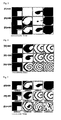
- FIG. 3 The propagation tendency of an action voltage in the imaginary heart depending on the shim number is shown Figs. 3 through 6 .
- Fig. 3 it can be noted that, even if an external disturbing wave is applied to give tachycardia-causing conditions to the imaginary hearts having the shim numbers [ Sh ] of 200, 400 and 600, the influence of the external disturbing wave disappears after a specified time has lapsed.
- Fig. 4 it can be appreciated that, when an external disturbing wave is applied to give tachycardia-causing conditions to the imaginary hearts having the shim numbers [Sh] of 800, 1000 and 2000, the influence of the external disturbing wave does not appear even after the lapse of a specified time and tachycardia is generated.
- FIG. 5 different results can be obtained when an external disturbing wave is applied to the imaginary hearts having the shim numbers [ Sh ] of 300, 800 and 1000 and having the instable cells added with disturbance of an ion current. More specifically, the imaginary heart having the shim number [Sh] of 300 comes back to a normal state after the lapse of 950 ms. The imaginary heart having the shim number [ Sh ] of 800 fails to come back to the normal state even after the lapse of 1500 ms, as a result of which tachycardia is generated. In the imaginary heart having the shim number [ Sh ] of 1000, tachycardia is induced with ease after the lapse of 2000 ms.
- Fig. 6 shows images of a normal cell model and a disturbed cell model when a specified time has lapsed after application of conditions for causing tachycardia.
- the shim number [ Sh ] is identical, the possibility of generation of tachycardia and fibrillation differs largely depending on whether the heart model is a normal cell model or a disturbed cell model. It also can be noted that the possibility of generation of tachycardia and fibrillation becomes higher as the shim number [ Sh ] grows larger.
- the colors in the images shown in Figs. 3 through 6 indicate the voltage distribution in a cell membrane. The red color indicates an excited state and the blue color indicates a pause period.
- the non-dimensional number [ Sh ] is a very important parameter in diagnosing arrhythmia. Description will now be made on a method and device for diagnosing arrhythmia through the use of the non-dimensional number [ Sh ].
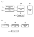
- Fig. 7 is a schematic block diagram showing an arrhythmia diagnosing device according to one embodiment of the present invention.
- the arrhythmia diagnosing device according to the present embodiment includes a first measuring unit 10, a second measuring unit 20, a third measuring unit 30, a storage unit 40, a display unit 50 and a determination unit 60.
- the first measuring unit 10 serves to measure a heart dimension L , i.e., the length of a route through which an electric wave passes in the heart.
- the heart dimension L is proportional to the size of the heart.
- the heart dimension L is also called a characteristic length of the heart.
- the first measuring unit 10 is similar to or identical with an echocardiograph and is configured to measure the heart dimension L .
- the first measuring unit 10 may directly measure the heart dimension or may measure the heart volume, if necessary.
- the heart dimension L is calculated by the first measuring unit 10 as a cubic root of the measured heart volume as represented by the following formula 4.
- the second measuring unit 20 serves to measure a frequency f of a cardiac electric wave.
- the second measuring unit 20 is similar to or identical with an electrocardiograph for outputting an electrocardiogram signal and is configured to measure a frequency of a cardiac electric wave.
- the electrocardiogram signal contains R waves and a time interval between the R waves. The reciprocal number of the time interval is the frequency of the cardiac electric wave as can be seen in the following formula 5.
- the third measuring unit 30 serves to measure a conduction velocity CV of a cardiac electric wave.
- the conduction velocity CV is calculated in the following manner using the electrocardiogram signal alone or the electrocardiogram signal and the heart dimension.
- the conduction velocity CV of the cardiac electric wave in the ventricle is calculated by the following formula 6.
- CV 1 QRS or RS
- the QRS is an interval of a QRS contained in the electrocardiogram signal
- the RS is an interval of an RS contained in the electrocardiogram signal
- the CV is a conduction velocity of a cardiac electric wave.
- the conduction velocity of the cardiac electric wave in the atrium is calculated by the following formula 7.
- CV L P where the L is a heart dimension and the P is a width of a P wave contained in the electrocardiogram signal.
- the third measuring unit 30 has the configurations of an echocardiograph and an electrocardiograph.
- the third measuring unit 30 is configured to calculate and output the conduction velocity of the cardiac electric wave using the electrocardiogram signal and the heart dimension.
- a reference value for the determination of arrhythmia is stored in the storage unit 40.
- the reference value is used to compare the magnitude thereof with the non-dimensional number [ Sh ].
- the reference value may be set to have many different values. In the present embodiment, the reference value is set to have a specific value in a range of 800 to 1200.
- the reference value can be changed by a user depending on a variety of factors such the age, sex, health, dietary habit, weight and stature of a patient.
- the display unit 50 serves to display the operating state of the arrhythmia diagnosing device.
- the display unit 50 is formed of an LCD and is capable of displaying the non-dimensional number [ Sh ] and the occurrence or absence of arrhythmia.
- the display unit 50 may be formed of a green LED and a red LED so that the red LED can be turned on in case of arrhythmia being diagnosed and the green LED can be turned on in case of arrhythmia not being diagnosed.
- the determination unit 60 determines the occurrence of absence of arrhythmia pursuant to the heart dimension, the frequency of the cardiac electric wave and the conduction velocity of the cardiac electric wave inputted from the first measuring unit 10, the second measuring unit 20 and the third measuring unit 30.
- the determination unit 60 outputs a control signal corresponding to the determination result to the display unit 50.
- the determination unit 60 calculates the non-dimensional number [ Sh ] according to the following formula 8.
- Sh f ⁇ L CV
- the [ Sh ] is a non-dimensional number
- the f is a frequency of a cardiac electric wave
- the L is a heart dimension
- the CV is a conduction velocity of a cardiac electric wave.
- the determination unit 60 compares the calculated non-dimensional number [ Sh ] with the reference value stored in the storage unit 40. If the calculated non-dimensional number [ Sh ] is equal to or larger than the reference value, the determination unit 60 determines that the heart suffers from arrhythmia. If the calculated non-dimensional number [ Sh ] is smaller than the reference value, the determination unit 60 determines that the heart does not suffer from arrhythmia.
- the determination unit 60 outputs a control signal corresponding to the determination result to the display unit 50. Responsive to the control signal, the display unit 50 displays the occurrence or absence of arrhythmia.
- Fig. 8 is a schematic block diagram showing an arrhythmia diagnosing device according to another embodiment of the present invention.
- the arrhythmia diagnosing device according to the present invention includes a second measuring unit 20a formed of an electrocardiograph. Accordingly, the second measuring unit 20a outputs an electrocardiogram signal.
- the first measuring unit 10 serves to measure a heart dimension. Needless to say, it may be possible to measure a heart volume instead of the heart dimension. In this case, the heart dimension is calculated by the first measuring unit 10 or the determination unit 60a.
- the determination unit 60a calculates a frequency of a cardiac electric wave and a conduction velocity thereof using the heart dimension and the electrocardiogram signal inputted. Based on this calculation result, the determination unit 60a calculates the non-dimensional number [Sh]. By comparing the calculated non-dimensional number [Sh] with the reference value, the determination unit 60a determines the occurrence or absence of arrhythmia.
- Fig. 9 is a schematic block diagram showing an arrhythmia diagnosing device according to a further embodiment of the present invention.
- the arrhythmia diagnosing device according to the present embodiment includes an interface unit 70, a storage unit 40, a display unit 50 and a determination unit 60b.
- the interface unit 70 is provided to make wire communication or wireless communication with an echocardiograph or an electrocardiograph.
- the heart dimension measured by the echocardiograph and the electrocardiogram signal (containing an RRinterval, a QRS interval, an RS interval and a P wave width) outputted from the electrocardiograph are inputted to the determination unit 60b through the interface unit 70.
- the determination unit 60b calculates a non-dimensional number [ Sh ] using the values inputted through the interface unit 70. By comparing the calculated non-dimensional number [ Sh ] with the reference value, the determination unit 60a determines the occurrence or absence of arrhythmia. The determination unit 60b outputs a control signal so that the determination result can be displayed on the display unit 50. If necessary, the arrhythmia diagnosing device may be configured such that the heart dimension, the frequency of the cardiac electric wave and the conduction velocity of the cardiac electric wave are inputted through the interface unit 70.
Landscapes
- Health & Medical Sciences (AREA)
- Life Sciences & Earth Sciences (AREA)
- Cardiology (AREA)
- Molecular Biology (AREA)
- Animal Behavior & Ethology (AREA)
- Pathology (AREA)
- Engineering & Computer Science (AREA)
- Biomedical Technology (AREA)
- Heart & Thoracic Surgery (AREA)
- Medical Informatics (AREA)
- Physics & Mathematics (AREA)
- Surgery (AREA)
- Biophysics (AREA)
- General Health & Medical Sciences (AREA)
- Public Health (AREA)
- Veterinary Medicine (AREA)
- Dentistry (AREA)
- Oral & Maxillofacial Surgery (AREA)
- Physiology (AREA)
- Measurement And Recording Of Electrical Phenomena And Electrical Characteristics Of The Living Body (AREA)
Applications Claiming Priority (2)
| Application Number | Priority Date | Filing Date | Title |
|---|---|---|---|
| KR1020100017665A KR101109738B1 (ko) | 2009-04-06 | 2010-02-26 | 심장 부정맥 진단방법 및 그 진단장치 |
| PCT/KR2011/001360 WO2011105859A2 (ko) | 2010-02-26 | 2011-02-25 | 심장 부정맥 진단방법 및 그 진단장치 |
Publications (2)
| Publication Number | Publication Date |
|---|---|
| EP2540222A2 true EP2540222A2 (de) | 2013-01-02 |
| EP2540222A4 EP2540222A4 (de) | 2014-04-16 |
Family
ID=44508410
Family Applications (1)
| Application Number | Title | Priority Date | Filing Date |
|---|---|---|---|
| EP11747749.7A Withdrawn EP2540222A4 (de) | 2010-02-26 | 2011-02-25 | Verfahren und vorrichtung zur diagnose von herzrhythmusstörungen |
Country Status (3)
| Country | Link |
|---|---|
| EP (1) | EP2540222A4 (de) |
| JP (1) | JP5718946B2 (de) |
| WO (1) | WO2011105859A2 (de) |
Cited By (8)
| Publication number | Priority date | Publication date | Assignee | Title |
|---|---|---|---|---|
| EP2762068A1 (de) * | 2013-02-05 | 2014-08-06 | Nihon Kohden Corporation | Elektrokardiogrammanalysator |
| WO2014201183A1 (en) * | 2013-06-11 | 2014-12-18 | Intelomed, Inc | Predicting hypovolemic hypotensive conditions using a pulse volume waveform |
| US9173579B2 (en) | 2010-07-08 | 2015-11-03 | Intelomed, Inc. | System and method for characterizing circulatory blood flow |
| EP2945534A4 (de) * | 2013-01-16 | 2016-08-31 | Univ Vermont | Systeme, katheter und zugehörige verfahren zur abbildung, minimierung und behandlung von vorhofflimmern |
| US9808160B2 (en) | 2013-08-12 | 2017-11-07 | Intelomed, Inc. | Systems and methods for monitoring and analyzing cardiovascular states |
| US10390767B2 (en) | 2013-06-04 | 2019-08-27 | Intelomed Inc. | Hemodynamic risk severity based upon detection and quantification of cardiac dysrhythmia behavior using a pulse volume waveform |
| US10912476B2 (en) | 2013-01-16 | 2021-02-09 | University Of Vermont | Catheters, systems, and related methods for mapping, minimizing, and treating cardiac fibrillation |
| US12582343B2 (en) | 2013-01-16 | 2026-03-24 | University Of Vermont And State Agricultural College | Methods and systems for wavelength mapping cardiac fibrillation and optimizing ablation lesion placement |
Family Cites Families (13)
| Publication number | Priority date | Publication date | Assignee | Title |
|---|---|---|---|---|
| JP3224569B2 (ja) * | 1990-09-21 | 2001-10-29 | 日本コーリン株式会社 | Ecg信号に基づく心臓不整脈検出装置 |
| JPH06149863A (ja) * | 1992-11-05 | 1994-05-31 | Yukio Tanaka | 時系列データ解析装置 |
| KR100400212B1 (ko) * | 1997-01-31 | 2003-11-14 | 삼성전자주식회사 | 부정맥검출기능을 가진 심전도장치 |
| JP4208275B2 (ja) * | 1997-10-30 | 2009-01-14 | 株式会社東芝 | 心臓内電気現象の診断装置およびその現象の表示方法 |
| US6301496B1 (en) * | 1998-07-24 | 2001-10-09 | Biosense, Inc. | Vector mapping of three-dimensionally reconstructed intrabody organs and method of display |
| US6556695B1 (en) * | 1999-02-05 | 2003-04-29 | Mayo Foundation For Medical Education And Research | Method for producing high resolution real-time images, of structure and function during medical procedures |
| KR100452748B1 (ko) * | 2001-05-08 | 2004-10-12 | 유닉스전자주식회사 | 전자 혈압계를 이용한 심전도 측정 방법 |
| US6773402B2 (en) * | 2001-07-10 | 2004-08-10 | Biosense, Inc. | Location sensing with real-time ultrasound imaging |
| CA2471655A1 (en) * | 2001-12-26 | 2003-07-17 | Mediwave Star Technology, Inc. | Method and system for evaluating arrhythmia risk with qt-rr interval data sets |
| KR20040107882A (ko) * | 2003-06-14 | 2004-12-23 | 주식회사 씨아이디 | 휴대형 부정맥 진단장치 |
| DE102005032755B4 (de) * | 2005-07-13 | 2014-09-04 | Siemens Aktiengesellschaft | System zur Durchführung und Überwachung minimal-invasiver Eingriffe |
| US8038625B2 (en) * | 2005-09-15 | 2011-10-18 | St. Jude Medical, Atrial Fibrillation Division, Inc. | System and method for three-dimensional mapping of electrophysiology information |
| US20100280366A1 (en) * | 2008-05-13 | 2010-11-04 | Lawrence Arne | Continuous field tomography systems and methods of using the same |
-
2011
- 2011-02-25 EP EP11747749.7A patent/EP2540222A4/de not_active Withdrawn
- 2011-02-25 WO PCT/KR2011/001360 patent/WO2011105859A2/ko not_active Ceased
- 2011-02-25 JP JP2012554940A patent/JP5718946B2/ja active Active
Cited By (15)
| Publication number | Priority date | Publication date | Assignee | Title |
|---|---|---|---|---|
| US9173579B2 (en) | 2010-07-08 | 2015-11-03 | Intelomed, Inc. | System and method for characterizing circulatory blood flow |
| US10912476B2 (en) | 2013-01-16 | 2021-02-09 | University Of Vermont | Catheters, systems, and related methods for mapping, minimizing, and treating cardiac fibrillation |
| US12582343B2 (en) | 2013-01-16 | 2026-03-24 | University Of Vermont And State Agricultural College | Methods and systems for wavelength mapping cardiac fibrillation and optimizing ablation lesion placement |
| US12036026B2 (en) | 2013-01-16 | 2024-07-16 | University Of Vermont And State Agricultural College | Catheters, systems, and related methods for mapping, minimizing, and treating cardiac fibrillation |
| EP2945534A4 (de) * | 2013-01-16 | 2016-08-31 | Univ Vermont | Systeme, katheter und zugehörige verfahren zur abbildung, minimierung und behandlung von vorhofflimmern |
| US9693699B2 (en) | 2013-01-16 | 2017-07-04 | University Of Vermont | Methods and systems for mapping cardiac fibrillation |
| US9706935B2 (en) | 2013-01-16 | 2017-07-18 | University Of Vermont | Catheter systems and related methods for mapping, minimizing, and treating cardiac fibrillation |
| US11089987B2 (en) | 2013-01-16 | 2021-08-17 | University Of Vermont | Catheter systems and related methods for mapping, minimizing, and treating cardiac fibrillation |
| US9386935B2 (en) | 2013-02-05 | 2016-07-12 | Nihon Kohden Corporation | Electrocardiogram analyzer |
| EP2762068A1 (de) * | 2013-02-05 | 2014-08-06 | Nihon Kohden Corporation | Elektrokardiogrammanalysator |
| US10390767B2 (en) | 2013-06-04 | 2019-08-27 | Intelomed Inc. | Hemodynamic risk severity based upon detection and quantification of cardiac dysrhythmia behavior using a pulse volume waveform |
| US10568583B2 (en) | 2013-06-11 | 2020-02-25 | Intelomed, Inc. | Methods and systems for predicting hypovolemic hypotensive conditions resulting from bradycardia behavior using a pulse volume waveform |
| WO2014201183A1 (en) * | 2013-06-11 | 2014-12-18 | Intelomed, Inc | Predicting hypovolemic hypotensive conditions using a pulse volume waveform |
| US9808160B2 (en) | 2013-08-12 | 2017-11-07 | Intelomed, Inc. | Systems and methods for monitoring and analyzing cardiovascular states |
| US11197617B2 (en) | 2013-08-12 | 2021-12-14 | Intelomed, Inc. | Systems and methods for monitoring and analyzing cardiovascular states |
Also Published As
| Publication number | Publication date |
|---|---|
| WO2011105859A3 (ko) | 2012-01-12 |
| JP2013520275A (ja) | 2013-06-06 |
| WO2011105859A2 (ko) | 2011-09-01 |
| EP2540222A4 (de) | 2014-04-16 |
| JP5718946B2 (ja) | 2015-05-13 |
Similar Documents
| Publication | Publication Date | Title |
|---|---|---|
| US9008763B2 (en) | Arrhythmia diagnosis method and device | |
| US7447543B2 (en) | Pathology assessment with impedance measurements using convergent bioelectric lead fields | |
| AU2021202438B2 (en) | Mobile cardiac monitoring device | |
| Brooks et al. | Electrical imaging of the heart | |
| US9282911B2 (en) | Linear display of ECG signals | |
| Harris | The Normal electrocardiogram: resting 12-Lead and electrocardiogram monitoring in the hospital | |
| JP2005531386A (ja) | 生体インピーダンス法を用いて鬱血性心不全患者をモニタするための装置 | |
| US20100023081A1 (en) | Comprehensive System for Detection of Coronary Syndrome, Cardiac Ischemia and Myocardial Infarction | |
| JP5718946B2 (ja) | 心臓不整脈診断装置 | |
| WO1992005831A1 (en) | Ekg system using statistic and topographic mapping | |
| CN108348186B (zh) | 用于标测心脏恢复的方法和系统 | |
| US20140066797A1 (en) | Systems and methods for analyzing changes in cardiac output | |
| KR20120108235A (ko) | 가상의 생체 모델을 이용하여 실제 생체 신호 측정에 이용되는 최적의 파라미터를 결정하는 방법 및 장치 | |
| JP5469574B2 (ja) | 電気的インピーダンス断層像測定装置。 | |
| Heena | Analysis and Classification of Heart Abnormalities | |
| RU2558973C2 (ru) | Способ оценки реакции сердечно - сосудистой системы на физическую нагрузку | |
| Rasoolzadeh | Solving the Inverse Problem of Electrocardiography for Spontaneous PVC Localization: Analysis of Clinical Electrocardiographic Data | |
| Franzone et al. | Basic Cardiac Anatomy and Electrocardiology | |
| Heena | 3 Analysis and | |
| Kozlíková et al. | IIM QRS in concentric left ventricular hypertrophy | |
| Hakomäki | ECG artefacts in EEG measurement | |
| Lyon | Computational detection of electrophysiological abnormalities in hypertrophic cardiomyopathy for risk stratification | |
| Woodcock | Ventricular Fibrillation waveform characteristics | |
| LaRocque | Application of least-squares minimization and PCA to the optimization of body surface potential maps derived from the standard precordial leads | |
| Bronzino et al. | 115.1 The Electroencephalogram |
Legal Events
| Date | Code | Title | Description |
|---|---|---|---|
| PUAI | Public reference made under article 153(3) epc to a published international application that has entered the european phase |
Free format text: ORIGINAL CODE: 0009012 |
|
| 17P | Request for examination filed |
Effective date: 20120924 |
|
| AK | Designated contracting states |
Kind code of ref document: A2 Designated state(s): AL AT BE BG CH CY CZ DE DK EE ES FI FR GB GR HR HU IE IS IT LI LT LU LV MC MK MT NL NO PL PT RO RS SE SI SK SM TR |
|
| DAX | Request for extension of the european patent (deleted) | ||
| RAP1 | Party data changed (applicant data changed or rights of an application transferred) |
Owner name: CU MECICAL SYSTEMS, INC. |
|
| RAP1 | Party data changed (applicant data changed or rights of an application transferred) |
Owner name: CU MEDICAL SYSTEMS, INC. |
|
| A4 | Supplementary search report drawn up and despatched |
Effective date: 20140317 |
|
| RIC1 | Information provided on ipc code assigned before grant |
Ipc: A61B 5/0464 20060101AFI20140311BHEP Ipc: A61B 5/107 20060101ALI20140311BHEP Ipc: A61B 5/04 20060101ALI20140311BHEP Ipc: A61B 8/08 20060101ALN20140311BHEP Ipc: A61B 6/00 20060101ALN20140311BHEP Ipc: A61B 5/00 20060101ALN20140311BHEP Ipc: A61B 5/0452 20060101ALN20140311BHEP Ipc: A61B 5/02 20060101ALN20140311BHEP |
|
| 17Q | First examination report despatched |
Effective date: 20160203 |
|
| STAA | Information on the status of an ep patent application or granted ep patent |
Free format text: STATUS: THE APPLICATION HAS BEEN WITHDRAWN |
|
| 18W | Application withdrawn |
Effective date: 20191011 |