EP2563462B1 - DISPOSITIF POUR DÉTECTER UNE SIGNATURE DE LA DOULEUR SUR LA BASE DE CHANGEMENTS DANS l'ÉMISSION DE TRAINS D'ONDES - Google Patents
DISPOSITIF POUR DÉTECTER UNE SIGNATURE DE LA DOULEUR SUR LA BASE DE CHANGEMENTS DANS l'ÉMISSION DE TRAINS D'ONDES Download PDFInfo
- Publication number
- EP2563462B1 EP2563462B1 EP11775539.7A EP11775539A EP2563462B1 EP 2563462 B1 EP2563462 B1 EP 2563462B1 EP 11775539 A EP11775539 A EP 11775539A EP 2563462 B1 EP2563462 B1 EP 2563462B1
- Authority
- EP
- European Patent Office
- Prior art keywords
- pain
- firing
- burst
- activity
- vpl
- Prior art date
- Legal status (The legal status is an assumption and is not a legal conclusion. Google has not performed a legal analysis and makes no representation as to the accuracy of the status listed.)
- Active
Links
Images











Classifications
-
- A—HUMAN NECESSITIES
- A61—MEDICAL OR VETERINARY SCIENCE; HYGIENE
- A61N—ELECTROTHERAPY; MAGNETOTHERAPY; RADIATION THERAPY; ULTRASOUND THERAPY
- A61N1/00—Electrotherapy; Circuits therefor
- A61N1/18—Applying electric currents by contact electrodes
- A61N1/32—Applying electric currents by contact electrodes alternating or intermittent currents
- A61N1/36—Applying electric currents by contact electrodes alternating or intermittent currents for stimulation
- A61N1/3605—Implantable neurostimulators for stimulating central or peripheral nerve system
- A61N1/3606—Implantable neurostimulators for stimulating central or peripheral nerve system adapted for a particular treatment
- A61N1/36071—Pain
-
- A—HUMAN NECESSITIES
- A61—MEDICAL OR VETERINARY SCIENCE; HYGIENE
- A61B—DIAGNOSIS; SURGERY; IDENTIFICATION
- A61B5/00—Measuring for diagnostic purposes; Identification of persons
- A61B5/24—Detecting, measuring or recording bioelectric or biomagnetic signals of the body or parts thereof
- A61B5/316—Modalities, i.e. specific diagnostic methods
- A61B5/369—Electroencephalography [EEG]
- A61B5/372—Analysis of electroencephalograms
-
- A—HUMAN NECESSITIES
- A61—MEDICAL OR VETERINARY SCIENCE; HYGIENE
- A61B—DIAGNOSIS; SURGERY; IDENTIFICATION
- A61B5/00—Measuring for diagnostic purposes; Identification of persons
- A61B5/24—Detecting, measuring or recording bioelectric or biomagnetic signals of the body or parts thereof
- A61B5/316—Modalities, i.e. specific diagnostic methods
- A61B5/369—Electroencephalography [EEG]
- A61B5/377—Electroencephalography [EEG] using evoked responses
- A61B5/383—Somatosensory stimuli, e.g. electric stimulation
-
- A—HUMAN NECESSITIES
- A61—MEDICAL OR VETERINARY SCIENCE; HYGIENE
- A61B—DIAGNOSIS; SURGERY; IDENTIFICATION
- A61B5/00—Measuring for diagnostic purposes; Identification of persons
- A61B5/48—Other medical applications
- A61B5/4824—Touch or pain perception evaluation
-
- A—HUMAN NECESSITIES
- A61—MEDICAL OR VETERINARY SCIENCE; HYGIENE
- A61N—ELECTROTHERAPY; MAGNETOTHERAPY; RADIATION THERAPY; ULTRASOUND THERAPY
- A61N1/00—Electrotherapy; Circuits therefor
- A61N1/18—Applying electric currents by contact electrodes
- A61N1/32—Applying electric currents by contact electrodes alternating or intermittent currents
- A61N1/36—Applying electric currents by contact electrodes alternating or intermittent currents for stimulation
- A61N1/3605—Implantable neurostimulators for stimulating central or peripheral nerve system
- A61N1/36125—Details of circuitry or electric components
-
- A—HUMAN NECESSITIES
- A61—MEDICAL OR VETERINARY SCIENCE; HYGIENE
- A61N—ELECTROTHERAPY; MAGNETOTHERAPY; RADIATION THERAPY; ULTRASOUND THERAPY
- A61N1/00—Electrotherapy; Circuits therefor
- A61N1/18—Applying electric currents by contact electrodes
- A61N1/32—Applying electric currents by contact electrodes alternating or intermittent currents
- A61N1/36—Applying electric currents by contact electrodes alternating or intermittent currents for stimulation
- A61N1/3605—Implantable neurostimulators for stimulating central or peripheral nerve system
- A61N1/36128—Control systems
- A61N1/36135—Control systems using physiological parameters
- A61N1/36139—Control systems using physiological parameters with automatic adjustment
-
- A—HUMAN NECESSITIES
- A61—MEDICAL OR VETERINARY SCIENCE; HYGIENE
- A61N—ELECTROTHERAPY; MAGNETOTHERAPY; RADIATION THERAPY; ULTRASOUND THERAPY
- A61N1/00—Electrotherapy; Circuits therefor
- A61N1/18—Applying electric currents by contact electrodes
- A61N1/32—Applying electric currents by contact electrodes alternating or intermittent currents
- A61N1/36—Applying electric currents by contact electrodes alternating or intermittent currents for stimulation
- A61N1/3605—Implantable neurostimulators for stimulating central or peripheral nerve system
- A61N1/36128—Control systems
- A61N1/36146—Control systems specified by the stimulation parameters
- A61N1/3615—Intensity
- A61N1/36153—Voltage
-
- A—HUMAN NECESSITIES
- A61—MEDICAL OR VETERINARY SCIENCE; HYGIENE
- A61N—ELECTROTHERAPY; MAGNETOTHERAPY; RADIATION THERAPY; ULTRASOUND THERAPY
- A61N1/00—Electrotherapy; Circuits therefor
- A61N1/18—Applying electric currents by contact electrodes
- A61N1/32—Applying electric currents by contact electrodes alternating or intermittent currents
- A61N1/36—Applying electric currents by contact electrodes alternating or intermittent currents for stimulation
- A61N1/3605—Implantable neurostimulators for stimulating central or peripheral nerve system
- A61N1/36128—Control systems
- A61N1/36146—Control systems specified by the stimulation parameters
- A61N1/3615—Intensity
- A61N1/36157—Current
-
- A—HUMAN NECESSITIES
- A61—MEDICAL OR VETERINARY SCIENCE; HYGIENE
- A61N—ELECTROTHERAPY; MAGNETOTHERAPY; RADIATION THERAPY; ULTRASOUND THERAPY
- A61N1/00—Electrotherapy; Circuits therefor
- A61N1/18—Applying electric currents by contact electrodes
- A61N1/32—Applying electric currents by contact electrodes alternating or intermittent currents
- A61N1/36—Applying electric currents by contact electrodes alternating or intermittent currents for stimulation
- A61N1/3605—Implantable neurostimulators for stimulating central or peripheral nerve system
- A61N1/36128—Control systems
- A61N1/36146—Control systems specified by the stimulation parameters
- A61N1/36167—Timing, e.g. stimulation onset
- A61N1/36171—Frequency
-
- A—HUMAN NECESSITIES
- A61—MEDICAL OR VETERINARY SCIENCE; HYGIENE
- A61N—ELECTROTHERAPY; MAGNETOTHERAPY; RADIATION THERAPY; ULTRASOUND THERAPY
- A61N1/00—Electrotherapy; Circuits therefor
- A61N1/18—Applying electric currents by contact electrodes
- A61N1/32—Applying electric currents by contact electrodes alternating or intermittent currents
- A61N1/36—Applying electric currents by contact electrodes alternating or intermittent currents for stimulation
- A61N1/3605—Implantable neurostimulators for stimulating central or peripheral nerve system
- A61N1/36128—Control systems
- A61N1/36146—Control systems specified by the stimulation parameters
- A61N1/36167—Timing, e.g. stimulation onset
- A61N1/36175—Pulse width or duty cycle
-
- A—HUMAN NECESSITIES
- A61—MEDICAL OR VETERINARY SCIENCE; HYGIENE
- A61N—ELECTROTHERAPY; MAGNETOTHERAPY; RADIATION THERAPY; ULTRASOUND THERAPY
- A61N1/00—Electrotherapy; Circuits therefor
- A61N1/18—Applying electric currents by contact electrodes
- A61N1/32—Applying electric currents by contact electrodes alternating or intermittent currents
- A61N1/36—Applying electric currents by contact electrodes alternating or intermittent currents for stimulation
- A61N1/3605—Implantable neurostimulators for stimulating central or peripheral nerve system
- A61N1/36128—Control systems
- A61N1/36146—Control systems specified by the stimulation parameters
- A61N1/36167—Timing, e.g. stimulation onset
- A61N1/36178—Burst or pulse train parameters
-
- A—HUMAN NECESSITIES
- A61—MEDICAL OR VETERINARY SCIENCE; HYGIENE
- A61B—DIAGNOSIS; SURGERY; IDENTIFICATION
- A61B5/00—Measuring for diagnostic purposes; Identification of persons
- A61B5/68—Arrangements of detecting, measuring or recording means, e.g. sensors, in relation to patient
- A61B5/6846—Arrangements of detecting, measuring or recording means, e.g. sensors, in relation to patient specially adapted to be brought in contact with an internal body part, i.e. invasive
- A61B5/6867—Arrangements of detecting, measuring or recording means, e.g. sensors, in relation to patient specially adapted to be brought in contact with an internal body part, i.e. invasive specially adapted to be attached or implanted in a specific body part
- A61B5/6868—Brain
-
- A—HUMAN NECESSITIES
- A61—MEDICAL OR VETERINARY SCIENCE; HYGIENE
- A61B—DIAGNOSIS; SURGERY; IDENTIFICATION
- A61B5/00—Measuring for diagnostic purposes; Identification of persons
- A61B5/72—Signal processing specially adapted for physiological signals or for diagnostic purposes
- A61B5/7235—Details of waveform analysis
-
- A—HUMAN NECESSITIES
- A61—MEDICAL OR VETERINARY SCIENCE; HYGIENE
- A61B—DIAGNOSIS; SURGERY; IDENTIFICATION
- A61B5/00—Measuring for diagnostic purposes; Identification of persons
- A61B5/72—Signal processing specially adapted for physiological signals or for diagnostic purposes
- A61B5/7235—Details of waveform analysis
- A61B5/7253—Details of waveform analysis characterised by using transforms
- A61B5/7257—Details of waveform analysis characterised by using transforms using Fourier transforms
Definitions
- Chronic pain is typically classified as pain lasting more than 6 months and generally divided into three main types: nociceptive, psychogenic or neuropathic (e.g., due to nerve injury) although the distinction between these types can be blurred.
- current treatment options including opioids and nonsteroidal anti-inflammatory drugs (e.g. COX inhibitors) are often ineffective, contraindicated or associated with significant gastrointestinal and cardiac side effects, sedation, respiratory depression, addiction and drug abuse.
- opioids and nonsteroidal anti-inflammatory drugs e.g. COX inhibitors
- COX inhibitors nonsteroidal anti-inflammatory drugs
- pharmacotherapy, surgical ablation, and externally applied non-drug therapies e.g. transcutaneous electrical nerve stimulation and acupuncture
- novel ideas are thus needed in pain research.
- the document US 2007/213783 A1 discloses an implantable neurostimulator with sensing means for detecting pain signatures.
- Document US-A-7 894 905 discloses a system of identifying a subject comprising chronic pain related to a peripheral nerve injury, wherein said system comprises a processor suitable to identify a pain signature comprising a pattern of burst-firing.
- Document " Pain and Temperature Encoding in the Human Thalamic Somatic Sensory Nucleus (Ventral caudal): Inhibition-Related Bursting Evoked by Somatic Stimuli, J.-I. Lee et al., Journal of neurophysiology 94: 1676-1686, 2005 relates to a method to determine the human thalamic response to mechanical and thermal stimuli including the steps a)-d) defined in claim 1.
- Chronic clinical pain includes neuropathic pain such as that associated with direct nerve damage, amputation, chemotherapy, diabetes, HIV infection or AIDS, Multiple Sclerosis, shingles, sciatic nerve compression or injury, as well as spine surgery.
- a method, not part of the invention, of identifying a subject suffering from chronic pain is carried out by detecting a pain signature comprising a pattern of neuronal firing different compared to a normal pattern of neuronal firing, e.g., a pattern obtained from a subject or a cohort of subjects that have been characterized as not suffering from pain.
- the pattern of firing is obtained from a single neuron or a plurality of neurons.
- the brain of a subject afflicted with chronic pain can present a pain signature.
- the pattern can comprise an elevated evoked response to stimuli, rhythmic after-discharge signaling, and/or increased spontaneous background firing.
- the pain and neuronal firing pattern subsists after an injury heals or is completely unrelated to a stimulus, e.g., an injury, or the degree of a stimulus.
- the pain signature comprises a pattern of neuronal burst-firing, each burst of the burst firing comprising at least 10 times, 50 times, 100 times or more, the number of spikes compared to a control non-pain pattern.
- An exemplary pain signature comprises a pattern of burst-firing that is characterized by one or more of the following measurable parameters: (a) a maximum interval signifying burst onset (6 ms); (b) a maximum interspike interval (9 ms); (c) longest increase in interspike interval within a burst (2 ms); (d) a minimum number of spikes within a burst (2).
- An aberrant pattern or pain signature is further characterized by a spontaneous high frequency rhythmic oscillation of long epoch.
- a method is disclosed, not part of the invention, of preventing or reducing pain perception involves identifying a subject using the criteria described above and administering to the subject at least one electrical pulse to the subject, the electrical pulses administered at a frequency of at least about 100, 150, or 200 Hz, at a voltage between about 1 and about 3 volts, through a current of between about 1 and about 3 milliampere, and between about 0.25 and about 1 second in duration.
- the electrical pulse of at least about 100 Hz jams or halts the pain circuitry at the level of the source.
- Subjects to be diagnosed and/or treated include human patients as well as animals such as companion animals (e.g., dogs, cats) as well as livestock and performance animals (e.g., horses, cattle).
- a system including a processor configured to be coupled to an electrical lead that is configured to sense electrical activity in a patient, a memory coupled to the processor, the memory containing computer readable instructions that, when executed by the processor, cause the processor to detect a pain signature in the sensed electrical activity, determine a treatment protocol in response to the detected pain signature, and cause treatment according to the treatment protocol to be delivered to the patient via the electrical lead.
- a system including a processor configured to provide an electrical treatment protocol to a patient, the electrical treatment protocol being configured to treat chronic pain in the patient, the treatment protocol including providing at least one electrical pulse to the patient, the electrical pulses to be provided at a frequency of at least about 150 Hz, at a voltage of between about 1 and about 3 volts, through a current of between about 1 and about 3 milliampere, and between about 0.25 and about 1 second in duration.
- a method not part of the invention, is disclosed of identifying a subject suffering from chronic pain, including detecting a pain signature comprising a pattern of neuronal firing, said pattern comprising an elevated evoked response to stimuli, rhythmic after-discharge signaling, and increased spontaneous background firing.
- a method, not part of the invention of identifying a subject suffering from chronic pain, the method comprising detecting a pain signature comprising a pattern of burst-firing, each burst of said burst firing comprising at least 10 times the number of spikes compared to a control non-pain pattern.
- a method not part of the invention of identifying a subject suffering from chronic pain, the method comprising detecting a pain signature including a pattern of burst-firing, wherein said pattern comprises burst-firing, said burst firing including (a) a maximum interval signifying burst onset (6 ms), (b) a maximum interspike interval (9 ms), (c) longest increase in interspike interval within a burst (2 ms), or (d) a minimum number of spikes within a burst (2).
- a method, not part of the invention of preventing or reducing pain perception, comprising identifying a subject, and administering to said subject at least one electrical pulse to the subject, the electrical pulses administered at a frequency of at least about 150 Hz, at a voltage of between about 1 and about 3 volts, through a current of between about 1 and about 3 milliampere, and between about 0.25 and about 1 second in duration.
- Various aspects may provide one or more of the following capabilities.
- the efficiency, battery life, and device life of devices used for neurostimulation can be improved.
- More physiologically relevant brain structures can be targeted. Side effects can be reduced. The temporal and overall amount of delivered current can be reduced. The likelihood of excessive tissue exposure, which has been thought to cause long-term changes and side effects, can be reduced. The necessity for combined pharmacologic intervention can be reduced, or possibly eliminated. Chronic pain can be empirically diagnosed.
- Chronic pain is a serious challenge in terms of pathophysiology, diagnosis, therapy and social burden.
- Studies in humans and laboratory animals suggest a relationship between intractable pain and ectopic neuronal activity in thalamic and cortical areas, leading to dysfunctional connectivity in the brain's pain network.
- Chronic pain is itself a disease process.
- Chronic pain is unrelenting and not self-limiting and as stated earlier, can persist for years and even decades after the initial injury. If not treated, chronic pain can lead to anxiety, fear, depression, sleeplessness and impairment of social interaction.
- Chronic, non-malignant pain is predominately neuropathic in nature and involves damage either to the peripheral or central nervous systems.
- Nociceptive and neuropathic pain are caused by different neurophysiological processes, and therefore respond to different treatment modalities.
- Nociceptive pain is mediated by receptors on A-delta and C-fibers which are located in skin, bone, connective tissue, muscle and viscera. These receptors serve a biologically useful role at localizing noxious chemical, thermal and mechanical stimuli.
- Nociceptive pain can be somatic or visceral in nature. Somatic pain tends to be well localized, constant pain that is described as sharp, aching, throbbing, or gnawing. Visceral pain, on the other hand, tends to be vague in distribution, paroxysmal in nature and is usually described as deep, aching, squeezing and colicky in nature.
- nociceptive pain examples include: post-operative pain, pain associated with trauma, and the chronic pain of arthritis. Nociceptive pain often responds to opioids and non-steroidal anti-inflammatories (NSAIDS). Neuropathic pain, in contrast to nociceptive pain, is described as burning, electric, tingling and shooting in nature and can be unrelated to a stimulus such as an injury. Examples of neuropathic pain include: monoradiculopathies, trigeminal neuralgia, postherpetic neuralgia, phantom limb pain, complex regional pain syndromes and the various peripheral neuropathies. Neuropathic pain tends to be only partially responsive to opioid therapy.
- VPL vasosus lateral
- DBS deep brain stimulation
- the data described herein was generated using an art-recognized pre-clinical rat model of peripheral neuropathy (chronic constriction injury, CCI).
- CCI peripheral neuropathy
- the brain area studied was in the sensory thalamus (ventroposterior lateral; VPL), a major nuclear relay for sensory information.
- VPL sensory thalamus
- the VPL on one side of the brain receives sensory input from the contralateral side of the body (neurons in the VPL contralateral to CCI were studied).
- Neuronal activity from single neurons in the VPL was recorded in live animals under deep anesthesia as extracellular action potentials. Neuronal patterns recorded were either evoked by stimuli on the corresponding receptive field in the body or spontaneous (no stimulation of the receptive field).
- Tissue collected from neuropathic rats showed local neuroinflammation in the VPL contralateral to CCI.
- DBS reversed all abnormal patterns of neuronal activity in the VPL (except spontaneous discharge, which remained high in neuropathic rats), with no side effects.
- DBS reversed thermal hyperalgesia in neuropathic rats, with no side effects.
- the neuronal activity patterns that make up the pain signature can be divided into two major categories: spontaneous and evoked.
- Spontaneous activity is further divided between baseline activity (on-going spontaneous discharge in the absence of overt bodily stimuli) and afterdischarge (on-going spontaneous discharge immediately following the cessation of a noxious bodily stimulus).
- Evoked activity is further divided between activity in response to noxious (e.g. painful high pressure or pinch) or non-noxious stimuli (e.g. gentle touch or brush).
- the signal could be either detected in an autonomous rigid manner (device with pre-programmed fixed set of parameters), an autonomous flexible manner (device capable of learning, i.e. with capacity to correct for errors to improve reliability of accurate detection), or recognized by an outside observer (experimenter, healthcare practitioner or self in the form of biofeedback).
- One way of objectively or empirically quantifying the signature is by computing the rate of firing (i.e. number of action potentials in time) for individual neurons.
- the data show, for example, that the firing rate during pinch under pain conditions is higher compared to normal.
- One point to consider is that, for example, the firing rate during pressure under pain conditions is lower compared to that during pinch under normal conditions.
- burst characteristics are based on parameters different than firing rate (e.g. number of bursts, mean spikes/burst), the same argument also applies (overlapping data between spontaneous and evoked activities).
- Another option for the design of a closed-loop device to detect pain signature, based on firing rate, is to couple the device to a mechanical sensing probe on the affected area of the body (superficially on the skin) capable of detecting mechanical energies such as touch, pressure and pinch stimuli, as well as thermal energies such as hot/cold surfaces, and relay this information to the closed-loop device in parallel to the main neuronal detector of the brain signature.
- a mechanical sensing probe on the affected area of the body (superficially on the skin) capable of detecting mechanical energies such as touch, pressure and pinch stimuli, as well as thermal energies such as hot/cold surfaces, and relay this information to the closed-loop device in parallel to the main neuronal detector of the brain signature.
- Such a design would enable an automated response while lessening the need for an observer or feedback from the subject to classify the type of neuronal activity (i.e. spontaneous or evoked).
- neuronal activities are also envisioned for the detection of the signature. These include neuronal activity recorded directly or indirectly at the level of Local Field Potential (LFP: i.e., sampling from a neuronal population) detecting shifts in power spectra using Fourier type analysis, absence or emergence of new spectral peaks, electroencephalogram (EEG), magnetoencephalogram (MEG), in addition to other types of imaging techniques and brain scans (for example Magnetic Resonance Imaging, MRI and fMRI and Positron Emission Tomography or PET).
- LFP Local Field Potential
- EEG electroencephalogram
- MEG magnetoencephalogram
- imaging techniques and brain scans for example Magnetic Resonance Imaging, MRI and fMRI and Positron Emission Tomography or PET.
- the sensor part of the closed-loop device for pain management, or an open loop sensor device for pain diagnosis depends on the capability of the sensor to record neuronal activity (from single neurons or a population of neurons, directly or indirectly using surrogate measurements such as blood flow or volume).
- neuronal activity from single neurons or a population of neurons, directly or indirectly using surrogate measurements such as blood flow or volume.
- Such a pain signature manifests high temporal and spatial resolutions, as the said neuronal activity is generated by a specific population of neurons in the brain (hence close proximity of the probe is needed for specific detection of the electric signal), and as the activity pattern or changes thereof occurs mostly in the order of milliseconds or seconds. While current technology allows such high temporal and spatial resolutions using implantable microelectrodes, the use of other sensor technologies, in particular non-invasive EEG functional imaging is useful.
- the design strategy considers the possibility of not only recording from a single area or structure in the brain, or looking at multiple areas or structures in the brain individually, but also studying the interaction between these regions under normal and pathological or pain conditions, as it is known that network connectivity in the brain is altered in chronic pain patients. Dysfunctional network connectivity will manifest by combined temporal and spatial analysis of neuronal activity in more than one brain area or structure at any of the recording or detection levels discussed above.
- a closed-loop device is programmed to detect the pain signature and operate upon detection of such signals to send a command to an operator that would deliver therapy with the aims of reversing the signature. For example, this device would be turned ON in wakeful states and OFF during sleep, depending on condition severity and need. Furthermore, the device is optionally set to deliver a therapeutic pulse periodically ( e.g., every 2 hrs) or intermittently.
- the device In addition to being used for analgesia (decreasing existent pain), the device is used for anesthesia during invasive or surgical procedures, in particular if anesthetics or sedatives are contraindicated. More importantly, the diagnostic aspects of the device are useful in cases where subjects or patients are non-cooperative, unable to respond, cognitively impaired, facing a language barrier, or where simply verbal reporting is unreliable (e.g., in the pediatric population or with adult drug-seekers).
- the techniques can include measuring electrical activity in a patient's brain to determine if a certain pain signature is present. This can involve the use of, for example, electrodes implanted into a patient's brain.
- the technique can also include providing therapeutic electrical stimulation to, for example, the brain of the patient at predefined times, frequencies, voltages, periodicities, and currents.
- the electrical stimulation can be provided in response to detecting the presence of the predefined pain signature in the patient in a closed-loop design, or can be provided on a periodic basis in a open loop system (e.g., every 1-2 hours).
- One case includes the use of a closed-loop design that can enable neurostimulation to be triggered upon detection of, for example, abnormal neuronal activity linked to (or immediately preceding) pain episodes, thus reversing aberrant neuronal activity and attenuating (or even preventing) pain, without interfering with 'normal' brain activity.
- An additional benefit can also be the delivery of high frequency current (e.g., >150 Hz) that blocks (or jams) neuronal firing with no reported side effects.
- a major relay station to ascending sensory information is preferably to be targeted in the thalamus, based on empirical evidence showing a characteristic burst firing pattern recorded from the thalamus of awake patients with neuropathic pain, which closely resembles that recorded from the thalamus of animal models of neuropathic pain.
- a thalamic neuronal activity pattern has been identified that is associated with neuropathic pain in anesthetized rats (pain signature); 2) an optimal stimulation protocol has been determined that reverses pain-related thalamic firing; 3) a reversal of pain-related behavior has been achieved by neurostimulation.
- a series of stimulation protocols have been tested and several have been identified that best achieve reversal of pain-related neuronal activity with the least amount of current delivered in duration and intensity.
- High frequency stimulation e.g., >150 Hz
- the brain circuitry targeted would preferably be the pain circuitry directly, mainly the sensory thalamus.
- the data show that a brief pulse train at high frequency typically effectively attenuates neuronal hyperexcitability in thalamic neurons associated with chronic pain, and attenuates pain behavior.
- a neuronal activity pattern has been characterized in thalamic neurons that is associated with chronic pain.
- the rationale for choosing thalamic neurons is at least partially based on tests showing that thalamic sensory neurons typically undergo distinctive plasticity changes under conditions of spinal cord injury-pain, and that reversal of these plasticity changes by pharmacologic treatment is linked to reversal of pain behavior.
- data suggest that thalamic sensory neurons with a receptive field in the dermatome of the injured sciatic nerve (a model of neuropathic pain) undergo distinct changes, including developing hyper-responsiveness to peripheral stimuli, increased spontaneous firing and an increased probability of after-discharge.
- these experiments have been validated in awake un-anesthetized rats, and the anti-nociceptive effects of neurostimulation on nociceptive behavior have been tested in a rat model of chronic pain.
- epochs During normal nociception (e.g., Fig. 1 ), information about stimulus location and intensity is encoded in precise patterns of action potential firing. Individual neurons produce, and dynamically switch between, a multitude of discrete firing modes such as single spikes, bursts (e.g., which can be configured in a variety of ways given differences in timing and patterning), spindle waves and spike motif trains termed epochs.
- Firing patterns are a product of (and influence) neurons in directly wired local circuits and in widely distributed circuits.
- One such circuit element, the thalamus serves as an important sensory relay to higher cortical circuits.
- An overall increase in thalamic gain is associated with an increased transfer ratio at the thalamocortical synapses that serve to more potently activate cortical circuits involved in pain sensation.
- the information content of bursts is typically higher than that of single spikes.
- the probability of generating at least one postsynaptic spike is higher for bursts than for single spikes.
- Thalamic nociceptive neurons undergo spontaneous firing activity in normal human subjects and rats, conferring distinct neuronal rhythmicity (oscillations) at defined resonant frequencies. Temporal coincidence of such activity patterns with cortical activity mediates functional states that characterize sensory experiences.
- VPL ventroposterior lateral
- Rhythmic network oscillation in the thalamus is modifiable by thalamic events and external synaptic input.
- Unstable or aberrantly processed nociceptive inputs lead to abnormal generation or amplification of nociceptive information. Therefore, neuromodulation by neurostimulation is an effective strategy to treat and/or manage chronic pain.
- a therapeutic system comprises the following: 1) a detector linked to a stimulator in a closed-loop device to detect and reduce abnormal brain activity, thus attenuating pain in an automated way; 2) a biocompatible closed-loop neurostimulation device specific for chronic pain, 3) a surgical brain implant and means for testing the device, and 4) clinical application means for chronic pain treatment.
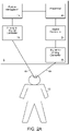
- a deep brain stimulation (DBS) system 5 for use with a patient 10 is shown.
- the DBS system 5 includes a pattern recognition system 15, a processor 20, a signal generator 25, an analog-to-digital converter 30, and a digital to analog converter 35.
- the DBS system 5 is shown in Fig. 2A as including separate discrete blocks (e.g., 15, 20, 25, 30, and 35), other configurations are possible.
- the functionality of one or more of the blocks 15, 20, 25, 30, and 35 can be combined into a single device and/or routine.
- 2A includes a number of discrete blocks (e.g., 15, 20, 25, 30, and 35), certain blocks may be omitted in some configurations (e.g., pattern recognition system 15 and analog-to-digital converter 30 can be omitted in non-closed loop systems).
- certain blocks may be omitted in some configurations (e.g., pattern recognition system 15 and analog-to-digital converter 30 can be omitted in non-closed loop systems).
- the analog-to-digital converter 30 is configured to receive signals from the brain of the patient 10 via an electrical lead 40, and/or any other device that can measure neuronal activity (e.g., functional scanners).
- the electrical lead 40 can be configured to be implanted intracranially in the brain of the patient 10, although, the electrical lead 40 can be configured to measure electrical activity of the patient 10 in other areas (e.g., the VPL, hippocampus, and/or brain stem).
- the electrical lead 40 is configured to be attached and/or in close proximity to a wide dynamic range (WDR) neuron in the brain of the patent 10, although other neurons can be used.
- WDR neuron is chosen as a function of or psychological correlate of the chronic pain being felt by the patient 10.
- the WDR neuron chosen can correspond to the portion of the body where the patient 10 feels chronic pain (e.g., a neuron corresponding to the right leg of a patient suffering from chronic pain in their right leg, technically defined as a receptive field).
- the electrical lead 40 is configured to detect electrical activity in the brain of the patient 10, and to relay the sensed information to the system 5.
- the analog-to-digital converter converts the signal into a form desired by the pattern recognition system 15.
- the pattern recognition system 15 is configured to monitor the signal provided by the electrical lead 40 to determine the presence of specific neuronal activity associated with chronic pain (e.g., the pain signatures identified in the exemplary data described herein).
- the pattern recognition system 15 can be configured to detect an increase in spontaneous background firing, an increase in rate of firing evoked by external stimulus (e.g., pressure or pinch), rhythmic after-discharge signaling, rhythmic oscillation, abnormal bursting.
- electrical lead 40 is configured to detect neuronal activity (e.g., a pain signature) in the sensory thalamus (ventral posterolateral, VPL) of the brain of the patient 10.
- the pattern recognition system 15 can be configured to detect at least two different major types of neuronal activity: spontaneous and evoked.
- Spontaneous activity is typically independent or temporally not associated with the presentation of an overt stimulus or identifiable cause. Spontaneous activity can best be described as an increase in the rate of spontaneous activity in pain subjects compared to naive/normal. Evoked activity is typically activity correlated with an overt stimulus or identifiable cause. Evoked activity can best be described as an increase in the rate of evoked activity in pain subjects in response to peripherally applied noxious and non-noxious cutaneous stimuli compared to naive/normal. In addition, abnormal bursting activity can occur during both spontaneous and evoked firing in pain compared to naive/normal.
- the pattern recognition system 15 is configured to communicate with the processor 20, and is configured to provide information to the processor 20 in a predetermined format over a network connection (e.g., a bus or network connection in cases where the pattern recognition system 15 is separate from the processor 20).
- the pattern recognition system 15 can be configured to perform various signal processing functions on the signals sensed from the patient 10 (e.g., frequency analysis, Fourier transform, inverse Fourier transform, filtering, denoising, threshold analysis, analysis of interspike intervals, analysis of burst cycle periods, analysis of spikes within bursts).
- the processor 20 can be configured to examine information provided by the pattern recognition system 15 to determine the appropriate response. For example, the processor 20 is configured to differentiate between various patterns that can be recognized by the pattern recognition system 15 and to determine an appropriate response. The processor 20 can differentiate between multiple recognized patterns, and determine an appropriate response strategy using, for example, a look-up table. The appropriate response can be nothing at all, or, for example, can be to cause an electrical signal to be provided to the brain of the patient 10 via an electrical lead 45.
- the processor 20 can be configured to reverse the pain signature in the brain of the patient 10 using neurostimulation (or more accurately, neuromodulation).
- neuromodulation can include the application of electricity at a predefined voltage, frequency, current, and of a predefined duration to the brain of a patient 10.
- the neuromodulation applied to the brain of the patient 10 is configured to jam the neuronal activity of the patient 10 (i.e., rather than further stimulating it).
- this neuromodulation is achieved by providing high frequency current (e.g., between 150-200 Hz, 1-3 volts, 1-3 mA, and substantially of 0.25-0.75 ms rectangular pulses of 1 second duration (assuming tissue impedance of 1000 ⁇ ).
- the pain signature can transiently be reversed back to normal.
- the neuromodulation protocol can be configured to transiently attenuate pain behavior in pain subjects to the level of that in naive/normal, while otherwise retaining tactile sensitivity. Electrical treatment is provided to the deep brain, the VPL, and/or WDR neurons.
- One exemplary treatment protocol includes electrical stimulation of the brain of the patient 10 using periodic pulses of electricity.
- intermittent pulses e.g., 1 pulse, every 1-3 hours
- each of the pulses has a brief (e.g., 1 sec) duration, high frequency (e.g., 150-200 Hz), and a low voltage (e.g., 1.5-2 V).
- each of these electrical pulses can jam the overactive circuitry in the brain of the patient 10, based on the temporal profile characterized herein. For example, for 2-3 hours, pain symptoms can be temporarily relieved after providing an electrical pulse.
- the system 5 can be open-loop and/or closed-loop.
- the system 5 can be programmed to provide electrical therapy according to a predetermined protocol (e.g., frequency, duration, voltage, amperage) without the use of the pattern recognition system 15 and the analog-to-digital converter 30.
- a predetermined protocol e.g., frequency, duration, voltage, amperage
- the treatment protocol can be stored in a memory that is connected to the processor 20.
- the system 5 preferably uses information received via, for example, the pattern recognition system 15 and the analog-to-digital converter 30 to treat cognitive, affective, and emotive neurological conditions, owing to the characterization of the pain signature described herein.
- the closed-loop system described herein discusses the use of an electrical lead 40 implanted in the brain of the patient 10, other configurations are possible (e.g., receiving diagnostic information from fMRI, or PET scanning). Additionally, while separate leads (e.g., leads 40, 45 are discussed, a single lead could instead be used for sensing and provision of the electrical signals. Additionally, the system 5 can be controlled manually (e.g., by actuating a button, or via a remote connection.
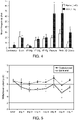
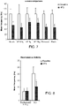
- Electrophysiological measurements of wide dynamic range (WDR) thalamic neurons in chronic constriction injury (CCI) rats indicate elevated evoked response to pressure and pinch stimuli in addition to rhythmic after-discharge signaling and an increase in spontaneous background firing ( Figs. 3 and 4 ), in addition to abnormal burst.
- Treatment efficacy was assessed in part based on the reversal of this known effect.
- Deep Brain Stimulation of CCI animals resulted in an attenuation of mean firing rate in response to all forms of mechanical stimuli, in addition to a statistically significant decrease in after-discharge ( Figs. 7 and 8 ). Furthermore, behavioral testing of awake DBS rats in group 6 revealed a corresponding increase in withdrawal latency following high frequency DBS ( Fig. 9 ; data represent values normalized to pre-HFS or Baseline 100%).
- Fig. 12 shows characterization of bursts in response to various stimuli.
- the requirements for defining bursts were:
- CCI Chronic Constrictive Injury
- the surgical procedure consisted of a modification of the original loose ligation model designed by Bennett and Xie. The process involved isolation of the sciatic nerve via blunt dissection of the biceps femoris followed by a unilateral loose ligation with 5-0 gauge chromic gut ligature at three sites above the branching of the nerve, 1 mm apart. The ligation initiates an inflammatory response that results in chromic gut constriction of the nerve. Following the surgery, overlying muscles and skin were closed with 4-0 nylon sutures and the rodents were allowed time for recovery. Thermal hyperalgesia resulting from CCI has been found to remain relatively constant for a period of 5-27 days following the injury.
- Electrophysiology Single unit firing -unit recording (i.e. sampling neuronal activity one at a time) was recorded under deep anesthesia (1.5% Isoflurane). Extracellular single-unit recordings in and were made with a 0.127 ⁇ m (0.005") 5M ⁇ Teflon-coated silver microelectrode (A-M Systems, Carlsborg, WA). DBS animals were implanted with a modified electrode as shown below ( Figs. 14 , 15, 16 ). Each subject was placed in a stereotaxic frame, and a limited craniotomy exposed the brain surface vertical to the recording sites within the VPL [Bregma (-3.3; -2.5); lateral (2.8; 3.6); vertical (5.4; 6.4)] ( Fig. 16 ). Electrical signals were amplified and filtered at 3000 Hz and processed with a CED micro 1401 data acquisition system and SPIKE-2 software (Cambridge Electronic Design, Cambridge, England).
- Waveforms were sorted to extract activity of a single neuron using automated template-matching.
- a hydraulic micropositioning device Karlin, Lav., Lav., Lav., Lav., Lav., Lav., a hydraulic micropositioning device was employed in all vertical electrode penetrations through nervous tissue. As the microelectrode is lowered into the estimated region, a single unit or neuron can be isolated by stimulating the suspected somatosensory receptive field via tapping, brushing, pinching the skin, or manipulating the limbs of the anesthetized subject until excitation at the location of the electrode tip is measured via changes in current. This process was used in order to identify VPL units innervated by the sciatic nerve. Spontaneous activity was then measured, followed by evoked responses to mechanical stimulation within the receptive field.
- DBS After measurements of at least 2 consecutive series of recording spontaneous and evoked activities, the electrode was disconnected from the recording equipment. The cathode of an isolated pulse stimulator was connected to the electrode and the anode was connected to the skin of the rat at the base of the head, acting as a body ground. Preliminary rectangular pulses 0.5 ms in width were applied at a frequency of 100 Hz at 0.5V for 1s. Immediately after stimulation, the electrode was disconnected from the stimulator and reconnected to the recording equipment and recording resumed. Background activity was recorded for 40s and progressive mechanical stimuli as described above were then applied for a duration of 20s each with 40s rest in between each.
- Behavioral tests of the CCI rats were performed with respect to thermal and mechanical stimulation in order to verify the presence of allodynia and hyperalgesia. Each animal was placed in a Plexiglas chamber situated on an elevated glass plate 30 minutes prior to testing for acclimatization.
- the thermal behavioral test consists of focusing a radiant heat source (4.7 amps) through the glass floor onto the plantar surface of the rat's hind limb, resulting in withdrawal behavior.
- the measured withdrawal latency is defined to begin at the onset of laser beam exposure and end upon movement of the rat hind paw from the floor surface. Five stimulation pulse events separated by 5 min were averaged for each hindpaw and reported as the withdrawal latency for a given session.
- DBS electrodes were held in place with orthodontic resin and microelectrode leads were stored in a small plastic container surgically implanted at the base of the skull during behavioral trials. All DBS behavioral testing animals received initial stimulation at 1.5V and 200 Hz within 6-8 days post surgery. Baseline pre-operative behavioral data was recorded for analysis beginning one day prior to initial neurostimulation. Following the DBS event, behavioral tests were repeated 5 minutes and 30 minutes post treatment.
- supplementary postmortem tissue analysis was used to identify the activation levels of glial cells in the region of interest. This provided the opportunity to assess microgliosis and astrogliosis associated with glial scarring.
- animals are anesthetized (5% isoflurane) and transcardially perfused with ice cold phosphate buffered saline (PBS) supplemented with 10 USP units of anticoagulant heparin sulfate for 5 minutes (10ml/min) followed by cooled 4% paraformaldehyde (PFA) in PBS for 5 minutes (10 ml/min). This fixation process was used in order to preserve nervous tissue from degradation.
- PBS ice cold phosphate buffered saline
- PFA paraformaldehyde
- the head was stored in PFA over night.
- the brains were then removed and stored in cold 30% sucrose until fully impregnated.
- the formalin-fixed brains were blocked in the desired orientation and placed in tissue-embedding media (O.C.T. Compound 4583, Tissue-Tek).
- Brains were stored at -62 degrees Celsius (-80 defrees Fahrenheit) and cut into 30 ⁇ m sections with a microtome. These sections (ranging from Bregma -2.12mm to -4.16mm) were mounted on slides, dried, and stained with OX-42 or GFAP for further analysis of microglia or actrocytes, respectively.
- the subject matter described herein can be implemented in digital electronic circuitry, or in computer software, firmware, or hardware, including the structural means disclosed in this specification and structural equivalents thereof, or in combinations of them.
- the subject matter described herein can be implemented as one or more computer program products, such as one or more computer programs tangibly embodied in an information carrier (e.g., in a machine-readable storage device), or embodied in a propagated signal, for execution by, or to control the operation of, a data processing apparatus (e.g., a programmable processor, a computer, or multiple computers).
- a computer program (also known as a program, software, software application, or code) can be written in any form of programming language, including compiled or interpreted languages, and it can be deployed in any form, including as a stand-alone program or as a module, component, subroutine, or other unit suitable for use in a computing environment.
- a computer program does not necessarily correspond to a file.
- a program can be stored in a portion of a file that holds other programs or data, in a single file dedicated to the program in question, or in multiple coordinated files (e.g., files that store one or more modules, sub-programs, or portions of code).
- a computer program can be deployed to be executed on one computer or on multiple computers at one site or distributed across multiple sites and interconnected by a communication network.
- processors suitable for the execution of a computer program include, by way of example, both general and special purpose microprocessors, and any one or more processor of any kind of digital computer.
- a processor will receive instructions and data from a read-only memory or a random access memory or both.
- the essential elements of a computer are a processor for executing instructions and one or more memory devices for storing instructions and data.
- a computer will also include, or be operatively coupled to receive data from or transfer data to, or both, one or more mass storage devices for storing data, e.g., magnetic, magneto-optical disks, or optical disks.
- Information carriers suitable for embodying computer program instructions and data include all forms of non-volatile memory, including by way of example semiconductor memory devices, (e.g., EPROM, EEPROM, and flash memory devices); magnetic disks, (e.g., internal hard disks or removable disks); magneto-optical disks; and optical disks (e.g., CD and DVD disks).
- semiconductor memory devices e.g., EPROM, EEPROM, and flash memory devices
- magnetic disks e.g., internal hard disks or removable disks
- magneto-optical disks e.g., CD and DVD disks
- optical disks e.g., CD and DVD disks.
- the processor and the memory can be supplemented by, or incorporated in, special purpose logic circuitry.
- the subject matter described herein can be implemented on a computer having a display device, e.g., a CRT (cathode ray tube) or LCD (liquid crystal display) monitor, for displaying information to the user and a keyboard and a pointing device, (e.g., a mouse or a trackball), by which the user can provide input to the computer.
- a display device e.g., a CRT (cathode ray tube) or LCD (liquid crystal display) monitor
- a keyboard and a pointing device e.g., a mouse or a trackball
- Other kinds of devices can be used to provide for interaction with a user as well.
- feedback provided to the user can be any form of sensory feedback, (e.g., visual feedback, auditory feedback, or tactile feedback), and input from the user can be received in any form, including acoustic, speech, or tactile input.
- the subject matter described herein can be implemented in a computing system that includes a back-end component (e.g., a data server), a middleware component (e.g., an application server), or a front-end component (e.g., a client computer having a graphical user interface or a web browser through which a user can interact with an implementation of the subject matter described herein), or any combination of such back-end, middleware, and front-end components.
- the components of the system can be interconnected by any form or medium of digital data communication, e.g., a communication network. Examples of communication networks include a local area network (LAN) and a wide area network (WAN), e.g., the Internet.
- LAN local area network
- WAN wide area network
- Example 1 Single-unit physiology in the ventral posterolateral nucleus of the thalamus in neuropathic rats
- Neuropathic pain secondary to nerve injury is often chronic and accompanied by dysesthesias. It is linked to long-term changes in neuronal physiology, known as neuroplasticity, which is well described in peripheral nerves and the spinal cord but relatively less understood in the brain. In spite of recent advances in pharmacotherapy, neuropathic pain remains poorly managed.
- VPL ventral posterolateral
- Nociceptive neurons in the VPL receive ascending projections mainly from spinothalamic tract neurons and project to several cortical areas including the primary somatosensory cortex.
- a group of neurons responds to a wide dynamic range (WDR) of mechanical stimuli, phenotypically homologous to WDR neurons at the spinal cord level whose role in central sensitization and chronic pain is well documented.
- WDR wide dynamic range
- DBS deep brain stimulation
- FDA Food and Drug Administration
- stimulation frequency represents a key factor, with high frequency stimulation (HFS, >100 Hz) mimicking the functional effects of ablation, also referred to as jamming of local circuitry.
- HFS in the VPL reduces mechanical allodynia in rats with peripheral neuropathy.
- HFS can be used to inhibit hyperactive VPL neurons, thus reversing neuroplasticity and, consequently, behavioral hypersensitivity.
- the firing of single units extracellularly from VPL neurons in naive rats was recorded.
- CCI chronic constriction injury
- CCI Chronic constriction injury
- the head was fixed in a stereotaxic apparatus (Kopf Instruments, Tujunga, CA, USA) and skin incision and a limited craniotomy exposed the brain surface vertical to the recording sites within the thalamus.
- Neuronal units were isolated from the VPL nuclei of the thalamus [respective stereotaxic coordinates in mm: Bregma (-3.3, -2.5); lateral (2.8, 3.6); vertical (5.4, 6.4)]. Extracellular single-unit recordings were made with a 5 M ⁇ Teflon-coated tungsten microelectrode (A-M Systems, Carlsborg, WA, USA).
- Cursors were also set at the beginning of the trace and after 40 s (baseline or un-evoked firing), and the spikes occurring during this period were summed to provide a measure of the background activity. The two sums were divided by the respective duration and the resulting averages (spikes/s) subtracted to yield the value attributed to the response (total number of spikes/s in excess of the background activity during the stimulus).
- spikes/s averages subtracted to yield the value attributed to the response (total number of spikes/s in excess of the background activity during the stimulus).
- One to two neurons with individually mapped receptive field were recorded from each rat. Neuronal activity was analyzed off-line using Spike2.
- the spinal cord was also exposed by laminectomy at thoracic (T4-T6) level and topical 2% lidocaine was applied using a cotton pledget to the dorsal and lateral surfaces of the spinal cord, followed 10 min later by complete cord transection using fine scissors while recording from the VPL neuron continued.
- Butson CR Maks CB, McIntyre CC (2006) Sources and effects of electrode impedance during deep brain stimulation. Clin Neurophysiol 117:447-454 ; Butson CR, McIntyre CC (2006) Role of electrode design on the volume of tissue activated during deep brain stimulation. J Neural Eng 3:1-8 ; Butson CR, McIntyre CC (2008) Current steering to control the volume of tissue activated during deep brain stimulation.
- Burst events were identified using Spike 2 Burst script using the following criteria: 6 ms of the maximum interval between two events that signifies the start of a burst, 9 ms of the longest interval between two events within a burst, and 2 of the minimum number of events in a burst. The following parameters were calculated during the recording periods of background activity, six natural mechanical stimuli evoked discharges and after-discharge: number
- Micro-stimulation in the VPL To test the effect of micro stimulation on neuronal firing in anesthetized rats, the same recording electrode was used for micro stimulation within the VPL. After identifying single units and recording pre-stimulation (baseline) firing rates, the electrode was disconnected from the recording equipment and connected to a stimulator (A-M Systems Isolated Pulse Stimulator). Rectangular pulses 0.5 ms width were applied at a frequency of 100 Hz at 0.5 V for 1 s. Immediately after stimulation, the electrode was disconnected from the stimulator and reconnected to the recording equipment and data acquisition resumed within 2-3 min after baseline measurements.
- A-M Systems Isolated Pulse Stimulator A-M Systems Isolated Pulse Stimulator
- bipolar silver wires (-1.5 k ⁇ ) were chronically implanted on the day of CCI. Electrode segments were fused together with Epoxy resin forming cathode and anode. Teflon coating was removed exposing 0.5 mm on each electrode tip, which were separated by 1 mm from tip-to-tip vertically. Initially, the longer tip was connected to the recording equipment and Local Field Potentials were recorded to localize the VPL area with a receptive field in the contralateral hindpaw (see description below for additional details on the orientation of the electrodes relative to the VPL for optimal steering of electric field and overlap with VPL nuclear structure).
- Rats were anesthetized with pentobarbital (60 mg/kg, i.p.) then perfused intracardially with ice-cold PBS followed by buffered 4% paraformaldehyde. Brains were post-fixed with buffered 4% paraformaldehyde overnight, equilibrated in 30% sucrose, and frozen to -80°C in OCT cryogenic compound (TissueTek Sakura). Coronal sections (30 ⁇ m) were adhered to glass slides and blocked with goat serum. Sections were stained for OX-42 (Santa Cruz Biotech, mouse IgG, 1:50), or GFAP (Chemicon International, mouse IgG, 1:100) overnight at 4°C.
- Extracellularly recorded action potentials were isolated from single units in the VPL nucleus of the thalamus under deep anesthesia using template matching techniques. All units discharged action potentials at relatively constant voltage amplitudes and responded with increased firing rates to contralateral mechanical stimuli.
- a peristimulus time histogram shows increased firing rate when noxious and non-noxious stimuli are applied to the receptive field in the contralateral hindpaw, noting a graded response to increased strengths of von Frey filaments, compared to almost absent spontaneous firing ( Fig 18A , upper panel). Seven days after CCI, evoked responses to pressure and pinch stimuli are increased, while spontaneous activity and afterdischarge are elevated ( Fig 18A , lower panel).
- Plasticity manifests as selective hyperexcitability in response to non-noxious pressure and noxious pinch, in addition to an emergence of un-evoked firing at a high rate.
- evoked responses to von Frey filaments 0.6, 8 and 15 g
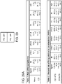
- pressure and pinch of VPL neurons with somatic receptive fields excluding the hindpaws in rats with CCI were not significantly different from corresponding values in naive rats, except for a significant increase in brush-evoked responses after CCI, whereas spontaneous activity and after-discharge were less than 1 Hz in both naive and CCI rats ( Fig. 25 ).
- Fig. 19A shows a peristimulus rate histogram of the same unit superimposed over a sinusoidal curve during the period of rhythmic oscillation (before transection) compared to nearly flat rate histogram after transection.
- HFS HFS was delivered through the recording electrode and the rate of firing was measured before and after micro stimulation.
- graded attenuation of evoked firing was achieved with incremental increase in voltage amplitude at 100 Hz and a prominent effect was noted at 1.5 V, whereas spontaneous activity was not affected ( Fig. 20A ).
- Results with 100 Hz and 200 Hz were comparable and therefore pooled together collectively. All mean firing rates were significantly decreased up to approximately -50% within 10-15 min after HFS for all firing modes except spontaneous activity ( Fig. 20C and Table 2).
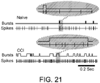
- Fig. 21 shows examples of burst firing during brush-evoked responses from a naive rat and another rat seven days after CCI.
- spontaneous bursts events were almost absent, whereas those in rats with CCI were increased in number ( Fig. 22 and Table 3).
- Spontaneous bursts were detected in only 2 out of 14 units from naive rats (mean burst events 0.4 ⁇ 0.3). Therefore burst parameters during spontaneous activity in naive rats were not analyzed.
- burst events were increased significantly for all firing modes in rats with CCI compared to those in naive rats, which were attenuated after HFS with significant changes for pressure and pinch-evoked responses ( FIG. 22 and Table 3). Similar trends were observed between groups for per cent spikes in burst values, whereas changes in the opposite directions were noted for mean interburst time values (i.e. values decreased for all firing modes in rats with CCI compared to those in naive rats and were increased after HFS). In general, therefore, bursting patterns consistently deviated from normal after sciatic neuropathy reaching significant levels. These changes were consistently reversed in the normal direction after HFS, indicating that HFS reverses several aberrant features of neuroplasticity including tonic and burst firing properties.
- withdrawal latency in the injured hindpaw remained decreased compared to the non-injured hindpaw ( Fig. 24B ).
- withdrawal latency in the injured hindpaw was transiently reversed 5 min after HFS with reproducible effects after 2 hr.
- the mean withdrawal latency was transiently and significantly reversed from 6.3 ⁇ 0.9 s to 9.1 ⁇ 0.1 s and 7.3 ⁇ 0.1 s at 5 min and 10 min after HFS, respectively, in the injured hindpaw.
- the withdrawal latency in the non-injured hindpaw also increased after HFS, however, this change was not significant ( Fig. 27 ).
- GFAP and OX-42 were quantified in tissue sections in or around the tip of the chronically implanted electrodes in the VPL, which revealed no significant changes in expression compared to the contralateral VPL ( Fig. 28 ), indicating limited glial reactivity to electrode implantation and HFS.
- Peripheral neuropathic injury is associated with neuroplasticity of VPL neurons with receptive fields in the contralateral injured hindpaw.
- Abnormal physiologic properties include higher rate of spontaneous firing, increased rates of afterdischarge and evoked firing in response to non-noxious and noxious mechanical stimuli.
- significant changes in burst firing and rhythmic oscillations were observed. Otherwise, the firing of neurons with receptive fields elsewhere in the body was not changed, except for increased responses to brush stimuli.
- VPL neurons Hyper-responsiveness, rhythmic oscillations and bursting in VPL neurons are observed in a rat model of central neuropathic pain following spinal cord contusion. Relative to neuronal firing in normal rats, firing in rats with spinal lesion alternated between simple, burst and spindle epochs, whereas the number of spikes per burst decreased and interburst interval was not changed. Compared to this study, different lesion sites (sciatic versus spinal) likely account for the incompletely matching patterns of aberrant firing in the VPL. However, spontaneous neuronal discharge in rats with peripheral or central neuropathy remained high after complete transection of the spinal cord, suggesting these changes in specific are likely intrinsic to VPL neurons, while rhythmic oscillations described in this study depend on on-going ascending input.
- burst spikes and isolated spikes both contribute to the coding of stimulus-related information ( Middleton JW, Yu N, Longtin A, Maler L (2011) Routing the flow of sensory signals using plastic responses to bursts and isolated spikes: experiment and theory. J Neurosci 31:2461-2473 ). Therefore, burst firing is an important feature of the neural code and can effectively impact postsynaptic neurons, including upstream from the VPL in the somatosensory cortex. Bursting can be modulated by the temporal frequency content of incoming signals, which in the case of peripheral neuropathy is significantly altered at peripheral and spinal cord levels.
- bursts and the relative timing of burst spikes can encode specific stimulus features. Neurons in the lateral geniculate nucleus discharge bursts and isolated spikes in response to visual stimuli, with different stimulus conditions preferentially evoking different bursting patterns. Bursting has also been reported in thalamocortical projection cells, including the auditory and the somatosensory systems. Although the functional significance of bursting in VPL neurons and modulation thereof under chronic pain conditions are unknown, interestingly, short-term plasticity of thalamocortical synapses is thought to accentuate the sensitivity of cortical responses to ascending sensory input.
- Burst firing has been documented in the ventral thalamus in a patient with root injury and occurred during episodes of self-reported touch-evoked allodynia.
- Direct micro stimulation within a thalamic nucleus that contains abnormally active neurons produces burning dysesthesia in patients with spinal deafferentation pain, as well as with root injury pain, whereas micro stimulation in the same area might also produce pain relief.
- These observations suggest that the aberrant physiology of thalamic cells may be directly related to the dysesthesias, whereas the functional outcome depends on the pain condition and the neurostimulation protocol. It is worth noting that changes in the patterns of burst firing, rather than burst firing pre se, might be a reliable marker for pain-related plasticity. For example, burst firing can occur in deafferented subjects without pain.
- HFS in CCI rats caused a 'reversal' effect on abnormal bursting patterns in the direction of normal condition, showing consistent results across all firing modalities. This is in agreement with the premise that HFS in the therapeutic range for motor disorders works mainly by inhibiting neuronal firing or neuronal jamming.
- the data described herein relates to hyperactive neurons in the VPL and a comparison of the effects of HFS and LFS on neuronal firing at the single unit level. Whereas HFS effectively reversed neuronal hyper-responsiveness, spontaneous activity did not change and overall responsiveness to mechanical stimuli was diminished but not abolished. Prior to the study no study compared the effects of varying stimulation frequency on analgesia.
- Neurons in the PAG known to mediate descending anti-nociceptive control unto lumbar dorsal horn neurons, could be activated by electrical stimulation in the VPL, an influence which is not affected by systemic administration of naloxone, suggesting that the VPL-PAG analgesic pathway is unlikely to involve the opioid system.
- This mechanism provides an alternative explanation to the widely held idea that the analgesic effects of subcortical brain stimulation is the result of activation of a descending pain suppressive system concentrated in the periventricular and periaqueductal regions, which blocks nociceptive transmission at the level of the spinal cord.
- VPL stimulation may cause sufficient input to the somatosensory cortex to activate corticofugal inhibitory pathways, following thalamocortical-corticofugal projections inhibiting spinal cord nociceptive neurons. It is also possible that VPL stimulation activates antidromically axon collaterals of STT neurons projecting to the medial brain stem, including the PAG and nucleus raphe magnus, thus resulting in analgesic effects.
- Low-voltage stimulation in the VPL at high frequency can locally inhibit VPL neurons and subsequently the transmission of nociceptive signals to cortical areas.
- the mechanisms include one or a combination of causes such as jamming of a local nociceptive circuitry, activation of inhibitory structures within a wider network, blockade of membrane ion channels such as voltage-gated currents, depolarization blockade, synaptic exhaustion, and induction of early genes.
- Neuropathic pain is a neurological disorder that previously lacked a reliable diagnostic.
- the systems presented provide a solution to a long-standing problem in pain management.
- An objective measure for pain leads to more accurate diagnosis, monitoring and optimizing therapeutics, thus ameliorating overall clinical outcome.
- a physiological correlate of pain is useful in the design of closed-loop neuromodulation systems.
- LFP Local field potential
- multiunit activity are extracellularly recorded signals from a local network of neurons.
- LFP represents the low-frequency ( ⁇ 0.5 kHz) content of the raw recording generated by membrane currents of the neurons in a local neighborhood of the recording electrode, whereas the high frequency (>1 kHz) content represents neuronal action potential spiking.
- the coordinated activity of cell populations is reflected by regular oscillations in LFPs, proposed as a mechanism to optimize the recording and analysis of network functions.
- LFP is a good candidate for a pain signature.
- field potentials Initially studied in great detail owing to their ease of recording with non-invasive macroelectrodes, field potentials contain informative signals measured out of the pool of a large number of spiking neurons while relating to sensory and motor phenomena. Changes in field potentials are triggered by external events (evoked potentials) when induced by a stimulus (e.g. noxious mechanical stimulus or non-noxious tactile stimulus) or by internal cortical dynamics (intrinsic oscillations or rhythms) due to thought processes (e.g. spontaneous pain).
- evoked potentials induced by a stimulus
- cortical dynamics intrinsic oscillations or rhythms
- LFP intracortically
- EcoG electrodes supracortically
- EEG scalp
- MEG magnetoencephalograhy
- the amount of information contained within a brain signal may diminish as it passes from intra- to extra- cellular, from single to multiunit to field potentials, and from LFP to ECoG to EEG to MEG. Many have recently argued in favor of balancing optimal information content with less invasive recording for translational purposes.
- Figs. 30 , 31 , 32 Data showing pain-related brain activity is described below ( Figs. 30 , 31 , 32 .
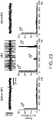
- Characteristic burst firing is recorded from the thalamus of patients with neuropathic pain ( Fig. 30 ). This bursting pattern closely resembles that recorded from the thalamus of an animal model of neuropathic deafferentation pain ( Fig. 31 ), whereby reversal of this neuronal activity was associated with attenuation of pain behavior.
- a series of psychiatric disorders may be associated with thalamocortical dysrhythmia (TCD).
- TCD thalamocortical dysrhythmia
- Synchrony of thalamic and cortical networks under normal conditions are thought to underlie consciousness, whereby state-dependant (sensory or cognitive state) rhythms of thalamocortical relay neurons are continuously driven by corticothalamic input resulting in oscillation, thus binding sensory or cognitive events in a unified percept.
- Some neurological disorders can lead to disregulation of calcium channels in the brain, thus causing abnormal calcium spiking and high frequency bursting in thalamic reticular nuclei, paradoxically driving the thalamocortical network into lower frequency oscillation (4-10 Hz) ( Fig. 32 ).
- LFP which is intermediate in terms of information content
- Pain-related LFP can be reversed by spinal cord stimulation (SCS) or peripheral nerve stimulation (PNS).
- the pain model utilized chronic constriction injury of the sciatic nerve. This is a widely used model of mononeuropathy that yields reliable pain behavior in rats. Rats are deeply anesthetized with isoflurane (1.5-2%) and the sciatic nerve is loosely ligated with 4 chromic gut (4-0) sutures. After CCI, the overlying muscles and skin are closed in layers with 4-0 nylon sutures.
- Pain behavioral is tested daily to determine mechanical and thermal sensory thresholds. Pre-operative testing generally begins 2 days prior to CCI and once per day for 10 days after CCI.
- Mechanical sensory thresholds are determined by the standard Dixon up-down method utilizing a series of von Frey filaments applied to the glabrous surface of the paw.
- Thermal hyperalgesia is measured by latency of paw withdrawal in response to a radiant heat source. Rats are placed in Plexiglas boxes on an elevated glass plate under which a radiant heat source (4.7 amps) is applied to the plantar surface of the paw and rats are free to escape from applied stimuli. The temperature that the paw can be subjected to can reach a maximum of 57°C for a transient time of less than 3 sec. Rats that do not display significant pain behavior are excluded from the study.
- Electrophysiology evaluations are carried out as follows. After confirming pain behavior, a minimal craniotomy at day 7 after CCI/sham under deep anesthesia (3% isoflurane) exposes the cortical surface vertically above the VPL or somatosensory cortex (SI) identified according to a rat brain atlas. LFP recordings (CED 1401) are made using a tungsten microelectrode (125 ⁇ m, 12 M ⁇ ) by low-pass filtering of the extracellular field potential below 300 Hz. Microelectrodes are positioned stereotaxically in areas of the VPL or SI with identifiable receptive fields in the injured dermatome (hindpaw on the side of CCI).
- LFP recordings were made under deep barbiturate anesthesia from the ventral posterolateral (VPL) nucleus of the thalamus bilaterally, with the side ipsilateral to injury serving as control (receiving ascending information predominantly from the un-injured paw).
- VPL ventral posterolateral
- the location of the microelectrode was confirmed by stereotaxic coordinates (according to a rat brain Atlas) and by increased background firing in response to gentle brushing of the corresponding receptive field in the contralateral paw ( Fig. 33 ).
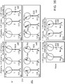
- Fig. 34 A reported leftward shift in MEG power spectrum of spontaneous activity in patients with chronic pain compared to control is in agreement with Fig. 34 (left panels).
- Thalamic LFP recordings from patients with Tourette Syndrome a cognitive/movement disorder, emphasized a significant correlation between the main LFP frequency and the frequency of single-unit interbursts.
- Data described herein shows that the mean interburst period of single-unit brush-evoked activity recorded from the VPL is approximately 400 ms for animals with CCI and 100 ms for naive, thus predicting a rightward shift in power spectra from 2.5 to 10 Hz after CCI, exactly matching with what we show in Fig. 34 (right panels).
Landscapes
- Health & Medical Sciences (AREA)
- Life Sciences & Earth Sciences (AREA)
- Animal Behavior & Ethology (AREA)
- Veterinary Medicine (AREA)
- Engineering & Computer Science (AREA)
- Biomedical Technology (AREA)
- Public Health (AREA)
- General Health & Medical Sciences (AREA)
- Radiology & Medical Imaging (AREA)
- Nuclear Medicine, Radiotherapy & Molecular Imaging (AREA)
- Neurosurgery (AREA)
- Neurology (AREA)
- Biophysics (AREA)
- Psychiatry (AREA)
- Molecular Biology (AREA)
- Medical Informatics (AREA)
- Heart & Thoracic Surgery (AREA)
- Pathology (AREA)
- Physics & Mathematics (AREA)
- Surgery (AREA)
- Pain & Pain Management (AREA)
- Psychology (AREA)
- Hospice & Palliative Care (AREA)
- Physiology (AREA)
- Electrotherapy Devices (AREA)
- Measurement And Recording Of Electrical Phenomena And Electrical Characteristics Of The Living Body (AREA)
- Medicines That Contain Protein Lipid Enzymes And Other Medicines (AREA)
Claims (5)
- Système comprenant :un processeur (20) configuré pour être couplé à un fil électrique (40) qui est configuré pour détecter une activité électrique chez un patient ;une mémoire couplée au processeur, la mémoire contenant des instructions lisibles par ordinateur qui, lorsqu'elles sont exécutées par le processeur, amènent le processeur à :déceler une signature de douleur dans l'activité électrique détectée ;déterminer un protocole de traitement en réponse à la signature de douleur décelée ; etamener le protocole de traitement à être administré au patient via le fil électrique ;dans lequel le processeur est apte à déceler la signature de douleur dans l'activité électrique détectée en décelant une signature de couleur comprenant un profil de déclenchement de salves, chaque salve dudit déclenchement de salves comprenant au moins 10 fois le nombres de pics par rapport à un profil de non-douleur de contrôle etdans lequel ledit déclenchement de salves comprend :(a) un intervalle maximal signifiant un début de salve de 6 ms ; et(b) un intervalle maximal inter-pic de 9 ms ; et(c) une augmentation la plus longue de l'intervalle inter-pic dans une salve de 2 ms ; et(d) un nombre minimal de pics dans une salve de 2.
- Système selon la revendication 1, dans lequel le profil de déclenchement neuronal est obtenu à partir d'un seul neurone ou d'une pluralité de neurones.
- Système selon la revendication 1 ou la revendication 2, dans lequel une signature de douleur aberrante est caractérisée par une oscillation de rythme spontané d'une période présentant environ 30-80 pics/salve.
- Système selon la revendication 1, dans lequel la signature de douleur est une activité spontanée ou activité suscitée.
- Système selon la revendication 1, dans lequel le processeur est en outre apte à déceler un profil de déclenchement neuronal, ledit profil comprenant une réponse suscitée élevée aux stimuli, une signalisation post-décharge rythmique et un déclenchement d'arrière-plan spontané augmenté.
Applications Claiming Priority (2)
| Application Number | Priority Date | Filing Date | Title |
|---|---|---|---|
| US32858310P | 2010-04-27 | 2010-04-27 | |
| PCT/US2011/034203 WO2011137193A1 (fr) | 2010-04-27 | 2011-04-27 | Gestion de la douleur |
Publications (3)
| Publication Number | Publication Date |
|---|---|
| EP2563462A1 EP2563462A1 (fr) | 2013-03-06 |
| EP2563462A4 EP2563462A4 (fr) | 2014-02-12 |
| EP2563462B1 true EP2563462B1 (fr) | 2022-12-28 |
Family
ID=44861905
Family Applications (1)
| Application Number | Title | Priority Date | Filing Date |
|---|---|---|---|
| EP11775539.7A Active EP2563462B1 (fr) | 2010-04-27 | 2011-04-27 | DISPOSITIF POUR DÉTECTER UNE SIGNATURE DE LA DOULEUR SUR LA BASE DE CHANGEMENTS DANS l'ÉMISSION DE TRAINS D'ONDES |
Country Status (6)
| Country | Link |
|---|---|
| US (2) | US8977362B2 (fr) |
| EP (1) | EP2563462B1 (fr) |
| JP (1) | JP2013525029A (fr) |
| AU (1) | AU2011245359B2 (fr) |
| CA (1) | CA2797701C (fr) |
| WO (1) | WO2011137193A1 (fr) |
Families Citing this family (73)
| Publication number | Priority date | Publication date | Assignee | Title |
|---|---|---|---|---|
| US9402558B2 (en) | 2007-04-05 | 2016-08-02 | New York University | System and method for pain detection and computation of a pain quantification index |
| US8750857B2 (en) * | 2010-06-04 | 2014-06-10 | Qualcomm Incorporated | Method and apparatus for wireless distributed computing |
| US11413458B2 (en) | 2011-05-19 | 2022-08-16 | Neuros Medical, Inc. | Nerve cuff electrode for neuromodulation in large human nerve trunks |
| US10279179B2 (en) | 2013-04-15 | 2019-05-07 | Neurometrix, Inc. | Transcutaneous electrical nerve stimulator with automatic detection of user sleep-wake state |
| US11247040B2 (en) | 2011-11-15 | 2022-02-15 | Neurometrix, Inc. | Dynamic control of transcutaneous electrical nerve stimulation therapy using continuous sleep detection |
| US11259744B2 (en) | 2011-11-15 | 2022-03-01 | Neurometrix, Inc. | Transcutaneous electrical nerve stimulator with automatic detection of leg orientation and leg motion for enhanced sleep analysis, including enhanced transcutaneous electrical nerve stimulation (TENS) using the same |
| US10112040B2 (en) | 2011-11-15 | 2018-10-30 | Neurometrix, Inc. | Transcutaneous electrical nerve stimulation using novel unbalanced biphasic waveform and novel electrode arrangement |
| US9827420B2 (en) | 2013-03-29 | 2017-11-28 | Neurometrix, Inc. | Transcutaneous electrical nerve stimulator with user gesture detector and electrode-skin contact detector, with transient motion detector for increasing the accuracy of the same |
| US10335595B2 (en) | 2011-11-15 | 2019-07-02 | Neurometrix, Inc. | Dynamic control of transcutaneous electrical nerve stimulation therapy using continuous sleep detection |
| CN107376115A (zh) | 2011-11-15 | 2017-11-24 | 神经系统检测公司 | 用于使用经皮电神经刺激减轻疼痛的设备和方法 |
| US9675801B2 (en) | 2011-11-15 | 2017-06-13 | Neurometrix, Inc. | Measuring the “on-skin” time of a transcutaneous electrical nerve stimulator (TENS) device in order to minimize skin irritation due to excessive uninterrupted wearing of the same |
| US9119965B2 (en) | 2012-04-09 | 2015-09-01 | Pacesetter, Inc. | Systems and methods for controlling spinal cord stimulation to improve stimulation efficacy for use by implantable medical devices |
| US9833614B1 (en) | 2012-06-22 | 2017-12-05 | Nevro Corp. | Autonomic nervous system control via high frequency spinal cord modulation, and associated systems and methods |
| KR20150085007A (ko) | 2012-11-13 | 2015-07-22 | 엘마인다 리미티드 | 시공간 분할을 이용한 신경 생리학적 데이터 분석 |
| US10940311B2 (en) | 2013-03-29 | 2021-03-09 | Neurometrix, Inc. | Apparatus and method for button-free control of a wearable transcutaneous electrical nerve stimulator using interactive gestures and other means |
| ES2879286T3 (es) | 2013-03-29 | 2021-11-22 | Gsk Consumer Healthcare Sarl | Detección de desprendimiento de los electrodos cutáneos mediante la impedancia electrocutánea |
| CN105431196B (zh) * | 2013-04-15 | 2019-02-01 | Gsk消费者健康有限公司 | 具有用户睡眠—觉醒状态的自动检测的经皮电神经刺激器 |
| US10327663B2 (en) * | 2013-08-31 | 2019-06-25 | Alpha Omega Neuro Technologies Ltd. | Evoked response probe and method of use |
| JP6033754B2 (ja) * | 2013-10-21 | 2016-11-30 | 株式会社クラレ | 多層中間膜 |
| EP3068295A4 (fr) * | 2013-11-13 | 2017-07-19 | Elminda Ltd. | Procédé et système de gestion de la douleur |
| EP4000684B1 (fr) | 2014-09-22 | 2025-05-07 | Boston Scientific Neuromodulation Corporation | Dispositifs d'utilisation d'un spectre d'énergie ou d'une association de signaux pour la gestion de la douleur |
| EP3197350B1 (fr) | 2014-09-28 | 2019-11-06 | Elminda Ltd. | Configuration d'outil de stimulation cérébrale |
| HK1245051A1 (zh) * | 2015-01-10 | 2018-08-24 | Deborah DULLEN | 用於诊断和验证患者治疗和结果的自动功能测量的方法和装置 |
| US11103696B2 (en) | 2015-05-05 | 2021-08-31 | Cosmo Haralambidis | Device for electrical stimulation of peridontal complex and surrounding tissue |
| EP3291880B1 (fr) | 2015-05-05 | 2021-07-07 | Haralambidis, Cosmo | Dispositif pour la stimulation électrique du complexe parodontal et du tissu environnant |
| US11400252B2 (en) * | 2015-11-23 | 2022-08-02 | Sana Heath Inc. | Non-pharmaceutical method of managing pain |
| EA035285B1 (ru) | 2015-11-23 | 2020-05-25 | Сана Хелт, Инк. | Способы и системы для предоставления стимулов для головного мозга |
| US11298502B2 (en) | 2015-11-23 | 2022-04-12 | Sana Health, Inc. | Non-pharmaceutical methods of mitigating addiction withdrawal symptoms |
| US11524135B2 (en) | 2015-11-23 | 2022-12-13 | Sana Health Inc. | Non-pharmaceutical systems and methods of treating the symptoms of fibromyalgia |
| EP3484576B1 (fr) | 2016-07-13 | 2023-07-12 | NeuroMetrix, Inc. | Appareil de compensation automatisée de stimulation nerveuse électrique transcutanée pour des fluctuations temporelles telles que les rythmes circadiens |
| AU2017334841B2 (en) | 2016-09-27 | 2020-03-19 | Boston Scientific Neuromodulation Corporation | Systems and methods for closed-loop pain management |
| AU2017335497B2 (en) | 2016-09-27 | 2020-01-16 | Boston Scientific Neuromodulation Corporation | System for pain management using objective pain measure |
| US11235142B2 (en) | 2016-12-23 | 2022-02-01 | Neurometrix, Inc. | “Smart” electrode assembly for transcutaneous electrical nerve stimulation (TENS) |
| WO2018132529A1 (fr) | 2017-01-11 | 2018-07-19 | Boston Scientific Neuromodulation Corporation | Gestion de la douleur basée sur la surveillance de l'activité cérébrale |
| WO2018132535A1 (fr) | 2017-01-11 | 2018-07-19 | Boston Scientific Neuromodulation Corporation | Prise en charge de la douleur sur la base de mesures de l'expression émotionnelle |
| US11089997B2 (en) | 2017-01-11 | 2021-08-17 | Boston Scientific Neuromodulation Corporation | Patient-specific calibration of pain quantification |
| US10631777B2 (en) | 2017-01-11 | 2020-04-28 | Boston Scientific Neuromodulation Corporation | Pain management based on functional measurements |
| US20180272132A1 (en) | 2017-01-19 | 2018-09-27 | Nevro Corp. | High frequency stimulation for treating sensory and/or motor deficits in patients with spinal cord injuries and/or peripheral polyneuropathy, and associated systems and methods |
| US10960210B2 (en) | 2017-02-10 | 2021-03-30 | Boston Scientific Neuromodulation Corporation | Method and apparatus for pain management with sleep detection |
| WO2018160903A1 (fr) | 2017-03-02 | 2018-09-07 | Sana Health, Inc. | Procédés et systèmes destinés à moduler des stimuli au cerveau avec des biocapteurs |
| US11058877B2 (en) | 2017-05-30 | 2021-07-13 | Neurometrix, Inc. | Apparatus and method for the automated control of transcutaneous electrical nerve stimulation based on current and forecasted weather conditions |
| USD837394S1 (en) | 2017-07-11 | 2019-01-01 | Neurometrix, Inc. | Transcutaneous electrical nerve stimulation (TENS) device |
| EP3936185A1 (fr) | 2017-07-18 | 2022-01-12 | Boston Scientific Neuromodulation Corporation | Systèmes et procédés de gestion de la douleur basés sur des capteurs |
| WO2019060298A1 (fr) | 2017-09-19 | 2019-03-28 | Neuroenhancement Lab, LLC | Procédé et appareil de neuro-activation |
| USD865986S1 (en) | 2017-09-21 | 2019-11-05 | Neurometrix, Inc. | Transcutaneous electrical nerve stimulation device strap |
| US10589089B2 (en) | 2017-10-25 | 2020-03-17 | Epineuron Technologies Inc. | Systems and methods for delivering neuroregenerative therapy |
| EP3700617A4 (fr) | 2017-10-25 | 2021-08-04 | Epineuron Technologies Inc. | Systèmes et procédés d'administration de thérapie anti-neurodégénérative |
| WO2019099887A1 (fr) | 2017-11-17 | 2019-05-23 | Boston Scientific Neuromodulation Corporation | Systèmes et procédés pour produire une stimulation intermittente au moyen de systèmes de stimulation électrique |
| US11717686B2 (en) | 2017-12-04 | 2023-08-08 | Neuroenhancement Lab, LLC | Method and apparatus for neuroenhancement to facilitate learning and performance |
| KR102648346B1 (ko) | 2017-12-13 | 2024-03-15 | 뉴로스 메디컬 인코포레이티드 | 신경 커프 배치 장치 |
| US11446496B2 (en) | 2017-12-20 | 2022-09-20 | Galvani Bioelectronics Limited | Treatment of disorders associated with inflammation |
| WO2019122903A2 (fr) | 2017-12-20 | 2019-06-27 | Galvani Bioelectronics Limited | Stimulation d'un nerf alimentant la rate |
| WO2019122906A1 (fr) | 2017-12-20 | 2019-06-27 | Galvani Bioelectronics Limited | Traitement d'états médicaux aigus |
| WO2019122907A1 (fr) | 2017-12-20 | 2019-06-27 | Galvani Bioelectronics Limited | Traitement d'états médicaux aigus |
| US12280219B2 (en) | 2017-12-31 | 2025-04-22 | NeuroLight, Inc. | Method and apparatus for neuroenhancement to enhance emotional response |
| US11478603B2 (en) | 2017-12-31 | 2022-10-25 | Neuroenhancement Lab, LLC | Method and apparatus for neuroenhancement to enhance emotional response |
| WO2019191601A1 (fr) | 2018-03-29 | 2019-10-03 | Nevro Corp. | Modulation thérapeutique pour traiter les anomalies de glycémie, y compris le diabète de type 2, et/ou pour réduire les niveaux d'hba1c, et systèmes et méthodes associés |
| CN112188915A (zh) | 2018-04-09 | 2021-01-05 | 纽若斯医疗公司 | 用于设置电剂量的装置和方法 |
| US11364361B2 (en) | 2018-04-20 | 2022-06-21 | Neuroenhancement Lab, LLC | System and method for inducing sleep by transplanting mental states |
| USD861903S1 (en) | 2018-05-15 | 2019-10-01 | Neurometrix, Inc. | Apparatus for transcutaneous electrical nerve stimulation |
| CA3100748A1 (fr) | 2018-05-22 | 2019-11-28 | Boston Scientific Neuromodulation Corporation | Dispositif de neurostimulation electrique implantee et systeme a utiliser pour offrir un soulagement adaptatif de la douleur chronique |
| AU2019275411B2 (en) | 2018-05-22 | 2022-01-06 | Boston Scientific Neuromodulation Corporation | Adaptive electrical neurostimulation treatment to reduce pain perception |
| WO2019226656A1 (fr) * | 2018-05-23 | 2019-11-28 | Sana Health, Inc. | Procédé non pharmaceutique de gestion de la douleur |
| CN108830806B (zh) * | 2018-05-29 | 2020-12-18 | 河南科技大学 | 一种感受野模型的敏感度及模型参数动态调控方法 |
| US11452839B2 (en) | 2018-09-14 | 2022-09-27 | Neuroenhancement Lab, LLC | System and method of improving sleep |
| WO2020115326A2 (fr) | 2018-12-07 | 2020-06-11 | GSK Consumer Healthcare S.A. | Détermination intelligente d'intensité de stimulation thérapeutique pour une stimulation nerveuse électrique transcutanée |
| US11786694B2 (en) | 2019-05-24 | 2023-10-17 | NeuroLight, Inc. | Device, method, and app for facilitating sleep |
| US11247043B2 (en) | 2019-10-01 | 2022-02-15 | Epineuron Technologies Inc. | Electrode interface devices for delivery of neuroregenerative therapy |
| EP4103267B1 (fr) | 2020-02-11 | 2025-06-04 | Neuros Medical, Inc. | Système de quantification d'ensembles de données qualitatives rapportées par un patient |
| AU2021274703A1 (en) | 2020-05-18 | 2022-12-22 | Sana Health, Inc. | Methods and systems for treating anxiety and depression |
| EP4429757A4 (fr) * | 2021-11-08 | 2025-07-30 | Univ New York | Interface neuronale en boucle fermée pour le contrôle de la douleur |
| EP4547322A1 (fr) | 2022-07-01 | 2025-05-07 | Nevro Corporation | Neurostimulation pour le traitement de déficits sensoriels, et systèmes et méthodes associés |
| WO2024081829A1 (fr) * | 2022-10-13 | 2024-04-18 | Case Western Reserve University | Gestion de la douleur par stimulation de la moelle épinière ventrale |
Family Cites Families (12)
| Publication number | Priority date | Publication date | Assignee | Title |
|---|---|---|---|---|
| US8008283B2 (en) | 1998-12-23 | 2011-08-30 | Neurotherapeutics Pharma, Inc. | Methods and compositions for the treatment of neuropsychiatric disorders |
| US6708064B2 (en) * | 2000-02-24 | 2004-03-16 | Ali R. Rezai | Modulation of the brain to affect psychiatric disorders |
| US20030100931A1 (en) * | 2001-11-28 | 2003-05-29 | Keith Mullett | Brain signal feedback for pain management |
| US20050010262A1 (en) * | 2002-02-01 | 2005-01-13 | Ali Rezai | Modulation of the pain circuitry to affect chronic pain |
| US7130691B2 (en) | 2002-06-27 | 2006-10-31 | Falci Scott P | Method for eradicating pain of central origin resulting from spinal cord injury |
| US7277748B2 (en) * | 2002-09-13 | 2007-10-02 | Neuropace, Inc. | Spatiotemporal pattern recognition for neurological event detection and prediction in an implantable device |
| WO2005007238A1 (fr) * | 2003-07-18 | 2005-01-27 | Campbell James N | Traitement de la douleur |
| WO2006033110A2 (fr) * | 2004-09-24 | 2006-03-30 | Dov Ehrlich | Procede et dispositif de traitement des acouphenes et d'autres troubles neurologiques par stimulation cerebrale dans le colliculus inferieur et/ou des zones adjacentes |
| WO2006057734A1 (fr) * | 2004-10-21 | 2006-06-01 | Advanced Neuromodulation Systems, Inc. | Nouvelle conception de stimulation pour la neuromodulation |
| WO2006122035A2 (fr) * | 2005-05-06 | 2006-11-16 | University Of Maryland, Baltimore | Methode de traitement de syndrome de douleur centrale ou d'induction de douleur generee centralement chez un modele animal |
| US7894905B2 (en) * | 2006-03-13 | 2011-02-22 | Neuropace, Inc. | Implantable system enabling responsive therapy for pain |
| US20070255176A1 (en) * | 2006-04-28 | 2007-11-01 | Medtronic, Inc. | Voiding detection with learning mode |
-
2011
- 2011-04-27 EP EP11775539.7A patent/EP2563462B1/fr active Active
- 2011-04-27 CA CA2797701A patent/CA2797701C/fr active Active
- 2011-04-27 JP JP2013508227A patent/JP2013525029A/ja not_active Withdrawn
- 2011-04-27 WO PCT/US2011/034203 patent/WO2011137193A1/fr not_active Ceased
- 2011-04-27 US US13/643,670 patent/US8977362B2/en active Active
- 2011-04-27 AU AU2011245359A patent/AU2011245359B2/en not_active Ceased
-
2015
- 2015-02-26 US US14/632,420 patent/US9486632B2/en active Active
Also Published As
| Publication number | Publication date |
|---|---|
| AU2011245359B2 (en) | 2015-07-09 |
| US9486632B2 (en) | 2016-11-08 |
| US20150231395A1 (en) | 2015-08-20 |
| EP2563462A1 (fr) | 2013-03-06 |
| US20130066394A1 (en) | 2013-03-14 |
| US8977362B2 (en) | 2015-03-10 |
| WO2011137193A1 (fr) | 2011-11-03 |
| EP2563462A4 (fr) | 2014-02-12 |
| CA2797701A1 (fr) | 2011-11-03 |
| CA2797701C (fr) | 2022-06-21 |
| JP2013525029A (ja) | 2013-06-20 |
Similar Documents
| Publication | Publication Date | Title |
|---|---|---|
| EP2563462B1 (fr) | DISPOSITIF POUR DÉTECTER UNE SIGNATURE DE LA DOULEUR SUR LA BASE DE CHANGEMENTS DANS l'ÉMISSION DE TRAINS D'ONDES | |
| AU2011245359A1 (en) | Pain management | |
| EP4299106B1 (fr) | Accès à un réseau vertébral pour permettre la fonction respiratoire | |
| US10835170B2 (en) | Methods for detecting neuronal oscillation in the spinal cord associated with pain and diseases or disorders of the nervous system | |
| US8918176B2 (en) | Assessing cognitive disorders based on non-motor epileptiform bioelectrical brain activity | |
| US10252056B2 (en) | Method and apparatus for preventing or terminating epileptic seizures | |
| Iwata et al. | High-frequency stimulation in the ventral posterolateral thalamus reverses electrophysiologic changes and hyperalgesia in a rat model of peripheral neuropathic pain | |
| US9399133B2 (en) | Non-invasive automated electrical control systems and methods for monitoring animal conditions | |
| Habaguchi et al. | Medullary reticulospinal tract mediating the generalized motor inhibition in cats: II. Functional organization within the medullary reticular formation with respect to postsynaptic inhibition of forelimb and hindlimb motoneurons | |
| Iyer et al. | Multi-limb acquisition of motor evoked potentials and its application in spinal cord injury | |
| MacIver et al. | Human subthalamic neuron spiking exhibits subtle responses to sedatives | |
| Lo et al. | Motor and somatosensory abnormalities are significant etiological factors for adolescent idiopathic scoliosis | |
| US20250256096A1 (en) | Systems, devices, and methods for delivering therapeutic agents for nerve stimulation | |
| Sanoja et al. | Enhanced excitability of thalamic sensory neurons and slow-wave EEG pattern after stimuli that induce spinal long-term potentiation | |
| Sabetian | Directionally Sensitive Low-Noise Neural Recording Using Tetrapolar Nerve Cuff Electrode | |
| Vespa | Neurophysiological biomarkers for vagus nerve stimulation in epilepsy | |
| Tsubokawa | The clinical value of multimodality spinal cord evoked potentials in the prognosis of spinal cord injury | |
| de Carrizosa et al. | A chronically implantable device for the controlled delivery of substances, and stimulation and recording of activity in severed nerves | |
| Lertwittayanon | Brainstem Seizures and Associated Cardiorespiratory Depression Following Intrahippocampal 4-AP Application in Freely Moving Rats | |
| Sala et al. | Intraoperative Neurophysiology During Spine and Spinal Cord Surgery in Children | |
| Alam | 2004 ANNUAL MEETING OF THE AMERICAN SOCIETY OF NEUROPHYSIOLOGICAL MONITORING | |
| Mills et al. | Use of Transcranial Magnetic Stimulation in the Investigation of Spinal and Transcortical | |
| Gholston | Microneurography as a Minimally Invasive Method to Assess Target Engagement During Neuromodulation | |
| Al Tzachmani | Regulation of wake-sleep states and state-dependent cardiovascular function in diet-induced obesity rats | |
| HK40018951B (en) | Accessing spinal network to enable respiratory function |
Legal Events
| Date | Code | Title | Description |
|---|---|---|---|
| PUAI | Public reference made under article 153(3) epc to a published international application that has entered the european phase |
Free format text: ORIGINAL CODE: 0009012 |
|
| 17P | Request for examination filed |
Effective date: 20121127 |
|
| AK | Designated contracting states |
Kind code of ref document: A1 Designated state(s): AL AT BE BG CH CY CZ DE DK EE ES FI FR GB GR HR HU IE IS IT LI LT LU LV MC MK MT NL NO PL PT RO RS SE SI SK SM TR |
|
| DAX | Request for extension of the european patent (deleted) | ||
| A4 | Supplementary search report drawn up and despatched |
Effective date: 20140114 |
|
| RIC1 | Information provided on ipc code assigned before grant |
Ipc: A61B 5/0476 20060101ALI20140108BHEP Ipc: A61N 1/36 20060101ALI20140108BHEP Ipc: A61B 5/00 20060101AFI20140108BHEP Ipc: A61B 5/0484 20060101ALI20140108BHEP |
|
| STAA | Information on the status of an ep patent application or granted ep patent |
Free format text: STATUS: EXAMINATION IS IN PROGRESS |
|
| 17Q | First examination report despatched |
Effective date: 20170519 |
|
| GRAP | Despatch of communication of intention to grant a patent |
Free format text: ORIGINAL CODE: EPIDOSNIGR1 |
|
| STAA | Information on the status of an ep patent application or granted ep patent |
Free format text: STATUS: GRANT OF PATENT IS INTENDED |
|
| INTG | Intention to grant announced |
Effective date: 20171103 |
|
| GRAJ | Information related to disapproval of communication of intention to grant by the applicant or resumption of examination proceedings by the epo deleted |
Free format text: ORIGINAL CODE: EPIDOSDIGR1 |
|
| STAA | Information on the status of an ep patent application or granted ep patent |
Free format text: STATUS: EXAMINATION IS IN PROGRESS |
|
| INTC | Intention to grant announced (deleted) | ||
| REG | Reference to a national code |
Ref country code: DE Ref legal event code: R079 Ref document number: 602011073561 Country of ref document: DE Free format text: PREVIOUS MAIN CLASS: A61N0001340000 Ipc: A61B0005000000 |
|
| GRAP | Despatch of communication of intention to grant a patent |
Free format text: ORIGINAL CODE: EPIDOSNIGR1 |
|
| STAA | Information on the status of an ep patent application or granted ep patent |
Free format text: STATUS: GRANT OF PATENT IS INTENDED |
|
| RIC1 | Information provided on ipc code assigned before grant |
Ipc: A61N 1/36 20060101ALI20220603BHEP Ipc: A61B 5/00 20060101AFI20220603BHEP |
|
| INTG | Intention to grant announced |
Effective date: 20220708 |
|
| GRAS | Grant fee paid |
Free format text: ORIGINAL CODE: EPIDOSNIGR3 |
|
| GRAA | (expected) grant |
Free format text: ORIGINAL CODE: 0009210 |
|
| STAA | Information on the status of an ep patent application or granted ep patent |
Free format text: STATUS: THE PATENT HAS BEEN GRANTED |
|
| AK | Designated contracting states |
Kind code of ref document: B1 Designated state(s): AL AT BE BG CH CY CZ DE DK EE ES FI FR GB GR HR HU IE IS IT LI LT LU LV MC MK MT NL NO PL PT RO RS SE SI SK SM TR |
|
| REG | Reference to a national code |
Ref country code: GB Ref legal event code: FG4D |
|
| REG | Reference to a national code |
Ref country code: CH Ref legal event code: EP |
|
| REG | Reference to a national code |
Ref country code: DE Ref legal event code: R096 Ref document number: 602011073561 Country of ref document: DE |
|
| REG | Reference to a national code |
Ref country code: AT Ref legal event code: REF Ref document number: 1539930 Country of ref document: AT Kind code of ref document: T Effective date: 20230115 |
|
| REG | Reference to a national code |
Ref country code: IE Ref legal event code: FG4D |
|
| REG | Reference to a national code |
Ref country code: LT Ref legal event code: MG9D |
|
| PG25 | Lapsed in a contracting state [announced via postgrant information from national office to epo] |
Ref country code: SE Free format text: LAPSE BECAUSE OF FAILURE TO SUBMIT A TRANSLATION OF THE DESCRIPTION OR TO PAY THE FEE WITHIN THE PRESCRIBED TIME-LIMIT Effective date: 20221228 Ref country code: NO Free format text: LAPSE BECAUSE OF FAILURE TO SUBMIT A TRANSLATION OF THE DESCRIPTION OR TO PAY THE FEE WITHIN THE PRESCRIBED TIME-LIMIT Effective date: 20230328 Ref country code: LT Free format text: LAPSE BECAUSE OF FAILURE TO SUBMIT A TRANSLATION OF THE DESCRIPTION OR TO PAY THE FEE WITHIN THE PRESCRIBED TIME-LIMIT Effective date: 20221228 Ref country code: FI Free format text: LAPSE BECAUSE OF FAILURE TO SUBMIT A TRANSLATION OF THE DESCRIPTION OR TO PAY THE FEE WITHIN THE PRESCRIBED TIME-LIMIT Effective date: 20221228 |
|
| REG | Reference to a national code |
Ref country code: NL Ref legal event code: MP Effective date: 20221228 |
|
| REG | Reference to a national code |
Ref country code: AT Ref legal event code: MK05 Ref document number: 1539930 Country of ref document: AT Kind code of ref document: T Effective date: 20221228 |
|
| PG25 | Lapsed in a contracting state [announced via postgrant information from national office to epo] |
Ref country code: RS Free format text: LAPSE BECAUSE OF FAILURE TO SUBMIT A TRANSLATION OF THE DESCRIPTION OR TO PAY THE FEE WITHIN THE PRESCRIBED TIME-LIMIT Effective date: 20221228 Ref country code: LV Free format text: LAPSE BECAUSE OF FAILURE TO SUBMIT A TRANSLATION OF THE DESCRIPTION OR TO PAY THE FEE WITHIN THE PRESCRIBED TIME-LIMIT Effective date: 20221228 Ref country code: HR Free format text: LAPSE BECAUSE OF FAILURE TO SUBMIT A TRANSLATION OF THE DESCRIPTION OR TO PAY THE FEE WITHIN THE PRESCRIBED TIME-LIMIT Effective date: 20221228 Ref country code: GR Free format text: LAPSE BECAUSE OF FAILURE TO SUBMIT A TRANSLATION OF THE DESCRIPTION OR TO PAY THE FEE WITHIN THE PRESCRIBED TIME-LIMIT Effective date: 20230329 |
|
| PGFP | Annual fee paid to national office [announced via postgrant information from national office to epo] |
Ref country code: GB Payment date: 20230309 Year of fee payment: 13 |
|
| PG25 | Lapsed in a contracting state [announced via postgrant information from national office to epo] |
Ref country code: NL Free format text: LAPSE BECAUSE OF FAILURE TO SUBMIT A TRANSLATION OF THE DESCRIPTION OR TO PAY THE FEE WITHIN THE PRESCRIBED TIME-LIMIT Effective date: 20221228 |
|
| P01 | Opt-out of the competence of the unified patent court (upc) registered |
Effective date: 20230605 |
|
| PG25 | Lapsed in a contracting state [announced via postgrant information from national office to epo] |
Ref country code: SM Free format text: LAPSE BECAUSE OF FAILURE TO SUBMIT A TRANSLATION OF THE DESCRIPTION OR TO PAY THE FEE WITHIN THE PRESCRIBED TIME-LIMIT Effective date: 20221228 Ref country code: RO Free format text: LAPSE BECAUSE OF FAILURE TO SUBMIT A TRANSLATION OF THE DESCRIPTION OR TO PAY THE FEE WITHIN THE PRESCRIBED TIME-LIMIT Effective date: 20221228 Ref country code: PT Free format text: LAPSE BECAUSE OF FAILURE TO SUBMIT A TRANSLATION OF THE DESCRIPTION OR TO PAY THE FEE WITHIN THE PRESCRIBED TIME-LIMIT Effective date: 20230428 Ref country code: ES Free format text: LAPSE BECAUSE OF FAILURE TO SUBMIT A TRANSLATION OF THE DESCRIPTION OR TO PAY THE FEE WITHIN THE PRESCRIBED TIME-LIMIT Effective date: 20221228 Ref country code: EE Free format text: LAPSE BECAUSE OF FAILURE TO SUBMIT A TRANSLATION OF THE DESCRIPTION OR TO PAY THE FEE WITHIN THE PRESCRIBED TIME-LIMIT Effective date: 20221228 Ref country code: CZ Free format text: LAPSE BECAUSE OF FAILURE TO SUBMIT A TRANSLATION OF THE DESCRIPTION OR TO PAY THE FEE WITHIN THE PRESCRIBED TIME-LIMIT Effective date: 20221228 Ref country code: AT Free format text: LAPSE BECAUSE OF FAILURE TO SUBMIT A TRANSLATION OF THE DESCRIPTION OR TO PAY THE FEE WITHIN THE PRESCRIBED TIME-LIMIT Effective date: 20221228 |
|
| PGFP | Annual fee paid to national office [announced via postgrant information from national office to epo] |
Ref country code: DE Payment date: 20230314 Year of fee payment: 13 |
|
| PG25 | Lapsed in a contracting state [announced via postgrant information from national office to epo] |
Ref country code: SK Free format text: LAPSE BECAUSE OF FAILURE TO SUBMIT A TRANSLATION OF THE DESCRIPTION OR TO PAY THE FEE WITHIN THE PRESCRIBED TIME-LIMIT Effective date: 20221228 Ref country code: PL Free format text: LAPSE BECAUSE OF FAILURE TO SUBMIT A TRANSLATION OF THE DESCRIPTION OR TO PAY THE FEE WITHIN THE PRESCRIBED TIME-LIMIT Effective date: 20221228 Ref country code: IS Free format text: LAPSE BECAUSE OF FAILURE TO SUBMIT A TRANSLATION OF THE DESCRIPTION OR TO PAY THE FEE WITHIN THE PRESCRIBED TIME-LIMIT Effective date: 20230428 Ref country code: AL Free format text: LAPSE BECAUSE OF FAILURE TO SUBMIT A TRANSLATION OF THE DESCRIPTION OR TO PAY THE FEE WITHIN THE PRESCRIBED TIME-LIMIT Effective date: 20221228 |
|
| REG | Reference to a national code |
Ref country code: DE Ref legal event code: R097 Ref document number: 602011073561 Country of ref document: DE |
|
| PG25 | Lapsed in a contracting state [announced via postgrant information from national office to epo] |
Ref country code: DK Free format text: LAPSE BECAUSE OF FAILURE TO SUBMIT A TRANSLATION OF THE DESCRIPTION OR TO PAY THE FEE WITHIN THE PRESCRIBED TIME-LIMIT Effective date: 20221228 |
|
| PLBE | No opposition filed within time limit |
Free format text: ORIGINAL CODE: 0009261 |
|
| STAA | Information on the status of an ep patent application or granted ep patent |
Free format text: STATUS: NO OPPOSITION FILED WITHIN TIME LIMIT |
|
| REG | Reference to a national code |
Ref country code: CH Ref legal event code: PL |
|
| 26N | No opposition filed |
Effective date: 20230929 |
|
| PG25 | Lapsed in a contracting state [announced via postgrant information from national office to epo] |
Ref country code: LU Free format text: LAPSE BECAUSE OF NON-PAYMENT OF DUE FEES Effective date: 20230427 |
|
| REG | Reference to a national code |
Ref country code: BE Ref legal event code: MM Effective date: 20230430 |
|
| PG25 | Lapsed in a contracting state [announced via postgrant information from national office to epo] |
Ref country code: MC Free format text: LAPSE BECAUSE OF FAILURE TO SUBMIT A TRANSLATION OF THE DESCRIPTION OR TO PAY THE FEE WITHIN THE PRESCRIBED TIME-LIMIT Effective date: 20221228 |
|
| PG25 | Lapsed in a contracting state [announced via postgrant information from national office to epo] |
Ref country code: SI Free format text: LAPSE BECAUSE OF FAILURE TO SUBMIT A TRANSLATION OF THE DESCRIPTION OR TO PAY THE FEE WITHIN THE PRESCRIBED TIME-LIMIT Effective date: 20221228 Ref country code: MC Free format text: LAPSE BECAUSE OF FAILURE TO SUBMIT A TRANSLATION OF THE DESCRIPTION OR TO PAY THE FEE WITHIN THE PRESCRIBED TIME-LIMIT Effective date: 20221228 Ref country code: LI Free format text: LAPSE BECAUSE OF NON-PAYMENT OF DUE FEES Effective date: 20230430 Ref country code: FR Free format text: LAPSE BECAUSE OF NON-PAYMENT OF DUE FEES Effective date: 20230430 Ref country code: CH Free format text: LAPSE BECAUSE OF NON-PAYMENT OF DUE FEES Effective date: 20230430 |
|
| REG | Reference to a national code |
Ref country code: IE Ref legal event code: MM4A |
|
| PG25 | Lapsed in a contracting state [announced via postgrant information from national office to epo] |
Ref country code: BE Free format text: LAPSE BECAUSE OF NON-PAYMENT OF DUE FEES Effective date: 20230430 |
|
| PG25 | Lapsed in a contracting state [announced via postgrant information from national office to epo] |
Ref country code: IE Free format text: LAPSE BECAUSE OF NON-PAYMENT OF DUE FEES Effective date: 20230427 |
|
| PG25 | Lapsed in a contracting state [announced via postgrant information from national office to epo] |
Ref country code: IE Free format text: LAPSE BECAUSE OF NON-PAYMENT OF DUE FEES Effective date: 20230427 |
|
| PG25 | Lapsed in a contracting state [announced via postgrant information from national office to epo] |
Ref country code: IT Free format text: LAPSE BECAUSE OF FAILURE TO SUBMIT A TRANSLATION OF THE DESCRIPTION OR TO PAY THE FEE WITHIN THE PRESCRIBED TIME-LIMIT Effective date: 20221228 |
|
| REG | Reference to a national code |
Ref country code: DE Ref legal event code: R119 Ref document number: 602011073561 Country of ref document: DE |
|
| PG25 | Lapsed in a contracting state [announced via postgrant information from national office to epo] |
Ref country code: BG Free format text: LAPSE BECAUSE OF FAILURE TO SUBMIT A TRANSLATION OF THE DESCRIPTION OR TO PAY THE FEE WITHIN THE PRESCRIBED TIME-LIMIT Effective date: 20221228 |
|
| PG25 | Lapsed in a contracting state [announced via postgrant information from national office to epo] |
Ref country code: BG Free format text: LAPSE BECAUSE OF FAILURE TO SUBMIT A TRANSLATION OF THE DESCRIPTION OR TO PAY THE FEE WITHIN THE PRESCRIBED TIME-LIMIT Effective date: 20221228 |
|
| GBPC | Gb: european patent ceased through non-payment of renewal fee |
Effective date: 20240427 |
|
| PG25 | Lapsed in a contracting state [announced via postgrant information from national office to epo] |
Ref country code: DE Free format text: LAPSE BECAUSE OF NON-PAYMENT OF DUE FEES Effective date: 20241105 |
|
| PG25 | Lapsed in a contracting state [announced via postgrant information from national office to epo] |
Ref country code: GB Free format text: LAPSE BECAUSE OF NON-PAYMENT OF DUE FEES Effective date: 20240427 |
|
| PG25 | Lapsed in a contracting state [announced via postgrant information from national office to epo] |
Ref country code: GB Free format text: LAPSE BECAUSE OF NON-PAYMENT OF DUE FEES Effective date: 20240427 Ref country code: DE Free format text: LAPSE BECAUSE OF NON-PAYMENT OF DUE FEES Effective date: 20241105 |
|
| PG25 | Lapsed in a contracting state [announced via postgrant information from national office to epo] |
Ref country code: CY Free format text: LAPSE BECAUSE OF FAILURE TO SUBMIT A TRANSLATION OF THE DESCRIPTION OR TO PAY THE FEE WITHIN THE PRESCRIBED TIME-LIMIT; INVALID AB INITIO Effective date: 20110427 |
|
| PG25 | Lapsed in a contracting state [announced via postgrant information from national office to epo] |
Ref country code: HU Free format text: LAPSE BECAUSE OF FAILURE TO SUBMIT A TRANSLATION OF THE DESCRIPTION OR TO PAY THE FEE WITHIN THE PRESCRIBED TIME-LIMIT; INVALID AB INITIO Effective date: 20110427 |
|
| PG25 | Lapsed in a contracting state [announced via postgrant information from national office to epo] |
Ref country code: TR Free format text: LAPSE BECAUSE OF FAILURE TO SUBMIT A TRANSLATION OF THE DESCRIPTION OR TO PAY THE FEE WITHIN THE PRESCRIBED TIME-LIMIT Effective date: 20221228 |