EP2613151B1 - Verfahren zur bestimmung des ausbruchs von krebs oder des risikos für den ausbruch von krebs - Google Patents
Verfahren zur bestimmung des ausbruchs von krebs oder des risikos für den ausbruch von krebs Download PDFInfo
- Publication number
- EP2613151B1 EP2613151B1 EP11821292.7A EP11821292A EP2613151B1 EP 2613151 B1 EP2613151 B1 EP 2613151B1 EP 11821292 A EP11821292 A EP 11821292A EP 2613151 B1 EP2613151 B1 EP 2613151B1
- Authority
- EP
- European Patent Office
- Prior art keywords
- fluorescent
- fluorescence
- image
- autofluorescence
- brightness
- Prior art date
- Legal status (The legal status is an assumption and is not a legal conclusion. Google has not performed a legal analysis and makes no representation as to the accuracy of the status listed.)
- Not-in-force
Links
Images



Classifications
-
- G—PHYSICS
- G01—MEASURING; TESTING
- G01N—INVESTIGATING OR ANALYSING MATERIALS BY DETERMINING THEIR CHEMICAL OR PHYSICAL PROPERTIES
- G01N21/00—Investigating or analysing materials by the use of optical means, i.e. using sub-millimetre waves, infrared, visible or ultraviolet light
- G01N21/62—Systems in which the material investigated is excited whereby it emits light or causes a change in wavelength of the incident light
- G01N21/63—Systems in which the material investigated is excited whereby it emits light or causes a change in wavelength of the incident light optically excited
- G01N21/64—Fluorescence; Phosphorescence
- G01N21/6486—Measuring fluorescence of biological material, e.g. DNA, RNA, cells
-
- G—PHYSICS
- G01—MEASURING; TESTING
- G01N—INVESTIGATING OR ANALYSING MATERIALS BY DETERMINING THEIR CHEMICAL OR PHYSICAL PROPERTIES
- G01N21/00—Investigating or analysing materials by the use of optical means, i.e. using sub-millimetre waves, infrared, visible or ultraviolet light
- G01N21/62—Systems in which the material investigated is excited whereby it emits light or causes a change in wavelength of the incident light
- G01N21/63—Systems in which the material investigated is excited whereby it emits light or causes a change in wavelength of the incident light optically excited
- G01N21/64—Fluorescence; Phosphorescence
- G01N21/6428—Measuring fluorescence of fluorescent products of reactions or of fluorochrome labelled reactive substances, e.g. measuring quenching effects, using measuring "optrodes"
-
- B—PERFORMING OPERATIONS; TRANSPORTING
- B82—NANOTECHNOLOGY
- B82Y—SPECIFIC USES OR APPLICATIONS OF NANOSTRUCTURES; MEASUREMENT OR ANALYSIS OF NANOSTRUCTURES; MANUFACTURE OR TREATMENT OF NANOSTRUCTURES
- B82Y15/00—Nanotechnology for interacting, sensing or actuating, e.g. quantum dots as markers in protein assays or molecular motors
-
- G—PHYSICS
- G01—MEASURING; TESTING
- G01N—INVESTIGATING OR ANALYSING MATERIALS BY DETERMINING THEIR CHEMICAL OR PHYSICAL PROPERTIES
- G01N21/00—Investigating or analysing materials by the use of optical means, i.e. using sub-millimetre waves, infrared, visible or ultraviolet light
- G01N21/62—Systems in which the material investigated is excited whereby it emits light or causes a change in wavelength of the incident light
- G01N21/63—Systems in which the material investigated is excited whereby it emits light or causes a change in wavelength of the incident light optically excited
- G01N21/64—Fluorescence; Phosphorescence
- G01N21/645—Specially adapted constructive features of fluorimeters
- G01N21/6456—Spatial resolved fluorescence measurements; Imaging
- G01N21/6458—Fluorescence microscopy
-
- G—PHYSICS
- G01—MEASURING; TESTING
- G01N—INVESTIGATING OR ANALYSING MATERIALS BY DETERMINING THEIR CHEMICAL OR PHYSICAL PROPERTIES
- G01N33/00—Investigating or analysing materials by specific methods not covered by groups G01N1/00 - G01N31/00
- G01N33/48—Biological material, e.g. blood, urine; Haemocytometers
- G01N33/50—Chemical analysis of biological material, e.g. blood, urine; Testing involving biospecific ligand binding methods; Immunological testing
- G01N33/53—Immunoassay; Biospecific binding assay; Materials therefor
- G01N33/575—Immunoassay; Biospecific binding assay; Materials therefor for cancer
- G01N33/57515—Immunoassay; Biospecific binding assay; Materials therefor for cancer of the breast
-
- G—PHYSICS
- G01—MEASURING; TESTING
- G01N—INVESTIGATING OR ANALYSING MATERIALS BY DETERMINING THEIR CHEMICAL OR PHYSICAL PROPERTIES
- G01N33/00—Investigating or analysing materials by specific methods not covered by groups G01N1/00 - G01N31/00
- G01N33/48—Biological material, e.g. blood, urine; Haemocytometers
- G01N33/50—Chemical analysis of biological material, e.g. blood, urine; Testing involving biospecific ligand binding methods; Immunological testing
- G01N33/58—Chemical analysis of biological material, e.g. blood, urine; Testing involving biospecific ligand binding methods; Immunological testing involving labelled substances
- G01N33/588—Chemical analysis of biological material, e.g. blood, urine; Testing involving biospecific ligand binding methods; Immunological testing involving labelled substances with semiconductor nanocrystal label, e.g. quantum dots
-
- G—PHYSICS
- G01—MEASURING; TESTING
- G01N—INVESTIGATING OR ANALYSING MATERIALS BY DETERMINING THEIR CHEMICAL OR PHYSICAL PROPERTIES
- G01N2800/00—Detection or diagnosis of diseases
- G01N2800/50—Determining the risk of developing a disease
Definitions
- the present invention relates to a method for determining cancer onset or cancer onset risk which uses a highly accurate, quantitatively analyzable tissue staining method from which the influence of autofluorescence is effectively eliminated.
- Cancer is a disease which splits the causes of adult death with vascular diseases represented by myocardial infarction and cerebral infarction.
- vascular diseases represented by myocardial infarction and cerebral infarction.
- the breast cancer incidence rate in Japanese is lower than those in European and American countries, but it tends to increase in recent years and has ranked No. 1 in the female incidence rate overtaking the incidence rate of stomach cancer in 1998.
- the year 2005 statistics projected by Ministry of Health, Labor and Welfare the annual incidence number of breast cancer exceeds 50000 cases.
- Cancer diagnosis commonly uses, in addition to image diagnoses such as X-ray CT and MRI, methods for detecting a cancer marker which is specifically expressed to a certain cancer or a cancer marker which leaks out in blood or tissues.
- the typical breast cancer screening test includes an interview, palpation, soft X-ray mammography (mammography) or ultrasonography examination and, when clinically suspected, cytodiagnosis or biopsy is performed to determine whether it is a breast cancer or not based on the pathological diagnosis.
- the pathological diagnosis is important to assess cancer treatment and prognostic conditions, and the leading diagnoses are "HE staining method for conducting the structure observation" and "immunohistochemical method using an antibody to a cancer marker factor". Particularly, owing to the antibody drugs developed in recent years, the immunohistochemistry has become far more important.
- Herceptin HER2 antibody: the antibody to Human Epidermal Growth Factor Receptor 2
- the immunohistochemical method using the HER2 antibody is clinically carried out as the criterion for the administration of this drug.
- the conventional methods by the immunohistochemical method have been performed by visual diagnosis by a pathologist using an enzyme method represented by DAB. For this reason, the detection sensitivity was low and quantitativity was problematic.
- Quantum dot has the number of particles and the fluorescent brightness in the proportional relationship, and thus has a potential for quantitatively analyzing an expression level of a cancer marker with high accuracy when a probe in which quantum dots are conjugated to an antibody of a cancer marker is used for the immunohistochemical method.
- PAR1 prote activated receptor 1
- MMP1 Matrix Metalloprotease 1
- the present inventors have documented that a PAR1 antibody, which inhibits the cleavage of PAR1 by MMP1 and thus inhibits the cancer cell mobility and infiltration ability, is produced and such an antibody inhibits the cell mobility activity and cell infiltration activity induced by MMP1 (see for example, patent document 1).
- Patent Document 1 International Publication No. 09/119455
- Fluorescent particles such as quantum dot exhibit a potential for the intrinsic light stability and quantitative analysis, and is thus highly expected as a new tool for the fluorescent immunostaining but, for the quantitative analysis, it is still limited to cultured cells.
- the major reason for this limit is because that the fluorescent brightness of quantum dot fluorescent particles can be calculated as a sufficiently high S (signal)/N (noise) ratio due to the weak cell autofluorescence in cultured cells, whereas, cells in biological tissues have extremely stronger autofluorescence than those in cultured cells and the fluorescent brightness of quantum dot fluorescent particles cannot be calculated as a sufficiently high S/N ratio.
- An object of the present invention is to provide a highly accurate and quantitative method for determining cancer onset or cancer onset risk by the quantitative tissue staining method in biological tissues using an antibody recognizing a cancer growth regulatory factor or metastasis regulatory factor such as PAR1 antibody which inhibits the mobility and infiltration ability of cancer cells.
- the above (b) contrast adjustment method has drawbacks in that, when the fluorescent brightness of quantum dot fluorescent particles is weaker than the brightness of autofluorescence, the fluorescent brightness of quantum dot fluorescent particles diminishes by the contrast adjustment to erase the autofluorescence image, and further a definite threshold cannot be determined because the autofluorescence widely varies depending on the type of and place in a sample.
- the present inventors conducted extensive studies on methods for eliminating the influence of autofluorescence in place of the above methods (a) and (b), and found that, after acquiring a fluorescence image of quantum dot fluorescent particles (fluorescence of quantum dot fluorescent particles + autofluorescence) by irradiating a breast cancer tissue sample, which is immunostained with a PAR1 antibody (PAR1ab-QD705) labeled with a quantum dot fluorescent particle (Qdot705), with excitation light having a wavelength of 488 nm through a band pass filter having an acquisition region of fluorescence wavelength of 695 to 740 nm, an autofluorescence image showing only autofluorescence is acquired in the exactly same field of vision and the same focal plane as those of the fluorescence image through a band pass filter having an acquisition region of fluorescence wavelength of 640 to 690 nm, followed by subtracting the corresponding fluorescent brightness of each pixel in the autofluorescence image from the fluorescent brightness of each pixel of the fluorescent still image, whereby
- the "blinking property" distinctive of the quantum dot is used to specify the particle number of quantum dot fluorescent particles included in the corrected fluorescence image.
- a single quantum dot fluorescent particle has about 4 seconds of off-state in excitation light irradiation time of 20 seconds, based on which the mean brightness of the particles having an about 4 second off-state is calculated to be the fluorescent brightness of a single quantum dot fluorescent particle, whereby the number of quantum dot fluorescent particles per cell in the corrected fluorescence image can be calculated.
- the present invention relates to: (1) a method for determining cancer onset or cancer onset risk, comprising using a tissue staining method comprising the steps of: (a) labeling an antibody which recognizes a cancer growth regulatory factor or cancer metastasis regulatory factor with a fluorescent material, and contacting the fluorescent-labeled antibody with a tissue sample; (b) irradiating a tissue site in contact with the antibody with excitation light of a given wavelength; (c1) acquiring from step (b) a fluorescence image in an acquisition region of fluorescence wavelength emitted by the fluorescent material; (c2) acquiring from step (b) an autofluorescence image in a vicinity region of a short wavelength side or long wavelength side of an acquisition region of fluorescence wavelength emitted by the fluorescent material, in the same field of vision and in the same focal point as those of the fluorescence image; (d) acquiring a corrected fluorescence image by image processing to eliminate a fluorescent brightness of the autofluorescence image from the fluorescent brightness of the fluorescence image
- the average value of the number of quantum dot fluorescent particles per cell in mammary tissues was 0.2 in 4 samples of normal mammary tissues from cancer patients, whereas 3.2 in 5 samples of tissues from recurrence-free breast cancer survivors for at least 5 years and who are HercepTest negative (detected using the HER2 antibody) and 14.8 in 3 samples of breast cancer tissues from recurrent patients (recurred within about 1 year and died within 4 years) who are HercepTest negative (detected using the HER2 antibody), thus acquiring significant differences.
- triple negatives which refers to any breast cancer that develops with no connection to three major breast cancer growth-related factors [estrogen receptor (ER; estrogen receptor), progesterone receptor (PgR; progesterone receptor), and human epidermal growth factor receptor 2 (HER2: human epidermal growth factor receptor 2)]
- ER estrogen receptor
- PgR progesterone receptor
- HER2 human epidermal growth factor receptor 2
- HercepTest is not valid, but the average value of the number of quantum dot fluorescent particles per cell (detected using the PAR1 antibody) was 6.0 in 8 samples of "triple negative" breast cancer tissues, obtaining a significant difference in comparison with the normal mammary tissues of cancer patients.
- cancer recurrence or cancer recurrence risk can be determined but cancer onset or cancer onset risk can also be determined.
- the method for determining cancer onset or cancer onset risk of the present invention is not particularly limited insofar as the method comprises using the tissue staining method (with the proviso that diagnosis by a doctor is excluded) comprising the steps of: (a) labeling an antibody which recognizes a cancer growth regulatory factor or cancer metastasis regulatory factor with a fluorescent material, and contacting the fluorescent-labeled antibody with a tissue sample; (b) irradiating a tissue site in contact with the antibody with excitation light of a given wavelength; (c1) acquiring from step (b) a fluorescence image in an acquisition region of fluorescence wavelength emitted by the fluorescent material; (c2) acquiring from step (b) an autofluorescence image in the vicinity region of the short wavelength side or long wavelength side of an acquisition region of fluorescence wavelength emitted by the fluorescent material, in the same field of vision and in the same focal point as those of the fluorescence image; (d) acquiring a corrected fluorescence image by image processing to eliminate a fluorescent brightness of the auto
- Examples of the cancer growth regulatory factor of the cancer growth regulatory factor or cancer metastasis regulatory factor in the step (a) may include factors which regulate the cell growth such as epidermal growth factor (EGF), EGF receptor (EGFR), platelet-derived growth factor (PDGF), PDGF receptor (PDGFR), insulin-like growth factor (IGF), IGF receptor (IGFR), fibroblast growth factor (FGF), FGF receptor (FGFR), vascular endotherial growth factor (VEGF), VEGF receptor (VEGFR), hepatocyte growth factor (HGF), HGF receptor (HGFR), neurotropin (NT), transforming growth factor- ⁇ (TGF ⁇ ) family, human epidermal growth factor receptor 1, 2, 3 or 4 (HER1, 2, 3 or 4), macrophage colony-stimulating factor 1 (CSF1) and CSF1 receptor (CSF1R), or factors which regulate the cell cycle such as cyclin, cyclin-dependent kinase (CDK), cyclin A, cyclin B, cyclin D, cyclin E,
- the antibody which recognizes a cancer growth regulatory factor or cancer metastasis regulatory factor in the above step (a) is preferably an antibody which specifically recognizes a growth regulatory factor or metastasis regulatory factor, and examples of the type of antibody may include the monoclonal antibody and polyclonal antibody.
- Class or subclass of the above antibodies are not particularly limited, and examples of the class may include IgA, IgG, IgE, IgD and IgM, and examples of the subclass may include IgG1, IgG2, IgG3, IgG4, IgA1 and IgA2.
- antibody used herein means to encompass any antibody fragments or derivatives, and examples thereof include Fab, Fab' 2 , CDR, humanized antibodies, multifunctional antibodies and single-chain variable fragments (ScFv). These antibodies can be produced by known methods (see for example, Harlow E. & Lane D., Antibody, Cold Spring Harbor Laboratory Press (1988 )) but commercial products may also be used. Examples of the antibody which recognizes the above PAR1 may include a commercial PAR1 antibody which recognizes the N-terminal region of PAR1 and the PAR1 antibody disclosed in International Publication No. 09/119455 .
- the fluorescent material in the above step (a) is not particularly limited insofar as the substance emits fluorescent in response to the irradiation with radiations of a selected wavelength such as ultraviolet rays and visible light
- examples of the organic fluorescent material may include fluorescein, rhodamine, Cy-5, Cy-3 and alexa
- examples of the inorganic fluorescent material may include fluorescent particles such as fluorescent silica nanoparticle and quantum dot fluorescent particle.
- a fluorescent material which emits fluorescence on the near infrared region side, particularly, of the near infrared region is used, and it is preferable that the acquisition region of the fluorescence wavelength emitted by the fluorescent material be the near infrared region side (570 to 800 nm), particularly the near infrared region (700 to 800 nm).
- the above organic fluorescent materials quickly bleach by excitation light and are hence not suitable for the analysis requiring irradiation with excitation light for an extended period of time.
- the fluorescent particle may be those which emit fluorescence when irradiated with excitation light, and the material thereof or the like is not limited and may be materials which emit fluorescence themselves or particles which contains fluorescent materials or coated with fluorescent materials so as to emit fluorescence.
- the shape and size of fluorescent particle are not particularly limited and examples thereof may include rectangle, disc form, polyhedron and spherical, but spherical is preferable and the size thereof has a diameter of 0.00001 nm to 1 mm, preferably a diameter of 0.001 nm to 100 ⁇ m, further preferably a diameter of 0.01 nm to 10 ⁇ m, and the wavelength of fluorescence emitted by the fluorescent particles can be suitably selected.
- the fluorescent particle can be produced by the methods disclosed in Japanese unexamined Patent Application Publication No. 9-241634 and Japanese unexamined Patent Application Publication No.
- SPHERO fluorescent particle a product of Bay biosciece Inc.
- SPHERO fluorescent particle a product of Bay biosciece Inc.
- suitable fluorescent dye or styrene fluorescent dye or fluorescent particle Estapor registered trademark
- standard microparticle a product of Merck Chime S.A.S.
- quantum dot fluorescent particle also called colloidal quantum dot (QD: quantum Dot)
- QD quantum Dot
- the quantum dot fluorescent particle is a fluorescent semiconductor nanoparticle with a diameter ranging from 1 to 20 nm, and thus has been used for fluorescence imaging in the field of biology and medical diagnosis.
- a quantum dot fluorescent particle electrons are quantumly confined in a clear-outlined 3D nano size semiconductor crystal, and since a smaller size quantum dot fluorescent particle has a wider semiconductor band gap, it is possible to adjust semiconductor photoluminescence emission wavelength throughout the visible spectrum in accordance with the size.
- the quantum dot fluorescent particle may be luminescent semiconductor nanoparticles, and it is desirable to suitably adjust the particle size thereof based on the excitation light and fluorescence wavelength.
- the quantum dot fluorescent particle which can be used may include nanocrystals of a semiconductive material (CdSe) coated with a semiconductor shell (ZnS) such as Qdot (registered trademark)525, Qdot565, Qdot585, Qdot605, Qdot625, Qdot655, Qdot705 and Qdot800 (all products of Invitrogen).
- Qdot registered trademark
- Qdot registered trademark
- Qdot565, Qdot585, Qdot605, Qdot625, Qdot655, Qdot705 and Qdot800 all products of Invitrogen.
- examples of the method for labeling the antibody with a fluorescent material may include a method which mediates an antibody (secondary antibody) having a specific affinity to the antibody, the biotin-avidin method, the coupling reaction of thiol group-maleimide group, a method which uses an existing chemical linker, a crosslinking reaction which uses a crosslinking agent (EDC, etc.) and ionic bonds.
- the antibody and quantum dot fluorescent particles are bonded in accordance with an instruction manual of a manufacturer, using Qdot Antibody Conjugation Kit (Invitrogen), etc.
- tissue sample in the above step (a) may include fixed tissue sample sections such as formalin-fixed paraffin sections and frozen sections; pathological tissue samples, peripheral blood tissue samples, cell block samples of cells isolated from tissues or cells isolated from body fluids, cell block samples prepared by precipitating or centrifuging floating samples of peripheral blood leukocytes or cells, imprint samples of tissues, and liquid smear samples containing peripheral bloods, body fluids, exudates or cells.
- fixed tissue sample sections such as formalin-fixed paraffin sections and frozen sections
- pathological tissue samples such as formalin-fixed paraffin sections and frozen sections
- pathological tissue samples such as formalin-fixed paraffin sections and frozen sections
- peripheral blood tissue samples cell block samples of cells isolated from tissues or cells isolated from body fluids
- cell block samples prepared by precipitating or centrifuging floating samples of peripheral blood leukocytes or cells imprint samples of tissues
- liquid smear samples containing peripheral bloods, body fluids, exudates or cells.
- the method for contacting the fluorescent-labeled antibody with a tissue sample is not particularly limited, but the temperature at the time of contact is preferably 0 to 40 °C since the contact induces the antigen-antibody reaction.
- the contact time varies depending on the type of tissue sample to be used, an antibody concentration, a titer height, the above contact (reaction) temperature, a level of target factor (antigen), the location and the like, and is suitably selected without particular limitation, but examples thereof may include 10 minutes to 12 hours, preferably 10 minutes to 2 hours.
- the blocking treatment using FBS, BSA, IgG or the like can be performed before the antibody is contacted with a tissue sample.
- excitation wavelength of excitation light in the above step (b) may include, in view of being a shorter wavelength than the fluorescence wavelength emitted by the fluorescent material and safety to the human body, preferably 400 to 700 nm, more preferably 450 to 650 nm, and specifically 488 nm, 532 nm or 635 nm used for typical excitation wavelength.
- Examples of the fluorescence image in the above step (c1) may include a fluorescent still image, a fluorescence videograph corresponding to a fluorescent still image and the fluorescent still image.
- the above fluorescent still image can be acquired using a computer such as a common PC as the controller and a fluorescence microscope which can, for example, regulate photographing conditions of a CCD camera, image and display a captured fluorescent still image and control a light intensity from a light source.
- Necessary settings such as the filter and dichroic mirrors on the excitation light side are done so that a fluorescent brightness emitted by the fluorescent material does not saturate, and it is further preferable that the exposure time of excitation light be set to be ranging between 0.0003 and 30 to 60 seconds.
- a brightness value is prespecified or a brightness value at a prespecified ratio to the maximum intensity value (for example, for an 8-bit image having the maximum value of 255, a numerical value of 220 or 90% of the maximum brightness) is decided and the saturation may be confirmed when this value is exceeded.
- the saturation may also be confirmed using a plurality of pixels at a prespecified ratio to the entire image instead of a single pixel.
- the band pass filter for acquiring the above fluorescence image it is desirable to use a band pass filter which selectively allows only fluorescence of specific wavelengths emitted by the fluorescent material such as red, cyan, orange, green or blue to penetrate and is capable of acquiring 25% or more, preferably 50% or more, more preferably 75% or more, of the total fluorescent brightness of the above fluorescent material.
- the band pass filter for acquiring the above fluorescence image it is desirable to use a band pass filter which can acquire 30% of the fluorescent brightness of the above fluorescent material in a wavelength range of within 30 nm, preferably within 15 nm, more preferably within 5 nm.
- the band pass filter for acquiring the above fluorescence image is preferably a band pass filter having an acquisition region of fluorescence wavelength of 695 to 740 nm.
- Examples of the method for acquiring an autofluorescence image in the above step (c2) may include a method wherein an image is acquired in the same field of vision and in the same focal point conditions as those of the above fluorescent still image using, in place of the band pass filter used to acquire the fluorescence wavelength emitted by the fluorescent material, a band pass filter which can acquire a wavelength in the vicinity region of the short wavelength side or long wavelength side of the band pass filter, preferably a wavelength in the vicinity region of the short wavelength side, whereby an image containing only the autofluorescence without including the fluorescence of the fluorescent material is acquired.
- the fluorescence of long wavelength has a smaller refraction than the fluorescence of short wavelength and is focused farther than the fluorescence of short wavelength.
- preferable examples of the difference in wavelengths between the acquisition region of fluorescence wavelength emitted by the fluorescent material and the vicinity region thereof may include regions of 130 nm or less, preferably 120 nm or less, more preferably 110 nm or less, and particularly 100 nm or less.
- band pass filter used to acquire the fluorescence image and autofluorescence image may include those having an acquisition region of fluorescence wavelength of specifically 505 to 545 nm, 565 to 595 nm, 585 to 630 nm, 640 to 690 nm, 695 to 740 nm or 760 to 800 nm, and, for example, when the above fluorescent still image is acquired using a band pass filter having an acquisition region of fluorescence wavelength of 695 to 740 nm, a band pass filter for acquiring an autofluorescence image is preferably a band pass filter having an acquisition region of fluorescence wavelength of 640 to 690 nm.
- the brightnesses of autofluorescence in the same region of the above fluorescent still image and the autofluorescence image are compared and any ROI may be set, for example, the maximum value of the autofluorescence having 25 x 25 pixels in the autofluorescence image becomes 1.1 to 1.3 times, preferably 1.2 times, of the fluorescent still image. More specifically, it is preferable that the autofluorescence image has a 10 to 30%, particularly about 20%, higher brightness value of autofluorescence than that of a fluorescent still image.
- Examples of the method for acquiring a corrected fluorescence image in the above step (d) may include a process wherein an fluorescent still image and autofluorescence image are converted to an image file such as JPEG or TIF, and then the brightness of the autofluorescence is eliminated from the brightness of the fluorescent still image based on the brightness of the autofluorescence image, using an image analysis software such as Image ⁇ J (http://rsb.info.nih.gov/ij/), Photoshop (a product of Adobe Systems Incorporated), After Effect (a product of Adobe Systems Incorporated) or G-Count (a product of G-Angstrom K.K.).
- image analysis software such as Image ⁇ J (http://rsb.info.nih.gov/ij/)
- Photoshop a product of Adobe Systems Incorporated
- After Effect a product of Adobe Systems Incorporated
- G-Count a product of G-Angstrom K.K.
- Examples of the method for counting the number of cells at the tissue site which was in contact with the antibody in the above step (e) may include a method wherein cell nucleus are stained with a nucleic acid staining agent which emits fluorescence when bonded to DNA such as DAPI, Hoechst or PI (Propidium Iodide), and then the total number of cell nucleus stained in the same field of vision as those of the fluorescence image is counted; however, when the acquisition region of fluorescence wavelength emitted by the fluorescent material is a near infrared region, it is more preferable to use DAPI or Hoechst which emits blue fluorescence than PI which emits red fluorescence whose fluorescence property is similar thereto.
- DAPI nucleic acid staining agent which emits fluorescence when bonded to DNA
- Hoechst or PI Propidium Iodide
- Examples of the method for identifying a single fluorescent particle in the above step (f) may include a method wherein a fluorescent brightness of the fluorescent particles in the fluorescence image is measured and, based on the brightness information of the fluorescence emitted by a single fluorescent particle, the number of particles to which the above fluorescent particles correspond is determined or a method wherein the "blinking property" distinctive of the fluorescent particles is used; however, when the fluorescent particle is the quantum dot fluorescent particle, the method which uses the blinking property is preferable.
- the "blinking property" used herein refers to the repetitious property that the off time (fading time), during which an emission intensity suddenly diminishes substantially to zero (off) to an irradiation time or an irradiation level when irradiated with excitation light, lasts for almost several msec to 5 sec and the previous emission intensity comes back again.
- the method for identifying a single fluorescent particle which uses the "blinking property” refers to a method for identifying a single fluorescent particle wherein the fading time of a fluorescent particle is measured when irradiated with excitation light for any time of period, for example, 10 to 100 seconds, based on the fluorescent still image or fluorescence videograph, and the fluorescent particle is evaluated whether it is a single particle or not.
- the fading time of a single quantum dot fluorescent particle when irradiated with excitation light for 20 seconds is about 4 seconds and thus a fluorescent particle having a fading time of about 4 seconds can be specified to be the quantum dot fluorescent particle derived from a single particle.
- the fluorescent brightness threshold of quantum dot fluorescent particle can be suitably determined, and, for example, 0 to 30%, preferably 5 to 15%, more preferably 10%, of the maximum fluorescent brightness of the quantum dot fluorescent particle is set as the threshold.
- the fluorescent brightness in the corrected fluorescence image corresponding to the specified single particle is measured using an image analysis software such as Image ⁇ J, Photoshop, After Effect or G-Count.
- the calculation of mean fluorescent brightness of a single particle by the above measurement is carried out using 2 to 200 fluorescent particles, preferably 5 to 10 fluorescent particles, in consideration of labor and accuracy.
- the number of fluorescent particles per cell is calculated.
- the determination method of the present invention comprising the above steps (a) to (d), corrected fluorescence images of a tissue sample to be determined and a normal tissue sample are respectively acquired, the fluorescent brightnesses in both corrected fluorescence images are compared, and when the fluorescent brightness in the corrected fluorescence image of the tissue sample to be determined is higher, a protein amount of the cancer growth regulatory factor or metastasis regulatory factor of the tissue sample to be determined is suggested to be large, whereby the tissue to be determined can be determined to have cancer onset or a cancer onset risk.
- the determination method of the present invention comprising the above steps (a) to (g), the numbers of fluorescent particles per cell in a tissue sample to be determined and in a normal tissue sample are respectively calculated, both numbers are compared, and when the number of fluorescent particles per cell in the tissue sample to be determined is larger, a protein amount of the cancer growth regulatory factor or metastasis regulatory factor of the tissue sample to be determined is suggested to be large, whereby the tissue to be determined can be determined to have cancer onset or a cancer onset risk.
- the antiPAR1 antibody which detects a breast cancer marker factor, is labeled with quantum dot fluorescent particles, using such a quantum dot fluorescent particle labeled antibody, the number of fluorescent particles per cell detected using the antiPAR1 antibody is calculated and the pathological diagnosis of breast cancer onset or a breast cancer onset risk can be performed using the calculated number as the index.
- the present invention may be carried out using (11) a system for determining cancer onset or cancer onset risk comprising: (A) an antibody which recognizes a cancer growth regulatory factor or cancer metastasis regulatory factor labeled with a fluorescent material; (B) excitation light irradiation means; (C) fluorescence image acquisition means; and (D) a band pass filter for acquiring a fluorescence image; (12) the determination system according to the above (11), wherein the antibody which recognizes a cancer growth regulatory factor or cancer metastasis regulatory factor is PAR1 (protease activated receptor 1); (13) the determination system according to the above (11) or (12), further comprising (E) a band pass filter for acquiring a cell nucleus fluorescence image; (14) the determination system according to any one of the above (11) to (13), wherein the fluorescent material is a fluorescent particle; and (15) the determination system according to the above (14), wherein the fluorescent particle is a quantum dot fluorescent particle.
- A an antibody which recognizes a cancer growth regulatory factor
- Examples of an excitation light irradiation means in the determination system which may be used to carry out the present invention include mercury lamp (100 V), mercury lamp (200 V), xenon lamp (75 V), xenon lamp (150 V), halogen lamp (12 V 100 W), tungsten lamp (6 V 30 W), xenon lamp [wavelength 250 to 1,000 nm], tungsten lamp [wavelength 250 to 1,000 nm], Cr: LiSAF lamp [wavelength 430 nm], helium-cadmium laser [wavelengths 325, 442 nm], UV argon laser [wavelengths 351, 364 nm], argon ion laser [wavelengths 488, 514 nm], helium neon laser [wavelengths 543, 594, 633 nm], Krypton ion laser [wavelengths 568, 647 nm], and LD excitation CW/Q-CW (continuation oscillation / semi-continuation oscillation) solid-state laser [wavelengths 375
- examples of the fluorescence image acquisition means in the determination system may include a fluorescence microscope and confocal laser scanning microscopy.
- examples of the fluorescence image acquired by such fluorescence image acquisition means may include the fluorescent still image and the autofluorescence image, and the fluorescence videograph as necessary.
- examples of the band pass filter for acquiring a fluorescence image in the determination system may include band pass filters for acquiring the fluorescent still image (the fluorescence videograph) and the autofluorescence image, and the band pass filter for acquiring a cell nucleus fluorescence image varies depending on the fluorescence properties of a nucleic acid staining agent to be used and, for example, since DAPI has the maximum fluorescence wavelength of 461 nm, Hoechst 33342 and Hoechst 33258 have the maximum fluorescence wavelength of 465 nm and PI (Propidium Iodide) has the maximum fluorescence wavelength of 617 nm, it is preferable to use a band pass filter which sufficiently covers these wavelengths.
- HercepTest detecting breast cancer tissues using HER2 antibody
- all tissue samples used as the human breast cancer pathological tissue were HercepTest negative.
- the "triple negative" breast cancer tissue samples were also studied.
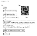
- cancer patient samples were fixed using formalin, dehydrated with alcohol, and then treated with xylene and immersed in paraffin at a high temperature for paraffin-embedding to prepare tissue samples ( Figure 1(a) ).
- 4 samples of normal mammary tissues from cancer patients were used as the control.
- the tissues samples from the above 20 samples were cut to a 2 to 4 ⁇ m sections, deparaffinized using xylene, treated with alcohol and then washed with deionized water.
- the above samples were retrieved in a 10 mM citric acid solution (pH 6.0) under the condition of 121°C for 15 minutes to loosen the fixed tissue structure.
- the above samples were washed in the order of deionized water and phosphate buffered saline (PBS), and the samples were then immersed in the order of in a 50 mM NH 4 Cl/PBS solution at 25°C for 30 minutes, a 10%FBS/PBS solution at 25°C for 60 minutes and a 10%FBS + 1 ⁇ M mouse IgG/PBS solution at 25°C for 3 hours for the blocking treatment to prevent the nonspecific binding of the PAR1 antibody.
- PBS phosphate buffered saline
- the DAPI staining (2 ⁇ g/ml PBS) was carried out at 25°C for 10 minutes and the cell nucleus was stained to count the number of cells. After washing 3 times with PBS, the cells were mounted and immunostained tissue samples were produced.
- the immunostained tissue samples produced in the above Example 1 were irradiated with 488 nm excitation light using a combination device of a confocal unit (a product of Yokogawa Electric Corporation), a fluorescence microscope (a product of OLYMPUS CORPORATION) and Electron-Multiplier CCD (EM-CCD) Camera (Andor Co., Ltd.), and then a fluorescence image (fluorescent still image) of quantum dot fluorescent particle (detected using PAR1ab-QD705) was acquired using a 695 to 740 nm band pass filter.
- a fluorescence image fluorescent still image
- Figure 1(b) shows the fluorescent still images of the tissue samples from recurrent breast cancer patients as an example, but the autofluorescence was so intense that the fluorescence of quantum dot fluorescent particle (detected using PAR1ab-QD705) was not identified on the first inspection.
- the fluorescent brightnesses of autofluorescence and quantum dot fluorescent particles (detected using PAR1 antibody) were compared, only about 2 to 3 times difference was found in reality. Further, since the autofluorescence significantly varies depending on the type of a tissue sample and the place in a tissue sample, a definite threshold cannot be determined. Consequently, the autofluorescence need to be eliminated to accurately measure the fluorescent brightness of quantum dot fluorescent particle (detected using PAR1 antibody).
- a fluorescent still image with a zero (0) fluorescent intensity of the background autofluorescence need to be acquired. Then, the total fluorescence of fluorescent still image can be calculated as the fluorescence derived from quantum dot fluorescent particles.
- the contrast adjustment method of the above Example 2 uses the division of fluorescent intensity, thus failing to produce zero (0) by the division, and accordingly an image processing method which uses the subtraction capable of producing zero (0) is required. Under the circumstances, the following method is proposed as the image processing method for eliminating the autofluorescence of a fluorescent still image.
- a breast cancer tissue sample immunostained with quantum dot fluorescent particles is irradiated with excitation light (laser) having an excitation wavelength of 488 nm
- excitation light laser
- an fluorescence image of quantum dot fluorescent particles fluorescence of the quantum dot fluorescent particles + autofluorescence
- a band pass filter having an acquisition region of fluorescence wavelength of 695 to 740 nm
- an autofluorescence image carrying only the autofluorescence is acquired in exactly the same focal plane and in the same field of vision as those of the acquired fluorescence image using a band pass filter having an acquisition region of fluorescence wavelength of 640 to 690 nm.
- the thus acquired fluorescence image (fluorescence of quantum dot fluorescent particles + autofluorescence) and the autofluorescence image are loaded in 512 x 512 pixels to convert to JPEG images in a 256-step gradation ( Figure 3(a) ), and then the brightness information (a value from 0 to 255) included in the autofluorescence image (autofluorescence) is subtracted from the brightness information (a value from 0 to 255) included in each pixel of the fluorescent still image (fluorescence of quantum dot fluorescent particles + autofluorescence) to produce an image showing only the fluorescence of quantum dot fluorescent particles (corrected fluorescence image).
- a 512 x 512 pixel image in the same region of each of the autofluorescence images acquired using the 6 types of band pass filters was converted to a JPEG image, and in the JPEG image, a 512 x 420 pixel (total 210540 pixels) image in the same region was cut out to determine the mean value of fluorescent brightnesses in the image, thereby calculating "fluorescent brightness mean value of autofluorescence image / pixel" (value 1)). Further, in each of the autofluorescence images acquired by the above 6 types of band pass filters, an image having the background with no autofluorescence was acquired as the background fluorescence image, the fluorescent brightness of the acquired background fluorescence image was measured.
- the fluorescent brightness in the 25 x 25 pixel background fluorescence image was determined in any 3 regions, and "fluorescent brightness mean value of the background fluorescence image / pixel" was calculated (value 2)). These 3 regions measured were all same regions in each of the autofluorescence images acquired using the above 6 types of band pass filters.
- the fluorescent brightness of autofluorescence of each of the above 6 types of band pass filters was calculated, which was then divided by the fluorescence wavelength width acquired by each of the band pass filters (505 to 545 nm: 40 nm, 565 to 595 nm: 30 nm, 585 to 630 nm: 45 nm, 640 to 690 nm: 50 nm, 695 to 740 nm: 45 nm, 760 to 800 nm: 40 nm) to calculate the fluorescent brightness mean value of autofluorescence per wavelength width.
- the chart showing the fluorescence wavelength behavior of the autofluorescence was made by plotting the fluorescent brightness of the autofluorescence per such the wavelength in the center of the fluorescence wavelength acquired by each of the band pass filters, and connecting with a dot line ( Figure 3(b) ).
- the breast cancer tissue sample immunostained with quantum dot fluorescent particles by the method described in the above Example 1 was excited with the 488 nm excitation light, and a fluorescent still image (quantum dot fluorescent particle + autofluorescence) was acquired using a band pass filter having an acquisition region of fluorescence wavelength of 695 to 740 nm.
- the fluorescent brightness of a pixel range (9 to 25 pixels) covering throughout the entire fluorescence of the single particle is determined and the "fluorescent brightness of fluorescent still image / pixel) was calculated (value 3)). Further, in the fluorescent still image, an image having the background with no autofluorescence or fluorescence of quantum dot fluorescent particles was acquired as the background fluorescence image, and the fluorescent brightness of the acquired background fluorescence image was measured.
- fluorescent brightness mean value of the background fluorescence image / pixel was calculated (value 4)).
- the fluorescent brightness of any 3 regions, which have the same number pixel as that when the fluorescent brightness of the single quantum dot fluorescent particle was measured, was determined, from the region showing only the autofluorescence not including the fluorescence of the quantum dot fluorescent particle, and the "fluorescent brightness mean value of the autofluorescence images / pixel" was calculated (value 5)).
- the mean value (the fluorescent brightness mean value of quantum dot fluorescent particles to the fluorescent brightness of the autofluorescence) was determined from the values of the acquired 15 fluorescent particles in total and thereby calculated as 1.88 ⁇ 0.10 (mean value ⁇ s.e.m.).
- the (an area representing the fluorescent brightness of Qdot705) / (an area representing the fluorescent brightness of autofluorescence) ratio of 1.88 suggests that the fluorescent brightness of autofluorescence to be the background accounts for over 50% of the total fluorescent brightness (the fluorescent brightness of quantum dot fluorescent particles + the fluorescent brightness of autofluorescence) in the fluorescent still image, and it was reconfirmed that the fluorescent brightness of autofluorescence needs to be eliminated to quantify the fluorescent brightness of quantum dot fluorescent particles.
- the corrected fluorescence image from which autofluorescence is eliminated was produced in accordance with the following procedure. More specifically, a tissue sample stained using PAR1ab-QD705 was irradiated with the 488 nm excitation light for 20 seconds, and a fluorescent still image (quantum dot fluorescent particles + autofluorescence) (20 s / frame x 1) acquired using a band pass filter having an acquisition region of fluorescence wavelength of 695 to 740 nm and an autofluorescence image (20 s / frame x 1) acquired in the same field of vision and the same focal point as those of the above fluorescent still image using a band pass filter having an acquisition region of fluorescence wavelength of 640 to 690 nm were acquired.
- a tissue sample stained using PAR1ab-QD705 was irradiated with the 488 nm excitation light for 20 seconds, and a fluorescent still image (quantum dot fluorescent particles + autofluorescence) (20 s / frame x 1) acquired using a band pass filter having an acquisition region
- the tissue sample was excited with the 400 nm excitation light, and a cell nucleus image (detected using DAPI) acquired using a band pass filter having an acquisition region of fluorescence wavelength of 420 to 500 nm was acquired ( Figure 4 , first from right).
- the autofluorescence image was adjusted to have an about 20% higher brightness value of autofluorescence than the fluorescent still image (quantum dot fluorescent particle + autofluorescence).
- the images are converted to a JPEG image ( Figure 4 , first from left and second from left).
- a threshold was set so that all fluorescence is imported.
- the treatment of "fluorescent still image (quantum dot fluorescent particle + autofluorescence) - autofluorescence image” was performed using a Photoshop image analysis software ( Figure 4 , second from right). At this treatment, it was confirmed in an optional plurality of regions that the brightness of a single quantum dot fluorescent particle is not diminished and the fluorescent intensity of the autofluorescence portion is zero (0) after the subtraction.
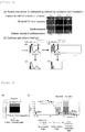
- a single quantum dot fluorescent particle has an off-state of about 4 seconds in 20 seconds of irradiation time ( Figure 6 ). Accordingly, a single particle fluorescent brightness can be acquired when the mean intensity of particles having an about 4 second-off-state is measured.
- a fluorescence videograph corresponding to a fluorescent still image a 100 consecutive series of fluorescent still image in 200 msec of resolution after irradiated with excitation light was acquired using a band pass filter having an acquisition region of fluorescence wavelength of 690 to 730 nm, and a fluorescence videograph (200 ms/frame x 100 images) was acquired.
- a particle having an off-state of about 4 seconds was identified as a single fluorescent particle and then the fluorescent brightness of the corresponding particle in the still image was determined using an Image J image analysis software. This procedure was carried out using 5 to 10 particles, and the mean fluorescent brightness per particle was calculated.
- the true number of particles detected using the PAR1 antibody had the mean value of 14.8 in 3 samples (see Samples 10 to 12, Table 1) of breast cancer tissues from recurrent patients (recurred within about 1 year and died within 4 years) which was higher than the mean value of 3.2 in 5 samples (see Samples 5 to 9, Table 1) of tissues from recurrence-free breast cancer survivors for at least 5 years, on the other hand, 4 samples (see Samples 1 to 4, Table 1) of normal mammary tissues from cancer patients had the mean value of 0.2, which was significantly lower than that of the breast cancer tissues (Table 1, Figure 7(a) ).
- the mean number of particle for 8 samples (see Samples 13 to 20, Table 1) of "triple negative” breast cancer tissues from recurrent patients (recurred within 4 years and died within 6 years) were calculated and found to have 6.0, which was higher value than that for normal mammary tissues from cancer patients and tissues from recurrence-free breast cancer survivors for at least 5 years (Table 1, Figure 7(a) ).
- the "triple negatives” is a serious issue for which Herceptin is not valid, and thus the immunohistostaining method which uses the PAR1 antibody of the present invention has the potential to be a breakthrough therein.
- the applicability of the excitation wavelength of excitation light other than 488 nm was studied using an excitation wavelength of 532 nm, and found that the number of particles detected using the PAR1 antibody per cell of Sample 13 tissue sample was 15.5 (the number of particles / the number of cells) with an excitation wavelength of 488 nm, whereas 15.8 (the number of particles / the number of cells) with the wavelength of 532 nm ( Figure 8 ), with the number of particles detected using the PAR1 antibody per cell calculated in both wavelengths being no notable difference ( Figure 8 ).
- the wavelength of excitation light applicable is not limited to 488 nm.
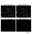
- the autofluorescence remained as indicated by the arrowheads when a corrected fluorescence image was acquired based on the autofluorescence image acquired in a wavelength range on the shorter wavelength side than the band pass filter having an acquisition region of fluorescence wavelength of 635 to 685 nm. This is because that when an autofluorescence image is acquired in a wavelength range apart from the wavelength range in which a fluorescent still image is acquired, the autofluorescence out of the focal plane is detected and the autofluorescence out of the focal plane still remains after the autofluorescence subtraction.
- the autofluorescence either on the short wavelength side or long wavelength side of the fluorescence wavelength of fluorescence particles is subtractable, but, due to the chromatic aberration problem, it is conceived that the subtraction is better performed at a vicinity wavelength of fluorescence wavelength of fluorescence particles.
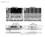
- the immunohistostaining method for calculating the number of quantum dot particles according to the present invention was studied to find any actual difference compared with the DAB staining method by performing a comparative experiment with the DAB staining method.
- Sample 12 tissue sample and Sample 14 tissue sample shown in Table 1 were subjected to the treatment steps up to the deparaffinization by the method described in the above Example 1, and then washed with deionized water.
- the samples were treated with a methanol solution containing 30% hydrogen peroxide solution (H 2 O 2 ) for 10 minutes, and then washed 3 times with distilled water.
- the above samples were retrieved in a 10 mM citric acid solution (pH 6.0) under the conditions of 121°C for 15 minutes to loosen the fixed tissue structure. Subsequently, the above samples were washed in the order of deionized water and phosphate buffered saline (PBS), and then the samples were treated in a 50 mM NH 4 Cl/PBS solution at 25°C for 30 minutes, further treated in a 10%FBS/PBS solution at 25°C for 2 hours for the blocking treatment to prevent the nonspecific binding of the PAR1 antibody.
- PBS phosphate buffered saline
- the samples were subjected to a primary antibody reaction in a 10%FBS/PBS solution containing 33nM PAR1 antibody at 25°C for 2 hours.
- a secondary antibody reaction was performed in a 10%FBS/PBS solution containing 22nM secondary antibodies to mouse antibody at 25°C for 1 hour.
- the DAB luminescence was carried out at a color developing time of 25°C for 5 minutes, using a peroxidase stain DAB kit (a product of NACALAI TESQUE, INC.). After washing the tissue samples with tap water, hematoxylin staining was carried out.
- the nonspecific PAR1 signal is not substantially detected in the cancer cell nucleus, and this presumably owes to the technical feature of the present invention which makes it possible to suitably eliminate the autofluorescence and specify the true quantum dot fluorescent particle using the blinking property.
- the determination method of the present invention is valid when, for example, the PAR1 antibody which detects the breast cancer marker factor is used as the antibody labeled with quantum dot fluorescent particles, the number of fluorescent particles per cell detected using the PAR1 antibody is determined, and, using the number as the indicator, the pathological diagnosis of breast cancer onset or breast cancer onset risk is performed.
Landscapes
- Health & Medical Sciences (AREA)
- Life Sciences & Earth Sciences (AREA)
- Chemical & Material Sciences (AREA)
- Engineering & Computer Science (AREA)
- Immunology (AREA)
- Molecular Biology (AREA)
- General Health & Medical Sciences (AREA)
- Physics & Mathematics (AREA)
- General Physics & Mathematics (AREA)
- Pathology (AREA)
- Biochemistry (AREA)
- Biomedical Technology (AREA)
- Analytical Chemistry (AREA)
- Hematology (AREA)
- Urology & Nephrology (AREA)
- Nanotechnology (AREA)
- Nuclear Medicine, Radiotherapy & Molecular Imaging (AREA)
- Crystallography & Structural Chemistry (AREA)
- Cell Biology (AREA)
- Biotechnology (AREA)
- Microbiology (AREA)
- Medicinal Chemistry (AREA)
- Food Science & Technology (AREA)
- Chemical Kinetics & Catalysis (AREA)
- Optics & Photonics (AREA)
- Materials Engineering (AREA)
- Investigating, Analyzing Materials By Fluorescence Or Luminescence (AREA)
- Investigating Or Analysing Biological Materials (AREA)
Claims (10)
- Verfahren zum Bestimmen des Ausbrechens von Krebs oder des Risikos des Ausbrechens von Krebs, umfassend das Verwenden eines Gewebefärbeverfahrens, das die folgenden Schritte (a) bis (d) umfasst:(a) Markieren eines Antikörpers, der einen Krebswachstums-Regulationsfaktor oder einen Krebsmetastase-Regulationsfaktor erkennt, mit einem fluoreszierenden Material und Inkontaktbringen des fluoreszenzmarkierten Antikörpers mit einer Gewebeprobe;(b) Bestrahlen eines Gewebeorts in Kontakt mit dem Antikörper mit Anregungslicht mit einer gegebenen Wellenlänge;(c1) Aufnehmen eines Fluoreszenzbilds von Schritt (b) in einem Aufnahmebereich mit der von dem fluoreszierenden Material emittierten Fluoreszenzwellenlänge;(c2) Aufnehmen eines Autofluoreszenzbilds von Schritt (b) in einem benachbarten Bereich an einer Kurzwellenlängenseite oder Langwellenlängenseite eines Aufnahmebereichs mit der von dem fluoreszierenden Material emittierten Fluoreszenzwellenlänge in dem gleichen Sehfeld und mit dem gleichen Brennpunkt wie das Fluoreszenzbild;(d) Aufnehmen eines korrigierten Fluoreszenzbilds durch Bildverarbeitung zum Entfernen von Fluoreszenzhelligkeit des Autofluoreszenzbilds von der Fluoreszenzhelligkeit des Fluoreszenzbilds.
- Bestimmungsverfahren gemäß Anspruch 1, wobei der Krebswachstums-Regulationsfaktor oder Krebsmetastase-Regulationsfaktor PAR1 (proteaseaktivierter Rezeptor 1) ist.
- Bestimmungsverfahren gemäß Anspruch 1 oder 2, wobei der Krebs ein Brustkrebs ist.
- Bestimmungsverfahren gemäß einem der Ansprüche 1 bis 3, wobei der Unterschied der Wellenlängen zwischen dem Aufnahmebereich mit der von dem fluoreszierenden Material emittierten Fluoreszenzwellenlänge und dem dazu benachbarten Bereich innerhalb von 100 nm liegt.
- Bestimmungsverfahren gemäß einem der Ansprüche 1 bis 4, wobei der Aufnahmebereich mit der von dem fluoreszierenden Material emittierten Fluoreszenzwellenlänge ein Nahinfrarotbereich ist.
- Bestimmungsverfahren gemäß einem der Ansprüche 1 bis 5, wobei die Wellenlänge des Anregungslichts 400 bis 700 nm beträgt.
- Bestimmungsverfahren gemäß einem der Ansprüche 1 bis 6, wobei das fluoreszierende Material ein fluoreszierendes Partikel ist.
- Bestimmungsverfahren gemäß Anspruch 7, wobei das fluoreszierende Partikel ein fluoreszierendes Quantenpunkt-Partikel ist.
- Bestimmungsverfahren gemäß Anspruch 7 oder 8, ferner umfassend die folgenden Schritte (e) bis (g):(e) Zählen der Anzahl von Zellen des Gewebeorts in Kontakt mit dem Antikörper;(f) Identifizieren eines einzelnen fluoreszierenden Partikels auf der Grundlage des Fluoreszenzbilds und Messen einer mittleren Fluoreszenzhelligkeit eines einzelnen fluoreszierenden Partikels in dem korrigierten Fluoreszenzbild, das dem einzelnen fluoreszierenden Partikel entspricht; und(g) Berechnen der Anzahl von fluoreszierenden Partikeln pro Zelle durch Teilen der Gesamtfluoreszenzhelligkeit in dem korrigierten Fluoreszenzbild durch die mittlere Fluoreszenzhelligkeit des einzelnen fluoreszierenden Partikels, um die Anzahl von fluoreszierenden Partikel zu bestimmen, und Teilen der Anzahl von fluoreszierenden Partikel durch die bei Schritt (e) gezählte Anzahl von Zellen.
- Bestimmungsverfahren gemäß Anspruch 9, wobei das einzelne fluoreszierende Partikel unter Verwendung einer Blinkeigenschaft der fluoreszierenden Partikel identifiziert wird.
Applications Claiming Priority (2)
| Application Number | Priority Date | Filing Date | Title |
|---|---|---|---|
| JP2010196442 | 2010-09-02 | ||
| PCT/JP2011/004762 WO2012029269A1 (ja) | 2010-09-02 | 2011-08-26 | がん発症又はがん発症リスクの判定方法 |
Publications (3)
| Publication Number | Publication Date |
|---|---|
| EP2613151A1 EP2613151A1 (de) | 2013-07-10 |
| EP2613151A4 EP2613151A4 (de) | 2015-02-18 |
| EP2613151B1 true EP2613151B1 (de) | 2016-06-29 |
Family
ID=45772395
Family Applications (1)
| Application Number | Title | Priority Date | Filing Date |
|---|---|---|---|
| EP11821292.7A Not-in-force EP2613151B1 (de) | 2010-09-02 | 2011-08-26 | Verfahren zur bestimmung des ausbruchs von krebs oder des risikos für den ausbruch von krebs |
Country Status (5)
| Country | Link |
|---|---|
| US (1) | US20130203082A1 (de) |
| EP (1) | EP2613151B1 (de) |
| JP (1) | JP5682975B2 (de) |
| CN (1) | CN103168242B (de) |
| WO (1) | WO2012029269A1 (de) |
Families Citing this family (34)
| Publication number | Priority date | Publication date | Assignee | Title |
|---|---|---|---|---|
| US20080161744A1 (en) | 2006-09-07 | 2008-07-03 | University Of Rochester Medical Center | Pre-And Intra-Operative Localization of Penile Sentinel Nodes |
| US8406860B2 (en) | 2008-01-25 | 2013-03-26 | Novadaq Technologies Inc. | Method for evaluating blush in myocardial tissue |
| JP5682976B2 (ja) * | 2010-09-17 | 2015-03-11 | 国立大学法人東北大学 | 抗体を成分として含む医薬品の有効性の判定方法 |
| WO2012133047A1 (ja) * | 2011-03-25 | 2012-10-04 | コニカミノルタエムジー株式会社 | 免疫組織染色法、およびこれを用いた抗体医薬の有効性を判定する方法 |
| US9189678B2 (en) * | 2012-03-30 | 2015-11-17 | Konica Minolta, Inc. | Medical image processor and storage medium |
| EP2863801B8 (de) | 2012-06-21 | 2024-06-12 | Stryker Corporation | Quantifizierung und analyse von angiografie und perfusionen |
| US10831013B2 (en) | 2013-08-26 | 2020-11-10 | S.D. Sight Diagnostics Ltd. | Digital microscopy systems, methods and computer program products |
| CN103589422A (zh) * | 2013-11-15 | 2014-02-19 | 无锡中科光远生物材料有限公司 | 一种可用于抑制癌细胞的荧光纳米颗粒的制备方法 |
| CN103760140B (zh) * | 2014-01-07 | 2016-01-20 | 李雁 | 一种基于量子点光谱分析及图像解析的肿瘤转移单元计数方法 |
| WO2015115151A1 (ja) * | 2014-01-31 | 2015-08-06 | オリンパス株式会社 | 蛍光観察装置 |
| US10732184B2 (en) | 2014-02-04 | 2020-08-04 | Celltrend Gmbh | Diagnosis of cancer by detecting auto-antibodies against PAR1 |
| JP5835536B1 (ja) * | 2014-03-26 | 2015-12-24 | コニカミノルタ株式会社 | 組織評価方法、画像処理装置、病理診断支援システム及びプログラム |
| WO2015145644A1 (ja) * | 2014-03-27 | 2015-10-01 | コニカミノルタ株式会社 | 画像処理装置および画像処理プログラム |
| EP3201607B1 (de) * | 2014-09-29 | 2020-12-30 | Novadaq Technologies ULC | Bildgebung eines zielfluorophors in einem biologischen material in anwesenheit von autofluoreszenz |
| EP3203902B1 (de) | 2014-10-09 | 2021-07-07 | Novadaq Technologies ULC | Quantifizierung des absoluten blutströmung in gewebe mittels fluoreszenzvermittelter photoplethysmographie |
| JP6547424B2 (ja) * | 2015-06-01 | 2019-07-24 | コニカミノルタ株式会社 | 蛍光画像の合焦システム、合焦方法および合焦プログラム |
| US10871447B2 (en) | 2015-06-30 | 2020-12-22 | Imec Vzw | Bleaching of dyes in luminescent detection |
| EP3859425B1 (de) | 2015-09-17 | 2024-04-17 | S.D. Sight Diagnostics Ltd. | Verfahren und vorrichtung zur detektion einer entität in einer körperprobe |
| US11733150B2 (en) | 2016-03-30 | 2023-08-22 | S.D. Sight Diagnostics Ltd. | Distinguishing between blood sample components |
| US11307196B2 (en) | 2016-05-11 | 2022-04-19 | S.D. Sight Diagnostics Ltd. | Sample carrier for optical measurements |
| AU2017263807B2 (en) | 2016-05-11 | 2023-02-02 | S.D. Sight Diagnostics Ltd | Performing optical measurements on a sample |
| CA3049922A1 (en) | 2017-02-10 | 2018-08-16 | Novadaq Technologies ULC | Open-field handheld fluorescence imaging systems and methods |
| EP3710810B1 (de) | 2017-11-14 | 2023-09-06 | S.D. Sight Diagnostics Ltd. | Probenträger für optische messungen |
| WO2020022394A1 (en) * | 2018-07-24 | 2020-01-30 | Sony Corporation | Information processing apparatus and microscope for separating the fluorescence of a fluorescent reageant from the autofluorescence of a specimen |
| JP2020020791A (ja) * | 2018-07-24 | 2020-02-06 | ソニー株式会社 | 情報処理装置、情報処理方法、情報処理システム、およびプログラム |
| WO2021005904A1 (ja) * | 2019-07-09 | 2021-01-14 | ソニー株式会社 | 情報処理装置およびプログラム |
| WO2021039592A1 (ja) * | 2019-08-29 | 2021-03-04 | コニカミノルタ株式会社 | 創薬支援方法、創薬支援装置及びプログラム |
| KR102931515B1 (ko) | 2019-10-22 | 2026-02-27 | 에스.디. 사이트 다이그노틱스 엘티디 | 광학 측정들에서의 오류들의 고려 |
| BR112022011233A2 (pt) | 2019-12-12 | 2022-08-30 | S D Sight Diagnostics Ltd | Detecção de plaquetas em uma amostra sanguínea |
| BR112022011312A2 (pt) | 2019-12-12 | 2022-08-23 | S D Sight Diagnostics Ltd | Análise de um analito disposto dentro de um meio |
| BR112022011354A2 (pt) | 2019-12-12 | 2022-08-23 | S D Sight Diagnostics Ltd | Geração artificial de uma imagem colorida de esfregaço sanguíneo |
| KR20260022484A (ko) | 2020-03-05 | 2026-02-19 | 캐노피 바이오사이언시스, 엘엘씨 | 자동화된 형광 이미징 및 단일 셀 분할 |
| JPWO2021230134A1 (de) * | 2020-05-13 | 2021-11-18 | ||
| AU2021416188A1 (en) | 2020-12-31 | 2023-07-06 | Orlucent, Inc. | Systems and methods for assessing tissue remodeling |
Family Cites Families (15)
| Publication number | Priority date | Publication date | Assignee | Title |
|---|---|---|---|---|
| US5733721A (en) * | 1992-11-20 | 1998-03-31 | The Board Of Regents Of The University Of Oklahoma | Cell analysis method using quantitative fluorescence image analysis |
| JPH08334466A (ja) * | 1995-06-06 | 1996-12-17 | Nikon Corp | 蛍光測光方法及び装置 |
| JPH09241634A (ja) | 1996-03-11 | 1997-09-16 | Osaka Gas Co Ltd | 蛍光粒子の製造方法 |
| JP3460673B2 (ja) * | 2000-02-04 | 2003-10-27 | 浜松ホトニクス株式会社 | 特定の遺伝子を発現した生細胞の選択的分離方法 |
| JP5005247B2 (ja) * | 2006-04-11 | 2012-08-22 | 浜松ホトニクス株式会社 | 光測定装置、光測定方法、及び光測定プログラム |
| JP5030513B2 (ja) * | 2006-09-11 | 2012-09-19 | オリンパス株式会社 | 生体組織用蛍光観測装置および内視鏡システム |
| JP2008261784A (ja) * | 2007-04-13 | 2008-10-30 | Olympus Corp | 生体観察方法 |
| CN100573106C (zh) * | 2007-06-28 | 2009-12-23 | 大连海事大学 | 一种光纤荧光生物传感器 |
| JP5574522B2 (ja) * | 2008-01-15 | 2014-08-20 | 徳宏 茶野 | 癌マーカー及び癌細胞の検査方法 |
| US20100069550A1 (en) * | 2008-03-17 | 2010-03-18 | Washington, University Of | Nanoparticle assemblies and methods for their preparation |
| US8674079B2 (en) * | 2008-03-27 | 2014-03-18 | Tohoku University | Cancer cell migration and cancer cell invasion inhibitor |
| CN101339188A (zh) * | 2008-08-08 | 2009-01-07 | 武汉大学 | 一种检测乳腺癌石蜡包埋组织抗原的量子点免疫荧光试剂盒及应用 |
| ES2432537T3 (es) * | 2008-09-01 | 2013-12-04 | National University Corporation Nagoya University | Pronóstico de cánceres de colon |
| US20110278471A1 (en) * | 2009-01-23 | 2011-11-17 | Mitsui Engineering & Shipbuilding Co., Ltd | Fluorescence detecting device and fluorescence detecting method |
| JP2010242059A (ja) | 2009-03-18 | 2010-10-28 | Tokyo Institute Of Technology | 蛍光粒子およびその製造方法 |
-
2011
- 2011-08-26 EP EP11821292.7A patent/EP2613151B1/de not_active Not-in-force
- 2011-08-26 JP JP2012531677A patent/JP5682975B2/ja not_active Expired - Fee Related
- 2011-08-26 US US13/819,008 patent/US20130203082A1/en not_active Abandoned
- 2011-08-26 CN CN201180041795.5A patent/CN103168242B/zh not_active Expired - Fee Related
- 2011-08-26 WO PCT/JP2011/004762 patent/WO2012029269A1/ja not_active Ceased
Also Published As
| Publication number | Publication date |
|---|---|
| EP2613151A4 (de) | 2015-02-18 |
| JP5682975B2 (ja) | 2015-03-11 |
| JPWO2012029269A1 (ja) | 2013-10-28 |
| EP2613151A1 (de) | 2013-07-10 |
| CN103168242A (zh) | 2013-06-19 |
| WO2012029269A1 (ja) | 2012-03-08 |
| CN103168242B (zh) | 2016-02-03 |
| US20130203082A1 (en) | 2013-08-08 |
Similar Documents
| Publication | Publication Date | Title |
|---|---|---|
| EP2613151A1 (de) | Verfahren zur bestimmung des ausbruchs von krebs oder des risikos für den ausbruch von krebs | |
| JP5682976B2 (ja) | 抗体を成分として含む医薬品の有効性の判定方法 | |
| US11435348B2 (en) | Tissue staining method | |
| Chen et al. | Quantum dots-based immunofluorescence technology for the quantitative determination of HER2 expression in breast cancer | |
| JP5900489B2 (ja) | 免疫組織染色法、およびこれを用いた抗体医薬の有効性を判定する方法 | |
| Xu et al. | Quantum dot-based, quantitative, and multiplexed assay for tissue staining | |
| JP2016029387A (ja) | 組織染色用蛍光標識体および生体物質検出方法 | |
| CA2623445A1 (en) | Comprehensive diagnostic testing procedures for personalized anticancer chemotherapy (pac) | |
| EP2833137B1 (de) | Gewebefärbungsverfahren | |
| EP3327427A1 (de) | Verfahren zur analyse einer biologischen zielsubstanz und analysesystem | |
| JP2008545499A (ja) | 生体内発現プロファイリング | |
| US20240402185A1 (en) | Method of analysing a sample for at least one analyte | |
| JP5863057B2 (ja) | 組織評価方法 | |
| Fakhrolmobasheri et al. | Compatibility of fast tissue clearing with targeted fluorescence imaging and pathology analysis using clinical head and neck cancer biopsies | |
| CN118613713A (zh) | 用于术中寿命成像的系统和方法 | |
| Rosbach et al. | Optical Molecular Imaging of Multiple Biomarkers of Epithelial Neoplasia: EGFR Expression and Metabolic Activity in Oral Mucosa | |
| Weigum | Development of a cell-based lab-on-a-chip sensor for detection of oral cancer biomarkers |
Legal Events
| Date | Code | Title | Description |
|---|---|---|---|
| PUAI | Public reference made under article 153(3) epc to a published international application that has entered the european phase |
Free format text: ORIGINAL CODE: 0009012 |
|
| 17P | Request for examination filed |
Effective date: 20130322 |
|
| AK | Designated contracting states |
Kind code of ref document: A1 Designated state(s): AL AT BE BG CH CY CZ DE DK EE ES FI FR GB GR HR HU IE IS IT LI LT LU LV MC MK MT NL NO PL PT RO RS SE SI SK SM TR |
|
| DAX | Request for extension of the european patent (deleted) | ||
| A4 | Supplementary search report drawn up and despatched |
Effective date: 20150119 |
|
| RIC1 | Information provided on ipc code assigned before grant |
Ipc: G01N 21/64 20060101ALI20150113BHEP Ipc: G01N 33/58 20060101ALI20150113BHEP Ipc: G01N 33/53 20060101ALI20150113BHEP Ipc: G01N 33/574 20060101AFI20150113BHEP Ipc: G01N 33/536 20060101ALI20150113BHEP Ipc: G01N 33/48 20060101ALI20150113BHEP |
|
| 17Q | First examination report despatched |
Effective date: 20150714 |
|
| GRAP | Despatch of communication of intention to grant a patent |
Free format text: ORIGINAL CODE: EPIDOSNIGR1 |
|
| INTG | Intention to grant announced |
Effective date: 20160415 |
|
| GRAS | Grant fee paid |
Free format text: ORIGINAL CODE: EPIDOSNIGR3 |
|
| GRAA | (expected) grant |
Free format text: ORIGINAL CODE: 0009210 |
|
| AK | Designated contracting states |
Kind code of ref document: B1 Designated state(s): AL AT BE BG CH CY CZ DE DK EE ES FI FR GB GR HR HU IE IS IT LI LT LU LV MC MK MT NL NO PL PT RO RS SE SI SK SM TR |
|
| REG | Reference to a national code |
Ref country code: GB Ref legal event code: FG4D |
|
| REG | Reference to a national code |
Ref country code: CH Ref legal event code: EP |
|
| REG | Reference to a national code |
Ref country code: AT Ref legal event code: REF Ref document number: 809510 Country of ref document: AT Kind code of ref document: T Effective date: 20160715 |
|
| REG | Reference to a national code |
Ref country code: IE Ref legal event code: FG4D |
|
| REG | Reference to a national code |
Ref country code: DE Ref legal event code: R096 Ref document number: 602011027826 Country of ref document: DE |
|
| REG | Reference to a national code |
Ref country code: FR Ref legal event code: PLFP Year of fee payment: 6 |
|
| REG | Reference to a national code |
Ref country code: LT Ref legal event code: MG4D |
|
| PG25 | Lapsed in a contracting state [announced via postgrant information from national office to epo] |
Ref country code: LT Free format text: LAPSE BECAUSE OF FAILURE TO SUBMIT A TRANSLATION OF THE DESCRIPTION OR TO PAY THE FEE WITHIN THE PRESCRIBED TIME-LIMIT Effective date: 20160629 Ref country code: NO Free format text: LAPSE BECAUSE OF FAILURE TO SUBMIT A TRANSLATION OF THE DESCRIPTION OR TO PAY THE FEE WITHIN THE PRESCRIBED TIME-LIMIT Effective date: 20160929 Ref country code: FI Free format text: LAPSE BECAUSE OF FAILURE TO SUBMIT A TRANSLATION OF THE DESCRIPTION OR TO PAY THE FEE WITHIN THE PRESCRIBED TIME-LIMIT Effective date: 20160629 |
|
| PGFP | Annual fee paid to national office [announced via postgrant information from national office to epo] |
Ref country code: GB Payment date: 20160819 Year of fee payment: 6 Ref country code: DE Payment date: 20160822 Year of fee payment: 6 |
|
| REG | Reference to a national code |
Ref country code: NL Ref legal event code: MP Effective date: 20160629 |
|
| PG25 | Lapsed in a contracting state [announced via postgrant information from national office to epo] |
Ref country code: RS Free format text: LAPSE BECAUSE OF FAILURE TO SUBMIT A TRANSLATION OF THE DESCRIPTION OR TO PAY THE FEE WITHIN THE PRESCRIBED TIME-LIMIT Effective date: 20160629 Ref country code: HR Free format text: LAPSE BECAUSE OF FAILURE TO SUBMIT A TRANSLATION OF THE DESCRIPTION OR TO PAY THE FEE WITHIN THE PRESCRIBED TIME-LIMIT Effective date: 20160629 Ref country code: NL Free format text: LAPSE BECAUSE OF FAILURE TO SUBMIT A TRANSLATION OF THE DESCRIPTION OR TO PAY THE FEE WITHIN THE PRESCRIBED TIME-LIMIT Effective date: 20160629 Ref country code: GR Free format text: LAPSE BECAUSE OF FAILURE TO SUBMIT A TRANSLATION OF THE DESCRIPTION OR TO PAY THE FEE WITHIN THE PRESCRIBED TIME-LIMIT Effective date: 20160930 Ref country code: LV Free format text: LAPSE BECAUSE OF FAILURE TO SUBMIT A TRANSLATION OF THE DESCRIPTION OR TO PAY THE FEE WITHIN THE PRESCRIBED TIME-LIMIT Effective date: 20160629 Ref country code: SE Free format text: LAPSE BECAUSE OF FAILURE TO SUBMIT A TRANSLATION OF THE DESCRIPTION OR TO PAY THE FEE WITHIN THE PRESCRIBED TIME-LIMIT Effective date: 20160629 |
|
| PGFP | Annual fee paid to national office [announced via postgrant information from national office to epo] |
Ref country code: FR Payment date: 20160822 Year of fee payment: 6 |
|
| REG | Reference to a national code |
Ref country code: AT Ref legal event code: MK05 Ref document number: 809510 Country of ref document: AT Kind code of ref document: T Effective date: 20160629 |
|
| PG25 | Lapsed in a contracting state [announced via postgrant information from national office to epo] |
Ref country code: BE Free format text: LAPSE BECAUSE OF NON-PAYMENT OF DUE FEES Effective date: 20160831 |
|
| PG25 | Lapsed in a contracting state [announced via postgrant information from national office to epo] |
Ref country code: SK Free format text: LAPSE BECAUSE OF FAILURE TO SUBMIT A TRANSLATION OF THE DESCRIPTION OR TO PAY THE FEE WITHIN THE PRESCRIBED TIME-LIMIT Effective date: 20160629 Ref country code: CZ Free format text: LAPSE BECAUSE OF FAILURE TO SUBMIT A TRANSLATION OF THE DESCRIPTION OR TO PAY THE FEE WITHIN THE PRESCRIBED TIME-LIMIT Effective date: 20160629 Ref country code: IT Free format text: LAPSE BECAUSE OF FAILURE TO SUBMIT A TRANSLATION OF THE DESCRIPTION OR TO PAY THE FEE WITHIN THE PRESCRIBED TIME-LIMIT Effective date: 20160629 Ref country code: IS Free format text: LAPSE BECAUSE OF FAILURE TO SUBMIT A TRANSLATION OF THE DESCRIPTION OR TO PAY THE FEE WITHIN THE PRESCRIBED TIME-LIMIT Effective date: 20161029 Ref country code: RO Free format text: LAPSE BECAUSE OF FAILURE TO SUBMIT A TRANSLATION OF THE DESCRIPTION OR TO PAY THE FEE WITHIN THE PRESCRIBED TIME-LIMIT Effective date: 20160629 Ref country code: EE Free format text: LAPSE BECAUSE OF FAILURE TO SUBMIT A TRANSLATION OF THE DESCRIPTION OR TO PAY THE FEE WITHIN THE PRESCRIBED TIME-LIMIT Effective date: 20160629 |
|
| PG25 | Lapsed in a contracting state [announced via postgrant information from national office to epo] |
Ref country code: BE Free format text: LAPSE BECAUSE OF FAILURE TO SUBMIT A TRANSLATION OF THE DESCRIPTION OR TO PAY THE FEE WITHIN THE PRESCRIBED TIME-LIMIT Effective date: 20160629 Ref country code: AT Free format text: LAPSE BECAUSE OF FAILURE TO SUBMIT A TRANSLATION OF THE DESCRIPTION OR TO PAY THE FEE WITHIN THE PRESCRIBED TIME-LIMIT Effective date: 20160629 Ref country code: SM Free format text: LAPSE BECAUSE OF FAILURE TO SUBMIT A TRANSLATION OF THE DESCRIPTION OR TO PAY THE FEE WITHIN THE PRESCRIBED TIME-LIMIT Effective date: 20160629 Ref country code: ES Free format text: LAPSE BECAUSE OF FAILURE TO SUBMIT A TRANSLATION OF THE DESCRIPTION OR TO PAY THE FEE WITHIN THE PRESCRIBED TIME-LIMIT Effective date: 20160629 Ref country code: PL Free format text: LAPSE BECAUSE OF FAILURE TO SUBMIT A TRANSLATION OF THE DESCRIPTION OR TO PAY THE FEE WITHIN THE PRESCRIBED TIME-LIMIT Effective date: 20160629 Ref country code: PT Free format text: LAPSE BECAUSE OF FAILURE TO SUBMIT A TRANSLATION OF THE DESCRIPTION OR TO PAY THE FEE WITHIN THE PRESCRIBED TIME-LIMIT Effective date: 20161031 |
|
| REG | Reference to a national code |
Ref country code: DE Ref legal event code: R097 Ref document number: 602011027826 Country of ref document: DE |
|
| PG25 | Lapsed in a contracting state [announced via postgrant information from national office to epo] |
Ref country code: MC Free format text: LAPSE BECAUSE OF FAILURE TO SUBMIT A TRANSLATION OF THE DESCRIPTION OR TO PAY THE FEE WITHIN THE PRESCRIBED TIME-LIMIT Effective date: 20160629 |
|
| REG | Reference to a national code |
Ref country code: CH Ref legal event code: PL |
|
| PG25 | Lapsed in a contracting state [announced via postgrant information from national office to epo] |
Ref country code: CH Free format text: LAPSE BECAUSE OF NON-PAYMENT OF DUE FEES Effective date: 20160831 Ref country code: LI Free format text: LAPSE BECAUSE OF NON-PAYMENT OF DUE FEES Effective date: 20160831 |
|
| PLBE | No opposition filed within time limit |
Free format text: ORIGINAL CODE: 0009261 |
|
| STAA | Information on the status of an ep patent application or granted ep patent |
Free format text: STATUS: NO OPPOSITION FILED WITHIN TIME LIMIT |
|
| PG25 | Lapsed in a contracting state [announced via postgrant information from national office to epo] |
Ref country code: DK Free format text: LAPSE BECAUSE OF FAILURE TO SUBMIT A TRANSLATION OF THE DESCRIPTION OR TO PAY THE FEE WITHIN THE PRESCRIBED TIME-LIMIT Effective date: 20160629 |
|
| REG | Reference to a national code |
Ref country code: IE Ref legal event code: MM4A |
|
| 26N | No opposition filed |
Effective date: 20170330 |
|
| PG25 | Lapsed in a contracting state [announced via postgrant information from national office to epo] |
Ref country code: IE Free format text: LAPSE BECAUSE OF NON-PAYMENT OF DUE FEES Effective date: 20160826 |
|
| PG25 | Lapsed in a contracting state [announced via postgrant information from national office to epo] |
Ref country code: LU Free format text: LAPSE BECAUSE OF NON-PAYMENT OF DUE FEES Effective date: 20160826 Ref country code: SI Free format text: LAPSE BECAUSE OF FAILURE TO SUBMIT A TRANSLATION OF THE DESCRIPTION OR TO PAY THE FEE WITHIN THE PRESCRIBED TIME-LIMIT Effective date: 20160629 Ref country code: BG Free format text: LAPSE BECAUSE OF FAILURE TO SUBMIT A TRANSLATION OF THE DESCRIPTION OR TO PAY THE FEE WITHIN THE PRESCRIBED TIME-LIMIT Effective date: 20160929 |
|
| REG | Reference to a national code |
Ref country code: DE Ref legal event code: R119 Ref document number: 602011027826 Country of ref document: DE |
|
| GBPC | Gb: european patent ceased through non-payment of renewal fee |
Effective date: 20170826 |
|
| REG | Reference to a national code |
Ref country code: FR Ref legal event code: ST Effective date: 20180430 |
|
| PG25 | Lapsed in a contracting state [announced via postgrant information from national office to epo] |
Ref country code: HU Free format text: LAPSE BECAUSE OF FAILURE TO SUBMIT A TRANSLATION OF THE DESCRIPTION OR TO PAY THE FEE WITHIN THE PRESCRIBED TIME-LIMIT; INVALID AB INITIO Effective date: 20110826 Ref country code: CY Free format text: LAPSE BECAUSE OF FAILURE TO SUBMIT A TRANSLATION OF THE DESCRIPTION OR TO PAY THE FEE WITHIN THE PRESCRIBED TIME-LIMIT Effective date: 20160629 |
|
| PG25 | Lapsed in a contracting state [announced via postgrant information from national office to epo] |
Ref country code: TR Free format text: LAPSE BECAUSE OF FAILURE TO SUBMIT A TRANSLATION OF THE DESCRIPTION OR TO PAY THE FEE WITHIN THE PRESCRIBED TIME-LIMIT Effective date: 20160629 Ref country code: MT Free format text: LAPSE BECAUSE OF NON-PAYMENT OF DUE FEES Effective date: 20160831 Ref country code: MK Free format text: LAPSE BECAUSE OF FAILURE TO SUBMIT A TRANSLATION OF THE DESCRIPTION OR TO PAY THE FEE WITHIN THE PRESCRIBED TIME-LIMIT Effective date: 20160629 |
|
| PG25 | Lapsed in a contracting state [announced via postgrant information from national office to epo] |
Ref country code: DE Free format text: LAPSE BECAUSE OF NON-PAYMENT OF DUE FEES Effective date: 20180301 Ref country code: GB Free format text: LAPSE BECAUSE OF NON-PAYMENT OF DUE FEES Effective date: 20170826 |
|
| PG25 | Lapsed in a contracting state [announced via postgrant information from national office to epo] |
Ref country code: FR Free format text: LAPSE BECAUSE OF NON-PAYMENT OF DUE FEES Effective date: 20170831 |
|
| PG25 | Lapsed in a contracting state [announced via postgrant information from national office to epo] |
Ref country code: AL Free format text: LAPSE BECAUSE OF FAILURE TO SUBMIT A TRANSLATION OF THE DESCRIPTION OR TO PAY THE FEE WITHIN THE PRESCRIBED TIME-LIMIT Effective date: 20160629 |