EP2620912A2 - Dreidimensionale Modellierung eines Objektfelds - Google Patents
Dreidimensionale Modellierung eines Objektfelds Download PDFInfo
- Publication number
- EP2620912A2 EP2620912A2 EP13156389.2A EP13156389A EP2620912A2 EP 2620912 A2 EP2620912 A2 EP 2620912A2 EP 13156389 A EP13156389 A EP 13156389A EP 2620912 A2 EP2620912 A2 EP 2620912A2
- Authority
- EP
- European Patent Office
- Prior art keywords
- recognition object
- recognition
- field
- view
- known dimensions
- Prior art date
- Legal status (The legal status is an assumption and is not a legal conclusion. Google has not performed a legal analysis and makes no representation as to the accuracy of the status listed.)
- Withdrawn
Links
Images

Classifications
-
- A—HUMAN NECESSITIES
- A61—MEDICAL OR VETERINARY SCIENCE; HYGIENE
- A61C—DENTISTRY; APPARATUS OR METHODS FOR ORAL OR DENTAL HYGIENE
- A61C9/00—Impression cups, i.e. impression trays; Impression methods
- A61C9/004—Means or methods for taking digitized impressions
- A61C9/0046—Data acquisition means or methods
-
- A—HUMAN NECESSITIES
- A61—MEDICAL OR VETERINARY SCIENCE; HYGIENE
- A61C—DENTISTRY; APPARATUS OR METHODS FOR ORAL OR DENTAL HYGIENE
- A61C1/00—Dental machines for boring or cutting ; General features of dental machines or apparatus, e.g. hand-piece design
- A61C1/08—Machine parts specially adapted for dentistry
- A61C1/082—Positioning or guiding, e.g. of drills
- A61C1/084—Positioning or guiding, e.g. of drills of implanting tools
-
- G—PHYSICS
- G06—COMPUTING OR CALCULATING; COUNTING
- G06T—IMAGE DATA PROCESSING OR GENERATION, IN GENERAL
- G06T17/00—Three-dimensional [3D] modelling for computer graphics
-
- G—PHYSICS
- G06—COMPUTING OR CALCULATING; COUNTING
- G06T—IMAGE DATA PROCESSING OR GENERATION, IN GENERAL
- G06T7/00—Image analysis
- G06T7/30—Determination of transform parameters for the alignment of images, i.e. image registration
-
- G—PHYSICS
- G06—COMPUTING OR CALCULATING; COUNTING
- G06V—IMAGE OR VIDEO RECOGNITION OR UNDERSTANDING
- G06V10/00—Arrangements for image or video recognition or understanding
- G06V10/20—Image preprocessing
- G06V10/24—Aligning, centring, orientation detection or correction of the image
-
- G—PHYSICS
- G06—COMPUTING OR CALCULATING; COUNTING
- G06T—IMAGE DATA PROCESSING OR GENERATION, IN GENERAL
- G06T2207/00—Indexing scheme for image analysis or image enhancement
- G06T2207/30—Subject of image; Context of image processing
- G06T2207/30004—Biomedical image processing
-
- G—PHYSICS
- G06—COMPUTING OR CALCULATING; COUNTING
- G06T—IMAGE DATA PROCESSING OR GENERATION, IN GENERAL
- G06T2207/00—Indexing scheme for image analysis or image enhancement
- G06T2207/30—Subject of image; Context of image processing
- G06T2207/30204—Marker
-
- G—PHYSICS
- G06—COMPUTING OR CALCULATING; COUNTING
- G06V—IMAGE OR VIDEO RECOGNITION OR UNDERSTANDING
- G06V2201/00—Indexing scheme relating to image or video recognition or understanding
- G06V2201/03—Recognition of patterns in medical or anatomical images
Definitions
- the invention is a method and system for determining the relative location of objects and features in a plurality of overlapping scanned images.
- the invention is particularly directed to medical and dental applications including those that require surgical and prosthetic devices to be designed and manufactured to precise dimensions dictated by the anatomy of individual patients.
- implant procedures must adhere to very strict placement, orientation and sizing requirements determined by existing bone structure and dentition, whereby the prosthetic components to be fitted onto surgically-placed implant anchors must preferably be designed, shaped and sized specifically to conform to the precise anatomical geometry of the patient, including the location, shape and size of adjoining teeth, and must transition to the precise orientation of the principal axis of the supporting implant anchor with a high degree of accuracy.
- impression copings are affixed to the external extremity of the inserted implant anchors, and serve to reference the location and angular orientation of the anchors.
- a model made from a mold based on said impression will incorporate so-called “analog” anchors to model the anchors in the patient's jaw, and prosthetic devices for said anchors will be designed and manufactured based on the geometry of the model created as described.
- U.S. Pat. No. 5,851,115 issued Dec. 22, 1998 to Carlsson, et al describes a photogrammetric method and system for imaging the mouth, for the purpose of creating a virtual model of the patient's mouth from which dental parts may be designed and made.
- a specialized camera is employed, comprising a set of mirrors that enable a single exposure to embody stereographic images from two different angles.
- the system of Carlsson further requires that the relative geometry of the virtual "lenses" created by the mirror system be known precisely.
- Carlsson teaches the use of reference markings, such as circles, applied to flat surfaces within the imaged field.
- U.S. Pat. No. 5,857,853 issued Jan. 12, 1999 to van Nifteric et al. also discloses a photogrammetry-based method for capturing the dimensional and orientation data required for the manufacture of dental prosthetic parts used in implant dentistry.
- the method of van Nifteric et al employs either a plurality of cameras having precisely-known relative positions, or a single camera mounted on a swiveling carriage that is movable between separated but accurately defined positions.
- van Nifteric et al. further teach the use of recognition objects and points, to serve as reference points used by the photogrammetry software in positioning features of the imaged scene within a coordinate frame.
- van Nifteric et al. thus disclose the use of a bar comprising measuring scale markings, and of two spheres mounted on a pin, as recognition objects.
- the present invention is a three-dimensional-based modeling method designed for dentistry and related medical applications.
- One aspect of the invention dispenses with the need for a specific camera and instead enables the use of any data capture means that produces a point cloud representing the three dimensional surface.
- data capture means may for example be a hand-held or frame-fixed three-dimensional laser scanner, an ordinary digital camera, or any other imaging means that is practically suited to the particular medical application.
- Another aspect of the invention is the use of three-dimensional recognition objects, to assist photogrammetry software to locate automatically, and to determine accurately the position and orientation of objects within the image field.
- Yet another aspect of the invention is the positioning of recognition objects having well-defined topography within those areas in the image field that have low image definition, and particularly in such of these areas that appear in overlapping portions of at least two images, to provide the photogrammetry software with position, angulation, and orientation information sufficient to enable highly accurate combining (or "stitching") of adjoining and overlapping images.
- recognition objects having a known three-dimensional geometry that comprises well defined features disposed at accurately known relative positions are fixedly positioned within the image field, and particularly in areas within the image field that have low feature definition.
- recognition objects include an object comprising three linked spheres having precisely known radii, fixed at precisely known positions on angled posts, as well as polygons of known dimensions such as pyramids.
- the image field is then scanned, such scanning effectively "panning" the image field to cover said image field in its entirety.
- Three-dimensional image processing software preferably comprising algorithms set forth in this specification, is then employed to interpret the image data acquired by the scanning means and to determine a virtual three dimensional model that reconstructs, to a high degree of accuracy, the geometry of the scanned image field.
- a method is provided, which method is suitable for use in medical, dental and veterinary applications, for developing a virtual three-dimensional model of a therapeutic field of view, said method comprising:
- a method according to the first aspect of the present invention wherein at least one of said recognition objects has a geometry that permits the determination of position and orientation of at least two images with respect to each other.
- a method according to the first aspect of the present invention wherein said imaging software comprises means for reconstructing a three-dimensional field of view from two images comprising views of a three-dimensional recognition object having precisely known dimensions.
- a method according to the first aspect of the present invention wherein at least one of said recognition objects comprises three spheres that have radii of known dimensions and that are affixed to angled posts, whereby the centers of said spheres form a triangle having precisely known dimensions.
- a method according to the first aspect of the present invention wherein at least one of said recognition objects is a polygon having known dimensions, such as a pyramid, suitably a pyramid that has a base in the form of an isosceles triangle.
- a method according to the first aspect of the present invention wherein at least one recognition object is positioned within an area in said field of view that is characterized by low feature definition, preferably said area of low feature definition being an edentulous portion of a dental arch of said patient.
- a method according to the first aspect of the present invention wherein the same recognition object appears in at least two of said images captured from different planes.
- a virtual modeling system for use in therapeutic medical, dental and veterinary applications, said system comprising:
- a system according to the second aspect of the present invention wherein said recognition object has a geometry that permits the determination of the relative position and orientation of at least two images with respect to each other.
- a system according to the second aspect of the present invention comprising at least one recognition object that comprises three spheres that have radii of known dimensions and that are affixed to angled posts whereby the centers of said spheres form a triangle having precisely known dimensions.
- a system comprising at least one recognition object that comprises at least one polygon having known dimensions, suitably the system comprising at least one recognition object that comprises at least two polygons that have known dimensions and that are joined by a linking means having a known dimension, or suitably the system comprising at least one polygonal recognition object having a pyramidal shape of known dimensions, suitably said at least one polygonal recognition object having a pyramidal shape of known dimensions being at least one recognition object having a pyramidal shape and a triangular base in the form of an isosceles triangle.
- a system according to the second aspect of the present invention comprising recognition objects including at least two pyramids having known dimensions, suitably said at least two pyramids being linked by means having known dimensions, suitably at least one of said two pyramids having a base in the form of an isosceles triangle.
- a system according to the second aspect of the present invention wherein said recognition object comprises a geometrical feature in the shape of a solid cylinder from which a portion parallel to the main axis of the cylinder has been sliced off.
- the present invention is a method and system for creating virtual three-dimensional models of a scanned field of view, using three-dimensional recognition objects as reference points in the "stitching" of overlapping captured images, and, additionally, positioning such recognition objects in areas of the field of view that are characterized by low feature definition in order to enhance the accuracy of the three-dimensional modeling of such areas.
- the invention is particularly suitable and intended for medical and dental applications, and it is particularly suited for use in the field of implant dentistry and related applications.
- Dental implants are used to support the restoration of missing teeth.
- Implant fixtures are surgically implanted by a dentist. These dental implants typically will be "restored” with abutments and crowns; that is, following successful implantation of implant fixtures into the jaw of the patient, complementary components including abutments and crowns will be affixed to the implanted fixtures to provide the patient with a restoration of the patient's natural teeth.
- the method and system of the present invention enable a manufacturer of dental restoration components to accurately measure the location and orientation of the implants in relation to the surrounding oral environment, and thereby to design and to machine restoration components that are, to a very high degree of precision and accuracy, customized to the anatomy and the existing dentition of the patient.
- the present invention dispenses with the need for specialized camera and camera mountings. Instead the invention enables the use of any data capture means that produces a point cloud representing the three dimensional surface.
- data capture means may for example be a hand-held or frame-fixed three-dimensional laser scanner, an ordinary digital camera, or any other imaging means that is practically suited to the particular medical application.
- Image-data capturing means usable with the invention are readily available from commercial sources, and would for example include three-dimensional laser scanners, such as the VIVID 900 model scanner marketed by the Minolta Corporation.
- An important aspect of the invention is the use of three-dimensional recognition objects, illustrated in FIGS. 1-4 , to assist photogrammetry software to locate automatically, and to determine accurately the position and orientation of objects within the image field.
- Yet another aspect of the invention is the positioning of recognition objects having well-defined topography within those areas in the image field that have low image definition, and particularly in such of these areas that appear in overlapping portions of at least two images, to provide the imaging software with position and orientation information sufficient to enable highly accurate combining (or "stitching") of adjoining and overlapping images.
- the presence of such areas of low feature definition is typical of edentulous dental conditions, and thus presents a chronic problem to the imaging of edentulous jaws, which the present invention is the first to address successfully.
- recognition objects having a known three-dimensional geometry that comprises well defined features disposed at accurately known relative positions are fixedly positioned within the image field, and particularly in areas within the image field that have low feature definition.
- the preferred embodiments of recognition objects for use with the invention include an object, as illustrated in FIG. 1 , that comprises three linked spheres having precisely known radii, fixed at precisely known positions on angled posts.
- Another preferred form of recognition object is shown in FIG. 3 and is a pyramidal polygon of known dimensions.
- recognition objects as shown in FIGS. 1-4 are positioned within the image field, preferably by securing such recognition objects to objects within the field of view.
- the preferred method is to secure one or more recognition objects to dental implants that have been surgically implanted into the jaw of the patient.
- the selected recognition objects each comprise an attachment feature that is complimentary to the interface features of the dental implant.
- the recognition objects can be secured with a fastener having a threaded shaft insertable within a bore of the recognition object that may be oriented along the principal axis of the implant fixture.
- the field of view is then scanned, using any suitable scanning means capable of capturing a cloud of data points representing features of the imaged field.
- Such scanning typically requires the taking of a plurality of overlapping images that collectively span the image field to cover said image field in its entirety.
- Various methods are currently employed at the time of scanning to recreate the entire model from these separate images.
- One such method uses precise information about the location of the model with respect to the camera to position and orient the multiple images.
- commercially available three-dimensional image processing software products also provide tools to combine discrete scans into a single model by matching the overlapping regions of the images.
- suitable image-processing software include the Studio software marketed by Raindrop Geomagic, Inc.
- the invention comprises a method and system for developing a virtual three-dimensional model of a dental restoration field.
- the invention provides means for determining the relative location of recognition objects, and of features of recognition objects, in one or a plurality of scanned images obtained with a three-dimensional scanning means, and for detecting the location and the orientation of a known feature in a three-dimensional scanned object.
- the recognition object could be an intrinsic feature of an original object (or set of objects), preferably the recognition object is an artifact which is added to the original object field and which has a precisely known geometry.
- software for use with the present invention enables the precise position and orientation of the recognition object to be identified with respect to the scanned data. Furthermore, multiple scans can be "registered” and their relative position and/or orientation precisely aligned, without any human intervention.
- the field of view to be scanned must therefore comprise at least one object having features of known geometry, said features being sufficient to completely define location and/or orientation. Examples of these types of objects are shown in FIGS. 1-5 .
- the location object is scanned using a three-dimensional scanner, and the scanned data is typically collected as unordered ASCII text format; however any collection of three-dimensional point data is applicable.
- the recognition object is detected by the imaging software, and the determination of its position and orientation (using its known geometry) enables also the determination of the position and orientation of all other objects and features captured in the scanned data.
- This process can be rapidly executed in a fully automated process employing efficient computer code.
- the centers of the three linked spheres (of known diameter and known distance from each other) are detected by the software.
- An origin is then calculated using the geometric relationship, and the location of the origin is typically output as a data point triple (x,y,z).
- the software may also calculate the orientation of the recognition object as two unit vectors, also expressed as data point triples. Similar methods are employed to determine the position and orientation defined by other objects of known geometry within the subject field, such as the spherical, polygonal, cylindrical and other shapes shown in FIGS. 1-5 . In this way a set of data is obtained that fully defines the position and orientation of each recognition object.
- Detecting an object of known geometry in a 3-dimensional scan has many potential applications.
- the medical and dental applications to which the present invention is principally directed involve a combination of organic surfaces and manufactured objects, and, in these applications, the ability to detect, to a high degree of accuracy, the position and orientation of an object of known geometry which is positioned within an anatomical field of view occasions the ability to design component parts that are customized to the topography of this anatomical field of view.
- the exact location and orientation of this part within the dental arch of the patient can be determined; in turn this determination permits a virtual assembly to be made that combines the scanned image and proposed replacement and supplemental part (i.e. a replacement tooth), in order to select, and then manufacture, replacement and supplemental parts that exactly complement the geometrical requirements of the patient's anatomical conditions.
- the existing manufactured part i.e. the implant itself
- the exact location and orientation of this part within the dental arch of the patient can be determined; in turn this determination permits a virtual assembly to be made that combines the scanned image and proposed replacement and supplemental part (i.e. a replacement tooth), in order to select, and then manufacture, replacement and supplemental parts that exactly complement the geometrical requirements of the patient's anatomical conditions.
- This method can also be employed in conjunction with and to supplement currently practiced "stitching” or “registration” methods. These methods align multiple scans without the use of known geometry, but are insufficiently accurate for many applications.
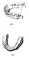
- FIG. 4 illustrates an example of this solution: in this case a dental arch is to be scanned.
- the known geometry introduced into the scan in the form of two polygons of known dimensions, linked by a bar also having a known dimension, enables detection of location and/or orientation of scans of portions of the teeth.
- Intra-oral scanning necessitates the use of small scanners, such that each scan enables the capture of only a portion of the arch.
- the present invention may be used in conjunction with the conventional practice whereby impressions are taken of the patient's dentition, and said dentition is replicated in the form of a master cast made from said impressions.
- the master cast will contain analogs of the dental implants. The accuracy with which the locations and orientations of these implant analogs can be determined, for the purpose of designing restoration component parts to be supported by the implants, is enhanced by using a recognition object according to the present invention.
- a preferred recognition object also for use when imaging a master cast of a patients dentition, is the object shown in FIG. 1 , which consists of three spheres sized and positioned to unambiguously define position and orientation. Following the securing of such a recognition object to an implant analog within a master cast, the cast is scanned to produce a data file of space-coordinate points of the surface. Then, imaging software comprising a feature location algorithm serves to deduce the defining surfaces of the recognition objects within the scanned image (in this case the spheres), and thus derives the locations and the precise orientation of the implants, which it reports to the system's users.
- a second preferred recognition object also for use in imaging a master cast of a patient's dentition, is a pyramid of known geometry.
Landscapes
- Health & Medical Sciences (AREA)
- Engineering & Computer Science (AREA)
- Physics & Mathematics (AREA)
- General Physics & Mathematics (AREA)
- Theoretical Computer Science (AREA)
- Oral & Maxillofacial Surgery (AREA)
- Public Health (AREA)
- Animal Behavior & Ethology (AREA)
- Veterinary Medicine (AREA)
- General Health & Medical Sciences (AREA)
- Dentistry (AREA)
- Epidemiology (AREA)
- Life Sciences & Earth Sciences (AREA)
- Software Systems (AREA)
- Computer Graphics (AREA)
- Multimedia (AREA)
- Geometry (AREA)
- Computer Vision & Pattern Recognition (AREA)
- Dental Tools And Instruments Or Auxiliary Dental Instruments (AREA)
- Apparatus For Radiation Diagnosis (AREA)
- Image Processing (AREA)
Applications Claiming Priority (2)
| Application Number | Priority Date | Filing Date | Title |
|---|---|---|---|
| US10/154,717 US6925198B2 (en) | 2002-05-24 | 2002-05-24 | Method and system for three-dimensional modeling of object fields |
| EP03736711A EP1512124A4 (de) | 2002-05-24 | 2003-05-23 | Dreidimensionales modellieren eines objektfeldes |
Related Parent Applications (1)
| Application Number | Title | Priority Date | Filing Date |
|---|---|---|---|
| EP03736711.7 Division | 2003-05-23 |
Publications (2)
| Publication Number | Publication Date |
|---|---|
| EP2620912A2 true EP2620912A2 (de) | 2013-07-31 |
| EP2620912A3 EP2620912A3 (de) | 2013-08-07 |
Family
ID=29548945
Family Applications (2)
| Application Number | Title | Priority Date | Filing Date |
|---|---|---|---|
| EP13156389.2A Withdrawn EP2620912A3 (de) | 2002-05-24 | 2003-05-23 | Dreidimensionale Modellierung eines Objektfelds |
| EP03736711A Withdrawn EP1512124A4 (de) | 2002-05-24 | 2003-05-23 | Dreidimensionales modellieren eines objektfeldes |
Family Applications After (1)
| Application Number | Title | Priority Date | Filing Date |
|---|---|---|---|
| EP03736711A Withdrawn EP1512124A4 (de) | 2002-05-24 | 2003-05-23 | Dreidimensionales modellieren eines objektfeldes |
Country Status (4)
| Country | Link |
|---|---|
| US (1) | US6925198B2 (de) |
| EP (2) | EP2620912A3 (de) |
| AU (1) | AU2003237245A1 (de) |
| WO (1) | WO2003100729A1 (de) |
Cited By (5)
| Publication number | Priority date | Publication date | Assignee | Title |
|---|---|---|---|---|
| CN109414304A (zh) * | 2016-03-22 | 2019-03-01 | 诺韦尔技术有限公司 | 面部特征扫描的系统和方法 |
| IT201900003379A1 (it) | 2019-03-08 | 2020-09-08 | De Goetzen S R L | Apparecchio e metodo per la creazione di immagini dentali |
| AT523132A2 (de) * | 2019-10-29 | 2021-05-15 | Fürhauser Lukas Dr | Scankörper mit langem arm |
| AT523131A2 (de) * | 2019-10-29 | 2021-05-15 | Fürhauser Nikolaus Dr | Scankörper mit orientierungsmarkierungen |
| EP4154844A1 (de) | 2021-09-27 | 2023-03-29 | Vigident | Verfahren, system, computerprogramm und vorrichtung zur fernanalyse einer zahnprothese |
Families Citing this family (49)
| Publication number | Priority date | Publication date | Assignee | Title |
|---|---|---|---|---|
| US7362890B2 (en) * | 2001-05-24 | 2008-04-22 | Astra Tech Inc. | Registration of 3-D imaging of 3-D objects |
| US7716024B2 (en) * | 2002-04-29 | 2010-05-11 | Geodigm Corporation | Method and apparatus for electronically generating a color dental occlusion map within electronic model images |
| US20030220778A1 (en) * | 2002-04-29 | 2003-11-27 | Hultgren Bruce Willard | Method and apparatus for electronically simulating jaw function within electronic model images |
| TWI310142B (en) * | 2003-05-28 | 2009-05-21 | Hon Hai Prec Ind Co Ltd | Cad-based cav system and method |
| US20050142517A1 (en) * | 2003-12-30 | 2005-06-30 | Howard Frysh | System for producing a dental implant and method |
| US7824346B2 (en) * | 2004-03-11 | 2010-11-02 | Geodigm Corporation | Determining condyle displacement utilizing electronic models of dental impressions having a common coordinate system |
| US7702492B2 (en) * | 2004-03-11 | 2010-04-20 | Geodigm Corporation | System and method for generating an electronic model for a dental impression having a common coordinate system |
| US7698068B2 (en) | 2004-06-17 | 2010-04-13 | Cadent Ltd. | Method for providing data associated with the intraoral cavity |
| US7270592B2 (en) * | 2004-08-12 | 2007-09-18 | D4D Technologies, Llc | Milling machine |
| US20060069591A1 (en) * | 2004-09-29 | 2006-03-30 | Razzano Michael R | Dental image charting system and method |
| DE102004054876B3 (de) * | 2004-11-12 | 2006-07-27 | Sirona Dental Systems Gmbh | Vermessungseinrichtung zur 3D-Vermessung von Zahnmodellen, Verschiebeplatte und Verfahren dazu |
| WO2006083875A2 (en) * | 2005-02-01 | 2006-08-10 | Friel Timothy P | Ocular prosthesis and fabrication method of same |
| US8185224B2 (en) | 2005-06-30 | 2012-05-22 | Biomet 3I, Llc | Method for manufacturing dental implant components |
| US7555403B2 (en) | 2005-07-15 | 2009-06-30 | Cadent Ltd. | Method for manipulating a dental virtual model, method for creating physical entities based on a dental virtual model thus manipulated, and dental models thus created |
| GB0608841D0 (en) | 2006-05-04 | 2006-06-14 | Isis Innovation | Scanner system and method for scanning |
| US8442283B2 (en) * | 2006-08-30 | 2013-05-14 | Anatomage Inc. | Patient-specific three-dimensional dentition model |
| US20080176182A1 (en) * | 2006-10-05 | 2008-07-24 | Bruce Willard Hultgren | System and method for electronically modeling jaw articulation |
| WO2008041943A1 (en) * | 2006-10-06 | 2008-04-10 | Biomain Ab | A measuring device and measuring method using said measuring device |
| US20080112610A1 (en) * | 2006-11-14 | 2008-05-15 | S2, Inc. | System and method for 3d model generation |
| DE502006005266D1 (de) * | 2006-11-28 | 2009-12-10 | Degudent Gmbh | Verfahren zur Herstellung einer zahnärztlichen Restauration |
| WO2009001298A2 (en) * | 2007-06-26 | 2008-12-31 | Densys Ltd. | Supplemental scene reference surface devices for three-dimensional mapping |
| DE102007043366A1 (de) | 2007-09-12 | 2009-03-19 | Degudent Gmbh | Verfahren zur Positionsbestimmung eines intraoral messenden Messgerätes |
| US8100692B2 (en) * | 2007-10-19 | 2012-01-24 | Cagenix Incorporated | Dental framework |
| DE102007056820A1 (de) * | 2007-11-23 | 2009-06-18 | Sirona Dental Systems Gmbh | Messkörper für ein Implantat und Verfahren zur Erstellung einer 3D-Messaufnahme |
| JP5250251B2 (ja) * | 2007-12-17 | 2013-07-31 | イマグノーシス株式会社 | 医用撮影用マーカーおよびその活用プログラム |
| DE102008006048A1 (de) * | 2008-01-25 | 2009-07-30 | Straumann Holding Ag | Verfahren zum Modellieren eines individuellen Zahnersatzes |
| US8682043B2 (en) * | 2008-02-29 | 2014-03-25 | Zimmer Dental, Inc. | Method of merging anatomical data and surface data of a patient's dentition |
| US8509932B2 (en) | 2008-07-17 | 2013-08-13 | Cadent Ltd. | Methods, systems and accessories useful for procedures relating to dental implants |
| US8640338B2 (en) | 2009-02-02 | 2014-02-04 | Viax Dental Technologies, LLC | Method of preparation for restoring tooth structure |
| US20100192375A1 (en) | 2009-02-02 | 2010-08-05 | Remedent Nv | Method for producing a dentist tool |
| DE102009038588A1 (de) * | 2009-08-26 | 2011-03-24 | Degudent Gmbh | Verfahren zur Ermittlung eines Gesamtdatensatzes eines zu messenden Objektes |
| DE102010023406A1 (de) * | 2010-06-11 | 2011-12-15 | Kevin Hintersehr | Verfahren und Datenverarbeitungsvorrichtung zum Bereitstellen von Geometriedaten von Zahn-und/oder Kieferbereichen |
| ES2848157T3 (es) | 2010-07-19 | 2021-08-05 | Align Technology Inc | Procedimientos y sistemas para crear e interactuar con modelos virtuales tridimensionales |
| US9157733B2 (en) * | 2010-09-10 | 2015-10-13 | Dimensional Photonics International, Inc. | Method of data acquisition for three-dimensional imaging |
| EP2739240B1 (de) | 2011-05-26 | 2020-07-15 | Viax Dental Technologies, Llc | Zahnärztliches instrument und führungsvorrichtungen |
| JP2016506842A (ja) | 2013-02-20 | 2016-03-07 | ゲーツェー ヨーロッパ | 事前調整された歯科インプラント補助器具 |
| EP2842493B1 (de) | 2013-08-30 | 2016-04-06 | Zfx GmbH | Intraoraler Bezugskörper |
| KR101444727B1 (ko) * | 2014-03-18 | 2014-09-26 | 주식회사 디오 | 임플란트용 가이드 스탠트 제조방법 |
| US10980618B2 (en) | 2014-05-08 | 2021-04-20 | Cagenix, Inc. | Dental framework and prosthesis |
| US10426711B2 (en) | 2014-05-08 | 2019-10-01 | Cagenix, Inc. | Dental implant framework |
| JP6262179B2 (ja) * | 2014-12-05 | 2018-01-17 | ディオ コーポレーションDio Corporation | デンタルインプラント施術のための手術ガイドおよび植立物の製造方法 |
| US9844426B2 (en) * | 2015-03-12 | 2017-12-19 | Align Technology, Inc. | Digital dental tray |
| CN107690303B (zh) * | 2015-06-04 | 2021-11-05 | 惠普发展公司,有限责任合伙企业 | 用于生成三维模型的计算设备和方法 |
| US11007035B2 (en) | 2017-03-16 | 2021-05-18 | Viax Dental Technologies Llc | System for preparing teeth for the placement of veneers |
| DE102017127128A1 (de) | 2017-11-17 | 2019-05-23 | Gustav Gerstenkamp | Verfahren zum virtuellen Modellieren eines Zahnbogens |
| CA3100495A1 (en) | 2018-05-16 | 2019-11-21 | Benevis Informatics, Llc | Systems and methods for review of computer-aided detection of pathology in images |
| US11364101B2 (en) | 2018-12-03 | 2022-06-21 | Cagenix, Inc. | Dental implant framework |
| WO2021087549A1 (en) * | 2019-11-04 | 2021-05-14 | Implant Solutions Pty Ltd | Apparatus for facilitating acquisition of a scan and an intraoral scanning procedure |
| CN111700698B (zh) * | 2020-05-14 | 2022-07-08 | 先临三维科技股份有限公司 | 齿科扫描方法、装置、系统和计算机可读存储介质 |
Citations (2)
| Publication number | Priority date | Publication date | Assignee | Title |
|---|---|---|---|---|
| US5851115A (en) | 1993-12-06 | 1998-12-22 | Nobel Biocare Ab | Method and arrangement for collecting data for production of replacement dental parts for the human body |
| US5857853A (en) | 1993-07-26 | 1999-01-12 | Nobel Biocare Ab | Method of manufacturing a prosthesis to be fixed to implants in the jawbone of a patient, and a system for manufacturing such prostheses |
Family Cites Families (9)
| Publication number | Priority date | Publication date | Assignee | Title |
|---|---|---|---|---|
| DE3636671A1 (de) * | 1986-09-11 | 1988-03-17 | Neumeyer Stefan | Verfahren zur dreidimensionalen bestimmung der relativbewegung zwischen zwei koerpern sowie messanordnung zur durchfuehrung dieses verfahrens |
| FR2639211A1 (fr) * | 1988-11-18 | 1990-05-25 | Hennson Int | Procede de correlation des saisies tridimensionnelles d'organes humains et dispositif pour sa mise en oeuvre |
| US5598515A (en) * | 1994-01-10 | 1997-01-28 | Gen Tech Corp. | System and method for reconstructing surface elements of solid objects in a three-dimensional scene from a plurality of two dimensional images of the scene |
| US5642293A (en) * | 1996-06-03 | 1997-06-24 | Camsys, Inc. | Method and apparatus for determining surface profile and/or surface strain |
| US6108497A (en) * | 1996-11-06 | 2000-08-22 | Asahi Kogaku Kogyo Kabushiki Kaisha | Standard measurement scale and markers for defining standard measurement scale |
| US6405071B1 (en) * | 2000-02-10 | 2002-06-11 | Advanced Research And Technology | Three dimensional imaging and analysis of a root canal |
| US7084868B2 (en) * | 2000-04-26 | 2006-08-01 | University Of Louisville Research Foundation, Inc. | System and method for 3-D digital reconstruction of an oral cavity from a sequence of 2-D images |
| US7027642B2 (en) * | 2000-04-28 | 2006-04-11 | Orametrix, Inc. | Methods for registration of three-dimensional frames to create three-dimensional virtual models of objects |
| US7065243B2 (en) * | 2001-06-28 | 2006-06-20 | Eastman Kodak Company | Method and system for creating dental models from imagery |
-
2002
- 2002-05-24 US US10/154,717 patent/US6925198B2/en not_active Expired - Fee Related
-
2003
- 2003-05-23 AU AU2003237245A patent/AU2003237245A1/en not_active Abandoned
- 2003-05-23 EP EP13156389.2A patent/EP2620912A3/de not_active Withdrawn
- 2003-05-23 WO PCT/US2003/016488 patent/WO2003100729A1/en not_active Ceased
- 2003-05-23 EP EP03736711A patent/EP1512124A4/de not_active Withdrawn
Patent Citations (2)
| Publication number | Priority date | Publication date | Assignee | Title |
|---|---|---|---|---|
| US5857853A (en) | 1993-07-26 | 1999-01-12 | Nobel Biocare Ab | Method of manufacturing a prosthesis to be fixed to implants in the jawbone of a patient, and a system for manufacturing such prostheses |
| US5851115A (en) | 1993-12-06 | 1998-12-22 | Nobel Biocare Ab | Method and arrangement for collecting data for production of replacement dental parts for the human body |
Non-Patent Citations (1)
| Title |
|---|
| "Manual of Photogrammetry", 1980, AMERICAN SOCIETY OF PHOTOGRA ETRY AND REMOTE SENSING |
Cited By (7)
| Publication number | Priority date | Publication date | Assignee | Title |
|---|---|---|---|---|
| CN109414304A (zh) * | 2016-03-22 | 2019-03-01 | 诺韦尔技术有限公司 | 面部特征扫描的系统和方法 |
| IT201900003379A1 (it) | 2019-03-08 | 2020-09-08 | De Goetzen S R L | Apparecchio e metodo per la creazione di immagini dentali |
| AT523132A2 (de) * | 2019-10-29 | 2021-05-15 | Fürhauser Lukas Dr | Scankörper mit langem arm |
| AT523132A3 (de) * | 2019-10-29 | 2021-05-15 | Fürhauser Lukas Dr | Scankörper mit langem arm |
| AT523131A2 (de) * | 2019-10-29 | 2021-05-15 | Fürhauser Nikolaus Dr | Scankörper mit orientierungsmarkierungen |
| AT523131A3 (de) * | 2019-10-29 | 2021-05-15 | Fuerhauser Nikolaus | Scankörper mit orientierungsmarkierungen |
| EP4154844A1 (de) | 2021-09-27 | 2023-03-29 | Vigident | Verfahren, system, computerprogramm und vorrichtung zur fernanalyse einer zahnprothese |
Also Published As
| Publication number | Publication date |
|---|---|
| EP2620912A3 (de) | 2013-08-07 |
| EP1512124A4 (de) | 2011-01-05 |
| EP1512124A1 (de) | 2005-03-09 |
| US20030219148A1 (en) | 2003-11-27 |
| AU2003237245A1 (en) | 2003-12-12 |
| US6925198B2 (en) | 2005-08-02 |
| WO2003100729A1 (en) | 2003-12-04 |
Similar Documents
| Publication | Publication Date | Title |
|---|---|---|
| US6925198B2 (en) | Method and system for three-dimensional modeling of object fields | |
| EP1904978B1 (de) | Erfassung von 3d-bildern von 3d-objekten | |
| US12201494B2 (en) | Methods and systems for dental procedures | |
| KR101878467B1 (ko) | 3차원 치아 영상 데이터와 광학식 스캔 치아 모델의 선택적 정합 방법, 장치 및 프로그램 | |
| CN113573662B (zh) | 将个体的牙弓的虚拟模型与所述个体的面部的数字模型配准的方法 | |
| KR101163808B1 (ko) | 인체정보 추출장치, 인체촬영정보의 기준면 변환방법 및단면정보 검출장치 | |
| EP3727189B1 (de) | Messgerät zur verifizierung von 3-d-bildern | |
| CN120280163A (zh) | 一种用于口腔无牙颌种植的多源数据拟合方法及系统 | |
| EP3753527A1 (de) | Entfernbare befestigungsvorrichtung für zahnlose fälle |
Legal Events
| Date | Code | Title | Description |
|---|---|---|---|
| PUAL | Search report despatched |
Free format text: ORIGINAL CODE: 0009013 |
|
| PUAI | Public reference made under article 153(3) epc to a published international application that has entered the european phase |
Free format text: ORIGINAL CODE: 0009012 |
|
| AC | Divisional application: reference to earlier application |
Ref document number: 1512124 Country of ref document: EP Kind code of ref document: P |
|
| AK | Designated contracting states |
Kind code of ref document: A2 Designated state(s): AT BE BG CH CY CZ DE DK EE ES FI FR GB GR HU IE IT LI LU MC NL PT RO SE SI SK TR |
|
| AK | Designated contracting states |
Kind code of ref document: A3 Designated state(s): AT BE BG CH CY CZ DE DK EE ES FI FR GB GR HU IE IT LI LU MC NL PT RO SE SI SK TR |
|
| RIC1 | Information provided on ipc code assigned before grant |
Ipc: G06T 7/00 20060101AFI20130701BHEP |
|
| STAA | Information on the status of an ep patent application or granted ep patent |
Free format text: STATUS: THE APPLICATION IS DEEMED TO BE WITHDRAWN |
|
| 18D | Application deemed to be withdrawn |
Effective date: 20140208 |