EP2628016B1 - Erfassung von mr-daten mit physiologischer überwachung - Google Patents
Erfassung von mr-daten mit physiologischer überwachung Download PDFInfo
- Publication number
- EP2628016B1 EP2628016B1 EP11776567.7A EP11776567A EP2628016B1 EP 2628016 B1 EP2628016 B1 EP 2628016B1 EP 11776567 A EP11776567 A EP 11776567A EP 2628016 B1 EP2628016 B1 EP 2628016B1
- Authority
- EP
- European Patent Office
- Prior art keywords
- patient
- imaging
- scan
- signal level
- physiological signal
- Prior art date
- Legal status (The legal status is an assumption and is not a legal conclusion. Google has not performed a legal analysis and makes no representation as to the accuracy of the status listed.)
- Active
Links
Images
Classifications
-
- A—HUMAN NECESSITIES
- A61—MEDICAL OR VETERINARY SCIENCE; HYGIENE
- A61B—DIAGNOSIS; SURGERY; IDENTIFICATION
- A61B5/00—Measuring for diagnostic purposes; Identification of persons
- A61B5/0033—Features or image-related aspects of imaging apparatus, e.g. for MRI, optical tomography or impedance tomography apparatus; Arrangements of imaging apparatus in a room
- A61B5/0037—Performing a preliminary scan, e.g. a prescan for identifying a region of interest
-
- A—HUMAN NECESSITIES
- A61—MEDICAL OR VETERINARY SCIENCE; HYGIENE
- A61B—DIAGNOSIS; SURGERY; IDENTIFICATION
- A61B5/00—Measuring for diagnostic purposes; Identification of persons
- A61B5/05—Detecting, measuring or recording for diagnosis by means of electric currents or magnetic fields; Measuring using microwaves or radio waves
- A61B5/055—Detecting, measuring or recording for diagnosis by means of electric currents or magnetic fields; Measuring using microwaves or radio waves involving electronic [EMR] or nuclear [NMR] magnetic resonance, e.g. magnetic resonance imaging
-
- A—HUMAN NECESSITIES
- A61—MEDICAL OR VETERINARY SCIENCE; HYGIENE
- A61B—DIAGNOSIS; SURGERY; IDENTIFICATION
- A61B5/00—Measuring for diagnostic purposes; Identification of persons
- A61B5/72—Signal processing specially adapted for physiological signals or for diagnostic purposes
- A61B5/7271—Specific aspects of physiological measurement analysis
- A61B5/7285—Specific aspects of physiological measurement analysis for synchronizing or triggering a physiological measurement or image acquisition with a physiological event or waveform, e.g. an ECG signal
-
- G—PHYSICS
- G01—MEASURING; TESTING
- G01R—MEASURING ELECTRIC VARIABLES; MEASURING MAGNETIC VARIABLES
- G01R33/00—Arrangements or instruments for measuring magnetic variables
- G01R33/20—Arrangements or instruments for measuring magnetic variables involving magnetic resonance
- G01R33/44—Arrangements or instruments for measuring magnetic variables involving magnetic resonance using nuclear magnetic resonance [NMR]
- G01R33/48—NMR imaging systems
- G01R33/54—Signal processing systems, e.g. using pulse sequences ; Generation or control of pulse sequences; Operator console
- G01R33/56—Image enhancement or correction, e.g. subtraction or averaging techniques, e.g. improvement of signal-to-noise ratio and resolution
- G01R33/567—Image enhancement or correction, e.g. subtraction or averaging techniques, e.g. improvement of signal-to-noise ratio and resolution gated by physiological signals, i.e. synchronization of acquired MR data with periodical motion of an object of interest, e.g. monitoring or triggering system for cardiac or respiratory gating
- G01R33/5673—Gating or triggering based on a physiological signal other than an MR signal, e.g. ECG gating or motion monitoring using optical systems for monitoring the motion of a fiducial marker
-
- G—PHYSICS
- G01—MEASURING; TESTING
- G01R—MEASURING ELECTRIC VARIABLES; MEASURING MAGNETIC VARIABLES
- G01R33/00—Arrangements or instruments for measuring magnetic variables
- G01R33/20—Arrangements or instruments for measuring magnetic variables involving magnetic resonance
- G01R33/44—Arrangements or instruments for measuring magnetic variables involving magnetic resonance using nuclear magnetic resonance [NMR]
- G01R33/48—NMR imaging systems
- G01R33/54—Signal processing systems, e.g. using pulse sequences ; Generation or control of pulse sequences; Operator console
- G01R33/56—Image enhancement or correction, e.g. subtraction or averaging techniques, e.g. improvement of signal-to-noise ratio and resolution
- G01R33/567—Image enhancement or correction, e.g. subtraction or averaging techniques, e.g. improvement of signal-to-noise ratio and resolution gated by physiological signals, i.e. synchronization of acquired MR data with periodical motion of an object of interest, e.g. monitoring or triggering system for cardiac or respiratory gating
- G01R33/5676—Gating or triggering based on an MR signal, e.g. involving one or more navigator echoes for motion monitoring and correction
-
- G—PHYSICS
- G01—MEASURING; TESTING
- G01R—MEASURING ELECTRIC VARIABLES; MEASURING MAGNETIC VARIABLES
- G01R33/00—Arrangements or instruments for measuring magnetic variables
- G01R33/20—Arrangements or instruments for measuring magnetic variables involving magnetic resonance
- G01R33/44—Arrangements or instruments for measuring magnetic variables involving magnetic resonance using nuclear magnetic resonance [NMR]
- G01R33/48—NMR imaging systems
- G01R33/54—Signal processing systems, e.g. using pulse sequences ; Generation or control of pulse sequences; Operator console
- G01R33/56—Image enhancement or correction, e.g. subtraction or averaging techniques, e.g. improvement of signal-to-noise ratio and resolution
- G01R33/563—Image enhancement or correction, e.g. subtraction or averaging techniques, e.g. improvement of signal-to-noise ratio and resolution of moving material, e.g. flow contrast angiography
- G01R33/56308—Characterization of motion or flow; Dynamic imaging
- G01R33/56325—Cine imaging
-
- G—PHYSICS
- G01—MEASURING; TESTING
- G01R—MEASURING ELECTRIC VARIABLES; MEASURING MAGNETIC VARIABLES
- G01R33/00—Arrangements or instruments for measuring magnetic variables
- G01R33/20—Arrangements or instruments for measuring magnetic variables involving magnetic resonance
- G01R33/44—Arrangements or instruments for measuring magnetic variables involving magnetic resonance using nuclear magnetic resonance [NMR]
- G01R33/48—NMR imaging systems
- G01R33/54—Signal processing systems, e.g. using pulse sequences ; Generation or control of pulse sequences; Operator console
- G01R33/56—Image enhancement or correction, e.g. subtraction or averaging techniques, e.g. improvement of signal-to-noise ratio and resolution
- G01R33/563—Image enhancement or correction, e.g. subtraction or averaging techniques, e.g. improvement of signal-to-noise ratio and resolution of moving material, e.g. flow contrast angiography
- G01R33/56366—Perfusion imaging
Definitions
- the invention relates to a method of performing magnetic resonance imaging scan of a patient and an MRI system.
- Image-forming MR methods which utilize the interaction between magnetic field and nuclear spins in order to form two-dimensional or three-dimensional images are widely used nowadays, notably in the field of medical diagnostics, because for the imaging of soft tissue they are superior to other imaging methods in many respects, they do not require ionizing radiation, and they are usually not invasive.
- MRI is used for example as imaging technique to visualize myocardial injury.
- Cardiac and respiratory triggered MR imaging can be used to image morphology, time resolved cine movies may reveal cardiac function, dynamic contrast enhanced imaging can be utilized to measure perfusion and MR tagging sequences can be used to study the contraction of the myocardium in detail.
- the magnetic field produces different energy levels for the individual nuclear spins in dependence on the applied magnetic field strength which spins can be excited (spin resonance) by application of an alternating electromagnetic field (RF field) of defined frequency, the so called Larmor frequency or MR frequency.
- RF field alternating electromagnetic field
- Larmor frequency or MR frequency the so called Larmor frequency or MR frequency.
- the distribution of the individual nuclear spins produces an overall magnetization which can be deflected out of the state of equilibrium by application of an electromagnetic pulse of appropriate frequency (RF pulse) while the magnetic field extends perpendicularly to the z-axis, so that the magnetization performs a precessional motion about the z-axis.
- Any variation of the magnetization can be detected by means of receiving RF antennas, which are arranged and oriented within an examination volume of the MR device in such a manner that the variation of the magnetization is measured in the direction perpendicularly to the z-axis.
- the signal picked up in the receiving antennas then contains components of different frequencies which can be associated with different locations in the body.
- a major criterion for obtaining high quality MR images is to ensure that the imaged region of interest is not moving during an MR scan.
- abdominal imaging this becomes a serious problem since physically necessary patient breathing and thus patient movement translates into blurring and ghosting of the acquired MR image. Consequently, a breath hold is required by the patient during the MR imaging scan in order to prevent any movement in the imaged region of interest.
- US 7,182,083 B2 discloses an integrated respiratory monitor and CT imaging device apparatus.
- the respiratory monitor system is adapted to engage a patient and generate a respiratory signal representative of a breath hold level of the patient during a breath hold.
- the imaging device is adapted to scan the patient during the breath hold and generate a volumetric image data set of the patient.
- the respiratory sensor and imaging device are operatively connected to associate the respiratory signal representative of the breath hold level of the patient together with the volumetric image data set of the patient.
- breath hold commands and starting an imaging scan when the patient has reached the breath hold state can introduce operator dependent variations. Breath hold sequences typically are started too early when the breath hold state has not been reached yet which translates into blurring and ghosting of the image. In the breath hold state, the breath hold may "drift away" also leading to motion related problems. Also automated breath hold commands are not patient dependent thus neglecting the capability to follow the breath hold commands and to address the overall patient situation.
- the patient's breath hold capabilities typically are related to the progress and the severity of a patient's disease.
- US patent 4,878,499 describes acquiring magnetic resonance data from a patient by a magnetic resonance imaging system, wherein the system has an announcement section for intermittently urging the patient to stop a body movement.
- a data acquisition section is operated under the control of a control section only while the patient stands still in response to the announcement of the announcement section, thereby intermittently acquiring magnetic resonance data in units of a predetermined volume.
- US patent application 2007/0172029 A1 describes a motion detection system for use on a patient undergoing a medical procedure where it is important for the subject to repeatedly re-establish a reference position.
- the motion detection system includes a motion detector for sensing the motion of the subject and producing a motion input signal, a control unit for receiving this signal and producing displacement data indicative of subject motion away from a reference position, and one or more displays for receiving the displacement data and indicating the displacement of the subject from the reference position.
- US patent 5,363,844 describes an NMR system in which a respiration monitor provides a visual feedback to the patient which enables the patient to perform a series of breath-holds with the patient's diaphragm positioned at the same reference point. This enables NMR data to be acquired over a series of breath-holds without introducing blurring or image artifacts. Between breath-holds a navigator pulse sequence is used to gather NMR data from which diaphragm position is measured, and during each breathhold the pulse sequence is changed to gather NMR image data.
- a dynamic scanning initiation means initiates dynamic scanning in which a plurality of scans is sequentially performed with a time interval between adjoining scans.
- a dynamic scanning suspending means suspends the dynamic scanning initiated by the dynamic scanning initiation means.
- a dynamic scanning resuming means resumes the dynamic scanning suspended by the dynamic scanning suspending means.
- US patent application 2008/0183475 describes an image diagnosing apparatus for shooting an image of the subject in an imaging space having a voice guidance unit which reproduces and outputs to the subject a prescribed voice guidance, and a voice output control unit which causes the output timing of the voice guidance outputted from the voice guidance unit to correspond with the timing of shooting the image of the subject.
- US 2008/0004518 A1 discloses a magnetic resonance sequence for quantitative T 1 mapping during free breathing.
- the known method includes: saturating or inverting an imaging region while leaving a navigation region unsaturated or non-inverted; generating navigation data from the navigation region; generating saturation or inversion recovery data from the imaging region; and creating a T 1 map from the saturation or inversion recovery data.
- the mentioned document discloses a magnetic resonance imaging system including respiratory monitoring.
- Stefan Bluemi et al MR imaging of newborns by using an MR compatible incubator with integrated radiofrequency coils initial experience, Radiology vol 231, no. 2,2004, pages 594-601 discloses MR imaging of newborns by using an MR-compatible incubator with integrated radiofrequency coils.
- the incubator comprises air, temperature and humidity regulators.
- Embodiments of the invention have the advantage that, with respect to the physiological situation of the patient, the patient can dynamically be instructed to optimize behavior such that a respective MR imaging scan can be performed in an optimized manner, i.e. without motion-related problems like blurring and ghosting in the reconstructed MR image.
- changing the environmental conditions of the patient comprises adapting visual and acoustic media exposed to the patient such that an in bore "Ambient Experience" changes.
- This also comprises changes in the air conditioning exposed to the patient.
- air conditioning may be adapted in accordance with air humidity, oxygen level, nitrogen level, temperature and/or air flow through the magnet bore. In either case this permits to comfort the patient both physically and emotionally by dynamically reacting on perceived needs and physiological reactions of the patient.
- the environment within the magnet bore is personalized for actual patient's needs. A consequence is that patients can be easier motivated and/or can be distracted to achieve a desired stable physiological condition.
- the physiological signal level comprises a breath hold level of the patient.
- the physiological signal level may comprise signal levels regarding breathing and/or body motion and/or cardiac activity of the patient.
- hyperventilation breath hold commands This enables for difficult breath holders to increase the breath hold time at a given breath hold level.
- command timings and patient comfort commands permit to achieve a desired level of cardiac activity.
- These instructions are dynamically chosen adapted to the actually monitored physiological signal level and thus guide the patient's physiological behavior in a desired manner.
- the timing of the breath in and breath out commands may be adapted to the actual breathing state while it is carefully checked if the commands are followed.
- the automated breath hold command advises to 'stop breathing'.
- another automated short 'stop breathing' command may be given.
- Patient motivation commands and in bore "Ambient Experience” changes psychologically motivate the patient to keep his actual breath hold level. For example, this may be performed by providing the patient information about a remaining required breath hold time or by visually stimulating the patient to suppress the upcoming breath reflex.
- analyzing the monitored physiological signal level does not only include an analysis on the actual physiological signal level, but may also comprise analysis of motion-relevant statistics over a certain time span in order to obtain general information for example on the patient's breath hold capabilities or on the patients cardiac activities.
- the breathing history of the patient can be analyzed in order to determine the capability to hold the breath after an automatic breath hold command has been given including the breath hold lengths and drifts during a breath hold.
- This may also include an analysis on the capability to follow an issued breath hold command including response times but also possible free breathing characteristics such as non-moving periods in the end expiration/inspiration phase and the presence of an apnea.
- the motion history statistics previously acquired can be recovered from a previous study.
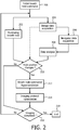
- the method further comprises adapting or optimizing the scan protocol for performing a magnetic resonance imaging scan, wherein the optimization is adapted to the monitored physiological signal level.
- an 'optimization' of the scan protocol can be understood as triggering or gating of an MR scan at selected points in the respiratory cycle of the patient. It however also can be seen as a change in individual parameters of the MR imaging sequence or substitute the imaging sequence by a different imaging sequence.
- the optimization of the scan protocol comprises calculating the expected maximum breath hold time within a predefined breath hold level of the patient and changing the actually selected scan protocol to a new imaging protocol, the new imaging protocol requiring a respective data acquisition time for completion of a respective magnetic resonance imaging scan, wherein the new imaging protocol is chosen in accordance with the data acquisition time being shorter then or matching the calculated expected maximum breath hold time.
- this actually selected scan protocol may be exchanged by a new imaging protocol which requires a data acquisition time of 7 seconds.
- the new imaging protocol may acquire the essential information in the first 7 seconds with the possibility to extend acquisition time in case that the patient can hold his breath longer successively improving image quality.
- the optimization of the scan protocol may comprise calculating the expected heart rate and changing the actually selected scan protocol to a new imaging protocol, the new imaging protocol requiring a respective data acquisition time for completion of a respective magnetic resonance imaging scan, wherein the new imaging protocol is chosen in accordance with the data acquisition time being shorter then or matching the calculated expected heart rate.
- the new imaging protocol may use centric k-space acquisition.
- acquisition points may be registered until a breath hold plateau with a specified breath hold level criterion is reached. These acquisition points can be re-acquired within the same breath hold and can be replaced to improve image quality. Another possibility is to not re-acquire but synthetically derive these k-space acquisition points using parallel imaging or compressed sensing methodologies.
- the acoustic gradient noise may be adapted to change dependent on the cardiac or breathing motion which in turn influences the patient's behavior such as breathing and heart beat.
- acoustically pretending continuous gradient noise helps to calm down the patient.
- the new imaging protocol comprises the actually selected scan protocol with instructions for an adapted image resolution, wherein the adapted image resolution results in the respective data acquisition time for completion of a magnetic resonance imaging scan.
- this may be performed by adapting for example the way the k-space is sampled.
- monitoring the breath hold level of the patient comprises repeatedly performing magnetic resonance navigator scans on the region of interest to be imaged during the magnetic resonance imaging scan, wherein the method further comprises during the navigator scans and/or the imaging scan to determine a physical movement within the region of interest from the navigator scans, wherein the movement results from a change in the patient's breath hold level, and adjusting the slice positions in accordance with the determined physical movement in order to compensate the acquired image data for the physical movement.
- the breath hold can be continuously monitored.
- this can be done by interleaving the navigator acquisition with the actual data acquisition.
- the advantage of the navigator acquisition is that in case the breath hold drifts, small drifts can thus be corrected by adapting the slice position (slice tracking) next to reminding the patient via an automatic command to 'further hold the breath'. Larger drifts or the capability not to hold the breath leads to acquisition points to be re-acquired later or derived synthetically.
- the invention relates to a magnetic resonance imaging system for performing a magnetic resonance imaging scan as defined in independent claim 7.
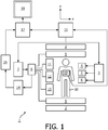
- an MR imaging system 1 comprises superconducting or resistive main magnet coils 2 such that a substantially uniform, temporarily constant main magnetic field B0 is created along a z-axis through an examination volume.
- the magnetic resonance system applies a series of RF pulses and switched magnetic field gradients to invert or excite nuclear magnetic spins, induce magnetic resonance, refocus magnetic resonance, manipulate magnetic resonance, spatially or otherwise encode the magnetic resonance, saturate spins and the like to perform MR imaging.
- a gradient pulse amplifier 3 applies current pulses to selected ones of whole body gradient coils 4, 5 and 6 along x, y and z-axes of the examination volume.
- An RF transmitter 7 transmits RF pulses or pulse packets, via a send/receive switch 8 to an RF antenna 9 to transmit RF pulses into the examination volume.
- a typical MR imaging sequence is composed of a packet of RF pulse sequences of short duration which taken together with each other and any applied magnetic field gradients achieve a selected manipulation of nuclear magnetic resonance.
- the RF pulses are used to saturate, excite resonance, invert magnetization, refocus resonance, or manipulate resonance and select a portion of a body 10 positioned in the examination volume.
- the MR signals may also be picked up by the RF antenna 9.
- a set of local array RF coils 11, 12 and 13 are placed contiguous to the region selected for imaging.
- the array coils 11, 12 and 13 can be used to receive MR signals induced by RF transmissions effected via the RF antenna.
- the resultant MR signals are picked up by the RF antenna 9 and/or by the array of RF coils 11, 12 and 13 and are demodulated by a receiver 14 preferably including a pre-amplifier (not shown).
- the receiver 14 is connected to the RF coils 9, 11, 12 and 13 via a send/receive switch 8.
- a host computer 15 controls the gradient pulse amplifier 3 and the transmitter 7 to generate any of a plurality of imaging sequences, such as echo planar imaging (EPI), echo volume imaging, gradient and spin echo imaging, fast spin echo imaging, imaging using ultra-short echo time acquisition pulse sequences and the like.
- EPI echo planar imaging
- echo volume imaging gradient and spin echo imaging
- fast spin echo imaging imaging using ultra-short echo time acquisition pulse sequences and the like.
- the receiver 14 receives a single or a plurality of MR data lines in a rapid succession following each RF excitation pulse.
- a data acquisition system 16 performs analogue to digital conversion of the received signals and converts each MR data line to a digital format suitable for further processing.
- the data acquisition system 16 is a separate computer which is specialized in acquisition of raw image data.
- the digital raw image data is reconstructed into an image representation by a reconstruction processor 17 which applies a Fourier transform or other appropriate reconstruction algorithms.
- the MR image may represent a planar slice through the patient, an array of parallel planar slices, a three-dimensional volume or the like.
- the image is then stored in an image memory where it may be accessed for converting slices or other portions of the image representation into appropriate formats for visualization, for example via a video monitor 18 which provides a man readable display of the resultant MR image.
Landscapes
- Health & Medical Sciences (AREA)
- Life Sciences & Earth Sciences (AREA)
- Physics & Mathematics (AREA)
- Engineering & Computer Science (AREA)
- Nuclear Medicine, Radiotherapy & Molecular Imaging (AREA)
- Biophysics (AREA)
- Radiology & Medical Imaging (AREA)
- General Health & Medical Sciences (AREA)
- Physiology (AREA)
- High Energy & Nuclear Physics (AREA)
- Animal Behavior & Ethology (AREA)
- Surgery (AREA)
- Biomedical Technology (AREA)
- Pathology (AREA)
- Public Health (AREA)
- Veterinary Medicine (AREA)
- Heart & Thoracic Surgery (AREA)
- Medical Informatics (AREA)
- Molecular Biology (AREA)
- Signal Processing (AREA)
- Power Engineering (AREA)
- Pulmonology (AREA)
- Cardiology (AREA)
- Condensed Matter Physics & Semiconductors (AREA)
- General Physics & Mathematics (AREA)
- Artificial Intelligence (AREA)
- Computer Vision & Pattern Recognition (AREA)
- Psychiatry (AREA)
- Magnetic Resonance Imaging Apparatus (AREA)
Claims (7)
- Verfahren zur Durchführung einer Magnetresonanz-Bildgebungsabtastung an einem Patienten, wobei das Verfahren die Schritte umfasst:- Überwachung (202) eines physiologischen Signalpegels des Patienten;- Analysieren (208) des überwachten physiologischen Signalpegels, wobei die Analyse des überwachten physiologischen Signalpegels auch die Analyse bewegungsrelevanter Statistiken über eine bestimmte Zeitspanne umfassen kann,- auf der Grundlage des analysierten physiologischen Signalpegels automatisches Führen des Patienten auf einen vordefinierten physiologischen Signalpegel, indem die Umgebungsbedingungen, denen der Patient ausgesetzt ist, dynamisch geändert werden; und- Durchführen (204) der Magnetresonanz-Bildgebungsabtastung gemäß einem ausgewählten Abtastprotokoll, wobei die Schritte des Überwachens (202), Analysierens (208) und Durchführens (204) der Magnetresonanz-Bildgebungsabtastung nach dem Durchführen (200) einer anfänglichen Führung des Patienten auf den vordefinierten physiologischen Signalpegel parallel durchgeführt werden;wobei das Verfahren weiter umfasst:- Anpassen (214) des ausgewählten Abtastprotokolls an den überwachten physiologischen Signalpegel während des Schritts des Durchführens (204) der Magnetresonanz-Bildgebungsabtastung, wenn der vordefinierte physiologische Signalpegel nicht erreicht wird, wobei das Anpassen an den überwachten physiologischen Signalpegel mindestens einen umfasst von- Auslösen oder Ansteuerung einer MR-Abtastung, die in das ausgewählte Abtastprotokoll an ausgewählten Punkten involviert ist;- Ändern eines einzelnen Parameters einer MR-Bildgebungssequenz, die an dem ausgewählten Abtastprotokoll beteiligt ist;- Ersetzen einer MR-Bildgebungssequenz, die an dem ausgewählten Abtastprotokoll beteiligt ist, durch eine andere Bildgebungssequenz;- Ändern des ausgewählten Abtastprotokolls in ein neues B ildgebung sprotokoll,wobei das neue Bildgebungsprotokoll eine entsprechende Datenerfassungszeit für den Abschluss einer entsprechenden MR-Abtastung erfordert.
- Verfahren nach Anspruch 1, wobei die Änderung der Umgebungsbedingungen, denen der Patient ausgesetzt ist, die Anpassung der visuellen und akustischen Medien oder eine Änderung der Klimaanlage umfasst.
- Verfahren nach Anspruch 1, wobei das physiologische Signal mindestens einen Atemanhaltepegel des Patienten, einen Signalpegel, der mit der Atmung des Patienten, der Körperbewegung des Patienten oder der Herzaktivität des Patienten verbunden ist, umfasst.
- Verfahren nach Anspruch 1, wobei das Führen des Patienten weiter eine Anweisung an den Patienten umfasst, die mindestens eine enthält:- die Zeitpunkte der Ein- und Ausatmungs-Kommandos,- ein "Atmen stoppen"-Kommando,- ein "Bitte anhalten"-Kommando,- ein Hyperventilationskommando,- ein Kommando zur Patientenmotivation, das mit der Aufrechterhaltung des Atemanhaltepegels verbunden ist, oder- ein Kommando für den Patientenkomfort.
- Verfahren nach Anspruch 1,
wobei das geänderte ausgewählte Abtastprotokoll umfasst:- Anwenden eines dem ausgewählten Abtastprotokoll vorausgehenden ausgeführten vorläufigen Bildgebungsprotokolls, bis der vordefinierte physiologische Signalpegel erreicht ist. - Verfahren nach Anspruch 1, weiter umfassend:- die akustische Emulation einer bildgebenden Abtastung des Patienten, wenn der vordefinierte physiologische Signalpegel nicht erreicht wird.
- Magnetresonanz-Bildgebungssystem (10) zum Durchführen einer Magnetresonanz-Bildgebungsabtastung an einem Patienten, wobei das System einen Host-Computer (15) und einen Sensor (19) zum Überwachen eines physiologischen Signalpegels des Patienten umfasst und das System so konfiguriert ist, dass es das Verfahren nach einem der Ansprüche 1-6 ausführt.
Priority Applications (1)
| Application Number | Priority Date | Filing Date | Title |
|---|---|---|---|
| EP11776567.7A EP2628016B1 (de) | 2010-10-14 | 2011-10-12 | Erfassung von mr-daten mit physiologischer überwachung |
Applications Claiming Priority (3)
| Application Number | Priority Date | Filing Date | Title |
|---|---|---|---|
| EP10187516 | 2010-10-14 | ||
| PCT/IB2011/054501 WO2012049634A1 (en) | 2010-10-14 | 2011-10-12 | Mr data acquisition using physiological monitoring |
| EP11776567.7A EP2628016B1 (de) | 2010-10-14 | 2011-10-12 | Erfassung von mr-daten mit physiologischer überwachung |
Publications (2)
| Publication Number | Publication Date |
|---|---|
| EP2628016A1 EP2628016A1 (de) | 2013-08-21 |
| EP2628016B1 true EP2628016B1 (de) | 2020-03-25 |
Family
ID=44898105
Family Applications (1)
| Application Number | Title | Priority Date | Filing Date |
|---|---|---|---|
| EP11776567.7A Active EP2628016B1 (de) | 2010-10-14 | 2011-10-12 | Erfassung von mr-daten mit physiologischer überwachung |
Country Status (5)
| Country | Link |
|---|---|
| US (1) | US10588578B2 (de) |
| EP (1) | EP2628016B1 (de) |
| CN (1) | CN103168248B (de) |
| RU (1) | RU2612859C2 (de) |
| WO (1) | WO2012049634A1 (de) |
Families Citing this family (27)
| Publication number | Priority date | Publication date | Assignee | Title |
|---|---|---|---|---|
| WO2014091374A2 (en) * | 2012-12-12 | 2014-06-19 | Koninklijke Philips N.V. | Motion detection and correction method for magnetic resonance diffusion weighted imaging (dwi) |
| DE102013219042A1 (de) * | 2013-09-23 | 2015-03-26 | Siemens Aktiengesellschaft | Verfahren zur Optimierung einer Bildgebungsmessung |
| DE102013222103A1 (de) * | 2013-10-30 | 2015-05-13 | Siemens Aktiengesellschaft | Verfahren zum Betrieb einer Magnetresonanzeinrichtung und Magnetresonanzeinrichtung |
| US9557397B2 (en) * | 2013-11-04 | 2017-01-31 | Aspect Imaging Ltd. | Method for manipulating the MRI's protocol of pulse-sequences |
| JP6408594B2 (ja) * | 2014-02-13 | 2018-10-17 | コーニンクレッカ フィリップス エヌ ヴェKoninklijke Philips N.V. | 時間効率的な4d磁気共鳴イメージング |
| US9472082B2 (en) * | 2014-06-23 | 2016-10-18 | Bruno Delean | Vision based system for detecting distress behavior |
| DE102015222835B4 (de) * | 2015-11-19 | 2019-06-13 | Siemens Healthcare Gmbh | Magnetresonanzbildgebungsverfahren mit simultaner Bildaufnahme von mehreren Teilvolumen mit einer synchronen Bildaufnahme von Navigatoren |
| GB2567975B (en) | 2016-05-27 | 2022-02-02 | Synaptive Medical Inc | Magnetic resonance imaging of different nuclear spin species with the same radio frequency coil |
| DE102016213632A1 (de) * | 2016-07-26 | 2018-02-01 | Siemens Healthcare Gmbh | Verbesserte Akquisition von MR-Messdaten bei einer Untersuchung mit angehaltenem Atem |
| EP3381353A1 (de) * | 2017-03-30 | 2018-10-03 | Koninklijke Philips N.V. | Verfahren zur planung eines bildabtastungsprotokolls |
| EP3524994A1 (de) * | 2018-02-08 | 2019-08-14 | Koninklijke Philips N.V. | Mrt mit akustischem klang im modus vor der datenerfassung |
| EP3575812A1 (de) * | 2018-05-28 | 2019-12-04 | Koninklijke Philips N.V. | Verfahren, computerprogrammprodukt und vorrichtung zum klassifizieren von schall in der mrt und zum trainieren eines patienten |
| EP3632298A1 (de) | 2018-10-05 | 2020-04-08 | Koninklijke Philips N.V. | Atemanpassungssystem und verfahren zur beeinflussung eines atemparameters |
| EP3805773A1 (de) | 2019-10-08 | 2021-04-14 | Koninklijke Philips N.V. | Respiratorisches biofeedback für die strahlentherapie |
| CN113100741B (zh) * | 2020-01-13 | 2024-02-27 | 上海联影医疗科技股份有限公司 | 一种磁共振扫描方法、设备及存储介质 |
| EP3862770A1 (de) | 2020-02-04 | 2021-08-11 | Koninklijke Philips N.V. | Vorrichtung zur überwachung eines patienten, der sich einer magnetresonanztomographie unterzieht |
| EP3889969A1 (de) * | 2020-04-02 | 2021-10-06 | Koninklijke Philips N.V. | System zur medizinischen bildgebung |
| CN111445990A (zh) * | 2020-04-13 | 2020-07-24 | 上海联影医疗科技有限公司 | 一种扫描方案的调整方法、装置、电子设备及存储介质 |
| EP3961238A1 (de) * | 2020-08-27 | 2022-03-02 | Siemens Healthcare GmbH | System und verfahren für standardisierte mrt-untersuchungen mit patientenzentrischen scan-arbeitsablaufanpassungen |
| EP3973865A1 (de) | 2020-09-29 | 2022-03-30 | Koninklijke Philips N.V. | Vorrichtung zur optimierung einer sequenz von magnetresonanz (mr)-scans einer mr-untersuchung |
| EP4079227B1 (de) * | 2021-04-23 | 2025-06-04 | Siemens Healthineers AG | Anpassung eines medizinischen bildgebungsverfahrens an ein individuelles atmungsverhalten eines patienten |
| EP4176796A1 (de) | 2021-11-05 | 2023-05-10 | Koninklijke Philips N.V. | Mehrsitzungsatmungsführung |
| EP4449983A1 (de) * | 2023-04-17 | 2024-10-23 | Siemens Healthineers AG | Verfahren zur vorhersage der atemanhaltedauer in einem medizinischen bildgebungsverfahren |
| EP4468009A1 (de) | 2023-05-22 | 2024-11-27 | Koninklijke Philips N.V. | Magnetresonanzfingerabdruckverfahren zur bildgebung während atemruhen |
| CN116602651A (zh) * | 2023-05-26 | 2023-08-18 | 中国人民解放军陆军军医大学第一附属医院 | 一种孕妇磁共振检查辅助装置 |
| EP4498382A1 (de) | 2023-07-24 | 2025-01-29 | Koninklijke Philips N.V. | Vorrichtung und verfahren zur umgebungserfahrungssteuerung |
| DE102024200268A1 (de) * | 2024-01-12 | 2025-07-17 | Siemens Healthineers Ag | Verfahren und Vorrichtung zur Überwachung und/oder Anpassung von atemangehaltenen MR-Untersuchungen |
Family Cites Families (20)
| Publication number | Priority date | Publication date | Assignee | Title |
|---|---|---|---|---|
| JPS62106755A (ja) | 1985-11-02 | 1987-05-18 | 株式会社東芝 | 磁気共鳴イメ−ジング装置 |
| US5363844A (en) * | 1993-08-13 | 1994-11-15 | Mayo Foundation For Medical Education And Research | Breath-hold monitor for MR imaging |
| US6201393B1 (en) * | 1997-12-15 | 2001-03-13 | General Electric Company | Reducing image artifacts caused by patient motion during MR imaging |
| US6889071B2 (en) * | 2000-12-19 | 2005-05-03 | General Electric Company | Acquisition of high-temporal free-breathing MR images |
| US7182083B2 (en) | 2002-04-03 | 2007-02-27 | Koninklijke Philips Electronics N.V. | CT integrated respiratory monitor |
| CN1929781A (zh) * | 2003-08-21 | 2007-03-14 | 依斯克姆公司 | 用于脉管斑块检测和分析的自动化方法和系统 |
| WO2005039390A2 (en) | 2003-10-20 | 2005-05-06 | Arthrocare Corporation | Electrosurgical method and apparatus for removing tissue within a bone body |
| CN2768671Y (zh) | 2004-12-30 | 2006-04-05 | 中国医学科学院北京协和医院 | 一种用于ct成像的呼吸监测装置 |
| JP4350679B2 (ja) * | 2005-05-26 | 2009-10-21 | ジーイー・メディカル・システムズ・グローバル・テクノロジー・カンパニー・エルエルシー | 磁気共鳴イメージング装置および画像撮影装置 |
| JP2007003264A (ja) | 2005-06-22 | 2007-01-11 | Shimadzu Corp | Pet装置 |
| US7678063B2 (en) * | 2006-01-06 | 2010-03-16 | Mayo Foundation For Medical Education And Research | Motion monitor system for use with imaging systems |
| US20080004518A1 (en) * | 2006-06-29 | 2008-01-03 | Koninklijke Philips Electronics N.V. | Magnetic resonance sequence for quantitative t1 mapping during free breathing |
| US20080051261A1 (en) * | 2006-08-25 | 2008-02-28 | Lewis Charles A | Exercise protocols for treadmills and bicycle ergometers for exercise, diagnostics and rehabilitation |
| JP4989166B2 (ja) | 2006-09-14 | 2012-08-01 | ジーイー・メディカル・システムズ・グローバル・テクノロジー・カンパニー・エルエルシー | 画像診断装置 |
| JP2008148918A (ja) | 2006-12-18 | 2008-07-03 | Ge Medical Systems Global Technology Co Llc | Mri装置およびその制御方法 |
| DE102007017269A1 (de) * | 2007-04-12 | 2008-10-16 | Siemens Ag | Bildakquisitionssystem und -verfahren zur Durchführung von CT- bwz. MRT-gesteuerten Interventionen in Korrelation mit der Atemlage eines Patienten |
| US20090192399A1 (en) * | 2008-01-25 | 2009-07-30 | Samsung Electronics Co., Ltd. | Apparatus and method to detect heart-rate and air conditioning system having the apparatus |
| US20100152600A1 (en) * | 2008-04-03 | 2010-06-17 | Kai Sensors, Inc. | Non-contact physiologic motion sensors and methods for use |
| US7880465B2 (en) * | 2008-06-04 | 2011-02-01 | General Electric Company | Method and apparatus for contrast inflow dynamic MR angiography |
| DE102009017775A1 (de) * | 2009-04-20 | 2010-12-02 | Siemens Aktiengesellschaft | Verfahren, Magnetresonanzgerät und Computerprogramm zur Anzeige eines Fortschritts einer Akquisition von Messdaten eines Untersuchungsbereichs eines Patienten während einer kontinuierlichen Fahrt des Untersuchungsbereichs durch ein Magnetresonanzgerät |
-
2011
- 2011-10-12 EP EP11776567.7A patent/EP2628016B1/de active Active
- 2011-10-12 US US13/879,417 patent/US10588578B2/en active Active
- 2011-10-12 RU RU2013121800A patent/RU2612859C2/ru active
- 2011-10-12 CN CN201180049681.5A patent/CN103168248B/zh active Active
- 2011-10-12 WO PCT/IB2011/054501 patent/WO2012049634A1/en not_active Ceased
Non-Patent Citations (5)
| Title |
|---|
| JOHANNES F.T. ARNOLD ET AL: "Lung MRI using an MR-compatible active breathing control (MR-ABC)", MAGNETIC RESONANCE IN MEDICINE, vol. 58, no. 6, 1 January 2007 (2007-01-01), pages 1092 - 1098, XP055021414, ISSN: 0740-3194, DOI: 10.1002/mrm.21424 * |
| LIU Y L ET AL: "A MONITORING, FEEDBACK, AND TRIGGERING SYSTEM FOR REPRODUCIBLE BREATH-HOLD MR IMAGING", MAGNETIC RESONANCE IN MEDICINE, JOHN WILEY & SONS, INC, US, vol. 30, no. 4, 1 October 1993 (1993-10-01), pages 507 - 511, XP000402838, ISSN: 0740-3194 * |
| STEFAN BLÜML ET AL: "MR Imaging of Newborns by Using an MR-compatible Incubator with Integrated Radiofrequency Coils: Initial Experience", RADIOLOGY, vol. 231, no. 2, 1 May 2004 (2004-05-01), US, pages 594 - 601, XP055224083, ISSN: 0033-8419, DOI: 10.1148/radiol.2312030166 * |
| YI WANG ET AL: "3D coronary MR angiography in multiple breath-holds using a respiratory feedback monitor", MAGNETIC RESONANCE IN MEDICINE., vol. 34, no. 1, 1 July 1995 (1995-07-01), US, pages 11 - 16, XP055224087, ISSN: 0740-3194, DOI: 10.1002/mrm.1910340104 * |
| YI WANG ET AL: "CORONARY MRI WITH A RESPIRATORY FEEDBACK MONITOR: THE 2D IMAGING CASE", MAGNETIC RESONANCE IN MEDICINE, JOHN WILEY & SONS, INC, US, vol. 33, no. 1, 1 January 1995 (1995-01-01), pages 116 - 121, XP000482975, ISSN: 0740-3194 * |
Also Published As
| Publication number | Publication date |
|---|---|
| RU2013121800A (ru) | 2014-11-20 |
| US20130211236A1 (en) | 2013-08-15 |
| US10588578B2 (en) | 2020-03-17 |
| EP2628016A1 (de) | 2013-08-21 |
| CN103168248B (zh) | 2016-06-08 |
| WO2012049634A1 (en) | 2012-04-19 |
| RU2612859C2 (ru) | 2017-03-13 |
| CN103168248A (zh) | 2013-06-19 |
Similar Documents
| Publication | Publication Date | Title |
|---|---|---|
| EP2628016B1 (de) | Erfassung von mr-daten mit physiologischer überwachung | |
| US8489174B2 (en) | Method to detect a breathing movement of an examination subject corresponding to signal data by magnetic resonance | |
| CN101354433B (zh) | 记录和处理时间上相继的图像数据组的序列的方法及设备 | |
| CN102743172B (zh) | 借助磁共振设备抑制不期望的信号触发地采集测量数据 | |
| RU2580189C2 (ru) | Интервенционная мр-томография с компенсацией движения | |
| JP5740307B2 (ja) | 磁気共鳴イメージング装置及び傾斜磁場印加方法 | |
| US9176210B2 (en) | Magnetic resonance imaging apparatus | |
| US10191131B2 (en) | Medical imaging apparatus having multiple subsystems, and operating method therefor | |
| US9713449B2 (en) | Magnetic resonance imaging device and magnetic resonance imaging method | |
| JP2001000417A (ja) | マルチ・スラブ及びマルチ・ウィンドウでの心臓の磁気共鳴イメージング法 | |
| US10871538B2 (en) | Planning support for selective arterial spin labeling MR imaging methods | |
| CN101862192B (zh) | 用于显示测量数据的采集的进展的方法和磁共振设备 | |
| CN112545482B (zh) | 用于创建医疗工作流程的路线图的方法和系统 | |
| CN101862191B (zh) | 用于采集测量数据的方法、磁共振设备 | |
| US8185187B2 (en) | Magnetic resonance lmethod and apparatus with gated shimming of the basic magnetic field | |
| JP6827813B2 (ja) | 医用画像診断装置 | |
| JP5337385B2 (ja) | 磁気共鳴映像装置 | |
| KR101625713B1 (ko) | 자기 공명 이미지들을 생성하는 방법 및 장치 | |
| JP5421600B2 (ja) | 核磁気共鳴イメージング装置および核磁気共鳴イメージング装置の作動方法 | |
| JP5371620B2 (ja) | 核磁気共鳴イメージング装置 | |
| KR101759083B1 (ko) | 자기 공명 영상 장치 및 자기 공명 영상 획득 방법 | |
| CN112824922B (zh) | 磁共振拍摄装置及其控制方法 | |
| CN108363027B (zh) | 采集心脏的诊断测量数据和检查心脏的方法及磁共振设备 | |
| JP2014237035A (ja) | 磁気共鳴映像装置 | |
| JP2013075199A (ja) | 磁気共鳴映像装置 |
Legal Events
| Date | Code | Title | Description |
|---|---|---|---|
| PUAI | Public reference made under article 153(3) epc to a published international application that has entered the european phase |
Free format text: ORIGINAL CODE: 0009012 |
|
| 17P | Request for examination filed |
Effective date: 20130514 |
|
| AK | Designated contracting states |
Kind code of ref document: A1 Designated state(s): AL AT BE BG CH CY CZ DE DK EE ES FI FR GB GR HR HU IE IS IT LI LT LU LV MC MK MT NL NO PL PT RO RS SE SI SK SM TR |
|
| RAP1 | Party data changed (applicant data changed or rights of an application transferred) |
Owner name: KONINKLIJKE PHILIPS N.V. |
|
| DAX | Request for extension of the european patent (deleted) | ||
| 17Q | First examination report despatched |
Effective date: 20140304 |
|
| REG | Reference to a national code |
Ref country code: DE Ref legal event code: R079 Ref document number: 602011065848 Country of ref document: DE Free format text: PREVIOUS MAIN CLASS: G01R0033280000 Ipc: G01R0033567000 |
|
| GRAP | Despatch of communication of intention to grant a patent |
Free format text: ORIGINAL CODE: EPIDOSNIGR1 |
|
| STAA | Information on the status of an ep patent application or granted ep patent |
Free format text: STATUS: GRANT OF PATENT IS INTENDED |
|
| RIC1 | Information provided on ipc code assigned before grant |
Ipc: G01R 33/567 20060101AFI20190912BHEP Ipc: G01R 33/563 20060101ALN20190912BHEP |
|
| RIC1 | Information provided on ipc code assigned before grant |
Ipc: G01R 33/567 20060101AFI20190913BHEP Ipc: G01R 33/563 20060101ALN20190913BHEP |
|
| INTG | Intention to grant announced |
Effective date: 20191004 |
|
| GRAS | Grant fee paid |
Free format text: ORIGINAL CODE: EPIDOSNIGR3 |
|
| GRAA | (expected) grant |
Free format text: ORIGINAL CODE: 0009210 |
|
| STAA | Information on the status of an ep patent application or granted ep patent |
Free format text: STATUS: THE PATENT HAS BEEN GRANTED |
|
| RAP1 | Party data changed (applicant data changed or rights of an application transferred) |
Owner name: KONINKLIJKE PHILIPS N.V. |
|
| AK | Designated contracting states |
Kind code of ref document: B1 Designated state(s): AL AT BE BG CH CY CZ DE DK EE ES FI FR GB GR HR HU IE IS IT LI LT LU LV MC MK MT NL NO PL PT RO RS SE SI SK SM TR |
|
| REG | Reference to a national code |
Ref country code: GB Ref legal event code: FG4D |
|
| REG | Reference to a national code |
Ref country code: AT Ref legal event code: REF Ref document number: 1249170 Country of ref document: AT Kind code of ref document: T Effective date: 20200415 Ref country code: IE Ref legal event code: FG4D |
|
| REG | Reference to a national code |
Ref country code: DE Ref legal event code: R096 Ref document number: 602011065848 Country of ref document: DE |
|
| REG | Reference to a national code |
Ref country code: DE Ref legal event code: R084 Ref document number: 602011065848 Country of ref document: DE |
|
| REG | Reference to a national code |
Ref country code: GB Ref legal event code: 746 Effective date: 20200519 |
|
| PG25 | Lapsed in a contracting state [announced via postgrant information from national office to epo] |
Ref country code: FI Free format text: LAPSE BECAUSE OF FAILURE TO SUBMIT A TRANSLATION OF THE DESCRIPTION OR TO PAY THE FEE WITHIN THE PRESCRIBED TIME-LIMIT Effective date: 20200325 Ref country code: NO Free format text: LAPSE BECAUSE OF FAILURE TO SUBMIT A TRANSLATION OF THE DESCRIPTION OR TO PAY THE FEE WITHIN THE PRESCRIBED TIME-LIMIT Effective date: 20200625 Ref country code: RS Free format text: LAPSE BECAUSE OF FAILURE TO SUBMIT A TRANSLATION OF THE DESCRIPTION OR TO PAY THE FEE WITHIN THE PRESCRIBED TIME-LIMIT Effective date: 20200325 |
|
| PG25 | Lapsed in a contracting state [announced via postgrant information from national office to epo] |
Ref country code: GR Free format text: LAPSE BECAUSE OF FAILURE TO SUBMIT A TRANSLATION OF THE DESCRIPTION OR TO PAY THE FEE WITHIN THE PRESCRIBED TIME-LIMIT Effective date: 20200626 Ref country code: SE Free format text: LAPSE BECAUSE OF FAILURE TO SUBMIT A TRANSLATION OF THE DESCRIPTION OR TO PAY THE FEE WITHIN THE PRESCRIBED TIME-LIMIT Effective date: 20200325 Ref country code: BG Free format text: LAPSE BECAUSE OF FAILURE TO SUBMIT A TRANSLATION OF THE DESCRIPTION OR TO PAY THE FEE WITHIN THE PRESCRIBED TIME-LIMIT Effective date: 20200625 Ref country code: LV Free format text: LAPSE BECAUSE OF FAILURE TO SUBMIT A TRANSLATION OF THE DESCRIPTION OR TO PAY THE FEE WITHIN THE PRESCRIBED TIME-LIMIT Effective date: 20200325 Ref country code: HR Free format text: LAPSE BECAUSE OF FAILURE TO SUBMIT A TRANSLATION OF THE DESCRIPTION OR TO PAY THE FEE WITHIN THE PRESCRIBED TIME-LIMIT Effective date: 20200325 |
|
| REG | Reference to a national code |
Ref country code: NL Ref legal event code: MP Effective date: 20200325 |
|
| REG | Reference to a national code |
Ref country code: LT Ref legal event code: MG4D |
|
| PG25 | Lapsed in a contracting state [announced via postgrant information from national office to epo] |
Ref country code: NL Free format text: LAPSE BECAUSE OF FAILURE TO SUBMIT A TRANSLATION OF THE DESCRIPTION OR TO PAY THE FEE WITHIN THE PRESCRIBED TIME-LIMIT Effective date: 20200325 |
|
| PG25 | Lapsed in a contracting state [announced via postgrant information from national office to epo] |
Ref country code: SK Free format text: LAPSE BECAUSE OF FAILURE TO SUBMIT A TRANSLATION OF THE DESCRIPTION OR TO PAY THE FEE WITHIN THE PRESCRIBED TIME-LIMIT Effective date: 20200325 Ref country code: IS Free format text: LAPSE BECAUSE OF FAILURE TO SUBMIT A TRANSLATION OF THE DESCRIPTION OR TO PAY THE FEE WITHIN THE PRESCRIBED TIME-LIMIT Effective date: 20200725 Ref country code: SM Free format text: LAPSE BECAUSE OF FAILURE TO SUBMIT A TRANSLATION OF THE DESCRIPTION OR TO PAY THE FEE WITHIN THE PRESCRIBED TIME-LIMIT Effective date: 20200325 Ref country code: LT Free format text: LAPSE BECAUSE OF FAILURE TO SUBMIT A TRANSLATION OF THE DESCRIPTION OR TO PAY THE FEE WITHIN THE PRESCRIBED TIME-LIMIT Effective date: 20200325 Ref country code: EE Free format text: LAPSE BECAUSE OF FAILURE TO SUBMIT A TRANSLATION OF THE DESCRIPTION OR TO PAY THE FEE WITHIN THE PRESCRIBED TIME-LIMIT Effective date: 20200325 Ref country code: PT Free format text: LAPSE BECAUSE OF FAILURE TO SUBMIT A TRANSLATION OF THE DESCRIPTION OR TO PAY THE FEE WITHIN THE PRESCRIBED TIME-LIMIT Effective date: 20200818 Ref country code: RO Free format text: LAPSE BECAUSE OF FAILURE TO SUBMIT A TRANSLATION OF THE DESCRIPTION OR TO PAY THE FEE WITHIN THE PRESCRIBED TIME-LIMIT Effective date: 20200325 Ref country code: CZ Free format text: LAPSE BECAUSE OF FAILURE TO SUBMIT A TRANSLATION OF THE DESCRIPTION OR TO PAY THE FEE WITHIN THE PRESCRIBED TIME-LIMIT Effective date: 20200325 |
|
| REG | Reference to a national code |
Ref country code: AT Ref legal event code: MK05 Ref document number: 1249170 Country of ref document: AT Kind code of ref document: T Effective date: 20200325 |
|
| REG | Reference to a national code |
Ref country code: DE Ref legal event code: R097 Ref document number: 602011065848 Country of ref document: DE |
|
| PG25 | Lapsed in a contracting state [announced via postgrant information from national office to epo] |
Ref country code: DK Free format text: LAPSE BECAUSE OF FAILURE TO SUBMIT A TRANSLATION OF THE DESCRIPTION OR TO PAY THE FEE WITHIN THE PRESCRIBED TIME-LIMIT Effective date: 20200325 Ref country code: IT Free format text: LAPSE BECAUSE OF FAILURE TO SUBMIT A TRANSLATION OF THE DESCRIPTION OR TO PAY THE FEE WITHIN THE PRESCRIBED TIME-LIMIT Effective date: 20200325 Ref country code: AT Free format text: LAPSE BECAUSE OF FAILURE TO SUBMIT A TRANSLATION OF THE DESCRIPTION OR TO PAY THE FEE WITHIN THE PRESCRIBED TIME-LIMIT Effective date: 20200325 Ref country code: ES Free format text: LAPSE BECAUSE OF FAILURE TO SUBMIT A TRANSLATION OF THE DESCRIPTION OR TO PAY THE FEE WITHIN THE PRESCRIBED TIME-LIMIT Effective date: 20200325 |
|
| PLBE | No opposition filed within time limit |
Free format text: ORIGINAL CODE: 0009261 |
|
| STAA | Information on the status of an ep patent application or granted ep patent |
Free format text: STATUS: NO OPPOSITION FILED WITHIN TIME LIMIT |
|
| PG25 | Lapsed in a contracting state [announced via postgrant information from national office to epo] |
Ref country code: PL Free format text: LAPSE BECAUSE OF FAILURE TO SUBMIT A TRANSLATION OF THE DESCRIPTION OR TO PAY THE FEE WITHIN THE PRESCRIBED TIME-LIMIT Effective date: 20200325 |
|
| 26N | No opposition filed |
Effective date: 20210112 |
|
| PG25 | Lapsed in a contracting state [announced via postgrant information from national office to epo] |
Ref country code: SI Free format text: LAPSE BECAUSE OF FAILURE TO SUBMIT A TRANSLATION OF THE DESCRIPTION OR TO PAY THE FEE WITHIN THE PRESCRIBED TIME-LIMIT Effective date: 20200325 |
|
| REG | Reference to a national code |
Ref country code: CH Ref legal event code: PL |
|
| PG25 | Lapsed in a contracting state [announced via postgrant information from national office to epo] |
Ref country code: MC Free format text: LAPSE BECAUSE OF FAILURE TO SUBMIT A TRANSLATION OF THE DESCRIPTION OR TO PAY THE FEE WITHIN THE PRESCRIBED TIME-LIMIT Effective date: 20200325 Ref country code: LU Free format text: LAPSE BECAUSE OF NON-PAYMENT OF DUE FEES Effective date: 20201012 |
|
| REG | Reference to a national code |
Ref country code: BE Ref legal event code: MM Effective date: 20201031 |
|
| PG25 | Lapsed in a contracting state [announced via postgrant information from national office to epo] |
Ref country code: LI Free format text: LAPSE BECAUSE OF NON-PAYMENT OF DUE FEES Effective date: 20201031 Ref country code: BE Free format text: LAPSE BECAUSE OF NON-PAYMENT OF DUE FEES Effective date: 20201031 Ref country code: CH Free format text: LAPSE BECAUSE OF NON-PAYMENT OF DUE FEES Effective date: 20201031 |
|
| PG25 | Lapsed in a contracting state [announced via postgrant information from national office to epo] |
Ref country code: IE Free format text: LAPSE BECAUSE OF NON-PAYMENT OF DUE FEES Effective date: 20201012 |
|
| PG25 | Lapsed in a contracting state [announced via postgrant information from national office to epo] |
Ref country code: TR Free format text: LAPSE BECAUSE OF FAILURE TO SUBMIT A TRANSLATION OF THE DESCRIPTION OR TO PAY THE FEE WITHIN THE PRESCRIBED TIME-LIMIT Effective date: 20200325 Ref country code: MT Free format text: LAPSE BECAUSE OF FAILURE TO SUBMIT A TRANSLATION OF THE DESCRIPTION OR TO PAY THE FEE WITHIN THE PRESCRIBED TIME-LIMIT Effective date: 20200325 Ref country code: CY Free format text: LAPSE BECAUSE OF FAILURE TO SUBMIT A TRANSLATION OF THE DESCRIPTION OR TO PAY THE FEE WITHIN THE PRESCRIBED TIME-LIMIT Effective date: 20200325 |
|
| PG25 | Lapsed in a contracting state [announced via postgrant information from national office to epo] |
Ref country code: MK Free format text: LAPSE BECAUSE OF FAILURE TO SUBMIT A TRANSLATION OF THE DESCRIPTION OR TO PAY THE FEE WITHIN THE PRESCRIBED TIME-LIMIT Effective date: 20200325 Ref country code: AL Free format text: LAPSE BECAUSE OF FAILURE TO SUBMIT A TRANSLATION OF THE DESCRIPTION OR TO PAY THE FEE WITHIN THE PRESCRIBED TIME-LIMIT Effective date: 20200325 |
|
| P01 | Opt-out of the competence of the unified patent court (upc) registered |
Effective date: 20230602 |
|
| PGFP | Annual fee paid to national office [announced via postgrant information from national office to epo] |
Ref country code: DE Payment date: 20251028 Year of fee payment: 15 |
|
| PGFP | Annual fee paid to national office [announced via postgrant information from national office to epo] |
Ref country code: GB Payment date: 20251023 Year of fee payment: 15 |
|
| PGFP | Annual fee paid to national office [announced via postgrant information from national office to epo] |
Ref country code: FR Payment date: 20251027 Year of fee payment: 15 |