EP2633457B1 - Verfahren für die myokardiale segmentarbeitsanalyse - Google Patents
Verfahren für die myokardiale segmentarbeitsanalyse Download PDFInfo
- Publication number
- EP2633457B1 EP2633457B1 EP11793651.8A EP11793651A EP2633457B1 EP 2633457 B1 EP2633457 B1 EP 2633457B1 EP 11793651 A EP11793651 A EP 11793651A EP 2633457 B1 EP2633457 B1 EP 2633457B1
- Authority
- EP
- European Patent Office
- Prior art keywords
- work
- segments
- segment
- pressure
- strain
- Prior art date
- Legal status (The legal status is an assumption and is not a legal conclusion. Google has not performed a legal analysis and makes no representation as to the accuracy of the status listed.)
- Active
Links
Images



Classifications
-
- A—HUMAN NECESSITIES
- A61—MEDICAL OR VETERINARY SCIENCE; HYGIENE
- A61B—DIAGNOSIS; SURGERY; IDENTIFICATION
- A61B5/00—Measuring for diagnostic purposes; Identification of persons
- A61B5/0033—Features or image-related aspects of imaging apparatus, e.g. for MRI, optical tomography or impedance tomography apparatus; Arrangements of imaging apparatus in a room
- A61B5/004—Features or image-related aspects of imaging apparatus, e.g. for MRI, optical tomography or impedance tomography apparatus; Arrangements of imaging apparatus in a room adapted for image acquisition of a particular organ or body part
- A61B5/0044—Features or image-related aspects of imaging apparatus, e.g. for MRI, optical tomography or impedance tomography apparatus; Arrangements of imaging apparatus in a room adapted for image acquisition of a particular organ or body part for the heart
-
- A—HUMAN NECESSITIES
- A61—MEDICAL OR VETERINARY SCIENCE; HYGIENE
- A61B—DIAGNOSIS; SURGERY; IDENTIFICATION
- A61B5/00—Measuring for diagnostic purposes; Identification of persons
- A61B5/02—Detecting, measuring or recording for evaluating the cardiovascular system, e.g. pulse, heart rate, blood pressure or blood flow
- A61B5/02028—Determining haemodynamic parameters not otherwise provided for, e.g. cardiac contractility or left ventricular ejection fraction
-
- A—HUMAN NECESSITIES
- A61—MEDICAL OR VETERINARY SCIENCE; HYGIENE
- A61B—DIAGNOSIS; SURGERY; IDENTIFICATION
- A61B5/00—Measuring for diagnostic purposes; Identification of persons
- A61B5/02—Detecting, measuring or recording for evaluating the cardiovascular system, e.g. pulse, heart rate, blood pressure or blood flow
- A61B5/021—Measuring pressure in heart or blood vessels
-
- A—HUMAN NECESSITIES
- A61—MEDICAL OR VETERINARY SCIENCE; HYGIENE
- A61B—DIAGNOSIS; SURGERY; IDENTIFICATION
- A61B5/00—Measuring for diagnostic purposes; Identification of persons
- A61B5/02—Detecting, measuring or recording for evaluating the cardiovascular system, e.g. pulse, heart rate, blood pressure or blood flow
- A61B5/021—Measuring pressure in heart or blood vessels
- A61B5/0215—Measuring pressure in heart or blood vessels by means inserted into the body
-
- A—HUMAN NECESSITIES
- A61—MEDICAL OR VETERINARY SCIENCE; HYGIENE
- A61B—DIAGNOSIS; SURGERY; IDENTIFICATION
- A61B5/00—Measuring for diagnostic purposes; Identification of persons
- A61B5/24—Detecting, measuring or recording bioelectric or biomagnetic signals of the body or parts thereof
- A61B5/316—Modalities, i.e. specific diagnostic methods
- A61B5/318—Heart-related electrical modalities, e.g. electrocardiography [ECG]
- A61B5/33—Heart-related electrical modalities, e.g. electrocardiography [ECG] specially adapted for cooperation with other devices
-
- A—HUMAN NECESSITIES
- A61—MEDICAL OR VETERINARY SCIENCE; HYGIENE
- A61B—DIAGNOSIS; SURGERY; IDENTIFICATION
- A61B5/00—Measuring for diagnostic purposes; Identification of persons
- A61B5/24—Detecting, measuring or recording bioelectric or biomagnetic signals of the body or parts thereof
- A61B5/316—Modalities, i.e. specific diagnostic methods
- A61B5/318—Heart-related electrical modalities, e.g. electrocardiography [ECG]
- A61B5/346—Analysis of electrocardiograms
- A61B5/349—Detecting specific parameters of the electrocardiograph cycle
- A61B5/366—Detecting abnormal QRS complex, e.g. widening
-
- A—HUMAN NECESSITIES
- A61—MEDICAL OR VETERINARY SCIENCE; HYGIENE
- A61B—DIAGNOSIS; SURGERY; IDENTIFICATION
- A61B5/00—Measuring for diagnostic purposes; Identification of persons
- A61B5/72—Signal processing specially adapted for physiological signals or for diagnostic purposes
- A61B5/7225—Details of analogue processing, e.g. isolation amplifier, gain or sensitivity adjustment, filtering, baseline or drift compensation
-
- A—HUMAN NECESSITIES
- A61—MEDICAL OR VETERINARY SCIENCE; HYGIENE
- A61B—DIAGNOSIS; SURGERY; IDENTIFICATION
- A61B5/00—Measuring for diagnostic purposes; Identification of persons
- A61B5/72—Signal processing specially adapted for physiological signals or for diagnostic purposes
- A61B5/7271—Specific aspects of physiological measurement analysis
- A61B5/7278—Artificial waveform generation or derivation, e.g. synthesizing signals from measured signals
-
- A—HUMAN NECESSITIES
- A61—MEDICAL OR VETERINARY SCIENCE; HYGIENE
- A61B—DIAGNOSIS; SURGERY; IDENTIFICATION
- A61B6/00—Apparatus or devices for radiation diagnosis; Apparatus or devices for radiation diagnosis combined with radiation therapy equipment
- A61B6/02—Arrangements for diagnosis sequentially in different planes; Stereoscopic radiation diagnosis
- A61B6/03—Computed tomography [CT]
- A61B6/032—Transmission computed tomography [CT]
-
- A—HUMAN NECESSITIES
- A61—MEDICAL OR VETERINARY SCIENCE; HYGIENE
- A61B—DIAGNOSIS; SURGERY; IDENTIFICATION
- A61B7/00—Instruments for auscultation
-
- A—HUMAN NECESSITIES
- A61—MEDICAL OR VETERINARY SCIENCE; HYGIENE
- A61B—DIAGNOSIS; SURGERY; IDENTIFICATION
- A61B8/00—Diagnosis using ultrasonic, sonic or infrasonic waves
- A61B8/02—Measuring pulse or heart rate
-
- A—HUMAN NECESSITIES
- A61—MEDICAL OR VETERINARY SCIENCE; HYGIENE
- A61B—DIAGNOSIS; SURGERY; IDENTIFICATION
- A61B8/00—Diagnosis using ultrasonic, sonic or infrasonic waves
- A61B8/04—Measuring blood pressure
-
- A—HUMAN NECESSITIES
- A61—MEDICAL OR VETERINARY SCIENCE; HYGIENE
- A61B—DIAGNOSIS; SURGERY; IDENTIFICATION
- A61B8/00—Diagnosis using ultrasonic, sonic or infrasonic waves
- A61B8/06—Measuring blood flow
- A61B8/065—Measuring blood flow to determine blood output from the heart
-
- A—HUMAN NECESSITIES
- A61—MEDICAL OR VETERINARY SCIENCE; HYGIENE
- A61B—DIAGNOSIS; SURGERY; IDENTIFICATION
- A61B8/00—Diagnosis using ultrasonic, sonic or infrasonic waves
- A61B8/48—Diagnostic techniques
- A61B8/485—Diagnostic techniques involving measuring strain or elastic properties
-
- A—HUMAN NECESSITIES
- A61—MEDICAL OR VETERINARY SCIENCE; HYGIENE
- A61B—DIAGNOSIS; SURGERY; IDENTIFICATION
- A61B8/00—Diagnosis using ultrasonic, sonic or infrasonic waves
- A61B8/52—Devices using data or image processing specially adapted for diagnosis using ultrasonic, sonic or infrasonic waves
- A61B8/5215—Devices using data or image processing specially adapted for diagnosis using ultrasonic, sonic or infrasonic waves involving processing of medical diagnostic data
- A61B8/5223—Devices using data or image processing specially adapted for diagnosis using ultrasonic, sonic or infrasonic waves involving processing of medical diagnostic data for extracting a diagnostic or physiological parameter from medical diagnostic data
-
- G—PHYSICS
- G16—INFORMATION AND COMMUNICATION TECHNOLOGY [ICT] SPECIALLY ADAPTED FOR SPECIFIC APPLICATION FIELDS
- G16H—HEALTHCARE INFORMATICS, i.e. INFORMATION AND COMMUNICATION TECHNOLOGY [ICT] SPECIALLY ADAPTED FOR THE HANDLING OR PROCESSING OF MEDICAL OR HEALTHCARE DATA
- G16H50/00—ICT specially adapted for medical diagnosis, medical simulation or medical data mining; ICT specially adapted for detecting, monitoring or modelling epidemics or pandemics
- G16H50/30—ICT specially adapted for medical diagnosis, medical simulation or medical data mining; ICT specially adapted for detecting, monitoring or modelling epidemics or pandemics for calculating health indices; for individual health risk assessment
-
- A—HUMAN NECESSITIES
- A61—MEDICAL OR VETERINARY SCIENCE; HYGIENE
- A61B—DIAGNOSIS; SURGERY; IDENTIFICATION
- A61B5/00—Measuring for diagnostic purposes; Identification of persons
- A61B5/72—Signal processing specially adapted for physiological signals or for diagnostic purposes
- A61B5/7271—Specific aspects of physiological measurement analysis
- A61B5/7285—Specific aspects of physiological measurement analysis for synchronizing or triggering a physiological measurement or image acquisition with a physiological event or waveform, e.g. an ECG signal
- A61B5/7289—Retrospective gating, i.e. associating measured signals or images with a physiological event after the actual measurement or image acquisition, e.g. by simultaneously recording an additional physiological signal during the measurement or image acquisition
-
- A—HUMAN NECESSITIES
- A61—MEDICAL OR VETERINARY SCIENCE; HYGIENE
- A61B—DIAGNOSIS; SURGERY; IDENTIFICATION
- A61B8/00—Diagnosis using ultrasonic, sonic or infrasonic waves
- A61B8/48—Diagnostic techniques
- A61B8/481—Diagnostic techniques involving the use of contrast agents, e.g. microbubbles introduced into the bloodstream
-
- G—PHYSICS
- G16—INFORMATION AND COMMUNICATION TECHNOLOGY [ICT] SPECIALLY ADAPTED FOR SPECIFIC APPLICATION FIELDS
- G16H—HEALTHCARE INFORMATICS, i.e. INFORMATION AND COMMUNICATION TECHNOLOGY [ICT] SPECIALLY ADAPTED FOR THE HANDLING OR PROCESSING OF MEDICAL OR HEALTHCARE DATA
- G16H50/00—ICT specially adapted for medical diagnosis, medical simulation or medical data mining; ICT specially adapted for detecting, monitoring or modelling epidemics or pandemics
- G16H50/20—ICT specially adapted for medical diagnosis, medical simulation or medical data mining; ICT specially adapted for detecting, monitoring or modelling epidemics or pandemics for computer-aided diagnosis, e.g. based on medical expert systems
Definitions
- the present invention relates to determination of time-resolved regional mechanical power or work for individual regions or segments of a ventricle of the heart.
- CRT cardiac resynchronization therapy
- ECG echocardiographic
- TDI tissue Doppler Imaging
- 2D imaging 2D imaging
- LV left ventricle
- WO 2004/066817 describes a device for measuring pressure and dimensions of the left ventricle of the heart, and calculating among other parameters, myocardial work as the differences between an integral of the product of pressure and volume over the systole and diastole, respectively. A value for the myocardial work is thereby determined for one full cardiac cycle and for an entire heart chamber (global work).
- Diamond et al. (Cardiokymography: Quantitative Analysis of Regional Ischemic Left Ventricular Dysfunction, The American Journal of Cardiol. 41 (1978): 1249-1257 ) describes estimating an area of a LVP (P) - relative cardiokymographic amplitude (K) loop, and reports this as an index of segment function analogous to stroke work. This index is thus determined for one full cardiac cycle.
- US 2009/0048513 describes a system and method for obtaining ultrasound information relating to a heart and determining a likelihood of myocardial remodeling of the heart based on the ultrasound information by predicting a probability of myocardial remodeling and measuring local myocardial strain measurements.
- STIG URHEIM ET AL Regional myocardial work by strain Doppler echocardiography and LV pressure: a new method for quantifying myocardial function
- Am J Physiol Heart Circ Physiol. 288 (2005): H2375-H2380 describes a study investigating the feasibility of quantifying regional function in terms of a segmental myocardial work index as derived from strain Doppler echocardiography (SDE) and invasive pressure.
- SDE strain Doppler echocardiography
- the invention relates to an apparatus as defined in claim 1 and a method as defined in claim 5. Further preferred embodiments are claimed in claims 2-4.
- an improved assessment of global left ventricular function, markers for regional electro-mechanical activation and function, assessment of ventricular synchronicity including classification of myocardial segments in the left ventricle, in patients with heart failure into normal, akinetic and dyskinetic segments due to late electrical activation, and assessment of viability in infarcted myocardium would be advantageous in the clinical setting.
- this could directly aid patient selection in order to avoid unnecessary pacemaker implantation in the about 30 - 40 % of non-responders that are selected for CRT on basis of QRS criteria.
- an object of the present invention relates to assessment of ventricular dyssynchrony in subjects that are potential candidates for cardiac resynchronisation therapy (CRT).
- CRT cardiac resynchronisation therapy
- the application provides a method for preparing and presenting data, the method comprising accessing individual strain traces for two or more myocardial segments in the left ventricle, obtained by medical imaging and a pressure trace proportional to a left ventricular pressure and in temporal synchrony with the strain traces, from a subject, wherein the strain and pressure traces are preferably obtained in a non-invasive fashion; the method further comprising the following to be carried out by an electronic processor: calculating mechanical power, P(t), and/or mechanical work, W(t), traces for the two or more individual myocardial segments in the left ventricle, from the strain traces and the pressure trace.
- the method according to the first aspect comprises calculating, on an electronic processor, mechanical power, P(t), and/or mechanical work, W(t), traces for individual myocardial segments in the left ventricle, as a function of time for a period comprising the time interval from the beginning of isovolumetric contraction and until the end of isovolumetric relaxation from ventricular tissue strain traces for individual myocardial segments in the left ventricle, and a pressure trace proportional to a left ventricular pressure and in temporal synchrony with the strain traces, strain and pressure traces preferably being obtained in a non-invasive fashion.
- the present invention is advantageous in that it provides such determination solely from a pressure estimate and a measurement of strain, preferably by echocardiography, such as speckle tracking ultrasound imaging. This allows for a fast, easy and non-invasive determination with high temporal and spatial resolution.
- the present invention is advantageous in that it provides such determination of power and/or work for individual myocardial segments in the left ventricle, as a function of time, and not simply the accumulated work over a full cardiac cycle. This allows for a detailed analysis of the positive and negative work contributions in the different periods of the cardiac cycle and thereby determination of more detailed indices.
- the method further comprises simultaneously presenting the entire calculated P(t) and/or W(t) traces for the two or more myocardial segments in the left ventricle, for a period comprising the time interval from the beginning of isovolumetric contraction and until the end of isovolumetric relaxation.
- the method according to the first aspect is a method for preparing and presenting data related to individual myocardial segment work from tissue strain imaging data, and further comprises the following to be carried out by an electronic processor: determining indices for segment work from calculated P(t) and/or W(t) traces for at least one of:
- this embodiment is advantageous in that these indices are indicative of the effect of the contraction of an individual myocardial segment in the left ventricle, on the other segments and in that they indicate whether any such adverse effect can be reduced by CRT.
- mechanical power development is used. This term is meant to define the changes in energy, i.e. the development, measured as the mechanical energy. As a muscle contracts a certain amount of mechanical work is performed, this may be termed 'mechanical power development' Generally power is the rate at which work is performed or energy is converted. Power can also be defined as the product of a force times the velocity of its point of application, in which case work is the integral of power as a function of time.
- Mechanical power development in the context of this document is the rate of production of energy by contraction of myocardial tissue against forces caused by the pressure inside the chambers of the heart.
- the mentioned standard or a mean curve is preferably a curve based on data from a population of patients so as to provide a statistical basis for certain values. These values could be stored in a data base or other suitable storage data structure and could e.g. be stored on a DVD/CD-ROM storage device, a hard disk or the like.
- the method according to the first aspect may further comprise the initial steps of:
- the application provides a computer program product for preparing data related to individual myocardial segment function in the left ventricle, the product being configured to provide the steps of the method according to the first aspect, which are to be carried out by an electronic processor.
- the invention provides a computer program product for updating a medical monitoring apparatus to prepare data related to individual myocardial segment function in the left ventricle, the product comprising means for installing the computer program product of the second aspect on a medical monitoring apparatus when executed.
- Such computer program product for updating could be embodied by an install suite for an operating system running on a computer, or a network application for executing installation on remote computer, such as over a network connection.
- the computer program products of the second and third aspects may be stored and distributed on appropriate storage media such as CD ROM or a memory stick. Alternatively, they may be stored on a server and distributed via the Internet by allowing downloads of the computer program product or by running the product in a web interface.
- the application provides an apparatus for preparing and presenting data related to individual myocardial segment work in the left ventricle, from tissue strain imaging data.
- the apparatus may for example be implemented as or form part of a medical monitoring apparatus, a medical imaging apparatus or a medical decision support system.
- the apparatus comprises an electronic processor capable of calculating mechanical power, P(t), and/or mechanical work, W(t), traces for two or more individual myocardial segments in the left ventricle as a function of time for period comprising the time interval from the beginning of isovolumetric contraction and until the end of isovolumetric relaxation from ventricular tissue strain traces for each of the two or more myocardial segments in the left ventricle and a preferably non-invasively determined pressure trace proportional to a left ventricular pressure and in temporal synchrony with the strain traces.
- the apparatus preferably further comprises a medical imaging device for recording tissue strain imaging data for two or more myocardial segments in the left ventricle.
- the apparatus may comprise a memory in relation to the electronic processor for holding computer program products to be executed by the electronic processor.
- the apparatus is implemented by a computer connected to access tissue strain imaging data and left ventricular pressure estimate data.
- the electronic processor is further capable of simultaneous presentation, such as on a display, of the entire calculated P(t) and/or W(t) traces in at least said time interval for the two or more myocardial segments.
- the electronic processor is further capable of preparing data related to individual myocardial segment work in the left ventricle from calculated P(t) and/or W(t) traces for at least one of:
- the application provides a method comprising:
- the application provides a method for assessing longitudinal changes in performance of myocardial segments in the left ventricle, the method comprising:
- the present application does not provide a diagnosis as part of the work or power traces, or the indices for segment work. Rather, the invention provides information that can assist a physician, a clinician, and/or a technician in reaching a diagnosis or decide on an appropriate treatment.
- the use of an embodiment of the application may thus include providing information as such which is then subsequently used for diagnosing a disease or determining a treatment.
- the present application provides a method for preparing and presenting indices related to individual myocardial segment work in the left ventricle, the method comprising non-invasively obtaining a medical image of a segment of a heart, determining strain rate of the segment of the heart based on the medical image, determining instantaneous left ventricular pressure, and calculating segment power based on strain rate of the segment of the heart and the instantaneous left ventricular pressure.
- this method is computer implemented so that a processor of the computer is used for determining strain rate of the segment of the heart.
- the processor of the computer is preferably used for determining instantaneous left ventricular pressure for the segment of the heart.
- the processor of the computer is preferably used for calculating segment power based on strain rate of the segment of the heart and the instantaneous left ventricular pressure for the segment of the heart.
- This method is particular advantageous when dealing with images of poor quality not allowing successful speckle tracking in all segments.
- the method allow calculations to be performed by using strain and pressure as the only, major, inputs.
- the method allow calculation of work indices without having to use segment strain measurements that cover full circular cross-section of the heart. Such a simplification will allow calculation of work from any number of segments,
- the strain rate is expressed in s -1 and left ventricular pressure in mmHg, and work in %mmHg.
- body data relating to the actual body of subject or patient being examined to e.g. a processor is likely to improve the result of the calculations.
- the data may include one or more of patient body weight, gender, age, body surface area to give more standardized values that may be compares across individuals.
- the method of correction might be determined by dimension analysis, multiple regression from data representing healthy individuals or both. Such a correction will make quantitative interpretation of work values easier.
- a medical monitoring apparatus may be an apparatus capable of measuring and analysing myocardial segment strain in the left ventricle, from a subject, or apparatus capable of receiving and analysing myocardial segment strain of a subject measured by other apparatus.
- Typical apparatus may be MRI apparatus, CT scanners, echocardiography machines, as well as image view workstations that may or may not be coupled to any such apparatus.
- the medical monitoring apparatus preferably comprises a display for enabling a user to interpret or evaluate displayed data.
- affix of trace to a quantity is meant a series of values of the quantity as a function of time, so that a strain trace consists of strain values measured as a function of time, and a work trace consists of work values calculated as a function of time.
- Temporal synchrony means that traces are synched in relation to temporal markers of the cardiac cycle. However, the strain and pressure traces do not need to be recorded simultaneously, but can be synched using temporal markers from a simultaneously recorded ECG, phonocardiogram, arterial peripheral blood pressure wave, plethysmogram or any other physiological variable that is known to be temporally related to the heart cycle.
- the parameter proportional to ventricular pressure as a function of time may be a directly measured pressure, or may be estimated from secondary data, or may be any analogue thereto which are proportional to the left ventricular pressure (LVP).
- LVP left ventricular pressure
- p(t) is measured non-invasively; several examples for such non-invasive determination of p(t) will be given.
- Strain and strain rate are cardiologic imaging modalities that can be made available in real time.
- Strain rate measures the rate of deformation of a tissue segment measured in s -1 and is determined by an algorithm calculating spatial differences in tissue velocities between neighbouring samples within the myocardium aligned along a direction of view. Strain is obtained by integrating strain rate over time and represents deformation of a tissue segment over time. Strain is expressed as the per cent change from the initial dimension.
- the strain s n (t) is the measured strain function of segment n (of N segments) as a function of time. As mentioned, the value is typically the measured per cent strain, but may depend on the output data interface of the imaging device or its associated post-processing software, e.g. echocardiography, MRI, CTI, and others. Different ways of determining s n (t) will be described later.
- s n (t) is measured non-invasively by speckle tracking ultrasound imaging.
- the power and/or work are calculated for a period comprising the time interval from the beginning of isovolumetric contraction and until the end of isovolumetric relaxation, i.e. including the entire systole.
- the period preferably comprises the time interval from onset of the QRS complex in a simultaneously recorded ECG until the time point of mitral valve opening following aortic valve closure.
- QRS refers to the QRS complex, which is a structure on the electrocardiogram (ECG) that corresponds to the depolarization of the ventricles.
- the period preferably comprises the time interval from mitral valve closure (MVO) to mitral valve opening (MVO).
- MVO mitral valve closure
- MVO mitral valve opening
- separate values of the power and/or work derived indices are calculated for the time interval only, i.e. between beginning of isovolumetric contraction (onset of the QRS complex) and the end of isovolumetric relaxation (time point of mitral valve opening), and not using power/work values from substantially outside this time interval.
- This is advantageous as the regional power/work in this period reveal important information so that the indices can be used as physiological markers to estimate the function of each myocardial segment in the left ventricle, as well as the potential improvement or benefit from CRT.
- indices derived from the mechanical power and/or work traces in some embodiments; that they are intuitively easy to understand and interpret. This greatly improves the applicability in a clinical setting.
- indices derived from modalities such as strain traces as in Delgado et al. are difficult to interpret as it is natural that the strain traces from different segments evolve differently due to their different placement and anatomy.
- s(t) and p(t) values should be measured for at least the same time interval.
- s n (t) and p(t) values are measured continuously over several heartbeats.
- s n (t) and p(t) are measured and values are stored in appropriate electronic memory or storage.
- s n (t) and p(t) data can be retrieved from this storage by the computer.
- the method is carried out by real-time processing during measurement of s n (t) and/or p(t).
- these are preferably measured non-invasively so as not to involve surgical steps.
- a work trace for the time interval typically begins and ends with a flat part and might have several increasing and decreasing parts therein between.
- a normal heart will have segment work traces with a monotonous rise during the time interval, without any decreasing parts.
- the following terminology is relied upon:
- the basis of the invention is calculation of temporal mechanical work (time integral of power) curves for individual myocardial segments based on recordings of local strain and an intraventricular pressure estimate.
- the work curves clearly discriminates myocardial segments in subjects with heart failure into normal, akinetic and dyskinetic segments due to late electrical activation.
- the present invention applies a novel algorithm for calculating power or work for individual myocardial segments.
- the algorithm is based on strain data from individual segments and pressure data common for all segments in the left ventricle, and does not require direct measurements of force.
- the pressure outside the cavity can be assumed to be zero (since blood pressures always are measured relative to atmospheric pressure) and the pressure inside the left ventricle or a parameter proportional to a ventricular pressure can be used as a substitute for ⁇ p(t), the pressure trace p(t).
- the regional mechanical power and work can be calculated using only measured strain traces from individual myocardial segments and a LV pressure estimate in temporal synchrony with the strain traces, both of which can be determined by non-invasive techniques.
- a simultaneously recorded ECG is used as a time marker to synchronize the data traces. The following describes different techniques for determining the strain and pressure traces.
- tissue strain imaging traces, s n (t) can be obtained non-invasively for individual myocardial segments using several different techniques.
- Tissue strain imaging can be performed with ultrasound imaging, notably ultrasound tissue Doppler, speckle tracking or by recording the local thickness of the myocardium from ultrasound images or M-mode recordings (assuming that tissue is volume incompressible).
- ultrasound speckle tracing imaging is used.
- Ultrasound speckle tracing imaging is based on processing of sequential greyscale ultrasound images of the myocardium, using correlation techniques to estimate the regional motion vector of the tissue. Spatial gradients in motion within a myocardial segment are used for calculation of strain.
- Tissue strain imaging can also be performed with MRI, by using "tissue tagging” in a similar manner as for ultrasound speckle tracking.
- Tissue can be "tagged” during MRI by a spatially located pre-saturation of the tissue, typically with a line or grid pattern, using an RF-pulse with defined spectral properties in combination with a spatial magnetic field gradient. Motion and deformation (strain) of the tissue can then be followed in image sequences in a similar manner as in ultrasound speckle tracking imaging.
- all the myocardial segments together represent the entire muscle of the left ventricle.
- myocardial segmentation or nomenclature rather, it is customary that the division and the number of segments depend on the imaging technology applied and the analysis one wants to make.
- a segmentation based on the coronary artery territories is used. More details as well as segmentations applicable in the present invention may be found in Cerqueira et al., Standardized Myocardial Segmentation and Nomenclature for Tomographic Imaging of the Heart, American Heart Association Circulation 105 (2002), 539 and references therein.
- the muscle of the left ventricle is divided into at least 4 segments, such as at least 6 segments or between 4 - 12 segments.
- regional work done by one segment can be monitored to assess regional response to treatment or as an assessment of disease progression. As an example, one can look at one segment in the right ventricle free wall and assess work done before and after e.g. load altering or drug treatment.
- the left ventricular pressure-proportional parameter, p(t) can be obtained non-invasively using several different techniques. Due to the inherent robustness of integration-based calculations (as in Equation 7), the pressure estimate does not have to be accurate. Correct timing of the recording of pressure and strain with respect to each other is however important.
- the pressure trace can be estimated non-invasively from systemic arterial pressure and pressure related temporal markers in the cardiac cycle such as cardiac valve opening and closures, apex cardiogram or phonocardiogram monitored for the subject.
- the pressure trace can be a standard pressure waveform brought into synchronization with the strain traces by non-invasively monitored, pressure related temporal markers in the cardiac cycle such as cardiac valve opening and closures.
- markers events are:
- the events listed above can be identified from ultrasound imaging of the heart, or from a phonocardiogram.
- a reasonable and useful estimate of the left ventricle pressure can be found by fitting a standard "average" curve to these points, such as a curve from systemic arterial pressure measurement.
- an apex cardiogram can be used as an aid for estimating the pressure waveform shape in early systole and for determination of start and end of systolic pressure elevation within the left ventricle.
- LVP can be estimated non-invasively utilizing micro bubble-based ultrasound contrast agents. Pressure dependant changes in the first, second, and subharmonic amplitudes of the ultrasound contrast agents may yield an applicable dynamic pressure estimate.
- an estimated p(t) can be determined in subjects with mitral regurgitation by estimating the velocity profile on the mitral regurgitation jet and a simplified Bernoulli equation. This may be performed on any image modality that allows estimation of the regurgitation jet velocity or similar. See e.g. Armstrong WF, Ryan T; Feigenbaum's Echocardiography; 7th ed. Lippincott Williams & Wilkins, 2009, pp 228-229 for more about this method.
- p(t) can be estimated via linear or non-linear (power, exponential or empirical) functions.
- LVEDP can be estimated depending of clinical condition (non-heart failure 10, heart failure 20 mmHg), by echo measurements (E/e') or neglected (0 mmHg).
- the time for EIVC can be measured by measuring aortic flow by Doppler.
- t OnsetQRS LVEDP and t EIVC , LVEIVC
- t OnsetQRS LVEDP and t EIVC
- LVEIVC LVEIVC
- Measurements of aortic pressure can be done by a blood pressure device and aortic valve opening pressure can be estimated. This is then time shifted so that systolic rise coincides with aortic opening by Doppler.
- indices for segment work are determined from the P(t) and/or W(t) traces.
- indices for segment work are determined from the P(t) and/or W(t) traces.
- This delay may be measured by comparing a time marker in the power traces or work traces for the individual segments.
- Another marker may be the time of peak power in the power trace.
- Yet another marker might be the center of gravity of the power curve along the time axis for each segment. In a preferred embodiment, these calculations might be restricted to be performed on the parts of the curve that has a positive power development. Other markers could also be defined.
- the standard deviation in t D or a similar parameter can be determined.
- a high value of the standard deviation in combination with adequate power development of individual segments means that activation of the segments is affected by dyssynchrony.
- a reduction in the standard deviation after treatment (CRT) indicates that the treatment was successful.
- the calculations above might be performed for any part of the heart cycle, but preferably for the time interval alone, the systole alone, or systole including the post-systolic isovolumetric relaxation phase alone.
- a high value of F indicates a low energetic efficacy, and a potential for improvement with CRT assuming that the myocardial segments that contribute to negative work delver a substantial net work (positive - negative) during systole and the post-systolic relaxation phase.
- the net (positive minus negative) work of a segment during systole and IVR phase is the difference between the work curve at start and end of the total time interval.
- a robust estimate of positive work is the difference between the highest and lowest extreme values of the work curve in the same time interval, assuming that the minimum value occurs before the maximum value.
- the negative work might then be calculated as the difference between positive work and net work.
- Negative work for individual myocardial segments may be expressed as a work value or alternatively as a fraction or percentage of the positive work, net work or total work as for the negative myocardial work above.
- a substantial value of negative work for a segment indicates that it is stretched by the contraction of another segment.
- Negative work occurring for a segment late in the systole or in the isovolumetric relaxation indicates that another segment is late activated so that this other segment keeps on contracting after the current segment has stopped contracting.
- Negative work may be accumulated for specific periods of the systole, such as for the early and late systole, or similarly for the isovolumic contraction (IVC) and isovolumic relaxation (IVR). Comparison or the distribution of negative work in these periods for the different segments can provide valuable information which can subsequently be used to identify late activated or ischemic segments.
- IVC isovolumic contraction
- IVR isovolumic relaxation
- accumulated negative work for IVC and IVR are compared with net positive work from the time interval for six LV segments.
- Figure 2A shows a bulls-eye plot of positive work and early/late systolic negative work in myocardial segments in a patient with left bundle branch block.
- the radial dimensions of the sectors have been scaled so that the wasted work fraction from each segment can be read directly from the concentric lines.
- the angular width of each sector has been scaled according to the total positive work in each segment.
- early systole has been defined as the time interval before left ventricular peak pressure is reached, and late systole is after the peak.
- Figure 2B shows the corresponding bulls-eye plot in the patient with left bundle branch block from Figure 2A , after treatment with a CRT pacemaker implant. Note the improvements in efficacy of the septal segments.
- Wasted work for a segment or for all segments may be expressed as a work value or alternatively as a fraction or percentage of the positive work, net work or total work as for the negative myocardial work above.
- Wasted work for a segment is a marker for delayed or extended contraction of the segment, typically due to late activation, or a marker of late activated segments in other parts of the heart.
- wasted work is in IVC (the actual segment is late activated) or in IVR (other segments are late activated)
- wasted work accumulated for all segments indicates the potential for improved pumping function if the contributing segments are brought into synchronization. Wasted work might also occur in early activated segments before opening of the aortic valve, since energy might be absorbed by late activated or non-contracting segments.
- Differences in the temporal development of segmental work might also be expressed by just comparing the shape of the segment work curves.
- One way of performing this is to calculate an average curve for all segments, and then calculate the root of mean squared sample point deviations from this curve for all segments. Since the segments of a normal heart might be expected to have differences in their power development due to geometric factors (e.g. different lengths of segments), the calculation above might also include a scaling of individual segment work curves to compensate for this.
- a high value of the calculated standard deviations, either for individual segments or for all segments indicates that the heart has either a dyssynchrony in systolic contraction, or that some of the segments are unable to generate a sufficient contraction force. The latter might be caused by lack of blood supply, local myocardial disease or scarring.
- the calculation above might also include a scaling of individual segment work curves to compensate for this.
- a standard nominal length can be assumed.
- the work or power is expressed per unit of length squared.
- a way of producing a dimensionless index that describe the similarity between each segment work curve with an arbitrary scaling and an average curve is to calculate the sample point correlation coefficient between the curves in the selected time interval. Values close to one means that the curves are very similar in their temporal development, while lower values means that the contractions are more or less asynchronous.
- indices all correlate with a positive response after CRT.
- a study in animals with surgical instrumentation for measurements of electrical activation, LVP and strain, inducing ischemia and conduction disturbances and combinations thereof will be used for identification of promising indices.
- a study where these indices are determined for strain trace data recorded prior to implantation of a pacemaker and then correlated with the observed response to CRT is planned.
- CRT device may turn off and on to confirm that wasted work is decreased when device is ON. This study will provide a qualification and comparison of the different indices.
- a further efficacy study is planned where users in a constructed clinical setting are asked to evaluate the indices on different parameters. This study will provide user response to the applicability of the different indices.
- Example 1 subject with left bundle branch block:
- a subject with cardiac failure and left bundle branch block (LBBB) was imaged with ultrasound strain imaging from a short axis view, with simultaneous recording of ECG.
- the strain traces (as per cent elongation values) were exported together with the ECG trace as a computer file.
- Left ventricle pressure and ECG was recorded in a separate session and stored as a computer file.
- Off-line analysis was performed by synchronizing the data from the two files by using the ECG R-wave as a marker, and segment work curves were calculated numerically as described above.
- FIG. 3A shows myocardial segment work in the LBBB subject
- Figure 4A shows the corresponding power traces.
- isovolumetric relaxation (IVR) ends at mitral valve opening, at about 0.5 s.
- segments BasPost and MidPost show a delayed activation with negative work (stretching) early in systole, and a concomitant delay in their systolic work curves.
- Segments BasPost and MidPost thus performs wasted work, which represents an unwanted energetic loss, increasing myocardial O 2 consumption without any useful pumping activity.
- the mechanical power output from these segments is considerable, strongly indicating that if they could be properly synchronized by pacemaker treatment, they would contribute normally to the pumping activity resulting in a considerable improvement in the pumping function of this heart.
- Example 2 subject with left bundle branch block and CRT pacemaker
- the LBBB subject in example 1 had a cardiac resynchronization (CRT) pacemaker implanted, and the measurements and calculations in example 1 were repeated.
- CRT cardiac resynchronization
- Example 3 subject with myocardial ischemia
- FIG. 1 A patient with myocardial ischemia was imaged with ultrasound and the left ventricular pressure was recorded as described in example 1. Work traces were calculated for the myocardial segments.
- Figure 5 shows work traces for a normal segment and an ischemic segment.
- the systole including the IVR phase is from 0.05 to 0.45 s on the time axis.
- the ischemic segment absorbs mechanical energy during the pressure rise in early systole, and only a small part of this energy is delivered back by elastic recoil as the left ventricular pressure drops in late systole. This behaviour clearly discriminates between ischemic segments and segments with late electrical activation.
- Example 4 dog with myocardial ischemia.
- a surgically instrumented dog was studied after induction of myocardial ischemia by ligation of the left anterior descending coronary artery for 15 minutes.
- a simultaneous measurement of LVP and short axis ultrasound strain imaging was performed. Segment work traces for a normal and an ischemic myocardial segment were calculated and are shown in Figure 6 .
- Systole including the isovolumetric relaxation phase last from about 0.03 to 0.27 seconds on the time axis.
- the ischemic segment absorbs mechanical energy in early systole, and delivers energy in late systole, probably by a combination of elastic recoil and active contraction force.
- the positive work contribution during systole strongly indicates that the segment still receives some blood supply, probably via collateral arteries.
- the total systolic work of the ischemic segment is 30% of the work provided by the normal segment, and energy loss (negative work) of the ischemic segment is 60% of the segments positive work contribution.
- a left bundle branch block was induced in a surgically instrumented dog by radiofrequency catheter ablation. Measurements and imaging was performed before and after the procedure as described in example 4.
- the standard deviations of the delays of power development of individual segments calculated by the centre of gravity method increased from 23 milliseconds to 78 milliseconds, and the mean coefficient of correlation between the individual curves and the average curve of the six was reduced from 0.98 to 0.95.
- the total negative work increased from 17% to 29%.
- the invention can be implemented as a method, as a computer program product (software), as software in a data analysis unit of a medical monitoring apparatus, or as software for updating a medical monitoring apparatus.
- a computer program product software
- software software
- a data analysis unit of a medical monitoring apparatus or as software for updating a medical monitoring apparatus.
- Figure 1 represents a flow chart 1 for illustrating the architecture of an embodiment of a software product in accordance with various aspects of the invention, such as a computer program product for preparing data related to onset of active force in left ventricular muscle segments.
- the flow chart 1 illustrates another embodiment of the method for preparing data related to individual myocardial segment power or work in left ventricular muscle segments. Some of the steps are optional or serve to illustrate the flow of data, and are thus not part of the broadest aspects of the invention as defined by the claims.
- tissue strain s n (t) and pressure traces p(t) are determined.
- mechanical power, P(t), and/or mechanical work, W(t), traces are calculated for each individual myocardial segment individually.
- indices for segment work from calculated P(t) and/or W(t) traces are also determined.
- the entire calculated P(t) and/or W(t) traces in the time interval are simultaneously presented for the myocardial segments.
- the determined indices are also presented.
- Subjects have previously been selected for CRT based on observation of a prolonged QRS complex from an electrocardiogram.
- the basic principles of the invention may be applied to select subjects eligible for CRT based on a comparison of the calculated power and/or work traces, or indices determined there from, for the segments in accordance to the invention.
- QRS complexes ⁇ 120 ms
- This might be related to regional differences in the delay from electrical activation to contraction, differences in force generation or differences in the duration of force development.
- These can also be identified based on a comparison of the calculated power and/or work traces, or indices determined there from, for the segments in accordance to the invention.
- More specific comparison criteria for the selection can comprise one or more of:
- the calculated power and/or work traces for each left ventricular segment may be applied to improve or optimize delay settings and electrode placements for a CRT device.
- Today's methods rely on global measures such as LVOT flow from echocardiography (interventricular delay) and E and A wave in mitral flow (atria-ventricular delay). These measures do not give information of the regional ventricular function.
- Electrode placement options are typically restricted. Currently, two electrodes are used, one at the septum and one in the lateral wall (inserted via cardiac veins). If the number of electrodes is increased, it is preferred to locate them in different segments, such as those with the longest delay in activation.
- power and/or work traces for individual segments are calculated based on strain and pressure traces determined from the subject during examinations on at least two different points in time, T 1 and T 2 . Power and/or work traces, or indices derived there from, from these points in time may then be compared, and the result of the comparison may used to assess longitudinal changes in the performance of myocardial segments in the left ventricle.
- the two examinations may be performed shortly after one another, such as separated by few minutes, or with longer intervals such as several weeks or months.
- a treatment of the subject or an adjustment of an existing treatment takes place between the examinations, so that the comparison can be used to observe and evaluate the effect. By repeating this, an observed effect can be used as feedback to a subsequent adjustment and the treatment can recursively be optimized.
- the condition of the subject is allowed time to evolve during the examinations, so that the comparison can be used for a monitoring of the subject's development.
- the current invention may be applied to assess work distribution after pacemaker implantation and could identify heterogeneous work distribution in this patient group. In case of heterogeneous work distribution, additional leads or replacement of the lead could be performed to avoid progression to heart failure.
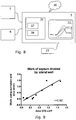
- the invention provides a medical monitoring apparatus or an ultrasonic or MR imaging system, both being illustrated in Figure 7 .
- Figure 7 illustrates the components applied in relation to the invention, and should be considered in combination with Figure 1 .
- a medical imaging device 5 such as an ultrasonic or MR imaging apparatus, performs an imaging sequence on a subject 4 to record myocardial strain data. Different imaging modalities and techniques have been described previously. The device determines tissue strain traces for individual myocardial segments, s n (t).
- Another device 6 prepares a pressure trace of a LV pressure-proportional parameter, p(t).
- the device 6 may comprise e.g. a systemic blood pressure measuring device, catheter and pressure sensor for measuring LVP invasively, ultrasound probe or phonocardiograph for determining timing events, as well as a computer for determining the pressure trace based on the measured data. There might be an overlap between devices 5 and 6.
- Devices 5 and 6 thus performs the function of Part I of Figure 1 , i.e. provides the strain and pressure traces.
- an electrocardiogram is also recorded for the subject to provide a temporal reference for the heart cycle.
- a medical monitoring apparatus 7 for preparing and presenting data is connected to devices 5 and 6 to receive the strain and pressure traces and calculate power and/or work traces according to the invention.
- the medical monitoring apparatus 7 comprises an electronic processor for executing computer programs and a memory for holding computer programs to be executed by the electronic processor, and preferably communication means for receiving at least measures strain data and means for presenting the calculated power/work traces and possible indices.
- Figure 8 A preferred implementation of the medical monitoring apparatus 7 is illustrated in Figure 8 .
- an embodiment of a medical monitoring apparatus 7, with a computer 21 for preparing and presenting data is shown.
- Computer program products for performing data preparation as described in relation to Figure 1 can be stored in memory 27 and executed by processor 28 of the computer 21.
- the apparatus will have a display 22 for presenting data to a user in a GUI, and a UI 23 such as a mouse and keyboard for receiving user input.
- Exemplary apparatuses could be medical imaging devices such as echocardiography machines or MRI apparatuses, or work stations (such as computers) for performing analysis of data obtained from such imaging devices.
- the medical monitoring apparatus 7 can comprise devices 5 and/or 6 that determine s n (t) and p(t), respectively.
- the medical monitoring apparatus 7 may access s n (t) and p(t) data stored remotely there from, either on an external imaging device 5 or 6 or on a server or data network 26.
- connection means to the electronic processor for receiving strain data and pressure related data to be used in the data preparation according to the invention.
- these connection means may be to units internal or external to the medical monitoring apparatus and may comprise a data bus and/or a wired connection and/or a wireless connection.
- the invention can also be embodied by a computer program product for updating a medical monitoring apparatus to prepare data related to work function in left ventricular muscle segments.
- a computer program product for updating a medical monitoring apparatus to prepare data related to work function in left ventricular muscle segments.
- Such product can be embodied as a packet managing system or an installation program for downloading and installing the software described in relation to Figure 1 on the medical monitoring apparatus 7 described in relation to Figure 7 and 8 , e.g. over the network connection 26.
- Such program can be stored and executed by memory 27 and processor 28, or stored and executed by a server (not shown) over network connection 26.
- the invention provides ultrasonic or MR imaging system operating.
- Such system can have a data processor corresponding to the computer 21 described above, and also involves an ultrasonic or MR imaging device configured to record ultrasound or MR data and to transmit recorded data to the data processor over the connection means.
- the individual elements of an embodiment of the invention may be physically, functionally and logically implemented in any suitable way such as in a single unit, in a plurality of units or as part of separate functional units.
- the invention may be implemented in a single unit, or be both physically and functionally distributed between different units and processors.
- a patient with left bundle branch block and without ischemic heart disease underwent ultrasound imaging with calculation of myocardial segmental work from strain and non-invasive left ventricular pressure estimation based on valvular timing and arm cuff measurement of systolic pressure.
- the work values were mapped into a standard 17-segment "bulls eye” plot, and normalized to per cent values relative to the segment with the highest value. These values were compared to the corresponding values obtained from FDG-PET imaging of the myocardium according to standard clinical PET imaging procedures of myocardial glucose metabolism.
- the coefficient of correlation between the segmental values was 0.87, indicating that work calculated from ultrasound imaging corresponds well with tissue glucose consumption.
- a scatterplot of the data is shown in Fig. 10 .
Landscapes
- Health & Medical Sciences (AREA)
- Life Sciences & Earth Sciences (AREA)
- Engineering & Computer Science (AREA)
- Medical Informatics (AREA)
- Public Health (AREA)
- General Health & Medical Sciences (AREA)
- Biomedical Technology (AREA)
- Veterinary Medicine (AREA)
- Surgery (AREA)
- Animal Behavior & Ethology (AREA)
- Molecular Biology (AREA)
- Heart & Thoracic Surgery (AREA)
- Pathology (AREA)
- Biophysics (AREA)
- Physics & Mathematics (AREA)
- Cardiology (AREA)
- Nuclear Medicine, Radiotherapy & Molecular Imaging (AREA)
- Radiology & Medical Imaging (AREA)
- Physiology (AREA)
- Computer Vision & Pattern Recognition (AREA)
- Hematology (AREA)
- Signal Processing (AREA)
- Vascular Medicine (AREA)
- Psychiatry (AREA)
- Artificial Intelligence (AREA)
- Theoretical Computer Science (AREA)
- Pulmonology (AREA)
- Power Engineering (AREA)
- High Energy & Nuclear Physics (AREA)
- Optics & Photonics (AREA)
- Data Mining & Analysis (AREA)
- Databases & Information Systems (AREA)
- Epidemiology (AREA)
- Primary Health Care (AREA)
- Ultra Sonic Daignosis Equipment (AREA)
- Magnetic Resonance Imaging Apparatus (AREA)
Claims (5)
- Vorrichtung zum Empfangen, Vorbereiten und Darlegen von Daten in Bezug auf die Arbeit von einzelnen Myokardsegmenten aus Gewebeverformung-Bildgebungsdaten, wobei die Vorrichtung Folgendes umfasst:eine medizinische Bildgebungseinrichtung zum nichtinvasiven Aufzeichnen von Gewebeverformung-Bildgebungsdaten für zwei oder mehr Myokardsegmente im linken Ventrikel; undeinen elektronischen Prozessor, der zu Folgendem in der Lage ist:• Berechnen von Verläufen der mechanischen Leistung, P(t), und/oder der mechanischen Arbeit, W(t), für zwei oder mehr einzelne Myokardsegmente im linken Ventrikel in Abhängigkeit von der Zeit für einen Zeitraum beinhaltend das Zeitintervall vom Beginn der isovolumetrischen Kontraktion und bis zum Ende der isovolumetrischen Relaxation aus ventrikulären Gewebeverformung-Verläufen für jedes der zwei oder mehr Myokardsegmente im linken Ventrikel und einem nichtinvasiv bestimmten Druckverlauf proportional zu einem linksventrikulären Druck und in zeitlicher Synchronität mit den Verformung-Verläufen,wobei die mechanische Leistung für das Segment n, Pn(t), aus Verformung-Verläufen, sn(t), und dem Druckverlauf, p(t), wie folgt berechnet wird:

oder wobei C1 eine Konstante ist und N die Anzahl von Segmenten ist, die in der Segmentierung des Ventrikels verwendet werden,
undwobei die mechanische Arbeit für das Segment n, Wn(t), wie folgt berechnet wird: wobei t' eine Integrationsvariable ist und C2n eine Konstante für das Segment n ist.
 - Medizinische Überwachungsvorrichtung nach Anspruch 1, wobei der elektronische Prozessor ferner in der Lage ist, den Druckverlauf aus Daten, die den systemischen arteriellen Druck darstellen, und druckbezogenen zeitlichen Markern im Herzzyklus zu bestimmen.
- Medizinische Überwachungsvorrichtung nach Anspruch 1, wobei der elektronische Prozessor ferner in der Lage ist, den Druckverlauf zu bestimmen, indem eine standardmäßige LV-Druck-Wellenform durch Verwendung von Daten, die nichtinvasiv überwachte, druckbezogene zeitliche Marker im Herzzyklus darstellen, in Synchronisation mit den Verformung-Verläufen gebracht wird.
- Medizinische Überwachungsvorrichtung nach einem der vorstehenden Ansprüche, wobei der elektronische Prozessor ferner in der Lage ist, Indizes für Segmentarbeit aus berechneten P(t)- und/oder W(t)-Verläufen für zumindest eines der Folgenden zu bestimmen:• negative Arbeit während des Zeitintervalls für einzelne Myokardsegmente im linken Ventrikel;• eine Summe von negativer Arbeit für alle Segmente als einen Bruchteil oder einen Prozentsatz der Summe von positiver Arbeit oder Nettoarbeit für alle Segmente;• eine Abweichung von einer Standard- oder einer Mittelwertkurve; und• verschwendete Arbeit, definiert als positive Arbeit, die erfolgt, wenn die Aortenklappe geschlossen ist.
- Computerimplementiertes Verfahren zum Vorbereiten und Darlegen von Daten in Bezug auf die Arbeit von einzelnen Myokardsegmenten aus Gewebeverformung-Bildgebungsdaten unter Verwendung einer Computereinrichtung umfassend einen Speicher, der Daten in Bezug auf eine Standard- oder eine Mittelwertkurve für Arbeit enthält, wobei das Verfahren Folgendes umfasst:• nichtinvasives Erhalten, durch Bildgebungsdatenwerte, die ventrikuläre Gewebeverformung-Verläufe für zwei oder mehr Myokardsegmente im linken Ventrikel angeben, und Speichern der Datenwerte;• Zugreifen auf Datenwerte, die einen Druckverlauf proportional zu einem linksventrikulären Druck angeben, in zeitlicher Synchronität mit den Verformung-Verläufen und auf eine nichtinvasive Weise erhalten; und• Verwenden eines elektronischen Prozessors, um Verläufe der mechanischen Leistung, P(t), und/oder der mechanischen Arbeit, W(t), für einzelne Myokardsegmente im linken Ventrikel in Abhängigkeit von der Zeit für einen Zeitraum beinhaltend das Zeitintervall vom Beginn der isovolumetrischen Kontraktion und bis zum Ende der isovolumetrischen Relaxation aus den ventrikulären Gewebeverformung-Verläufen für jedes der zwei oder mehr Myokardsegmente im linken Ventrikel und dem Druckverlauf zu berechnen,wobei die mechanische Leistung für das Segment n, Pn(t), aus Verformung-Verläufen, sn(t), und dem Druckverlauf, p(t), wie folgt berechnet wird:

oder •
und
wobei die mechanische Arbeit für das Segment n, Wn(t), wie folgt berechnet wird:• ,•wobei t' eine Integrationsvariable ist und C2n eine Konstante für das Segment n ist.
Priority Applications (1)
| Application Number | Priority Date | Filing Date | Title |
|---|---|---|---|
| EP11793651.8A EP2633457B1 (de) | 2010-10-26 | 2011-10-17 | Verfahren für die myokardiale segmentarbeitsanalyse |
Applications Claiming Priority (4)
| Application Number | Priority Date | Filing Date | Title |
|---|---|---|---|
| EP10188827 | 2010-10-26 | ||
| US40776010P | 2010-10-28 | 2010-10-28 | |
| PCT/EP2011/005198 WO2012055498A1 (en) | 2010-10-26 | 2011-10-17 | Method for myocardial segment work analysis |
| EP11793651.8A EP2633457B1 (de) | 2010-10-26 | 2011-10-17 | Verfahren für die myokardiale segmentarbeitsanalyse |
Publications (2)
| Publication Number | Publication Date |
|---|---|
| EP2633457A1 EP2633457A1 (de) | 2013-09-04 |
| EP2633457B1 true EP2633457B1 (de) | 2021-10-06 |
Family
ID=43821988
Family Applications (1)
| Application Number | Title | Priority Date | Filing Date |
|---|---|---|---|
| EP11793651.8A Active EP2633457B1 (de) | 2010-10-26 | 2011-10-17 | Verfahren für die myokardiale segmentarbeitsanalyse |
Country Status (5)
| Country | Link |
|---|---|
| US (1) | US20130211256A1 (de) |
| EP (1) | EP2633457B1 (de) |
| JP (1) | JP5860470B2 (de) |
| CN (1) | CN103260524B (de) |
| WO (1) | WO2012055498A1 (de) |
Cited By (1)
| Publication number | Priority date | Publication date | Assignee | Title |
|---|---|---|---|---|
| RU2806486C1 (ru) * | 2022-11-23 | 2023-11-01 | Федеральное государственное бюджетное учреждение науки институт иммунологии и физиологии Уральского отделения Российской академии наук | Способ прогнозирования эффективности сердечной ресинхронизирующей терапии с использованием оптимизации расположения стимулирующих электродов |
Families Citing this family (40)
| Publication number | Priority date | Publication date | Assignee | Title |
|---|---|---|---|---|
| EP2633457B1 (de) * | 2010-10-26 | 2021-10-06 | Oslo Universitetssykehus HF | Verfahren für die myokardiale segmentarbeitsanalyse |
| WO2012087842A1 (en) | 2010-12-23 | 2012-06-28 | The Foundry, Llc | System for mitral valve repair and replacement |
| WO2012177942A2 (en) | 2011-06-21 | 2012-12-27 | Hanson Gifford, Iii | Prosthetic heart valve devices and associated systems and methods |
| CN103889369B (zh) | 2011-10-19 | 2016-09-14 | 托尔福公司 | 用于心脏瓣膜置换术的装置 |
| US9655722B2 (en) | 2011-10-19 | 2017-05-23 | Twelve, Inc. | Prosthetic heart valve devices, prosthetic mitral valves and associated systems and methods |
| US9763780B2 (en) | 2011-10-19 | 2017-09-19 | Twelve, Inc. | Devices, systems and methods for heart valve replacement |
| US11202704B2 (en) | 2011-10-19 | 2021-12-21 | Twelve, Inc. | Prosthetic heart valve devices, prosthetic mitral valves and associated systems and methods |
| JP6133309B2 (ja) | 2011-10-19 | 2017-05-24 | トゥエルヴ, インコーポレイテッド | 人工心臓弁デバイス |
| US9039757B2 (en) | 2011-10-19 | 2015-05-26 | Twelve, Inc. | Prosthetic heart valve devices, prosthetic mitral valves and associated systems and methods |
| US9579198B2 (en) | 2012-03-01 | 2017-02-28 | Twelve, Inc. | Hydraulic delivery systems for prosthetic heart valve devices and associated methods |
| CN105246431B (zh) | 2013-05-20 | 2018-04-06 | 托尔福公司 | 可植入心脏瓣膜装置、二尖瓣修复装置以及相关系统和方法 |
| WO2015073927A2 (en) * | 2013-11-15 | 2015-05-21 | The Regents Of The University Of California | Compositions, devices and methods for diagnosing heart failure and for patient-specific modeling to predict outcomes of cardiac resynchronization therapy |
| JP6646646B2 (ja) * | 2014-07-17 | 2020-02-14 | アンスティチュ ナショナル ドゥ ラ サンテ エ ドゥ ラ ルシェルシュ メディカル | 筋肉の機能パラメータを入手する方法、装置及びシステム |
| JP6640444B2 (ja) * | 2014-09-30 | 2020-02-05 | キヤノンメディカルシステムズ株式会社 | 超音波診断装置、超音波画像処理装置及び超音波画像処理プログラム |
| CN107205653B (zh) * | 2015-01-09 | 2020-11-20 | 马克斯-普朗克科学促进学会 | 用于表征可激励以变形的介质的空间-时空动态的方法和设备 |
| WO2017035002A1 (en) | 2015-08-21 | 2017-03-02 | Twelve Inc. | Implantable heart valve devices, mitral valve repair devices and associated systems and methods |
| AU2016379418A1 (en) | 2015-12-22 | 2018-06-14 | The Regents Of The University Of California | Computational localization of fibrillation sources |
| JP7163278B2 (ja) * | 2016-04-27 | 2022-10-31 | マイオカーディアル ソリューションズ インク. | Mriのストレイン測定値による心機能の迅速な定量評価 |
| CN109069272A (zh) | 2016-04-29 | 2018-12-21 | 美敦力瓦斯科尔勒公司 | 具有带系绳的锚定件的假体心脏瓣膜设备以及相关联的系统和方法 |
| US10575950B2 (en) | 2017-04-18 | 2020-03-03 | Twelve, Inc. | Hydraulic systems for delivering prosthetic heart valve devices and associated methods |
| US10702378B2 (en) | 2017-04-18 | 2020-07-07 | Twelve, Inc. | Prosthetic heart valve device and associated systems and methods |
| US10433961B2 (en) | 2017-04-18 | 2019-10-08 | Twelve, Inc. | Delivery systems with tethers for prosthetic heart valve devices and associated methods |
| US10792151B2 (en) | 2017-05-11 | 2020-10-06 | Twelve, Inc. | Delivery systems for delivering prosthetic heart valve devices and associated methods |
| US10646338B2 (en) | 2017-06-02 | 2020-05-12 | Twelve, Inc. | Delivery systems with telescoping capsules for deploying prosthetic heart valve devices and associated methods |
| US10709591B2 (en) | 2017-06-06 | 2020-07-14 | Twelve, Inc. | Crimping device and method for loading stents and prosthetic heart valves |
| US10729541B2 (en) | 2017-07-06 | 2020-08-04 | Twelve, Inc. | Prosthetic heart valve devices and associated systems and methods |
| US10786352B2 (en) | 2017-07-06 | 2020-09-29 | Twelve, Inc. | Prosthetic heart valve devices and associated systems and methods |
| GB2565583A (en) * | 2017-08-17 | 2019-02-20 | Cardiaccs As | Estimating ventricular pressure |
| US11547369B2 (en) | 2018-04-26 | 2023-01-10 | Vektor Medical, Inc. | Machine learning using clinical and simulated data |
| EP3586758A1 (de) * | 2018-06-28 | 2020-01-01 | Koninklijke Philips N.V. | Verfahren und systeme zur durchführung der quantifizierung von transvalvularem druck |
| EP3818541B1 (de) | 2018-07-05 | 2025-04-09 | The Regents of the University of California | Rechensimulationen von anatomischen strukturen und elektrodenpositionierung auf körperoberflächen |
| CN112654302B (zh) * | 2018-10-23 | 2023-11-03 | 深圳迈瑞生物医疗电子股份有限公司 | 一种心脏运动定量分析方法和超声系统 |
| US12558015B2 (en) | 2018-12-31 | 2026-02-24 | The Vektor Group, Inc. | Enhanced computational heart simulations |
| US11872019B2 (en) | 2019-06-05 | 2024-01-16 | Myocardial Solutions, Inc. | MRI-derived strain-based measurements and related image data acquisitions, image data processing, patient evaluations and monitoring methods and systems |
| JP2022183393A (ja) * | 2019-11-07 | 2022-12-09 | テルモ株式会社 | 心臓再同期療法治療効果予測装置、方法およびプログラム |
| CN114041828B (zh) * | 2022-01-13 | 2022-04-29 | 深圳瀚维智能医疗科技有限公司 | 超声扫查控制方法、机器人及存储介质 |
| CN115089215B (zh) * | 2022-07-20 | 2025-09-30 | 武汉中旗生物医疗电子有限公司 | 左心室参数确定方法、装置及超声成像设备 |
| CN117838190B (zh) * | 2024-03-08 | 2024-05-28 | 中国医学科学院北京协和医院 | 一种心肌功能指数测量方法及装置 |
| WO2025259951A1 (en) * | 2024-06-14 | 2025-12-18 | The Cooper Health System | Method and appratus for the integrated assessment of cardiac function and vascular physiology |
| US12496044B1 (en) | 2024-09-12 | 2025-12-16 | GE Precision Healthcare LLC | System and method for real-time correction of strain traces in patients with conduction delays |
Family Cites Families (13)
| Publication number | Priority date | Publication date | Assignee | Title |
|---|---|---|---|---|
| JPH09313486A (ja) * | 1996-06-02 | 1997-12-09 | Noritoshi Nakabachi | 超音波生体計測装置 |
| EP1585441A4 (de) | 2003-01-24 | 2008-05-21 | Proteus Biomedical Inc | Verfahren und systeme zur messung von herzparametern |
| WO2004068406A2 (en) * | 2003-01-30 | 2004-08-12 | Chase Medical, L.P. | A method and system for image processing and contour assessment |
| US7877128B2 (en) * | 2005-08-02 | 2011-01-25 | Biosense Webster, Inc. | Simulation of invasive procedures |
| US7666144B2 (en) * | 2006-02-21 | 2010-02-23 | Board Of Trustees Operating Michigan State University | Methods and apparatus for determining cardiac output and left atrial pressure |
| US7912270B2 (en) * | 2006-11-21 | 2011-03-22 | General Electric Company | Method and system for creating and using an impact atlas |
| US8214011B2 (en) * | 2007-08-13 | 2012-07-03 | General Electric Company | System and method for remodeling prediction using ultrasound |
| JP5443719B2 (ja) * | 2007-09-07 | 2014-03-19 | 株式会社東芝 | 超音波診断装置、超音波画像処理装置及び超音波画像処理プログラム |
| WO2009079344A1 (en) * | 2007-12-14 | 2009-06-25 | The Cleveland Clinic Foundation | System and method to characterize cardiac function |
| CN101278828A (zh) * | 2008-05-20 | 2008-10-08 | 上海大学 | 测量心室壁微面积功的方法 |
| JP5491836B2 (ja) * | 2009-01-30 | 2014-05-14 | 株式会社東芝 | 超音波診断装置、超音波画像処理装置、医用画像診断装置及び医用画像処理装置 |
| JP5597455B2 (ja) * | 2010-06-25 | 2014-10-01 | 株式会社東芝 | 超音波診断装置、超音波画像処理装置及び超音波画像処理プログラム |
| EP2633457B1 (de) * | 2010-10-26 | 2021-10-06 | Oslo Universitetssykehus HF | Verfahren für die myokardiale segmentarbeitsanalyse |
-
2011
- 2011-10-17 EP EP11793651.8A patent/EP2633457B1/de active Active
- 2011-10-17 US US13/881,011 patent/US20130211256A1/en not_active Abandoned
- 2011-10-17 JP JP2013535297A patent/JP5860470B2/ja active Active
- 2011-10-17 CN CN201180059482.2A patent/CN103260524B/zh active Active
- 2011-10-17 WO PCT/EP2011/005198 patent/WO2012055498A1/en not_active Ceased
Non-Patent Citations (1)
| Title |
|---|
| None * |
Cited By (1)
| Publication number | Priority date | Publication date | Assignee | Title |
|---|---|---|---|---|
| RU2806486C1 (ru) * | 2022-11-23 | 2023-11-01 | Федеральное государственное бюджетное учреждение науки институт иммунологии и физиологии Уральского отделения Российской академии наук | Способ прогнозирования эффективности сердечной ресинхронизирующей терапии с использованием оптимизации расположения стимулирующих электродов |
Also Published As
| Publication number | Publication date |
|---|---|
| EP2633457A1 (de) | 2013-09-04 |
| US20130211256A1 (en) | 2013-08-15 |
| WO2012055498A4 (en) | 2012-06-28 |
| CN103260524A (zh) | 2013-08-21 |
| WO2012055498A1 (en) | 2012-05-03 |
| JP2013541385A (ja) | 2013-11-14 |
| CN103260524B (zh) | 2015-06-10 |
| JP5860470B2 (ja) | 2016-02-16 |
| WO2012055498A8 (en) | 2013-05-30 |
Similar Documents
| Publication | Publication Date | Title |
|---|---|---|
| US20130211256A1 (en) | Method for myocardial segment work analysis | |
| Wilkenshoff et al. | Regional mean systolic myocardial velocity estimation by real-time color Doppler myocardial imaging: a new technique for quantifying regional systolic function | |
| Mor-Avi et al. | Current and evolving echocardiographic techniques for the quantitative evaluation of cardiac mechanics: ASE/EAE consensus statement on methodology and indications endorsed by the Japanese Society of Echocardiography | |
| D'hooge et al. | Echocardiographic strain and strain-rate imaging: a new tool to study regional myocardial function | |
| US8428687B2 (en) | Systems and methods for matching and imaging tissue characteristics | |
| JP5906234B2 (ja) | 診断ecgにおける心筋梗塞サイズの可視化 | |
| JP3875581B2 (ja) | 超音波診断システム | |
| US20210085284A1 (en) | System and method for electromechanical activation of arrhythmias | |
| CA2954573C (en) | Method for obtaining a functional parameter of a muscle | |
| JP2015512292A (ja) | マルチプルイメージデータループを獲得して分析する方法及びシステム | |
| JP6273419B2 (ja) | 生体情報測定装置および生体情報測定装置もしくは生体情報測定システムの作動方法 | |
| Morbach et al. | Clinical application of three-dimensional echocardiography | |
| JP2015526150A (ja) | 画像データ処理方法 | |
| Williams et al. | Strain rate imaging after dynamic stress provides objective evidence of persistent regional myocardial dysfunction in ischaemic myocardium: regional stunning identified? | |
| Citro et al. | Myocardial postsystolic motion in ischemic and not ischemic myocardium: the clinical value of tissue Doppler | |
| CN114667101B (zh) | 心脏装置 | |
| Sutton et al. | Assessment of left ventricular systolic function by echocardiography | |
| D'hooge et al. | Deformation imaging by ultrasound for the assessment of regional myocardial function | |
| Rozzi | VIDEO KINEMATIC EVALUATION OF THE HEART (VI. KI. E.): AN IDEA, A PROJECT, A REALITY | |
| Aufan | Analytical Methods Using Cardiac MRI to Assess Systolic and Diastolic Ventricular Function | |
| Fixsen | Ultrasound strain imaging of the failing and supported heart: experimental and clinical studies | |
| Bradway | Transthoracic Cardiac Acoustic Radiation Force Impulse Imaging | |
| REDDINGTON | Noninvasive Hemodynamic Monitoring: Left Ventricular Pressure-Volume Loop Reconstruction | |
| Johnson | The Cardiac State Diagram: A new method for assessing cardiac mechanics | |
| CN120643252A (zh) | 组织超声成像系统及方法、组织力学特性参数测量方法 |
Legal Events
| Date | Code | Title | Description |
|---|---|---|---|
| PUAI | Public reference made under article 153(3) epc to a published international application that has entered the european phase |
Free format text: ORIGINAL CODE: 0009012 |
|
| 17P | Request for examination filed |
Effective date: 20130419 |
|
| AK | Designated contracting states |
Kind code of ref document: A1 Designated state(s): AL AT BE BG CH CY CZ DE DK EE ES FI FR GB GR HR HU IE IS IT LI LT LU LV MC MK MT NL NO PL PT RO RS SE SI SK SM TR |
|
| DAX | Request for extension of the european patent (deleted) | ||
| STAA | Information on the status of an ep patent application or granted ep patent |
Free format text: STATUS: EXAMINATION IS IN PROGRESS |
|
| 17Q | First examination report despatched |
Effective date: 20170420 |
|
| REG | Reference to a national code |
Ref country code: DE Ref legal event code: R079 Ref document number: 602011071893 Country of ref document: DE Free format text: PREVIOUS MAIN CLASS: G06F0019000000 Ipc: G16H0050300000 |
|
| RIC1 | Information provided on ipc code assigned before grant |
Ipc: G16H 50/30 20180101AFI20210308BHEP Ipc: A61B 8/04 20060101ALI20210308BHEP Ipc: A61B 5/0215 20060101ALI20210308BHEP Ipc: A61B 5/318 20210101ALI20210308BHEP Ipc: A61B 8/08 20060101ALI20210308BHEP Ipc: A61B 5/02 20060101ALI20210308BHEP Ipc: A61B 8/06 20060101ALI20210308BHEP |
|
| GRAP | Despatch of communication of intention to grant a patent |
Free format text: ORIGINAL CODE: EPIDOSNIGR1 |
|
| STAA | Information on the status of an ep patent application or granted ep patent |
Free format text: STATUS: GRANT OF PATENT IS INTENDED |
|
| INTG | Intention to grant announced |
Effective date: 20210416 |
|
| GRAS | Grant fee paid |
Free format text: ORIGINAL CODE: EPIDOSNIGR3 |
|
| GRAA | (expected) grant |
Free format text: ORIGINAL CODE: 0009210 |
|
| STAA | Information on the status of an ep patent application or granted ep patent |
Free format text: STATUS: THE PATENT HAS BEEN GRANTED |
|
| AK | Designated contracting states |
Kind code of ref document: B1 Designated state(s): AL AT BE BG CH CY CZ DE DK EE ES FI FR GB GR HR HU IE IS IT LI LT LU LV MC MK MT NL NO PL PT RO RS SE SI SK SM TR |
|
| REG | Reference to a national code |
Ref country code: GB Ref legal event code: FG4D |
|
| REG | Reference to a national code |
Ref country code: CH Ref legal event code: EP Ref country code: AT Ref legal event code: REF Ref document number: 1436931 Country of ref document: AT Kind code of ref document: T Effective date: 20211015 |
|
| REG | Reference to a national code |
Ref country code: DE Ref legal event code: R096 Ref document number: 602011071893 Country of ref document: DE |
|
| REG | Reference to a national code |
Ref country code: IE Ref legal event code: FG4D |
|
| REG | Reference to a national code |
Ref country code: LT Ref legal event code: MG9D |
|
| REG | Reference to a national code |
Ref country code: NL Ref legal event code: MP Effective date: 20211006 |
|
| REG | Reference to a national code |
Ref country code: AT Ref legal event code: MK05 Ref document number: 1436931 Country of ref document: AT Kind code of ref document: T Effective date: 20211006 |
|
| PG25 | Lapsed in a contracting state [announced via postgrant information from national office to epo] |
Ref country code: RS Free format text: LAPSE BECAUSE OF FAILURE TO SUBMIT A TRANSLATION OF THE DESCRIPTION OR TO PAY THE FEE WITHIN THE PRESCRIBED TIME-LIMIT Effective date: 20211006 Ref country code: LT Free format text: LAPSE BECAUSE OF FAILURE TO SUBMIT A TRANSLATION OF THE DESCRIPTION OR TO PAY THE FEE WITHIN THE PRESCRIBED TIME-LIMIT Effective date: 20211006 Ref country code: FI Free format text: LAPSE BECAUSE OF FAILURE TO SUBMIT A TRANSLATION OF THE DESCRIPTION OR TO PAY THE FEE WITHIN THE PRESCRIBED TIME-LIMIT Effective date: 20211006 Ref country code: BG Free format text: LAPSE BECAUSE OF FAILURE TO SUBMIT A TRANSLATION OF THE DESCRIPTION OR TO PAY THE FEE WITHIN THE PRESCRIBED TIME-LIMIT Effective date: 20220106 Ref country code: AT Free format text: LAPSE BECAUSE OF FAILURE TO SUBMIT A TRANSLATION OF THE DESCRIPTION OR TO PAY THE FEE WITHIN THE PRESCRIBED TIME-LIMIT Effective date: 20211006 |
|
| REG | Reference to a national code |
Ref country code: CH Ref legal event code: PL |
|
| PG25 | Lapsed in a contracting state [announced via postgrant information from national office to epo] |
Ref country code: IS Free format text: LAPSE BECAUSE OF FAILURE TO SUBMIT A TRANSLATION OF THE DESCRIPTION OR TO PAY THE FEE WITHIN THE PRESCRIBED TIME-LIMIT Effective date: 20220206 Ref country code: SE Free format text: LAPSE BECAUSE OF FAILURE TO SUBMIT A TRANSLATION OF THE DESCRIPTION OR TO PAY THE FEE WITHIN THE PRESCRIBED TIME-LIMIT Effective date: 20211006 Ref country code: PT Free format text: LAPSE BECAUSE OF FAILURE TO SUBMIT A TRANSLATION OF THE DESCRIPTION OR TO PAY THE FEE WITHIN THE PRESCRIBED TIME-LIMIT Effective date: 20220207 Ref country code: PL Free format text: LAPSE BECAUSE OF FAILURE TO SUBMIT A TRANSLATION OF THE DESCRIPTION OR TO PAY THE FEE WITHIN THE PRESCRIBED TIME-LIMIT Effective date: 20211006 Ref country code: NO Free format text: LAPSE BECAUSE OF FAILURE TO SUBMIT A TRANSLATION OF THE DESCRIPTION OR TO PAY THE FEE WITHIN THE PRESCRIBED TIME-LIMIT Effective date: 20220106 Ref country code: NL Free format text: LAPSE BECAUSE OF FAILURE TO SUBMIT A TRANSLATION OF THE DESCRIPTION OR TO PAY THE FEE WITHIN THE PRESCRIBED TIME-LIMIT Effective date: 20211006 Ref country code: LV Free format text: LAPSE BECAUSE OF FAILURE TO SUBMIT A TRANSLATION OF THE DESCRIPTION OR TO PAY THE FEE WITHIN THE PRESCRIBED TIME-LIMIT Effective date: 20211006 Ref country code: HR Free format text: LAPSE BECAUSE OF FAILURE TO SUBMIT A TRANSLATION OF THE DESCRIPTION OR TO PAY THE FEE WITHIN THE PRESCRIBED TIME-LIMIT Effective date: 20211006 Ref country code: GR Free format text: LAPSE BECAUSE OF FAILURE TO SUBMIT A TRANSLATION OF THE DESCRIPTION OR TO PAY THE FEE WITHIN THE PRESCRIBED TIME-LIMIT Effective date: 20220107 Ref country code: ES Free format text: LAPSE BECAUSE OF FAILURE TO SUBMIT A TRANSLATION OF THE DESCRIPTION OR TO PAY THE FEE WITHIN THE PRESCRIBED TIME-LIMIT Effective date: 20211006 |
|
| REG | Reference to a national code |
Ref country code: BE Ref legal event code: MM Effective date: 20211031 |
|
| REG | Reference to a national code |
Ref country code: DE Ref legal event code: R097 Ref document number: 602011071893 Country of ref document: DE |
|
| PG25 | Lapsed in a contracting state [announced via postgrant information from national office to epo] |
Ref country code: SM Free format text: LAPSE BECAUSE OF FAILURE TO SUBMIT A TRANSLATION OF THE DESCRIPTION OR TO PAY THE FEE WITHIN THE PRESCRIBED TIME-LIMIT Effective date: 20211006 Ref country code: SK Free format text: LAPSE BECAUSE OF FAILURE TO SUBMIT A TRANSLATION OF THE DESCRIPTION OR TO PAY THE FEE WITHIN THE PRESCRIBED TIME-LIMIT Effective date: 20211006 Ref country code: RO Free format text: LAPSE BECAUSE OF FAILURE TO SUBMIT A TRANSLATION OF THE DESCRIPTION OR TO PAY THE FEE WITHIN THE PRESCRIBED TIME-LIMIT Effective date: 20211006 Ref country code: MC Free format text: LAPSE BECAUSE OF FAILURE TO SUBMIT A TRANSLATION OF THE DESCRIPTION OR TO PAY THE FEE WITHIN THE PRESCRIBED TIME-LIMIT Effective date: 20211006 Ref country code: LU Free format text: LAPSE BECAUSE OF NON-PAYMENT OF DUE FEES Effective date: 20211017 Ref country code: EE Free format text: LAPSE BECAUSE OF FAILURE TO SUBMIT A TRANSLATION OF THE DESCRIPTION OR TO PAY THE FEE WITHIN THE PRESCRIBED TIME-LIMIT Effective date: 20211006 Ref country code: DK Free format text: LAPSE BECAUSE OF FAILURE TO SUBMIT A TRANSLATION OF THE DESCRIPTION OR TO PAY THE FEE WITHIN THE PRESCRIBED TIME-LIMIT Effective date: 20211006 Ref country code: CZ Free format text: LAPSE BECAUSE OF FAILURE TO SUBMIT A TRANSLATION OF THE DESCRIPTION OR TO PAY THE FEE WITHIN THE PRESCRIBED TIME-LIMIT Effective date: 20211006 Ref country code: BE Free format text: LAPSE BECAUSE OF NON-PAYMENT OF DUE FEES Effective date: 20211031 |
|
| PLBE | No opposition filed within time limit |
Free format text: ORIGINAL CODE: 0009261 |
|
| STAA | Information on the status of an ep patent application or granted ep patent |
Free format text: STATUS: NO OPPOSITION FILED WITHIN TIME LIMIT |
|
| PG25 | Lapsed in a contracting state [announced via postgrant information from national office to epo] |
Ref country code: LI Free format text: LAPSE BECAUSE OF NON-PAYMENT OF DUE FEES Effective date: 20211031 Ref country code: CH Free format text: LAPSE BECAUSE OF NON-PAYMENT OF DUE FEES Effective date: 20211031 |
|
| 26N | No opposition filed |
Effective date: 20220707 |
|
| GBPC | Gb: european patent ceased through non-payment of renewal fee |
Effective date: 20220106 |
|
| PG25 | Lapsed in a contracting state [announced via postgrant information from national office to epo] |
Ref country code: IE Free format text: LAPSE BECAUSE OF NON-PAYMENT OF DUE FEES Effective date: 20211017 Ref country code: GB Free format text: LAPSE BECAUSE OF NON-PAYMENT OF DUE FEES Effective date: 20220106 Ref country code: AL Free format text: LAPSE BECAUSE OF FAILURE TO SUBMIT A TRANSLATION OF THE DESCRIPTION OR TO PAY THE FEE WITHIN THE PRESCRIBED TIME-LIMIT Effective date: 20211006 |
|
| PG25 | Lapsed in a contracting state [announced via postgrant information from national office to epo] |
Ref country code: SI Free format text: LAPSE BECAUSE OF FAILURE TO SUBMIT A TRANSLATION OF THE DESCRIPTION OR TO PAY THE FEE WITHIN THE PRESCRIBED TIME-LIMIT Effective date: 20211006 Ref country code: FR Free format text: LAPSE BECAUSE OF NON-PAYMENT OF DUE FEES Effective date: 20211206 |
|
| PG25 | Lapsed in a contracting state [announced via postgrant information from national office to epo] |
Ref country code: IT Free format text: LAPSE BECAUSE OF FAILURE TO SUBMIT A TRANSLATION OF THE DESCRIPTION OR TO PAY THE FEE WITHIN THE PRESCRIBED TIME-LIMIT Effective date: 20211006 Ref country code: HU Free format text: LAPSE BECAUSE OF FAILURE TO SUBMIT A TRANSLATION OF THE DESCRIPTION OR TO PAY THE FEE WITHIN THE PRESCRIBED TIME-LIMIT; INVALID AB INITIO Effective date: 20111017 Ref country code: CY Free format text: LAPSE BECAUSE OF FAILURE TO SUBMIT A TRANSLATION OF THE DESCRIPTION OR TO PAY THE FEE WITHIN THE PRESCRIBED TIME-LIMIT Effective date: 20211006 |
|
| PG25 | Lapsed in a contracting state [announced via postgrant information from national office to epo] |
Ref country code: MK Free format text: LAPSE BECAUSE OF FAILURE TO SUBMIT A TRANSLATION OF THE DESCRIPTION OR TO PAY THE FEE WITHIN THE PRESCRIBED TIME-LIMIT Effective date: 20211006 |
|
| PG25 | Lapsed in a contracting state [announced via postgrant information from national office to epo] |
Ref country code: TR Free format text: LAPSE BECAUSE OF FAILURE TO SUBMIT A TRANSLATION OF THE DESCRIPTION OR TO PAY THE FEE WITHIN THE PRESCRIBED TIME-LIMIT Effective date: 20211006 |
|
| PG25 | Lapsed in a contracting state [announced via postgrant information from national office to epo] |
Ref country code: MT Free format text: LAPSE BECAUSE OF FAILURE TO SUBMIT A TRANSLATION OF THE DESCRIPTION OR TO PAY THE FEE WITHIN THE PRESCRIBED TIME-LIMIT Effective date: 20211006 |
|
| PGFP | Annual fee paid to national office [announced via postgrant information from national office to epo] |
Ref country code: DE Payment date: 20251021 Year of fee payment: 15 |