EP2641609A1 - Agent thérapeutique pour une dystrophie musculaire - Google Patents
Agent thérapeutique pour une dystrophie musculaire Download PDFInfo
- Publication number
- EP2641609A1 EP2641609A1 EP11841477.0A EP11841477A EP2641609A1 EP 2641609 A1 EP2641609 A1 EP 2641609A1 EP 11841477 A EP11841477 A EP 11841477A EP 2641609 A1 EP2641609 A1 EP 2641609A1
- Authority
- EP
- European Patent Office
- Prior art keywords
- mice
- mdx
- csf
- muscle
- csf3r
- Prior art date
- Legal status (The legal status is an assumption and is not a legal conclusion. Google has not performed a legal analysis and makes no representation as to the accuracy of the status listed.)
- Ceased
Links
- 239000003814 drug Substances 0.000 title claims abstract description 31
- 229940124597 therapeutic agent Drugs 0.000 title claims abstract description 28
- 201000006938 muscular dystrophy Diseases 0.000 title claims abstract description 26
- 108010017080 Granulocyte Colony-Stimulating Factor Proteins 0.000 claims abstract description 120
- 102000004269 Granulocyte Colony-Stimulating Factor Human genes 0.000 claims abstract description 120
- 210000002027 skeletal muscle Anatomy 0.000 claims abstract description 37
- 206010028289 Muscle atrophy Diseases 0.000 claims abstract description 25
- 201000010099 disease Diseases 0.000 claims abstract description 24
- 208000037265 diseases, disorders, signs and symptoms Diseases 0.000 claims abstract description 24
- 239000004480 active ingredient Substances 0.000 claims abstract description 7
- 206010013801 Duchenne Muscular Dystrophy Diseases 0.000 claims description 31
- 241000699670 Mus sp. Species 0.000 description 156
- 210000003205 muscle Anatomy 0.000 description 82
- -1 polyoxyethylene Polymers 0.000 description 36
- 210000001087 myotubule Anatomy 0.000 description 33
- 210000000107 myocyte Anatomy 0.000 description 32
- 101100372319 Rattus norvegicus Utrn gene Proteins 0.000 description 29
- 108010069091 Dystrophin Proteins 0.000 description 24
- 210000004027 cell Anatomy 0.000 description 23
- 102000001039 Dystrophin Human genes 0.000 description 20
- 150000001413 amino acids Chemical group 0.000 description 19
- 229920003171 Poly (ethylene oxide) Polymers 0.000 description 17
- 108010054017 Granulocyte Colony-Stimulating Factor Receptors Proteins 0.000 description 16
- 102100039622 Granulocyte colony-stimulating factor receptor Human genes 0.000 description 16
- 230000001172 regenerating effect Effects 0.000 description 16
- 230000009756 muscle regeneration Effects 0.000 description 14
- 230000001965 increasing effect Effects 0.000 description 13
- 102000011856 Utrophin Human genes 0.000 description 12
- 108010075653 Utrophin Proteins 0.000 description 12
- 235000014113 dietary fatty acids Nutrition 0.000 description 12
- 239000000194 fatty acid Substances 0.000 description 12
- 229930195729 fatty acid Natural products 0.000 description 12
- 230000007850 degeneration Effects 0.000 description 10
- 238000010172 mouse model Methods 0.000 description 10
- 108090000623 proteins and genes Proteins 0.000 description 10
- 230000037396 body weight Effects 0.000 description 9
- 230000000295 complement effect Effects 0.000 description 9
- 230000004220 muscle function Effects 0.000 description 9
- 238000000034 method Methods 0.000 description 8
- 230000035772 mutation Effects 0.000 description 8
- 235000002639 sodium chloride Nutrition 0.000 description 8
- 230000004083 survival effect Effects 0.000 description 8
- 241000699666 Mus <mouse, genus> Species 0.000 description 7
- 239000004359 castor oil Substances 0.000 description 7
- 238000006243 chemical reaction Methods 0.000 description 7
- 230000008929 regeneration Effects 0.000 description 7
- 238000011069 regeneration method Methods 0.000 description 7
- FWBHETKCLVMNFS-UHFFFAOYSA-N 4',6-Diamino-2-phenylindol Chemical compound C1=CC(C(=N)N)=CC=C1C1=CC2=CC=C(C(N)=N)C=C2N1 FWBHETKCLVMNFS-UHFFFAOYSA-N 0.000 description 6
- 108700028369 Alleles Proteins 0.000 description 6
- 235000019438 castor oil Nutrition 0.000 description 6
- 230000003247 decreasing effect Effects 0.000 description 6
- 238000009826 distribution Methods 0.000 description 6
- 230000000694 effects Effects 0.000 description 6
- ZEMPKEQAKRGZGQ-XOQCFJPHSA-N glycerol triricinoleate Natural products CCCCCC[C@@H](O)CC=CCCCCCCCC(=O)OC[C@@H](COC(=O)CCCCCCCC=CC[C@@H](O)CCCCCC)OC(=O)CCCCCCCC=CC[C@H](O)CCCCCC ZEMPKEQAKRGZGQ-XOQCFJPHSA-N 0.000 description 6
- 239000000203 mixture Substances 0.000 description 6
- 102000004169 proteins and genes Human genes 0.000 description 6
- 230000002441 reversible effect Effects 0.000 description 6
- FBPFZTCFMRRESA-FSIIMWSLSA-N D-Glucitol Natural products OC[C@H](O)[C@H](O)[C@@H](O)[C@H](O)CO FBPFZTCFMRRESA-FSIIMWSLSA-N 0.000 description 5
- 241001465754 Metazoa Species 0.000 description 5
- 208000010428 Muscle Weakness Diseases 0.000 description 5
- 206010028372 Muscular weakness Diseases 0.000 description 5
- 229920001213 Polysorbate 20 Polymers 0.000 description 5
- FAPWRFPIFSIZLT-UHFFFAOYSA-M Sodium chloride Chemical compound [Na+].[Cl-] FAPWRFPIFSIZLT-UHFFFAOYSA-M 0.000 description 5
- 238000004458 analytical method Methods 0.000 description 5
- 239000000872 buffer Substances 0.000 description 5
- 230000034994 death Effects 0.000 description 5
- 238000009472 formulation Methods 0.000 description 5
- 230000014509 gene expression Effects 0.000 description 5
- 230000002401 inhibitory effect Effects 0.000 description 5
- 238000011813 knockout mouse model Methods 0.000 description 5
- 230000017074 necrotic cell death Effects 0.000 description 5
- 235000010486 polyoxyethylene sorbitan monolaurate Nutrition 0.000 description 5
- 239000000256 polyoxyethylene sorbitan monolaurate Substances 0.000 description 5
- 229920002503 polyoxyethylene-polyoxypropylene Polymers 0.000 description 5
- 239000000600 sorbitol Substances 0.000 description 5
- 239000004094 surface-active agent Substances 0.000 description 5
- 208000024891 symptom Diseases 0.000 description 5
- 238000002560 therapeutic procedure Methods 0.000 description 5
- COXVTLYNGOIATD-HVMBLDELSA-N CC1=C(C=CC(=C1)C1=CC(C)=C(C=C1)\N=N\C1=C(O)C2=C(N)C(=CC(=C2C=C1)S(O)(=O)=O)S(O)(=O)=O)\N=N\C1=CC=C2C(=CC(=C(N)C2=C1O)S(O)(=O)=O)S(O)(=O)=O Chemical compound CC1=C(C=CC(=C1)C1=CC(C)=C(C=C1)\N=N\C1=C(O)C2=C(N)C(=CC(=C2C=C1)S(O)(=O)=O)S(O)(=O)=O)\N=N\C1=CC=C2C(=CC(=C(N)C2=C1O)S(O)(=O)=O)S(O)(=O)=O COXVTLYNGOIATD-HVMBLDELSA-N 0.000 description 4
- WZUVPPKBWHMQCE-UHFFFAOYSA-N Haematoxylin Chemical compound C12=CC(O)=C(O)C=C2CC2(O)C1C1=CC=C(O)C(O)=C1OC2 WZUVPPKBWHMQCE-UHFFFAOYSA-N 0.000 description 4
- 101001053946 Homo sapiens Dystrophin Proteins 0.000 description 4
- 239000002202 Polyethylene glycol Substances 0.000 description 4
- 229920001214 Polysorbate 60 Polymers 0.000 description 4
- 230000002950 deficient Effects 0.000 description 4
- 239000012895 dilution Substances 0.000 description 4
- 238000010790 dilution Methods 0.000 description 4
- 229960003699 evans blue Drugs 0.000 description 4
- 238000007490 hematoxylin and eosin (H&E) staining Methods 0.000 description 4
- 102000057878 human DMD Human genes 0.000 description 4
- 238000005259 measurement Methods 0.000 description 4
- 229920001223 polyethylene glycol Polymers 0.000 description 4
- 235000010482 polyoxyethylene sorbitan monooleate Nutrition 0.000 description 4
- 239000000244 polyoxyethylene sorbitan monooleate Substances 0.000 description 4
- 229920000053 polysorbate 80 Polymers 0.000 description 4
- 210000003314 quadriceps muscle Anatomy 0.000 description 4
- 150000003839 salts Chemical class 0.000 description 4
- GEHJYWRUCIMESM-UHFFFAOYSA-L sodium sulfite Chemical compound [Na+].[Na+].[O-]S([O-])=O GEHJYWRUCIMESM-UHFFFAOYSA-L 0.000 description 4
- 239000000243 solution Substances 0.000 description 4
- 229910052717 sulfur Inorganic materials 0.000 description 4
- CIWBSHSKHKDKBQ-JLAZNSOCSA-N Ascorbic acid Chemical compound OC[C@H](O)[C@H]1OC(=O)C(O)=C1O CIWBSHSKHKDKBQ-JLAZNSOCSA-N 0.000 description 3
- CURLTUGMZLYLDI-UHFFFAOYSA-N Carbon dioxide Chemical compound O=C=O CURLTUGMZLYLDI-UHFFFAOYSA-N 0.000 description 3
- 108020004414 DNA Proteins 0.000 description 3
- 241000282412 Homo Species 0.000 description 3
- 102000005604 Myosin Heavy Chains Human genes 0.000 description 3
- 108010084498 Myosin Heavy Chains Proteins 0.000 description 3
- 238000010222 PCR analysis Methods 0.000 description 3
- NINIDFKCEFEMDL-UHFFFAOYSA-N Sulfur Chemical compound [S] NINIDFKCEFEMDL-UHFFFAOYSA-N 0.000 description 3
- 230000005856 abnormality Effects 0.000 description 3
- 125000000217 alkyl group Chemical group 0.000 description 3
- 210000002469 basement membrane Anatomy 0.000 description 3
- 230000004071 biological effect Effects 0.000 description 3
- 239000003795 chemical substances by application Substances 0.000 description 3
- 210000004978 chinese hamster ovary cell Anatomy 0.000 description 3
- 230000007423 decrease Effects 0.000 description 3
- 230000037430 deletion Effects 0.000 description 3
- 238000012217 deletion Methods 0.000 description 3
- 229940079593 drug Drugs 0.000 description 3
- 238000011987 exercise tolerance test Methods 0.000 description 3
- 230000006870 function Effects 0.000 description 3
- 229910052739 hydrogen Inorganic materials 0.000 description 3
- 238000012744 immunostaining Methods 0.000 description 3
- 230000007246 mechanism Effects 0.000 description 3
- 229920000609 methyl cellulose Polymers 0.000 description 3
- 239000001923 methylcellulose Substances 0.000 description 3
- 235000010981 methylcellulose Nutrition 0.000 description 3
- 201000000585 muscular atrophy Diseases 0.000 description 3
- 210000004940 nucleus Anatomy 0.000 description 3
- 229940068977 polysorbate 20 Drugs 0.000 description 3
- 229940068968 polysorbate 80 Drugs 0.000 description 3
- 230000011664 signaling Effects 0.000 description 3
- 239000011780 sodium chloride Substances 0.000 description 3
- 239000011593 sulfur Substances 0.000 description 3
- 238000012360 testing method Methods 0.000 description 3
- IIZPXYDJLKNOIY-JXPKJXOSSA-N 1-palmitoyl-2-arachidonoyl-sn-glycero-3-phosphocholine Chemical compound CCCCCCCCCCCCCCCC(=O)OC[C@H](COP([O-])(=O)OCC[N+](C)(C)C)OC(=O)CCC\C=C/C\C=C/C\C=C/C\C=C/CCCCC IIZPXYDJLKNOIY-JXPKJXOSSA-N 0.000 description 2
- CFKMVGJGLGKFKI-UHFFFAOYSA-N 4-chloro-m-cresol Chemical compound CC1=CC(O)=CC=C1Cl CFKMVGJGLGKFKI-UHFFFAOYSA-N 0.000 description 2
- 206010010356 Congenital anomaly Diseases 0.000 description 2
- FBPFZTCFMRRESA-KVTDHHQDSA-N D-Mannitol Chemical compound OC[C@@H](O)[C@@H](O)[C@H](O)[C@H](O)CO FBPFZTCFMRRESA-KVTDHHQDSA-N 0.000 description 2
- KCXVZYZYPLLWCC-UHFFFAOYSA-N EDTA Chemical compound OC(=O)CN(CC(O)=O)CCN(CC(O)=O)CC(O)=O KCXVZYZYPLLWCC-UHFFFAOYSA-N 0.000 description 2
- IAYPIBMASNFSPL-UHFFFAOYSA-N Ethylene oxide Chemical compound C1CO1 IAYPIBMASNFSPL-UHFFFAOYSA-N 0.000 description 2
- 101000746367 Homo sapiens Granulocyte colony-stimulating factor Proteins 0.000 description 2
- 239000004166 Lanolin Substances 0.000 description 2
- 208000029549 Muscle injury Diseases 0.000 description 2
- 206010049816 Muscle tightness Diseases 0.000 description 2
- DFPAKSUCGFBDDF-UHFFFAOYSA-N Nicotinamide Chemical compound NC(=O)C1=CC=CN=C1 DFPAKSUCGFBDDF-UHFFFAOYSA-N 0.000 description 2
- 108020004485 Nonsense Codon Proteins 0.000 description 2
- ISWSIDIOOBJBQZ-UHFFFAOYSA-N Phenol Chemical compound OC1=CC=CC=C1 ISWSIDIOOBJBQZ-UHFFFAOYSA-N 0.000 description 2
- WCUXLLCKKVVCTQ-UHFFFAOYSA-M Potassium chloride Chemical compound [Cl-].[K+] WCUXLLCKKVVCTQ-UHFFFAOYSA-M 0.000 description 2
- 241000700159 Rattus Species 0.000 description 2
- UIIMBOGNXHQVGW-UHFFFAOYSA-M Sodium bicarbonate Chemical compound [Na+].OC([O-])=O UIIMBOGNXHQVGW-UHFFFAOYSA-M 0.000 description 2
- 208000027418 Wounds and injury Diseases 0.000 description 2
- 238000007792 addition Methods 0.000 description 2
- 150000005215 alkyl ethers Chemical class 0.000 description 2
- VREFGVBLTWBCJP-UHFFFAOYSA-N alprazolam Chemical compound C12=CC(Cl)=CC=C2N2C(C)=NN=C2CN=C1C1=CC=CC=C1 VREFGVBLTWBCJP-UHFFFAOYSA-N 0.000 description 2
- 125000000539 amino acid group Chemical group 0.000 description 2
- 238000010171 animal model Methods 0.000 description 2
- 239000003963 antioxidant agent Substances 0.000 description 2
- 235000006708 antioxidants Nutrition 0.000 description 2
- 235000013871 bee wax Nutrition 0.000 description 2
- 239000012166 beeswax Substances 0.000 description 2
- 230000002051 biphasic effect Effects 0.000 description 2
- 230000001488 breeding effect Effects 0.000 description 2
- 229910002092 carbon dioxide Inorganic materials 0.000 description 2
- 230000000747 cardiac effect Effects 0.000 description 2
- 210000000170 cell membrane Anatomy 0.000 description 2
- 239000003638 chemical reducing agent Substances 0.000 description 2
- 150000001875 compounds Chemical class 0.000 description 2
- 230000006378 damage Effects 0.000 description 2
- 230000006735 deficit Effects 0.000 description 2
- 230000003412 degenerative effect Effects 0.000 description 2
- 238000011161 development Methods 0.000 description 2
- 230000018109 developmental process Effects 0.000 description 2
- 238000003745 diagnosis Methods 0.000 description 2
- 239000002552 dosage form Substances 0.000 description 2
- 238000002651 drug therapy Methods 0.000 description 2
- YQGOJNYOYNNSMM-UHFFFAOYSA-N eosin Chemical compound [Na+].OC(=O)C1=CC=CC=C1C1=C2C=C(Br)C(=O)C(Br)=C2OC2=C(Br)C(O)=C(Br)C=C21 YQGOJNYOYNNSMM-UHFFFAOYSA-N 0.000 description 2
- RTZKZFJDLAIYFH-UHFFFAOYSA-N ether Substances CCOCC RTZKZFJDLAIYFH-UHFFFAOYSA-N 0.000 description 2
- 238000002474 experimental method Methods 0.000 description 2
- 150000004665 fatty acids Chemical class 0.000 description 2
- 108020001507 fusion proteins Proteins 0.000 description 2
- 102000037865 fusion proteins Human genes 0.000 description 2
- 239000007789 gas Substances 0.000 description 2
- 238000010353 genetic engineering Methods 0.000 description 2
- RWSXRVCMGQZWBV-WDSKDSINSA-N glutathione Chemical compound OC(=O)[C@@H](N)CCC(=O)N[C@@H](CS)C(=O)NCC(O)=O RWSXRVCMGQZWBV-WDSKDSINSA-N 0.000 description 2
- 238000010166 immunofluorescence Methods 0.000 description 2
- 238000003125 immunofluorescent labeling Methods 0.000 description 2
- 239000003112 inhibitor Substances 0.000 description 2
- 208000014674 injury Diseases 0.000 description 2
- 238000007918 intramuscular administration Methods 0.000 description 2
- FZWBNHMXJMCXLU-BLAUPYHCSA-N isomaltotriose Chemical compound O[C@@H]1[C@@H](O)[C@H](O)[C@@H](CO)O[C@@H]1OC[C@@H]1[C@@H](O)[C@H](O)[C@@H](O)[C@@H](OC[C@@H](O)[C@@H](O)[C@H](O)[C@@H](O)C=O)O1 FZWBNHMXJMCXLU-BLAUPYHCSA-N 0.000 description 2
- 235000019388 lanolin Nutrition 0.000 description 2
- 229940039717 lanolin Drugs 0.000 description 2
- 235000010445 lecithin Nutrition 0.000 description 2
- 239000000787 lecithin Substances 0.000 description 2
- 229940067606 lecithin Drugs 0.000 description 2
- 230000003902 lesion Effects 0.000 description 2
- 230000007774 longterm Effects 0.000 description 2
- 210000004379 membrane Anatomy 0.000 description 2
- 239000012528 membrane Substances 0.000 description 2
- 238000012986 modification Methods 0.000 description 2
- 230000004048 modification Effects 0.000 description 2
- 125000004573 morpholin-4-yl group Chemical group N1(CCOCC1)* 0.000 description 2
- 230000003274 myotonic effect Effects 0.000 description 2
- 230000037434 nonsense mutation Effects 0.000 description 2
- 108020004707 nucleic acids Proteins 0.000 description 2
- 102000039446 nucleic acids Human genes 0.000 description 2
- 150000007523 nucleic acids Chemical class 0.000 description 2
- 230000001575 pathological effect Effects 0.000 description 2
- 239000008363 phosphate buffer Substances 0.000 description 2
- 239000002953 phosphate buffered saline Substances 0.000 description 2
- 229910052698 phosphorus Inorganic materials 0.000 description 2
- 235000010483 polyoxyethylene sorbitan monopalmitate Nutrition 0.000 description 2
- 239000000249 polyoxyethylene sorbitan monopalmitate Substances 0.000 description 2
- 235000010989 polyoxyethylene sorbitan monostearate Nutrition 0.000 description 2
- 239000001818 polyoxyethylene sorbitan monostearate Substances 0.000 description 2
- 229920001184 polypeptide Polymers 0.000 description 2
- 229910052700 potassium Inorganic materials 0.000 description 2
- 239000003755 preservative agent Substances 0.000 description 2
- 102000004196 processed proteins & peptides Human genes 0.000 description 2
- 108090000765 processed proteins & peptides Proteins 0.000 description 2
- 230000000750 progressive effect Effects 0.000 description 2
- 238000011084 recovery Methods 0.000 description 2
- 230000000241 respiratory effect Effects 0.000 description 2
- 230000004044 response Effects 0.000 description 2
- 230000000979 retarding effect Effects 0.000 description 2
- 239000001509 sodium citrate Substances 0.000 description 2
- NLJMYIDDQXHKNR-UHFFFAOYSA-K sodium citrate Chemical compound O.O.[Na+].[Na+].[Na+].[O-]C(=O)CC(O)(CC([O-])=O)C([O-])=O NLJMYIDDQXHKNR-UHFFFAOYSA-K 0.000 description 2
- 235000010265 sodium sulphite Nutrition 0.000 description 2
- 239000002904 solvent Substances 0.000 description 2
- 238000001179 sorption measurement Methods 0.000 description 2
- 239000003381 stabilizer Substances 0.000 description 2
- 238000010186 staining Methods 0.000 description 2
- 238000006467 substitution reaction Methods 0.000 description 2
- 239000000375 suspending agent Substances 0.000 description 2
- CWERGRDVMFNCDR-UHFFFAOYSA-N thioglycolic acid Chemical compound OC(=O)CS CWERGRDVMFNCDR-UHFFFAOYSA-N 0.000 description 2
- LWIHDJKSTIGBAC-UHFFFAOYSA-K tripotassium phosphate Chemical compound [K+].[K+].[K+].[O-]P([O-])([O-])=O LWIHDJKSTIGBAC-UHFFFAOYSA-K 0.000 description 2
- GVJHHUAWPYXKBD-IEOSBIPESA-N α-tocopherol Chemical compound OC1=C(C)C(C)=C2O[C@@](CCC[C@H](C)CCC[C@H](C)CCCC(C)C)(C)CCC2=C1C GVJHHUAWPYXKBD-IEOSBIPESA-N 0.000 description 2
- JNYAEWCLZODPBN-JGWLITMVSA-N (2r,3r,4s)-2-[(1r)-1,2-dihydroxyethyl]oxolane-3,4-diol Chemical compound OC[C@@H](O)[C@H]1OC[C@H](O)[C@H]1O JNYAEWCLZODPBN-JGWLITMVSA-N 0.000 description 1
- DYIOSHGVFJTOAR-JGWLITMVSA-N (2r,3r,4s,5r)-6-sulfanylhexane-1,2,3,4,5-pentol Chemical compound OC[C@@H](O)[C@@H](O)[C@H](O)[C@@H](O)CS DYIOSHGVFJTOAR-JGWLITMVSA-N 0.000 description 1
- REYLLNRLWCBKCM-YFKPBYRVSA-N (2s)-2-acetamido-4-sulfanylbutanoic acid Chemical compound CC(=O)N[C@H](C(O)=O)CCS REYLLNRLWCBKCM-YFKPBYRVSA-N 0.000 description 1
- AGBQKNBQESQNJD-SSDOTTSWSA-N (R)-lipoic acid Chemical compound OC(=O)CCCC[C@@H]1CCSS1 AGBQKNBQESQNJD-SSDOTTSWSA-N 0.000 description 1
- VBICKXHEKHSIBG-UHFFFAOYSA-N 1-monostearoylglycerol Chemical compound CCCCCCCCCCCCCCCCCC(=O)OCC(O)CO VBICKXHEKHSIBG-UHFFFAOYSA-N 0.000 description 1
- SPSPIUSUWPLVKD-UHFFFAOYSA-N 2,3-dibutyl-6-methylphenol Chemical compound CCCCC1=CC=C(C)C(O)=C1CCCC SPSPIUSUWPLVKD-UHFFFAOYSA-N 0.000 description 1
- QAQJMLQRFWZOBN-UHFFFAOYSA-N 2-(3,4-dihydroxy-5-oxo-2,5-dihydrofuran-2-yl)-2-hydroxyethyl hexadecanoate Chemical compound CCCCCCCCCCCCCCCC(=O)OCC(O)C1OC(=O)C(O)=C1O QAQJMLQRFWZOBN-UHFFFAOYSA-N 0.000 description 1
- FPNZBYLXNYPRLR-UHFFFAOYSA-N 2-(4-carbamimidoylphenyl)-1h-indole-6-carboximidamide;hydron;dichloride Chemical compound Cl.Cl.C1=CC(C(=N)N)=CC=C1C1=CC2=CC=C(C(N)=N)C=C2N1 FPNZBYLXNYPRLR-UHFFFAOYSA-N 0.000 description 1
- QTWJRLJHJPIABL-UHFFFAOYSA-N 2-methylphenol;3-methylphenol;4-methylphenol Chemical compound CC1=CC=C(O)C=C1.CC1=CC=CC(O)=C1.CC1=CC=CC=C1O QTWJRLJHJPIABL-UHFFFAOYSA-N 0.000 description 1
- TVIMZSOUQXNWHO-UHFFFAOYSA-N 2-tetradecanoylglycerol Chemical compound CCCCCCCCCCCCCC(=O)OC(CO)CO TVIMZSOUQXNWHO-UHFFFAOYSA-N 0.000 description 1
- OOUGLTULBSNHNF-UHFFFAOYSA-N 3-[5-(2-fluorophenyl)-1,2,4-oxadiazol-3-yl]benzoic acid Chemical compound OC(=O)C1=CC=CC(C=2N=C(ON=2)C=2C(=CC=CC=2)F)=C1 OOUGLTULBSNHNF-UHFFFAOYSA-N 0.000 description 1
- MOMKYJPSVWEWPM-UHFFFAOYSA-N 4-(chloromethyl)-2-(4-methylphenyl)-1,3-thiazole Chemical compound C1=CC(C)=CC=C1C1=NC(CCl)=CS1 MOMKYJPSVWEWPM-UHFFFAOYSA-N 0.000 description 1
- XZIIFPSPUDAGJM-UHFFFAOYSA-N 6-chloro-2-n,2-n-diethylpyrimidine-2,4-diamine Chemical compound CCN(CC)C1=NC(N)=CC(Cl)=N1 XZIIFPSPUDAGJM-UHFFFAOYSA-N 0.000 description 1
- 244000215068 Acacia senegal Species 0.000 description 1
- 235000006491 Acacia senegal Nutrition 0.000 description 1
- 102000007469 Actins Human genes 0.000 description 1
- 108010085238 Actins Proteins 0.000 description 1
- 239000004261 Ascorbyl stearate Substances 0.000 description 1
- 241000416162 Astragalus gummifer Species 0.000 description 1
- 201000006935 Becker muscular dystrophy Diseases 0.000 description 1
- UXVMQQNJUSDDNG-UHFFFAOYSA-L Calcium chloride Chemical compound [Cl-].[Cl-].[Ca+2] UXVMQQNJUSDDNG-UHFFFAOYSA-L 0.000 description 1
- 208000031229 Cardiomyopathies Diseases 0.000 description 1
- ZAKOWWREFLAJOT-CEFNRUSXSA-N D-alpha-tocopherylacetate Chemical compound CC(=O)OC1=C(C)C(C)=C2O[C@@](CCC[C@H](C)CCC[C@H](C)CCCC(C)C)(C)CCC2=C1C ZAKOWWREFLAJOT-CEFNRUSXSA-N 0.000 description 1
- CIWBSHSKHKDKBQ-DUZGATOHSA-N D-araboascorbic acid Natural products OC[C@@H](O)[C@H]1OC(=O)C(O)=C1O CIWBSHSKHKDKBQ-DUZGATOHSA-N 0.000 description 1
- FBPFZTCFMRRESA-JGWLITMVSA-N D-glucitol Chemical compound OC[C@H](O)[C@@H](O)[C@H](O)[C@H](O)CO FBPFZTCFMRRESA-JGWLITMVSA-N 0.000 description 1
- 108010014303 DNA-directed DNA polymerase Proteins 0.000 description 1
- 102000016928 DNA-directed DNA polymerase Human genes 0.000 description 1
- 229920002307 Dextran Polymers 0.000 description 1
- 239000003109 Disodium ethylene diamine tetraacetate Substances 0.000 description 1
- 241000588724 Escherichia coli Species 0.000 description 1
- FPVVYTCTZKCSOJ-UHFFFAOYSA-N Ethylene glycol distearate Chemical compound CCCCCCCCCCCCCCCCCC(=O)OCCOC(=O)CCCCCCCCCCCCCCCCC FPVVYTCTZKCSOJ-UHFFFAOYSA-N 0.000 description 1
- 108700024394 Exon Proteins 0.000 description 1
- 108010037362 Extracellular Matrix Proteins Proteins 0.000 description 1
- 102000010834 Extracellular Matrix Proteins Human genes 0.000 description 1
- 238000001134 F-test Methods 0.000 description 1
- 108010010803 Gelatin Proteins 0.000 description 1
- 108010024636 Glutathione Proteins 0.000 description 1
- PEDCQBHIVMGVHV-UHFFFAOYSA-N Glycerol Natural products OCC(O)CO PEDCQBHIVMGVHV-UHFFFAOYSA-N 0.000 description 1
- 102000003886 Glycoproteins Human genes 0.000 description 1
- 108090000288 Glycoproteins Proteins 0.000 description 1
- 229920000084 Gum arabic Polymers 0.000 description 1
- 206010019280 Heart failures Diseases 0.000 description 1
- 241000238631 Hexapoda Species 0.000 description 1
- 102000008100 Human Serum Albumin Human genes 0.000 description 1
- 108091006905 Human Serum Albumin Proteins 0.000 description 1
- 239000004354 Hydroxyethyl cellulose Substances 0.000 description 1
- 229920000663 Hydroxyethyl cellulose Polymers 0.000 description 1
- 229920002153 Hydroxypropyl cellulose Polymers 0.000 description 1
- DGAQECJNVWCQMB-PUAWFVPOSA-M Ilexoside XXIX Chemical compound C[C@@H]1CC[C@@]2(CC[C@@]3(C(=CC[C@H]4[C@]3(CC[C@@H]5[C@@]4(CC[C@@H](C5(C)C)OS(=O)(=O)[O-])C)C)[C@@H]2[C@]1(C)O)C)C(=O)O[C@H]6[C@@H]([C@H]([C@@H]([C@H](O6)CO)O)O)O.[Na+] DGAQECJNVWCQMB-PUAWFVPOSA-M 0.000 description 1
- PWKSKIMOESPYIA-BYPYZUCNSA-N L-N-acetyl-Cysteine Chemical compound CC(=O)N[C@@H](CS)C(O)=O PWKSKIMOESPYIA-BYPYZUCNSA-N 0.000 description 1
- 239000002211 L-ascorbic acid Substances 0.000 description 1
- 235000000069 L-ascorbic acid Nutrition 0.000 description 1
- 239000011786 L-ascorbyl-6-palmitate Substances 0.000 description 1
- 235000000072 L-ascorbyl-6-palmitate Nutrition 0.000 description 1
- 101001053945 Mus musculus Dystrophin Proteins 0.000 description 1
- 208000021642 Muscular disease Diseases 0.000 description 1
- 201000009623 Myopathy Diseases 0.000 description 1
- 206010035226 Plasma cell myeloma Diseases 0.000 description 1
- RVGRUAULSDPKGF-UHFFFAOYSA-N Poloxamer Chemical compound C1CO1.CC1CO1 RVGRUAULSDPKGF-UHFFFAOYSA-N 0.000 description 1
- 229920001219 Polysorbate 40 Polymers 0.000 description 1
- 108010029485 Protein Isoforms Proteins 0.000 description 1
- 102000001708 Protein Isoforms Human genes 0.000 description 1
- 208000004756 Respiratory Insufficiency Diseases 0.000 description 1
- 240000004808 Saccharomyces cerevisiae Species 0.000 description 1
- VMHLLURERBWHNL-UHFFFAOYSA-M Sodium acetate Chemical compound [Na+].CC([O-])=O VMHLLURERBWHNL-UHFFFAOYSA-M 0.000 description 1
- DWAQJAXMDSEUJJ-UHFFFAOYSA-M Sodium bisulfite Chemical compound [Na+].OS([O-])=O DWAQJAXMDSEUJJ-UHFFFAOYSA-M 0.000 description 1
- DBMJMQXJHONAFJ-UHFFFAOYSA-M Sodium laurylsulphate Chemical compound [Na+].CCCCCCCCCCCCOS([O-])(=O)=O DBMJMQXJHONAFJ-UHFFFAOYSA-M 0.000 description 1
- IYFATESGLOUGBX-YVNJGZBMSA-N Sorbitan monopalmitate Chemical compound CCCCCCCCCCCCCCCC(=O)OC[C@@H](O)[C@H]1OC[C@H](O)[C@H]1O IYFATESGLOUGBX-YVNJGZBMSA-N 0.000 description 1
- 239000004147 Sorbitan trioleate Substances 0.000 description 1
- PRXRUNOAOLTIEF-ADSICKODSA-N Sorbitan trioleate Chemical compound CCCCCCCC\C=C/CCCCCCCC(=O)OC[C@@H](OC(=O)CCCCCCC\C=C/CCCCCCCC)[C@H]1OC[C@H](O)[C@H]1OC(=O)CCCCCCC\C=C/CCCCCCCC PRXRUNOAOLTIEF-ADSICKODSA-N 0.000 description 1
- 229910000831 Steel Inorganic materials 0.000 description 1
- 229930006000 Sucrose Natural products 0.000 description 1
- 108010006785 Taq Polymerase Proteins 0.000 description 1
- 229920001615 Tragacanth Polymers 0.000 description 1
- 239000013504 Triton X-100 Substances 0.000 description 1
- 229920004890 Triton X-100 Polymers 0.000 description 1
- 229930003779 Vitamin B12 Natural products 0.000 description 1
- 210000001766 X chromosome Anatomy 0.000 description 1
- 230000002159 abnormal effect Effects 0.000 description 1
- 235000010489 acacia gum Nutrition 0.000 description 1
- 239000008351 acetate buffer Substances 0.000 description 1
- DPXJVFZANSGRMM-UHFFFAOYSA-N acetic acid;2,3,4,5,6-pentahydroxyhexanal;sodium Chemical compound [Na].CC(O)=O.OCC(O)C(O)C(O)C(O)C=O DPXJVFZANSGRMM-UHFFFAOYSA-N 0.000 description 1
- 229960004308 acetylcysteine Drugs 0.000 description 1
- 239000002253 acid Substances 0.000 description 1
- 150000007513 acids Chemical class 0.000 description 1
- 230000001154 acute effect Effects 0.000 description 1
- 125000001931 aliphatic group Chemical group 0.000 description 1
- 125000005037 alkyl phenyl group Chemical group 0.000 description 1
- 150000008051 alkyl sulfates Chemical class 0.000 description 1
- 229940087168 alpha tocopherol Drugs 0.000 description 1
- AGBQKNBQESQNJD-UHFFFAOYSA-N alpha-Lipoic acid Natural products OC(=O)CCCCC1CCSS1 AGBQKNBQESQNJD-UHFFFAOYSA-N 0.000 description 1
- 150000001408 amides Chemical class 0.000 description 1
- 229940126575 aminoglycoside Drugs 0.000 description 1
- 239000003945 anionic surfactant Substances 0.000 description 1
- 230000000692 anti-sense effect Effects 0.000 description 1
- 239000012062 aqueous buffer Substances 0.000 description 1
- 125000003118 aryl group Chemical group 0.000 description 1
- 229960005070 ascorbic acid Drugs 0.000 description 1
- 238000003556 assay Methods 0.000 description 1
- 210000004369 blood Anatomy 0.000 description 1
- 239000008280 blood Substances 0.000 description 1
- CZBZUDVBLSSABA-UHFFFAOYSA-N butylated hydroxyanisole Chemical compound COC1=CC=C(O)C(C(C)(C)C)=C1.COC1=CC=C(O)C=C1C(C)(C)C CZBZUDVBLSSABA-UHFFFAOYSA-N 0.000 description 1
- 235000010354 butylated hydroxytoluene Nutrition 0.000 description 1
- 239000001110 calcium chloride Substances 0.000 description 1
- 229910001628 calcium chloride Inorganic materials 0.000 description 1
- 235000011148 calcium chloride Nutrition 0.000 description 1
- 150000001720 carbohydrates Chemical group 0.000 description 1
- 229910052799 carbon Inorganic materials 0.000 description 1
- 125000004432 carbon atom Chemical group C* 0.000 description 1
- 239000001569 carbon dioxide Substances 0.000 description 1
- 239000001768 carboxy methyl cellulose Substances 0.000 description 1
- 229960001777 castor oil Drugs 0.000 description 1
- 230000008859 change Effects 0.000 description 1
- 239000002738 chelating agent Substances 0.000 description 1
- 229960002242 chlorocresol Drugs 0.000 description 1
- 239000007979 citrate buffer Substances 0.000 description 1
- 238000010367 cloning Methods 0.000 description 1
- AGVAZMGAQJOSFJ-WZHZPDAFSA-M cobalt(2+);[(2r,3s,4r,5s)-5-(5,6-dimethylbenzimidazol-1-yl)-4-hydroxy-2-(hydroxymethyl)oxolan-3-yl] [(2r)-1-[3-[(1r,2r,3r,4z,7s,9z,12s,13s,14z,17s,18s,19r)-2,13,18-tris(2-amino-2-oxoethyl)-7,12,17-tris(3-amino-3-oxopropyl)-3,5,8,8,13,15,18,19-octamethyl-2 Chemical compound [Co+2].N#[C-].[N-]([C@@H]1[C@H](CC(N)=O)[C@@]2(C)CCC(=O)NC[C@@H](C)OP(O)(=O)O[C@H]3[C@H]([C@H](O[C@@H]3CO)N3C4=CC(C)=C(C)C=C4N=C3)O)\C2=C(C)/C([C@H](C\2(C)C)CCC(N)=O)=N/C/2=C\C([C@H]([C@@]/2(CC(N)=O)C)CCC(N)=O)=N\C\2=C(C)/C2=N[C@]1(C)[C@@](C)(CC(N)=O)[C@@H]2CCC(N)=O AGVAZMGAQJOSFJ-WZHZPDAFSA-M 0.000 description 1
- 230000000052 comparative effect Effects 0.000 description 1
- 239000002299 complementary DNA Substances 0.000 description 1
- 210000002808 connective tissue Anatomy 0.000 description 1
- 230000008602 contraction Effects 0.000 description 1
- 230000036461 convulsion Effects 0.000 description 1
- 229920001577 copolymer Polymers 0.000 description 1
- 239000003246 corticosteroid Substances 0.000 description 1
- 229960001334 corticosteroids Drugs 0.000 description 1
- 229930003836 cresol Natural products 0.000 description 1
- 229940013361 cresol Drugs 0.000 description 1
- UFULAYFCSOUIOV-UHFFFAOYSA-N cysteamine Chemical compound NCCS UFULAYFCSOUIOV-UHFFFAOYSA-N 0.000 description 1
- 210000004292 cytoskeleton Anatomy 0.000 description 1
- 229940119744 dextran 40 Drugs 0.000 description 1
- MTHSVFCYNBDYFN-UHFFFAOYSA-N diethylene glycol Chemical compound OCCOCCO MTHSVFCYNBDYFN-UHFFFAOYSA-N 0.000 description 1
- 239000003085 diluting agent Substances 0.000 description 1
- LOKCTEFSRHRXRJ-UHFFFAOYSA-I dipotassium trisodium dihydrogen phosphate hydrogen phosphate dichloride Chemical compound P(=O)(O)(O)[O-].[K+].P(=O)(O)([O-])[O-].[Na+].[Na+].[Cl-].[K+].[Cl-].[Na+] LOKCTEFSRHRXRJ-UHFFFAOYSA-I 0.000 description 1
- 235000019301 disodium ethylene diamine tetraacetate Nutrition 0.000 description 1
- BNIILDVGGAEEIG-UHFFFAOYSA-L disodium hydrogen phosphate Chemical compound [Na+].[Na+].OP([O-])([O-])=O BNIILDVGGAEEIG-UHFFFAOYSA-L 0.000 description 1
- FFQUUCADLBSLBR-UHFFFAOYSA-L disodium;2-dodecyl-2-sulfobutanedioate Chemical compound [Na+].[Na+].CCCCCCCCCCCCC(S(O)(=O)=O)(C([O-])=O)CC([O-])=O FFQUUCADLBSLBR-UHFFFAOYSA-L 0.000 description 1
- 101150015424 dmd gene Proteins 0.000 description 1
- 230000005584 early death Effects 0.000 description 1
- 230000002708 enhancing effect Effects 0.000 description 1
- 235000010350 erythorbic acid Nutrition 0.000 description 1
- 239000004318 erythorbic acid Substances 0.000 description 1
- BEFDCLMNVWHSGT-UHFFFAOYSA-N ethenylcyclopentane Chemical compound C=CC1CCCC1 BEFDCLMNVWHSGT-UHFFFAOYSA-N 0.000 description 1
- 125000001495 ethyl group Chemical group [H]C([H])([H])C([H])([H])* 0.000 description 1
- 229940071106 ethylenediaminetetraacetate Drugs 0.000 description 1
- 239000013604 expression vector Substances 0.000 description 1
- 210000002744 extracellular matrix Anatomy 0.000 description 1
- 239000000835 fiber Substances 0.000 description 1
- ZTHYODDOHIVTJV-UHFFFAOYSA-N gallic acid propyl ester Natural products CCCOC(=O)C1=CC(O)=C(O)C(O)=C1 ZTHYODDOHIVTJV-UHFFFAOYSA-N 0.000 description 1
- 229920000159 gelatin Polymers 0.000 description 1
- 239000008273 gelatin Substances 0.000 description 1
- 235000019322 gelatine Nutrition 0.000 description 1
- 235000011852 gelatine desserts Nutrition 0.000 description 1
- 238000001415 gene therapy Methods 0.000 description 1
- 238000003205 genotyping method Methods 0.000 description 1
- 239000003862 glucocorticoid Substances 0.000 description 1
- 229960003180 glutathione Drugs 0.000 description 1
- 235000011187 glycerol Nutrition 0.000 description 1
- 150000002327 glycerophospholipids Chemical class 0.000 description 1
- 229940100608 glycol distearate Drugs 0.000 description 1
- 150000002334 glycols Chemical class 0.000 description 1
- 208000014951 hematologic disease Diseases 0.000 description 1
- 210000004408 hybridoma Anatomy 0.000 description 1
- 230000002209 hydrophobic effect Effects 0.000 description 1
- 125000002887 hydroxy group Chemical group [H]O* 0.000 description 1
- 235000019447 hydroxyethyl cellulose Nutrition 0.000 description 1
- 229940071826 hydroxyethyl cellulose Drugs 0.000 description 1
- 235000010977 hydroxypropyl cellulose Nutrition 0.000 description 1
- 239000001863 hydroxypropyl cellulose Substances 0.000 description 1
- 229960003444 immunosuppressant agent Drugs 0.000 description 1
- 239000003018 immunosuppressive agent Substances 0.000 description 1
- 230000006872 improvement Effects 0.000 description 1
- 238000001727 in vivo Methods 0.000 description 1
- 238000011534 incubation Methods 0.000 description 1
- 230000002757 inflammatory effect Effects 0.000 description 1
- 150000002484 inorganic compounds Chemical class 0.000 description 1
- 230000004068 intracellular signaling Effects 0.000 description 1
- 229940026239 isoascorbic acid Drugs 0.000 description 1
- 231100000518 lethal Toxicity 0.000 description 1
- 230000001665 lethal effect Effects 0.000 description 1
- 231100000225 lethality Toxicity 0.000 description 1
- 210000000265 leukocyte Anatomy 0.000 description 1
- 239000003446 ligand Substances 0.000 description 1
- 230000000670 limiting effect Effects 0.000 description 1
- 235000019136 lipoic acid Nutrition 0.000 description 1
- 210000004962 mammalian cell Anatomy 0.000 description 1
- 235000010355 mannitol Nutrition 0.000 description 1
- 238000004519 manufacturing process Methods 0.000 description 1
- 239000003550 marker Substances 0.000 description 1
- 230000001404 mediated effect Effects 0.000 description 1
- 229960003151 mercaptamine Drugs 0.000 description 1
- 125000002496 methyl group Chemical group [H]C([H])([H])* 0.000 description 1
- 229960002900 methylcellulose Drugs 0.000 description 1
- 230000004784 molecular pathogenesis Effects 0.000 description 1
- 239000003068 molecular probe Substances 0.000 description 1
- 229910000403 monosodium phosphate Inorganic materials 0.000 description 1
- 235000019799 monosodium phosphate Nutrition 0.000 description 1
- PJUIMOJAAPLTRJ-UHFFFAOYSA-N monothioglycerol Chemical compound OCC(O)CS PJUIMOJAAPLTRJ-UHFFFAOYSA-N 0.000 description 1
- 201000000050 myeloid neoplasm Diseases 0.000 description 1
- 210000003098 myoblast Anatomy 0.000 description 1
- 210000003365 myofibril Anatomy 0.000 description 1
- 210000005036 nerve Anatomy 0.000 description 1
- 230000002232 neuromuscular Effects 0.000 description 1
- 239000002547 new drug Substances 0.000 description 1
- 235000005152 nicotinamide Nutrition 0.000 description 1
- 239000011570 nicotinamide Substances 0.000 description 1
- 229910052757 nitrogen Inorganic materials 0.000 description 1
- 239000002736 nonionic surfactant Substances 0.000 description 1
- 238000001543 one-way ANOVA Methods 0.000 description 1
- 150000002894 organic compounds Chemical class 0.000 description 1
- 230000008520 organization Effects 0.000 description 1
- 230000036284 oxygen consumption Effects 0.000 description 1
- 239000003002 pH adjusting agent Substances 0.000 description 1
- 230000037361 pathway Effects 0.000 description 1
- 230000002085 persistent effect Effects 0.000 description 1
- 239000000546 pharmaceutical excipient Substances 0.000 description 1
- 230000000144 pharmacologic effect Effects 0.000 description 1
- 229960003742 phenol Drugs 0.000 description 1
- 230000037081 physical activity Effects 0.000 description 1
- 229940068196 placebo Drugs 0.000 description 1
- 239000000902 placebo Substances 0.000 description 1
- 229920001983 poloxamer Polymers 0.000 description 1
- 229920001993 poloxamer 188 Polymers 0.000 description 1
- 229920000259 polyoxyethylene lauryl ether Polymers 0.000 description 1
- 235000010988 polyoxyethylene sorbitan tristearate Nutrition 0.000 description 1
- 239000001816 polyoxyethylene sorbitan tristearate Substances 0.000 description 1
- 229920000136 polysorbate Polymers 0.000 description 1
- 229940101027 polysorbate 40 Drugs 0.000 description 1
- 229940113124 polysorbate 60 Drugs 0.000 description 1
- 229940068965 polysorbates Drugs 0.000 description 1
- 239000001103 potassium chloride Substances 0.000 description 1
- 235000011164 potassium chloride Nutrition 0.000 description 1
- 239000001508 potassium citrate Substances 0.000 description 1
- 229960002635 potassium citrate Drugs 0.000 description 1
- QEEAPRPFLLJWCF-UHFFFAOYSA-K potassium citrate (anhydrous) Chemical compound [K+].[K+].[K+].[O-]C(=O)CC(O)(CC([O-])=O)C([O-])=O QEEAPRPFLLJWCF-UHFFFAOYSA-K 0.000 description 1
- 235000011082 potassium citrates Nutrition 0.000 description 1
- 229910000160 potassium phosphate Inorganic materials 0.000 description 1
- 235000011009 potassium phosphates Nutrition 0.000 description 1
- 239000000843 powder Substances 0.000 description 1
- 230000008569 process Effects 0.000 description 1
- 208000026526 progressive weakness Diseases 0.000 description 1
- 230000001737 promoting effect Effects 0.000 description 1
- 239000000473 propyl gallate Substances 0.000 description 1
- 235000010388 propyl gallate Nutrition 0.000 description 1
- 229940075579 propyl gallate Drugs 0.000 description 1
- GHBFNMLVSPCDGN-UHFFFAOYSA-N rac-1-monooctanoylglycerol Chemical compound CCCCCCCC(=O)OCC(O)CO GHBFNMLVSPCDGN-UHFFFAOYSA-N 0.000 description 1
- 238000011160 research Methods 0.000 description 1
- 201000004193 respiratory failure Diseases 0.000 description 1
- 230000004202 respiratory function Effects 0.000 description 1
- 208000037974 severe injury Diseases 0.000 description 1
- 230000009528 severe injury Effects 0.000 description 1
- 238000002741 site-directed mutagenesis Methods 0.000 description 1
- 210000002363 skeletal muscle cell Anatomy 0.000 description 1
- 239000011734 sodium Substances 0.000 description 1
- 229910052708 sodium Inorganic materials 0.000 description 1
- 239000001632 sodium acetate Substances 0.000 description 1
- 235000017281 sodium acetate Nutrition 0.000 description 1
- 235000017557 sodium bicarbonate Nutrition 0.000 description 1
- 229910000030 sodium bicarbonate Inorganic materials 0.000 description 1
- 235000019812 sodium carboxymethyl cellulose Nutrition 0.000 description 1
- 229920001027 sodium carboxymethylcellulose Polymers 0.000 description 1
- 229940080236 sodium cetyl sulfate Drugs 0.000 description 1
- 235000011083 sodium citrates Nutrition 0.000 description 1
- AJPJDKMHJJGVTQ-UHFFFAOYSA-M sodium dihydrogen phosphate Chemical compound [Na+].OP(O)([O-])=O AJPJDKMHJJGVTQ-UHFFFAOYSA-M 0.000 description 1
- FQENQNTWSFEDLI-UHFFFAOYSA-J sodium diphosphate Chemical compound [Na+].[Na+].[Na+].[Na+].[O-]P([O-])(=O)OP([O-])([O-])=O FQENQNTWSFEDLI-UHFFFAOYSA-J 0.000 description 1
- HRZFUMHJMZEROT-UHFFFAOYSA-L sodium disulfite Chemical compound [Na+].[Na+].[O-]S(=O)S([O-])(=O)=O HRZFUMHJMZEROT-UHFFFAOYSA-L 0.000 description 1
- 235000010267 sodium hydrogen sulphite Nutrition 0.000 description 1
- 235000019333 sodium laurylsulphate Nutrition 0.000 description 1
- 235000019983 sodium metaphosphate Nutrition 0.000 description 1
- 239000001488 sodium phosphate Substances 0.000 description 1
- 229910000162 sodium phosphate Inorganic materials 0.000 description 1
- 235000011008 sodium phosphates Nutrition 0.000 description 1
- 229940048086 sodium pyrophosphate Drugs 0.000 description 1
- AKHNMLFCWUSKQB-UHFFFAOYSA-L sodium thiosulfate Chemical compound [Na+].[Na+].[O-]S([O-])(=O)=S AKHNMLFCWUSKQB-UHFFFAOYSA-L 0.000 description 1
- 235000019345 sodium thiosulphate Nutrition 0.000 description 1
- MWZFQMUXPSUDJQ-KVVVOXFISA-M sodium;[(z)-octadec-9-enyl] sulfate Chemical compound [Na+].CCCCCCCC\C=C/CCCCCCCCOS([O-])(=O)=O MWZFQMUXPSUDJQ-KVVVOXFISA-M 0.000 description 1
- GGHPAKFFUZUEKL-UHFFFAOYSA-M sodium;hexadecyl sulfate Chemical compound [Na+].CCCCCCCCCCCCCCCCOS([O-])(=O)=O GGHPAKFFUZUEKL-UHFFFAOYSA-M 0.000 description 1
- 235000010199 sorbic acid Nutrition 0.000 description 1
- 239000004334 sorbic acid Substances 0.000 description 1
- 229940075582 sorbic acid Drugs 0.000 description 1
- 229940035044 sorbitan monolaurate Drugs 0.000 description 1
- 235000011071 sorbitan monopalmitate Nutrition 0.000 description 1
- 239000001570 sorbitan monopalmitate Substances 0.000 description 1
- 229940031953 sorbitan monopalmitate Drugs 0.000 description 1
- 235000019337 sorbitan trioleate Nutrition 0.000 description 1
- 229960000391 sorbitan trioleate Drugs 0.000 description 1
- 238000007619 statistical method Methods 0.000 description 1
- 239000010959 steel Substances 0.000 description 1
- 210000000130 stem cell Anatomy 0.000 description 1
- 238000009168 stem cell therapy Methods 0.000 description 1
- 150000003431 steroids Chemical class 0.000 description 1
- 238000005728 strengthening Methods 0.000 description 1
- 238000007920 subcutaneous administration Methods 0.000 description 1
- 239000005720 sucrose Substances 0.000 description 1
- 230000001629 suppression Effects 0.000 description 1
- 229940037128 systemic glucocorticoids Drugs 0.000 description 1
- 210000002435 tendon Anatomy 0.000 description 1
- 235000019818 tetrasodium diphosphate Nutrition 0.000 description 1
- 239000001577 tetrasodium phosphonato phosphate Substances 0.000 description 1
- 230000001225 therapeutic effect Effects 0.000 description 1
- 229960002663 thioctic acid Drugs 0.000 description 1
- YODZTKMDCQEPHD-UHFFFAOYSA-N thiodiglycol Chemical compound OCCSCCO YODZTKMDCQEPHD-UHFFFAOYSA-N 0.000 description 1
- 229950006389 thiodiglycol Drugs 0.000 description 1
- 229940035024 thioglycerol Drugs 0.000 description 1
- 125000003396 thiol group Chemical group [H]S* 0.000 description 1
- 210000001519 tissue Anatomy 0.000 description 1
- 229960000984 tocofersolan Drugs 0.000 description 1
- 229940042585 tocopherol acetate Drugs 0.000 description 1
- 238000012549 training Methods 0.000 description 1
- 238000012546 transfer Methods 0.000 description 1
- 238000002054 transplantation Methods 0.000 description 1
- RYFMWSXOAZQYPI-UHFFFAOYSA-K trisodium phosphate Chemical compound [Na+].[Na+].[Na+].[O-]P([O-])([O-])=O RYFMWSXOAZQYPI-UHFFFAOYSA-K 0.000 description 1
- 210000004881 tumor cell Anatomy 0.000 description 1
- 235000019163 vitamin B12 Nutrition 0.000 description 1
- 239000011715 vitamin B12 Substances 0.000 description 1
- 239000002076 α-tocopherol Substances 0.000 description 1
- 235000004835 α-tocopherol Nutrition 0.000 description 1
Images


Classifications
-
- A—HUMAN NECESSITIES
- A61—MEDICAL OR VETERINARY SCIENCE; HYGIENE
- A61K—PREPARATIONS FOR MEDICAL, DENTAL OR TOILETRY PURPOSES
- A61K38/00—Medicinal preparations containing peptides
- A61K38/16—Peptides having more than 20 amino acids; Gastrins; Somatostatins; Melanotropins; Derivatives thereof
- A61K38/17—Peptides having more than 20 amino acids; Gastrins; Somatostatins; Melanotropins; Derivatives thereof from animals; from humans
- A61K38/19—Cytokines; Lymphokines; Interferons
- A61K38/193—Colony stimulating factors [CSF]
-
- A—HUMAN NECESSITIES
- A61—MEDICAL OR VETERINARY SCIENCE; HYGIENE
- A61P—SPECIFIC THERAPEUTIC ACTIVITY OF CHEMICAL COMPOUNDS OR MEDICINAL PREPARATIONS
- A61P21/00—Drugs for disorders of the muscular or neuromuscular system
-
- A—HUMAN NECESSITIES
- A61—MEDICAL OR VETERINARY SCIENCE; HYGIENE
- A61P—SPECIFIC THERAPEUTIC ACTIVITY OF CHEMICAL COMPOUNDS OR MEDICINAL PREPARATIONS
- A61P21/00—Drugs for disorders of the muscular or neuromuscular system
- A61P21/04—Drugs for disorders of the muscular or neuromuscular system for myasthenia gravis
Definitions
- the present invention relates to therapeutic agents for diseases associated with skeletal muscle degeneration, especially therapeutic agents for muscular dystrophy.
- the therapeutic agents for diseases associated with skeletal muscle degeneration of the present invention comprise a granulocyte colony-stimulating factor (G-CSF) as an active ingredient.
- G-CSF granulocyte colony-stimulating factor
- Muscular dystrophy collectively refers to inheritable diseases characterized by main symptoms such as progressive muscle weakness and muscular atrophy caused by degeneration and necrosis of myofiber and currently classified by their mode of inheritance and clinical features into ten or more types including Duchenne, Becker, limb-girdle, congenital, distal, facioscapulohumeral, myotonic, etc. Among them, Duchenne patients are the most common, and previous studies have been focused on Duchenne type.
- Duchenne muscular dystrophy is inherited in an X-linked recessive pattern and predominately affects males. It is said to affect one in 3,500 male newborns. The patients show symptoms of muscle weakness during childhood, and once the disease develops, muscular atrophy proceeds relentlessly. Muscle tissue is gradually replaced by fat and fibrous tissue, and the patients lose the ability to walk at around 10 years of age, and die of progressive weakness, respiratory failure or heart failure at around 20 years of age on average.
- DMD is caused by abnormality (deletion or mutation or the like) of the dystrophin gene located in the short arm of the X chromosome (Xp21) (non-patent document 1).
- the Dystrophin protein (Mw 427 kD), that is the gene product of the dystrophin gene is localized just beneath the plasma membrane of myocytes and functions to maintain the cytoskeleton of myocytes by binding intracellularly to actin among proteins of which myofibrils are composed and also binding to laminin in the extracellular matrix through the dystrophin-associated glycoprotein complex at the plasma membrane (non-patent document 2).
- any effective therapy for DMD has not been found yet, and importance is placed on rehabilitation for retarding the progression of symptoms or respiratory management using mechanical ventilators or the like.
- Drug therapy includes corticosteroids (steroids), but they have strong side effects and have not produced sufficient therapeutic effect.
- Experimental therapies such as regenerative therapy (transplantation of stem cells and myoblasts) (non-patent document 3), gene therapy (functional dystrophin gene transfer, antisense morpholino-mediated skipping of mutated exons) (non-patent document 4), new drug therapy (read-through of nonsense mutations) (non-patent document 5) and the like have been tried, but have not been practically applied.
- any immunosuppressants have not actually shown reliable effect except for glucocorticoids, which in turn have not been explained about their muscle strengthening mechanism and which should be further studied to verify their effects.
- a drug acting suppressively on myofiber disruption in muscular dystrophy would be expected to be effective for treating or retarding the progression of muscular dystrophy.
- Non-patent document 1 Koenig, M. et al. Complete cloning of the Duchenne muscular dystrophy (DMD) cDNA and preliminary genomic organization of the DMD gene in normal and affected individuals. Cell 50, 509-517 (1987 ).
- Non-patent document 2 Matsumura, K. & Campbell, K. P. Dystrophin-glycoprotein complex: its role in the molecular pathogenesis of muscular dystrophies. Muscle Nerve 17, 2-15 (1994 ).
- Non-patent document 3 Meregalli, M. et al. Stem cell therapies to treat muscular dystrophy: progress to date. BioDrugs 24, 237-247 (2010 ).
- Non-patent document 4 Kinali, M. et al.
- the present invention aims to provide a therapeutic agent for a disease associated with skeletal muscle degeneration comprising a G-CSF, especially a therapeutic agent for muscular dystrophy. Specifically, it aims to elucidate the effect of G-CSF on injured muscles and the underlying mechanism and further to determine the route and frequency of administration and dose of G-CSF suitable for use as a therapeutic agent for muscular dystrophy, thereby providing a therapeutic agent that can be used practically for treating muscular dystrophy.
- G-CSFR G-CSF receptor
- the present invention provides the following.
- the therapeutic agents for diseases associated with skeletal muscle degeneration of the present invention were shown to be especially effective as therapeutic agents for muscular dystrophy and to act suppressively on myofiber disruption in muscular dystrophy. They can be expected to be used for treating muscular dystrophy that is an intractable disease for which no efficacious therapy has been available so far.
- Skeletal muscles are composed of long thin cells called myofibers and connective tissue filling the gaps between the cells to hold them together.
- the myofibers are each one cell called myocyte.
- the myocyte is a multinuclear cell having multiple nuclei.
- An assembly of myofibers forms a muscle bundle, and an assembly of muscle bundles forms a skeletal muscle.
- the treatment of a disease associated with skeletal muscle degeneration refers to any of inhibiting degeneration of muscles and promoting their regeneration; inhibiting the progression of muscle weakness; inhibiting myofiber disruption; increasing the ratio of regenerated myofibers; increasing muscles; or enhancing the muscle's ability to move.
- the disease associated with skeletal muscle degeneration is not specifically limited, but typically muscular dystrophy including Duchenne, Becker, limb-girdle, congenital, distal, facioscapulohumeral and myotonic types.
- the therapeutic agents of the present invention are especially useful for Duchenne muscular dystrophy (DMD).
- the therapeutic agents for diseases associated with skeletal muscle degeneration of the present invention comprise a granulocyte colony-stimulating factor (G-CSF) as an active ingredient.
- G-CSF granulocyte colony-stimulating factor
- the G-CSF used as an active ingredient of the therapeutic agents for diseases associated with skeletal muscle degeneration of the present invention can be any G-CSF, but preferably a highly purified G-CSF, more specifically having biological activities substantially identical to those of mammalian G-CSFs, especially human G-CSF.
- the source from which the G-CSF is derived is not specifically limited, and naturally derived or genetically engineered G-CSFs or the like can be used, preferably genetically engineered G-CSFs.
- the genetically engineered G-CSFs may have an amino acid sequence identical to those of naturally derived G-CSFs or may have a variant of such an amino acid sequence in which one or more amino acids are modified by deletion, substitution, addition or the like while retaining similar biological activities to those of naturally derived G-CSFs.
- the amino acid deletion, substitution, addition or the like can be made by a method known to those skilled in the art.
- a polypeptide functionally comparable to a G-CSF can be prepared by those skilled in the art by introducing an amino acid variation into the G-CSF as appropriate using site-directed mutagenesis ( Gotoh, T. et al. (1995) Gene 152, 271-275 ; Zoller, M. J. and Smith, M.
- amino acids are classified by the properties of their side chains into hydrophobic amino acids (A, I, L, M, F, P, W, Y, V), hydrophilic amino acids (R, D, N, C, E, Q, G, H, K, S, T), amino acids having aliphatic side chains (G, A, V, L, I, P), amino acids having hydroxyl-containing side chains (S, T, Y), amino acids having sulfur-containing side chains (C, M), amino acids having carboxylate- and amide-containing side chains (D, N, E, Q), amino acids having base-containing side chains (R, K, H), and amino acids having aromatic-containing side chains (H, F, Y, W) (examples are shown by one-letter amino acid codes in each set of parentheses).
- hydrophobic amino acids A, I, L, M, F, P, W, Y, V
- hydrophilic amino acids R, D, N, C, E, Q, G, H, K, S, T
- polypeptides having a variant of an amino acid sequence in which one or more amino acid residues are deleted, added and/or substituted by another amino acid retain their biological activities ( Mark, D. F. et al., Proc. Natl. Acad. Sci. USA (1984) 81, 5662-5666 ; Zoller, M. J. & Smith, M. Nucleic Acids Research (1982) 10, 6487-6500 ; Wang, A. et al., Science 224, 1431-1433 ; Daibadie-McFarland, G. et al., Proc. Natl. Acad. Sci. USA (1982) 79, 6409-6413 ).
- Fusion proteins of a G-CSF with another protein can also be used.
- the DNA encoding a G-CSF can be linked in-frame with the DNA encoding another protein and inserted into an expression vector and then expressed in a host, for example.
- the another protein to be fused with a G-CSF of the present invention is not specifically limited.
- Chemically modified G-CSFs can also be used.
- Examples of chemically modified G-CSFs include, for example, G-CSFs manipulated to change the conformation of or add or remove a carbohydrate chain, G-CSFs conjugated to polyethylene glycol, vitamin B12 or a compound such as an inorganic or organic compound and the like.
- the G-CSFs that can be used in the present invention may be those prepared by any process and include, for example, G-CSFs extracted and isolated/purified by various methods from cultures of a cell line of a human tumor cell or a human G-CSF-producing hybridoma or G-CSFs produced by genetic engineering techniques in E.coli, yeast, Chinese Hamster Ovary cells (CHO cells), C127 cells, COS cells, myeloma cells, BHK cells, insect cells or the like and extracted and isolated/purified by various methods.
- G-CSFs extracted and isolated/purified by various methods from cultures of a cell line of a human tumor cell or a human G-CSF-producing hybridoma or G-CSFs produced by genetic engineering techniques in E.coli, yeast, Chinese Hamster Ovary cells (CHO cells), C127 cells, COS cells, myeloma cells, BHK cells, insect cells or the like and extracted and isolated/purified by various methods.
- the G-CSFs used in the present invention are preferably G-CSFs prepared by genetic engineering techniques, preferably G-CSFs prepared by using mammalian cells (especially CHO cells) (e.g., see JPB H-1-44200 , JPB H-2-5395 , JPA S-62-129298 , JPA S-62-132899 , JPA S-62-236488 , and JPA S-64-85098 ).
- mammalian cells especially CHO cells
- the therapeutic agents for diseases associated with skeletal muscle degeneration of the present invention can optionally contain suspending agents, solubilizers, stabilizers, isotonizing agents, preservatives, adsorption inhibitors, surfactants, diluents, excipients, pH modifiers, soothing agents, buffers, sulfur-containing reducing agents, antioxidants and the like as appropriate depending on the mode of administration or dosage form.
- suspending agents examples include methylcellulose, Polysorbate 80, Polysorbate 20, hydroxyethylcellulose, gum acacia, gum tragacanth powder, sodium carboxymethylcellulose, polyoxyethylene sorbitan monolaurate, etc.
- Solubilizers include polyoxyethylene hardened castor oil, Polysorbate 80, Polysorbate 20, nicotinic acid amide, macrogols, castor oil fatty acid ethyl esters, etc.
- Stabilizers include dextran 40, methylcellulose, gelatin, sodium sulfite, sodium metasulfite, etc.
- Isotonizing agents include e.g., D-mannitol, sorbitol, etc.
- Preservatives include e.g., methyl paraoxybenzoate, ethyl paraoxybenzoate, sorbic acid, phenol, cresol, chlorocresol, etc.
- Adsorption inhibitors include e.g., human serum albumin, lecithin, dextran, ethylene oxide/propylene oxide copolymers, hydroxypropyl cellulose, methylcellulose, polyoxyethylene hardened castor oil, polyethylene glycol, etc.
- Typical examples of surfactants include:
- Sulfur-containing reducing agents include e.g., sulfhydryl-containing compounds such as N-acetylcysteine, N-acetylhomocysteine, thioctic acid, thiodiglycol, thioethanolamine, thioglycerol, thiosorbitol, thioglycolic acid and salts thereof, sodium thiosulfate, glutathione, and thioalkanoic acids having 1 to 7 carbon atoms.
- sulfhydryl-containing compounds such as N-acetylcysteine, N-acetylhomocysteine, thioctic acid, thiodiglycol, thioethanolamine, thioglycerol, thiosorbitol, thioglycolic acid and salts thereof, sodium thiosulfate, glutathione, and thioalkanoic acids having 1 to 7 carbon atoms.
- Antioxidants include e.g., erythorbic acid, dibutylhydroxytoluene, butylhydroxyanisole, ⁇ -tocopherol, tocopherol acetate, L-ascorbic acid and salts thereof, L-ascorbyl palmitate, L-ascorbyl stearate, sodium bisulfite, sodium sulfite, triamyl gallate, propyl gallate or chelating agents such as disodium ethylenediamine tetraacetate (EDTA), sodium pyrophosphate and sodium metaphosphate.
- EDTA disodium ethylenediamine tetraacetate
- inorganic salts such as sodium chloride, potassium chloride, calcium chloride, sodium phosphate, potassium phosphate and sodium bicarbonate
- organic salts such as sodium citrate, potassium citrate and sodium acetate
- the therapeutic agents of the present invention can be administered in dosage forms suitable as injectables for local administration (e.g., subcutaneous, intracutaneous, intramuscular, etc.). Especially preferred local administration is intramuscular administration.
- Injectables are prepared by dissolving the components described above in an aqueous buffer known in the field of solution formulations such as a phosphate buffer (preferably a sodium monohydrogen phosphate / sodium dihydrogen phosphate system) and/or a citrate buffer (preferably sodium citrate buffer) and/or an acetate buffer to prepare a solution formulation.
- a phosphate buffer preferably a sodium monohydrogen phosphate / sodium dihydrogen phosphate system
- a citrate buffer preferably sodium citrate buffer
- an acetate buffer preferably sodium citrate buffer
- concentration of the buffer is normally 1-500 mM, preferably 5-100 mM, more preferably 10-20 mM.
- Injectables may be solution formulations or freeze-dried formulations.
- the dose and the time and frequency of administration of the therapeutic agents for diseases associated with skeletal muscle degeneration containing a G-CSF as an active ingredient of the present invention can be determined as appropriate by those skilled in the art based on the pathological condition of the patient with a disease to be treated.
- the therapeutic agents for diseases associated with skeletal muscle degeneration of the present invention are preferably administered at a dose of 0.1 to 500 ⁇ g/kg/day, more preferably 1 to 50 ⁇ g/kg/day when they are administered to humans.
- the present invention is not limited by the G-CSF doses to humans.
- the therapeutic agents for diseases associated with skeletal muscle degeneration of the present invention may also be used in combination with other drugs.
- G-CSF is obviously effective for muscle regeneration and functional recovery in DMD model mice carrying a dystrophin mutation. Further, mice not only carrying a dystrophin mutation but also haploinsufficient for G-CSFR (abnormal expression of one copy of the gene) tend to die early, showing that G-CSF, which is a ligand for G-CSFR, is critical for inhibiting the progression of DMD. Moreover, G-CSF administration dramatically improved the muscle regeneration, functional recovery and survival of dystrophin / utrophin double knockout mice known as another type of DMD model mice showing severe symptoms similar to human DMD and resulting in early death. These results strongly suggest that G-CSF plays an important role in the skeletal muscle regeneration and survival of the DMD model mice and open the way to future clinical applications.
- the present invention can be expected to be industrially applicable as therapeutic agents for muscular dystrophy against which any effective therapy has not existed so far.
- mice and utrophin heterozygous mice were a kind gift from Dr. Shin'ichi Takeda (the National Center of Neurology and Psychiatry of Japan).
- the csf3r - / - mice were a kind gift from Dr. Daniel C. Link (Washington University School of Medicine, St. Louis) ( Richards, M. K., Liu, F., Iwasaki, H., Akashi, K. & Link, D. C. Pivotal role of granulocyte colony-stimulating factor in the development of progenitors in the common myeloid pathway.
- Knockout mice deficient in both dystrophin and csf3r were obtained by the following breeding program: male csf3r knockout mice were mated with female C57B/6 mdx mice (deficient in dystrophin ) to produce F1 generation mice of all possible genotypes, i.e., heterozygous for csf3r (females and males) and either heterozygous for the mdx mutation (females) or hemizygous for the mdx mutation (males), and the resulting offspring were crossed among themselves.
- Knockout mice deficient in both dystrophin and utrophin were obtained by the following breeding program: male utrophin heterozygous mice were mated with female C57B/6 mdx mice to produce F1 generation mice heterozygous for utrophin (females and males) and either heterozygous for the mdx mutation (females) or hemizygous for the mdx mutation (males), and the resulting offspring were crossed among themselves ( Deconinck, A. E. et al. Utrophin-Dystrophin-Deficient Mice as a Model for Duchenne Muscular Dystrophy. Cell 90, 717-727, doi:Doi: 10.1016/ s0092-8674 (00) 80532-2 (1997 )).
- DNA was isolated from the tail of each mouse using the Easy-DNA TM Kit (Invitrogen).
- a forward primer (5' CATAGTTATTAATGCATAGATATTCAG 3') complementary to the mouse dystrophin and a reverse primer (5' GTCACTCAGATAGTTGAAGCCATTTAG 3') complementary to the wild-type allele or a reverse primer (5'GTCACTCAGATAGTTGAAGCCATTTAA3') complementary to a mouse mutant dystrophin allele were used for PCR analysis to determine the mutant dystrophin allele.
- Both PCR reactions were performed under conditions of 95 °C, 4 min; then 34 cycles of 95 °C, 1 min, 55 °C, 1 min and 72 °C, 1 min followed by 72 °C, 10 min.
- PCR reactions were performed by using the GoTaq DNA Polymerase kit (Promega, Madison, WI, USA).
- a forward primer (5' GTGAAGGATGTCATGAAAG 3') complementary to exon 7 of the mouse utrophin and a reverse primer (5' TGAAGTCCGAAAGAGATACC 3') complementary to intron 7 or a reverse primer (5' ACGAGACTAGTGAGACGTGC 3') complementary to the PGK promoter located within the Neo-knockout cassette were used for PCR analysis to determine the utrophin -knockout status.
- PCR reactions were performed under conditions of 35 cycles of 94 °C, 30 sec, 57 °C, 30 sec and 72 °C, 25 sec.
- a forward primer (5' AGTTCACCAGGCAGGTGAGT 3') complementary to the mouse csf3r and a reverse primer (5' GTAGGCCTAGTTCATACCTG 3') complementary to the wild-type allele or a reverse primer (5' TCCAGACTGCCTTGGGAAAA 3') complementary to a mouse mutant csf3r allele were used for PCR analysis to determine the mutant csf3r allele. Both PCR reactions were performed under conditions of 95 °C, 4 min; then 30 cycles of 95 °C, 30 sec, 60 °C, 30 sec and 72 °C, 45 sec. Each reaction yielded a 275 bp product. All PCR reactions were performed by using the Taq DNA Polymerase kit (TaKaRa, Japan).
- G-CSF NeuronTM, 100 ⁇ g; Chugai Pharmaceutical Co., Ltd.
- G-CSF was intraperitoneally administered to 3-week-old male mice at a dose of 5 ⁇ g/day for two weeks by using a 27-gauge needle. Mice of the G-CSF-treated group and control group were sacrificed at 5 weeks of age.
- Frozen sections (10 ⁇ m) of tibialis anterior (TA) muscles were stained with hematoxylin and eosin (H&E stain). H&E staining was performed on the sections to determine the percentage of regenerated muscle.
- the fiber cross area (FSA) was determined on tibialis anterior sections (7 ⁇ m) immunofluorescently stained for laminin-2 alpha using software (BZ-HIC: KEYENE). Digital photographs of each section were taken with a digital camera (BIOREVO: KEYENCE) at a magnification of 20x.
- Evans blue purchased from Sigma was dissolved in phosphate buffered saline (PBS: 0.15 M NaCl, 10 mM phosphate buffer, pH 7.0). Using a 27-gauge needle, 0.05 mL of 1 % Evans blue was intraperitoneally administered at postnatal day 2. The mice were sacrificed 3 hours later.
- DAPI 4',6-diamidino-2-phenylindole dihydrochloride
- Extensor digitorum longus (EDL) muscles were isolated and removed from 5 week-old mice. The muscles were carefully mounted in a chamber filled with PSS buffer (95 % O 2 , 5 % CO 2 ) and maintained at 30 °C. One muscle tendon was attached to a steel hook in the chamber, and the other was tied with 5-0 surgical silk to the lever arm of a dual-mode servomotor system (Electronic Stimulator; NIHON KOHDEN CORPORATION, Tokyo, Japan). The muscles were stretched to the length at which the maximum single twitch and tetanic forces were produced (optimal length; Lo).
- PSS buffer 95 % O 2 , 5 % CO 2
- One muscle tendon was attached to a steel hook in the chamber, and the other was tied with 5-0 surgical silk to the lever arm of a dual-mode servomotor system (Electronic Stimulator; NIHON KOHDEN CORPORATION, Tokyo, Japan).
- the muscles were stretched to the length
- the corresponding tetanic force was then measured at 150 Hz, 5 sec duration for 20 minutes with a rest period of 120 seconds for changing the buffer.
- the muscles were adjusted to the optimum length (Lo) before all the measurements.
- all the measurements were normalized to the muscle cross-sectional area (CSA), which was calculated by dividing the muscle mass by the product of the length and the mammalian skeletal muscle density (1.056 mg/mm 3 ).
- CSA muscle cross-sectional area
- the specific force (N/cm 2 ) was calculated with muscle density assumed as 1.056 mg/mm 3 .
- mice were subjected to an exhaustion treadmill test. Each mouse was placed on the belt of a one-lane motorized treadmill (MK-680S TREADMILL for RATS &MICE; Muromachi). The test was run at an inclination of 0° initially at 5 m/min for 5 minutes, and the speed was then increased by 1 m/min every minute. The endpoint of the test was when the mouse remained in the electrical stimulus-applying zone for 20 seconds or more.
- MK-680S TREADMILL for RATS &MICE; Muromachi
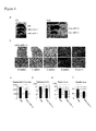
- Example 1 G-CSF receptor is expressed in regenerative myocytes in mdx mice.
- myofibers are initially degenerated and then regenerated to nearly compensate for the degeneration.
- G-CSFR G-CSF receptor
- Example 2 Influence of G-CSF on skeletal muscle function in mdx mice through skeletal muscle regeneration.
- G-CSFR-expressing cells appear most frequently at 3 weeks to 5 weeks of age in regenerating myocytes in mdx mice.
- G-CSF was intraperitoneally administered to mdx mice daily between 3 weeks and 5 weeks of age ( Figure 2a ) and examined gross appearance and functional differences.
- laminin 2 ⁇ which is the main isoform in the basement membrane of muscle
- Laminin 2 ⁇ is a protein found in the basement membrane of myofibers so that the size of each myofiber can be observed by staining with laminin 2 ⁇ .
- the results show that the number of centrally nucleated myofibers (regenerated myofibers) significantly increased at 5 weeks of age in the G-CSF-treated group as compared with the control group ( Figure 2b, c ).
- RNMy2/9D2 recognizes a myosin heavy chain (MHC) present during the embryonic and neonatal period in the development of skeletal muscle and also occurring in newly formed adult regenerating myocytes ( Bigard, et al. Changes in myosin heavy chain profile of mature regenerated muscle with endurance training in rat. Acta Physiologica Scandinavica 165, 185-192 (1999 )). Immunostaining with RNMy2/9D2 at 4 weeks of age showed that G-CSF significantly increased the number of regenerating myocytes ( Figure 2d, e ).
- MHC myosin heavy chain
- Muscle weakness is one of the most prominent features of muscular dystrophy ( Bushby, K. et al. Diagnosis and management of Duchenne muscular dystrophy, part 1: diagnosis, and pharmacological and psychosocial management. The Lancet Neurology 9, 77-93, doi:Doi: 10.1016/s1474-4422 (09) 70271-6 (2010 )). Thus, an exercise tolerance test was performed to evaluate in vivo muscle function. The results showed that G-CSF treatment significantly improved exercise tolerance at 5 weeks of age ( Figure 2h , the distribution of the number of individuals vs. tolerance time) (left bars: control group, right bars: G-CSF-treated group).
- Example 3 The role of G-CSF signaling in the survival of muscular dystrophy model mice through diaphragm degeneration.
- G-CSFR-knockout mice were used. The life span of the csf3r - /- mice was normal though they showed a mild hematological disorder.
- G-CSFR homozygous knockout mice further carrying an mdx mutation would show severe injury in muscle function, but surprisingly, a biphasic death response was observed immediately after birth and at postnatal week 3 to 5 (the stage during which muscles are acutely degenerated/regenerated) even in G-CSF heterozygous mdx ( mdx / csf3r +/- ) mice ( Figure 3a ).
- the surprising fact that death was observed in mdx / csf3r +/- mice indicates the importance of G-CSF signaling in muscular dystrophy.
- the thickness of diaphragms was significantly smaller in mdx / csf3r +/- mice ( Figure 3e, f ).
- the average body weight of the remaining survivors was consistently lower in mdx / csf3r +/- mice than csf3r +/- mice ( Figure 3g ).
- the difference between csf3r +/- mice and mdx / csf3r +/- mice apparently decreases on and after 5 weeks of age on the graph because mdx / csf3r +/- mice having a strong phenotype die by the age of 5 weeks while relatively healthy mdx / csf3r +/- mice survive.
- Example 4 The role of G-CSF in the late survival of muscular dystrophy model mice.
- Example 5 G-CSFR is expressed in regenerative myocytes in severe muscular dystrophy model mice.
- DMD Duchenne muscular dystrophy
- Mdx / utrn - /- mice that express neither dystrophin nor its homolog utrophin show severe dystrophy similar to human DMD patients so that they can be an animal model of DMD ( Grady, R. M. et al.
- H&E stainings of TA muscle sections at 2-12 weeks of age show that muscle injury occurred more acutely in mdx / utrn - /- mice at 3 weeks / 4 weeks / 5 weeks of age as compared with mdx mice ( Figure 1b ) and that this injury was persistent later ( Figure 5b ).
- Example 6 G-CSF improves skeletal muscle regeneration and function in severe muscular dystrophy model mice.
- G-CSF was administered to mdx / utrn -/- mice according to the administration schedule described in Figure 6a . Specifically, G-CSF was intraperitoneally administered once daily at the stage during which muscles were acutely injured and regenerated (at 3-5 weeks of age).
- Photographs of H&E stained TA muscle sections ( Figure 6b ), and the perimeters of TA muscles ( Figure 6c ) are shown (left bar: control group, right bar: G-CSF-treated group). It is shown that muscle size was increased by G-CSF administration. Moreover, the ratio of muscle wet weight to body weight tended to increase in tibialis anterior muscle and significantly increased in gastrocnemius muscle (Gastrocnemius) and quadriceps muscle (Quadriceps) by G-CSF administration ( Figure 6d ) (TA: tibialis anterior muscle, Gas: gastrocnemius muscle, Quad: quadriceps muscle) (left bars: control group, right bars: G-CSF-treated group).
Landscapes
- Health & Medical Sciences (AREA)
- Life Sciences & Earth Sciences (AREA)
- Pharmacology & Pharmacy (AREA)
- Animal Behavior & Ethology (AREA)
- General Health & Medical Sciences (AREA)
- Medicinal Chemistry (AREA)
- Chemical & Material Sciences (AREA)
- Veterinary Medicine (AREA)
- Bioinformatics & Cheminformatics (AREA)
- Public Health (AREA)
- Engineering & Computer Science (AREA)
- Gastroenterology & Hepatology (AREA)
- Zoology (AREA)
- Immunology (AREA)
- Proteomics, Peptides & Aminoacids (AREA)
- Epidemiology (AREA)
- General Chemical & Material Sciences (AREA)
- Nuclear Medicine, Radiotherapy & Molecular Imaging (AREA)
- Neurology (AREA)
- Organic Chemistry (AREA)
- Chemical Kinetics & Catalysis (AREA)
- Physical Education & Sports Medicine (AREA)
- Orthopedic Medicine & Surgery (AREA)
- Medicines That Contain Protein Lipid Enzymes And Other Medicines (AREA)
- Peptides Or Proteins (AREA)
Applications Claiming Priority (2)
| Application Number | Priority Date | Filing Date | Title |
|---|---|---|---|
| JP2010256873 | 2010-11-17 | ||
| PCT/JP2011/077113 WO2012067262A1 (fr) | 2010-11-17 | 2011-11-17 | Agent thérapeutique pour une dystrophie musculaire |
Publications (2)
| Publication Number | Publication Date |
|---|---|
| EP2641609A1 true EP2641609A1 (fr) | 2013-09-25 |
| EP2641609A4 EP2641609A4 (fr) | 2014-07-23 |
Family
ID=46084178
Family Applications (1)
| Application Number | Title | Priority Date | Filing Date |
|---|---|---|---|
| EP11841477.0A Ceased EP2641609A4 (fr) | 2010-11-17 | 2011-11-17 | Agent thérapeutique pour une dystrophie musculaire |
Country Status (4)
| Country | Link |
|---|---|
| US (1) | US20130302272A1 (fr) |
| EP (1) | EP2641609A4 (fr) |
| JP (2) | JP5944828B2 (fr) |
| WO (1) | WO2012067262A1 (fr) |
Families Citing this family (1)
| Publication number | Priority date | Publication date | Assignee | Title |
|---|---|---|---|---|
| EP2641609A4 (fr) * | 2010-11-17 | 2014-07-23 | Keiichi Fukuda | Agent thérapeutique pour une dystrophie musculaire |
Family Cites Families (8)
| Publication number | Priority date | Publication date | Assignee | Title |
|---|---|---|---|---|
| JPH06102021B2 (ja) | 1985-12-03 | 1994-12-14 | 中外製薬株式会社 | 新規なポリペプチド |
| JPS62129298A (ja) | 1985-12-02 | 1987-06-11 | Chugai Pharmaceut Co Ltd | 新規ポリペプチド |
| JPH0657152B2 (ja) | 1985-09-17 | 1994-08-03 | 中外製薬株式会社 | Csf遺伝子類 |
| JPS6444200A (en) | 1987-08-12 | 1989-02-16 | Kenji Hoshino | Stereophonic sound field recording and reproducing system |
| JP2005206544A (ja) * | 2004-01-23 | 2005-08-04 | Yasuyoshi Uchida | 筋肉再生剤 |
| JPWO2008020638A1 (ja) * | 2006-08-15 | 2010-01-07 | 国立大学法人神戸大学 | 靭帯損傷治療剤 |
| ES2544453T3 (es) * | 2009-05-14 | 2015-08-31 | Keiichi Fukuda | Uso de G-CSF en el tratamiento de lesiones musculares |
| EP2641609A4 (fr) * | 2010-11-17 | 2014-07-23 | Keiichi Fukuda | Agent thérapeutique pour une dystrophie musculaire |
-
2011
- 2011-11-17 EP EP11841477.0A patent/EP2641609A4/fr not_active Ceased
- 2011-11-17 US US13/988,255 patent/US20130302272A1/en not_active Abandoned
- 2011-11-17 WO PCT/JP2011/077113 patent/WO2012067262A1/fr not_active Ceased
- 2011-11-17 JP JP2012544338A patent/JP5944828B2/ja not_active Expired - Fee Related
-
2016
- 2016-04-05 JP JP2016075582A patent/JP2016183154A/ja active Pending
Non-Patent Citations (3)
| Title |
|---|
| None * |
| See also references of WO2012067262A1 * |
| SIMOES G F ET AL: "Granulocyte colony stimulating factor (G-CSF) reduces loss of inputs to alpha-motoneurons during the course of muscular distrophy and after axotomy in mdx mice", SOCIETY FOR NEUROSCIENCE ABSTRACT VIEWER AND ITINERARY PLANNER, vol. 40, 16 November 2010 (2010-11-16), & 40TH ANNUAL MEETING OF THE SOCIETY-FOR-NEUROSCIENCE; SAN DIEGO, CA, USA; NOVEMBER 16, 2010, XP002724622, Retrieved from the Internet <URL:http://www.abstractsonline.com/Plan/ViewAbstract.aspx?sKey=4190a374-0288-44ad-bbd9-871c1b739aa9&cKey=77bd8d5e-42be-4360-b1ad-7f38a582911b&mKey=%7bE5D5C83F-CE2D-4D71-9DD6-FC7231E090FB%7d> [retrieved on 20140520] * |
Also Published As
| Publication number | Publication date |
|---|---|
| EP2641609A4 (fr) | 2014-07-23 |
| US20130302272A1 (en) | 2013-11-14 |
| JPWO2012067262A1 (ja) | 2014-05-19 |
| WO2012067262A1 (fr) | 2012-05-24 |
| JP2016183154A (ja) | 2016-10-20 |
| JP5944828B2 (ja) | 2016-07-05 |
Similar Documents
| Publication | Publication Date | Title |
|---|---|---|
| EP2063905B1 (fr) | Traitement des troubles hépatiques par l'administration de conjugués de protéine associée au récepteur (rap) | |
| JP2017523140A (ja) | 潰瘍を処置するための方法および組成物 | |
| CA2874042C (fr) | Une levure recombinante exprimant la h ferritine humaine et ses utilisations | |
| EA007967B1 (ru) | Защита, восстановление и усиление эритропоэтинреактивных клеток, тканей и органов | |
| EA015672B1 (ru) | Тканезащитные пептиды и их применения | |
| KR20160009617A (ko) | 트레할로스의 비경구 투여에 의한 단백질 응집 근병증 및 신경퇴행성 질환의 치료 | |
| CN112135639A (zh) | 细胞组装介导的癌症免疫治疗检查点抑制剂的递送 | |
| US20250332223A1 (en) | Ghrh antagonists for use in a method of treating sarcoidosis | |
| US20210145922A1 (en) | Ang (1-7) derviative oligopeptides for the treatment of pain | |
| US11987608B2 (en) | Composition for accelerating cell proliferation comprising erythropoietin-derived peptide | |
| TW200815026A (en) | Treatment of ischemic diseases using erythropoietin | |
| US20210008164A1 (en) | Erythropoietin for Gastrointestinal Dysfunction | |
| AU2013335678B2 (en) | Therapeutic agent for amyotrophic lateral sclerosis | |
| EP2641609A1 (fr) | Agent thérapeutique pour une dystrophie musculaire | |
| CA2586213A1 (fr) | Formulations, procedes de production et utilisations du fgf-20 | |
| KR20210109812A (ko) | 부갑상선 호르몬 약제의 경구전달용 나노 복합체 | |
| US20190358307A1 (en) | Therapeutic agent for cerebral infarction | |
| CN106317217B (zh) | 一种促红细胞生成素模拟肽及其制备方法和应用 | |
| WO2025091016A1 (fr) | Compositions comprenant des inhibiteurs de p75 ntr et un facteur de croissance nerveuse recombinant et méthodes de traitement de maladies cornéennes et de troubles l'utilisant | |
| HK40110225A (zh) | 用於治疗结节病的方法的ghrh拮抗剂 | |
| WO2024054892A1 (fr) | Peptides thérapeutiques | |
| JP2025040825A (ja) | 間質性肺炎を予防又は治療するための医薬及び医薬組成物 | |
| HK40068602B (zh) | 用於治疗结节病的方法的ghrh拮抗剂 | |
| JP2009249292A (ja) | ポリリン酸を安定化剤とするエリスロポエチン溶液製剤 | |
| HK40068602A (en) | Ghrh antagonists for use in a method of treating sarcoidosis |
Legal Events
| Date | Code | Title | Description |
|---|---|---|---|
| PUAI | Public reference made under article 153(3) epc to a published international application that has entered the european phase |
Free format text: ORIGINAL CODE: 0009012 |
|
| 17P | Request for examination filed |
Effective date: 20130528 |
|
| AK | Designated contracting states |
Kind code of ref document: A1 Designated state(s): AL AT BE BG CH CY CZ DE DK EE ES FI FR GB GR HR HU IE IS IT LI LT LU LV MC MK MT NL NO PL PT RO RS SE SI SK SM TR |
|
| DAX | Request for extension of the european patent (deleted) | ||
| RIC1 | Information provided on ipc code assigned before grant |
Ipc: A61K 38/00 20060101AFI20140612BHEP Ipc: A61P 21/00 20060101ALI20140612BHEP |
|
| A4 | Supplementary search report drawn up and despatched |
Effective date: 20140625 |
|
| 17Q | First examination report despatched |
Effective date: 20160125 |
|
| REG | Reference to a national code |
Ref country code: DE Ref legal event code: R003 |
|
| STAA | Information on the status of an ep patent application or granted ep patent |
Free format text: STATUS: THE APPLICATION HAS BEEN REFUSED |
|
| 18R | Application refused |
Effective date: 20170307 |