EP2657331A1 - Substrat de culture et film de culture - Google Patents
Substrat de culture et film de culture Download PDFInfo
- Publication number
- EP2657331A1 EP2657331A1 EP10861209.4A EP10861209A EP2657331A1 EP 2657331 A1 EP2657331 A1 EP 2657331A1 EP 10861209 A EP10861209 A EP 10861209A EP 2657331 A1 EP2657331 A1 EP 2657331A1
- Authority
- EP
- European Patent Office
- Prior art keywords
- culture
- sheet
- cells
- projections
- region
- Prior art date
- Legal status (The legal status is an assumption and is not a legal conclusion. Google has not performed a legal analysis and makes no representation as to the accuracy of the status listed.)
- Withdrawn
Links
- 239000000758 substrate Substances 0.000 title claims abstract description 63
- 238000005192 partition Methods 0.000 claims abstract description 53
- 239000000463 material Substances 0.000 claims description 24
- 238000012258 culturing Methods 0.000 claims description 7
- 238000000034 method Methods 0.000 abstract description 29
- 239000002061 nanopillar Substances 0.000 abstract description 19
- 239000000126 substance Substances 0.000 abstract description 13
- 238000013508 migration Methods 0.000 abstract description 2
- 230000005012 migration Effects 0.000 abstract description 2
- 230000003993 interaction Effects 0.000 abstract 1
- 210000004027 cell Anatomy 0.000 description 118
- 210000003494 hepatocyte Anatomy 0.000 description 34
- 238000003466 welding Methods 0.000 description 20
- 230000015572 biosynthetic process Effects 0.000 description 15
- 238000004113 cell culture Methods 0.000 description 15
- 230000000717 retained effect Effects 0.000 description 14
- 230000000694 effects Effects 0.000 description 12
- 239000004793 Polystyrene Substances 0.000 description 11
- 229920002223 polystyrene Polymers 0.000 description 11
- 230000033001 locomotion Effects 0.000 description 10
- 230000008569 process Effects 0.000 description 10
- 239000000243 solution Substances 0.000 description 10
- 102000012422 Collagen Type I Human genes 0.000 description 9
- 108010022452 Collagen Type I Proteins 0.000 description 9
- 230000006837 decompression Effects 0.000 description 9
- 241001465754 Metazoa Species 0.000 description 8
- 238000005304 joining Methods 0.000 description 8
- 230000010412 perfusion Effects 0.000 description 8
- 238000012360 testing method Methods 0.000 description 8
- 239000000853 adhesive Substances 0.000 description 7
- 230000001070 adhesive effect Effects 0.000 description 7
- 238000004519 manufacturing process Methods 0.000 description 7
- 102000029816 Collagenase Human genes 0.000 description 6
- 108060005980 Collagenase Proteins 0.000 description 6
- 230000009087 cell motility Effects 0.000 description 6
- 229960002424 collagenase Drugs 0.000 description 6
- 206010067482 No adverse event Diseases 0.000 description 5
- 230000002411 adverse Effects 0.000 description 5
- 238000010586 diagram Methods 0.000 description 5
- 239000003814 drug Substances 0.000 description 5
- 230000002900 effect on cell Effects 0.000 description 5
- 238000002474 experimental method Methods 0.000 description 5
- 238000001878 scanning electron micrograph Methods 0.000 description 5
- 241000196324 Embryophyta Species 0.000 description 4
- 238000012136 culture method Methods 0.000 description 4
- 238000011081 inoculation Methods 0.000 description 4
- NOESYZHRGYRDHS-UHFFFAOYSA-N insulin Chemical compound N1C(=O)C(NC(=O)C(CCC(N)=O)NC(=O)C(CCC(O)=O)NC(=O)C(C(C)C)NC(=O)C(NC(=O)CN)C(C)CC)CSSCC(C(NC(CO)C(=O)NC(CC(C)C)C(=O)NC(CC=2C=CC(O)=CC=2)C(=O)NC(CCC(N)=O)C(=O)NC(CC(C)C)C(=O)NC(CCC(O)=O)C(=O)NC(CC(N)=O)C(=O)NC(CC=2C=CC(O)=CC=2)C(=O)NC(CSSCC(NC(=O)C(C(C)C)NC(=O)C(CC(C)C)NC(=O)C(CC=2C=CC(O)=CC=2)NC(=O)C(CC(C)C)NC(=O)C(C)NC(=O)C(CCC(O)=O)NC(=O)C(C(C)C)NC(=O)C(CC(C)C)NC(=O)C(CC=2NC=NC=2)NC(=O)C(CO)NC(=O)CNC2=O)C(=O)NCC(=O)NC(CCC(O)=O)C(=O)NC(CCCNC(N)=N)C(=O)NCC(=O)NC(CC=3C=CC=CC=3)C(=O)NC(CC=3C=CC=CC=3)C(=O)NC(CC=3C=CC(O)=CC=3)C(=O)NC(C(C)O)C(=O)N3C(CCC3)C(=O)NC(CCCCN)C(=O)NC(C)C(O)=O)C(=O)NC(CC(N)=O)C(O)=O)=O)NC(=O)C(C(C)CC)NC(=O)C(CO)NC(=O)C(C(C)O)NC(=O)C1CSSCC2NC(=O)C(CC(C)C)NC(=O)C(NC(=O)C(CCC(N)=O)NC(=O)C(CC(N)=O)NC(=O)C(NC(=O)C(N)CC=1C=CC=CC=1)C(C)C)CC1=CN=CN1 NOESYZHRGYRDHS-UHFFFAOYSA-N 0.000 description 4
- 210000004185 liver Anatomy 0.000 description 4
- 238000003556 assay Methods 0.000 description 3
- 230000004956 cell adhesive effect Effects 0.000 description 3
- 230000001771 impaired effect Effects 0.000 description 3
- 238000011065 in-situ storage Methods 0.000 description 3
- 230000004060 metabolic process Effects 0.000 description 3
- 239000002547 new drug Substances 0.000 description 3
- 238000012216 screening Methods 0.000 description 3
- 230000004083 survival effect Effects 0.000 description 3
- 230000009772 tissue formation Effects 0.000 description 3
- 230000001988 toxicity Effects 0.000 description 3
- 231100000419 toxicity Toxicity 0.000 description 3
- 210000003462 vein Anatomy 0.000 description 3
- 241000282412 Homo Species 0.000 description 2
- 102000004877 Insulin Human genes 0.000 description 2
- 108090001061 Insulin Proteins 0.000 description 2
- XUIMIQQOPSSXEZ-UHFFFAOYSA-N Silicon Chemical compound [Si] XUIMIQQOPSSXEZ-UHFFFAOYSA-N 0.000 description 2
- 238000013459 approach Methods 0.000 description 2
- 239000008280 blood Substances 0.000 description 2
- 210000004369 blood Anatomy 0.000 description 2
- 230000021164 cell adhesion Effects 0.000 description 2
- 230000001413 cellular effect Effects 0.000 description 2
- 238000010276 construction Methods 0.000 description 2
- 210000004748 cultured cell Anatomy 0.000 description 2
- 238000011161 development Methods 0.000 description 2
- UREBDLICKHMUKA-CXSFZGCWSA-N dexamethasone Chemical compound C1CC2=CC(=O)C=C[C@]2(C)[C@]2(F)[C@@H]1[C@@H]1C[C@@H](C)[C@@](C(=O)CO)(O)[C@@]1(C)C[C@@H]2O UREBDLICKHMUKA-CXSFZGCWSA-N 0.000 description 2
- 229960003957 dexamethasone Drugs 0.000 description 2
- 230000006870 function Effects 0.000 description 2
- 238000013537 high throughput screening Methods 0.000 description 2
- 238000000099 in vitro assay Methods 0.000 description 2
- 229940125396 insulin Drugs 0.000 description 2
- WEXRUCMBJFQVBZ-UHFFFAOYSA-N pentobarbital Chemical compound CCCC(C)C1(CC)C(=O)NC(=O)NC1=O WEXRUCMBJFQVBZ-UHFFFAOYSA-N 0.000 description 2
- 230000002093 peripheral effect Effects 0.000 description 2
- 238000002360 preparation method Methods 0.000 description 2
- 230000001172 regenerating effect Effects 0.000 description 2
- 229910052710 silicon Inorganic materials 0.000 description 2
- 239000010703 silicon Substances 0.000 description 2
- 239000003894 surgical glue Substances 0.000 description 2
- XLYOFNOQVPJJNP-UHFFFAOYSA-N water Chemical compound O XLYOFNOQVPJJNP-UHFFFAOYSA-N 0.000 description 2
- 206010002091 Anaesthesia Diseases 0.000 description 1
- YCKRFDGAMUMZLT-UHFFFAOYSA-N Fluorine atom Chemical compound [F] YCKRFDGAMUMZLT-UHFFFAOYSA-N 0.000 description 1
- FAPWRFPIFSIZLT-UHFFFAOYSA-M Sodium chloride Chemical compound [Na+].[Cl-] FAPWRFPIFSIZLT-UHFFFAOYSA-M 0.000 description 1
- GLNADSQYFUSGOU-GPTZEZBUSA-J Trypan blue Chemical compound [Na+].[Na+].[Na+].[Na+].C1=C(S([O-])(=O)=O)C=C2C=C(S([O-])(=O)=O)C(/N=N/C3=CC=C(C=C3C)C=3C=C(C(=CC=3)\N=N\C=3C(=CC4=CC(=CC(N)=C4C=3O)S([O-])(=O)=O)S([O-])(=O)=O)C)=C(O)C2=C1N GLNADSQYFUSGOU-GPTZEZBUSA-J 0.000 description 1
- 210000001015 abdomen Anatomy 0.000 description 1
- 239000003929 acidic solution Substances 0.000 description 1
- 230000037005 anaesthesia Effects 0.000 description 1
- 239000012298 atmosphere Substances 0.000 description 1
- QVGXLLKOCUKJST-UHFFFAOYSA-N atomic oxygen Chemical compound [O] QVGXLLKOCUKJST-UHFFFAOYSA-N 0.000 description 1
- 210000000741 bile canaliculi Anatomy 0.000 description 1
- 230000008827 biological function Effects 0.000 description 1
- 230000010261 cell growth Effects 0.000 description 1
- 239000006285 cell suspension Substances 0.000 description 1
- 210000002236 cellular spheroid Anatomy 0.000 description 1
- 230000008859 change Effects 0.000 description 1
- 238000006243 chemical reaction Methods 0.000 description 1
- 239000003795 chemical substances by application Substances 0.000 description 1
- 210000000038 chest Anatomy 0.000 description 1
- 230000003247 decreasing effect Effects 0.000 description 1
- 238000007865 diluting Methods 0.000 description 1
- 229940079593 drug Drugs 0.000 description 1
- 238000009509 drug development Methods 0.000 description 1
- 238000012362 drug development process Methods 0.000 description 1
- 210000001671 embryonic stem cell Anatomy 0.000 description 1
- DEFVIWRASFVYLL-UHFFFAOYSA-N ethylene glycol bis(2-aminoethyl)tetraacetic acid Chemical compound OC(=O)CN(CC(O)=O)CCOCCOCCN(CC(O)=O)CC(O)=O DEFVIWRASFVYLL-UHFFFAOYSA-N 0.000 description 1
- 238000011156 evaluation Methods 0.000 description 1
- 230000007717 exclusion Effects 0.000 description 1
- 238000001914 filtration Methods 0.000 description 1
- 229910052731 fluorine Inorganic materials 0.000 description 1
- 239000011737 fluorine Substances 0.000 description 1
- 239000011521 glass Substances 0.000 description 1
- 230000005484 gravity Effects 0.000 description 1
- 238000001727 in vivo Methods 0.000 description 1
- 210000004263 induced pluripotent stem cell Anatomy 0.000 description 1
- 229940042040 innovative drug Drugs 0.000 description 1
- 230000001678 irradiating effect Effects 0.000 description 1
- 230000014759 maintenance of location Effects 0.000 description 1
- 239000007769 metal material Substances 0.000 description 1
- 238000001000 micrograph Methods 0.000 description 1
- 238000000465 moulding Methods 0.000 description 1
- 231100000989 no adverse effect Toxicity 0.000 description 1
- 229910052760 oxygen Inorganic materials 0.000 description 1
- 239000001301 oxygen Substances 0.000 description 1
- 229960001412 pentobarbital Drugs 0.000 description 1
- 239000004033 plastic Substances 0.000 description 1
- 210000003240 portal vein Anatomy 0.000 description 1
- 230000002028 premature Effects 0.000 description 1
- 230000001737 promoting effect Effects 0.000 description 1
- 210000005245 right atrium Anatomy 0.000 description 1
- 238000000926 separation method Methods 0.000 description 1
- 210000002966 serum Anatomy 0.000 description 1
- 239000002356 single layer Substances 0.000 description 1
- 239000011780 sodium chloride Substances 0.000 description 1
- 238000000638 solvent extraction Methods 0.000 description 1
- 239000008223 sterile water Substances 0.000 description 1
- 239000000725 suspension Substances 0.000 description 1
- 238000005406 washing Methods 0.000 description 1
Images


Classifications
-
- C—CHEMISTRY; METALLURGY
- C12—BIOCHEMISTRY; BEER; SPIRITS; WINE; VINEGAR; MICROBIOLOGY; ENZYMOLOGY; MUTATION OR GENETIC ENGINEERING
- C12N—MICROORGANISMS OR ENZYMES; COMPOSITIONS THEREOF; PROPAGATING, PRESERVING, OR MAINTAINING MICROORGANISMS; MUTATION OR GENETIC ENGINEERING; CULTURE MEDIA
- C12N5/00—Undifferentiated human, animal or plant cells, e.g. cell lines; Tissues; Cultivation or maintenance thereof; Culture media therefor
-
- G—PHYSICS
- G01—MEASURING; TESTING
- G01N—INVESTIGATING OR ANALYSING MATERIALS BY DETERMINING THEIR CHEMICAL OR PHYSICAL PROPERTIES
- G01N33/00—Investigating or analysing materials by specific methods not covered by groups G01N1/00 - G01N31/00
- G01N33/48—Biological material, e.g. blood, urine; Haemocytometers
- G01N33/50—Chemical analysis of biological material, e.g. blood, urine; Testing involving biospecific ligand binding methods; Immunological testing
- G01N33/5005—Chemical analysis of biological material, e.g. blood, urine; Testing involving biospecific ligand binding methods; Immunological testing involving human or animal cells
- G01N33/5008—Chemical analysis of biological material, e.g. blood, urine; Testing involving biospecific ligand binding methods; Immunological testing involving human or animal cells for testing or evaluating the effect of chemical or biological compounds, e.g. drugs, cosmetics
-
- C—CHEMISTRY; METALLURGY
- C12—BIOCHEMISTRY; BEER; SPIRITS; WINE; VINEGAR; MICROBIOLOGY; ENZYMOLOGY; MUTATION OR GENETIC ENGINEERING
- C12M—APPARATUS FOR ENZYMOLOGY OR MICROBIOLOGY; APPARATUS FOR CULTURING MICROORGANISMS FOR PRODUCING BIOMASS, FOR GROWING CELLS OR FOR OBTAINING FERMENTATION OR METABOLIC PRODUCTS, i.e. BIOREACTORS OR FERMENTERS
- C12M25/00—Means for supporting, enclosing or fixing the microorganisms, e.g. immunocoatings
- C12M25/06—Plates; Walls; Drawers; Multilayer plates
-
- C—CHEMISTRY; METALLURGY
- C12—BIOCHEMISTRY; BEER; SPIRITS; WINE; VINEGAR; MICROBIOLOGY; ENZYMOLOGY; MUTATION OR GENETIC ENGINEERING
- C12M—APPARATUS FOR ENZYMOLOGY OR MICROBIOLOGY; APPARATUS FOR CULTURING MICROORGANISMS FOR PRODUCING BIOMASS, FOR GROWING CELLS OR FOR OBTAINING FERMENTATION OR METABOLIC PRODUCTS, i.e. BIOREACTORS OR FERMENTERS
- C12M3/00—Tissue, human, animal or plant cell, or virus culture apparatus
Definitions
- the present invention relates to a technique of culturing animal and plant cells using a culture substrate, and graphically forming spheroids (3D tissues), and monolayer tissues (2D tissues) of the cells.
- Non-Patent Literature 1 the construction of 3D tissues which exhibits functions more similar to those of living bodies has been attempted so far, and 3D tissues has been successfully formed for various cell strains.
- the 3D tissues formed As a substrate for forming 3D tissues of cells, a sheet (nanopillar sheet) for culture in which regularly arranged ultrafine pillar structures or protrusions are formed on the surface of a sheet has been developed, the 3D tissues formed has the problems that they have high release properties from the substrate (Patent Literature 1), and that they are lost in the process of medium change. Moreover, since it is impossible to control the diameter of formed 3D tissues, it entails the problem that their sizes are not uniform, and therefore the performance of each of the 3D tissues is varied. It is thus still premature as a practical formation method.
- Patent Literature 2 Non-Patent Literature 2
- a feature of this technique is that by applying a substance having adhesion to a predetermined region around the center of at the bottom of the cavity, a cell adhesive region and a cell non-adhesive region are defined, and the cavity itself is rotated by a rotation drive apparatus or the like to perform rotation culture, so that cultured cells are retained around the center of the bottom of the cavity which is the cell adhesive region.
- the cell adhesive region and cell non-adhesive region need to be defined by applying a chemically synthesized substance on the surface of the substrate, which entails some problems.
- inoculated cells fall into non-adhesive regions, they are inevitably disposed of along with the medium when the medium is changed during culture, which is hardly considered as an efficient culture method. Furthermore, it is suspected that the cells which have fallen into the adhesion region are caused to form tissues compulsorily by rotation culture, and therefore stress is exerted on cells, which leads to a lowered activity.
- known nanopillar sheets also have the problems that it is difficult to control the cell movement on the substrate plane, and that it is impossible to control the dimension and diameter of the 3D tissues formed. At the same time, it also has the problem that it is impossible to retain the formed 3D tissues in a target position.
- An object of the present invention is to provide a culture sheet, a culture substrate, and a cell culture method using the same which enable forming 3D tissues having a uniform diameter without applying chemicals on the surface of the culture substrate, and further retaining the 3D tissues in a target position.
- the present invention provides a culture substrate and a culture sheet in which a culture region is provided, a plurality of projections are formed in the culture region, a partition which partitions the culture region and is taller than the projections around the culture region form, and the constitutional proportion of the projections in the culture region is in the range from 20% to 75%.
- the present invention provides a culture substrate and a culture sheet in which a culture region is provided, a plurality of projections are formed in the culture region, a partition which partitions the culture region and is taller than the projections around the culture region is formed, and the constitutional proportion of the projections in the culture region is in the range from 40% to 50%.
- a limited region i.e., a partition
- cells inoculated within the limited region are all involved in the formation of a single 3D tissue.
- This achieves a very efficient culture method, and also leads to the expectation that the sizes of a plurality of 3D tissues formed for the respective limited regions are uniform and homogenous, which is effective in cell assays.
- the 3D tissues are retained in a target position within the limited region, i.e., the partition. Furthermore, 2D tissues can be formed depending on the purpose. Similar effects are also expected on the 2D tissues.
- Example 1 shows an example in which the culture sheet is applied to the chamber slide which is a culture sheet retaining member.
- a sheet which has a partition structure which forms the culture region in the present invention on a known nanopillar sheet, and on which a plurality of projections are formed inside the partition structure is referred to as a culture sheet.
- the culture sheet is formed from a material which has no adverse effect on cells, in this example, it is polystyrene. However, it goes without saying that the material is not limited to polystyrene.
- Fig. 1 is a schematic diagram of a scanning electron micrograph of a culture sheet 100 prepared in this Example. Simultaneously, it shows the structure of one of holes 101 (hereinafter referred to as hole) constituted by a plurality of partition structures 102 existing in a single culture sheet. The inside of the hole 101 constitutes a culture region by cell tissue formation unit.
- a plurality of projections 102 retained at the bottom of the hole 101 includes a plurality of microprojections 103 (hereinafter also referred to as projections, pillars or nanopillars). Moreover, the diameter of this hole 101 is a hole diameter 105.
- the hole 101 including the above-mentioned partition wall 102 and a plurality of projections 103 formed inside the hole 101 are formed from the same material integrally. It should be noted that the shape of this hole 101 is not limited to round, but may have other shapes such as a square shape.
- the hole 101 and the plurality of projections 103 formed inside the hole 101 including the partition wall 102 are formed integrally as the culture sheet 100 from a single material which has no adverse effects on cells, whereby cells can be grown without foreign substances bonding to the cells in the culture steps. Furthermore, since cells are grown in each of the partitions, cells of a homogeneous size can be formed.
- a plurality of projections are provided within the partition wall 102 arranged in a surrounding manner, and therefore the cell movement which is the ability inherent to the cells is promoted, and cells are grown by the movement so that cell culture which can maintain the cell activity is possible with no influence of disturbance (stress) by rotation culture or the like.
- the inner diameter of the hole 101 is a hyperfine region diameter on the cell formation level, and therefore it is very difficult to perform welding while forming a target cell region and not damaging the partitions and projections.
- the partitions and projections have damages and deformations, unwanted stress may be applied on the cells in the process of cell formation, and the movement of the cells themselves may be impaired.
- the hole bottom 104, partition wall 102 and projections 103 constituting the holes 101 which forms the culture region are preferably formed integrally.
- integrally it is preferable because culture excluding the influence of unwanted components other than those required for cell culture can be performed.
- FIG. 2 An enlarged view of the projection 103 is shown in Fig. 2 .
- a pillar diameter indicates a diameter 106 of the tip of the projection.
- a pillar pitch indicates a distance 107 from the center of the tip of the projection to the center of the tip of the adjacent projection.
- a pillar height indicates a height 108 from the tip of a nanopillar to the bottom thereof.
- Fig. 2(a) and Fig. 2(b) indicate a square arrangement and a triangle arrangement, respectively, of nanopillars of this Example.
- culture sheets in which the pillar diameter, pillar pitch and pillar height are 2.0 ⁇ m, 4.0 ⁇ m, and 1.0 ⁇ m, respectively, were used, but as will be described later, such culture sheets are not necessarily used.
- the height of the partition structure is 70 ⁇ m in this Example, but this value is not necessarily used, and suitably the height may be such that the formed cells do not get over the partition.
- the culture sheet 100 in this Example is produced by the method described below.
- a mold in which round holes each having a diameter of 200 ⁇ m and depth of 70 ⁇ m are arranged in the form of squares, and micropores each having a diameter of 2.0 ⁇ m and a depth of 1.0 ⁇ m are formed at the bottom at a pitch of 4.0 ⁇ m was pressed against a polystyrene film having a thickness of 400 ⁇ m at 135°C and a pressure of 2 MPa.
- the film was took out from a press machine after being cooled to room temperature, and the mold was peeled off from the polystyrene film, whereby a culture sheet retaining a plurality of holes each having a hole diameter of 200 ⁇ m and having a plurality of projections at the bottom thereof can be produced.
- a mold material is silicon wafer, and in order to prevent adhesion with the polystyrene film during the production of the culture sheet, a mold releasing process is performed in advance with a fluorine-based mold releasing agent.
- Silicon wafer was used as the mold material in this Example, but a mold made from other metal materials and the like may be also used.
- 109a represents a frame for partitioning the culture sheets 100.
- This frame 109a is formed from, for example, a plastic material or the like.
- the shape of a frame body such as this frame 109a is not limited to square, but may be other shapes such as a round shape.
- Figs. 13A , 13B , 13C show the overall constitution diagram and principal part cross-sectional view of the chamber slide with the culture sheet of this Example affixed thereto.
- Fig. 13A is an appearance perspective view, top view, and upper and lower side views of the culture substrate in this Example. Illustration of left and right side elevational views is omitted since its form is obvious from the perspective view.
- Fig. 13B is a partially enlarged view, which shows an A-A, B-B partially enlarged view, and a C-C, D-D partially enlarged view.
- Fig. 13C is a partially enlarged view and end view, which shows an E-E, F-F partially enlarged view, and a line G-G end view.
- the article shown in Figs. 13A to 13C is a culture device (culture containers) for culturing cells of humans, animals, plants and others, and are each constituted by the culture sheet 100 and a retaining member (chamber slide) 109 which retains the culture sheet 100.
- a plurality of partition portions 102 are formed on the surface of the culture sheet 100, and is provided at the bottom of the inside of a cylindrical hole portion 109a formed on the retaining member 109.
- culture regions having a plurality of minute projection portions 103 within the partition portion are formed respectively.
- target cells to be cultured are added to the inside of the hole portion 109a, as added to the sheet surface forming the culture regions within the partition portion 102, the target cell is retained in the plurality of minute projection portions 103 and cultured.
- Example 2 will be described with reference to Figs. 4 and 5 .
- Fig. 4(a) is a bottom view of a frame body 111 constituting the multiwell plate.
- the frame body 111 which is a culture sheet retaining member is such that has 24 cylindrical hole portions 111a in total, arranged in 4 rows and 6 columns, formed in an area measuring about 125 mm in width, about 80 mm in length, and about 20 mm in height.
- the material used is polystyrene.
- the number of holes formed on the frame body normally ranges from 6 to 1536, varied depending on the use, and therefore the number of holes on this frame body is not limited to 24.
- the material of the frame body is not limited to polystyrene either.
- the frame body 111 and the culture sheet 100 is joined by ultrasonic welding.
- the following processes are performed on the frame body 111 in advance.
- a projection for fixing film 112 is processed at the bottom of the frame body 111 for the purpose of preventing the cell culture sheet and the plate from being shifted due to the vibration of ultrasonic waves provided when the frame body 111 and the culture sheet 100 are welded.
- a rib structure 113 is provided to weld the culture sheet by ultrasonic waves.
- Figs. 4(b) and 4(c) are shows the cross-sectional views at lines B-B' and A-A', respectively, in Fig. 4(a) .
- holes 114 having the same diameter are provided in the culture sheet in the same position when both are overlapped so that the projection engages with the projection for fixing film. Successively, this frame body and the culture sheet 100 are adhered by ultrasonic welding.
- the step of the welding is shown in Fig. 5 .
- the holes of the projection for fixing film of the frame body and of the culture sheet are placed together and stacked ( Fig. 5(a) ).
- ultrasonic waves are produced from the culture sheet side from an ultrasonic wave oscillator via a converter, a booster, or further a horn, and both are welded ( Fig. 5(b) ).
- a horn is an apparatus for welding by irradiating an appropriate position with ultrasonic waves of an appropriate energy.
- a specific apparatus designed so that ultrasonic waves are generated appropriately along the position of the rib structure was produced and used.
- 115 shows a top view of the thus-produced plate.
- this Example is a culture sheet which is applicable and useful not only to toxicity and metabolism tests in new drug development processes, but also to the formation of organizations intended for regenerative medicine.
- the culture substrate can be produced by a joining method similar to this Example.
- a plurality of the holes 101 are formed on the culture sheet 100 formed at the bottom of the frame body 111, and a plurality of projections constituted at the bottom 104 of the hole include a plurality of microprojections 103 (hereinafter also referred to as projections, pillars or nanopillars). Moreover, the diameter of this hole 101 is used as a hole diameter 105.
- the hole 101 including the above-mentioned partition wall 102 and the plurality of projections 103 formed inside the hole 101 are formed from the same material integrally. It should be noted that the shape of this hole 101 is not limited to round, but may have other shapes such as a square shape.
- the hole 101 including the partition wall 102 and the plurality of projections 103 formed inside the hole 101 are formed integrally from a single material which has no adverse effects on cells as a culture sheet, whereby cells can be grown with no foreign substances adhering to cells in the culture step. Furthermore, since cells are grown in each of the partitions, cells of a homogeneous size can be formed.
- a plurality of projections are provided within the partition arranged in a surrounding manner, and therefore cell movement, which is the ability inherent to the cells, is promoted, and cells are grown by the movement so that cell culture which can maintain the cell activity is possible with no influence of disturbance (stress) by rotation culture or the like.
- the inner diameter of the hole 101 is a hyperfine region diameter on the cell formation level, and therefore it is very difficult to perform welding while forming a target cell region and not damaging the partitions and projections.
- the partitions and projections have damages and deformations, unwanted stress may be applied on the cells in the process of cell formation, and the movement of the cells themselves may be impaired.
- the hole 101 which forms the culture region and the projections 103 are preferably formed integrally by forming integrally in such a manner, it is preferable because culture excluding the influence of unwanted components other than those required for cell culture can be performed.
- FIGs. 14A , 14B , 14C , and 14D an overall constitution diagram and a principal part cross-sectional view of a multiwell plate with the culture sheet of this Example are shown.
- Fig. 14A shows an appearance perspective view and a bottom view of the culture substrate in this Example.
- Fig. 14B shows a top view and upper and lower side views of the culture substrate.
- Fig. 14C is a partially enlarged view and a partial cross-sectional view, which show an A-A, a B-B partially enlarged view, a C-C, D-D partially enlarged view, and an H-H cross-sectional view.
- Fig. 14D is a partially enlarged view, and an end view, which show an E-E, F-F partially enlarged view, and a line G-G end view.
- the article shown in Figs. 14A , 14B , 14C , 14D is a culture device (culture container) for culturing cells of humans, animals, plants and others, and is constituted by the culture sheet 100 and a retaining member (frame body) 111 which retains the culture sheet 100.
- a plurality of the holes 101 are formed on the surface of the culture sheet 100, and is provided at the bottom of the inside of a cylindrical hole portion 111a formed in the retaining member. Furthermore, culture regions having a plurality of minute projection portion 103 within the partition portion are formed respectively. When target cells to be cultured are added to the inside of the hole portion 111a, as added to the sheet surface forming the culture regions within the hole 101, the target cell is retained in the plurality of minute projection portions 103 and cultured.
- the culture substrate of this example shows an example in which the culture sheet is welded from the back side of the frame body 111, and the frame body 111 which is a retaining member and the culture sheet 100 are welded via a joint 1112.
- the joint 1112 is provided on the outside of the hole portion 111a, and the culture region is not affected by the welding. Therefore, although welding was shown as an example in this example, the joining method is not limited to this, and other joining methods can be also employed since joining does not affect the culture region itself with other joining methods.
- the frame body 111 has the form of a square, and at least of the four apexes is cut off.
- This cut surface 1113 facilitates specification of the position of the hole portion of the substrate by the operator who performs culture.
- This cut face is not essential, and of course may be or may not be present.
- a non-slip portion 1111 is formed on the culture substrate, which can prevent the operator from unexpectedly shaking and dropping the substrate and prevent other accidents during the operation.
- Example 3 shows an example of application of cells to tissue culture using the culture substrates produced in Examples 1 and 2.
- the construction of the 3D tissues which reflects biological functions has demands for various evaluations utilizing cells substituting animal experiments.
- the thus-formed 3D tissues are subjected to various tests in screening of pharmaceuticals or development of new drugs, it is necessary to verify in advance whether the 3D tissues retain activities to withstand the tests. In this case, if the formed spheroids are held in the predetermined position with high reproducibility, it is expected that they are suitable for high through-put screening and various tests.
- 3D tissues need to be formed before culturing induced pluripotent stem cells (iPS cells) and embryonic stem cells (ES cells) to cause them to differentiate into target cells. Therefore, a technique of easily constructing 3D tissues has been demanded also in the field of regenerative medicine. From such a background, an example of forming 3D tissues using the chamber slide in particular is shown herein, but the essential part of cell culture is not especially different even for a multiwell plate. In this example, an example using rat hepatocytes is shown, but as mentioned above, it is applicable to cell strains of various animals and plants, and cell strains are not especially limited.
- Preparation of hepatocytes is performed according to in situ collagenase perfusion technique. The detail is as follows: The abdomen of a Fisher 344 male rat (7 to 10 weeks old) is opened under pentobarbital anesthesia, and a catheter is inserted into the portal vein to inject a preperfusate (Hanks' solution not including Ca 2+ or Mg 2+ and containing EGTA).
- a preperfusate Hanks' solution not including Ca 2+ or Mg 2+ and containing EGTA.
- the postcaval vein in the lower liver is simultaneously incised to discharge blood.
- the thorax is opened, the postcaval vein which goes into the right atrium is incised, and the postcaval vein in the lower liver is clipped with a clamp to perform perfusion.
- Perfusion is stopped after it is confirmed that the blood removal from the liver has been fully conducted.
- the perfusate is exchanged to a collagenase solution to perform perfusion.
- Perfusion is performed using the Hanks' solution containing 0.05% of collagenase in this example, but this solution is not necessarily used. Perfusion is stopped after it is confirmed that intercellular tissues have been digested by collagenase.
- the liver is separated, cut into small pieces in a cooled Hanks' solution, and is dispersed into cells by pipetting. Subsequently, undigested tissues are removed by gauze filtration.
- the cell suspension is repeatedly centrifuged at 50 G for 1 minute several times to remove nonparenchymal cells. Subsequently, damaged hepatocytes are removed by centrifugal separation at 500 G for 5 minutes using an isotonic Percoll solution.
- the survival rate of the obtained hepatocytes is measured by the trypan blue exclusion method, and the hepatocytes with a survival rate of 85% or higher are used for culture.
- the hepatocytes with a survival rate of 85% or higher are used for culture, but it goes without saying that this condition is not necessarily used.
- Preparation of the hepatocytes is not necessarily limited to the in situ collagenase perfusion technique.
- FIG. 6 A flowchart of the culture using the thus-obtained hepatocytes is shown in Fig. 6 .
- type I collagen 116 is applied to the culture sheet of the chamber slide type produced in Example 1.
- a 1 to 1.5-ml portion of a diluted solution which has been produced by diluting type I collagen dissolved in a weakly acidic solution with sterile water to a predetermined concentration is added to the chamber slide mentioned above ( Fig. 6(a) ).
- a decompression operation is performed in order to cause the added type I collagen to be adsorbed onto the nanopillar sheet 100 completely ( Fig. 6(b) ).
- the decompression operation is performed at 0.04 atmosphere or lower using a decompression container 117 and a decompression pump 118.
- the decompression time is not particularly limited, but the decompression is performed for 10 minutes in this Example.
- the constitution of the apparatus used for decompression is not particularly limited.
- the range of the predetermined concentration of the diluted solution is 100 (ng/ml) or higher and 10 ( ⁇ g/ml) or lower.
- the concentration is not necessarily limited to this range, but this range is suitable for spherical 3D tissues to form.
- an excess of type I collagen is removed, and PBS(-) 119 is added thereto ( Fig. 6(c) ). This operation is performed three times, and an excess of type I collagen is washed.
- Hepatocytes 120 prepared by the in situ collagenase perfusion technique as above-mentioned are suspended in a medium 121, and the suspension is inoculated on the NP sheet with Type I collagen prepared as stated above applied thereto similarly ( Fig. 6(d) ).
- the medium is not particularly limited, but, a Williams E medium including a medium containing serum (FCS), insulin, and dexamethasone (hereinafter referred to as medium (including 10% FCS)) is used.
- medium including 10% FCS
- a Williams E medium containing 10% FCS, 8.6 nM insulin, and 255 nM dexamethasone is particularly used.
- medium used for the culture after the 18th hour after the inoculation is not particularly limited, in this example, a medium (hereinafter referred to as medium (containing no FCS) with FCS removed from a medium (containing 10% FCS) is used.
- the inoculation density of hepatocytes was set to 1 ⁇ 10 5 cells/ml in this Example, but is not limited to this concentration.
- the culture sheet 100 used for culture has a pillar height, pillar diameter and pillar pitch of 1.0 ⁇ m, 2.0 ⁇ m, and 4.0 ⁇ m, respectively, but the values are not limited to these.
- the concentration of Type I collagen added to the culture sheet is set to 100 (ng/ml) in this Example, but may be a concentration other than this.
- Spheroids may be formed at a concentration other than this concentration depending on the conditions of the cells.
- the cells are cultures for 96 hours in total, whereby 3D tissues 122 are formed ( Fig. 6(e) ).
- Fig. 7 shows a photograph of the results of actual culture of hepatocytes using the above-mentioned culture sheet having a hole diameter of 200 ⁇ m.
- spherical 3D tissues 71 having such similar sizes are formed in the holes 70 with no special chemical applied onto the surface of the culture sheet and by stationary culture having little stress on cells. This culture method supposedly does not deteriorate the activity of the cells originally retained, and is therefore effective for cell assays and the like.
- Fig. 8 shows a variant of Example of the culture sheet 100 mentioned above as Example 4.
- a culture sheet 123 by arranging the arrangement pattern of projections which provides differences in the migration and adhesion of cells in two stages, as in Fig. 8(a) , in a manner of surrounding a first arrangement pattern 125a with a second arrangement pattern 125b, 3D tissues or 2D tissues are formed on the first arrangement pattern 125a (for example, near the center of the hole).
- Fig. 8(d) shows a culture sheet 128 in which the arrangement pattern is set to be multi-stage patterns 129c, 129b, 129a.
- pillar patterns types of the arrangement patterns (hereinafter referred to as pillar patterns) of the projections in the above-mentioned Example.
- 11 types of arrangement patterns have been shown as examples.
- the pillar diameter and pillar pitch ranging from 0.18 to 20.0 ⁇ m and from 0.36 to 40.0 ⁇ m, respectively, but the pillar diameter and pillar pitch are not limited to these.
- FIG. 10A and 10B An example of hepatocytes cultured under these pillar patterns is shown in Figs. 10A and 10B .
- Fig. 10A are figures which show the states of the cells when culture is performed using the culture sheet 100 with the double pitch relative to the pillar diameter.
- the pillar diameter is 0.18 ⁇ m, 0.5 ⁇ m, and 1.0 ⁇ m, flat tissues which are not spherical are adhered at the bottom of the substrate, while, when it is 2.0 ⁇ m and 5.0 ⁇ m, 3D tissues which are spherical are formed on the substrate.
- the substrate with the pillar diameter of 2.0 ⁇ m had more cells adhered onto the substrate, indicating that it is in a stable state. That is, it can be seen that as for the cell adhesion, the greater the pillar diameter, the lower the adhesion and the more promoted the movement by cells.
- Fig. 10B is a graph which shows, as for the number of 3D tissues (spheroids) of the hepatocytes formed on the sheets with each of the pillar diameters, the results grouped by diameter of the spheroids formed.
- the area of the sheet is 4 square cm (2 cm ⁇ 2 cm).
- the pillar diameter is 2.0 ⁇ m is preferable in order to form cells having diameters of 50 to 100 microns, but the pillar diameter is not limited to this, and for all the pillar diameters used in this examination, it was found that a greater number of cells with stable shapes are formed compared to the flat state with no pillar formed.
- the form or adhesion to the substrate of cells or tissues formed from cells can be freely changed by the difference in pillar pattern.
- tissue having target shapes can be formed in target positions within the holes utilizing cell adhesion and the motion characteristics of cells themselves.
- Fig. 11(a) shows a culture sheet 130 which is a variant in which a difference is provided in the heights of the nanopillars gradually. At this time, unlike in a normal U-shaped culture container, there is produced an effect that the cells are retained in the center by the presence of the pillars.
- the culture sheet 131 of Fig. 11(b) it is also possible to promote the effect stated above by providing a difference in pillar diameter even in the inclination.
- the height is changed gradually to smoothen the inclination, but a constitution in which the height is sequentially changed stepwise may be also employed.
- a plurality of holes gather to form a culture surface (square shape in the case of the chamber slide, round shape in the case of and the plate), but in the culture, a difference occurs in how 3D tissues are formed in the central portion and peripheral portion of the culture surface by the influence of the surface tension. That is, although 3D tissues are formed in the central portion of the culture surface, 3D tissues may not be formed in some events for the reason that the amount of the medium is increased of the portion by the surface tension in the peripheral portion, the amount of oxygen supplied is lowered, or the high water pressure is applied. In order to avoid this phenomenon, the culture sheets 132, 133 retaining the hole structure may be produced only in the central portion of the culture surface as shown in Figs. 12 (a), (b) .
- the culture substrate having high culture efficiency and little production load can be achieved.
- Fig. 15A , Fig. 15B , Fig. 15C, and Fig. 15D show the culture sheet of Example 5.
- the first arrangement pattern 125a is a flat structure, and a culture sheet having a pattern in which projections are arranged in the central portion of the hole is applied to the chamber slide which is a culture sheet retaining member.
- projections are arranged near the center in the culture region, but the center need not be necessarily included, and it goes without saying that projections may be arranged in a desired region in the culture region.
- the projection region of an approximate rhombus shape and circle shape is formed is shown, it goes without saying that the projection region may be in the shape of a square or a polygon.
- Fig. 15A shows an example of the culture sheet prepared in this Example.
- Fig. 15A shows one structure of holes 151 constituted by a plurality of partition structures 152 in a single culture sheet.
- the configuration that the inside of the hole 101 constitutes a culture region by a cell tissue formation unit partitioned by a partition wall is the same as in the above-mentioned Example.
- a plurality of projections 153 retained at the bottom 154 of the hole 151 includes a plurality of microprojections.
- the diameter of this hole 151 is set tube a hole diameter 155.
- the hole 151 including the above-mentioned partition wall 152 and a plurality of projections 153 formed within the hole 151 are formed from the same material integrally.
- the shape of this hole 151 is not limited to round, but may be another shape such as a square, as in the above-mentioned Example.
- a culture substrate in which the formation region (constitutional proportion) of projections by in the hole that is, a cell tissue formation unit partitioned by a partition wall constituted in multi-stages is effective as a test substrate for grasping an optimum formation rate of projection regions in Example 7 described later.
- An optimum constitutional proportion may vary depending on the cell strains and desired size intended for culture, and therefore performing a culture test in advance using such a culture substrate is useful since it affects the culture efficiency thereafter.
- the hole 151a indicates a hole in which no projection is formed.
- the culture sheet 150a is formed from a material which does not adversely affect cells, and it is, in this example, polystyrene. However, it goes without saying that the material is not limited to polystyrene.
- a SEM image in which the diameter of the assembly of projection portions is 80 ⁇ m is shown in Fig. 15C
- an SEM image in which the diameter is 20 ⁇ m is shown in Fig. 15D
- 156 and 158 represent a hole
- 157 and 159 represent a projection assembly.
- the hole 151 including the partition wall 152 and the plurality of projections 153 formed inside the hole 151 are formed integrally from a single material which has no adverse effects on cells as culture sheets 150, 150a, whereby cells can be grown with no foreign substances adhering to cells in the culture step. Furthermore, since cells are grown in each of the partitions, cells of a homogeneous size can be formed.
- a plurality of projections are provided within the partition wall 152 arranged in a surrounding manner. Therefore, cell movement, which is the ability inherent to the cells, is promoted, and cells are grown by the movement so that a cell culture which can maintain the cell activity is possible with no influence of disturbance (stress) by rotation culture or the like.
- these holes 151 and projection assembly 153 When a culture region is to be formed while these holes 151 and projection assembly 153 are provided separately, they need to be joined by adhesion or welding. For example, when these are joined by adhesion, adhesive components enter into the culture region, which may adversely affect generated cells.
- the inner diameter of the hole 151 is a hyperfine region diameter on the cell formation level, and therefore it is very difficult to perform welding while forming a target cell region and not damaging the partitions and projections.
- the partitions and projections have damages and deformations, unwanted stress may be applied on the cells in the process of cell formation, and the movement of the cells themselves may be impaired.
- the hole bottom 154, partition wall 152 and projections 153 constituting the holes 151 which form the culture region are preferably formed integrally by forming integrally in such a manner, it is preferable because culture excluding the influence of unwanted components other than those required for cell culture can be performed.
- the culture sheet in which the pillar diameter, pillar pitch and pillar height are 1.0 ⁇ m or 2.0 ⁇ m, 2.0 ⁇ m or 4.0 ⁇ m, 1.0 ⁇ m, respectively, was used, but as will be described later, the culture sheet may be one with other specifications than these.
- the height of the partition structure is 70 ⁇ m in this Example, but this value is not necessarily used, and suitably the height may be such that the formed cells do not get over the partition.
- the culture sheets 150, 150a in this Example are produced by a method similar to that in Example 1, and therefore detailed description of the production method will be omitted herein.
- the chamber slide 109 with the culture sheet 150 affixed as shown in Fig. 3 can be produced, and it goes without saying that a chamber slide having an overall constitution and a principal part cross section similar to those in Fig. 13A , Fig. 13B , Fig. 13C can be obtained, and therefore explanation will be omitted herein.
- Example 6 will be described with reference to Figs. 4 and 5 .
- This Example shows the constitution of a multiwell plate with a culture sheet using the culture sheets 150, 150a described in Example 5, and a production example thereof.
- the constitution of the multiwell plate and a production example of the same have been described in Figs. 4 and 5 , but this Example is basically similar to Example 2 except that the culture sheets 150, 150a are used in place of the culture sheet 100 used in Example 2.
- Fig. 4(a) is a bottom view of the frame body 111 constituting the multiwell plate.
- the frame body 111 which is a culture sheet retaining member is such that has 24 cylindrical hole portions 111a in total, arranged in 4 rows and 6 columns, are formed in an area measuring about 125 mm in width, about 80 mm in length, and about 20 mm in height.
- the material used is polystyrene.
- the number of holes formed on the frame body normally ranges from 6 to 1536, varied depending on the use, and therefore the number of holes on this frame body is not limited to 24.
- the material of the frame body is not limited to polystyrene either.
- the frame body 111 and the culture sheets 150, 150a in Figs. 15A and 15B are joined by ultrasonic welding.
- the process and constitution mentioned above are the same as those in Example 2, and their explanation will be therefore omitted herein.
- a plurality of holes 151 are formed on the culture sheets 150, 150a used in place of the culture sheet 100 formed at the bottom of the frame body 111, and a plurality of projections constituted at the bottom 154 of the hole are constituted by a plurality of microprojections 153.
- the holes 151 including the above-mentioned partition wall 152 and the plurality of projections 153 formed within the holes 151 are formed from the same material integrally. It should be noted that the shape of this hole 151 is not limited to round, and may have other shapes such as a square shape.
- the hole 151 including the partition wall 152 and the plurality of projections 153 formed inside the hole 151 are formed integrally from a single material which has no adverse effects on cells as a culture sheet, whereby cells can be grown with no foreign substances adhering to cells in the culture step. Furthermore, since cells are grown in each of the partitions, cells of a homogeneous size can be formed. Moreover, a plurality of projections are provided within the partition arranged in a surrounding manner. Therefore, cell movement, which is the ability inherent to the cells, is promoted, and cells are grown by the movement so that a cell culture which can maintain the cell activity is possible with no influence of disturbance (stress) by rotation culture or the like.
- the holes 151 which form the culture region and the projections 153 are preferably formed integrally.
- culture excluding the influence of unwanted components other than those required for cell culture can be favorably performed.
- Example 7 shows an example of application of cells to tissue culture using the culture substrate produced in Examples 5 and 6.
- An example of application of cells to tissue culture using the culture substrates produced in Examples 1 and 2 was shown previously as Example 3 using Figs. 6 and 7 .
- the difference between this Example and Example 3 is that a culture substrate in which the culture sheets 150, 150a are used in place of the culture sheet 100 is used. Since explanation is common for other point, explanation will be therefore omitted herein.
- the inoculation density of hepatocytes is set to 5 ⁇ 10 5 cells/ml, but is not limited to this concentration.
- the culture sheets 150, 150a used for culture as previously explained have a pillar height, pillar diameter and pillar pitch of 1.0 ⁇ m, 1.0 ⁇ m, 2.0 ⁇ m and, 1.0 ⁇ m, 2.0 ⁇ m, 4.0 ⁇ m, respectively, but the values are not limited to these.
- the concentration of Type I collagen added to the culture sheets 150, 150a was set to Example 100 (ng/ml) in this Example, but may be a concentration other than this. Spheroids may be formed at a concentration other than this concentration depending on the conditions of the cells. In this Example, as shown in Fig.
- culture sheets having a pillar height, pillar diameter and pillar pitch of 1.0 ⁇ m, 1.0 ⁇ m, 2.0 ⁇ m and, 1.0 ⁇ m, 2.0 ⁇ m, 4.0 ⁇ m, respectively, a hole diameter of 200 ⁇ m, a diameter of the assembly of projection portions of 200 ⁇ m (nanopillars on the entire surface), 150 ⁇ m, 120 ⁇ m, 100 ⁇ m, 80 ⁇ m, 60 ⁇ m, 40 ⁇ m, 20 ⁇ m, respectively was used. Needless to say, the hole diameter and the diameter of the assembly of projection portions are not limited to these values.
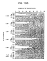
- Figs. 16A , 16B , 16C , 16D show the results of the cell culture for 96 hours in total using the culture sheets having these patterns. That is, the graphs of the results of measuring the distance between the center of the hole and the center of the spheroid and verifying the hole center retention rate of the spheroids are shown.
- Figs. 16A , 16B , 16C , 16D as illustrated, correspond to a square arrangement with a pillar diameter of 2.0 ⁇ m, a triangle arrangement with a pillar diameter of 2.0 ⁇ m, a square arrangement with a pillar diameter of 1.0 ⁇ m, and a triangle arrangement with a pillar diameter of 1.0 ⁇ m, respectively.
- the distance between the centers of the hole center and spheroid is indicated in 3 steps of 0 to 19 ⁇ m, 20 to 39 ⁇ m, 40 ⁇ m or more on the horizontal axis of each graph, and the proportion of the number of spheroids occupying each range in the total number of spheroids is indicated on the vertical axis.
- Figs. 17 and 18 show the results of the culture sheets in which a pillar height, pillar diameter, and a pillar pitch of 1.0 ⁇ m, 2.0 ⁇ m, 4.0 ⁇ m, respectively, and a diameter of the assembly of projection portions of 80 ⁇ m and 20 ⁇ m, respectively, as typical examples of phase-contrast micrographs of the results of culture of the culture sheet of this Example.
- the numbers 170, 180 in the holes 156, 158 in the figures represent spheroids.
- the diameter of the assembly of projection portions is 80 ⁇ m
- the spheroids 170 having almost the same diameter were retained in the central portions of the holes 156.
- the spheroids 180 were not retained in the central portions.
Landscapes
- Health & Medical Sciences (AREA)
- Life Sciences & Earth Sciences (AREA)
- Engineering & Computer Science (AREA)
- Chemical & Material Sciences (AREA)
- Bioinformatics & Cheminformatics (AREA)
- Organic Chemistry (AREA)
- Wood Science & Technology (AREA)
- Zoology (AREA)
- Biomedical Technology (AREA)
- Biotechnology (AREA)
- Immunology (AREA)
- Microbiology (AREA)
- Biochemistry (AREA)
- General Health & Medical Sciences (AREA)
- Genetics & Genomics (AREA)
- General Engineering & Computer Science (AREA)
- Sustainable Development (AREA)
- Cell Biology (AREA)
- Molecular Biology (AREA)
- Urology & Nephrology (AREA)
- Hematology (AREA)
- Food Science & Technology (AREA)
- Tropical Medicine & Parasitology (AREA)
- Toxicology (AREA)
- Medicinal Chemistry (AREA)
- Physics & Mathematics (AREA)
- Analytical Chemistry (AREA)
- General Physics & Mathematics (AREA)
- Pathology (AREA)
- Virology (AREA)
- Apparatus Associated With Microorganisms And Enzymes (AREA)
- Micro-Organisms Or Cultivation Processes Thereof (AREA)
Applications Claiming Priority (1)
| Application Number | Priority Date | Filing Date | Title |
|---|---|---|---|
| PCT/JP2010/073127 WO2012086028A1 (fr) | 2010-12-22 | 2010-12-22 | Substrat de culture et film de culture |
Publications (2)
| Publication Number | Publication Date |
|---|---|
| EP2657331A1 true EP2657331A1 (fr) | 2013-10-30 |
| EP2657331A4 EP2657331A4 (fr) | 2015-03-04 |
Family
ID=46313332
Family Applications (1)
| Application Number | Title | Priority Date | Filing Date |
|---|---|---|---|
| EP10861209.4A Withdrawn EP2657331A4 (fr) | 2010-12-22 | 2010-12-22 | Substrat de culture et film de culture |
Country Status (6)
| Country | Link |
|---|---|
| US (1) | US20130323839A1 (fr) |
| EP (1) | EP2657331A4 (fr) |
| JP (1) | JP5722347B2 (fr) |
| KR (1) | KR101522120B1 (fr) |
| CN (1) | CN103261393A (fr) |
| WO (1) | WO2012086028A1 (fr) |
Cited By (2)
| Publication number | Priority date | Publication date | Assignee | Title |
|---|---|---|---|---|
| EP3406700A4 (fr) * | 2016-01-20 | 2019-01-23 | Soken Chemical & Engineering Co., Ltd. | Substrat de culture cellulaire et son procédé de production |
| EP3766957A1 (fr) * | 2019-07-18 | 2021-01-20 | Axenoll Life Sciences AG | Procédé de génération et / ou d'agencement de cultures cellulaires |
Families Citing this family (14)
| Publication number | Priority date | Publication date | Assignee | Title |
|---|---|---|---|---|
| WO2014100199A1 (fr) * | 2012-12-19 | 2014-06-26 | The Regents Of The University Of California | Ensembles de micropiliers destinés à analyser une myélinisation |
| WO2015118873A1 (fr) * | 2014-02-05 | 2015-08-13 | Toyo Gosei Co., Ltd. | Plaques pour culture d'échantillons biologiques |
| JP5909543B2 (ja) * | 2014-12-26 | 2016-04-26 | 株式会社日立製作所 | 培養器材 |
| CN108368468A (zh) * | 2015-07-08 | 2018-08-03 | 巴黎科学与文学联大-拉丁区 | 细胞培养装置 |
| JP2017046649A (ja) * | 2015-09-02 | 2017-03-09 | 大日本印刷株式会社 | 細胞凍結保存容器 |
| CN108026498A (zh) * | 2015-09-17 | 2018-05-11 | Agc科技玻璃株式会社 | 细胞培养容器 |
| CN108407156B (zh) * | 2017-02-09 | 2020-06-02 | 广东乾晖生物科技有限公司 | 组织工程人工肝样组织构建模具和制造该模具的注塑模具的方法 |
| KR102038068B1 (ko) | 2017-06-28 | 2019-10-29 | 충남대학교산학협력단 | 세포배양용 용기의 제조방법 및 이를 이용한 세포배양 방법 |
| CN108467837B (zh) * | 2018-06-15 | 2023-08-18 | 上海天引生物科技有限公司 | 一种可视多通道流体剪切力细胞培养装置及其方法 |
| JP2020138489A (ja) * | 2019-02-28 | 2020-09-03 | 国立大学法人愛知教育大学 | 構造体 |
| KR102366651B1 (ko) * | 2019-12-19 | 2022-02-28 | 한국생산기술연구원 | 일체형 멀티웰 플레이트 및 이의 제조방법 |
| KR102379490B1 (ko) | 2020-04-24 | 2022-03-28 | 재단법인대구경북과학기술원 | 세포 배양 장치 |
| CN115782190B (zh) * | 2022-10-21 | 2026-02-24 | 广州洁特生物过滤股份有限公司 | 一种温敏型细胞培养装置的制备方法 |
| TWI834527B (zh) * | 2023-02-02 | 2024-03-01 | 新原生細胞製備股份有限公司 | 三維培養裝置及生物培養器具 |
Family Cites Families (8)
| Publication number | Priority date | Publication date | Assignee | Title |
|---|---|---|---|---|
| DK1412725T3 (en) * | 2001-06-29 | 2019-03-25 | Meso Scale Technologies Llc | Multi-well plates for LUMINESCENSE TEST MEASUREMENTS |
| EP1756568A1 (fr) * | 2004-04-05 | 2007-02-28 | Proteome Systems Intellectual Property Pty Ltd. | Appareil de test diagnostique ameliore |
| JP4507686B2 (ja) * | 2004-04-28 | 2010-07-21 | 株式会社日立製作所 | 観察用容器及び培養用容器,培養細胞 |
| JP3981929B2 (ja) * | 2004-10-29 | 2007-09-26 | 財団法人北九州産業学術推進機構 | 細胞組織体マイクロチップ |
| JP4918755B2 (ja) * | 2005-05-30 | 2012-04-18 | 株式会社日立製作所 | 細胞培養容器,細胞培養容器の製造方法、及び培養細胞 |
| US7689269B2 (en) * | 2007-05-10 | 2010-03-30 | Actis, Ltd. | System, method and apparatus for the detection of patient-borne fluorescing nanocrystals |
| CN101815782B (zh) * | 2007-09-12 | 2014-02-05 | 公益财团法人北九州产业学术推进机构 | 细胞培养器具和使用该器具的细胞培养方法 |
| KR101454580B1 (ko) * | 2009-06-23 | 2014-10-28 | 가부시키가이샤 히타치세이사쿠쇼 | 배양 기재 |
-
2010
- 2010-12-22 CN CN2010800708392A patent/CN103261393A/zh active Pending
- 2010-12-22 WO PCT/JP2010/073127 patent/WO2012086028A1/fr not_active Ceased
- 2010-12-22 JP JP2012549527A patent/JP5722347B2/ja not_active Expired - Fee Related
- 2010-12-22 EP EP10861209.4A patent/EP2657331A4/fr not_active Withdrawn
- 2010-12-22 KR KR1020137015979A patent/KR101522120B1/ko not_active Expired - Fee Related
- 2010-12-22 US US13/996,074 patent/US20130323839A1/en not_active Abandoned
Cited By (3)
| Publication number | Priority date | Publication date | Assignee | Title |
|---|---|---|---|---|
| EP3406700A4 (fr) * | 2016-01-20 | 2019-01-23 | Soken Chemical & Engineering Co., Ltd. | Substrat de culture cellulaire et son procédé de production |
| EP3766957A1 (fr) * | 2019-07-18 | 2021-01-20 | Axenoll Life Sciences AG | Procédé de génération et / ou d'agencement de cultures cellulaires |
| WO2021009320A1 (fr) * | 2019-07-18 | 2021-01-21 | Axenoll Life Sciences Ag | Procédé pour générer et/ou agencer des cultures cellulaires |
Also Published As
| Publication number | Publication date |
|---|---|
| EP2657331A4 (fr) | 2015-03-04 |
| US20130323839A1 (en) | 2013-12-05 |
| CN103261393A (zh) | 2013-08-21 |
| WO2012086028A1 (fr) | 2012-06-28 |
| JP5722347B2 (ja) | 2015-05-20 |
| KR101522120B1 (ko) | 2015-05-20 |
| KR20130110200A (ko) | 2013-10-08 |
| JPWO2012086028A1 (ja) | 2014-05-22 |
Similar Documents
| Publication | Publication Date | Title |
|---|---|---|
| EP2657331A1 (fr) | Substrat de culture et film de culture | |
| KR101454580B1 (ko) | 배양 기재 | |
| JP5730472B2 (ja) | 培養基材、及び細胞培養方法 | |
| JP5583312B2 (ja) | 組織体形成用基材、組織体形成キット、それを用いた組織体形成法、及び該組織体形成法により形成された三次元組織体 | |
| US20150313704A1 (en) | Cardiac tissue constructs and methods of fabrication thereof | |
| US20140093953A1 (en) | Non-adherent cell support and manufacturing method | |
| US20130045530A1 (en) | Self-folding sub-centimeter structures | |
| JP6256853B1 (ja) | 3次元細胞構造体の製造方法およびそれに用いる支持体 | |
| KR20140113139A (ko) | 세포 스페로이드 배양판 | |
| KR20210024342A (ko) | 약물의 심장 효능 및 독성 시험을 위한 심근내막 수준 생체모방 심장칩 | |
| KR20150051199A (ko) | 세포 스페로이드 배양판 | |
| JP5730499B2 (ja) | 培養器材 | |
| US20080254535A1 (en) | Cell Separation Apparatus | |
| KR101569619B1 (ko) | 수축 및 팽창 기능을 하는 인체장기를 모사한 실험모델장치 | |
| JP5984998B2 (ja) | 培養基材 | |
| CN119120196B (zh) | 一种微流控装置及其应用 | |
| JP5909543B2 (ja) | 培養器材 | |
| EP4438713A1 (fr) | Dispositif et procédé de génération de réseaux ordonnés de microsupports de cellules au niveau d'un substrat | |
| Lee et al. | Microfabricated cell culture system for the live cell observation of the multilayered proliferation of undifferentiated HT-29 cells | |
| CN116685669A (zh) | 微腔板 | |
| Takeuchi | Cell-laden hydrogel beads, fibers and plates for 3D tissue construction | |
| TW202530392A (zh) | 細胞培養平台 |
Legal Events
| Date | Code | Title | Description |
|---|---|---|---|
| PUAI | Public reference made under article 153(3) epc to a published international application that has entered the european phase |
Free format text: ORIGINAL CODE: 0009012 |
|
| 17P | Request for examination filed |
Effective date: 20130722 |
|
| AK | Designated contracting states |
Kind code of ref document: A1 Designated state(s): AL AT BE BG CH CY CZ DE DK EE ES FI FR GB GR HR HU IE IS IT LI LT LU LV MC MK MT NL NO PL PT RO RS SE SI SK SM TR |
|
| DAX | Request for extension of the european patent (deleted) | ||
| A4 | Supplementary search report drawn up and despatched |
Effective date: 20150129 |
|
| RIC1 | Information provided on ipc code assigned before grant |
Ipc: C12M 3/00 20060101AFI20150123BHEP |
|
| STAA | Information on the status of an ep patent application or granted ep patent |
Free format text: STATUS: EXAMINATION IS IN PROGRESS |
|
| 17Q | First examination report despatched |
Effective date: 20170508 |
|
| STAA | Information on the status of an ep patent application or granted ep patent |
Free format text: STATUS: THE APPLICATION IS DEEMED TO BE WITHDRAWN |
|
| 18D | Application deemed to be withdrawn |
Effective date: 20170919 |