EP2660746A1 - Système de prédicateur de modèle de résultats médicaux dose-réponse - Google Patents
Système de prédicateur de modèle de résultats médicaux dose-réponse Download PDFInfo
- Publication number
- EP2660746A1 EP2660746A1 EP20120166187 EP12166187A EP2660746A1 EP 2660746 A1 EP2660746 A1 EP 2660746A1 EP 20120166187 EP20120166187 EP 20120166187 EP 12166187 A EP12166187 A EP 12166187A EP 2660746 A1 EP2660746 A1 EP 2660746A1
- Authority
- EP
- European Patent Office
- Prior art keywords
- bcl
- protein
- bak
- bax
- cancer
- Prior art date
- Legal status (The legal status is an assumption and is not a legal conclusion. Google has not performed a legal analysis and makes no representation as to the accuracy of the status listed.)
- Withdrawn
Links
Images





Classifications
-
- G—PHYSICS
- G16—INFORMATION AND COMMUNICATION TECHNOLOGY [ICT] SPECIALLY ADAPTED FOR SPECIFIC APPLICATION FIELDS
- G16B—BIOINFORMATICS, i.e. INFORMATION AND COMMUNICATION TECHNOLOGY [ICT] SPECIALLY ADAPTED FOR GENETIC OR PROTEIN-RELATED DATA PROCESSING IN COMPUTATIONAL MOLECULAR BIOLOGY
- G16B5/00—ICT specially adapted for modelling or simulations in systems biology, e.g. gene-regulatory networks, protein interaction networks or metabolic networks
-
- G—PHYSICS
- G01—MEASURING; TESTING
- G01N—INVESTIGATING OR ANALYSING MATERIALS BY DETERMINING THEIR CHEMICAL OR PHYSICAL PROPERTIES
- G01N33/00—Investigating or analysing materials by specific methods not covered by groups G01N1/00 - G01N31/00
- G01N33/48—Biological material, e.g. blood, urine; Haemocytometers
- G01N33/50—Chemical analysis of biological material, e.g. blood, urine; Testing involving biospecific ligand binding methods; Immunological testing
- G01N33/53—Immunoassay; Biospecific binding assay; Materials therefor
- G01N33/575—Immunoassay; Biospecific binding assay; Materials therefor for cancer
- G01N33/5758—Immunoassay; Biospecific binding assay; Materials therefor for cancer involving compounds serving as markers for tumours, cancers or neoplasias, e.g. cellular determinants, receptors, heat shock/stress proteins, A-protein, oligosaccharides or metabolites
- G01N33/57595—Immunoassay; Biospecific binding assay; Materials therefor for cancer involving compounds serving as markers for tumours, cancers or neoplasias, e.g. cellular determinants, receptors, heat shock/stress proteins, A-protein, oligosaccharides or metabolites involving intracellular compounds
-
- G—PHYSICS
- G16—INFORMATION AND COMMUNICATION TECHNOLOGY [ICT] SPECIALLY ADAPTED FOR SPECIFIC APPLICATION FIELDS
- G16B—BIOINFORMATICS, i.e. INFORMATION AND COMMUNICATION TECHNOLOGY [ICT] SPECIALLY ADAPTED FOR GENETIC OR PROTEIN-RELATED DATA PROCESSING IN COMPUTATIONAL MOLECULAR BIOLOGY
- G16B20/00—ICT specially adapted for functional genomics or proteomics, e.g. genotype-phenotype associations
-
- G—PHYSICS
- G01—MEASURING; TESTING
- G01N—INVESTIGATING OR ANALYSING MATERIALS BY DETERMINING THEIR CHEMICAL OR PHYSICAL PROPERTIES
- G01N2333/00—Assays involving biological materials from specific organisms or of a specific nature
- G01N2333/82—Translation products from oncogenes
-
- G—PHYSICS
- G16—INFORMATION AND COMMUNICATION TECHNOLOGY [ICT] SPECIALLY ADAPTED FOR SPECIFIC APPLICATION FIELDS
- G16C—COMPUTATIONAL CHEMISTRY; CHEMOINFORMATICS; COMPUTATIONAL MATERIALS SCIENCE
- G16C20/00—Chemoinformatics, i.e. ICT specially adapted for the handling of physicochemical or structural data of chemical particles, elements, compounds or mixtures
- G16C20/30—Prediction of properties of chemical compounds, compositions or mixtures
Definitions
- the invention relates to a computer-implemented system and method for predicting a patient's response to a treatment.
- the invention relates to a quantitative method and system for predicting a patient's response to cancer treatment and calculates effective treatment dosages.
- Cancer treatments are ineffective with poor overall 5 year survival rates of just 55% for all cancers.
- Treatment regimes that do not improve the patient's health or mitigate patient burden pose significant and unnecessary costs to the health care sector that could have been directed to more efficient therapies.
- the decision for the treatment that has the best success rate in individual patients and minimizes patient risk and burden is therefore decisive. To decide on the best individually suitable treatment, reliable clinical tools are needed.
- Chemotherapeutic treatment shall kill cancer cells while preserving healthy tissue. Finding the right treatment dose is therefore decisive.
- the dose where cancer cells are killed and healthy tissue is preserved influences the therapeutic window.
- the size of this therapeutic window depends on the cancer type, cancer stage and patient individual characteristics.
- Mathematical modelling allows assembling existing knowledge into a holistic model. This facilitates the establishment and in-silico tests of new hypotheses before model predictions are eventually validated in wet lab experiments. Using tools from control theory, they may supplement quantitative data that otherwise cannot be determined experimentally. State of the art models put emphasis on pre-MOMP or post-MOMP processes or try to establish a holistic model of extrinsic or intrinsic stress or apoptosis. As a recent example, Zhang and co-workers ( T. Zhang, P. Brazhnik, and J. J. Tyson, 'Computational analysis of dynamical responses to the intrinsic pathway of programmed cell death', Biophys J, vol. 97, p.
- a computer-implemented method for predicting quantitatively whether an adjuvant or neoadjuvant chemotherapeutic treatment will be or is being successful in treating an individual suffering from cancer comprising the steps of:
- the method comprises the further step of outputting a quantitative, dose-dependent prediction value of the likelihood of treatment success using novel apoptosis sensitizers.
- the method further comprises the step of estimating a minimal quantitative dose value of a chemotherapeutic required to induce the mitochondrial outer membrane permeabilisation apoptosis pathway in the sample, such that any cancer cell is killed while preserving healthy cells of the individual suffering from cancer.
- the quantitative minimal dose is estimated based on the concentration values of the BCL-2 family members required to induce the process of mitochondrial outer membrane permeabilisation in the cancer cell.
- the invention has established a comprehensive model that is applicable to individual stress situations and protein expression levels. This has allowed investigations of how the combination of BH-3 only family members and their mimetics are apoptotically processed using the well-characterized specificity between certain pro-apoptotic BCL2 members and their anti-apoptotic counterparts.
- VDAC2 voltage dependent ion channels
- the concentration values inputted into the computational model are processed to provide an apoptosis status, which is correlated with a known response to provide said quantitative, dose-dependent prediction value to predict the individual's response to treatment and/or the minimal dose necessary to kill cancer cells.
- said processing step calculates an activation profile over time of pro-apoptotic effector BCL-2 family member proteins and compares the result with known results to quantitatively predict the individual's response to treatment and/or the minimal dose necessary to kill cancer cells.
- the BCL-2 family members are selected from BAK, BAX, BCL-2, BCL-(X)L, BCL-(X)S, BCL-W, MCL-1, BIM, BID, BAD, PUMA and NOXA.
- the pro-apoptotic effector BCL-2 proteins are BAK and BAX.
- the concentration value for each of the at least two BCL-2 family member is representative of protein levels for the sample.
- Absolute protein levels can be obtained from biopsies, resected tumor material, or formalin-fixed, paraffin-embedded histopathology material using reverse phase protein arrays, quantitative Western Blotting, tissue microarray immunostaining or immunohistochemistry. This protein data will feed into the computational model that analyses protein-protein interaction.
- the biological sample is a blood sample, especially blood serum or plasma.
- other biological samples may also be employed, for example, cerebrospinal fluid, saliva, urine, lymphatic fluid, or cell or tissue extracts or biopsy or tissue biopsy.
- the individual is a human, although the computer-implemented method of the invention is applicable to other higher mammals.
- cancer should be understood to mean a cancer that is treated by chemotherapeutic regimens.
- An example of such a cancer include multiple myeloma, prostate cancer, glioblastoma, lymphoma, fibrosarcoma; myxosarcoma; liposarcoma; chondrosarcom; osteogenic sarcoma; chordoma; angiosarcoma; endotheliosarcoma; lymphangiosarcoma; lymphangioendotheliosarcoma; synovioma; mesothelioma; Ewing's tumor; leiomyosarcoma; rhabdomyosarcoma; colon carcinoma; pancreatic cancer; breast cancer; ovarian cancer; squamous cell carcinoma; basal cell carcinoma; adenocarcinoma; sweat gland carcinoma; sebaceous gland carcinoma; papillary carcinoma; papillary adenocarcinomas; cystadenocarcinom
- a computer-implemented system for quantitatively predicting whether an adjuvant or neoadjuvant chemotherapeutic treatment will be or is being successful in treating an individual suffering from cancer comprising:
- said processing step calculates an activation profile over time of pro-apoptotic effector BCL-2 family member proteins and compares the result with known results to predict the individual's response to treatment and/or the minimal dose necessary to kill cancer cells.
- the BCL-2 family members are selected from BAK, BAX, BCL-2, BCL-(X)L, BCL-(X)S, BCL-W, A1, MCL-1, BIM, BID, BAD, PUMA and NOXA.
- positive outcome should be understood to mean a patient who responds positively to chemotherapeutic treatment
- negative outcome should be understood to mean a patient who does not respond to chemotherapeutic treatment
- treatment should be understood to mean standard chemotherapeutic treatment, novel co-treatment regimes and/or novel adjuvant treatments (including non-standard/experimental treatments).
- Chemotherapeutic treatments are those using compounds such as for example chemotherapeutic agents fluorouracil (5FU), oxaliplatin, irinotecan, etoposide, cisplatin, doxorubicin, and receptor kinase inhibitors such as cetuximab and sorafenib.
- stage 2 non metastatic colorectal cancer
- FOLFOX 5FU/oxaliplatin and leucovorin
- FOLFIRI 5FU/ironotecan
- These drugs exert their beneficial activities by inducing DNA damage ('genotoxic drugs').
- FOLFOX or FOLFIRI chemotherapeutic treatment is applied in the presence or absence of the angiogenesis inhibitor bevacizumab (Avastin) or the HER2 inhibitor Cetuximab (kinase inhibition).
- Novel adjuvant, non-standard and experimental treatments include BH3-only mimetics Abbot's Navitoclax and ABT-737, Gemin X's Obatoclax, Ascenta's AT-101, and Gossypol.
- pro-apoptotic biomarker should be understood to mean a molecule involved in promoting and/or progressing the process of programmed cell death (biochemical events leading to characteristic cell changes including blebbing, cell shrinkage, nuclear fragmentation, chromatin condensation, and chromosomal DNA fragmentation) and cell death by modifying the so-called effector proteins (Bak and Bax) from an inactive to an active form.
- Pro-apoptotic biomarkers can be selected from the group comprising BIM, BID, BAD, PUMA, NOXA, and BCL-(X)S.
- BCL2 anti-apoptotic proteins should be understood to mean a molecule involved in preventing and/or stopping the process of programmed cell death selected from the group comprising BCL-2, BCL-(X)L, BCL-W, A1 and MCL-1.
- a computer program comprising program instructions for causing a computer program to carry out the above method which may be embodied on a record medium, carrier signal or read-only memory.
- the initial concentrations of the BCL2-family proteins BAK, BAX, BCL-2, BCL-(X)L, MCL-1 were obtained by quantitative Western Blotting in HeLa cells, the colorectal cancer cell lines (COLO-205, DLD-1, HCT-116 (wild type), HCT-116 puma - / - , HCT-116 p53 - / - , HT-29 and LoVo), Glioblastoma Multiforme (GBM) cell lines (A172, MZ18, MZ51, MZ256, MZ256, MZ294, MZ304, MZ327, U87, U251, U343 and U373) and all colon cancer patients as described below.
- GBM Glioblastoma Multiforme
- the anti-apoptotic proteins BCL-2, BCL-(X)L, and MCL-1 were modelled to underlie a permanent turnover.
- cell specific production rates were chosen and together with assumed degradation constants for each protein (Table 2) resulted in equilibrium concentrations of these proteins in the absence of stress.
- MCL-1 degradation was modelled to drop from 45 minutes to 17 minutes simultaneously with the induction of BIM, PUMA and NOXA production.
- the model output was the concentration of the BAX and BAK pores. These pores were assumed to be homo-oligomers in complexes equal or larger than hexamers. MOMP was assumed to occur once 10% of total BAK and BAX were homo-oligomerised in pores.
- the signalling pathway of the BCL2 family was modelled by creating a set of ordinary differential equation (ODEs) that were based on the law of mass action.
- ODEs ordinary differential equation
- Each ODE described the changes in concentration of a single protein or a single protein complex at a time point t.
- the protein concentrations of modelled proteins at time point t as well as the kinetic constants were used as arguments in each ODE.
- the ODE of a protein or protein complex was composed from sub-ODEs which were translated from protein reactions (Table 1) in which the particular protein (or the complex) was involved as reagent or product.
- Table 1 protein reactions
- For the translation of the protein reaction to a mathematical equation a distinction is made at the type of protein reactions between protein degradations, protein turnovers, irreversible reactions as well as reversible reactions.
- the degradation rate of a protein or protein complex which was degraded was calculated by multiplying the protein concentration with the degradation constant k deg (Table 1A). The resulting degradation rate was subtracted from the protein concentration of the degraded protein.
- To determine the protein concentration change caused by a protein turnover we added the production rate k prod to the degradation rate of the protein that underlay the turnover (Table 1B).
- the equilibrium concentration of the protein which underlay a protein turnover was calculated by dividing its production rate k prod by its degradation constant k deg (Table 1B).
- the change in concentration due to an irreversible reaction was modelled by multiplying the reagent protein concentration by the reaction constant k.
- the concentration of the involved reagent is reduced by the protein change, whereas the resulting product was increased by the protein change (Table 1C).
- the product of all reagent concentration was multiplied with the reaction constant k (Table 1D).
- Reversible reactions were modelled by considering a forward and backward reaction.
- the protein changed caused by the protein forward reaction was modelled to be the product of involved reagents multiplied with the forward constant k forward . This change was added to the concentration of the product and subtracted from the concentrations of each reagent.
- the change caused by the backward reaction was modelled to be the concentration of the product protein complex (or protein) multiplied by the backward constant k backward .
- the resulting protein concentration change was added to each reagent concentration and subtracted from the product concentration (Table 1E).
- VDAC2, BAK and BAX monomers, BAK and BAX hetero-dimers, and BAK and BAX homo-oligomers were assumed to be stable and not to get degraded. All other proteins were subjected to degradation (Table 2).
- the anti-apoptotic proteins BCL-2, BCL-(X)L, BCL-W and MCL-1 were the only protein that underlay a constant protein production.
- the production rates are set to values that hold the cell specific initial concentrations ( Figure 6A ) of the anti-apoptotic BCL2 proteins in the absence of stress.
- the cell, as well as BCL2 protein specific production rate, is determined by dividing the cell specific initial concentration by the anti-apoptotic BCL2 protein specific degradation constant k deg .
- the core of the model was the specific binding of anti-apoptotic proteins BCL-2, BCL-(X)L and MCL-1 to the pro-apoptotic proteins BAK, BAX, BIM, tBID, PUMA and NOXA (see Table 3).
- the binding affinities are described by the dissociation constant K D .
- a low K D value indicates high protein affinity whereas a high K D value indicates low protein affinity.
- the dissociation constants were frequently used in literature whereas we did not obtain all required forward and backward kinetic constant.
- Chen et. al. L. Chen, S. N. Willis, A. Wei, B. J. Smith, J. I. Fletcher, M. G. Hinds, P. M.
- the backward constant of the reversible binding of the anti-apoptotic BCL-2, BCL-(X)L, BCL-W and MCL-1 to BAK, BAX, PUMA, tBID and NOXA is assumed in this model to be identical to the backward constant of binding of BCL-2, BCL-(X)L, BCL-W and MCL-1 to BIM, in respect of BCL-2, BCL-(X)L, BCL-W and MCL-1.
- the associated forward constants k forward were determined by dividing the assumed backward constants by the obtained dissociation constants.
- Inactive BAX c was modelled to be initially present only in the cytosol and to translocate into the mitochondrial outer membrane (MOM) after its activation (Table 4A-C). It is assumed that no interaction of inactive BAX c with anti-apoptotic BCL2 family proteins. BH3-only proteins BIM, PUMA and tBID were able to bind to inactive BAX c .
- a dissociation constant K D of 100 nM, a backward constant of 2.57E-04 s -1 (which correspond with a half-life time of 45 minutes) and a forward constant of 2.57E-06 nM -1 s -1 are assumed.
- the constant of the enzymatic activation of inactive BAX c in the hetero-dimer was assumed to be 1.16E-01 s -1 with a subsequent instant dissociation from the dimer.
- the dissociated BAX c * is modelled to translocate to the MOM with a constant of 1.16E-01 s -1 (which is equivalent to a half-life time of six seconds).
- BAK and BAX are similar in form and function, the activation of BAK is modelled in the direct activation model with the same reactions and the same kinetic constants (see Table 4A-B). Based on the fact that BAK is exclusively located to the MOM, BAK translocation from the cytosol to the membrane was not modelled. In contrast to BAX, BAK is modelled to interact with the Voltage-dependent anion-selective channel protein 2 (VDAC2). In each calculation, VDAC2 was assumed to be expressed with the same concentration as BAK in the cell. In the direct activation model, only inactive BAK is assumed to bind to VDAC2 with a dissociation constant of 1,000 nM.
- VDAC2 Voltage-dependent anion-selective channel protein 2
- the forward constant k forward was set to 2.31E-06 nM -1 s -1 (see Table 4D).
- the BAK activation step was removed and only constitutive active BAK * was assumed.
- VDAC2 binds to active BAK * with the same constants as in the direct activation model. It was assumed that the cytosolic BAX activation step was still required in the indirect activation model.
- BAK * and BAX * were modelled to homo-oligomerise to dimers.
- the dimers further homo-oligomerised to larger homo-oligomers (see Table 5).
- K D 15 nM
- k forward 1.28E-05 nM -1 s -1
- BAK and BAX homo-oligomerisation was modelled up to twelve proteins in one homo-oligomer.
- hexamers or larger homo-oligomers were assumed to be pores that cause the mitochondrial outer membrane permeabilisation (MOMP) once 10% of total effectors homo-oligomerised to pores.
- MOMP mitochondrial outer membrane permeabilisation
- Proteins of the BCL2 protein family were assumed to be mainly located and to interact at the MOM. As the only exception, BAX c was assumed to have a cytosolic fraction which was modelled to be inactive and only able to interact with the BH3-only proteins, but not with anti-apoptotic proteins.
- MATLAB 7.3 (MathWorks, USA, R2007b, 7.5.0.342) and its function ode15s was used.
- tBID BAK and tBID BAX Two recently developed tBID chimeras, tBID BAK and tBID BAX whose tBID BH3 domains were replaced with that of BAK or BAX were modelled.
- the specific binding characteristic of these chimeras allowed studying how easy anti-apoptotic proteins that either bind to activator or effector (BAK and BAX) can be de-repressed from their binding partners.
- the binding affinities (forward and backward constants) of tBID chimeras to the anti-apoptotic protein were assumed to be the same as for BAK or BAX.
- the same kinetics as for wild type tBID activating of the effectors were assumed.
- a half-life time of 75 minutes was assumed, identical to the one considered for tBID.
- BAK and BAX expressing cells two sets of calculations each were performed. In each set, the concentration of a single anti-apoptotic protein, BCL-2 or MCL-1, was set to 0; 10; 25; 50; 75; 100; 250; 500; 750; 1,000 and 2,000 nM and performed a calculation to determine the maximum amount of BAK or BAX pores within 48 hours. In the BAK expressing cells, a protein dose of 1 ⁇ M tBID BAX chimeras was applied in each calculation, and in the BAX expressing cells a protein dose of 1 ⁇ M tBID BAK chimeras was applied in each calculation.
- Mode 1 activator inhibition
- Mode 2 effector inhibition
- F. Llambi, T. Moldoveanu, S. W. G. Tait, L. Bouchier-Hayes, J. Temirov, L. L. McCormick, C. P. Dillon, and D. R. Green 'A unified model of mammalian BCL-2 protein family interactions at the mitochondria', Mol. Cell, vol. 44, no. 4, pp. 517-531, Nov. 2011 ).
- Results for both calculations were plotted as simulated Western Blot.
- the area of the bar was proportional to the amount of pores and normalised to 500 nM in the first calculations (see Figure 3B ) and 1 ,500 nM in the second calculations (see Figure 3C ).
- Colorectal cancer patient tissue was collected and stored from the Departments of Surgery, Gastroenterology and Pathology, Beaumont Hospital, Dublin, Ireland. Matched adjacent normal colorectal tissue was available for all of these patients. Clinical follow-up was obtained through a review of medical records by a dedicated clinical research nurse. For classification purposes, patients without disease recurrence and/or cancer mortality within 4 years were classified as a good outcome; patients who recurred/died from colorectal cancer were classified as bad outcome. Patients with hereditary forms of colorectal cancer were excluded. Ethical approval has been obtained for this study by the Beaumont Hospital Ethics (Medical Research) Committee and informed consent was obtained from all patients. Tissue was stored for use as snap frozen (-80°C) or in RNAlater (Ambion, Abington, UK) (-20°C).
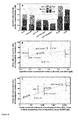
- a correlation analysis was performed to determine the relationship between model prediction and amount of cell survival in a cell population of a CRC or GBM cell line subjected to stress (5FU/Oxaliplatin, survival obtained by FACS).
- the minimal protein dose that the model predicted that is required to induce MOMP in a prototypical cell of above cell line was correlated with the amount of cell survival subsequent to drug administration.
- the amount of total concentration of all anti-apoptotic proteins was correlated to the same amount of cell survival.
- the grade of linear dependency of two variables was assessed by the Pearson's correlation coefficient.
- the MATLAB 7.3 MathWorks, USA, R2007b, 7.5.0.342
- the Wilcoxon rank-sum test was applied using the MATLAB routine ranksum. For comparing whether the same model predictor indicates that the protein dose required to induce apoptosis in tumour tissue and in matched normal tissue are independent from each other, the Wilcoxon signed rank-sum test for paired data sets was applied and the MATLAB routine signrank was used therefore.
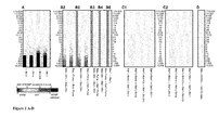
- a computational model was constructed that allows a study of temporal BCL2 protein profiles and an investigation of how pro- and anti-apoptotic BCL2 interact to induce MOMP in response to genotoxic stress ( Figure 1 ) in individual cell lines.
- Genotoxic stress was assumed to induce the BH3-only proteins BIM, PUMA and NOXA ( Figure 1 . Red boxes), leading to an interaction network of pro- and anti-apoptotic proteins, resulting of BAK and BAX homo-oligomerisation (blue boxes) and finally MOMP (grey box, denoted as output).
- Oligomers larger or equal than hexamers were defined as pores which caused MOMP once 10% of BAK or BAX were bound in oligomers.
- BAX is assumed to translocate to the mitochondria upon activation, while BAK is assumed to be constitutively present at the mitochondrial membrane where inactive BAK (or constitutively active BAK in the indirect activation model) was modelled to bind additionally to the inhibitor VDAC2 (Table 4).
- active BAK and BAX may be bound by anti-apoptotic proteins which prevent BAK and BAX oligomerisation.
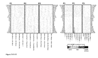
- the anti-apoptotic proteins BCL-2, BCL-(X)L and MCL-1 may bind BIM, PUMA, and NOXA in a reversible manner and we therefore investigated how changes of the forward and backward constant of this binding influenced the time point of MOMP.
- changing BIM and PUMA inhibition by their anti-apoptotic binding partners did also not markedly change time of MOMP occurrence suggesting the reference binding affinity as low ( Figure 2E1-E3 ).
- Anti-apoptotic BCL2 proteins are promiscuous inhibitors of apoptosis as they bind to apoptotic effectors as well as to BH3-only activators and sensitizers.
- chimera proteins of the activator tBID that mimicked the BH3-domain of either BAK and BAX (tBID BAK and tBID BAX )
- Llambi et. al. F. Llambi, T. Moldoveanu, S. W. G. Tait, L. Bouchier-Hayes, J. Temirov, L. L. McCormick, C. P. Dillon, and D. R.
- tBID BAK and tBID BAX allowed selectively studying activator and effector repression by using specific anti-apoptotic proteins.
- tBID chimera only inhibited the respective activator and not the effector (denoted as (I)) was studied. This was done by modelling BAX -/- cells (expressing BAK) to be exposed only to tBID BAK and MCL-1 and by modelling BAK -/- cells (expressing BAX) be exposed only to tBID BAX and BCL-2.
- effector repression was investigated by assuming either BAX deficient cells to be exposed to tBID BAK and MCL-1 or BAK deficient cells to be exposed to tBID BAX and BCL-2.
- BCL-X(L) and BCL-W were disregarded and set to zero.
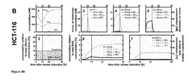
- the direct activation model predicts HeLa cells as susceptible and HCT-116 cells as resistant to MOMP under milder genotoxic stress
- BCL-2 levels depleted from 240 nM to 80 nM, BCL-X(L) and from 110 nM to 10 nM.
- MCL-1 levels from 80 nM to 30 nM at time point of 12 hr ( Figure 4A1 dotted, dashed-dotted and dashed line, respectively).
- the indirect model does not assume an activation step for BAK.
- the amount of pro-apoptotic BCL2 family members can be severely higher than the amount of anti-apoptotic proteins ( Figure 6A ).
- Figure 5AB dashed line for BAK pores. This result suggests that repression of BAK by binding to anti-apoptotic proteins was not sufficient to prevent MOMP in the absence of stress ( Figure 5CD for hetero-dimers between anti-apoptotic proteins, and BAK, Figure 5EF for free anti-apoptotic proteins).
- the backward reaction rate was increased in order to change the dissociation constant of BAK homo-oligomers (ratio between backward and forward constant) from 15 nM ( Figures 5G,H,I , black solid line) to values of 30 nM (long dashed line, Figures 4B1-4B3 ), 60 nM (long/short-dashed), 120 nM (short-dashed), 240 nM (grained dotted) and 480 nM (fine dotted line). Indeed, it was found that BAK pore formation ( Figure 4B ) in HeLa cells was substantially reduced in absence of stress and no MOMP was observed when the dissociation constant was decreased to values below 120 nM.
- HCT-116 and LoVo cells were predicted to undergo MOMP in the absence of stress for dissociation constants lower or equal than 120 nM ( Figure 5H,I ).
- Figure 5H,I in order to predict stability for LoVo cells in the absence of stress, an even higher backward reaction rate, and therefore an even weaker binding of BAK and BAX homo-oligomers of about 480 nM had to be assumed.
- CRC colorectal cancer
- extracts were compared to purified proteins, and absolute protein levels were determined by densitometry.
- Levels of above proteins for Colo-205, DLD-1, HCT-116, HCT-116 puma - / - , HCT-116 p53 - / - , HT-29 and LoVo CRC cells were obtained relative to HeLa cells by co-plotting the respective cell extracts.
- HeLa cells were determined to express 1 ⁇ M BAK, 519 nM BAX, 239 nM BCL-2, 110 nM BCL-(X)L and 83 nM MCL-1. Concentrations for the colorectal cancer cell lines are depicted in Figure 6A .
- HCT-116 wild type and HCT-116 p53 - / - and HCT-116 puma -l- mutant cell lines showed similar total levels of pro- and anti-apoptotic proteins.
- the inequality of pro- and anti-apoptotic proteins suggests that the mere inhibition of BAK and BAX by anti-apoptotic BCL2 proteins may not be sufficient to prevent MOMP in the absence of stress (unless BAK and BAX homo-oligomerisation is instable as modelled by a high dissociation constant in the previous section).
- Protein expression of the BCL2 family members may determine whether or not apoptosis is executed upon stress such as upon genotoxic stress following chemotherapeuic treatment.
- Colo-205, HCT-116, HCT-116 p53 - / - , HT-29 and LoVo cells were subjected to 30 ⁇ g/ml 5FU and 10 ⁇ g/ml Oxaliplatin, a drug combination being the current standard of care in treatment of colorectal cancer.
- FACS fluorescence-activated cell sorting
- cell death in the p53-mutated cell lines was low (40%, 23% and 20% for Colo-205, HCT-116 p53 -l- , and HT-29 cells, respectively) while a larger amount of cell death of 70% was observed in the p53-competent cell line HCT-116.
- Amount of cell death in the p53-competent cell line LoVo was 42%.
- a higher amount of apoptotic stress due to 5FU/Oxaliplatin would induce a higher expression of BIM, PUMA and NOXA in p53 and PUMA competent cells, a higher expression of BIM and NOXA in HCT-116 puma - / - cells (no PUMA expression assumed) and a higher expression of BIM in p53-mutated or deficient cells (no PUMA and NOXA expression assumed). It was therefore assumed that the minimal amount of protein dose in response to genotoxic stress that is required to induce MOMP in a typical cell of the population may be a good predictor for the apoptosis susceptibility of a cell population.
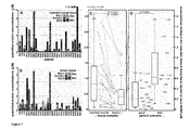
- Model predicts patient tumour cells more susceptible to chemotherapeutically induced cell death than cells from matched normal tissue
- a therapeutic window is defined by the higher susceptibility of tumour cells to therapeutic stimuli compared to other cells of the same tissue (matched normal tissue).
- chemotherapeutic drugs together with synthetic mimetics of BH3-only proteins, Certo et. al. ( M. Certo, V. Del Gaizo Moore, M. Nishino, G. Wei, S. Korsmeyer, S. A. Armstrong, and A. Letai, 'Mitochondria primed by death signals determine cellular addiction to antiapoptotic BCL-2 family members', Cancer Cell, vol. 9, no. 5, pp. 351-365, May 2006 ) have recently shown that cancer cells may be indeed more susceptible to undergo drug induced MOMP than normal tissue.
- the model of the present invention was used to test their findings in a clinical setting.
- BIM/PUMA/NOXA median value of 410 nM
- p 0.02, Wilcoxon-signed rank test
- Figure 7C a lower dosage of 5FU/oxaliplatin-based chemotherapy is required to induce tumour cell death in these CRC patients.
- This lower dose to induce cancer cell death was found in the majority (21 out of 27) of patients as illustrated by the lines between data points that indicate correspondence between healthy matched normal and tumour tissue of an individual patient ( Figure 7C ).
- the required protein dose to induce cell death in healthy tissue showed a much higher variability than the dose required to induce cancer cell death (indicated by the whiskers of both box-plots), suggesting that
- BH3-mimetics are drugs that sensitise cancer cells to apoptosis by mimicking the BH3-domain of BH3-only proteins and are currently in clinical trial for adjuvant chemotherapy.
- the synthetic drug ABT-737 has been shown to induce apoptosis by binding with strong affinity to BCL-2 and BCL-(X)L leading to a de-repression of BH3-only activators.
- the lipid Gossypol that naturally occurs in cotton seed sensitises cell to apoptosis by its binding to the anti-apoptotic proteins BCL-2 and MCL-1.
- the computational model of the present invention allows assessing for optimal drugs that sensitise cells to apoptosis by mimicking pro-apoptotic BCL2 proteins.
- the computational model of the present invention can be applied for clinical decision making.
- the computational model may aide in the decision whether or not compounds with certain molecular interaction characteristics may go into clinical trial.
- the computational model of the present invention allows a patient specific investigation of the treatment dose and may point to the most suitable co-treatments that are applied in support of classical 5FU/Oxaliplatin based chemotherapy.
Landscapes
- Health & Medical Sciences (AREA)
- Life Sciences & Earth Sciences (AREA)
- Engineering & Computer Science (AREA)
- Physics & Mathematics (AREA)
- Bioinformatics & Cheminformatics (AREA)
- Molecular Biology (AREA)
- General Health & Medical Sciences (AREA)
- Biotechnology (AREA)
- Bioinformatics & Computational Biology (AREA)
- Biophysics (AREA)
- Medical Informatics (AREA)
- Chemical & Material Sciences (AREA)
- Evolutionary Biology (AREA)
- Spectroscopy & Molecular Physics (AREA)
- Theoretical Computer Science (AREA)
- Immunology (AREA)
- Analytical Chemistry (AREA)
- Proteomics, Peptides & Aminoacids (AREA)
- Genetics & Genomics (AREA)
- Urology & Nephrology (AREA)
- Hematology (AREA)
- Physiology (AREA)
- Biomedical Technology (AREA)
- Cell Biology (AREA)
- Microbiology (AREA)
- Pathology (AREA)
- General Physics & Mathematics (AREA)
- Biochemistry (AREA)
- Medicinal Chemistry (AREA)
- Food Science & Technology (AREA)
- Medicines That Contain Protein Lipid Enzymes And Other Medicines (AREA)
- Investigating Or Analysing Biological Materials (AREA)
Priority Applications (4)
| Application Number | Priority Date | Filing Date | Title |
|---|---|---|---|
| EP20120166187 EP2660746A1 (fr) | 2012-04-30 | 2012-04-30 | Système de prédicateur de modèle de résultats médicaux dose-réponse |
| US14/397,697 US20150119272A1 (en) | 2012-04-30 | 2013-04-30 | Dose-response medical outcome model predictor system and method |
| EP13728324.8A EP2845136A2 (fr) | 2012-04-30 | 2013-04-30 | Système et procédé prédicteur de modèle de résultat médical d'une dose-réponse |
| PCT/EP2013/059051 WO2013164365A2 (fr) | 2012-04-30 | 2013-04-30 | Système et procédé prédicteur de modèle de résultat médical d'une dose-réponse |
Applications Claiming Priority (1)
| Application Number | Priority Date | Filing Date | Title |
|---|---|---|---|
| EP20120166187 EP2660746A1 (fr) | 2012-04-30 | 2012-04-30 | Système de prédicateur de modèle de résultats médicaux dose-réponse |
Publications (1)
| Publication Number | Publication Date |
|---|---|
| EP2660746A1 true EP2660746A1 (fr) | 2013-11-06 |
Family
ID=46201387
Family Applications (2)
| Application Number | Title | Priority Date | Filing Date |
|---|---|---|---|
| EP20120166187 Withdrawn EP2660746A1 (fr) | 2012-04-30 | 2012-04-30 | Système de prédicateur de modèle de résultats médicaux dose-réponse |
| EP13728324.8A Withdrawn EP2845136A2 (fr) | 2012-04-30 | 2013-04-30 | Système et procédé prédicteur de modèle de résultat médical d'une dose-réponse |
Family Applications After (1)
| Application Number | Title | Priority Date | Filing Date |
|---|---|---|---|
| EP13728324.8A Withdrawn EP2845136A2 (fr) | 2012-04-30 | 2013-04-30 | Système et procédé prédicteur de modèle de résultat médical d'une dose-réponse |
Country Status (3)
| Country | Link |
|---|---|
| US (1) | US20150119272A1 (fr) |
| EP (2) | EP2660746A1 (fr) |
| WO (1) | WO2013164365A2 (fr) |
Cited By (2)
| Publication number | Priority date | Publication date | Assignee | Title |
|---|---|---|---|---|
| CN108694191A (zh) * | 2017-04-08 | 2018-10-23 | 大连万达集团股份有限公司 | 在bim模型中快速检索构件信息的方法 |
| EP3274467A4 (fr) * | 2015-03-24 | 2018-10-31 | Eutropics Pharmaceuticals, Inc. | Biomarqueur fonctionnel de substitution pour un cancer à tumeur solide |
Families Citing this family (3)
| Publication number | Priority date | Publication date | Assignee | Title |
|---|---|---|---|---|
| KR102062416B1 (ko) * | 2012-05-10 | 2020-01-03 | 유트로픽스 파마슈티컬스, 인크. | 실용 가능한 암 진단 대체 법 |
| WO2017007961A1 (fr) | 2015-07-08 | 2017-01-12 | Accelerated Medical Diagnostics, Inc. | Procédés, systèmes et nécessaires pour tests prédictifs basés sur une chimiothérapie cytotoxique |
| WO2018132766A1 (fr) * | 2017-01-12 | 2018-07-19 | The Regents Of The University Of California | Dosages prédictifs basés sur une chimiothérapie cytotoxique pour la leucémie myéloïde aiguë |
-
2012
- 2012-04-30 EP EP20120166187 patent/EP2660746A1/fr not_active Withdrawn
-
2013
- 2013-04-30 EP EP13728324.8A patent/EP2845136A2/fr not_active Withdrawn
- 2013-04-30 WO PCT/EP2013/059051 patent/WO2013164365A2/fr not_active Ceased
- 2013-04-30 US US14/397,697 patent/US20150119272A1/en not_active Abandoned
Non-Patent Citations (18)
| Title |
|---|
| A. LETAI; M. C. BASSIK; L. D. WALENSKY; M. D. SORCINELLI; S. WEILER; S. J. KORSMEYER: "Distinct BH3 domains either sensitize or activate mitochondrial apoptosis, serving as prototype cancer therapeutics", CANCER CELL, vol. 2, no. 3, September 2002 (2002-09-01), pages 183 - 192, XP002380485, DOI: doi:10.1016/S1535-6108(02)00127-7 |
| AYERS M. ET AL: "GENE EXPRESSION PROFILES PREDICT COMPLETE PATHOLOGIC RESPONSE TO NEOADJUVANT PACLITAXEL AND FLUOROURACIL, DOXORUBICIN, AND CYCLOPHOSPHAMIDE CHEMOTHERAPY IN BREAST CANCER", JOURNAL OF CLINICAL ONCOLOGY, AMERICAN SOCIETY OF CLINICAL ONCOLOGY, US, vol. 22, no. 12, 15 June 2004 (2004-06-15), pages 2284 - 2293, XP008043042, ISSN: 0732-183X, DOI: 10.1200/JCO.2004.05.166 * |
| CONCANNON CG; KOEHLER BF; REIMERTZ C; MURPHY BM; BONNER C; THUROW N; WARD MW; VILLUNGER A; STRASSER A; KOGEL D: "Apoptosis induced by proteasome inhibition in cancer cells: predominant role of the p53/PUMA pathway.", ONCOGENE, vol. 26, no. 12, 15 March 2007 (2007-03-15), pages 1681 - 92 |
| D. C. HUANG; A. STRASSER: "BH3-Only proteins-essential initiators of apoptotic cell death", CELL, vol. 103, no. 6, December 2000 (2000-12-01), pages 839 - 842, XP002440760, DOI: doi:10.1016/S0092-8674(00)00187-2 |
| F. LLAMBI; T. MOLDOVEANU; S. W. G. TAIT; L. BOUCHIER-HAYES; J. TEMIROV; L. L. MCCORMICK; C. P. DILLON; D. R. GREEN: "A unified model of mammalian BCL-2 protein family interactions at the mitochondria", MOL. CELL, vol. 44, no. 4, November 2011 (2011-11-01), pages 517 - 531, XP028120587, DOI: doi:10.1016/j.molcel.2011.10.001 |
| FANEYTE I. F. ET AL: "Breast cancer response to neoadjuvant chemotherapy: predictive markers and relation with outcome", BRITISH JOURNAL OF CANCER, vol. 88, no. 3, 10 February 2003 (2003-02-10), pages 406 - 412, XP055049959, ISSN: 0007-0920, DOI: 10.1038/sj.bjc.6600749 * |
| G. LESSENE; P. E. CZABOTAR; P. M. COLMAN: "BCL-2 family antagonists for cancer therapy", NAT REV DRUG DISCOV, vol. 7, no. 12, December 2008 (2008-12-01), pages 989 - 1000, XP055030263, DOI: doi:10.1038/nrd2658 |
| GAFFNEY E. A.: "The application of mathematical modelling to aspects of adjuvant chemotherapy scheduling", JOURNAL OF MATHEMATICAL BIOLOGY, vol. 48, no. 4, 1 April 2004 (2004-04-01), pages 375 - 422, XP055050488, ISSN: 0303-6812, DOI: 10.1007/s00285-003-0246-2 * |
| IGNEY F. H. ET AL: "Death and anti-death: Tumour resistance to apoptosis", NATURE REVIEWS. CANCER, NATUR PUBLISHING GROUP, LONDON, GB, vol. 2, no. 4, 20 April 2002 (2002-04-20), pages 277 - 288, XP002251126, ISSN: 1474-175X, DOI: 10.1038/NRC776 * |
| JAIN H. V. ET AL: "The Molecular Basis of Synergism between Carboplatin and ABT-737 Therapy Targeting Ovarian Carcinomas", CANCER RESEARCH, vol. 71, no. 3, 1 February 2011 (2011-02-01), pages 705 - 715, XP055048765, ISSN: 0008-5472, DOI: 10.1158/0008-5472.CAN-10-3174 * |
| L. CHEN; S. N. WILLIS; A. WEI; B. J. SMITH; J. 1. FLETCHER; M. G. HINDS; P. M. COLMAN; C. L. DAY; J. M. ADAMS; D. C. S. HUANG: "Differential targeting of prosurvival Bcl-2 proteins by their BH3-only ligands allows complementary apoptotic function", MOL. CELL, vol. 17, no. 3, February 2005 (2005-02-01), pages 393 - 403, XP055051154, DOI: doi:10.1016/j.molcel.2004.12.030 |
| L. CHEN; S. N. WILLIS; A. WEI; B. J. SMITH; J. I. FLETCHE; M. G. HINDS; P. M. COLMAN; C. L. DAY; J. M. ADAMS; D. C. S. HUANG: "Differential targeting of prosurvival Bcl-2 proteins by their BH3-only ligands allows complementary apoptotic function", MOL. CELL, vol. 17, no. 3, February 2005 (2005-02-01), pages 393 - 403, XP055051154, DOI: doi:10.1016/j.molcel.2004.12.030 |
| L. H. CHU; B.S. CHEN, BMC SYSTEMS BIOLOGY, vol. 2, 2008, pages 56 |
| M. CERTO; V. DEL GAIZO MOORE; M. NISHINO; G. WEI; S. KORSMEYER; S. A. ARMSTRONG; A. LETAI: "Mitochondria primed by death signals determine cellular addiction to antiapoptotic BCL-2 family members", CANCER CELL, vol. 9, no. 5, May 2006 (2006-05-01), pages 351 - 365 |
| M. REHM; H. J. HUBER; H. DUSSMANN; J. H. M. PREHN: "Systems analysis of effector caspase activation and its control by X-linked inhibitor of apoptosis protein", EMBO J., vol. 25, no. 18, September 2006 (2006-09-01), pages 4338 - 4349 |
| SJÖSTRÖM J. ET AL: "The predictive value of bcl-2, bax, bcl-xL, bag-1, fas, and fasL for chemotherapy response in advanced breast cancer", CLINICAL CANCER RESEARCH, vol. 8, no. 3, 1 January 2002 (2002-01-01), pages 811 - 816, XP055051548, ISSN: 1078-0432 * |
| T. ZHANG; P. BRAZHNIK; J. J. TYSON: "Computational analysis of dynamical responses to the intrinsic pathway of programmed cell death", BIOPHYS J, vol. 97, 2009, pages 415 - 34, XP055041016, DOI: doi:10.1016/j.bpj.2009.04.053 |
| WELLER M.: "Predicting response to cancer chemotherapy: the role of p53", CELL AND TISSUE RESEARCH, vol. 292, no. 3, 20 May 1998 (1998-05-20), pages 435 - 445, XP055051549, ISSN: 0302-766X, DOI: 10.1007/s004410051072 * |
Cited By (2)
| Publication number | Priority date | Publication date | Assignee | Title |
|---|---|---|---|---|
| EP3274467A4 (fr) * | 2015-03-24 | 2018-10-31 | Eutropics Pharmaceuticals, Inc. | Biomarqueur fonctionnel de substitution pour un cancer à tumeur solide |
| CN108694191A (zh) * | 2017-04-08 | 2018-10-23 | 大连万达集团股份有限公司 | 在bim模型中快速检索构件信息的方法 |
Also Published As
| Publication number | Publication date |
|---|---|
| WO2013164365A3 (fr) | 2014-02-27 |
| EP2845136A2 (fr) | 2015-03-11 |
| WO2013164365A2 (fr) | 2013-11-07 |
| US20150119272A1 (en) | 2015-04-30 |
Similar Documents
| Publication | Publication Date | Title |
|---|---|---|
| Trudel et al. | Human epididymis protein 4 (HE4) and ovarian cancer prognosis | |
| Jain et al. | Population pharmacokinetic analysis of sorafenib in patients with solid tumours | |
| Karim et al. | Generalisability of common oncology clinical trial eligibility criteria in the real world | |
| Zheng | Serum albumin: a pharmacokinetic marker for optimizing treatment outcome of immune checkpoint blockade | |
| JP6105491B2 (ja) | 照合性細胞ベースのアッセイ及びその使用 | |
| Park et al. | Role of mass spectrometry-based serum proteomics signatures in predicting clinical outcomes and toxicity in patients with cancer treated with immunotherapy | |
| Steward et al. | Impact of radiation therapy on survival in patients with triple-negative breast cancer | |
| Cheung et al. | The predictive and prognostic value of sex in early-stage colon cancer: a pooled analysis of 33,345 patients from the ACCENT database | |
| Melo-Hanchuk et al. | Expression of the NEK family in normal and cancer tissue: an immunohistochemical study | |
| Guttà et al. | Low expression of pro-apoptotic proteins Bax, Bak and Smac indicates prolonged progression-free survival in chemotherapy-treated metastatic melanoma | |
| de Lima et al. | STK11 and KEAP1 mutations in non-small cell lung cancer patients: Descriptive analysis and prognostic value among Hispanics (STRIKE registry-CLICaP) | |
| EP2660746A1 (fr) | Système de prédicateur de modèle de résultats médicaux dose-réponse | |
| Hohla et al. | Female gender may predict response to FOLFIRINOX in patients with unresectable pancreatic cancer: a single institution retrospective review | |
| Shah et al. | Very high PD‐L1 expression as a prognostic indicator of overall survival among patients with advanced non‐small cell lung cancer receiving anti‐PD‐(L) 1 monotherapies in routine practice | |
| Schlicke et al. | How mathematical modeling could contribute to the quantification of metastatic tumor burden under therapy: insights in immunotherapeutic treatment of non-small cell lung cancer | |
| Castellano et al. | A simple and reproducible prognostic index in luminal ER-positive breast cancers | |
| Richard et al. | Diagnostic performance of peroxiredoxin 1 to determine time-of-onset of acute cerebral infarction | |
| Di Noia et al. | Blood serum amyloid A as potential biomarker of pembrolizumab efficacy for patients affected by advanced non-small cell lung cancer overexpressing PD-L1: results of the exploratory “FoRECATT” study | |
| Zhu et al. | Prognostic value of routine laboratory variables in prediction of breast cancer recurrence | |
| Venugopal et al. | Early clinical experience with cabozantinib for advanced renal cell carcinoma in the UK: real-world treatment pathways and clinical outcomes | |
| Ng et al. | Correlation of changes between 2-year disease-free survival and 5-year overall survival in adjuvant breast cancer trials from 1966 to 2006 | |
| Yu et al. | Paradoxical effect of myosteatosis on the immune checkpoint inhibitor response in metastatic renal cell carcinoma | |
| Zhang et al. | Modeling the overall survival of patients with advanced‐stage non‐small cell lung cancer using data of routine laboratory tests | |
| Ito et al. | The impact of high PD-L1 expression on the surrogate endpoints and clinical outcomes of anti-PD-1/PD-L1 antibodies in non-small cell lung cancer | |
| Yang et al. | DR6 as a diagnostic and predictive biomarker in adult sarcoma |
Legal Events
| Date | Code | Title | Description |
|---|---|---|---|
| PUAI | Public reference made under article 153(3) epc to a published international application that has entered the european phase |
Free format text: ORIGINAL CODE: 0009012 |
|
| AK | Designated contracting states |
Kind code of ref document: A1 Designated state(s): AL AT BE BG CH CY CZ DE DK EE ES FI FR GB GR HR HU IE IS IT LI LT LU LV MC MK MT NL NO PL PT RO RS SE SI SK SM TR |
|
| AX | Request for extension of the european patent |
Extension state: BA ME |
|
| STAA | Information on the status of an ep patent application or granted ep patent |
Free format text: STATUS: THE APPLICATION IS DEEMED TO BE WITHDRAWN |
|
| 18D | Application deemed to be withdrawn |
Effective date: 20140507 |