EP2671505A2 - Procédé et appareil d'analyse pour la mesure d'une cornée - Google Patents
Procédé et appareil d'analyse pour la mesure d'une cornée Download PDFInfo
- Publication number
- EP2671505A2 EP2671505A2 EP13170059.3A EP13170059A EP2671505A2 EP 2671505 A2 EP2671505 A2 EP 2671505A2 EP 13170059 A EP13170059 A EP 13170059A EP 2671505 A2 EP2671505 A2 EP 2671505A2
- Authority
- EP
- European Patent Office
- Prior art keywords
- cornea
- change
- topography
- measured
- measurement
- Prior art date
- Legal status (The legal status is an assumption and is not a legal conclusion. Google has not performed a legal analysis and makes no representation as to the accuracy of the status listed.)
- Granted
Links
Images

Classifications
-
- A—HUMAN NECESSITIES
- A61—MEDICAL OR VETERINARY SCIENCE; HYGIENE
- A61B—DIAGNOSIS; SURGERY; IDENTIFICATION
- A61B3/00—Apparatus for testing the eyes; Instruments for examining the eyes
- A61B3/10—Objective types, i.e. instruments for examining the eyes independent of the patients' perceptions or reactions
- A61B3/107—Objective types, i.e. instruments for examining the eyes independent of the patients' perceptions or reactions for determining the shape or measuring the curvature of the cornea
-
- A—HUMAN NECESSITIES
- A61—MEDICAL OR VETERINARY SCIENCE; HYGIENE
- A61B—DIAGNOSIS; SURGERY; IDENTIFICATION
- A61B5/00—Measuring for diagnostic purposes; Identification of persons
- A61B5/72—Signal processing specially adapted for physiological signals or for diagnostic purposes
- A61B5/7203—Signal processing specially adapted for physiological signals or for diagnostic purposes for noise prevention, reduction or removal
- A61B5/7207—Signal processing specially adapted for physiological signals or for diagnostic purposes for noise prevention, reduction or removal of noise induced by motion artifacts
- A61B5/721—Signal processing specially adapted for physiological signals or for diagnostic purposes for noise prevention, reduction or removal of noise induced by motion artifacts using a separate sensor to detect motion or using motion information derived from signals other than the physiological signal to be measured
-
- A—HUMAN NECESSITIES
- A61—MEDICAL OR VETERINARY SCIENCE; HYGIENE
- A61B—DIAGNOSIS; SURGERY; IDENTIFICATION
- A61B5/00—Measuring for diagnostic purposes; Identification of persons
- A61B5/02—Detecting, measuring or recording for evaluating the cardiovascular system, e.g. pulse, heart rate, blood pressure or blood flow
- A61B5/021—Measuring pressure in heart or blood vessels
-
- A—HUMAN NECESSITIES
- A61—MEDICAL OR VETERINARY SCIENCE; HYGIENE
- A61B—DIAGNOSIS; SURGERY; IDENTIFICATION
- A61B5/00—Measuring for diagnostic purposes; Identification of persons
- A61B5/02—Detecting, measuring or recording for evaluating the cardiovascular system, e.g. pulse, heart rate, blood pressure or blood flow
- A61B5/024—Measuring pulse rate or heart rate
Definitions
- the invention relates to a method and an ophthalmological analyzer for measuring a cornea on an eye to be examined, in particular a person, with an ophthalmological analysis system, wherein the ophthalmological analysis system a topography of a cornea is measured, wherein the ophthalmological analysis system in a measuring period a row is obtained from image data sets of a surface area of the cornea, wherein in the measuring period, a change of an intraocular pressure of an eye by a cardiovascular activity, whereby this change in the intraocular pressure causes a recurrent change in the topography of the cornea in the measuring period.
- Ophthalmological analysis systems for measuring a topography of a cornea are well known and are used in various variants, for example for measuring the cornea or for determining astigmatism. Furthermore, central and peripheral curvature radii of the cornea can be measured, which can then be used to adapt a contact lens. So-called Among other things, keratometric analyzers are used to determine keratoconus and to calculate an intraocular lens. By measuring a topography of the cornea, it becomes possible to describe a surface course of a cornea surface and, for example, to determine a keratoconus. Especially with a keratoconus, the problem is that it can be reliably detected only after its formation.
- intraocular pressure of an eye of the subject undergoes minimal change due to cardiovascular activity of a subject or person.
- an intraocular pressure of the eye is increased and decreased at essentially regular intervals. This is caused by the related increase in blood pressure in the head, over which a force is exerted on the eye.
- This effect of changing the intraocular pressure can be measured, for example, with a so-called pascal tonometer.
- a Pascal Tonometer includes a concave Tonometer head that is placed against a cornea and an integrated, electronic pressure sensor with which an ocular pulse amplitude or change in intraocular pressure as a result of cardiovascular activity can be measured.
- influences due to properties of the cornea as well as the ocular pulse amplitude can be excluded from the measurement of the intraocular pressure, whereby the intraocular pressure is very accurately measurable.
- the disadvantage is that this is an invasive measuring method. In a measurement of the intraocular pressure with a non-contact tonometer, however, a measurement result is influenced by the cardiovascular activity or an ocular pulse amplitude. Thus, a change in the intraocular pressure leads to a likewise minimal change in a topography of the cornea.
- the present invention has for its object to propose a method and an analyzer for measuring a cornea, with which a measurement of keratoconus is possible in an early stage.
- a topography of a cornea is measured with the ophthalmological analysis system, wherein with the ophthalmological analysis system in a measurement period a series of image data sets of a Surface area of the cornea is obtained, wherein in the measuring period a change of an intraocular pressure of the eye takes place through a cardiovascular activity of the person or the person, whereby this change of the intraocular pressure causes a recurring change of the topography of the cornea in the measuring period being the recurrent change of the topography of the Cornea is determined by means of the ophthalmic analysis system from the series of image data sets, wherein a change in the topography of the cornea is measured, and wherein in each case measured for the topography points of the surface area of the cornea, the change is measured.
- the otherwise undesirable effect of a recurring change in the topography of the cornea due to the cardiovascular activity is used to analyze a condition of the cornea in more detail. It is envisaged to obtain a series of image data sets from the surface region of the cornea within the measurement time segment. The measuring period is at least so long that a change in the topography of the cornea can be determined or a period can be measured. It is possible for each image data set to measure a respective topography of the cornea and, depending on the design of the ophthalmological analysis system, to carry out this measurement for a plurality of points on a corneal surface.
- the topography measurement can be carried out for up to each pixel of the respective image.
- the topography measurement can be carried out for up to each pixel of the respective image.
- This measurement makes it possible to draw conclusions about a changed geometry of the cornea.
- an increase in intraocular pressure as a result of cardiovascular activity in the area concerned leads to an increased deflection or protuberance of the cornea due to the increase in intraocular pressure change in the topography of the cornea.
- the cornea of an increase in pressure can only lower Resist resistance.
- areas of the corneal surface with deviating material properties, for example with a reduced rigidity of the cornea are also measurable.
- a probability of emergence of keratoconus can be derived from the measurement results of the respective changes in the points. Even before a symptom of an eccentric cornea steepening surface areas of the cornea can be determined, which may form a corneal distruction in the future, for example, due to altered material properties.
- the ophthalmic analysis system may include a database containing data from constellations of different persons. By comparing the measurement results with the stored measurement results of the persons, it is then possible to calculate a probability of emergence of a keratoconus. Furthermore, it is also possible to identify first symptoms of keratoconus due to the measurement of the cornea as such, for example, and not to confuse it with astigmatism.
- an amplitude of the change in the topography of the cornea can be measured, in which case the amplitude is measured in each case for the points measured during the topography measurement.
- a maximum and / or minimum amplitude of the movement of a measured point can be located particularly easily.
- the respective amplitudes are therefore particularly well suited as a reference for detection a cyclical, recurrent movement of the cornea. Further, the respective amplitudes can be used as a measure of the movement of the cornea.
- An amplitude is here understood to mean a maximum of a deflection of an alternating variable.
- At least one upper or maximum amplitude of the change in the topography of the cornea that is, the amplitude which is caused by a systolic cardiovascular activity, is determined.
- a lower or minimum amplitude is thus understood here the amplitude which is caused by a diastolic cardiovascular activity.
- a comparison of average values of the respective recurring changes of the points can be carried out.
- the measurement period can be chosen so that a variety of recurrent changes in the topography of the cornea can be measured.
- a measurement accuracy can be further improved, since then repeatedly occurring changes in the surface area can be determined safely.
- it is possible to measure a period of the change in the topography of the cornea wherein for the respective measurement of the period, for example, a maximum amplitude, a minimum amplitude, a root mean square or an effective value of the respective amplitude or period can be determined.
- the mean values for the measured points of the surface area can then be measured under a maximum intraocular or systolic pressure to localize areas of the surface area of the cornea in which the mean values deviate significantly from mean values of comparable points or areas of the cornea.
- a comparison of the measured change in the topography of the cornea in relation to a presumed change in the topography of the cornea for the respective points can be carried out. This can be done, for example, by comparing the measured maximum amplitude for a point of the surface area of the cornea with a value for a presumed amplitude. This can be done for all measured points of the surface area of the cornea.
- the prerequisite values for the respective points can, for example, be taken from a database of the ophthalmic analysis system.
- the comparison can be done alone or in combination with averaging the recurrent changes in the points.
- a cardiovascular activity of a person to be examined can be measured, in which case a series of measurement data records can be obtained from a cardiovascular frequency of the person in the measurement period with the second analysis system, the recurrent change of the topography the cornea from the series of image data sets can be synchronized with the measurement data of the cardiovascular frequency in the measurement period.
- Cardiovascular activity can be determined by a variety of measurement methods.
- the cardiovascular frequency of the person to be examined can be measured and a series of measurement data sets in the measurement period can be obtained and recorded.
- the measurement data sets can be synchronized by means of a processing device for data processing with the image data sets of the ophthalmic analysis system.
- An adaptation of the measurement data sets to the image data sets or vice versa can take place here.
- the synchronization may be performed such that a change or an amplitude or period of the cardiovascular activity with a change or amplitude or period of change in the topography of the cornea is substantially the same. This is particularly easy because the change in corneal topography is or is dependent on cardiovascular activity.
- Synchronization has the advantage that the change or amplitude of the change in the topography of the cornea can be determined particularly accurately and any other influences that can cause a change in the topography of the cornea, such as a tear film, blinking or an altered fixation of the eye , excluded or filtered out.
- a measuring period comprising several periods, the respective changes or amplitudes of the change in the topography of the cornea due to the cardiovascular activity can then be determined particularly accurately.
- a phase of the change in the topography relative to a phase of the cardiovascular frequency. This can be done, for example, in relation to an absolute level of an amplitude or a frequency or period. Any changes in cardiovascular activity during the measurement period may thus be taken into account in an interpretation of the corneal topography change. As a result, possible measurement errors can be further minimized.
- a measurement period may be up to one minute in length.
- the second analysis system can be a sphygmological analysis system.
- a sphygmological analysis system for measuring a pulse is particularly easy and inexpensive to obtain and can be easily combined with an ophthalmic analysis system, for example in a common housing.
- a pulse measurement can be made on a finger or an ear of a person to be examined. The person is then hardly affected by the pulse measurement.
- the systolic, arterial pulse is measured.
- This pulse provides, inter alia, information about an absolute pressure of the vessels and is therefore particularly suitable for synchronization with a maximum amplitude of the topography measurement.
- a blood pressure change is measured continuously. Then it is possible to continuously monitor a pulse or the change in blood pressure, whereby a determination of the systolic and / or diastolic pulse is particularly easy. Also, certain recurring time periods of the blood pressure change can then be used for a measurement or the synchronization of the recurrent change in the topography of the cornea. Furthermore, the continuous measurement of the blood pressure change can also be stored for a later evaluation.
- the change in topography between a beginning and an end of systole can be measured. Based on a change in blood pressure, this means that in the region of an increase in blood pressure, ie at one end of the diastole or beginning of systole, and a maximum amplitude or in the region of a maximum value of the blood pressure of the systole concerned, ie an end of the systole or At the beginning of the distole, a change in the topography in the respective points of the cornea can be measured.
- the movement change can be represented for example in a path-time diagram, wherein for the change of the topography between the beginning and the end of the systole in each case a slope of a curve of the change of motion can be determined.
- a change in curvature .DELTA.K of the cornea From the change in the topography of the cornea can be further derived a change in curvature .DELTA.K of the cornea.
- a change in curvature may result, in particular, from an increase in intraocular pressure, as described above, by cardiovascular activity.
- curvature changes ⁇ K of the cornea result from the movement of the point.
- a relative movement of the surface area of the cornea in the direction of a longitudinal axis of the examined eye of at least 0.01 mm can be measured.
- This measurement accuracy it becomes possible to carry out the procedure for measuring the cornea.
- This measurement accuracy can be achieved, for example, when a keratometer is used as the ophthalmological analyzer.
- an average value for the respective changes of curvature ⁇ K of the points can be formed over the measuring period.
- the mean would take on the value zero over a longer measurement period since, then, almost all sections of a curve would move one point in a path-time. Diagram would be considered.
- corneal rigidity can be determined for each of the measured points of the surface area of the cornea, and the rigidity of the cornea can be guided from a slope of the change in curvature ⁇ K.
- a distribution of the rigidity of the cornea over the measured surface area can be obtained as a measurement result, from which conclusions can be drawn on a disease of the cornea or of the eye. For example, a particularly pronounced change of curvature ⁇ K in a corneal region compared to otherwise low curvature changes ⁇ K in the other corneal regions allows the conclusion that the corneal region with the high or high change in curvature is particularly flexible and therefore less stiff.
- Stiffness here is understood to mean the resistance of the corneal material to deformation by a force.
- the stiffness of the cornea depends on the elasticity of the corneal material or modulus of elasticity or of the shear modulus, but also on the size and shape of the relevant cross-sectional area of the cornea.
- a person to be examined is particularly little impaired in the measurement of the cornea, when a non-invasive measurement of cardiovascular frequency is performed.
- a measurement of a pulse A finger or ear is essentially a non-invasive measurement. Basically, the non-invasive measurement of the cardiovascular frequency pursues the goal of measuring an ocular pulse cycle. Although this can also be done by an invasive measurement directly on the eye to be examined, but this is perceived by the patient or persons concerned as unpleasant.
- a measurement of a tear film in the measurement period can be performed from the image data sets. If the measurement period is selected to be long enough for the existing tear film to undergo a change in the surface area of the cornea recorded with the ophthalmic analysis system, for example tearing, the surface area topography measurement may be corrupted inter alia. Although a change of the tear film compared to a cardiovascular activity and thus to a dependent change in the topography of the cornea is comparatively slow, and is thus easily recognizable. Further, a cardiovascular change in the topography of the cornea is smaller than a change in topography due to a tear film change. Therefore, the change or the amplitude of the change of the topography of the cornea can be easily corrected according to the measurement data of the tear film or a change of the tear film. This allows the measurement of the cornea to be performed even more accurately.
- the measurement results of the changes of the points can be graphically displayed as a map of a corneal surface.
- the graphical representation may include isolines or a gradient plot to indicate the isolines.
- perspective views and representations of deviations in a surface area of an eye are possible.
- the presentation of the measurement results can be made by the ophthalmic analysis system take place so that immediately after a measurement of a cornea of an operator, a measurement result is immediately visible.
- the ophthalmological analyzer for measuring a cornea on an eye to be examined, in particular a person, comprises an ophthalmological analysis system, wherein the ophthalmological analysis system is used to measure a topography of a cornea, wherein the ophthalmological analysis system is designed such that in a measuring period a row of image data sets can be obtained from a surface area of the cornea, in which case a change in intraocular pressure of the eye occurs through cardiovascular activity in the measurement period, and this change in intraocular pressure causes a recurrent change in the topography of the cornea in the measurement period; wherein the ophthalmic analysis system comprises a processing device, by means of which the recurrent change in the topography of the cornea from the series of image data sets can be determined, wherein by means of the processing device a change in the topography of the cornea is measurable, and wherein in each case for measured at the Topographiefug points of the surface area of the cornea, the change is measurable.
- the ophthalmic analyzer may be a keratometer.
- a keratometer With a keratometer a topography of a cornea can be measured and determined very easily.
- Fig. 1 shows a diagrammatic representation of a change in cardiovascular activity or a pulse of a person.
- a pressure in mmHg and on an abscissa axis 11 a time in seconds of a recurrent change of the pulse in the form of a wave-shaped, solid line 12 is shown.
- a distance S in mm is shown for a wave-shaped, dashed line 13.
- the time segment shown here corresponds to a measurement period of a measurement.
- the distance S is referenced at a point P on a surface 14 of a cornea 15, the line 13 representing an offset or movement of the point P in the direction of an optical axis 16 of an eye 17.
- the line 13 here corresponds to an improved illustration of a substantially idealized representation of a movement without the movement falsifying Influences, such as a tear film change, blinking or a fixation change or eye movement.
- Fig. 2 is shown by an increase in intraocular pressure of the eye 17 shown here only in sections, the surface 14 of the cornea 15 is moved substantially in the direction of the optical axis 16 such that the point P from a position P1 by the distance S in a position P2 emotional.
- a keratometer likewise not illustrated here during the measuring time section by taking a series of image data records.
- the point P is in the position P1 at a minimum amplitude A min and in the position P2 at a maximum amplitude A max of the line 13.
- the line 13, which represents the displacement of the point P relative to the measuring period, is synchronized with the line 12, which represents a pulse or a blood pressure change relative to the measuring period.
- the line 12 therefore passes with a maximum amplitude Ap max and a minimum amplitude Ap min of a pulse as far as possible with the line 13 in registration.
- a surface region 18 of the cornea 15 is shown, which is everted during a maximum deflection of the cornea 15 with respect to a presumed, here shown with a dashed line 19 deflection in the surface region 18 and has a relative to the optical axis 16 eccentric corneal Versannon.
- This protuberance 20 is due to a weakening of a corneal tissue in the surface area 18 and a first significant indication of the formation of keratoconus in the surface area 18.
- the cornea 15 is flattened in a surface region 21 or forms a flattening 22 relative to the presumed deflection or line 19.
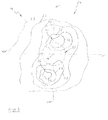
- FIG. 12 shows a diagram of the cornea 15 with isolines 23, which respectively represent different magnitudes of an average value for the respective measured points of the cornea 15.
- the diagram representation comprises a substantially entire surface area 24 of the cornea 15, which corresponds to a measuring range of the keratometer. Coaxially to the optical axis 16, a pupil 25 is indicated. From the different mean values of the respective amplitudes of the change in the topography of the cornea 15 shown with the isolines 23, it can be seen from the exemplary illustration shown here that comparatively high average values are measured in a surface region 26. These mean values are due to a greater deflection of the cornea 15 in the surface area 24, which makes it possible to conclude an emerging keratoconus. Next results in a surface region 27 analogous to the example of the
- Fig. 2 a reduced excursion of the cornea 15, which is reflected in comparatively low averages. From this, a flattening in the surface area 27 can be concluded.
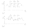
- FIG. 1 Diagram representations of a change in cardiovascular activity or a change in curvature .DELTA.K.
- Fig. 4 There is a continuous blood pressure measurement, in each case the maximum and minimum amplitudes and the start and end points of a systole are determined.
- a measurement of a change in curvature ⁇ K takes place in the corresponding, coinciding period T A.
- T A coinciding period
- FIG. 5 shows a changing curvature change .DELTA.K with respect to the illustrated measuring period, in which the change in curvature is not ideal, but further influences, such as a change a tear film, by blinking or changing a fixation of the eye is subjected.
- the change in curvature at the point P 1.1 in the diagram of the curvature change is determined at the same time, and subsequently for the point Ap max1 the change in curvature at the point P 1.2 . From this, it is possible to determine a slope of the change in curvature between the point P 1.1 and the point P 1.2 .
- This slope of the change in curvature is determined for the respective time segments T A1 , T A2 to T An and an average value of the change in curvature is formed.
- the mean value of the change in curvature thus forms a comparable measurand for all measured points P of the surface area of the cornea. In particular, it is thus possible to analyze precisely the change in the topography of the cornea or of the point P shown here caused by the cardiovascular activity and other influences, and to filter out any changes caused by other influences, if necessary, overshadowing this change.
Landscapes
- Health & Medical Sciences (AREA)
- Life Sciences & Earth Sciences (AREA)
- Engineering & Computer Science (AREA)
- Surgery (AREA)
- General Health & Medical Sciences (AREA)
- Biophysics (AREA)
- Biomedical Technology (AREA)
- Heart & Thoracic Surgery (AREA)
- Medical Informatics (AREA)
- Molecular Biology (AREA)
- Physics & Mathematics (AREA)
- Animal Behavior & Ethology (AREA)
- Veterinary Medicine (AREA)
- Public Health (AREA)
- Signal Processing (AREA)
- Ophthalmology & Optometry (AREA)
- Artificial Intelligence (AREA)
- Computer Vision & Pattern Recognition (AREA)
- Physiology (AREA)
- Psychiatry (AREA)
- Pathology (AREA)
- Eye Examination Apparatus (AREA)
- Measuring Pulse, Heart Rate, Blood Pressure Or Blood Flow (AREA)
Applications Claiming Priority (1)
| Application Number | Priority Date | Filing Date | Title |
|---|---|---|---|
| DE102012209667A DE102012209667A1 (de) | 2012-06-08 | 2012-06-08 | Verfahren und Analysegerät zur Messung einer Hornhaut |
Publications (3)
| Publication Number | Publication Date |
|---|---|
| EP2671505A2 true EP2671505A2 (fr) | 2013-12-11 |
| EP2671505A3 EP2671505A3 (fr) | 2017-07-05 |
| EP2671505B1 EP2671505B1 (fr) | 2023-08-02 |
Family
ID=48607038
Family Applications (1)
| Application Number | Title | Priority Date | Filing Date |
|---|---|---|---|
| EP13170059.3A Active EP2671505B1 (fr) | 2012-06-08 | 2013-05-31 | Procédé et appareil d'analyse pour la mesure d'une cornée |
Country Status (8)
| Country | Link |
|---|---|
| US (1) | US9615738B2 (fr) |
| EP (1) | EP2671505B1 (fr) |
| JP (1) | JP5860005B2 (fr) |
| KR (1) | KR101738205B1 (fr) |
| CN (1) | CN103479326B (fr) |
| DE (1) | DE102012209667A1 (fr) |
| ES (1) | ES2961576T3 (fr) |
| PL (1) | PL2671505T3 (fr) |
Cited By (1)
| Publication number | Priority date | Publication date | Assignee | Title |
|---|---|---|---|---|
| CN106361265A (zh) * | 2015-07-21 | 2017-02-01 | 明达医学科技股份有限公司 | 量测装置及其运作方法 |
Families Citing this family (11)
| Publication number | Priority date | Publication date | Assignee | Title |
|---|---|---|---|---|
| US9125724B2 (en) | 2012-03-09 | 2015-09-08 | John Berdahi | Intraocular pressure modification |
| DE102015009642A1 (de) * | 2015-07-24 | 2017-01-26 | Carl Zeiss Meditec Ag | Verfahren zur Bestimmung der Topografie der Kornea eines Auges |
| BR112018003838A2 (pt) * | 2015-08-27 | 2018-09-25 | Equinox Inc | modificação e identificação de pressão intracorporal relacionada ao olho |
| JP2019154764A (ja) * | 2018-03-13 | 2019-09-19 | 株式会社トプコン | 涙液層厚み測定装置及び方法 |
| KR20250069675A (ko) | 2018-08-09 | 2025-05-19 | 밸런스 오프탈믹스, 인크. | 눈의 혈류를 조절하기 위한 장치 및 방법 |
| US10945598B2 (en) | 2019-02-06 | 2021-03-16 | Jichi Medical University | Method for assisting corneal severity identification using unsupervised machine learning |
| JP6707239B1 (ja) * | 2019-03-13 | 2020-06-10 | 株式会社中京メディカル | 円錐角膜判定装置及びプログラム |
| CN121867677A (zh) * | 2019-12-10 | 2026-04-17 | 爱尔康公司 | 用于测量光学参数的方法和系统 |
| GB2619264A (en) * | 2022-04-11 | 2023-12-06 | Paul Aviram David | Device for measuring the function of a Schlemm's canal |
| KR102853382B1 (ko) | 2023-09-20 | 2025-09-01 | 가톨릭대학교 산학협력단 | 눈물막 파괴 검출 방법, 장치 및 이를 포함하는 컴퓨터 프로그램 |
| KR102944487B1 (ko) * | 2024-01-19 | 2026-03-26 | 가톨릭대학교 산학협력단 | 각막의 형상 측정을 통해 인공 수정체의 선택에 도움을 주면서 주시 방향의 추적이 가능한 시스템, 서버 및 방법 |
Family Cites Families (15)
| Publication number | Priority date | Publication date | Assignee | Title |
|---|---|---|---|---|
| US5116115A (en) | 1990-05-09 | 1992-05-26 | Wyko Corporation | Method and apparatus for measuring corneal topography |
| JP3313309B2 (ja) * | 1997-08-21 | 2002-08-12 | 株式会社トプコン | 眼科装置 |
| US7083609B2 (en) * | 2002-06-13 | 2006-08-01 | Visx, Incorporated | Corneal topography-based target warping |
| JP3939217B2 (ja) * | 2002-08-01 | 2007-07-04 | 株式会社ニデック | 非接触式眼圧計 |
| JP3986406B2 (ja) | 2002-09-25 | 2007-10-03 | 株式会社ニデック | 非接触式眼圧計 |
| JP3986407B2 (ja) * | 2002-09-25 | 2007-10-03 | 株式会社ニデック | 非接触式眼圧計 |
| JP3986408B2 (ja) * | 2002-09-25 | 2007-10-03 | 株式会社ニデック | 非接触式眼圧計 |
| CA2561287C (fr) * | 2004-04-01 | 2017-07-11 | William C. Torch | Biocapteurs,communicateurs et controleurs permettant de surveiller le mouvement de l'oeil et methodes d'utilisation de ces derniers |
| JP4492858B2 (ja) * | 2004-07-20 | 2010-06-30 | 株式会社ニデック | 眼科装置及び眼内屈折力分布算出プログラム |
| US7281801B2 (en) * | 2004-09-15 | 2007-10-16 | University Of Rochester | Tear dynamics measured with optical coherence tomography |
| US7325927B2 (en) * | 2005-01-11 | 2008-02-05 | The University Of Houston | Method of filtering data for the objective classification of eyes |
| DE202005002562U1 (de) * | 2005-02-16 | 2005-06-09 | Oculus Optikgeräte GmbH | Ophthalmisches Analysesystem zur Messung des intraocularen Drucks im Auge |
| US8226235B2 (en) * | 2006-02-14 | 2012-07-24 | Vision Optimization, Llc | Method and apparatus for determining dynamic deformation characteristics of an object |
| US7798641B2 (en) | 2008-09-05 | 2010-09-21 | Heidelberg Engineering Gmbh | Finite element model of a keratoconic cornea |
| US20110237999A1 (en) * | 2010-03-19 | 2011-09-29 | Avedro Inc. | Systems and methods for applying and monitoring eye therapy |
-
2012
- 2012-06-08 DE DE102012209667A patent/DE102012209667A1/de active Pending
-
2013
- 2013-05-28 US US13/903,423 patent/US9615738B2/en active Active
- 2013-05-31 PL PL13170059.3T patent/PL2671505T3/pl unknown
- 2013-05-31 ES ES13170059T patent/ES2961576T3/es active Active
- 2013-05-31 EP EP13170059.3A patent/EP2671505B1/fr active Active
- 2013-06-03 KR KR1020130063526A patent/KR101738205B1/ko active Active
- 2013-06-05 CN CN201310221600.6A patent/CN103479326B/zh active Active
- 2013-06-06 JP JP2013119406A patent/JP5860005B2/ja active Active
Non-Patent Citations (1)
| Title |
|---|
| None |
Cited By (1)
| Publication number | Priority date | Publication date | Assignee | Title |
|---|---|---|---|---|
| CN106361265A (zh) * | 2015-07-21 | 2017-02-01 | 明达医学科技股份有限公司 | 量测装置及其运作方法 |
Also Published As
| Publication number | Publication date |
|---|---|
| ES2961576T3 (es) | 2024-03-12 |
| US9615738B2 (en) | 2017-04-11 |
| JP2013255791A (ja) | 2013-12-26 |
| EP2671505B1 (fr) | 2023-08-02 |
| KR20130138117A (ko) | 2013-12-18 |
| PL2671505T3 (pl) | 2024-04-15 |
| CN103479326A (zh) | 2014-01-01 |
| US20130329187A1 (en) | 2013-12-12 |
| CN103479326B (zh) | 2017-04-12 |
| EP2671505A3 (fr) | 2017-07-05 |
| KR101738205B1 (ko) | 2017-05-29 |
| JP5860005B2 (ja) | 2016-02-16 |
| DE102012209667A1 (de) | 2013-12-12 |
Similar Documents
| Publication | Publication Date | Title |
|---|---|---|
| EP2671505B1 (fr) | Procédé et appareil d'analyse pour la mesure d'une cornée | |
| EP2397068B1 (fr) | Analyse de la pression intra-oculaire | |
| DE102006018445B4 (de) | Vorrichtung und Verfahren zur Bestimmung arterio-venöser Verhältniswerte durch quantitative Analyse retinaler Gefäße | |
| DE60312200T2 (de) | Verfahren zur analyse von einzelpuls-druckwellen | |
| DE69720274T2 (de) | Anzeige des auftretens von herzschlägen | |
| DE102004011779A1 (de) | Zurückdrängen von Artefakten mittels Pulsqualitätswerten | |
| DE102007016357A1 (de) | System und Verfahren zur Überwachung präeklamptischer Patienten | |
| DE102010061580A1 (de) | Verwendung des Frequenzspektrums eines Artefaktes in der Oszillometrie | |
| DE102008003978A1 (de) | Druckmesser, Blutdruckmesser, Verfahren zum Bestimmen von Druckwerten, Verfahren zum Kalibrieren eines Druckmessers und Computerprogramm | |
| EP2529662B1 (fr) | Procédé d'analyse ophtalmologique et système d'analyse | |
| DE112009003560T5 (de) | Blutdruckinformation-Messeinrichtung zum Messen der Pulswellen-Ausbreitungsgeschwindigkeit als Blutdruckinformation | |
| DE102019101409B4 (de) | Verfahren und Sehprüfsystem zum Überprüfen der Augen | |
| EP3773151A1 (fr) | Dispositif et procédé permettant de déterminer des valeurs de pression artérielle rétiniennes et de mapper des valeurs de pression artérielle rétiniennes et des valeurs de pression de perfusion rétiniennes | |
| EP2258256B1 (fr) | Procédé de détermination d'une lentille de contact | |
| DE102010016035A1 (de) | System und Verfahren für eine nichtinvasive Blutdruckmessung | |
| DE102004009879A1 (de) | Verfahren und System zum Modifizieren des Manschettendrucks | |
| WO2017001023A1 (fr) | Dispositif et procédé pour la surveillance et le diagnostic du mécanisme d'autorégulation de la pression sanguine chez un être vivant | |
| WO2013060322A1 (fr) | Procédé et dispositif de surveillance non invasive et de détermination de l'intensité de la douleur et de la profondeur de l'anesthésie | |
| EP2805300A1 (fr) | Procédé et dispositif destinés à mesurer la pression intra-oculaire de manière continue | |
| Lewandowska et al. | Saying “yes” when you want to say “no”-pupil dilation reflects evidence accumulation in a visual working memory recognition task | |
| EP2269502A1 (fr) | Procédé et dispositif de détermination d'une pression de fermeture et/ou d'ouverture artérielle dans un vaisseau sanguin | |
| DE2622990A1 (de) | Einrichtung zur reihenuntersuchung des glaukomas in der augenheilkunde | |
| DE102019214784A1 (de) | Verfahren zur Ermittlung physiologisch korrekter, biometrischer Daten eines Auges | |
| EP3801226A1 (fr) | Procédé et appareil permettant de déterminer au moins un paramètre physiologique | |
| DE102017001677B3 (de) | Verfahren zur Bestimmung des Auftretens eines Gefäßkollapses bei einem Blutgefäß im oder am Auge |
Legal Events
| Date | Code | Title | Description |
|---|---|---|---|
| PUAI | Public reference made under article 153(3) epc to a published international application that has entered the european phase |
Free format text: ORIGINAL CODE: 0009012 |
|
| AK | Designated contracting states |
Kind code of ref document: A2 Designated state(s): AL AT BE BG CH CY CZ DE DK EE ES FI FR GB GR HR HU IE IS IT LI LT LU LV MC MK MT NL NO PL PT RO RS SE SI SK SM TR |
|
| AX | Request for extension of the european patent |
Extension state: BA ME |
|
| PUAL | Search report despatched |
Free format text: ORIGINAL CODE: 0009013 |
|
| AK | Designated contracting states |
Kind code of ref document: A3 Designated state(s): AL AT BE BG CH CY CZ DE DK EE ES FI FR GB GR HR HU IE IS IT LI LT LU LV MC MK MT NL NO PL PT RO RS SE SI SK SM TR |
|
| AX | Request for extension of the european patent |
Extension state: BA ME |
|
| RIC1 | Information provided on ipc code assigned before grant |
Ipc: A61B 3/107 20060101AFI20170530BHEP |
|
| STAA | Information on the status of an ep patent application or granted ep patent |
Free format text: STATUS: REQUEST FOR EXAMINATION WAS MADE |
|
| 17P | Request for examination filed |
Effective date: 20171128 |
|
| RBV | Designated contracting states (corrected) |
Designated state(s): AL AT BE BG CH CY CZ DE DK EE ES FI FR GB GR HR HU IE IS IT LI LT LU LV MC MK MT NL NO PL PT RO RS SE SI SK SM TR |
|
| STAA | Information on the status of an ep patent application or granted ep patent |
Free format text: STATUS: EXAMINATION IS IN PROGRESS |
|
| 17Q | First examination report despatched |
Effective date: 20191119 |
|
| REG | Reference to a national code |
Ref legal event code: R079 Free format text: PREVIOUS MAIN CLASS: A61B0003107000 Ipc: A61B0005021000 Ref country code: DE Ref legal event code: R079 Ref document number: 502013016434 Country of ref document: DE Free format text: PREVIOUS MAIN CLASS: A61B0003107000 Ipc: A61B0005021000 |
|
| GRAP | Despatch of communication of intention to grant a patent |
Free format text: ORIGINAL CODE: EPIDOSNIGR1 |
|
| STAA | Information on the status of an ep patent application or granted ep patent |
Free format text: STATUS: GRANT OF PATENT IS INTENDED |
|
| RIC1 | Information provided on ipc code assigned before grant |
Ipc: A61B 3/107 20060101ALI20230126BHEP Ipc: A61B 5/00 20060101ALI20230126BHEP Ipc: A61B 5/024 20060101ALI20230126BHEP Ipc: A61B 5/021 20060101AFI20230126BHEP |
|
| INTG | Intention to grant announced |
Effective date: 20230216 |
|
| GRAS | Grant fee paid |
Free format text: ORIGINAL CODE: EPIDOSNIGR3 |
|
| GRAA | (expected) grant |
Free format text: ORIGINAL CODE: 0009210 |
|
| STAA | Information on the status of an ep patent application or granted ep patent |
Free format text: STATUS: THE PATENT HAS BEEN GRANTED |
|
| AK | Designated contracting states |
Kind code of ref document: B1 Designated state(s): AL AT BE BG CH CY CZ DE DK EE ES FI FR GB GR HR HU IE IS IT LI LT LU LV MC MK MT NL NO PL PT RO RS SE SI SK SM TR |
|
| REG | Reference to a national code |
Ref country code: GB Ref legal event code: FG4D Free format text: NOT ENGLISH |
|
| REG | Reference to a national code |
Ref country code: CH Ref legal event code: EP |
|
| REG | Reference to a national code |
Ref country code: DE Ref legal event code: R096 Ref document number: 502013016434 Country of ref document: DE |
|
| REG | Reference to a national code |
Ref country code: IE Ref legal event code: FG4D Free format text: LANGUAGE OF EP DOCUMENT: GERMAN |
|
| REG | Reference to a national code |
Ref country code: LT Ref legal event code: MG9D |
|
| REG | Reference to a national code |
Ref country code: NL Ref legal event code: MP Effective date: 20230802 |
|
| PG25 | Lapsed in a contracting state [announced via postgrant information from national office to epo] |
Ref country code: GR Free format text: LAPSE BECAUSE OF FAILURE TO SUBMIT A TRANSLATION OF THE DESCRIPTION OR TO PAY THE FEE WITHIN THE PRESCRIBED TIME-LIMIT Effective date: 20231103 |
|
| PG25 | Lapsed in a contracting state [announced via postgrant information from national office to epo] |
Ref country code: IS Free format text: LAPSE BECAUSE OF FAILURE TO SUBMIT A TRANSLATION OF THE DESCRIPTION OR TO PAY THE FEE WITHIN THE PRESCRIBED TIME-LIMIT Effective date: 20231202 |
|
| PG25 | Lapsed in a contracting state [announced via postgrant information from national office to epo] |
Ref country code: SE Free format text: LAPSE BECAUSE OF FAILURE TO SUBMIT A TRANSLATION OF THE DESCRIPTION OR TO PAY THE FEE WITHIN THE PRESCRIBED TIME-LIMIT Effective date: 20230802 Ref country code: RS Free format text: LAPSE BECAUSE OF FAILURE TO SUBMIT A TRANSLATION OF THE DESCRIPTION OR TO PAY THE FEE WITHIN THE PRESCRIBED TIME-LIMIT Effective date: 20230802 Ref country code: PT Free format text: LAPSE BECAUSE OF FAILURE TO SUBMIT A TRANSLATION OF THE DESCRIPTION OR TO PAY THE FEE WITHIN THE PRESCRIBED TIME-LIMIT Effective date: 20231204 Ref country code: NO Free format text: LAPSE BECAUSE OF FAILURE TO SUBMIT A TRANSLATION OF THE DESCRIPTION OR TO PAY THE FEE WITHIN THE PRESCRIBED TIME-LIMIT Effective date: 20231102 Ref country code: NL Free format text: LAPSE BECAUSE OF FAILURE TO SUBMIT A TRANSLATION OF THE DESCRIPTION OR TO PAY THE FEE WITHIN THE PRESCRIBED TIME-LIMIT Effective date: 20230802 Ref country code: LV Free format text: LAPSE BECAUSE OF FAILURE TO SUBMIT A TRANSLATION OF THE DESCRIPTION OR TO PAY THE FEE WITHIN THE PRESCRIBED TIME-LIMIT Effective date: 20230802 Ref country code: LT Free format text: LAPSE BECAUSE OF FAILURE TO SUBMIT A TRANSLATION OF THE DESCRIPTION OR TO PAY THE FEE WITHIN THE PRESCRIBED TIME-LIMIT Effective date: 20230802 Ref country code: IS Free format text: LAPSE BECAUSE OF FAILURE TO SUBMIT A TRANSLATION OF THE DESCRIPTION OR TO PAY THE FEE WITHIN THE PRESCRIBED TIME-LIMIT Effective date: 20231202 Ref country code: HR Free format text: LAPSE BECAUSE OF FAILURE TO SUBMIT A TRANSLATION OF THE DESCRIPTION OR TO PAY THE FEE WITHIN THE PRESCRIBED TIME-LIMIT Effective date: 20230802 Ref country code: GR Free format text: LAPSE BECAUSE OF FAILURE TO SUBMIT A TRANSLATION OF THE DESCRIPTION OR TO PAY THE FEE WITHIN THE PRESCRIBED TIME-LIMIT Effective date: 20231103 Ref country code: FI Free format text: LAPSE BECAUSE OF FAILURE TO SUBMIT A TRANSLATION OF THE DESCRIPTION OR TO PAY THE FEE WITHIN THE PRESCRIBED TIME-LIMIT Effective date: 20230802 |
|
| REG | Reference to a national code |
Ref country code: ES Ref legal event code: FG2A Ref document number: 2961576 Country of ref document: ES Kind code of ref document: T3 Effective date: 20240312 |
|
| P01 | Opt-out of the competence of the unified patent court (upc) registered |
Effective date: 20240304 |
|
| PG25 | Lapsed in a contracting state [announced via postgrant information from national office to epo] |
Ref country code: SM Free format text: LAPSE BECAUSE OF FAILURE TO SUBMIT A TRANSLATION OF THE DESCRIPTION OR TO PAY THE FEE WITHIN THE PRESCRIBED TIME-LIMIT Effective date: 20230802 Ref country code: RO Free format text: LAPSE BECAUSE OF FAILURE TO SUBMIT A TRANSLATION OF THE DESCRIPTION OR TO PAY THE FEE WITHIN THE PRESCRIBED TIME-LIMIT Effective date: 20230802 Ref country code: EE Free format text: LAPSE BECAUSE OF FAILURE TO SUBMIT A TRANSLATION OF THE DESCRIPTION OR TO PAY THE FEE WITHIN THE PRESCRIBED TIME-LIMIT Effective date: 20230802 Ref country code: DK Free format text: LAPSE BECAUSE OF FAILURE TO SUBMIT A TRANSLATION OF THE DESCRIPTION OR TO PAY THE FEE WITHIN THE PRESCRIBED TIME-LIMIT Effective date: 20230802 Ref country code: CZ Free format text: LAPSE BECAUSE OF FAILURE TO SUBMIT A TRANSLATION OF THE DESCRIPTION OR TO PAY THE FEE WITHIN THE PRESCRIBED TIME-LIMIT Effective date: 20230802 Ref country code: SK Free format text: LAPSE BECAUSE OF FAILURE TO SUBMIT A TRANSLATION OF THE DESCRIPTION OR TO PAY THE FEE WITHIN THE PRESCRIBED TIME-LIMIT Effective date: 20230802 |
|
| REG | Reference to a national code |
Ref country code: DE Ref legal event code: R097 Ref document number: 502013016434 Country of ref document: DE |
|
| PLBE | No opposition filed within time limit |
Free format text: ORIGINAL CODE: 0009261 |
|
| STAA | Information on the status of an ep patent application or granted ep patent |
Free format text: STATUS: NO OPPOSITION FILED WITHIN TIME LIMIT |
|
| 26N | No opposition filed |
Effective date: 20240503 |
|
| PG25 | Lapsed in a contracting state [announced via postgrant information from national office to epo] |
Ref country code: SI Free format text: LAPSE BECAUSE OF FAILURE TO SUBMIT A TRANSLATION OF THE DESCRIPTION OR TO PAY THE FEE WITHIN THE PRESCRIBED TIME-LIMIT Effective date: 20230802 |
|
| PG25 | Lapsed in a contracting state [announced via postgrant information from national office to epo] |
Ref country code: BG Free format text: LAPSE BECAUSE OF FAILURE TO SUBMIT A TRANSLATION OF THE DESCRIPTION OR TO PAY THE FEE WITHIN THE PRESCRIBED TIME-LIMIT Effective date: 20230802 |
|
| PG25 | Lapsed in a contracting state [announced via postgrant information from national office to epo] |
Ref country code: BG Free format text: LAPSE BECAUSE OF FAILURE TO SUBMIT A TRANSLATION OF THE DESCRIPTION OR TO PAY THE FEE WITHIN THE PRESCRIBED TIME-LIMIT Effective date: 20230802 |
|
| REG | Reference to a national code |
Ref country code: CH Ref legal event code: PL |
|
| PG25 | Lapsed in a contracting state [announced via postgrant information from national office to epo] |
Ref country code: MC Free format text: LAPSE BECAUSE OF FAILURE TO SUBMIT A TRANSLATION OF THE DESCRIPTION OR TO PAY THE FEE WITHIN THE PRESCRIBED TIME-LIMIT Effective date: 20230802 |
|
| PG25 | Lapsed in a contracting state [announced via postgrant information from national office to epo] |
Ref country code: LU Free format text: LAPSE BECAUSE OF NON-PAYMENT OF DUE FEES Effective date: 20240531 |
|
| PG25 | Lapsed in a contracting state [announced via postgrant information from national office to epo] |
Ref country code: MC Free format text: LAPSE BECAUSE OF FAILURE TO SUBMIT A TRANSLATION OF THE DESCRIPTION OR TO PAY THE FEE WITHIN THE PRESCRIBED TIME-LIMIT Effective date: 20230802 Ref country code: LU Free format text: LAPSE BECAUSE OF NON-PAYMENT OF DUE FEES Effective date: 20240531 Ref country code: CH Free format text: LAPSE BECAUSE OF NON-PAYMENT OF DUE FEES Effective date: 20240531 |
|
| REG | Reference to a national code |
Ref country code: BE Ref legal event code: MM Effective date: 20240531 |
|
| PG25 | Lapsed in a contracting state [announced via postgrant information from national office to epo] |
Ref country code: IE Free format text: LAPSE BECAUSE OF NON-PAYMENT OF DUE FEES Effective date: 20240531 |
|
| PG25 | Lapsed in a contracting state [announced via postgrant information from national office to epo] |
Ref country code: BE Free format text: LAPSE BECAUSE OF NON-PAYMENT OF DUE FEES Effective date: 20240531 |
|
| PGFP | Annual fee paid to national office [announced via postgrant information from national office to epo] |
Ref country code: PL Payment date: 20250418 Year of fee payment: 13 |
|
| PGFP | Annual fee paid to national office [announced via postgrant information from national office to epo] |
Ref country code: GB Payment date: 20250522 Year of fee payment: 13 Ref country code: ES Payment date: 20250616 Year of fee payment: 13 |
|
| PGFP | Annual fee paid to national office [announced via postgrant information from national office to epo] |
Ref country code: IT Payment date: 20250530 Year of fee payment: 13 |
|
| REG | Reference to a national code |
Ref country code: AT Ref legal event code: MM01 Ref document number: 1593721 Country of ref document: AT Kind code of ref document: T Effective date: 20240531 |
|
| PGFP | Annual fee paid to national office [announced via postgrant information from national office to epo] |
Ref country code: FR Payment date: 20250523 Year of fee payment: 13 |
|
| PG25 | Lapsed in a contracting state [announced via postgrant information from national office to epo] |
Ref country code: AT Free format text: LAPSE BECAUSE OF NON-PAYMENT OF DUE FEES Effective date: 20240531 |
|
| PG25 | Lapsed in a contracting state [announced via postgrant information from national office to epo] |
Ref country code: CY Free format text: LAPSE BECAUSE OF FAILURE TO SUBMIT A TRANSLATION OF THE DESCRIPTION OR TO PAY THE FEE WITHIN THE PRESCRIBED TIME-LIMIT; INVALID AB INITIO Effective date: 20130531 |
|
| PGFP | Annual fee paid to national office [announced via postgrant information from national office to epo] |
Ref country code: DE Payment date: 20250722 Year of fee payment: 13 |
|
| PG25 | Lapsed in a contracting state [announced via postgrant information from national office to epo] |
Ref country code: HU Free format text: LAPSE BECAUSE OF FAILURE TO SUBMIT A TRANSLATION OF THE DESCRIPTION OR TO PAY THE FEE WITHIN THE PRESCRIBED TIME-LIMIT; INVALID AB INITIO Effective date: 20130531 |