EP2684045B2 - Verfahren zur charakterisierung von patienten mit hämolyse - Google Patents
Verfahren zur charakterisierung von patienten mit hämolyse Download PDFInfo
- Publication number
- EP2684045B2 EP2684045B2 EP12710056.8A EP12710056A EP2684045B2 EP 2684045 B2 EP2684045 B2 EP 2684045B2 EP 12710056 A EP12710056 A EP 12710056A EP 2684045 B2 EP2684045 B2 EP 2684045B2
- Authority
- EP
- European Patent Office
- Prior art keywords
- sad
- heme
- microparticles
- cells
- cell
- Prior art date
- Legal status (The legal status is an assumption and is not a legal conclusion. Google has not performed a legal analysis and makes no representation as to the accuracy of the status listed.)
- Active
Links
Images






Classifications
-
- G—PHYSICS
- G01—MEASURING; TESTING
- G01N—INVESTIGATING OR ANALYSING MATERIALS BY DETERMINING THEIR CHEMICAL OR PHYSICAL PROPERTIES
- G01N33/00—Investigating or analysing materials by specific methods not covered by groups G01N1/00 - G01N31/00
- G01N33/48—Biological material, e.g. blood, urine; Haemocytometers
- G01N33/50—Chemical analysis of biological material, e.g. blood, urine; Testing involving biospecific ligand binding methods; Immunological testing
- G01N33/72—Chemical analysis of biological material, e.g. blood, urine; Testing involving biospecific ligand binding methods; Immunological testing involving blood pigments, e.g. haemoglobin, bilirubin or other porphyrins; involving occult blood
- G01N33/721—Haemoglobin
-
- G—PHYSICS
- G01—MEASURING; TESTING
- G01N—INVESTIGATING OR ANALYSING MATERIALS BY DETERMINING THEIR CHEMICAL OR PHYSICAL PROPERTIES
- G01N2800/00—Detection or diagnosis of diseases
- G01N2800/22—Haematology
-
- G—PHYSICS
- G01—MEASURING; TESTING
- G01N—INVESTIGATING OR ANALYSING MATERIALS BY DETERMINING THEIR CHEMICAL OR PHYSICAL PROPERTIES
- G01N2800/00—Detection or diagnosis of diseases
- G01N2800/56—Staging of a disease; Further complications associated with the disease
Definitions
- the present disclosure relates to methods for determining the risk of severe complications in hemolytic diseases. More particularly the present disclosure relates to a method for determining whether a patient suffering from sickle cell disease is at risk for a vaso-occlusive crisis.
- Sickle cell disease is the first genetic disease and a public health challenge in France.
- SCD was designated public health priority by UNESCO in 2003, the French Ministry of Health in 2004 and WHO in 2006.
- UNESCO European Ministry of Health in 2004
- WHO World Health Organization
- a sharp increase in patient numbers put sickle cell disease at the top position of genetic diseases in French metropolitan areas, concerning 5,000 to 6,000 patients up to 18 yo, with an estimated 20,000 patients by 2020.
- Carrier frequency reaches 12% in French West Indies and rises in the USA and developed countries. Demographic trends and the systematic diagnosis at birth for ethnics at risk are partly responsible for this sharp increase in cases to treat.
- SCD the most common genetic disease of our time, will soon become a public health challenge in western countries and France in particular.
- SCD results from a point mutation (HbS) in the hemoglobin gene, causing hemoglobin to polymerize, mediating drastic and irreversible remodeling of red blood cells (RBC).
- HbS point mutation
- RBC red blood cells
- sickle cell disease refers to the homozygous HbSS phenotype.
- Remodeled RBC aggregate and become trapped in microvessels, favoring painful vaso-occlusive crises (VOC).
- VOC vaso-occlusive crises
- Ensuing hemolysis liberates 'free' hemoglobin and heme into the bloodstream, and RBC membrane fragments called microparticles (MP), which carry adhesion receptors and bind avidly to vascular and circulating cells.
- MP microparticles
- the physiopathology of SCD involves an intricate combination of circulating and cardiovascular factors that have only been envisaged recently.
- the primary focus has been the biology of the erythrocyte for the past decades, aimed at blocking RBC remodeling to prevent anemia.
- the present invention is defined in the claims.
- the present disclosure relates to methods for determining the risk of developing vaso-occlusives crises, vascular injury and severe degenerative complications in hemolytic diseases.
- the present disclosure relates to a method for determining whether a patient suffering from sickle cell disease is at risk for a vaso-occlusive crisis comprising the steps consisting of i) determining the level of cell microparticles in a blood sample obtained from said patient, and ii) determining the level of heme and/or hemoglobin contained in said cell microparticles.
- the present disclosure also relates to a method for determining whether a patient suffering from sickle cell disease is at risk for a vaso-occlusive crisis comprising the steps consisting of i) determining the level of cell microparticles in a blood sample obtained from said patient, and ii) determining the level of heme and/or hemoglobin contained in said cell microparticles and iii) determining the the level of heme and/or hemoglobin totally contained in the blood sample
- the present disclosure also relates to a method for determining whether a patient suffering from a hemolytic disease is at risk for vascular injury comprising the steps consisting of i) determining the level of circulating microparticles in a blood sample obtained from said patient, and ii) determining the level of heme and/or hemoglobin contained in said cell microparticles and iii) determining the level of heme and/or hemoglobin totally contained in said blood sample.
- Also disclosed is a method for determining whether a patient suffering from a hemolytic disease is at risk for generative complications comprising the steps consisting of i) determining the level of circulating microparticles in a blood sample obtained from said patient, and ii) determining the level of heme and/or hemoglobin contained in said cell microparticles and iii) determining the level of heme and/or hemoglobin totally contained in said blood sample.
- the present disclosure also relates to a method for preventing a vaso-occlusive crisis in a patient suffering from sickle cell disease comprising depleting the blood-borne microparticles laden in heme and hemoglobin from the blood of said patient.
- the present disclosure also relates to a method for limiting vascular injury in a patient suffering from hemolysis comprising depleting the blood-borne microparticles laden in heme and hemoglobin from the blood of said patient.
- the inventors have demonstrated a practical method to evaluate the relative charge of circulating or experimental microparticles in heme and hemoglobin.
- the disclosure is a novel technique to characterize patients with sickle cell disease, other major hemolytic syndromes, or diseases otherwise associated with hemolysis.
- the technique aims at helping the diagnosis of the severity of vaso-occlusive crises, and the likeliness to develop complications associated with vascular dysfunction, vascular injury and vascular remodeling.
- the disclosure may also be useful to help predict the occurrence of vaso-occlusive crises, and better characterize their severity.
- the disclosure may also be useful to provide patients at risk with better, more appropriate and more immediate care.
- the disclosure may also be useful to help therapeutic decision making and to better manage the side effects associated with current treatments for hemolytic disorders, including blood transfusion programs and hydroxyura.
- microparticles in the plasma from sickle cell disease can be separated by ultracentrifugation.
- the inventors have shown that separating and discarding said microparticles by ultracentrifugation also removes a significant proportion of heme present in plasma.
- the inventors have shown that the level and the proportion of heme associated with microparticles is particularly elevated in sickle cell disease plasma.
- patients that suffer from vaso-occlusive crises and vascular injury display high levels of microparticle-associated heme.
- the inventors confirmed these observations in plasma from transgenic SAD mice suffering from a murine form of sickle cell disease, compared to healthy mice. Futhermore, the inventors found that removing circulating microparticles by filtration (0.1 mm pore size) is also efficient to deplete a significant part of heme in sickle cell disease plasma.
- the MP shed by red blood cells with the technique appear very similar to circulating MP in sickle cell disease in terms of heme and hemoglobin contents, they are representative of circulating microparticles in steady state patients and microparticles that could be shed during VOC.
- microparticle preparations generated from diseased red blood cells do not always bear the same characteristics (size, granulosity, cell surface determinants, toxicity for the endothelium), nor carry the same loads of heme and hemoglobin. They also showed that microparticles shed by red blood cells purified from a selected individual using said technique have specific characteristics that other MP generated by different means do not always share.
- microparticles generated by said technique or purified from body fluids could be used for the diagnosis of the severity of sickle cell disease, for the diagnosis of vascular injury, and for the prognostic of further VOC as well as degenerative complications.
- heme- and hemoglobin-laden MP are toxic for cultured endothelium, inducing more radical oxygen species production and more apoptosis.
- microparticles released by sickle cell disease red blood cells were more toxic for cultured endothelium, inducing more radical oxygen species production and more apoptosis.
- synthetic microparticles made with phosphatidylcholine and phosphatidylserine, artificially loaded with heme are particularly toxic for cultured endothelium, inducing more radical oxygen species production and more apoptosis than synthetic microparticles devoid of heme or hemoglobin.
- the inventors also showed that mesenteric micro-arteries perfused with heme-loaded microparticles loose their ability to vasodilate in response to acethycholine, consistent with vascular injury and endothelial dysfunction.
- heme- and hemoglobin-laden MP are toxic for red blood cells, favor their aggregation and enhance the bonds linking them to each other.
- Heme- and hemoglobin-laden MP function as information vectors from a parent cell to a target cell, carrying toxic moieties and potentiating their effects on target cells.
- Heme- and hemoglobin-laden MP may thus participate in the induction of VOC by promoting small vessel obstruction by red blood cell and red blood cell fragment aggregates.
- the inventors have found that the intravascular injection of purified heme-loaded microparticles to transgenic SAD mice suffering from a murine form of sickle cell disease is sufficient to trigger immediate VOC, characterized by a drastic drop in the perfusion of sensitive organs such as kidneys.
- heme- and hemoglobin-laden MP are toxic for the endothelium and promote the adhesion of red blood cells to vascular endothelial cells.
- heme- and hemoglobin-laden MP may promote the anchorage of red blood cell aggregates to the vascular wall and further enhance the plugging of small vessels.
- heme- and hemoglobin-laden MP my favor deposits of red blood cells, red blood cell membranes or red blood cell fragments against the wall of blood vessels.
- heme- and hemoglobin-laden MP have several of the specific characteristics necessary to enhance or even trigger VOC in sickle cell disease.
- red blood cell MP to SAD transgenic mice with sickle cell disease provided the in vivo demonstration that heme- and hemoglobin-laden MP triggered immediate VOC, as revealed by a local increase in vascular resistance in specific territories such as the kidney.
- the inventors have also demonstrated that the 'free' heme and 'free' hemoglobin present in plasma can be virtually entirely depleted from plasma by ultracentrifugation. This finding suggests that therapeutic benefits may be obtained by the specific depletion of patient plasma from heme- and hemoglobin-laden MP.
- the quantification of MP levels coupled to the evaluation of their relative contents in heme and hemoglobin can thus be used as a novel clinical index of disease severity, predictive of vascular damage, remodeling and dysfunction. This index may help better diagnose and stratify patients according to the predicted severity of their disease and the likeliness to develop severe complications associated with sickle cell disease.
- the characterization of the levels of heme- and hemoglobin-carrying microparticles, as well as their relative contents in heme and hemoglobin may constitute a major improvement for the care of patients with sickle cell disease, major hemolytic disorders and other diseases associated with hemolysis.
- the present disclosure relates to a method for determining whether a patient suffering from sickle cell disease is at risk for a vaso-occlusive crisis comprising the steps consisting of i) determining the level of cell microparticles in a blood sample obtained from said patient and ii) determining the level of heme and/or hemoglobin contained in said cell microparticles.
- Also disclosed is a method for determining whether a patient suffering from sickle cell disease is at risk for a vaso-occlusive crisis comprising the steps consisting of i) isolating the cell microparticles in a blood sample obtained from said patient and ii) determining the level of heme and/or hemoglobin contained in said cell microparticles.
- the methods as above described comprise the step consisting of ii) determining the level of heme and/or hemoglobin totally contained in the blood sample obtained from the patient.
- level of heme and/or hemoglobin totally contained in the blood sample refers to the level of heme and/or hemoglobin that is free and associated with micropaticles in the blood sample.
- blood sample means a whole blood, serum, or plasma sample obtained from the patient.
- the blood sample according to the disclosure is a plasma sample.
- a plasma sample may be obtained using methods well known in the art. For example, blood may be drawn from the patient following standard venipuncture procedure on tri-sodium citrate buffer. Plasma may then be obtained from the blood sample following standard procedures including but not limited to, centrifuging the blood sample at about 1,500*g for about 15-20 minutes (room temperature), followed by pipeting of the plasma layer. Platelet-free plasma (PFP) will be obtained following centrifugation at about 13,000*g for 5 min. In order to collect or discard the microparticles, the plasma sample may be centrifuged in a range of from about 15,000 to about 20,000*g.
- the plasma sample is ultra-centrifuged at around 17,570*g at a temperature of about 4°C.
- buffers may be considered appropriate for resuspending the pelleted cellular debris, which contains the microparticles.
- buffers include reagent grade (distilled or deionized) water and phosphate buffered saline (PBS) pH 7.4.
- PBS buffer Sheath fluid
- the blood sample obtained from the patient is a platelet free platelet sample (PFP) sample.
- PFP may be separated from 10 ml citrated whole blood drawn from the fistula-free arm, 72 hours after the last dialysis. PFP may be obtained after citrate blood centrifugation at 1500*g (15 min), followed by 13000*g centrifugation (5 min, room temperature).
- cell microparticle denotes a plasma membrane vesicle shed from an apoptotic, senescent or activated cell ( Boulanger and Dignat-George, 2011, Arterioscler Thromb Vasc Biol. 2011;31:2-3 ).
- the size of cell microparticle ranges from 0,05 ⁇ m to 1 ⁇ m in diameter.
- said cell microparticle expresses different cell surface markers that are the same as the parent cells.
- microparticles are phosphatidylserine-positive microparticles.
- Standard methods for isolating microparticles are well known in the art.
- the methods may consist in collecting the population of microparticles present in the blood sample as above prepared and using differential binding partners directed against the specific surface markers of the microparticles of interest, wherein microparticles are bound by said binding partners to said surface markers.
- Annexin V binds to externalized phosphatidylserine in a clacium dependent manner and thus labels microparticles expressing phosphatidylserine irrespective of the cellular origin.
- the methods of the disclosure comprise contacting the blood sample with a set of binding partners capable of selectively interacting with microparticles present in said blood sample.
- the binding partner may be an antibody that may be polyclonal or monoclonal, preferably monoclonal, directed against the specific surface marker of the microparticles.
- the binding partners may be a set of aptamers.
- Polyclonal antibodies of the disclosure or a fragment thereof can be raised according to known methods by administering the appropriate antigen or epitope to a host animal selected, e.g., from pigs, cows, horses, rabbits, goats, sheep, and mice, among others.
- a host animal selected, e.g., from pigs, cows, horses, rabbits, goats, sheep, and mice, among others.
- Various adjuvants known in the art can be used to enhance antibody production.
- antibodies useful in practicing the invention can be polyclonal, monoclonal antibodies are preferred.
- Monoclonal antibodies of the disclosure or a fragment thereof can be prepared and isolated using any technique that provides for the production of antibody molecules by continuous cell lines in culture.
- Techniques for production and isolation include but are not limited to the hybridoma technique originally described by Kohler and Milstein (1975); the human B-cell hybridoma technique (Cote et al., 1983); and the EBV-hybridoma technique (Cole et al. 1985).
- the binding partner may be an aptamer.
- Aptamers are a class of molecule that represents an alternative to antibodies in term of molecular recognition. Aptamers are oligonucleotide or oligopeptide sequences with the capacity to recognize virtually any class of target molecules with high affinity and specificity. Such ligands may be isolated through Systematic Evolution of Ligands by EXponential enrichment (SELEX) of a random sequence library. The random sequence library is obtainable by combinatorial chemical synthesis of DNA. In this library, each member is a linear oligomer, eventually chemically modified, of a unique sequence. Possible modifications, uses and advantages of this class of molecules have been reviewed in Jayasena S.D., 1999. Peptide aptamers consist of conformationally constrained antibody variable regions displayed by a platform protein, such as E. coli Thioredoxin A, that are selected from combinatorial libraries by two hybrid methods.
- a platform protein such as E. coli Thioredoxin A
- binding partners of the disclosure such as antibodies or aptamers, may be labelled with a detectable molecule or substance, such as a fluorescent molecule, a radioactive molecule or any others labels known in the art.
- Labels are known in the art that generally provide (either directly or indirectly) a signal.
- the term "labelled", with regard to the antibody or aptamer, is intended to encompass direct labelling of the antibody or aptamer by coupling (i.e., physically linking) a detectable substance, such as a radioactive agent or a fluorophore (e.g. fluorescein isothiocyanate (FITC) or phycoerythrin (PE) or Indocyanine (Cy5)) to the antibody or aptamer, as well as indirect labelling of the probe or antibody by reactivity with a detectable substance.
- a detectable substance such as a radioactive agent or a fluorophore (e.g. fluorescein isothiocyanate (FITC) or phycoerythrin (PE) or Indocyanine (Cy5)
- FITC fluorescein isothiocyanate
- PE phycoerythrin
- Indocyanine Indocyanine
- An antibody or aptamer of the disclosure may be labelled with a radioactive molecule by any method known
- the antibodies against the surface markers are already conjugated to a fluorophore (e.g. FITC-conjugated and/or PE-conjugated).
- a fluorophore e.g. FITC-conjugated and/or PE-conjugated.
- examples include monoclonal anti-human CD62E-FITC, CDC105-FITC, CD51-FITC, CD106-PE, CD31-PE, and CD54-PE, available through Ancell Co. (Bayport, Minn.).
- the aforementioned assays may involve the binding of the binding partners (ie. Antibodies or aptamers) to a solid support.
- Solid supports which can be used in the practice of the invention include substrates such as nitrocellulose (e. g., in membrane or microtiter well form); polyvinylchloride (e. g., sheets or microtiter wells); polystyrene latex (e.g., beads or microtiter plates); polyvinylidine fluoride; diazotized paper; nylon membranes; activated beads, magnetically responsive beads, and the like.
- the solid surfaces are preferably beads.
- the beads for use in the present invention should have a diameter larger than 1 ⁇ m.
- Beads may be made of different materials, including but not limited to glass, plastic, polystyrene, and acrylic.
- the beads are preferably fluorescently labelled.
- fluorescent beads are those contained in TruCount(TM) tubes, available from Becton Dickinson Biosciences, (San Jose, California).
- methods of flow cytometry are preferred methods for determining the level of microparticles in the blood sample obtained from the patient.
- FACS fluorescence activated cell sorting
- magnetic beads may be used to isolate microparticles (MACS).
- beads labelled with monoclonal specific antibodies may be used for the positive selection of microparticles.
- Other methods can include the isolation of microparticles by depletion the microparticles that are not of interest (negative selection).
- cell microparticle isolation methods may also consist in collecting the population of microparticles present in the blood sample of the cells and using size exclusion columns or filters to purify microparticles of specific sizes out of the blood sample as above prepared (filtration). Ultracentrifugation may also be performed, for example before filtration. For example, microparticles can be isolated from plasma by filtration, using 0,1 ⁇ m pore filters for instance.
- Standard methods for the level of heme and hemoglobin are well known. Typically said levels may be determined by spectrophotometry.
- the classical method for determining the level of hemoglobin in a sample utilizes the method of Drabkin. ( D. L. Drabkin and J. H. Austin, Spectre/photometric Studies, J. BIOL. CHEM., 112:51, 1935 ). Briefly, potassium ferricyanide is then added to the sample, which results in oxidation of the heme iron to, produce cyanomethemoglobin. Cyanide ions then convert the methemoglobin to cyanomethemoglobin, a more stable chromagen.
- the hemoglobin concentration of the sample is then determined by measuring the absorbance of the cyanomethemoglobin at 540 nm.
- An other method is also described in Kahn SE, Watkins BF, Bermes EW Jr (1981). Ann Clin Lab Sci 11:126-31 ; Fairbanks VF, Wiesmer SC, O'Brien PC (1992). Cli Chem 38:132-140 .
- the EXAMPLE provides an illustration of the methods that may be used for determining the level of heme and hemoglobin contained in the microparticles isolated as above described. Another method consists at measuring absorbance at 575 nm, the wavelength of an absorption peak specific to heme and hemoglobin. Spectrophotometric methods preferably include an approach to correct for the enhanced turbidity of the plasmas in sickle cell disease and other hemolytic diseases. Such methods include substracting absorbance values measured at 600 nm or 650 nm.
- the levels determined by the present method are then compared to reference levels for determining whether the patient is a risk for a vaso-occlusive crises.
- the reference level refers to the levels of microparticles in blood sample obtained from a selected population of patient who underwent or not a vaso-occlusive crisis.
- the reference level may be of the level of microparticles in blood sample obtained from patients who underwent a vaso-occlusive crisis.
- the reference level may be of the level of microparticles in blood sample obtained from patients who did not undergo a vaso-occlusive crisis.
- the reference level can be a threshold value, or a range.
- the reference level can be established based upon comparative measurements between patients who underwent a vaso-occlusive crisis and patients who did not undergo a vaso-occlusive crisis.
- the reference level refers to the levels of heme and hemoglobin in the cell microparticle sample obtained from a selected population of patient who underwent or not a vaso-occlusive crisis.
- the reference level may be of the level of heme and/or hemoglobin obtained from patients who underwent a vaso-occlusive crisis.
- the reference level may be of the level of heme and/or hemoglobin obtained from patients who did not undergo a vaso-occlusive crisis.
- the reference level can be a threshold value, or a range.
- the reference level can be established based upon comparative measurements between patients who underwent a vaso-occlusive crisis and patients who did not undergo a vaso-occlusive crisis.
- the level of heme and/or hemoglobin contained in said microparticles is compared to the level of heme and/or hemoglobin totally contained in the blood sample, and the ratio accordingly determined indicates whether the patient is at risk for a vaso-occlusive crisis
- the combined measurement of the level of microparticles and the level of heme and/or hemoglobin contained in said microparticles and the level of heme and/or hemoglobin totally contained in the blood sample indicate whether the patient is at risk for a vaso-occlusive crisis.
- the method of the present invention may further comprise a step consisting of determining whether said microparticles are toxic for the endothelium.
- said step may be performed as described in the EXAMPLE. Briefly, endothelial cells may be incubated with said microparticles and toxicity toward said endothelial cells is then determined.
- the method of the disclosure is thus suitable for the stratification of patients with the severity of vaso-occlusive crises and the extent of vascular injury.
- the method of the invention may also particularly useful for determining whether a patient is at risk for a vasculopathy.
- said method may be useful to better diagnose patients afflicted with major hemolytic disorders, or other diseases associated with hemolysis.
- the method of the invention may allow the stratification of patients with respect to their likeliness to develop complications associated with vascular damage, remodeling or dysfunction. These complications include:
- the present disclosure relates to a method for determining whether a patient is at risk for severe complication in hemolytic diseases comprising the steps consisting of i) determining the level of cell microparticles in a blood sample obtained from said patient and ii) determining the level of heme and/or hemoglobin contained in said cell microparticles.
- the present disclosure also relates to a kit or an apparatus for performing the methods of the present invention comprising means for isolating microparticles from a blood sample, means for quantifying said microparticles and means for determining the the level of heme and/or hemoglobin contained in said cell microparticles
- the present disclosure also relates to a method for preventing a vaso-occlusive crisis in a patient afflicted with by sickle cell disease comprising depleting the blood-borne microparticles laden in heme and hemoglobin from the blood of said patient.
- the patient has been determined at risk for a vaso-occlusive crisis by the diagnostic method of the disclosure described herein.
- Depletion of the blood-borne microparticles laden in heme or hemoglobin may be performed by any well-known method in the art. Typically, plasmapheresis coupled to ultracentrifugation may be used.
- the therapeutic method described herein may also be applied to patient who has been determined at risk for a vasculopathy as above described.
- the present disclosure relates to a method for preventing or treating a vaso-occlusive crisis in a patient suffering from sickle cell disease comprising the step of administering said patient with an agent selected from the group consisting of a phosphatidylserine receptor antagonist (such as recombinant annaxin a5 or anti-Del-1 neutralizing antibodies) or an inhibitor of phosphatidylserine receptor expression.
- an agent selected from the group consisting of a phosphatidylserine receptor antagonist (such as recombinant annaxin a5 or anti-Del-1 neutralizing antibodies) or an inhibitor of phosphatidylserine receptor expression.
- the present disclosure also relates to a method for preventing or treating vascular complications in patients afflicted with major hemolytic disorders comprising the step of administering said patient with an agent selected from the group consisting of a phosphatidylserine receptor antagonist or an inhibitor of phosphatidylserine receptor expression.
- phophatidylserine has its general meaning in the art and refers to the most abundant negatively charged phospholipid in eukaryotic membranes. Phophatidylserine refers to the anionic phospholipid present exclusively in the inner leaflet of the plasma membrane of a normal cell, phophatidylserine is externalized following cell activation with both physiological and pathologic stimuli such apoptosis.
- phosphatidylserine receptor antagonist refers to any agent that inhibits the binding of phosphatidylserine to phosphatidylserine receptor.
- Said antagonist may be selected form the group consisting of small molecule, antibodies, aptamers, and polypeptides.
- phosphatidylserine receptor antagonist which may consist in an antibody or antibody fragment directed against phosphatidylserine or phosphatidylserine receptor.
- antibody includes both naturally occurring and non-naturally occurring antibodies. Specifically, “antibody” includes polyclonal and monoclonal antibodies, and monovalent and divalent fragments thereof. Furthermore, “antibody” includes chimeric antibodies, wholly synthetic antibodies, single chain antibodies, and fragments thereof. The antibody may be a human or nonhuman antibody. A nonhuman antibody may be humanized by recombinant methods to reduce its immunogenicity in man.
- Antibodies may be prepared according to conventional methodology. Monoclonal antibodies may be generated using the method of Kohler and Milstein (1975). To prepare monoclonal antibodies useful in the invention, a mouse or other appropriate host animal is immunized at suitable intervals (e.g., twice-weekly, weekly, twice-monthly or monthly) with antigenic forms of phosphatidylserine receptor. The animal may be administered a final "boost" of antigen within one week of sacrifice. It is often desirable to use an immunologic adjuvant during immunization.
- Suitable immunologic adjuvants include Freund's complete adjuvant, Freund's incomplete adjuvant, alum, Ribi adjuvant, Hunter's Titermax, saponin adjuvants such as QS21 or Quil A, or CpG-containing immunostimulatory oligonucleotides.
- Other suitable adjuvants are well-known in the field.
- the animals may be immunized by subcutaneous, intraperitoneal, intramuscular, intravenous, intranasal or other routes. A given animal may be immunized with multiple forms of the antigen by multiple routes.
- the antigen may be provided as synthetic peptides corresponding to antigenic regions of interest in phosphatidylserine receptor.
- lymphocytes are isolated from the spleen, lymph node or other organ of the animal and fused with a suitable myeloma cell line using an agent such as polyethylene glycol to form a hydridoma.
- cells are placed in media permissive for growth of hybridomas but not the fusion partners using standard methods, as described ( Coding, Monoclonal Antibodies: Principles and Practice: Production and Application of Monoclonal Antibodies in Cell Biology, Biochemistry and Immunology, 3rd edition, Academic Press, New York, 1996 ).
- cell supernatants are analyzed for the presence of antibodies of the desired specificity, i.e., that selectively bind the antigen.
- Suitable analytical techniques include ELISA, flow cytometry, immunoprecipitation, and western blotting. Other screening techniques are well-known in the field. Preferred techniques are those that confirm binding of antibodies to conformationally intact, natively folded antigen, such as non-denaturing ELISA, flow cytometry, and immunoprecipitation.
- an antibody from which the pFc' region has been enzymatically cleaved, or which has been produced without the pFc' region designated an F(ab')2 fragment, retains both of the antigen binding sites of an intact antibody.
- an antibody from which the Fc region has been enzymatically cleaved, or which has been produced without the Fc region designated an Fab fragment, retains one of the antigen binding sites of an intact antibody molecule.
- Fab fragments consist of a covalently bound antibody light chain and a portion of the antibody heavy chain denoted Fd.
- the Fd fragments are the major determinant of antibody specificity (a single Fd fragment may be associated with up to ten different light chains without altering antibody specificity) and Fd fragments retain epitope-binding ability in isolation.
- CDRs complementarity determining regions
- FRs framework regions
- CDR1 through CDRS complementarity determining regions
- compositions and methods that include humanized forms of antibodies.
- “humanized” describes antibodies wherein some, most or all of the amino acids outside the CDR regions are replaced with corresponding amino acids derived from human immunoglobulin molecules.
- Methods of humanization include, but are not limited to, those described in U.S. Pat. Nos. 4,816,567 , 5,225,539 , 5,585,089 , 5,693,761 , 5,693,762 and 5,859,205 , which are hereby incorporated by reference.
- U.S. Pat. Nos. 5,585,089 and 5,693,761 , and WO 90/07861 also propose four possible criteria which may used in designing the humanized antibodies.
- the first proposal was that for an acceptor, use a framework from a particular human immunoglobulin that is unusually homologous to the donor immunoglobulin to be humanized, or use a consensus framework from many human antibodies.
- the second proposal was that if an amino acid in the framework of the human immunoglobulin is unusual and the donor amino acid at that position is typical for human sequences, then the donor amino acid rather than the acceptor may be selected.
- the third proposal was that in the positions immediately adjacent to the 3 CDRs in the humanized immunoglobulin chain, the donor amino acid rather than the acceptor amino acid may be selected.
- the fourth proposal was to use the donor amino acid reside at the framework positions at which the amino acid is predicted to have a side chain atom within 3A of the CDRs in a three dimensional model of the antibody and is predicted to be capable of interacting with the CDRs.
- the above methods are merely illustrative of some of the methods that one skilled in the art could employ to make humanized antibodies.
- One of ordinary skill in the art will be familiar with other methods for antibody humanization.
- the present disclosure also relates to humanized forms of the antibodies, some, most or all of the amino acids outside the CDR regions have been replaced with amino acids from human immunoglobulin molecules but where some, most or all amino acids within one or more CDR regions are unchanged. Small additions, deletions, insertions, substitutions or modifications of amino acids are permissible as long as they would not abrogate the ability of the antibody to bind a given antigen. Suitable human immunoglobulin molecules would include IgG1, IgG2, IgG3, IgG4, IgA and IgM molecules.
- a "humanized" antibody retains a similar antigenic specificity as the original antibody. However, using certain methods of humanization, the affinity and/or specificity of binding of the antibody may be increased using methods of "directed evolution", as described by Wu et al., (1999), the contents of which are incorporated herein by reference.
- Fully human monoclonal antibodies also can be prepared by immunizing mice transgenic for large portions of human immunoglobulin heavy and light chain loci. See, e.g., U.S. Pat. Nos. 5,591,669 , 5,598,369 , 5,545,806 , 5,545,807 , 6,150,584 , and references cited therein, the contents of which are incorporated herein by reference. These animals have been genetically modified such that there is a functional deletion in the production of endogenous (e.g., murine) antibodies. The animals are further modified to contain all or a portion of the human germ-line immunoglobulin gene locus such that immunization of these animals will result in the production of fully human antibodies to the antigen of interest.
- monoclonal antibodies can be prepared according to standard hybridoma technology. These monoclonal antibodies will have human immunoglobulin amino acid sequences and therefore will not provoke human anti-mouse antibody (KAMA) responses when administered to humans.
- KAMA human anti-mouse antibody
- the present disclosure also provides for F(ab') 2 Fab, Fv and Fd fragments; chimeric antibodies in which the Fc and/or FR and/or CDR1 and/or CDR2 and/or light chain CDR3 regions have been replaced by homologous human or non-human sequences; chimeric F(ab')2 fragment antibodies in which the FR and/or CDR1 and/or CDR2 and/or light chain CDR3 regions have been replaced by homologous human or non-human sequences; chimeric Fab fragment antibodies in which the FR and/or CDR1 and/or CDR2 and/or light chain CDR3 regions have been replaced by homologous human or non-human sequences; and chimeric Fd fragment antibodies in which the FR and/or CDR1 and/or CDR2 regions have been replaced by homologous human or non-human sequences.
- the present disclosure also includes so-called single chain antibodies.
- the various antibody molecules and fragments may derive from any of the commonly known immunoglobulin classes, including but not limited to IgA, secretory IgA, IgE, IgG and IgM.
- IgG subclasses are also well known to those in the art and include but are not limited to human IgG1, IgG2, IgG3 and IgG4.
- the antibody is a single domain antibody.
- single domain antibody sdAb or “VHH” refers to the single heavy chain variable domain of antibodies of the type that can be found in Camelid mammals which are naturally devoid of light chains. Such VHH are also called “nanobody ® ". According to the invention, sdAb can particularly be llama sdAb.
- the phosphatidylserine receptor antagonist is an aptamer.
- Aptamers are a class of molecule that represents an alternative to antibodies in term of molecular recognition.
- Aptamers are oligonucleotide sequences with the capacity to recognize virtually any class of target molecules with high affinity and specificity.
- Such ligands may be isolated through Systematic Evolution of Ligands by EXponential enrichment (SELEX) of a random sequence library.
- the random sequence library is obtainable by combinatorial chemical synthesis of DNA. In this library, each member is a linear oligomer, eventually chemically modified, of a unique sequence.
- Peptide aptamers consists of a conformationally constrained antibody variable region displayed by a platform protein, such as E.
- the present disclosure also relates to a phosphatidylserine receptor antagonist which is a polypeptide, especially a polypeptide having the ability to bind posphatidylserine.
- the polypeptide derives from a polypeptide selected from the group consisting of annexin V, annexin peptides, synaptotagmin I, lactadherin, T cell immunoglobulin mucin 1 and 4 (TIM-1, TIM-4), c-carboxyglutamic acid (Gla) containing proteins such as vitamin K-dependent blood coagulation factors.
- a polypeptide selected from the group consisting of annexin V, annexin peptides, synaptotagmin I, lactadherin, T cell immunoglobulin mucin 1 and 4 (TIM-1, TIM-4), c-carboxyglutamic acid (Gla) containing proteins such as vitamin K-dependent blood coagulation factors.
- the phosphatidylserine receptor antagonist is a modified annexin V polypeptide.
- the modified annexin V polypeptide is a polymer of annexin V that has an increased effective size. It is believed that the increase in effective size results in prolonged half-life in the vascular compartment.
- One such modified annexin V is a dimer of annexin V.
- the dimer of annexin V is a homodimer of annexin V. Said homodimer of human annexin V may prepared in using well-established methods of recombinant DNA technology. The annexin V molecules of the homodimer are joined through peptide bonds to a flexible linker.
- the flexible linker contains a sequence of amino acids flanked by a glycine and a serine residue at either end to serve as swivels.
- the linker preferably comprises one or more such "swivels.”
- the linker comprises 2 swivels which may be separated by at least 2 amino acids, more particularly by at least 4 amino acids, more particularly by at least 6 amino acids, more particularly by at least 8 amino acids, more particularly by at least 10 amino acids.
- the overall length of the linker is 5-30 amino acids, 5-20 amino acids, 5-10 amino acids, 10-15 amino acids, or 10-20 amino acids.
- the dimer can fold in such a way that the convex surfaces of the monomer which bind phosphatidylserine, can both gain access to externalized phosphatidylserine.
- Flexible linkers are well known in the art.
- a homodimer of annexin V is diannexin as described in Kuypers FA, Larkin SK, Emeis JJ, Allison AC. Interaction of an annexin V homodimer (Diannexin) with phosphatidylserine on cell surfaces and consequent antithrombotic activity. Thromb Haemost. 2007 Mar;97(3):478-86 .
- modified annexin V polypeptide may consist on a recombinant annexin V expressed with, or chemically coupled to, another protein such as the Fc portion of immunoglobulin. Such expression or coupling increases the effective size of the molecule, preventing the loss of annexin V from the vascular compartment and prolonging the half life of said modified annexin V polypetide.
- the polypeptide is a functional equivalent of phosphatidylserine receptor.
- a “functional equivalent of phosphatidylserine receptor” is a compound which is capable of binding to phosphatidylserine, thereby preventing its interaction with phosphatidylserine receptor.
- the term “functional equivalent” includes fragments, mutants, and muteins of phosphatidylserine receptor.
- the term “functionally equivalent” thus includes any equivalent of phosphatidylserine receptor obtained by altering the amino acid sequence, for example by one or more amino acid deletions, substitutions or additions such that the protein analogue retains the ability to bind to phosphatidylserine. Amino acid substitutions may be made, for example, by point mutation of the DNA encoding the amino acid sequence.
- Functional equivalents include molecules that bind phosphatidylserine and comprise all or a portion of the extracellular domains of phosphatidylserine receptor. Typically, said functional equivalents may comprise binding domain of phosphatidylserine receptor or a portion thereof.
- the functional equivalents include soluble forms of the phosphatidylserine receptor.
- a suitable soluble form of these proteins, or functional equivalents thereof, might comprise, for example, a truncated form of the protein from which the transmembrane domain has been removed by chemical, proteolytic or recombinant methods.
- the functional equivalent is at least 80% homologous to the corresponding protein. In a preferred embodiment, the functional equivalent is at least 90% homologous as assessed by any conventional analysis algorithm such as for example, the Pileup sequence analysis software ( Program Manual for the Wisconsin Package, 1996 )..
- a functionally equivalent fragment as used herein also may mean any fragment or assembly of fragments of phosphatidylserine receptor that binds to phosphatidylserine. Accordingly the present disclosure provides a polypeptide capable of inhibiting binding of phosphatidylserine receptor to phosphatidylserine, which polypeptide comprises consecutive amino acids having a sequence which corresponds to the sequence of at least a portion of an extracellular domain of phosphatidylserine receptor, which portion binds to phosphatidylserine.
- Functionally equivalent fragments may belong to the same protein family as the human phosphatidylserine receptor identified herein.
- protein family is meant a group of proteins that share a common function and exhibit common sequence homology.
- homology between functionally equivalent protein sequences is at least 25% across the whole of amino acid sequence of the complete protein. More preferably, the homology is at least 50%, even more preferably 75% across the whole of amino acid sequence of the protein or protein fragment. More preferably, homology is greater than 80% across the whole of the sequence. More preferably, homology is greater than 90% across the whole of the sequence. More preferably, homology is greater than 95% across the whole of the sequence.
- polypeptides may be produced by any suitable means, as will be apparent to those of skill in the art.
- expression may conveniently be achieved by culturing under appropriate conditions recombinant host cells containing the polypeptide of the invention.
- the polypeptide is produced by recombinant means, by expression from an encoding nucleic acid molecule.
- Systems for cloning and expression of a polypeptide in a variety of different host cells are well known.
- the polypeptide When expressed in recombinant form, the polypeptide is preferably generated by expression from an encoding nucleic acid in a host cell.
- a host cell Any host cell may be used, depending upon the individual requirements of a particular system. Suitable host cells include bacteria mammalian cells, plant cells, yeast and baculovirus systems. Mammalian cell lines available in the art for expression of a heterologous polypeptide include Chinese hamster ovary cells. HeLa cells, baby hamster kidney cells and many others. Bacteria are also preferred hosts for the production of recombinant protein, due to the ease with which bacteria may be manipulated and grown. A common, preferred bacterial host is E coli.
- polypeptides used in the therapeutic methods described herein may be modified in order to improve their therapeutic efficacy. Such modification may be used to decrease toxicity, increase circulatory time, or modify biodistribution. For example, the toxicity of potentially important therapeutic compounds can be decreased significantly by combination with a variety of drug carrier vehicles that modify biodistribution.
- a strategy for improving drug viability is the utilization of water-soluble polymers.
- Various water-soluble polymers have been shown to modify biodistribution, improve the mode of cellular uptake, change the permeability through physiological barriers; and modify the rate of clearance from the body.
- water-soluble polymers have been synthesized that contain drug moieties as terminal groups, as part of the backbone, or as pendent groups on the polymer chain.
- PEG Polyethylene glycol
- Attachment to various drugs, proteins, and liposomes has been shown to improve residence time and decrease toxicity.
- PEG can be coupled to active agents through the hydroxyl groups at the ends of the chain and via other chemical methods; however, PEG itself is limited to at most two active agents per molecule.
- copolymers of PEG and amino acids were explored as novel biomaterials which would retain the biocompatibility properties of PEG, but which would have the added advantage of numerous attachment points per molecule (providing greater drug loading), and which could be synthetically designed to suit a variety of applications.
- PEGylation techniques for the effective modification of drugs.
- drug delivery polymers that consist of alternating polymers of PEG and tri-functional monomers such as lysine have been used by VectraMed (Plainsboro, N.J.).
- the PEG chains typically 2000 daltons or less
- Such copolymers retain the desirable properties of PEG, while providing reactive pendent groups (the carboxylic acid groups of lysine) at strictly controlled and predetermined intervals along the polymer chain.
- the reactive pendent groups can be used for derivatization, cross-linking, or conjugation with other molecules.
- These polymers are useful in producing stable, long-circulating pro-drugs by varying the molecular weight of the polymer, the molecular weight of the PEG segments, and the cleavable linkage between the drug and the polymer.
- the molecular weight of the PEG segments affects the spacing of the drug/linking group complex and the amount of drug per molecular weight of conjugate (smaller PEG segments provides greater drug loading).
- increasing the overall molecular weight of the block co-polymer conjugate will increase the circulatory half-life of the conjugate. Nevertheless, the conjugate must either be readily degradable or have a molecular weight below the threshold-limiting glomular filtration (e.g., less than 60 kDa).
- linkers may be used to maintain the therapeutic agent in a pro-drug form until released from the backbone polymer by a specific trigger, typically enzyme activity in the targeted tissue.
- a specific trigger typically enzyme activity in the targeted tissue.
- tissue activated drug delivery is particularly useful where delivery to a specific site of biodistribution is required and the therapeutic agent is released at or near the site of pathology.
- Linking group libraries for use in activated drug delivery are known to those of skill in the art and may be based on enzyme kinetics, prevalence of active enzyme, and cleavage specificity of the selected disease-specific enzymes. Such linkers may be used in modifying the protein or fragment of the protein described herein for therapeutic delivery.
- a gene product can be the direct transcriptional product of a gene (e.g., mRNA, tRNA, rRNA, antisense RNA, ribozyme, structural RNA or any other type of RNA) or a protein produced by translation of a mRNA.
- Gene products also include messenger RNAs which are modified, by processes such as capping, polyadenylation, methylation, and editing, and proteins (e.g., phosphatidylserine receptor) modified by, for example, methylation, acetylation, phosphorylation, ubiquitination, SUMOylation, ADP-ribosylation, myristilation, and glycosylation.
- proteins e.g., phosphatidylserine receptor
- An 'inhibitor of expression refers to a natural or synthetic compound that has a biological effect to inhibit the expression of a gene.
- Inhibitors of phosphatidylserine receptor expression for use as disclosed may be based on antisense oligonucleotide constructs.
- Anti-sense oligonucleotides including anti-sense RNA molecules and anti-sense DNA molecules, would act to directly block the translation of phosphatidylserine receptor mRNA by binding thereto and thus preventing protein translation or increasing mRNA degradation, thus decreasing the level of phosphatidylserine receptor proteins, and thus activity, in a cell.
- antisense oligonucleotides of at least about 15 bases and complementary to unique regions of the mRNA transcript sequence encoding phosphatidylserine receptor can be synthesized, e.g., by conventional phosphodiester techniques and administered by e.g., intravenous injection or infusion.
- Methods for using antisense techniques for specifically alleviating gene expression of genes whose sequence is known are well known in the art (e.g. see U.S. Pat. Nos. 6,566,135 ; 6,566,131 ; 6,365,354 ; 6,410,323 ; 6,107,091 ; 6,046,321 ; and 5,981,732 ).
- Small inhibitory RNAs can also function as inhibitors of phosphatidylserine receptor expression for use in the present disclosure.
- phosphatidylserine receptor gene expression can be reduced by contacting the patient or cell with a small double stranded RNA (dsRNA), or a vector or construct causing the production of a small double stranded RNA, such that phosphatidylserine receptor expression is specifically inhibited (i.e. RNA interference or RNAi).
- dsRNA small double stranded RNA
- RNAi RNA interference
- Methods for selecting an appropriate dsRNA or dsRNA-encoding vector are well known in the art for genes whose sequence is known (e.g. see Tuschl, T. et al. (1999); Elbashir, S. M. et al.
- Ribozymes can also function as inhibitors of phosphatidylserine receptor expression for use in the present disclosure.
- Ribozymes are enzymatic RNA molecules capable of catalyzing the specific cleavage of RNA. The mechanism of ribozyme action involves sequence specific hybridization of the ribozyme molecule to complementary target RNA, followed by endonucleolytic cleavage.
- Engineered hairpin or hammerhead motif ribozyme molecules that specifically and efficiently catalyze endonucleolytic cleavage of phosphatidylserine receptor mRNA sequences are thereby useful within the scope of the present disclosure.
- ribozyme cleavage sites within any potential RNA target are initially identified by scanning the target molecule for ribozyme cleavage sites, which typically include the following sequences, GUA, GuU, and GUC. Once identified, short RNA sequences of between about 15 and 20 ribonucleotides corresponding to the region of the target gene containing the cleavage site can be evaluated for predicted structural features, such as secondary structure, that can render the oligonucleotide sequence unsuitable. The suitability of candidate targets can also be evaluated by testing their accessibility to hybridization with complementary oligonucleotides, using, e.g., ribonuclease protection assays.
- antisense oligonucleotides and ribozymes useful as inhibitors of phosphatidylserine receptor expression can be prepared by known methods. These include techniques for chemical synthesis such as, e.g., by solid phase phosphoramadite chemical synthesis. Alternatively, anti-sense RNA molecules can be generated by in vitro or in vivo transcription of DNA sequences encoding the RNA molecule. Such DNA sequences can be incorporated into a wide variety of vectors that incorporate suitable RNA polymerase promoters such as the T7 or SP6 polymerase promoters. Various modifications to the oligonucleotides of the disclosure can be introduced as a means of increasing intracellular stability and half-life.
- Possible modifications include but are not limited to the addition of flanking sequences of ribonucleotides or deoxyribonucleotides to the 5' and/or 3' ends of the molecule, or the use of phosphorothioate or 2'-O-methyl rather than phosphodiesterase linkages within the oligonucleotide backbone.
- Antisense oligonucleotides siRNAs and ribozymes of the disclosure may be delivered in vivo alone or in association with a vector.
- a "vector" is any vehicle capable of facilitating the transfer of the antisense oligonucleotide siRNA or ribozyme nucleic acid to the cells and preferably cells expressing phosphatidylserine receptor.
- the vector transports the nucleic acid to cells with reduced degradation relative to the extent of degradation that would result in the absence of the vector.
- the vectors useful in the disclosure include, but are not limited to, plasmids, phagemids, viruses, other vehicles derived from viral or bacterial sources that have been manipulated by the insertion or incorporation of the the antisense oligonucleotide siRNA or ribozyme nucleic acid sequences.
- Viral vectors are a preferred type of vector and include, but are not limited to nucleic acid sequences from the following viruses: retrovirus, such as moloney murine leukemia virus, harvey murine sarcoma virus, murine mammary tumor virus, and rouse sarcoma virus; adenovirus, adeno-associated virus; SV40-type viruses; polyoma viruses; Epstein-Barr viruses; papilloma viruses; herpes virus; vaccinia virus; polio virus; and RNA virus such as a retrovirus.
- retrovirus such as moloney murine leukemia virus, harvey murine sarcoma virus, murine mammary tumor virus, and rouse sarcoma virus
- retrovirus such as moloney murine leukemia virus, harvey murine sarcoma virus, murine mammary tumor virus, and rouse sarcoma virus
- adenovirus adeno
- Non-cytopathic viruses include retroviruses (e.g., lentivirus), the life cycle of which involves reverse transcription of genomic viral RNA into DNA with subsequent proviral integration into host cellular DNA. Retroviruses have been approved for human gene therapy trials. Most useful are those retroviruses that are replication-deficient (i.e., capable of directing synthesis of the desired proteins, but incapable of manufacturing an infectious particle). Such genetically altered retroviral expression vectors have general utility for the high-efficiency transduction of genes in vivo.
- adeno-viruses and adeno-associated viruses are double-stranded DNA viruses that have already been approved for human use in gene therapy.
- the adeno-associated virus can be engineered to be replication deficient and is capable of infecting a wide range of cell types and species. It further has advantages such as, heat and lipid solvent stability; high transduction frequencies in cells of diverse lineages, including hemopoietic cells; and lack of superinfection inhibition thus allowing multiple series of transductions.
- the adeno-associated virus can integrate into human cellular DNA in a site-specific manner, thereby minimizing the possibility of insertional mutagenesis and variability of inserted gene expression characteristic of retroviral infection.
- adeno-associated virus infections have been followed in tissue culture for greater than 100 passages in the absence of selective pressure, implying that the adeno-associated virus genomic integration is a relatively stable event.
- the adeno-associated virus can also function in an extrachromosomal fashion.
- Plasmid vectors have been extensively described in the art and are well known to those of skill in the art. See e.g., SANBROOK et al., "Molecular Cloning: A Laboratory Manual," Second Edition, Cold Spring Harbor Laboratory Press, 1989 . In the last few years, plasmid vectors have been used as DNA vaccines for delivering antigen-encoding genes to cells in vivo. They are particularly advantageous for this because they do not have the same safety concerns as with many of the viral vectors. These plasmids, however, having a promoter compatible with the host cell, can express a peptide from a gene operatively encoded within the plasmid.
- Plasmids may be delivered by a variety of parenteral, mucosal and topical routes.
- the DNA plasmid can be injected by intramuscular, intradermal, subcutaneous, or other routes. It may also be administered by intranasal sprays or drops, rectal suppository and orally.
- the plasmids may be given in an aqueous solution, dried onto gold particles or in association with another DNA delivery system including but not limited to liposomes, dendrimers, cochleate and microencapsulation.
- FIGURES are a diagrammatic representation of FIGURES.
- Human recombinant TSP1 was produced by EMP-Genetech (Germany). Synthetic peptides (17) 4N1-1 (RFYVVMWK) (SEQ ID NO:1) and 4N1-2 (RFYVVM) (SEQ ID NO:2) were obtained from Bachem, 4NGG (RFYGGMWK) (SEQ ID NO:3) is manufactured by Genecust (France). Di-aminidophenylindol (DAPI), RNase A, and propidium iodide were from Sigma-Aldrich.
- DAPI Di-aminidophenylindol
- RNase A RNase A
- propidium iodide were from Sigma-Aldrich.
- mice were anesthetized with isoflurane and monitored to prevent any cardiorespiratory depression. Mice were shaved and placed in the decubitus position on a heating blanket (38°C).
- a Vivid 7 echograph (GE Medical Systems ® , Horten, Norway) equipped with a 12-MHz linear transducer (12L).
- the ultrasound probe was placed on the left side of the abdomen for examination of renal arteries, or in left lateral decubitus for cardiac output acquisition with transducer on the chest. Data were transferred on-line to an EchoPAC ultrasound image analysis workstation (GE Medical Systems ® ). Two-Dimensional ultrasound imaging of the abdomen in left-sided longitudinal B-mode allowed kidney width and height measurements. Color-coded blood flow detection by Doppler enabled renal arteries to be localized. A pulsed Doppler spectrum was recorded and peak systolic, end-diastolic and time-average mean BFV were measured in the renal artery, with Doppler beam angle correction.
- CO [(V mean .60). ( ⁇ . (Dpa/2) 2 )], where CO is the cardiac output in ml/minute, V mean is the mean time-averaged BFV in cm/s and Dpa is the pulmonary artery diameter in cm.
- Kidney sizes and BFV were determined by the same investigator, as means of 5 to 8 measurements. Repeatability for cardiac output measurements was verified. We controlled and confirmed that kidney size was similar between SAD and wild type mice. After sacrifice, kidneys were dissected, dehydrated, mounted in paraffin, sectioned and stained by Masson trichrome.
- Mouse blood was obtained through retro-orbital sinus puncture, mixed with anti-coagulant heparin buffer (5 UI/ml), and collected on citrated tubes.
- Blood diluted 1:1 with PBS (Gibco-Life Sciences) was placed onto histopaque-1083 (Sigma) density gradients for centrifugation (600 g, 25 minutes). Erythrocyte pellets were resuspended, washed in DMEM and stored for up to 4 hours at room temperature. Erythrocyte surface CD47 expression was assessed by FACS.
- erythrocyte were suspended in polyvinylpyrrolidone (31 mPa.s) and placed in a rotating type LORCA ektacytometer (R&R Mechatronics, Hoorn, The Netherlands) in the absence or presence of TSP1 (25 ⁇ g/ml), or TSP1-derived peptides 4N1-1 or 4N1-2 (25 ⁇ M). Shear rate was applied according to the builder's program (1500 s -1 ; 2.5 minutes). Erythrocytes were immediately separated and supernatant PS + MP quantified by FACS.
- MP stocks were generated from 10 to 20 ml of supernatant from erythrocytes suspended in RPMI-1640 (4.10 6 /ml) and treated with 4N1-1. Suspensions were centrifuged at 400 g for 15 min to discard cells, and ultracentrifuged at 20500 g for 45 min to pellet MP, which were resuspended in filtered (0.2 ⁇ m) DMEM and stored at -80°C. MP concentrations were determined as previously described (20). Briefly, MP were labeled with 2 ⁇ l FITC-conjugated annexin-V (Roche Diagnostics, France) diluted in 100 ⁇ l reaction buffer with 5 mM CaCl 2 .
- MP were analyzed on a Coulter EPICS XL flow cytometer (Beckman Coulter). MP were identified in forward light scatter (FSc) and side-angle light scatter (SSc) intensity dot plots set at logarithmic gain, as events of 0.1-1 ⁇ m in diameter. MP-size events were analyzed in FL/FSC fluorescence dot plots to determine annexin-V labeling. MP concentration was determined with respect to calibrated fluorescent microbeads (Flowcount). To determine erythrocyte origin, MP were double labeled with rat PE-Cy7-coupled mouse-specific anti-Ter119 antibody (Biolegend via Ozyme, France), or matched irrelevant IgG 2b ⁇ isotype.
- FSc forward light scatter
- SSc side-angle light scatter
- SV40-transformed mouse lymph node endothelial cells (SVEC4-10 from ATCC) were obtained from LGC Promochem, France and grown in Dulbecco's Modified Eagle Medium (DMEM) supplemented with 20% neonatal calf serum (NCS ; Miguel Dutscher) in 24 well plaques at 37°C with 5% CO 2 . Confluent SVEC monolayers in 96 well plates were incubated with 25 MP/ ⁇ l for up to 2 h.
- DMEM Dulbecco's Modified Eagle Medium
- NCS neonatal calf serum
- SVEC were cultured up to 70% confluency, washed and placed in DMEM with 0.05% NCS with or without RBC MP. After 16 hours, SVEC were trypsinized, fixed overnight in ice-cold 70% ethanol-H2O, and incubated in 0.05% Triton X100, 100 ⁇ g/ml RNase A plus 50 ⁇ g/ml propidium iodide for 1 h in the dark. Total DNA content was determined by FACS. DNA profiles were divided as sub-G1 (apoptosis), G0/G1 (resting) and G2/M (proliferation) phases of the cell cycle. Data shown is the mean percentage of cells in each phase, in 3 to 6 independent experiments.
- Mesenteric arteries were removed from terminally anesthetized mice and placed them in ice-cold physiological salt solution (PSS). 4-6 arterial segments from each mouse were used. A segment of mesenteric artery was cannulated and mounted in a video-monitored perfusion system as described (21). Arterial segments were bathed in a 5 ml organ bath containing a PSS maintained at pH 7.4, with pO 2 and pCO 2 of 160 mm Hg and 37 mm Hg, respectively. Arterial diameter was continuously recorded by video (Living System Instrumentation Inc., Burlington, VT). Pressure and flow rates could be changed independently. Diameter changes were measured in each segment when the intraluminal pressure was set at 75 mm Hg in order to measure dilation.
- PSS ice-cold physiological salt solution
- TSP1 triggered vaso-occlusive crises in SAD mice
- TSP1 influences VOC during SCD in vivo.
- SAD transgenic mice that express a mutated human hemoglobin ⁇ -chain known to cause sickle cell formation and sensitivity to VOC (18, 19).
- VOC are not identified by the expression of pain as in humans, but on a reduction of perfusion in organs sensitive to vaso-occlusion, revealed by a drop in local mean blood flow velocities measured by Echo-Doppler.
- TSP1 upregulated plasma MP in vivo
- TSP1 injections triggered a 3 fold raise in plasma PS + MP within 5 minutes in wild type mice ( p ⁇ 0.05 vs control).
- SAD mice displayed a doubling in circulating PS + MP levels at rest versus wild type mice ( p ⁇ 0.05). They also responded to TSP1 with a 3 fold increase in circulating PS + MP ( p ⁇ 0.05 vs control SAD and versus TSP1-treated wild type mice).
- TSP1 was injected a CD47 agonist, the TSP1 carboxyterminal peptide 4N1-1. Its effects were comparable to whole TSP1.
- CD47 agonist the TSP1 carboxyterminal peptide 4N1-1. Its effects were comparable to whole TSP1.
- TSP1 induced RBC conversion into echinocytes and MP shedding
- TSP1 could mediate significant modifications in isolated erythrocyte phenotype, or in MP shedding.
- Fresh wild type and SAD mouse erythrocytes were placed under shear in a Lorca ektacytometer.
- Phase contrast and electron microscopic analysis revealed that echinocytes were rather rare in healthy wild type RBC.
- the addition of TSP1 raised the proportion of wild type echinocytes over 10 fold to reach 30% within minutes (p ⁇ 0.05 vs control).
- the pattern of short spicules of the healthy echinocytes was predictable by surface tension analysis (22, 23).
- SAD erythrocytes at rest displayed a great variety of shapes with elongated deformed bodies, including about 5% of typical sickle erythrocytes.
- TSP1 triggered a 50% increase in SAD echinocytes ( p ⁇ 0.05 vs control), but did not affect sickle cells. Conversion into SAD echinocytes was accompanied by PS + MP shedding ( p ⁇ 0.05).
- TSP1 carboxyterminal peptides including the CD47 agonist 4N1-1, truncated control 4N1-2, and mutated control 4NGG devoid of the original VVM motif.
- 4N1-1 proved particularly strong stimulus of echinocytosis with a doubling in wild type echinocytes and a 50% increase in SAD echinocytes ( p ⁇ 0.05), similar to recombinant TSP1.
- 4N1-1 did not affect sickle cells.
- 4N1-1 also triggered intense MP shedding with a 16 fold increase in wild type erythrocytes and a 40 fold raise in SAD erythrocytes ( p ⁇ 0.05 versus control peptides), whereas 4N1-2 and 4NGG had no effect.
- SAD RBC shed about twice as many MP than healthy erythrocytes whether stimulated or at rest, and more particularly when TSP1 or 4N1-1 are applied ( p ⁇ 0.05 wild type vs SAD).
- Scanning electron microscopy of SAD erythrocytes revealed that echinocytosis occurred as a drastic phenotypic change with the multiplication of unusually thin, long and apparently fragile spicules, as shown after treatment with 4N1-1. In contrast spicule dimensions appeared strikingly homogenous in wild type cells.
- 4N1-1 increased spicule numbers in SAD RBC by 20% ( p ⁇ 0.05 vs wild type) and not in healthy wild type erythrocytes.
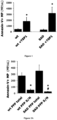
- SAD RBC MP induced a 30 to 35% reduction in mean blood flow velocity in SAD mice renal arteries ( Fig. 1A ; p ⁇ 0.05 vs none).
- SAD mice systolic blood flow velocity was unchanged after MP injection, but diastolic and mean blood flow velocities were dramatically decreased. No changes occurred in cardiac output or heart rate.
- wild type RBC MP had very similar effects ( p ⁇ 0.05 vs controls).
- Renal histology after Masson trichrome stain revealed red blood cells aggregates and vascular congestion in SAD mice.
- Erythrocyte MP activate endothelial cells
- ROS generation induced by SAD RBC MP was entirely inhibited when endothelial NOS was blocked with 100 ⁇ M L-NAME, whereas its enantiomer D-NAME had no effects ( p ⁇ 0.05).
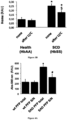
- vasodilation in response to the NO donor SNP.
- exposure to wild type or SAD RBC MP reduced SNP vasodilation down to 69+5% and 59+6% of maximal vessel diameter, respectively ( p ⁇ 0.05 vs control).
- two thirds of impact of RBC MP on vasodilation could be attributed to endothelial injury.
- Circulating MP have long been reported as a key feature of SCD in conjunction with hemolysis. However, the mechanisms of their generation and their physiopathological impact remained unknown.
- TSP1 triggers vaso-occlusive crises
- TSP1 is mainly stored in platelets and released upon activation to generate steep and local gradients. TSP1 can also be secreted by vascular cells challenged with thrombin (26), hypoxia (27, 28), mechanical injury (29), or abnormal blood flow (30-32). With platelet counts of 250.10 6 platelets/ml and 80 ng TSP1 per 10 6 platelets, 1 ml of normal human blood contains over 20 ⁇ g TSP1 (10). Coagulated human blood contains 17.5 ⁇ g/ml of TSP1 (33). Moreover, basal systemic plasma TSP1 concentrations are similar in health and steady state SCD patients (about 0.5 ⁇ g/ml). However, TSP1 increases sharply during VOC (up to 5 fold) (10).
- TSP1 injection induced a 30% drop in renal perfusion within minutes, but TSP1 had no impact on systemic hemodynamic parameters such as cardiac output or heart rate.
- the drop in SAD renal perfusion is thus consistent with a local increase in renal vascular resistance and related to VOC (18, 19). This was confirmed by histological assessment of severe renal capillary congestion and obvious signs of pain in injected SAD mice, including temporary prostration and reduced mobility after the end of anesthesia.
- TSP1 did not modify renal perfusion in wild type mice. The deleterious effects of TSP1 were only revealed in transgenic SAD mice, in territories susceptible to vaso-occlusion. At rest, SAD PFP contained twice as many MP than wild type PFP (880 vs.
- TSP1 signals conversion into echinocytes and MP shedding
- Transgenic SAD erythrocytes at rest displayed a great variety of elongated and deformed shapes. True sickle cells represented less than 5% of SAD erythrocytes, whereas echinocytes accounted for up to 30%. TSP1 and shear stress induced PS externalization, doubled the proportion of SAD echinocytes ( p ⁇ 0.05 vs control) and doubled MP shedding (p ⁇ 0.05 vs control). SAD echinocytes displayed highly unpredictable phenotypes and a heterogeneous number of thin and long spicules. Moreover, SAD echinocytes shed twice more MP than healthy cells with TSP1. MP shedding correlated with the proportion of echinocytes, and the numbers and putative fragility of SAD spicules might also participate in greater MP shedding.
- Carboxyterminal peptides reproduce the stimulation of MP shedding by TSP1
- CD47 also known as integrin-associated protein (IAP) in platelets and leukocytes, is a key constituent of the Rhesus protein complex. CD47 activation is known to enhance integrin avidity and recapitulate the pro-adhesive effects of TSP1 on sickle erythrocytes (13-16). Synthetic peptides including 4N1-1 (17) mimic these carboxyterminal sequences and reproduce some of the functions of TSP1, although the peptides may have additional effects (39-42).
- IAP integrin-associated protein
- TSP1-CD47 signaling was previously reported to include rapid cytosolic Ca 2+ mobilization (43), cytoskeletal remodeling (44, 45) and membrane PS externalization (36, 45, 46) in fibroblasts and lymphocytes. CD47 may thus mediate the mechanical properties of the RBC membrane and trigger vesiculation.
- TSP1 and 4N1-1 One singular difference between TSP1 and 4N1-1 is that the presence of shear stress was necessary to unmask the effects of TSP1, whereas 4N1-1 remained active at rest. Shear might thus synergize with the TSP1 carboxyterminus to activate CD47. Indeed, CD47 has already been proposed as a mechanosensor, and CD47 signaling is strongly enhanced by shear in SCD erythrocytes (15). However, the nature of this synergy is still not understood, dedicated molecular studies will be needed to determine how shear influences CD47 activation by TSP1.
- Erythrocyte MP trigger vaso-occlusive crises
- Erythrocyte MP activate the endothelium
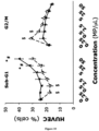
- perfused erythrocyte MP greatly reduced ACH-mediated endothelial-dependent vasodilation.
- SAD MP were more deleterious than wild type MP, coherent with the increased ROS stimulation in cultured endothelium.
- the effects of MP on endothelial function were mediated ROS and the phosphatidylserine carried at the MP surface.
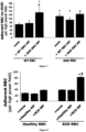
- Endothelial incubation with MP also increased the adhesion of erythrocytes.
- SAD erythrocytes are twice more adherent than wild type erythrocytes. Priming the endothelium with SAD RBC MP for 2 hours resulted in a 3 fold rise in subsequent SAD erythrocyte adhesion, but had little effects with wild type erythrocytes.
- SAD MP may thus synergize with intrinsic SAD erythrocyte features to maximize their adhesion.
- Endothelial activation by erythrocyte MP may represent a critical step in the initiation of vaso-occlusions. In the long term, SAD erythrocyte MP left on endothelial monolayers for 16 hours induced significant apoptosis, whereas wild type MP did not.
- RBC MP may be able to enhance several processes thought to be critical to VOC induction, namely endothelial damage, RBC adhesion and impaired vasodilation. These effects may also contribute to the progression of vasculopathy and the degenerative manifestations associated with sickle cell disease. ROS production and impaired endothelial function could be prevented by saturating PS with recombinant annexin-V. Annexin-V also abrogated the induction of VOC by RBC MP in vivo, suggesting that PS is a key functional feature of circulating MP and may open novel therapeutic possibilities to treat VOC. Further experiments will be necessary to determine the importance of this pathway in human SCD.
Landscapes
- Health & Medical Sciences (AREA)
- Life Sciences & Earth Sciences (AREA)
- Hematology (AREA)
- Engineering & Computer Science (AREA)
- Molecular Biology (AREA)
- Biomedical Technology (AREA)
- Chemical & Material Sciences (AREA)
- Immunology (AREA)
- Urology & Nephrology (AREA)
- Biotechnology (AREA)
- Biochemistry (AREA)
- Cell Biology (AREA)
- Food Science & Technology (AREA)
- Medicinal Chemistry (AREA)
- Physics & Mathematics (AREA)
- Analytical Chemistry (AREA)
- Microbiology (AREA)
- General Health & Medical Sciences (AREA)
- General Physics & Mathematics (AREA)
- Pathology (AREA)
- Peptides Or Proteins (AREA)
- Investigating Or Analysing Biological Materials (AREA)
- Medicines That Contain Protein Lipid Enzymes And Other Medicines (AREA)
Claims (3)
- Verfahren zur Bestimmung, ob bei einem an Sichelzellkrankheit leidenden Patienten ein Risiko für eine gefäßokklusive Krise besteht, umfassend die folgenden Schritte: i) Bestimmen des Niveaus von Zellmikropartikeln in einer vom Patienten erhaltenen Probe und ii) Bestimmen des Niveaus von in den Zellmikropartikeln enthaltenem Häm und/oder Hämoglobin, wobei die in den Schritten i) und ii) bestimmten Niveaus dann mit Referenzniveaus verglichen werden, um zu bestimmen, ob bei dem Patienten ein Risiko für eine gefäßokklusive Krise besteht.
- Phosphatidylserin-Rezeptorantagonist zur Verwendung in einem Verfahren zur Behandlung einer gefäßokklusiven Krise bei einem an Sichelzellkrankheit leidenden Patienten, wobei der Phosphatidylserin-Rezeptorantagonist ein Annexin-V-Polypeptid ist.
- Wirkstoff zur Verwendung nach Anspruch 2, wobei das Annexin-V-Polypeptid ein Dimer von Annexin V ist.
Priority Applications (1)
| Application Number | Priority Date | Filing Date | Title |
|---|---|---|---|
| EP12710056.8A EP2684045B2 (de) | 2011-03-09 | 2012-03-09 | Verfahren zur charakterisierung von patienten mit hämolyse |
Applications Claiming Priority (4)
| Application Number | Priority Date | Filing Date | Title |
|---|---|---|---|
| EP11305250 | 2011-03-09 | ||
| EP11305261 | 2011-03-10 | ||
| EP12710056.8A EP2684045B2 (de) | 2011-03-09 | 2012-03-09 | Verfahren zur charakterisierung von patienten mit hämolyse |
| PCT/EP2012/054153 WO2012120130A1 (en) | 2011-03-09 | 2012-03-09 | Methods to characterize patients suffering from hemolysis |
Publications (3)
| Publication Number | Publication Date |
|---|---|
| EP2684045A1 EP2684045A1 (de) | 2014-01-15 |
| EP2684045B1 EP2684045B1 (de) | 2015-09-02 |
| EP2684045B2 true EP2684045B2 (de) | 2023-03-29 |
Family
ID=45875936
Family Applications (1)
| Application Number | Title | Priority Date | Filing Date |
|---|---|---|---|
| EP12710056.8A Active EP2684045B2 (de) | 2011-03-09 | 2012-03-09 | Verfahren zur charakterisierung von patienten mit hämolyse |
Country Status (3)
| Country | Link |
|---|---|
| US (1) | US10139420B2 (de) |
| EP (1) | EP2684045B2 (de) |
| WO (1) | WO2012120130A1 (de) |
Families Citing this family (9)
| Publication number | Priority date | Publication date | Assignee | Title |
|---|---|---|---|---|
| EP2684045B2 (de) | 2011-03-09 | 2023-03-29 | INSERM (Institut National de la Santé et de la Recherche Médicale) | Verfahren zur charakterisierung von patienten mit hämolyse |
| JP6471385B2 (ja) * | 2013-03-15 | 2019-02-20 | グラディエーター・バイオサイエンシーズ・インコーポレイテッド | 標的化剤としてのGlaドメイン |
| EP3433619A1 (de) | 2016-03-22 | 2019-01-30 | INSERM (Institut National de la Santé et de la Recherche Médicale) | Funktionelle freie annexinspiegel in plasma als biomarker von herz-kreislauf-risiken |
| EA201990373A1 (ru) * | 2016-08-04 | 2019-07-31 | Баксалта Инкорпорэйтед | Применение adamts13 для лечения, уменьшения интенсивности и/или предупреждения вазоокклюзивного криза при серповидноклеточной анемии, остром повреждении легкого и/или остром респираторном дистресс-синдроме |
| PE20201462A1 (es) | 2017-09-05 | 2020-12-17 | Gladiator Biosciences Inc | Metodo de direccionamiento de exosomas |
| WO2019057972A1 (en) * | 2017-09-25 | 2019-03-28 | INSERM (Institut National de la Santé et de la Recherche Médicale) | METHODS AND KITS FOR DETERMINING A PATIENT SUFFERING FROM DEEPANOCYTOSIS TO VASO-OCCLUSIONS OR A RISK OF VASO OCCLUSIONS |
| US12331320B2 (en) | 2018-10-10 | 2025-06-17 | The Research Foundation For The State University Of New York | Genome edited cancer cell vaccines |
| EP4065152A1 (de) * | 2019-11-25 | 2022-10-05 | Institut National de la Santé et de la Recherche Médicale (INSERM) | Trem-1 inhibitoren zur behandlung von gefässverschlüssen und gewebeläsionen bei patienten mit sichelzellkrankheit |
| US20240141001A1 (en) | 2021-06-14 | 2024-05-02 | Institut National de la Santé et de la Recherche Médicale | Mutated annexin a5 polypeptides and uses thereof for therapeutic purposes |
Family Cites Families (28)
| Publication number | Priority date | Publication date | Assignee | Title |
|---|---|---|---|---|
| US4816567A (en) | 1983-04-08 | 1989-03-28 | Genentech, Inc. | Recombinant immunoglobin preparations |
| US5225539A (en) | 1986-03-27 | 1993-07-06 | Medical Research Council | Recombinant altered antibodies and methods of making altered antibodies |
| US5567610A (en) | 1986-09-04 | 1996-10-22 | Bioinvent International Ab | Method of producing human monoclonal antibodies and kit therefor |
| GB8823869D0 (en) | 1988-10-12 | 1988-11-16 | Medical Res Council | Production of antibodies |
| US5175384A (en) | 1988-12-05 | 1992-12-29 | Genpharm International | Transgenic mice depleted in mature t-cells and methods for making transgenic mice |
| IL162181A (en) | 1988-12-28 | 2006-04-10 | Pdl Biopharma Inc | A method of producing humanized immunoglubulin, and polynucleotides encoding the same |
| US5530101A (en) | 1988-12-28 | 1996-06-25 | Protein Design Labs, Inc. | Humanized immunoglobulins |
| US5859205A (en) | 1989-12-21 | 1999-01-12 | Celltech Limited | Humanised antibodies |
| US6150584A (en) | 1990-01-12 | 2000-11-21 | Abgenix, Inc. | Human antibodies derived from immunized xenomice |
| US5229275A (en) | 1990-04-26 | 1993-07-20 | Akzo N.V. | In-vitro method for producing antigen-specific human monoclonal antibodies |
| US5545806A (en) | 1990-08-29 | 1996-08-13 | Genpharm International, Inc. | Ransgenic non-human animals for producing heterologous antibodies |
| US5565332A (en) | 1991-09-23 | 1996-10-15 | Medical Research Council | Production of chimeric antibodies - a combinatorial approach |
| US5573905A (en) | 1992-03-30 | 1996-11-12 | The Scripps Research Institute | Encoded combinatorial chemical libraries |
| EP0690452A3 (de) | 1994-06-28 | 1999-01-07 | Advanced Micro Devices, Inc. | Elektrisch löschbarer Speicher und Löschverfahren |
| US6506559B1 (en) | 1997-12-23 | 2003-01-14 | Carnegie Institute Of Washington | Genetic inhibition by double-stranded RNA |
| AUPP249298A0 (en) | 1998-03-20 | 1998-04-23 | Ag-Gene Australia Limited | Synthetic genes and genetic constructs comprising same I |
| US6566131B1 (en) | 2000-10-04 | 2003-05-20 | Isis Pharmaceuticals, Inc. | Antisense modulation of Smad6 expression |
| US6410323B1 (en) | 1999-08-31 | 2002-06-25 | Isis Pharmaceuticals, Inc. | Antisense modulation of human Rho family gene expression |
| US6107091A (en) | 1998-12-03 | 2000-08-22 | Isis Pharmaceuticals Inc. | Antisense inhibition of G-alpha-16 expression |
| US5981732A (en) | 1998-12-04 | 1999-11-09 | Isis Pharmaceuticals Inc. | Antisense modulation of G-alpha-13 expression |
| US6046321A (en) | 1999-04-09 | 2000-04-04 | Isis Pharmaceuticals Inc. | Antisense modulation of G-alpha-i1 expression |
| GB9927444D0 (en) | 1999-11-19 | 2000-01-19 | Cancer Res Campaign Tech | Inhibiting gene expression |
| WO2001068836A2 (en) | 2000-03-16 | 2001-09-20 | Genetica, Inc. | Methods and compositions for rna interference |
| US6365354B1 (en) | 2000-07-31 | 2002-04-02 | Isis Pharmaceuticals, Inc. | Antisense modulation of lysophospholipase I expression |
| US6566135B1 (en) | 2000-10-04 | 2003-05-20 | Isis Pharmaceuticals, Inc. | Antisense modulation of caspase 6 expression |
| US6982154B2 (en) * | 2002-02-21 | 2006-01-03 | Surromed, Inc. | Modified annexin proteins and methods for treating vaso-occlusive sickle-cell disease |
| WO2004013303A2 (en) | 2002-08-02 | 2004-02-12 | Surromed, Inc. | Modified annexin proteins and methods for treating vaso-occlusive sickle-cell disease |
| EP2684045B2 (de) | 2011-03-09 | 2023-03-29 | INSERM (Institut National de la Santé et de la Recherche Médicale) | Verfahren zur charakterisierung von patienten mit hämolyse |
-
2012
- 2012-03-09 EP EP12710056.8A patent/EP2684045B2/de active Active
- 2012-03-09 WO PCT/EP2012/054153 patent/WO2012120130A1/en not_active Ceased
- 2012-03-09 US US14/003,919 patent/US10139420B2/en active Active
Also Published As
| Publication number | Publication date |
|---|---|
| EP2684045B1 (de) | 2015-09-02 |
| US20140044735A1 (en) | 2014-02-13 |
| EP2684045A1 (de) | 2014-01-15 |
| WO2012120130A1 (en) | 2012-09-13 |
| US10139420B2 (en) | 2018-11-27 |
Similar Documents
| Publication | Publication Date | Title |
|---|---|---|
| EP2684045B1 (de) | Verfahren zur charakterisierung von patienten mit hämolyse | |
| US10570208B2 (en) | Antagonistic anti-OX40L antibodies and methods of their use | |
| JP5850943B2 (ja) | 狼瘡を治療又は予防するための組成物及び方法 | |
| UA123390C2 (uk) | Терапія і діагностика на основі білків тау-опосередковуваної патології при хворобі альцгеймера | |
| JP2021100408A (ja) | 自己免疫疾患の治療及び/又は診断におけるアプタマーの使用 | |
| US7906331B2 (en) | Methods for identifying stem cells expressing breast cancer resistance protein (BCRP) | |
| JP2016515208A (ja) | Als関連ジアミノ酸リピート含有タンパク質 | |
| JP7813234B2 (ja) | 抗tmprss6抗体及びその用途 | |
| WO2012170071A1 (en) | Mcam antagonists and methods of treatment | |
| MX2011003428A (es) | Inhibicion del factor de crecimiento placentario (plgf) para tratar la leucemia con cromosoma filadelfia positivo. | |
| US9957576B2 (en) | Methods for determining responsiveness to an anti-CD47 agent | |
| EP2838562A1 (de) | Menschliche antikörper und spezifische bindungssequenzen davon zur verwendung bei apoplexie und ischämie oder ischämischen zuständen | |
| WO2019158512A1 (en) | Methods for the prognosis and the treatment of glioblastoma | |
| EP3430404A1 (de) | Frühes und nichtinvasives verfahren zur beurteilung des risikos einer person eines duktalen adenokarzinoms des pankreas und verfahren zur behandlung solch einer erkrankung | |
| US20190271702A1 (en) | Methods of treatement of cancer disease by targetting tumor associated macrophage | |
| AU2001263496A1 (en) | Method of identifying and/or isolating stem cells and prognosing responsiveness to leukemia treatment | |
| WO2014064192A1 (en) | Method and pharmaceutical composition for use in the treatment and prediction of myocardial infraction | |
| WO2019150309A1 (en) | Modulators of gpr68 and uses thereof for treating and preventing diseases | |
| JP2016515092A (ja) | 骨転移の治療のための方法及び医薬組成物 | |
| CA2957314C (en) | Antagonistic anti-ox40l antibodies and methods of their use | |
| WO2019014744A1 (en) | ASPRV1 AS A NEUTROPHIL SPECIFIC MARKER AND THERAPEUTIC TARGET FOR INFLAMMATORY DISEASES | |
| US20120171194A1 (en) | Method of Cell Differentiation and Uses Therefor in Screening, Diagnosis and Therapy | |
| EP4727570A1 (de) | Verwendung eines cxcl-10-hemmers zur behandlung von transplantat-vaskulopathie | |
| KR101753457B1 (ko) | 암 줄기 유사세포 검출용 바이오마커 gpr50 및 이의 용도 | |
| IL303120A (en) | Anti-clic1 protein antibodies and the diagnostic and therapeutic uses thereof |
Legal Events
| Date | Code | Title | Description |
|---|---|---|---|
| PUAI | Public reference made under article 153(3) epc to a published international application that has entered the european phase |
Free format text: ORIGINAL CODE: 0009012 |
|
| 17P | Request for examination filed |
Effective date: 20130905 |
|
| AK | Designated contracting states |
Kind code of ref document: A1 Designated state(s): AL AT BE BG CH CY CZ DE DK EE ES FI FR GB GR HR HU IE IS IT LI LT LU LV MC MK MT NL NO PL PT RO RS SE SI SK SM TR |
|
| DAX | Request for extension of the european patent (deleted) | ||
| 17Q | First examination report despatched |
Effective date: 20140618 |
|
| REG | Reference to a national code |
Ref country code: DE Ref legal event code: R079 Ref document number: 602012010222 Country of ref document: DE Free format text: PREVIOUS MAIN CLASS: G01N0033500000 Ipc: G01N0033720000 |
|
| GRAP | Despatch of communication of intention to grant a patent |
Free format text: ORIGINAL CODE: EPIDOSNIGR1 |
|
| RIC1 | Information provided on ipc code assigned before grant |
Ipc: G01N 33/72 20060101AFI20150310BHEP |
|
| INTG | Intention to grant announced |
Effective date: 20150401 |
|
| GRAS | Grant fee paid |
Free format text: ORIGINAL CODE: EPIDOSNIGR3 |
|
| GRAA | (expected) grant |
Free format text: ORIGINAL CODE: 0009210 |
|
| AK | Designated contracting states |
Kind code of ref document: B1 Designated state(s): AL AT BE BG CH CY CZ DE DK EE ES FI FR GB GR HR HU IE IS IT LI LT LU LV MC MK MT NL NO PL PT RO RS SE SI SK SM TR |
|
| REG | Reference to a national code |
Ref country code: GB Ref legal event code: FG4D |
|
| REG | Reference to a national code |
Ref country code: AT Ref legal event code: REF Ref document number: 746954 Country of ref document: AT Kind code of ref document: T Effective date: 20150915 Ref country code: CH Ref legal event code: EP |
|
| REG | Reference to a national code |
Ref country code: IE Ref legal event code: FG4D |
|
| REG | Reference to a national code |
Ref country code: DE Ref legal event code: R096 Ref document number: 602012010222 Country of ref document: DE |
|
| REG | Reference to a national code |
Ref country code: AT Ref legal event code: MK05 Ref document number: 746954 Country of ref document: AT Kind code of ref document: T Effective date: 20150902 |
|
| PG25 | Lapsed in a contracting state [announced via postgrant information from national office to epo] |
Ref country code: GR Free format text: LAPSE BECAUSE OF FAILURE TO SUBMIT A TRANSLATION OF THE DESCRIPTION OR TO PAY THE FEE WITHIN THE PRESCRIBED TIME-LIMIT Effective date: 20151203 Ref country code: FI Free format text: LAPSE BECAUSE OF FAILURE TO SUBMIT A TRANSLATION OF THE DESCRIPTION OR TO PAY THE FEE WITHIN THE PRESCRIBED TIME-LIMIT Effective date: 20150902 Ref country code: LT Free format text: LAPSE BECAUSE OF FAILURE TO SUBMIT A TRANSLATION OF THE DESCRIPTION OR TO PAY THE FEE WITHIN THE PRESCRIBED TIME-LIMIT Effective date: 20150902 Ref country code: NO Free format text: LAPSE BECAUSE OF FAILURE TO SUBMIT A TRANSLATION OF THE DESCRIPTION OR TO PAY THE FEE WITHIN THE PRESCRIBED TIME-LIMIT Effective date: 20151202 Ref country code: LV Free format text: LAPSE BECAUSE OF FAILURE TO SUBMIT A TRANSLATION OF THE DESCRIPTION OR TO PAY THE FEE WITHIN THE PRESCRIBED TIME-LIMIT Effective date: 20150902 |
|
| REG | Reference to a national code |
Ref country code: LT Ref legal event code: MG4D Ref country code: NL Ref legal event code: MP Effective date: 20150902 |
|
| REG | Reference to a national code |
Ref country code: FR Ref legal event code: PLFP Year of fee payment: 5 |
|
| PG25 | Lapsed in a contracting state [announced via postgrant information from national office to epo] |
Ref country code: PL Free format text: LAPSE BECAUSE OF FAILURE TO SUBMIT A TRANSLATION OF THE DESCRIPTION OR TO PAY THE FEE WITHIN THE PRESCRIBED TIME-LIMIT Effective date: 20150902 Ref country code: AT Free format text: LAPSE BECAUSE OF FAILURE TO SUBMIT A TRANSLATION OF THE DESCRIPTION OR TO PAY THE FEE WITHIN THE PRESCRIBED TIME-LIMIT Effective date: 20150902 Ref country code: ES Free format text: LAPSE BECAUSE OF FAILURE TO SUBMIT A TRANSLATION OF THE DESCRIPTION OR TO PAY THE FEE WITHIN THE PRESCRIBED TIME-LIMIT Effective date: 20150902 Ref country code: RS Free format text: LAPSE BECAUSE OF FAILURE TO SUBMIT A TRANSLATION OF THE DESCRIPTION OR TO PAY THE FEE WITHIN THE PRESCRIBED TIME-LIMIT Effective date: 20150902 Ref country code: SE Free format text: LAPSE BECAUSE OF FAILURE TO SUBMIT A TRANSLATION OF THE DESCRIPTION OR TO PAY THE FEE WITHIN THE PRESCRIBED TIME-LIMIT Effective date: 20150902 |
|
| PG25 | Lapsed in a contracting state [announced via postgrant information from national office to epo] |
Ref country code: NL Free format text: LAPSE BECAUSE OF FAILURE TO SUBMIT A TRANSLATION OF THE DESCRIPTION OR TO PAY THE FEE WITHIN THE PRESCRIBED TIME-LIMIT Effective date: 20150902 Ref country code: SK Free format text: LAPSE BECAUSE OF FAILURE TO SUBMIT A TRANSLATION OF THE DESCRIPTION OR TO PAY THE FEE WITHIN THE PRESCRIBED TIME-LIMIT Effective date: 20150902 Ref country code: EE Free format text: LAPSE BECAUSE OF FAILURE TO SUBMIT A TRANSLATION OF THE DESCRIPTION OR TO PAY THE FEE WITHIN THE PRESCRIBED TIME-LIMIT Effective date: 20150902 Ref country code: IT Free format text: LAPSE BECAUSE OF FAILURE TO SUBMIT A TRANSLATION OF THE DESCRIPTION OR TO PAY THE FEE WITHIN THE PRESCRIBED TIME-LIMIT Effective date: 20150902 Ref country code: IS Free format text: LAPSE BECAUSE OF FAILURE TO SUBMIT A TRANSLATION OF THE DESCRIPTION OR TO PAY THE FEE WITHIN THE PRESCRIBED TIME-LIMIT Effective date: 20160102 Ref country code: CZ Free format text: LAPSE BECAUSE OF FAILURE TO SUBMIT A TRANSLATION OF THE DESCRIPTION OR TO PAY THE FEE WITHIN THE PRESCRIBED TIME-LIMIT Effective date: 20150902 |
|
| REG | Reference to a national code |
Ref country code: DE Ref legal event code: R026 Ref document number: 602012010222 Country of ref document: DE |
|
| PLBI | Opposition filed |
Free format text: ORIGINAL CODE: 0009260 |
|
| PG25 | Lapsed in a contracting state [announced via postgrant information from national office to epo] |
Ref country code: PT Free format text: LAPSE BECAUSE OF FAILURE TO SUBMIT A TRANSLATION OF THE DESCRIPTION OR TO PAY THE FEE WITHIN THE PRESCRIBED TIME-LIMIT Effective date: 20160104 Ref country code: RO Free format text: LAPSE BECAUSE OF FAILURE TO SUBMIT A TRANSLATION OF THE DESCRIPTION OR TO PAY THE FEE WITHIN THE PRESCRIBED TIME-LIMIT Effective date: 20150902 |
|
| 26 | Opposition filed |
Opponent name: POTTER CLARKSON LLP Effective date: 20160518 |
|
| PLAX | Notice of opposition and request to file observation + time limit sent |
Free format text: ORIGINAL CODE: EPIDOSNOBS2 |
|
| PG25 | Lapsed in a contracting state [announced via postgrant information from national office to epo] |
Ref country code: DK Free format text: LAPSE BECAUSE OF FAILURE TO SUBMIT A TRANSLATION OF THE DESCRIPTION OR TO PAY THE FEE WITHIN THE PRESCRIBED TIME-LIMIT Effective date: 20150902 Ref country code: SI Free format text: LAPSE BECAUSE OF FAILURE TO SUBMIT A TRANSLATION OF THE DESCRIPTION OR TO PAY THE FEE WITHIN THE PRESCRIBED TIME-LIMIT Effective date: 20150902 Ref country code: BE Free format text: LAPSE BECAUSE OF NON-PAYMENT OF DUE FEES Effective date: 20160331 |
|
| PG25 | Lapsed in a contracting state [announced via postgrant information from national office to epo] |
Ref country code: LU Free format text: LAPSE BECAUSE OF FAILURE TO SUBMIT A TRANSLATION OF THE DESCRIPTION OR TO PAY THE FEE WITHIN THE PRESCRIBED TIME-LIMIT Effective date: 20160309 Ref country code: MC Free format text: LAPSE BECAUSE OF FAILURE TO SUBMIT A TRANSLATION OF THE DESCRIPTION OR TO PAY THE FEE WITHIN THE PRESCRIBED TIME-LIMIT Effective date: 20150902 |
|
| PLAF | Information modified related to communication of a notice of opposition and request to file observations + time limit |
Free format text: ORIGINAL CODE: EPIDOSCOBS2 |
|
| REG | Reference to a national code |
Ref country code: CH Ref legal event code: PL |
|
| REG | Reference to a national code |
Ref country code: IE Ref legal event code: MM4A |
|
| PG25 | Lapsed in a contracting state [announced via postgrant information from national office to epo] |
Ref country code: BE Free format text: LAPSE BECAUSE OF FAILURE TO SUBMIT A TRANSLATION OF THE DESCRIPTION OR TO PAY THE FEE WITHIN THE PRESCRIBED TIME-LIMIT Effective date: 20150902 |
|
| PLBB | Reply of patent proprietor to notice(s) of opposition received |
Free format text: ORIGINAL CODE: EPIDOSNOBS3 |
|
| PG25 | Lapsed in a contracting state [announced via postgrant information from national office to epo] |
Ref country code: IE Free format text: LAPSE BECAUSE OF NON-PAYMENT OF DUE FEES Effective date: 20160309 Ref country code: CH Free format text: LAPSE BECAUSE OF NON-PAYMENT OF DUE FEES Effective date: 20160331 Ref country code: LI Free format text: LAPSE BECAUSE OF NON-PAYMENT OF DUE FEES Effective date: 20160331 |
|
| REG | Reference to a national code |
Ref country code: FR Ref legal event code: PLFP Year of fee payment: 6 |
|
| PG25 | Lapsed in a contracting state [announced via postgrant information from national office to epo] |
Ref country code: MT Free format text: LAPSE BECAUSE OF FAILURE TO SUBMIT A TRANSLATION OF THE DESCRIPTION OR TO PAY THE FEE WITHIN THE PRESCRIBED TIME-LIMIT Effective date: 20150902 |
|
| REG | Reference to a national code |
Ref country code: FR Ref legal event code: PLFP Year of fee payment: 7 |
|
| PLAY | Examination report in opposition despatched + time limit |
Free format text: ORIGINAL CODE: EPIDOSNORE2 |
|
| PG25 | Lapsed in a contracting state [announced via postgrant information from national office to epo] |
Ref country code: SM Free format text: LAPSE BECAUSE OF FAILURE TO SUBMIT A TRANSLATION OF THE DESCRIPTION OR TO PAY THE FEE WITHIN THE PRESCRIBED TIME-LIMIT Effective date: 20150902 Ref country code: CY Free format text: LAPSE BECAUSE OF FAILURE TO SUBMIT A TRANSLATION OF THE DESCRIPTION OR TO PAY THE FEE WITHIN THE PRESCRIBED TIME-LIMIT Effective date: 20150902 Ref country code: HU Free format text: LAPSE BECAUSE OF FAILURE TO SUBMIT A TRANSLATION OF THE DESCRIPTION OR TO PAY THE FEE WITHIN THE PRESCRIBED TIME-LIMIT; INVALID AB INITIO Effective date: 20120309 |
|
| APBM | Appeal reference recorded |
Free format text: ORIGINAL CODE: EPIDOSNREFNO |
|
| APBP | Date of receipt of notice of appeal recorded |
Free format text: ORIGINAL CODE: EPIDOSNNOA2O |
|
| PLAP | Information related to despatch of examination report in opposition + time limit deleted |
Free format text: ORIGINAL CODE: EPIDOSDORE2 |
|
| APAH | Appeal reference modified |
Free format text: ORIGINAL CODE: EPIDOSCREFNO |
|
| PG25 | Lapsed in a contracting state [announced via postgrant information from national office to epo] |
Ref country code: HR Free format text: LAPSE BECAUSE OF FAILURE TO SUBMIT A TRANSLATION OF THE DESCRIPTION OR TO PAY THE FEE WITHIN THE PRESCRIBED TIME-LIMIT Effective date: 20150902 Ref country code: MK Free format text: LAPSE BECAUSE OF FAILURE TO SUBMIT A TRANSLATION OF THE DESCRIPTION OR TO PAY THE FEE WITHIN THE PRESCRIBED TIME-LIMIT Effective date: 20150902 Ref country code: MT Free format text: LAPSE BECAUSE OF FAILURE TO SUBMIT A TRANSLATION OF THE DESCRIPTION OR TO PAY THE FEE WITHIN THE PRESCRIBED TIME-LIMIT Effective date: 20160331 Ref country code: TR Free format text: LAPSE BECAUSE OF FAILURE TO SUBMIT A TRANSLATION OF THE DESCRIPTION OR TO PAY THE FEE WITHIN THE PRESCRIBED TIME-LIMIT Effective date: 20150902 |
|
| PG25 | Lapsed in a contracting state [announced via postgrant information from national office to epo] |
Ref country code: BG Free format text: LAPSE BECAUSE OF FAILURE TO SUBMIT A TRANSLATION OF THE DESCRIPTION OR TO PAY THE FEE WITHIN THE PRESCRIBED TIME-LIMIT Effective date: 20150902 |
|
| APBQ | Date of receipt of statement of grounds of appeal recorded |
Free format text: ORIGINAL CODE: EPIDOSNNOA3O |
|
| PG25 | Lapsed in a contracting state [announced via postgrant information from national office to epo] |
Ref country code: AL Free format text: LAPSE BECAUSE OF FAILURE TO SUBMIT A TRANSLATION OF THE DESCRIPTION OR TO PAY THE FEE WITHIN THE PRESCRIBED TIME-LIMIT Effective date: 20150902 |
|
| APAH | Appeal reference modified |
Free format text: ORIGINAL CODE: EPIDOSCREFNO |
|
| APBU | Appeal procedure closed |
Free format text: ORIGINAL CODE: EPIDOSNNOA9O |
|
| PUAH | Patent maintained in amended form |
Free format text: ORIGINAL CODE: 0009272 |
|
| STAA | Information on the status of an ep patent application or granted ep patent |
Free format text: STATUS: PATENT MAINTAINED AS AMENDED |
|
| 27A | Patent maintained in amended form |
Effective date: 20230329 |
|
| AK | Designated contracting states |
Kind code of ref document: B2 Designated state(s): AL AT BE BG CH CY CZ DE DK EE ES FI FR GB GR HR HU IE IS IT LI LT LU LV MC MK MT NL NO PL PT RO RS SE SI SK SM TR |
|
| REG | Reference to a national code |
Ref country code: DE Ref legal event code: R102 Ref document number: 602012010222 Country of ref document: DE |
|
| P01 | Opt-out of the competence of the unified patent court (upc) registered |
Effective date: 20240327 |
|
| PGFP | Annual fee paid to national office [announced via postgrant information from national office to epo] |
Ref country code: GB Payment date: 20260219 Year of fee payment: 15 |
|
| PGFP | Annual fee paid to national office [announced via postgrant information from national office to epo] |
Ref country code: DE Payment date: 20260219 Year of fee payment: 15 |
|
| PGFP | Annual fee paid to national office [announced via postgrant information from national office to epo] |
Ref country code: FR Payment date: 20260220 Year of fee payment: 15 |