EP2693399A1 - Procédés et appareil pour imagerie par tomographie - Google Patents
Procédés et appareil pour imagerie par tomographie Download PDFInfo
- Publication number
- EP2693399A1 EP2693399A1 EP12178557.0A EP12178557A EP2693399A1 EP 2693399 A1 EP2693399 A1 EP 2693399A1 EP 12178557 A EP12178557 A EP 12178557A EP 2693399 A1 EP2693399 A1 EP 2693399A1
- Authority
- EP
- European Patent Office
- Prior art keywords
- image
- feature
- position data
- tomography
- additional image
- Prior art date
- Legal status (The legal status is an assumption and is not a legal conclusion. Google has not performed a legal analysis and makes no representation as to the accuracy of the status listed.)
- Granted
Links
Images


Classifications
-
- G—PHYSICS
- G06—COMPUTING OR CALCULATING; COUNTING
- G06T—IMAGE DATA PROCESSING OR GENERATION, IN GENERAL
- G06T7/00—Image analysis
- G06T7/10—Segmentation; Edge detection
- G06T7/174—Segmentation; Edge detection involving the use of two or more images
-
- G—PHYSICS
- G06—COMPUTING OR CALCULATING; COUNTING
- G06T—IMAGE DATA PROCESSING OR GENERATION, IN GENERAL
- G06T2207/00—Indexing scheme for image analysis or image enhancement
- G06T2207/10—Image acquisition modality
- G06T2207/10072—Tomographic images
- G06T2207/10101—Optical tomography; Optical coherence tomography [OCT]
-
- G—PHYSICS
- G06—COMPUTING OR CALCULATING; COUNTING
- G06T—IMAGE DATA PROCESSING OR GENERATION, IN GENERAL
- G06T2207/00—Indexing scheme for image analysis or image enhancement
- G06T2207/20—Special algorithmic details
- G06T2207/20212—Image combination
- G06T2207/20221—Image fusion; Image merging
-
- G—PHYSICS
- G06—COMPUTING OR CALCULATING; COUNTING
- G06T—IMAGE DATA PROCESSING OR GENERATION, IN GENERAL
- G06T2207/00—Indexing scheme for image analysis or image enhancement
- G06T2207/30—Subject of image; Context of image processing
- G06T2207/30004—Biomedical image processing
- G06T2207/30041—Eye; Retina; Ophthalmic
Definitions
- the present invention relates to a method and apparatus for determining the position of a feature of an object under test in a tomography image in an imaging system using optical coherence tomography (OCT).
- OCT optical coherence tomography
- the invention relates to detecting boundaries between particular layers of an eye as well as some eye-specific features, such as the optic disc (blind spot) or the fovea centre during optical tomography data acquisition, especially OCT scans.
- OCT Optical Coherence Tomography
- A-scan A series of A-scans across the structure permits a cross-sectional reconstruction of a plane through the anterior or posterior segment of the tissue. This is known as a B-scan.
- a layer segmentation and features identification (segmentation/features) algorithm that uses optical coherence tomography results (structural data) is an essential function provided by most commercially available OCT devices (e.g. SOCT Copernicus, Optopol; RS-3000, Nidek; OCT-2000, Topcon; Cirrus HD-OCT, Carl Zeiss Meditec).
- OCT devices e.g. SOCT Copernicus, Optopol; RS-3000, Nidek; OCT-2000, Topcon; Cirrus HD-OCT, Carl Zeiss Meditec).
- the segmentation/features algorithm attempts to detect boundaries between particular layers as well as some eye-specific features, such as the optic disc (blind spot) or the fovea centre. In many cases it is possible to use a model of a healthy eye to perform accurate layer segmentation. However, this approach fails when imaging some pathologic eyes. There is a further drawback in known methods in that it is difficult to perform accurate layer segmentation and/or features detection on a single tomogram (i.e. a single B-scan taken in XZ or YZ plane, where the Z axis denotes the depth of the imaging sample). Such difficulties are due to lack of information about the position of the scanned area of a retina. In this case and in case of some pathologies, such as a hole in the retina, a false non-anatomical area may be recognized as an optic disc area.
- a single tomogram i.e. a single B-scan taken in XZ or YZ plane, where the Z axis denotes the depth of the imaging sample.
- additional information can be obtained by analyzing features of a fundus-like image (i.e. a projection image on the XY plane) reconstructed from 3D OCT data.
- the additional information about location of optic disc is very useful for improving the performance of the segmentation/features algorithm.
- the additional information can be used to determine the location of a single B-scan in relation to the location of optic disc and based on this information adjust and/or modify parameters of the segmentation algorithm.
- the disc detection on 3D OCT data volume cannot be performed because of one or more of the following reasons:
- a method for identifying a feature of an object of interest in a tomography image comprising:
- the feature in the tomography image and the feature in the additional image are the same feature of the object of interest.
- the additional image is not a tomography image.
- the additional image could be obtained by imaging with a scanning laser ophthalmoscope, a fundus camera or other retina imaging device.
- guiding the identification comprises correcting an initial identification of the feature.
- Preferably guiding the identification comprises fitting the position data of the additional image to position data of the tomography image.
- guiding the identification further comprises utilizing the fitted position data to determine a region of interest in the tomography image in which to search for the feature to be identified in the tomography image.
- fitting the position data of the additional image to position data of the tomography image is performed by aligning the additional image and tomography image with use of an image fitting algorithm.
- fitting the position data of the additional image to position data of the tomography image is performed by manually aligning the additional image with tomography image.
- fitting the position data of the additional image to position data of the tomography image is performed by determining the relationship between the additional image and tomography image and using determined relationship to aligning the additional image with tomography image.
- fitting the position data of the additional image to position data of the tomography image is performed by determining the relationship between hardware components, and using determined relationship to aligning the additional image with tomography image.
- tomography image may be converted to a two-dimensional image.
- the object of interest is an eye and the feature relates to the optic disc of the retina in the eye.
- the feature related to optic disc is used to define area of correction of the thickness of layers of retina.
- the feature related to optic disc is used to define an area of converging layers of the retina.
- the feature related to optic disc is used to define area of removing false positive detections of tractions.
- apparatus is adapted to perform the method for identifying a feature of an object of interest in a tomography image.
- a computer readable medium preferably comprises instructions which upon execution cause a device perform the method for identifying a feature of an object of interest in a tomography image.
- Data gained with the method and apparatus according to present invention may be used to improve the results of further analyses, e.g. the position and shape of the disc and position of the fovea may be detected regardless of the OCT.
- the search area for the OCT optic disc detection algorithm may be reduced in order to improve the performance and quality of the results.
- an image of the retina is taken by a second imaging apparatus simultaneously with OCT data acquisition.
- the second imaging apparatus comprises a scanning laser ophthalmoscope (SLO)w which obtains an image of the retina, but it is to be understood that the invention is not so limited.
- SLO scanning laser ophthalmoscope
- Any suitable imaging apparatus can be used that is suitable for obtaining an image of the feature of interest.
- a fundus camera or any other similar device suitable for retina imaging may be used.
- the locations of specific features of the imaged retina are obtained based on the taken image.
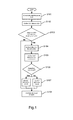
- Fig. 1 a flowchart of an embodiment of the present invention is shown.
- the optic disc position is detected on the additional image. This may be achieved by applying common image processing techniques, such as edge detection filters, transforms detecting circular regions etc.
- the additional image is aligned to the OCT scanning area, such that position of retina features visible on the additional image are aligned with the position of those elements on the OCT image.
- Such an operation may be performed by preparing a two-dimensional projection of the OCT data and using any suitable mathematical optimization technique to find a best fit of these two images. If such a best fit transformation is found, the data retrieved from the additional image is transformed to the space of OCT data, thus enabling its usage in OCT detection algorithms.
- step S101 the disc on retina image is detected by applying a series of image filters to enhance the image and remove noise (such as Gaussian blur or median.
- a series of image filters to enhance the image and remove noise (such as Gaussian blur or median.
- noise such as Gaussian blur or median.
- This is followed by using any algorithm designed to detect circular areas on the image, such as the Hough transform or a set of directional filters to detect disc edges.
- the result comprises coordinates of the centre of the determined optic disc along with a polygon describing the optic disc boundaries on the image of the retina.
- step S102 the coordinates determined in step S101 are fitted or translated to the OCT image space.
- the fitting process in step S102 from may be performed in one of the following ways:
- step S103 When the disc is detected on the retina image and the retina image is properly aligned with OCT data, it is possible to carry out step S103 and check, if the OCT contains the disc area. If so, the disc detection on the OCT image may be performed. Otherwise, all successive algorithms for feature detection may use the retina image disc detection as a reference during the layer detection.
- the OCT data is converted to a two-dimensional image resembling the fundus image, namely the projection image.
- the disc detection on the projection image (steps S104 and S105 from FIG. 1 ) may be carried out in a similar manner to detecting the disc on the retina image.
- disc detection on the retina image detection may be used in several ways to improve and simplify the OCT disc detection algorithm. For instance:
- the retrieved SLO position data may, for example, be used for at least the following purposes:
- Vitreous/RNFL border there are no tractions inside the optic disc area, so one can clearly determine the location of Vitreous/RNFL border there. This information can then be used to distinguish tractions from a Vitreous/RNFL layer (both have similar gradients) to improve the analysis results.
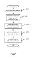
- step S201 of figure 2 initial layer detection is performed. This is required before the translated disc position information from the retina image can be utilised to guide the layer detection result in the OCT image.
- the detection may be performed, for example, by one or more of the following methods:
- the result of the optic disc detection may be usedin the initial detection, such that the algorithm omits the areas, where layer detection cannot be performed reliably.
- the detected layers of the OCT image are corrected basing on anatomical layer thickness in the vicinity of optic disc.
- layers usually do not exceed a defined range of thicknesses in different regions of an eye.
- Information about optic disc may provide a way to correct erroneously detected layers by checking, if the layer fits the appropriate thickness range and making some adjustments, if necessary.
- pathologies may be detected as layers. For example, tractions have very similar noise characteristics as the RNFL layer. Therefore, tractions can be incorrectly detected as a RNFL layer, resulting in faulty detection of all remaining layers. However, tractions usually do not exist in the vicinity of the optic disc, so the detected disc position can be used to distinguish the RNFL layer from these pathologies. This property is utilized by using the disc position to remove false-positive detection on tractions in step S203 of the method of figure 2 .
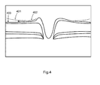
- FIG. 4 shows a traction 401 which is closely aligned to an RNFL layer 403 which is to be detected.
- the detected position of the optic disc can be used to determine a tracking path 402 which starts inside the detected disc region and ends at the edges of the tomogram.
- the algorithm may then try to track the RNFL layer from the detected disc area to the edges of the tomogram, thus avoiding the tractions.
- it may assign values to detected border fragments to choose the valid layer borders instead of tractions.
- step S204 The algorithm may omit this region when detecting layer boundaries in order to avoid false positive detections.
- step S205 information based on the detected optic disc position may be used to make necessary adjustments to adapt the detected layers (other than the RNFL layer) to reflect this known property. For example, the layer boundaries of the non-RNFL layers may be forced to converge to RPE ends on both sides.
- the shape of the SLO disc polygon can be used to evaluate scaling ratios for an OCT projection image.
- the OCT projection image can then be used to detect the optic disc. After detection, there is a high probability that the disc on the OCT image will have a circular shape, which in effect simplifies the detection process.
- Such a program may be provided by itself or may be carried in, by or on a carrier medium.
- the carrier medium may be a storage medium or recording medium, in particular a computer-readable storage medium. Examples include a hard disk drive, DVD, or memory device.
- the carrier medium may also be a transmission medium such as a signal.
- a program embodying the present invention may be distributed, downloaded or uploaded in the form of a signal through a network including the Internet.
- the program may be non-transitory.
Landscapes
- Engineering & Computer Science (AREA)
- Computer Vision & Pattern Recognition (AREA)
- Physics & Mathematics (AREA)
- General Physics & Mathematics (AREA)
- Theoretical Computer Science (AREA)
- Eye Examination Apparatus (AREA)
Priority Applications (4)
| Application Number | Priority Date | Filing Date | Title |
|---|---|---|---|
| EP12178557.0A EP2693399B1 (fr) | 2012-07-30 | 2012-07-30 | Procédés et appareil pour imagerie par tomographie |
| JP2013154963A JP5769766B2 (ja) | 2012-07-30 | 2013-07-25 | 画像処理装置、画像処理方法およびプログラム |
| US13/953,291 US9582732B2 (en) | 2012-07-30 | 2013-07-29 | Method and apparatus for tomography imaging |
| CN201310326084.3A CN103565404B (zh) | 2012-07-30 | 2013-07-30 | 识别关注对象的特征及处理断层图像的方法和设备 |
Applications Claiming Priority (1)
| Application Number | Priority Date | Filing Date | Title |
|---|---|---|---|
| EP12178557.0A EP2693399B1 (fr) | 2012-07-30 | 2012-07-30 | Procédés et appareil pour imagerie par tomographie |
Publications (2)
| Publication Number | Publication Date |
|---|---|
| EP2693399A1 true EP2693399A1 (fr) | 2014-02-05 |
| EP2693399B1 EP2693399B1 (fr) | 2019-02-27 |
Family
ID=46982378
Family Applications (1)
| Application Number | Title | Priority Date | Filing Date |
|---|---|---|---|
| EP12178557.0A Not-in-force EP2693399B1 (fr) | 2012-07-30 | 2012-07-30 | Procédés et appareil pour imagerie par tomographie |
Country Status (4)
| Country | Link |
|---|---|
| US (1) | US9582732B2 (fr) |
| EP (1) | EP2693399B1 (fr) |
| JP (1) | JP5769766B2 (fr) |
| CN (1) | CN103565404B (fr) |
Families Citing this family (8)
| Publication number | Priority date | Publication date | Assignee | Title |
|---|---|---|---|---|
| EP2693399B1 (fr) * | 2012-07-30 | 2019-02-27 | Canon Kabushiki Kaisha | Procédés et appareil pour imagerie par tomographie |
| JP6118698B2 (ja) * | 2013-09-30 | 2017-04-19 | 株式会社Ihi | 画像解析装置及びプログラム |
| CN104434026B (zh) * | 2014-12-17 | 2016-08-17 | 深圳市斯尔顿科技有限公司 | 视网膜注视点偏离黄斑中心凹的检测方法 |
| US10123689B2 (en) | 2015-10-28 | 2018-11-13 | Oregon Health & Science University | Systems and methods for retinal layer segmentation in OCT imaging and OCT angiography |
| CN106447663A (zh) * | 2016-09-30 | 2017-02-22 | 深圳市莫廷影像技术有限公司 | 一种去除重影的眼科oct图像高清配准方法及装置 |
| US10346723B2 (en) * | 2016-11-01 | 2019-07-09 | Snap Inc. | Neural network for object detection in images |
| US10832402B2 (en) * | 2017-03-10 | 2020-11-10 | Carl Zeiss Meditec, Inc. | Methods for detection and enhanced visualization of pathologies in a human eye |
| CN112526608B (zh) * | 2020-11-16 | 2022-08-02 | 哈尔滨工程大学 | 一种基于射线简正波的深海完整声道会聚区分类方法 |
Citations (3)
| Publication number | Priority date | Publication date | Assignee | Title |
|---|---|---|---|---|
| US20080100612A1 (en) * | 2006-10-27 | 2008-05-01 | Dastmalchi Shahram S | User interface for efficiently displaying relevant oct imaging data |
| US20120050308A1 (en) * | 2010-08-30 | 2012-03-01 | Canon Kabushiki Kaisha | Image processing apparatus, image processing method, image processing system, and computer readable memory |
| US20120070049A1 (en) * | 2009-06-02 | 2012-03-22 | Canon Kabushiki Kaisha | Image processing apparatus, control method thereof, and computer program |
Family Cites Families (27)
| Publication number | Priority date | Publication date | Assignee | Title |
|---|---|---|---|---|
| US7668342B2 (en) * | 2005-09-09 | 2010-02-23 | Carl Zeiss Meditec, Inc. | Method of bioimage data processing for revealing more meaningful anatomic features of diseased tissues |
| JP4916779B2 (ja) * | 2005-09-29 | 2012-04-18 | 株式会社トプコン | 眼底観察装置 |
| US7524061B2 (en) * | 2005-10-12 | 2009-04-28 | Siemens Corporate Research, Inc. | System and method for robust optic disk detection in retinal images using vessel structure and radon transform |
| JP5192394B2 (ja) * | 2006-01-19 | 2013-05-08 | オプトビュー,インコーポレーテッド | 眼を光コヒーレンス断層撮影によって検査する方法 |
| US7782464B2 (en) * | 2006-05-12 | 2010-08-24 | The General Hospital Corporation | Processes, arrangements and systems for providing a fiber layer thickness map based on optical coherence tomography images |
| JP5095167B2 (ja) * | 2006-09-19 | 2012-12-12 | 株式会社トプコン | 眼底観察装置、眼底画像表示装置及び眼底観察プログラム |
| US7992999B2 (en) * | 2008-04-23 | 2011-08-09 | University Of Pittsburgh - Of The Commonwealth System Of Higher Education | Automated assessment of optic nerve head with spectral domain optical coherence tomography |
| JP4819851B2 (ja) * | 2008-07-31 | 2011-11-24 | キヤノン株式会社 | 診断支援装置およびその方法、プログラム、記録媒体 |
| WO2010138645A2 (fr) * | 2009-05-29 | 2010-12-02 | University Of Pittsburgh - Of The Commonwealth System Of Higher Education | Segmentation de vaisseau sanguin avec tomographie par cohérence optique du domaine spectral 3d |
| JP4850927B2 (ja) * | 2009-06-02 | 2012-01-11 | キヤノン株式会社 | 画像処理装置、画像処理方法及びコンピュータプログラム |
| JP2011087672A (ja) * | 2009-10-21 | 2011-05-06 | Topcon Corp | 眼底画像処理装置及び眼底観察装置 |
| JP5582772B2 (ja) * | 2009-12-08 | 2014-09-03 | キヤノン株式会社 | 画像処理装置及び画像処理方法 |
| WO2011074078A1 (fr) * | 2009-12-15 | 2011-06-23 | キヤノン株式会社 | Dispositif de traitement d'image, procédé de traitement d'image, système d'imagerie, et programme |
| JP5631032B2 (ja) * | 2010-03-19 | 2014-11-26 | キヤノン株式会社 | 画像処理装置、画像処理システム、画像処理方法、及び画像処理をコンピュータに実行させるためのプログラム |
| JP5645432B2 (ja) * | 2010-03-19 | 2014-12-24 | キヤノン株式会社 | 画像処理装置、画像処理システム、画像処理方法、及び画像処理をコンピュータに実行させるためのプログラム |
| JP5451492B2 (ja) * | 2010-03-31 | 2014-03-26 | キヤノン株式会社 | 画像処理装置、その制御方法及びプログラム |
| JP5702991B2 (ja) | 2010-11-19 | 2015-04-15 | キヤノン株式会社 | 画像処理装置及び画像処理方法 |
| JP5751815B2 (ja) * | 2010-12-09 | 2015-07-22 | キヤノン株式会社 | 画像処理装置、撮影システム、画像処理方法及びプログラム |
| US8801187B1 (en) * | 2010-12-23 | 2014-08-12 | Carl Zeiss Meditec, Inc. | Methods to reduce variance in OCT analysis of the macula |
| JP5822485B2 (ja) * | 2011-02-25 | 2015-11-24 | キヤノン株式会社 | 画像処理装置、画像処理方法、画像処理システム、slo装置、およびプログラム |
| JP5792967B2 (ja) * | 2011-02-25 | 2015-10-14 | キヤノン株式会社 | 画像処理装置及び画像処理システム |
| EP2690581A1 (fr) * | 2012-07-27 | 2014-01-29 | Canon Kabushiki Kaisha | Procédé et appareil pour la détection d'une pupille |
| EP2693399B1 (fr) * | 2012-07-30 | 2019-02-27 | Canon Kabushiki Kaisha | Procédés et appareil pour imagerie par tomographie |
| EP2692284A1 (fr) * | 2012-07-30 | 2014-02-05 | Canon Kabushiki Kaisha | Procédé et appareil pour correction géométrique des données de tomographie par cohérence optique représentant un balayage obtenu par imagerie OCT d'un échantillon |
| JP6099903B2 (ja) * | 2012-08-30 | 2017-03-22 | キヤノン株式会社 | 画像処理装置および画像処理方法 |
| US9107610B2 (en) * | 2012-11-30 | 2015-08-18 | Kabushiki Kaisha Topcon | Optic neuropathy detection with three-dimensional optical coherence tomography |
| JP2016002380A (ja) * | 2014-06-18 | 2016-01-12 | キヤノン株式会社 | 画像処理装置、その作動方法及びプログラム |
-
2012
- 2012-07-30 EP EP12178557.0A patent/EP2693399B1/fr not_active Not-in-force
-
2013
- 2013-07-25 JP JP2013154963A patent/JP5769766B2/ja active Active
- 2013-07-29 US US13/953,291 patent/US9582732B2/en not_active Expired - Fee Related
- 2013-07-30 CN CN201310326084.3A patent/CN103565404B/zh not_active Expired - Fee Related
Patent Citations (3)
| Publication number | Priority date | Publication date | Assignee | Title |
|---|---|---|---|---|
| US20080100612A1 (en) * | 2006-10-27 | 2008-05-01 | Dastmalchi Shahram S | User interface for efficiently displaying relevant oct imaging data |
| US20120070049A1 (en) * | 2009-06-02 | 2012-03-22 | Canon Kabushiki Kaisha | Image processing apparatus, control method thereof, and computer program |
| US20120050308A1 (en) * | 2010-08-30 | 2012-03-01 | Canon Kabushiki Kaisha | Image processing apparatus, image processing method, image processing system, and computer readable memory |
Non-Patent Citations (3)
| Title |
|---|
| B.J. ANTONY ET AL: "Automated 3-D Segmentation of Intraretinal Layers fromOptic Nerve Head Optical Coherence Tomography Images", MEDICAL IMAGING 2010: BIOMEDICAL APPLICATIONS IN MOLECULAR, STRUCTURAL, AND FUNCTIONAL IMAGING, 13 February 2010 (2010-02-13), pages 1 - 12, XP040546962 * |
| M. D. ABRAMOFF ET AL: "Automated Segmentation of the Cup and Rim from Spectral Domain OCT of the Optic Nerve Head", INVESTIGATIVE OPHTHALMOLOGY & VISUAL SCIENCE, vol. 50, no. 12, 1 December 2009 (2009-12-01), pages 5778 - 5784, XP055049457, ISSN: 0146-0404, DOI: 10.1167/iovs.09-3790 * |
| MICHAEL D ABRAMOFF ET AL: "Retinal Imaging and Image Analysis", IEEE REVIEWS IN BIOMEDICAL ENGINEERING, IEEE, USA, vol. 3, 1 January 2010 (2010-01-01), pages 169 - 208, XP011339903, ISSN: 1937-3333, DOI: 10.1109/RBME.2010.2084567 * |
Also Published As
| Publication number | Publication date |
|---|---|
| CN103565404B (zh) | 2016-08-10 |
| CN103565404A (zh) | 2014-02-12 |
| JP5769766B2 (ja) | 2015-08-26 |
| US9582732B2 (en) | 2017-02-28 |
| JP2014023933A (ja) | 2014-02-06 |
| US20140029825A1 (en) | 2014-01-30 |
| EP2693399B1 (fr) | 2019-02-27 |
Similar Documents
| Publication | Publication Date | Title |
|---|---|---|
| US9582732B2 (en) | Method and apparatus for tomography imaging | |
| JP7579372B2 (ja) | 医用画像処理装置、医用画像処理システム、医用画像処理方法及びプログラム | |
| US10251548B2 (en) | Image processing apparatus, estimation method, system, and medium | |
| US9585560B2 (en) | Image processing apparatus, image processing method, and program | |
| US10194866B2 (en) | Methods and apparatus for reducing artifacts in OCT angiography using machine learning techniques | |
| US20110137157A1 (en) | Image processing apparatus and image processing method | |
| JP4909377B2 (ja) | 画像処理装置及びその制御方法、コンピュータプログラム | |
| US9418423B2 (en) | Motion correction and normalization of features in optical coherence tomography | |
| JP5631339B2 (ja) | 画像処理装置、画像処理方法、眼科装置、眼科システム及びコンピュータプログラム | |
| CN102858230A (zh) | 断层图像观察设备、处理方法和非瞬态计算机可读存储介质 | |
| US10573007B2 (en) | Image processing apparatus, image processing method, and image processing program | |
| EP3417401B1 (fr) | Procédé de réduction d'artefacts dans une oct grâce à des techniques d'apprentissage automatique | |
| Niemeijer et al. | Registration of 3D spectral OCT volumes combining ICP with a graph-based approach | |
| US10916012B2 (en) | Image processing apparatus and image processing method | |
| JP7514344B2 (ja) | Oct血管造影のためのoct-bスキャンの前処理 | |
| WO2015154200A1 (fr) | Procédé destiné à l'analyse de données d'image représentant un volume tridimensionnel de tissu biologique | |
| CN116030042B (zh) | 一种针对医生目诊的诊断装置、方法、设备及存储介质 | |
| JP2022062620A (ja) | 画像処理装置、画像処理方法及びプログラム | |
| US20180240240A1 (en) | Image processing apparatus, image processing method, and image processing program | |
| Medhi et al. | Improved analysis of diabetic maculopathy using level set spatial fuzzy clustering | |
| JP2022062619A (ja) | 画像処理装置、画像処理方法及びプログラム | |
| Muramatsu et al. | Detection of Eye Diseases | |
| JP2015144963A (ja) | 画像処理装置、画像処理方法及びプログラム |
Legal Events
| Date | Code | Title | Description |
|---|---|---|---|
| AK | Designated contracting states |
Kind code of ref document: A1 Designated state(s): AL AT BE BG CH CY CZ DE DK EE ES FI FR GB GR HR HU IE IS IT LI LT LU LV MC MK MT NL NO PL PT RO RS SE SI SK SM TR |
|
| AX | Request for extension of the european patent |
Extension state: BA ME |
|
| PUAI | Public reference made under article 153(3) epc to a published international application that has entered the european phase |
Free format text: ORIGINAL CODE: 0009012 |
|
| 17P | Request for examination filed |
Effective date: 20140709 |
|
| RBV | Designated contracting states (corrected) |
Designated state(s): AL AT BE BG CH CY CZ DE DK EE ES FI FR GB GR HR HU IE IS IT LI LT LU LV MC MK MT NL NO PL PT RO RS SE SI SK SM TR |
|
| STAA | Information on the status of an ep patent application or granted ep patent |
Free format text: STATUS: EXAMINATION IS IN PROGRESS |
|
| 17Q | First examination report despatched |
Effective date: 20170324 |
|
| REG | Reference to a national code |
Ref country code: DE Ref legal event code: R079 Ref document number: 602012057053 Country of ref document: DE Free format text: PREVIOUS MAIN CLASS: G06T0007000000 Ipc: G06T0007174000 |
|
| GRAP | Despatch of communication of intention to grant a patent |
Free format text: ORIGINAL CODE: EPIDOSNIGR1 |
|
| STAA | Information on the status of an ep patent application or granted ep patent |
Free format text: STATUS: GRANT OF PATENT IS INTENDED |
|
| RIC1 | Information provided on ipc code assigned before grant |
Ipc: G06T 7/174 20170101AFI20180921BHEP |
|
| INTG | Intention to grant announced |
Effective date: 20181022 |
|
| GRAS | Grant fee paid |
Free format text: ORIGINAL CODE: EPIDOSNIGR3 |
|
| GRAA | (expected) grant |
Free format text: ORIGINAL CODE: 0009210 |
|
| STAA | Information on the status of an ep patent application or granted ep patent |
Free format text: STATUS: THE PATENT HAS BEEN GRANTED |
|
| RIC1 | Information provided on ipc code assigned before grant |
Ipc: G06T 7/174 20170101AFI20180921BHEP |
|
| AK | Designated contracting states |
Kind code of ref document: B1 Designated state(s): AL AT BE BG CH CY CZ DE DK EE ES FI FR GB GR HR HU IE IS IT LI LT LU LV MC MK MT NL NO PL PT RO RS SE SI SK SM TR |
|
| REG | Reference to a national code |
Ref country code: GB Ref legal event code: FG4D |
|
| REG | Reference to a national code |
Ref country code: CH Ref legal event code: EP |
|
| REG | Reference to a national code |
Ref country code: AT Ref legal event code: REF Ref document number: 1102456 Country of ref document: AT Kind code of ref document: T Effective date: 20190315 |
|
| REG | Reference to a national code |
Ref country code: IE Ref legal event code: FG4D |
|
| REG | Reference to a national code |
Ref country code: DE Ref legal event code: R096 Ref document number: 602012057053 Country of ref document: DE |
|
| REG | Reference to a national code |
Ref country code: NL Ref legal event code: MP Effective date: 20190227 |
|
| REG | Reference to a national code |
Ref country code: LT Ref legal event code: MG4D |
|
| PG25 | Lapsed in a contracting state [announced via postgrant information from national office to epo] |
Ref country code: NL Free format text: LAPSE BECAUSE OF FAILURE TO SUBMIT A TRANSLATION OF THE DESCRIPTION OR TO PAY THE FEE WITHIN THE PRESCRIBED TIME-LIMIT Effective date: 20190227 Ref country code: SE Free format text: LAPSE BECAUSE OF FAILURE TO SUBMIT A TRANSLATION OF THE DESCRIPTION OR TO PAY THE FEE WITHIN THE PRESCRIBED TIME-LIMIT Effective date: 20190227 Ref country code: PT Free format text: LAPSE BECAUSE OF FAILURE TO SUBMIT A TRANSLATION OF THE DESCRIPTION OR TO PAY THE FEE WITHIN THE PRESCRIBED TIME-LIMIT Effective date: 20190627 Ref country code: LT Free format text: LAPSE BECAUSE OF FAILURE TO SUBMIT A TRANSLATION OF THE DESCRIPTION OR TO PAY THE FEE WITHIN THE PRESCRIBED TIME-LIMIT Effective date: 20190227 Ref country code: FI Free format text: LAPSE BECAUSE OF FAILURE TO SUBMIT A TRANSLATION OF THE DESCRIPTION OR TO PAY THE FEE WITHIN THE PRESCRIBED TIME-LIMIT Effective date: 20190227 Ref country code: NO Free format text: LAPSE BECAUSE OF FAILURE TO SUBMIT A TRANSLATION OF THE DESCRIPTION OR TO PAY THE FEE WITHIN THE PRESCRIBED TIME-LIMIT Effective date: 20190527 |
|
| PG25 | Lapsed in a contracting state [announced via postgrant information from national office to epo] |
Ref country code: IS Free format text: LAPSE BECAUSE OF FAILURE TO SUBMIT A TRANSLATION OF THE DESCRIPTION OR TO PAY THE FEE WITHIN THE PRESCRIBED TIME-LIMIT Effective date: 20190627 Ref country code: BG Free format text: LAPSE BECAUSE OF FAILURE TO SUBMIT A TRANSLATION OF THE DESCRIPTION OR TO PAY THE FEE WITHIN THE PRESCRIBED TIME-LIMIT Effective date: 20190527 Ref country code: LV Free format text: LAPSE BECAUSE OF FAILURE TO SUBMIT A TRANSLATION OF THE DESCRIPTION OR TO PAY THE FEE WITHIN THE PRESCRIBED TIME-LIMIT Effective date: 20190227 Ref country code: RS Free format text: LAPSE BECAUSE OF FAILURE TO SUBMIT A TRANSLATION OF THE DESCRIPTION OR TO PAY THE FEE WITHIN THE PRESCRIBED TIME-LIMIT Effective date: 20190227 Ref country code: GR Free format text: LAPSE BECAUSE OF FAILURE TO SUBMIT A TRANSLATION OF THE DESCRIPTION OR TO PAY THE FEE WITHIN THE PRESCRIBED TIME-LIMIT Effective date: 20190528 Ref country code: HR Free format text: LAPSE BECAUSE OF FAILURE TO SUBMIT A TRANSLATION OF THE DESCRIPTION OR TO PAY THE FEE WITHIN THE PRESCRIBED TIME-LIMIT Effective date: 20190227 |
|
| REG | Reference to a national code |
Ref country code: AT Ref legal event code: MK05 Ref document number: 1102456 Country of ref document: AT Kind code of ref document: T Effective date: 20190227 |
|
| PG25 | Lapsed in a contracting state [announced via postgrant information from national office to epo] |
Ref country code: CZ Free format text: LAPSE BECAUSE OF FAILURE TO SUBMIT A TRANSLATION OF THE DESCRIPTION OR TO PAY THE FEE WITHIN THE PRESCRIBED TIME-LIMIT Effective date: 20190227 Ref country code: IT Free format text: LAPSE BECAUSE OF FAILURE TO SUBMIT A TRANSLATION OF THE DESCRIPTION OR TO PAY THE FEE WITHIN THE PRESCRIBED TIME-LIMIT Effective date: 20190227 Ref country code: SK Free format text: LAPSE BECAUSE OF FAILURE TO SUBMIT A TRANSLATION OF THE DESCRIPTION OR TO PAY THE FEE WITHIN THE PRESCRIBED TIME-LIMIT Effective date: 20190227 Ref country code: AL Free format text: LAPSE BECAUSE OF FAILURE TO SUBMIT A TRANSLATION OF THE DESCRIPTION OR TO PAY THE FEE WITHIN THE PRESCRIBED TIME-LIMIT Effective date: 20190227 Ref country code: DK Free format text: LAPSE BECAUSE OF FAILURE TO SUBMIT A TRANSLATION OF THE DESCRIPTION OR TO PAY THE FEE WITHIN THE PRESCRIBED TIME-LIMIT Effective date: 20190227 Ref country code: EE Free format text: LAPSE BECAUSE OF FAILURE TO SUBMIT A TRANSLATION OF THE DESCRIPTION OR TO PAY THE FEE WITHIN THE PRESCRIBED TIME-LIMIT Effective date: 20190227 Ref country code: ES Free format text: LAPSE BECAUSE OF FAILURE TO SUBMIT A TRANSLATION OF THE DESCRIPTION OR TO PAY THE FEE WITHIN THE PRESCRIBED TIME-LIMIT Effective date: 20190227 Ref country code: RO Free format text: LAPSE BECAUSE OF FAILURE TO SUBMIT A TRANSLATION OF THE DESCRIPTION OR TO PAY THE FEE WITHIN THE PRESCRIBED TIME-LIMIT Effective date: 20190227 |
|
| REG | Reference to a national code |
Ref country code: DE Ref legal event code: R097 Ref document number: 602012057053 Country of ref document: DE |
|
| PG25 | Lapsed in a contracting state [announced via postgrant information from national office to epo] |
Ref country code: SM Free format text: LAPSE BECAUSE OF FAILURE TO SUBMIT A TRANSLATION OF THE DESCRIPTION OR TO PAY THE FEE WITHIN THE PRESCRIBED TIME-LIMIT Effective date: 20190227 Ref country code: PL Free format text: LAPSE BECAUSE OF FAILURE TO SUBMIT A TRANSLATION OF THE DESCRIPTION OR TO PAY THE FEE WITHIN THE PRESCRIBED TIME-LIMIT Effective date: 20190227 |
|
| PG25 | Lapsed in a contracting state [announced via postgrant information from national office to epo] |
Ref country code: AT Free format text: LAPSE BECAUSE OF FAILURE TO SUBMIT A TRANSLATION OF THE DESCRIPTION OR TO PAY THE FEE WITHIN THE PRESCRIBED TIME-LIMIT Effective date: 20190227 |
|
| PLBE | No opposition filed within time limit |
Free format text: ORIGINAL CODE: 0009261 |
|
| STAA | Information on the status of an ep patent application or granted ep patent |
Free format text: STATUS: NO OPPOSITION FILED WITHIN TIME LIMIT |
|
| 26N | No opposition filed |
Effective date: 20191128 |
|
| PG25 | Lapsed in a contracting state [announced via postgrant information from national office to epo] |
Ref country code: SI Free format text: LAPSE BECAUSE OF FAILURE TO SUBMIT A TRANSLATION OF THE DESCRIPTION OR TO PAY THE FEE WITHIN THE PRESCRIBED TIME-LIMIT Effective date: 20190227 Ref country code: MC Free format text: LAPSE BECAUSE OF FAILURE TO SUBMIT A TRANSLATION OF THE DESCRIPTION OR TO PAY THE FEE WITHIN THE PRESCRIBED TIME-LIMIT Effective date: 20190227 |
|
| REG | Reference to a national code |
Ref country code: CH Ref legal event code: PL |
|
| GBPC | Gb: european patent ceased through non-payment of renewal fee |
Effective date: 20190730 |
|
| PG25 | Lapsed in a contracting state [announced via postgrant information from national office to epo] |
Ref country code: TR Free format text: LAPSE BECAUSE OF FAILURE TO SUBMIT A TRANSLATION OF THE DESCRIPTION OR TO PAY THE FEE WITHIN THE PRESCRIBED TIME-LIMIT Effective date: 20190227 |
|
| REG | Reference to a national code |
Ref country code: BE Ref legal event code: MM Effective date: 20190731 |
|
| PG25 | Lapsed in a contracting state [announced via postgrant information from national office to epo] |
Ref country code: GB Free format text: LAPSE BECAUSE OF NON-PAYMENT OF DUE FEES Effective date: 20190730 |
|
| PG25 | Lapsed in a contracting state [announced via postgrant information from national office to epo] |
Ref country code: LI Free format text: LAPSE BECAUSE OF NON-PAYMENT OF DUE FEES Effective date: 20190731 Ref country code: CH Free format text: LAPSE BECAUSE OF NON-PAYMENT OF DUE FEES Effective date: 20190731 Ref country code: LU Free format text: LAPSE BECAUSE OF NON-PAYMENT OF DUE FEES Effective date: 20190730 Ref country code: BE Free format text: LAPSE BECAUSE OF NON-PAYMENT OF DUE FEES Effective date: 20190731 |
|
| PG25 | Lapsed in a contracting state [announced via postgrant information from national office to epo] |
Ref country code: FR Free format text: LAPSE BECAUSE OF NON-PAYMENT OF DUE FEES Effective date: 20190731 |
|
| PG25 | Lapsed in a contracting state [announced via postgrant information from national office to epo] |
Ref country code: IE Free format text: LAPSE BECAUSE OF NON-PAYMENT OF DUE FEES Effective date: 20190730 |
|
| PG25 | Lapsed in a contracting state [announced via postgrant information from national office to epo] |
Ref country code: CY Free format text: LAPSE BECAUSE OF FAILURE TO SUBMIT A TRANSLATION OF THE DESCRIPTION OR TO PAY THE FEE WITHIN THE PRESCRIBED TIME-LIMIT Effective date: 20190227 |
|
| PG25 | Lapsed in a contracting state [announced via postgrant information from national office to epo] |
Ref country code: MT Free format text: LAPSE BECAUSE OF FAILURE TO SUBMIT A TRANSLATION OF THE DESCRIPTION OR TO PAY THE FEE WITHIN THE PRESCRIBED TIME-LIMIT Effective date: 20190227 Ref country code: HU Free format text: LAPSE BECAUSE OF FAILURE TO SUBMIT A TRANSLATION OF THE DESCRIPTION OR TO PAY THE FEE WITHIN THE PRESCRIBED TIME-LIMIT; INVALID AB INITIO Effective date: 20120730 |
|
| PGFP | Annual fee paid to national office [announced via postgrant information from national office to epo] |
Ref country code: DE Payment date: 20210622 Year of fee payment: 10 |
|
| PG25 | Lapsed in a contracting state [announced via postgrant information from national office to epo] |
Ref country code: MK Free format text: LAPSE BECAUSE OF FAILURE TO SUBMIT A TRANSLATION OF THE DESCRIPTION OR TO PAY THE FEE WITHIN THE PRESCRIBED TIME-LIMIT Effective date: 20190227 |
|
| REG | Reference to a national code |
Ref country code: DE Ref legal event code: R119 Ref document number: 602012057053 Country of ref document: DE |
|
| PG25 | Lapsed in a contracting state [announced via postgrant information from national office to epo] |
Ref country code: DE Free format text: LAPSE BECAUSE OF NON-PAYMENT OF DUE FEES Effective date: 20230201 |