EP2703005A1 - Inhibiteurs d'intégrine alpha6/E-cadhérine complexe - Google Patents
Inhibiteurs d'intégrine alpha6/E-cadhérine complexe Download PDFInfo
- Publication number
- EP2703005A1 EP2703005A1 EP12182994.9A EP12182994A EP2703005A1 EP 2703005 A1 EP2703005 A1 EP 2703005A1 EP 12182994 A EP12182994 A EP 12182994A EP 2703005 A1 EP2703005 A1 EP 2703005A1
- Authority
- EP
- European Patent Office
- Prior art keywords
- integrin
- cadherin
- seq
- inhibiting agent
- cells
- Prior art date
- Legal status (The legal status is an assumption and is not a legal conclusion. Google has not performed a legal analysis and makes no representation as to the accuracy of the status listed.)
- Withdrawn
Links
Images


Classifications
-
- A—HUMAN NECESSITIES
- A61—MEDICAL OR VETERINARY SCIENCE; HYGIENE
- A61K—PREPARATIONS FOR MEDICAL, DENTAL OR TOILETRY PURPOSES
- A61K38/00—Medicinal preparations containing peptides
- A61K38/16—Peptides having more than 20 amino acids; Gastrins; Somatostatins; Melanotropins; Derivatives thereof
- A61K38/17—Peptides having more than 20 amino acids; Gastrins; Somatostatins; Melanotropins; Derivatives thereof from animals; from humans
- A61K38/177—Receptors; Cell surface antigens; Cell surface determinants
- A61K38/1777—Integrin superfamily
-
- A—HUMAN NECESSITIES
- A61—MEDICAL OR VETERINARY SCIENCE; HYGIENE
- A61P—SPECIFIC THERAPEUTIC ACTIVITY OF CHEMICAL COMPOUNDS OR MEDICINAL PREPARATIONS
- A61P35/00—Antineoplastic agents
- A61P35/04—Antineoplastic agents specific for metastasis
-
- C—CHEMISTRY; METALLURGY
- C07—ORGANIC CHEMISTRY
- C07K—PEPTIDES
- C07K14/00—Peptides having more than 20 amino acids; Gastrins; Somatostatins; Melanotropins; Derivatives thereof
- C07K14/435—Peptides having more than 20 amino acids; Gastrins; Somatostatins; Melanotropins; Derivatives thereof from animals; from humans
- C07K14/705—Receptors; Cell surface antigens; Cell surface determinants
- C07K14/70546—Integrin superfamily
-
- C—CHEMISTRY; METALLURGY
- C07—ORGANIC CHEMISTRY
- C07K—PEPTIDES
- C07K7/00—Peptides having 5 to 20 amino acids in a fully defined sequence; Derivatives thereof
- C07K7/04—Linear peptides containing only normal peptide links
- C07K7/06—Linear peptides containing only normal peptide links having 5 to 11 amino acids
-
- G—PHYSICS
- G01—MEASURING; TESTING
- G01N—INVESTIGATING OR ANALYSING MATERIALS BY DETERMINING THEIR CHEMICAL OR PHYSICAL PROPERTIES
- G01N33/00—Investigating or analysing materials by specific methods not covered by groups G01N1/00 - G01N31/00
- G01N33/48—Biological material, e.g. blood, urine; Haemocytometers
- G01N33/50—Chemical analysis of biological material, e.g. blood, urine; Testing involving biospecific ligand binding methods; Immunological testing
- G01N33/53—Immunoassay; Biospecific binding assay; Materials therefor
- G01N33/575—Immunoassay; Biospecific binding assay; Materials therefor for cancer
- G01N33/57535—Immunoassay; Biospecific binding assay; Materials therefor for cancer of the large intestine, e.g. colon, rectum or anus
-
- G—PHYSICS
- G01—MEASURING; TESTING
- G01N—INVESTIGATING OR ANALYSING MATERIALS BY DETERMINING THEIR CHEMICAL OR PHYSICAL PROPERTIES
- G01N33/00—Investigating or analysing materials by specific methods not covered by groups G01N1/00 - G01N31/00
- G01N33/48—Biological material, e.g. blood, urine; Haemocytometers
- G01N33/50—Chemical analysis of biological material, e.g. blood, urine; Testing involving biospecific ligand binding methods; Immunological testing
- G01N33/53—Immunoassay; Biospecific binding assay; Materials therefor
- G01N33/575—Immunoassay; Biospecific binding assay; Materials therefor for cancer
- G01N33/57557—Immunoassay; Biospecific binding assay; Materials therefor for cancer of other specific parts of the body, e.g. brain
-
- G—PHYSICS
- G01—MEASURING; TESTING
- G01N—INVESTIGATING OR ANALYSING MATERIALS BY DETERMINING THEIR CHEMICAL OR PHYSICAL PROPERTIES
- G01N2333/00—Assays involving biological materials from specific organisms or of a specific nature
- G01N2333/435—Assays involving biological materials from specific organisms or of a specific nature from animals; from humans
- G01N2333/705—Assays involving receptors, cell surface antigens or cell surface determinants
- G01N2333/70546—Integrin superfamily, e.g. VLAs, leuCAM, GPIIb/GPIIIa, LPAM
-
- G—PHYSICS
- G01—MEASURING; TESTING
- G01N—INVESTIGATING OR ANALYSING MATERIALS BY DETERMINING THEIR CHEMICAL OR PHYSICAL PROPERTIES
- G01N2500/00—Screening for compounds of potential therapeutic value
-
- G—PHYSICS
- G01—MEASURING; TESTING
- G01N—INVESTIGATING OR ANALYSING MATERIALS BY DETERMINING THEIR CHEMICAL OR PHYSICAL PROPERTIES
- G01N2800/00—Detection or diagnosis of diseases
- G01N2800/52—Predicting or monitoring the response to treatment, e.g. for selection of therapy based on assay results in personalised medicine; Prognosis
Definitions
- the present invention relates to inhibiting agents of a ⁇ 6 integrin/E-cadherin molecular complex for use as a medicament, particularly for the prevention or/and treatment of metastases of a primary cancer disease, and a method of determining the prognosis of metastatic homing of a primary cancer disease.
- Cancer known medically as a malignant neoplasm is a term for a large group of different diseases, all involving unregulated cell growth.
- cells divide and grow uncontrollably, forming malignant tumors, and invade nearby parts of the body.
- the cancer may also spread to more distant parts of the body through the lymphatic system or bloodstream.
- a primary tumor When the area of cancer cells at the originating site becomes clinically detectable, it is called a primary tumor.
- Some cancer cells acquire the ability to penetrate and infiltrate surrounding normal tissues in the local area, forming a new tumor.
- the newly formed tumor within the tissue is called a local metastasis.
- Some cancer cells acquire the ability to penetrate the walls of lymphatic and/or blood vessels, after which they are able to circulate through the blood stream (circulating tumor cells) to other sites and tissues in the body. This process is known as lymphatic and hematogenous spread, respectively.
- This new tumor is known as a metastatic (or secondary) tumor.
- the impact of secondary tumors is often more fatal than that of the primary tumor.
- CRC Advanced colorectal cancer
- a pivotal contribution to metastatic colonization comes from components of the host tissue and stroma. Therefore, targeting cancer microenvironments provides a promising strategy for the prevention or/and treatment of metastases.
- Angiopoietin-like 6 is a secreted factor whose mRNA has been detected particularly in the liver of humans. Although this protein shares a common structure with other members of the angiopoietin family, and particularly a coiled-coil domain in the N-terminal portion and a fibrinogen-like domain in the C-terminal portion, it does not bind to the Tie1 or Tie2 receptor and is currently considered an orphan ligand ( Kim et al. 2000, Biochem J, 346 Pt 3:603-610 ; Oike et al. 2003, Proc Natl Acad Sci USA, 100:9494-9499 ; Oike et al. 2004, Blood, 103:3760-3765 ).
- Angiopoietin-like 6 regulates angiogenesis by preventing endothelial cell apoptosis, inducing endothelial cell migration and vascular leakiness and enhancing blood flow (Kim et al; Oike et al; Urano et al. 2008, Arterioscler Thromb Vasc Biol, 28:827-834 ).
- Some evidence suggests that RGD-binding integrins might be involved in angiopoietin-like 6-mediated cell adhesion, spreading and migration, although a direct interaction with integrins has not been described thus far ( Zhang et al. 2006, Biochem Biophys Res Commun, 347:100-108 ).
- Integrin ⁇ 6 complexed with either ⁇ 1 or ⁇ 4 subunit, is a receptor for laminin with an emerging role in regulating angiogenesis and cancer progression through both direct and indirect mechanisms ( Humphries et al. 2006, J Cell Sci, 119:3901-3903 ; Primo et al. 2010, Cancer Res, 70:5759-5769 , Lee et al. 2006, J Biol Chem, 281:40450-40460 ; Gonzalez et al. 2002, Proc Natl Acad Sci U S A, 99:16075-16080 , Rabinovitz et al. 2001, Mol Biol Cell, 12:4030-4043 ; Robertson et al. 2008, Curr Pharm Des, 14:296-305 ).
- E-cadherin is a well-described oncosuppressor protein, whose expression in the primary tumor counteracts cell detachment and is therefore associated with a better outcome ( Christofori, 2003, Embo J, 22:2318-2323 ).
- Decreased production of E-cadherin is one of the central events underlying epithelial-mesenchymal transition and carcinoma progression, in response to different cellular events such as the acquisition of loss-of-function mutations and loss-of-heterozygosis for the mutant allele, transcriptional or epigenetic repression and aberrant cellular localization ( Ilyas et al. 1997, Gut, 40:654-659 ; Natalwala et al.
- angiopoietin-like 6 acts as a ligand for cells that express a receptor complex of ⁇ 6 integrin and E-cadherin.
- the interaction between the angiopoietin-like 6 and the ⁇ 6 integrin/E-cadherin complex is found to have significant influence in metastasis homing and colonization.
- Experimental results show that inhibition of the ⁇ 6 integrin/E-cadherin molecular complex may inhibit/reduce metastases on different levels.
- a first aspect of the present invention refers to an inhibiting agent of the ⁇ 6 integrin/E-cadherin molecular complex for use as a medicament, particularly for the prevention or/and treatment of metastasis of a primary cancer disease such as colorectal, bone, brain, breast, cervix, colon, gastric, liver, lung, pancreas, exocrine pancreas, duodenum, ovarian, renal, prostate, stomach, soft tissue, bone marrow, esophagus, skin cancer or lymphoma, particularly colorectal, stomach and lung cancer, more particularly colorectal cancer.
- the inhibiting agent may be used e.g. in human or veterinary medicine.
- the ⁇ 6 integrin/E-cadherin molecular complex is formed by direct and/or indirect molecular interaction between the full length ⁇ 6 integrin protein (140 kD, SEQ ID No 16) or a proteolytic fragment thereof and the full length E-cadherin protein (120 kD, SEQ ID No 17) or a proteolytic fragment thereof.
- Proteolytic fragments of ⁇ 6 integrin protein preferably have a molecular weight of 10 to 130 kDa, preferably 20 to 120 kDa. More preferably, the proteolytic fragments include the fragments of aa24-1073, aa24-899, aa903-1073, aa24-594, aa595-899 and/or aa595-1073 of the full-length ⁇ 6 integrin protein (SEQ ID No 16).
- Proteolytic fragments of E-cadherin protein preferably have a molecular weight of 20 to 100 kDa, preferably 30 to 97 kDa, particularly 30 kDa, 40 kDa, 80 kDa or 97 kDa. More preferably, the proteolytic fragments of E-Cadherin include amino acids aa36-882 of the full-length sequence (SEQ ID No 17). Full length or proteolytic fragments of E-cadherin are described by Solanas et al. Nat Cell Biol. 2011 ; Céspedes et al.
- direct molecular interaction means a covalent bond or noncovalent interactions, such as electrostatic or van-der-Waals interactions or hydrogen bonds, particularly van-der-Waals interactions.
- indirect molecular interactions refers to domains and/or regions where the complex partners ⁇ 6 integrin as well as E-cadherin are accumulated, i.e. where the concentration of both complex partners ( ⁇ 6 integrin + E-cadherin) is increased as compared to the average concentration of ( ⁇ 6 integrin + E-cadherin).
- the ⁇ 6 integrin/E-cadherin molecular complex is preferably expressed by a plurality of tumor cells, preferably by metastatic tumor cells, preferably metastatic tumor cells of primary colorectal, bone, brain, breast, cervix, colon, gastric, liver, lung, pancreas, exocrine pancreas, duodenum, ovarian, renal, prostate, stomach, soft tissue, bone marrow, esophagus, skin cancer or metastatic tumor cells of lymphoma.
- metastatic tumor cells preferably metastatic tumor cells of primary colorectal, bone, brain, breast, cervix, colon, gastric, liver, lung, pancreas, exocrine pancreas, duodenum, ovarian, renal, prostate, stomach, soft tissue, bone marrow, esophagus, skin cancer or metastatic tumor cells of lymphoma.
- metastatic tumor cells preferably metastatic tumor cells of primary colorectal, stomach and lung cancer, more particularly primary colore
- the molecular complex is expressed on the surface of metastatic tumor cells.
- the inhibiting agent of the ⁇ 6 integrin/E-cadherin molecular complex may be selected from inhibitors acting on the protein level or on the nucleic acid level.
- the complex inhibitor acts on the protein level.
- the inhibitor binds to the ⁇ 6 integrin/E-cadherin complex.
- the inhibitor may be selected from an antibody, an antibody fragment or an antigen binding fragment specific for ⁇ 6 integrin or/and E-cadherin or/and E-cadherin/ ⁇ 6 integrin complex, preferably for E-cadherin/ ⁇ 6 integrin complex, or an aptamer directed against E-cadherin or/and ⁇ 6 integrin or/and E-cadherin/ ⁇ 6 integrin complex, preferably an aptamer directed against E-cadherin/ ⁇ 6 integrin complex.
- the inhibitor is an antibody.
- the antibody may be selected from a polyclonal antibody, a monoclonal antibody, a chimeric antibody, a humanized antibody, a human antibody, a recombinant antibody or a fragment thereof, preferably Fab' fragments, F(ab') 2 fragments or single-chain Fv fragments.
- a host animal e.g. a mouse or rabbit
- E-cadherin or/and ⁇ 6 integrin or/and E-cadherin/ ⁇ 6 integrin antigen optionally together with an adjuvant to increase the immunological response.
- a monoclonal antibody may be prepared by using known techniques, including but not limited to the hybridoma technique developed by Köhler and Millstein.
- Chimeric antibodies may be obtained from monoclonal antibodies by replacing non-human constant regions by appropriate human constant regions.
- Humanized antibodies may be obtained by replacing non-human framework regions in the variable antibody domains by appropriate human sequences.
- Human antibodies may be obtained from host animals, e.g. mice, comprising a xenogenic human immune system.
- Recombinant antibodies may be obtained by phage display and affinity maturation of given antibody sequences.
- Recombinant antibodies may be single-chain antibodies, bispecific antibodies etc.
- Antibody fragments which contain at least one binding site for E-cadherin or/and ⁇ 6 integrin or/and ⁇ 6 integrin/E-cadherin complex may be selected from Fab fragments, Fab' fragments, F(ab') 2 fragments or single-chain Fv fragments.
- Aptamers directed against E-cadherin or/and ⁇ 6 integrin or/and ⁇ 6 integrin/E-cadherin complex may be obtained by affinity selection of nucleic acid and/or peptidic sequences according to known protocols.
- WO 2008/064910 discloses peptides capable of selectively binding to metastatic cells having a sequence motif LRS and a length of 6 to 100 amino acids.
- the peptides if labeled, can be used for the detection of hepatic metastases already in pre-clinical stages. The authors further suggest conjugating the peptides with chemotherapeutic drugs for target therapy.
- WO 2008/064910 does not give any hint to use these peptides alone, i.e. in non-conjugated form, as a medicament. It has now surprisingly been found that such peptides - without conjugated drugs or diagnostic agents - effectively inhibit the E-cadherin/ ⁇ 6 integrin complex.
- the inhibiting agent of the E-cadherin/ ⁇ 6 integrin complex is a peptide having the sequence motif LRS and a length of 6 to 100, preferably to 70, more preferably to 40, most preferably to 35, amino acids.
- such peptides are not conjugated, e.g. chemically or physically, to other active agents, such as drugs or diagnostic agents, which are preferably different from the inhibitors according to the invention.
- peptide includes amino acid sequences constituted by at least one of the 20 common amino acids that can be found in natural proteins, modified, e.g. non genetically encoded, amino acids, amino acid mimetics known in the art or unusual amino acids such as Aad, 2- Aminoadipic acid; EtAsn, N-Ethylasparagine; Baad, 3-Aminoadipic acid, Hyl, Hydroxylysine; Bala, beta-alanine, beta-Amino-propionic acid; AHyl, allo- Hydroxylysine; Abu, 2-Aminobutyric acid; 3Hyp, 3-Hydroxyproline; 4Abu, 4- Aminobutyric acid, piperidinic acid; 4Hyp, 4-Hydroxyproline; Acp, 6-Aminocaproic acid, Ide, Isodesmosine; Ahe, 2-Aminoheptanoic acid; Alle, allo-Isoleucine; Aib,
- the amino acid sequence may include one or more non-amino acids.
- the sequence of a peptide of the present invention may be interrupted by one or more non-amino acids.
- the peptides of the present invention may be linear or cyclic peptides, preferably linear.
- peptides inhibiting the E-cadherin/ ⁇ 6 integrin complex comprise an amino acid sequence selected from the group consisting of ARPGLRS (SEQ ID NO. 1), MRYALRS (SEQ ID NO. 2), LRPGLRS (SEQ ID NO. 3), LRSGSGS (SEQ ID NO. 4), GIYRLRS (SEQ ID NO. 5), GVYSLRS (SEQ ID NO. 6), LRSGRGS (SEQ ID NO. 7), RREGLRS (SEQ ID NO. 8), SWYTLRS (SEQ ID NO. 9), LAYRLRS (SEQ ID NO. 10), LTYRLRS (SEQ ID NO. 11), VRPGLRS (SEQ ID NO. 12), LRSGRGS (SEQ ID NO. 13), preferably GIYRLRS (SEQ ID NO. 5) and GVYSLRS (SEQ ID NO. 6).
- ARPGLRS SEQ ID NO. 1
- MRYALRS SEQ ID NO. 2
- LRPGLRS SEQ ID NO.
- the inhibiting agent is a peptide comprising the amino acid sequence CGIYRLRSC (SEQ ID NO. 14) and CGVYSLRSC (SEQ ID NO. 15).
- the peptides according to the invention can be synthesized in solution or on solid supports, according to well known techniques. Short peptides, generally from about 6 to 35-40 amino acids, can be easily produced with these techniques.
- recombinant cDNA technology can be used, in which a nucleotidic sequence coding for a peptide of the invention is inserted in an expression vector, transformed or transfected in proper host cells, and cultured in conditions suitable for protein expression.
- the inhibitor of E-cadherin/ ⁇ 6 integrin complex acts on the nucleic acid level, e.g. by inhibiting E-cadherin or/and ⁇ 6 integrin or/and E-cadherin/ ⁇ 6 integrin complex transcription and/or translation.
- the inhibitor of E-cadherin/ ⁇ 6 integrin complex nucleic acid may be an ⁇ 6 integrin or/and an E-cadherin gene expression inhibitor, preferably selected from nucleic acid effector molecules directed against E-cadherin or/and ⁇ 6 integrin mRNA, such as RNAi molecules or precursors or templates thereof, antisense molecules or ribozymes.
- RNAi molecules are RNA molecules or RNA analogues capable of mediating an interference of a target mRNA molecule.
- RNAi molecules may be siRNA molecules (short-interfering RNA molecules), which are short, doublestranded RNA molecules with a length of preferably 18-30 nucleotides and optionally at least one 3' overhang.
- Further RNAi molecules may be shRNA molecules (short hairpin RNA molecules) having a length of e.g. 14-50 nucleotides.
- the RNAi molecules may comprise ribonucleotide analogues in order to enhance the stability against degradation.
- the invention also encompasses precursors of RNAi molecules, i.e. RNA molecules which are processed by cellular mechanisms into active RNAi molecules.
- the invention encompasses DNA templates of RNAi molecules or precursors thereof, wherein the templates are operatively linked to an expression control sequence.
- the RNAi molecules have sufficient complementarity to the ⁇ 6 integrin or/and E-cadherin mRNA to allow specific degradation thereof, thereby inhibiting ⁇ 6 integrin or/and E-cadherin expression.
- the siRNA molecule has a sense strand selected from
- the nucleic acid inhibitor molecule may be an antisense molecule, i.e. an antisense RNA, DNA or nucleic acid analogue molecule, which blocks translation of ⁇ 6 integrin or/and E-cadherin mRNA by binding thereto and preventing translation.
- Antisense molecules may be single-stranded and preferably have a length of 14-30 nucleotides. Antisense molecules directed against the translation initiation site of E-cadherin or/and ⁇ 6 integrin mRNA are preferred.
- the E-cadherin or/and ⁇ 6 integrin nucleic acid inhibitor may be a ribozyme.
- Ribozymes are enzymatic RNA molecules which catalyze specific cleavage of RNA, e.g. hammerhead ribozymes.
- the inhibiting agent of the present invention is used as a medicament, particularly as a medicament for the prevention or/and treatment of metastases of a primary cancer disease.
- the primary cancer disease may preferably selected from the group consisting of colorectal, bone, brain, breast, cervix, colon, gastric, liver, lung, pancreas, exocrine pancreas, duodenum, ovarian, renal, prostate, stomach, soft tissue, bone marrow, esophagus or skin cancer or lymphoma, particularly colorectal, stomach or lung cancer, preferably colorectal cancer.
- the inhibiting agent is used to prevent or/and reduce metastases in liver tissue, breast tissue, lung tissue, lymph nodes, bone tissue or brain tissue, preferably in liver tissue.
- the inhibiting agent is used for the prevention or/and treatment of metastases deriving from primary colorectal cancer in liver tissue.
- the inhibiting agent may be used to prevent or/and reduce secondary cancer, particularly in liver tissue, breast tissue, lung tissue, lymph nodes, brain tissue or bone tissue, preferably in liver tissue.
- the inhibiting agent of the invention may be used in combination with another (other than the inhibiting agent) anti-cancer or/and anti-viral therapy, preferably anti-cancer therapy.
- the anti-cancer therapy may be selected from chemotherapy, radiation therapy, surgical intervention, immunotherapy, gene therapy, target therapy or combinations thereof.
- the inhibiting agent is preferably used in combination with at least another additional chemotherapeutic or/and antiviral agent.
- the chemotherapeutic agent may be selected from antimetabolites, DNA-fragmenting agents, DNA-crosslinking agents, intercalating agents, protein synthesis inhibitors, Topoisomerase 1 and 2 inhibitors, microtubule-directed agents, kinase inhibitors, hormones and hormone antagonists, anti-tumor antibodies, or any combination thereof.
- the anti-cancer agent is selected from platinum compounds (oxaliplatinum), fluoropyrimidines (inhibitors of the thymidylate synthetase, such as capecitabine and its derivative 5-fluorouracil), alkaloids (inhibitors of the topoisomerase I, such as campthotecin and its derivative irinotecan).
- platinum compounds oxaliplatinum
- fluoropyrimidines inhibitors of the thymidylate synthetase, such as capecitabine and its derivative 5-fluorouracil
- alkaloids inhibitors of the topoisomerase I, such as campthotecin and its derivative irinotecan.
- the anti-viral agent may be selected from a protease inhibitor, a polymerase inhibitor, an integrase inhibitor, an entry inhibitor, an assembly secretion inhibitor, a translation inhibitor, an immunostimulant or any combination thereof.
- the inhibiting agent may be co-administered with at least another chemotherapeutic or/and anti-viral agent.
- the inhibiting agent and the chemotherapeutic or/and anti-viral agent may be administered separately.
- a further aspect of the invention is a pharmaceutical composition or kit which comprises as an active agent at least one inhibiting agent of ⁇ 6 integrin or/and E-cadherin or/and ⁇ 6 integrin/E-cadherin complex as described above, together with a pharmaceutically acceptable carrier, diluent and/or adjuvant.
- the pharmaceutical composition is preferably for use in medicine, e.g. in human or veterinary medicine.
- pharmaceutically acceptable carrier preferably includes sterile water, buffers or isotonic saline.
- solvent and adjuvant preferably includes solvents such as ethanol, antioxidants and/or preservatives.
- the pharmaceutical composition may be formulated e.g. as tablets, pills, capsules, liquids, sirups, slurries, suspensions, injectable solutions etc.
- the composition may be administered systemically or locally.
- Suitable routes may e.g. include oral, rectal, transmucosal, intestinal, intranasal, intraocular or pulmonal administration or parenteral delivery, including intramuscular, subcutaneous, intrathecal, intravenous or intraperitoneal injection or infusion.
- the pharmaceutical composition comprises the active agent in an effective dose, sufficient to achieve its intended purpose. Determination of an effective dose can be carried out by the skilled person. For example, the effective dose may be estimated from cell culture assays and animal models. Usual dosages for administration in human medicine may range from e.g. 0.01 to 2000 mg/day, commonly from 0.1 to 1000 mg/day and typically from 1 to 500 mg/day.
- the pharmaceutical composition according to the present invention may further comprise at least one other active agent, such as an anti-cancer, e.g. a chemotherapeutic agent or/and an anti-viral agent.
- an anti-cancer e.g. a chemotherapeutic agent or/and an anti-viral agent.
- the anti-cancer agent may or/and the anti-viral agent may be as defined above.
- Another aspect of the invention is directed to a method of screening for an inhibiting agent for the ⁇ 6 integrin/E-cadherin molecular complex, comprising the steps of:
- ⁇ 6 integrin/E-cadherin molecular complex and cells expressing the ⁇ 6 integrin/E-cadherin molecular complex, respectively are incubated with a candidate agent. Incubation preferably takes place at 2-10° C, preferably 4-6° C, in phosphate buffer saline or in cell culture Hepes-buffered medium (such as Iscove's Modified Dulbecco's Minimal Essential Medium) for 0.5-4 hours, preferably 1.5-2.5 hours.
- the binding of the candidate agent to the ⁇ 6 integrin/E-cadherin molecular complex is detected via phage displayed peptide binding assay, radio- or dye-labelled ligand binding or/and surface plasmon resonance assay, preferably by phage displayed peptide binding assay.
- phage displayed peptide binding assay radio- or dye-labelled ligand binding or/and surface plasmon resonance assay, preferably by phage displayed peptide binding assay.
- the results obtained are compared to the extent of binding of a known agent to the molecular complex.
- the predetermined binding score is a quantitative parameter of the binding of a known substance (standard) to the complex.
- the predetermined binding score is selected such that any agent which has the same or a higher binding score than that of the standard can be regarded as an inhibitor.
- Preferred standard substances are e.g. laminin 332 or E-cadherin.
- the present invention provides a method for determining the prognosis of metastatic homing of a primary cancer disease, in particular the aggressiveness of the metastatic potential of a primary cancer disease, comprising the steps of:
- a sample e.g. a blood sample, tissue sample or lymph liquid from a patient suffering from metastases of, e.g. colorectal, bone, brain, breast, cervix, colon, gastric, liver, lung, pancreas, ovarian, renal, pancreas, prostate, stomach, soft tissue, bone marrow or skin primary cancer or lymphoma primary cancer, particularly colorectal primary cancer, is provided.
- the sample may be a blood sample, tissue sample or lymph liquid.
- the sample may be a blood sample or a tissue sample of the organs affected by the primary cancer, e.g. a colorectum sample, or/and a tissue sample of the organ suspicious to suffer from a secondary cancer organ, e.g. liver tissue, breast tissue, lung tissue or lymph liquid, preferably liver tissue.
- Determination of the expression or/and amount of the ⁇ 6 integrin/E-cadherin molecular complex or/and angiopoietin-like 6 protein is carried out by conventional assays as known in the art, e.g. immunofluorescence staining.
- ⁇ 6 integrin/E-cadherin molecular complex or/and angiopoietin-like 6 are usually associated with advanced metastasis homing and shorter disease-free survival.
- ⁇ 6 integrin/E-cadherin molecular complex or/and angiopoietin like 6 may be classified in predetermined disease states.
- Antibodies, recombinant proteins, and synthetic peptides Goat polyclonal anti- ⁇ 6 integrin N-19 (used for immunoblot) (sc-6597) and anti-vinculin N-19 (sc-7649), rabbit polyclonal anti- ⁇ 4 integrin H-101 (used for ELISA) (sc-9090), and horseradish peroxidase (HRP)-conjugated donkey anti-goat IgG (sc-2033) were from Santa Cruz Biotechnology (Santa Cruz, CA).
- Mouse monoclonal anti- ⁇ 6 integrin clone BQ16 was from Calbiochem (San Diego, CA).
- Rat monoclonal anti- ⁇ 6 integrin clone GoH3 was from AbD Serotec (Raleigh, NC).
- Mouse monoclonal anti- ⁇ 4 integrin clone 7 (used for immunoblot) and anti-E-cadherin clone 36 were from BD Transduction Laboratories (Franklin Lakes, NJ).
- Mouse monoclonal anti- ⁇ 1 integrin clone P4G11 was from Chemicon (Millipore, Billerica, MA).
- Rabbit polyclonal anti-fd bacteriophage (B-7786) was from Sigma (St. Louis, MO).
- Alexa Fluor 488 anti-rat IgG and 555 anti-mouse IgG were from Invitrogen (Carlsbad, CA).
- HRP-conjugated donkey anti-mouse IgG was from Jackson ImmunoResearch (West Grove, PA).
- Mouse monoclonal anti-CD31 clone JC70A and HRP-conjugated anti-rabbit EnVision were from DAKO (Glostrup, Denmark).
- Rabbit polyclonal (used for immunostaining) and mouse monoclonal clone Kairos-60 (used for immunoblot) anti-angiopoietin-like 6, and recombinant angiopoietin-like 6 were from Alexis Biochemicals (Enzo Life Sciences, Farmingdale, NY).
- Rabbit polyclonal anti-PRL3 (62) was a gift of Dr. Alberto Bardelli (Institute for Cancer Research and Treatment, Candiolo, Italy).
- Laminin (L-2020) was from Sigma.
- Targeting (CGIYRLRSC) and control (CARAC) peptides were from New England Peptides (Gardner, MA).
- SW620 ATCC CCL-227), NCI-H630 (ATCC CRL-5833), HepG2 (ATCC HB-8065), NCI-N87 (ATCC CRL-5822), A549 (ATCC CCL-185), HCT-116 (ATCC CCL-247), HT-29 (ATCC HTB-38), DLD-1 (ATCC CCL-221 ), SW-48 (ATCC CCL-231), LS-174T (ATCC CL-188), and U293 (ATCC CRL-1573) cell lines were from LGC-Promochem (Sesto San Giovanni, Italy), and were cultured according to the purchaser's instructions.
- HCT-116m A variant of HCT-116, selected in vivo for its ability to metastasize to the liver in pseudo-orthotopic models (here named HCT-116m), was provided by Dr. Alberto Bardelli.
- Fresh (grossly normal livers from CRC patients, primary CRCs, liver metastases secondary to CRC) and paraffin-embedded (grossly normal livers from CRC patients, primary CRCs, liver metastases of various origins) human specimens were collected by the Units of Surgical Oncology and of Pathology at the Institute for Cancer Research and Treatment. Paraffin-embedded human specimens of normal liver from healthy donors, of lung metastasis secondary to CRC, and of different healthy tissues were collected by the Unit of Pathology at the Molinette Hospital (Turin, Italy).
- the resulting suspension was passed through a 40 ⁇ m nylon cell strainer (BD Labware, Franklin Lakes, NJ), and cells were resuspended in binding medium (IMDM supplemented with 2% Fetal Calf Serum, FCS).
- binding medium IMDM supplemented with 2% Fetal Calf Serum, FCS.
- 1010 transducing units (TU) of a CX7C, CX9C, or CX3CX3CX3C phage library was added to 5x10⁵ liver metastasis cells in binding medium and cells were incubated overnight (first round).
- phage was first pre-adsorbed on normal liver cells for 1 hour at 4°C and was subsequently incubated with liver metastasis cells for 2 hours at 4°C.
- Cells were washed 5 times with binding medium, and bound phages were recovered and amplified by infection of K91 Kan Escherichia coli bacteria in log-phase. Purification of phage particles and DNA sequencing of phage-displayed inserts were performed as described ( Scott et al. 1990. Science, 249:386-390 ; Smith et al. 1993. Methods Enzymol 217:228-257 ).
- the corresponding fusion peptide was purified to homogeneity from BL-21 Escherichia coli cell lysates by affinity chromatography on glutathione-sepharose beads (GE Healthcare), according to the manufacturer's protocol. HepG2 and NCI-H630 cells were lysed in 50 mM Tris-HCl (pH 7.4), 150 mM NaCl, 0.1 % NP-40, 10% glycerol, and a protease inhibitor cocktail (SIGMA-Aldrich).
- MALDI Matrix-assisted laser desorption/ionization
- Liquid chromatography (LC)-mass spectrometry (MS)/MS analyses were performed on a CHIP MS Ion Trap XCT Ultra equipped with a 1100 high pressure liquid chromatography (HPLC) system and a chip cube (Agilent Technologies, Palo Alto, CA). Peptide analysis was performed by data-dependent acquisition of one MS scan (mass range from 400 to 2000 m/z) followed by MS/MS scans of the three most abundant ions in each MS scan. Raw data from nanoLC-MS/MS analyses were introduced into the MASCOT software to search the human proteome.
- Binding of single phage clones on whole cells was performed with a 10 9 TU input of each phage on 5x10 5 suspended cells in binding medium as described ( Chambers et al. 2002. Nat Rev Cancer, 2:563-572 ).
- 5 x10 9 TU/ml of each phage was incubated with 10 ⁇ m tissue sections of OCT-frozen tissues and detected as described , with the EnVision system (DAKO) and 3-amino-9-ethylcarbazole (AEC) as substrate ( Arap et al 2002, Nat Med, 8:121-127 ; Padua et al. 2008, Cell, 133:66-77 ).
- Phage overlay images were acquired with an EC3 Leica camera (Leica Microsystems, Milan, Italy).
- CGIYRLRSC Click-through receptor 3
- 5 mg of synthetic peptide was immobilized on column-packed diaminodipropylamine-agarose (CarboxyLink Kit, Pierce, Euroclone, Milan, Italy) according to the manufacturer's protocol. After equilibration in PBS, the column was loaded with 10 mg of total protein from 7 pooled human samples of liver metastasis secondary to CRC, allowing recirculation for 30 minutes at 4°C.
- the column was washed with 10 ml of column buffer (PBS, 1 mM CaCl 2 , 1 mM MgCl 2 , 50 mM ⁇ -octyl-D-glucosylpyranoside, 1 mM PMSF and protease inhibitor cocktail), followed by salt elution of unspecific proteins in column buffer supplemented with 50 mM NaCl.
- Control and target protein elution was obtained with 2 mM of the control and CGIYRLRSC peptide, respectively, and the column was finally cleared with 0.1 M NaCl, 0.1 M Glycine pH 3.00.
- Protein amounts in collected fractions were followed by reading their OD at 280 nm, and selected fractions were concentrated by the use of Microcon centrifugal filter devices with cut-off 10,000 (Millipore) to remove residual synthetic peptides. Proteins were quantified with the Coomassie (Bradford) Protein Assay Kit (Pierce), and 500 ng of each sample was evaluated for the presence of specific integrin subunits with a standard ELISA assay. In parallel, the relative amount of targeted proteins was assessed by phage binding as described (66), on 2 ⁇ g of each sample and with an input of 10 9 TU of fd-tet or CGIYRLRSC-phage. Binding to BSA-coated microwells was used for normalization.
- Neo vector pcDNA3.CAD1
- Dr. C. Gottardi North Western University Medical School, Chicago, IL ( Bos, et al.
- HCT-116m, SW-48, HT-29, or DLD-1 cells were transfected with shRNA plasmid pools targeting ITGA6 (sc-43129-SH) or CDH1 (sc-35242-SH), or with non-targeting control shRNA plasmid pool A (sc-108060) (all from Santa Cruz Biotechnologies), according to the manufacturer's protocol.
- shRNA plasmid pools targeting ITGA6 (sc-43129-SH) or CDH1 (sc-35242-SH), or with non-targeting control shRNA plasmid pool A (sc-108060) (all from Santa Cruz Biotechnologies), according to the manufacturer's protocol.
- 6 clones for each experimental point were subjected to dotblot analysis to confirm selective protein down-regulation.
- cell lysates (1 ⁇ g each) were spotted onto PVDF membranes; after drying, membranes were subjected to specific antibody staining with standard procedures.
- cDNAs from cell lines and from biopsies of human liver metastases secondary to CRC were subjected to end-point PCR amplification with the following primer pair:
- Visible images were acquired with either an EC3 Leica (immunostaining of frozen tissues) or a High-Performance IEEE 1394 FireWire Digital CCD Camera (QIMAGING, Surrey, BC, Canada) (immunostaining of paraffin-embedded tissues).
- 3 confocal images (1024x1024 pixels, equivalent to 375x375 ⁇ m) for each sample, acquired by keeping all the parameters constant, were divided in two 8-bit images corresponding to the red and green fluorescence channels.
- Cytofluorimetric analyses were performed with the use of the Cytofix/CytopermTM Kit (BD Transduction Laboratories), following the manufacturer's protocol.
- lysates were pre-cleared for 1 hour at 4°C on Protein G-Sepharose (GE Healthcare, Chalfont St. Giles, UK), followed by incubation with specific antibodies for 1 hour at 4°C and addition of Protein G-Sepharose for another 2 hours at 4°C. Proteins were separated on 10% SDS-polyacrylamide gels and were blotted onto polyvinylidene fluoride (PVDF) membranes (Millipore, Billerica, MA). For protein quantification, densitometric analysis of the detected bands was performed with the QuantityOne software (BioRad, Hercules, CA); values were normalized to the intensity of vinculin at each experimental point.
- PVDF polyvinylidene fluoride
- Adhesion, proliferation, and migration assays All the described in vitro tests were performed at least in triplicate.
- 1 ⁇ g of each substrate was incubated per well of a 96 well-plate for 1 hour at 37°C.
- 2% FCS for 1 hour at 37°C
- 10 4 cells were allowed to adhere for 1 hour at 37°C.
- Samples were washed gently in PBS, and cells were fixed in 8% glutaraldehyde and stained in 0.25% crystal violet in 10% methanol.
- OCT-frozen grossly normal liver samples were cut into 10 ⁇ m sections.
- Tissues were blocked in IMDM, 2% FCS for 30 minutes at 37°C, followed by incubation with 5x10 4 cells in 5% CO 2 at 37°C, for the indicated periods of time. Samples were washed 4 times in the same medium and once in PBS, fixed in 4% para-formaldehyde, and stained with hematoxylin (BioOptica). Adhered cells were counted manually under a light microscope.
- mice Animal models of human metastatic CRC.
- Six-week female CD1-nude mice were purchased from Charles River (Lecco, Italy). Animals were subjected to intraperitoneal anaesthesia with a mixture composed by 0.75 mg/ml xylazine (Xilor ® , BIO98, Milan, Italy), 1 mg/ml tiletamine - 1mg/ml zolazepam (Zoletil ® , Virbac, Milan, Italy), in physiological solution. After the mice were deeply asleep, a midline incision was performed and target organs were gently exposed. Two or five million suspended cells were injected in 50 ⁇ l of culture medium intrasplenically ( Tibbettset al. 1993.
- mice were divided in two arms, receiving medium alone (vehicle) or supplemented with 100 ⁇ M CGIYRLRSC peptide.
- the wound was closed by a double suture and each animal was given 0.1 mg caprofen (Rymadil ® , Pfizer, Milan, Italy) in a physiological solution to allow post-operative pain relief and rehydration.
- Mice were strictly monitored until completely awake, and oral ampicillin was administered for 5 days following the surgery. Mice were euthanized at the indicated time points, and organs were photographed with a PL-200 digital photocamera (Samsung Electronics, Milan, Italy). External metastatic areas were quantified using ImageJ software.
- Example 1 Towards an extracellular protein signature of human liver metastases from CRC.
- elegans 7399 usher syndrome 2a (autosomal recessive, mild) 2066 v-erb-a erythroblastic leukemia viral oncogene homolog 4 (avian) 2065 v-erb-b2 erythroblastic leukemia viral oncogene homolog 3 (avian) 7450 von willebrand factor 6098 v-ros ur2 sarcoma virus oncogene homolog 1 (avian)
- Example 2 The extracellular signature: selectivity of LRS-peptides and tissue distribution of their targets.
- CGIYRLRSC-phage targets human liver metastases with high selectivity
- Ten micrometer cryostatic tissue sections were incubated with 10 8 TU of either the insertless fd-tet or the CGIYRLRSC-phage. The intensity of the immunostaining was estimated by comparison with the controls.
- Patient lDs for liver metastasis were P24, P29, P30, P33, P34, P35, P38, P39, P40, P41, P42, P43, P45, P47; patient lDs for ovary cancer/sigma metastasis were PO218, P0219, P0226, P0229, P0235 and PO .
- Human tissues Positive/total samples Metastatic CRC (liver) 11/14(79%) Ovary cancer 1/6 (17%) Metastatic ovary cancer (sigma) 1/7 (14%)
- Example 3 The receptor side of the the signature: ⁇ 6 integrin and E-cadherin are targets for an LRS-peptide and form a molecular complex in human liver metastases from CRC.
- CGIYRLRSC-GST soluble CGIYRLRSC as a fusion peptide with Glutathione S-Transferase
- Swiss Prot entries protein names and MASCOT identification scores of the identified proteins are listed. Examples of protein local izations/functions are also shown in the table.
- Swiss pro Protein name Score Localization Function P12830 E-cadherin 173 cell surface adhesion P23229 alpha-6 integrin 98 cell surface adhesion P56470 galectin-4 75 cell surface adhesion P16444 microsomal dipeptidase 117 cell surface protease P05026 Na/K-ATPase ⁇ 1 chain 94 cell surface channel P07900, P08238 HSP 90- ⁇ , HSP 90- ⁇ 125 cell surface/ cytoplasm chaperone P19338 nucleolin 236 cell surface/nucleus chromatin binding Q00839 hnRNP U 124 cell surface/nucleus DNA/RNA binding P35579 myosin-9 4750 cytoplasm cytoskeleton Q7Z406 myosin-14 2408 cytoplasm cytoskeleton Q01082 spectrin ⁇ chain 1989
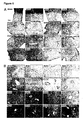
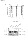
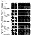
- ⁇ 6 integrin and E-cadherin were evaluated by confocal microscopy imaging ( Figure 3A ). There was co-localization of these proteins in the liver metastasis cell line NCI-H630, in which both ⁇ 6 integrin and E-cadherin were highly represented on cell membranes; in contrast, barely detectable immunostaining of ⁇ 6 integrin and no co-localization with E-cadherin were observed in the primary tumor cell line HepG2. It was suspected that these proteins could be part of a supramolecular complex in liver metastasis cells, as indicated by mass spectrometry and by their coincident location on the cell surface.
- Co-immunoprecipitation assays confirmed that ⁇ 6 integrin and E-cadherin, physically interact in NCI-H630 cells ( Figure 3A and Figure 6A for protein quantification in cell lines).
- Confocal imaging analyses performed on liver metastasis samples from CRC patients (n 6) (see also Figure 4 ) revealed that ⁇ 6 integrin and E-cadherin were expressed by selected groups of cells, with regions of overlap; in contrast, ⁇ 6 integrin was barely detectable and the two proteins did not co-localize in matched normal livers.
- Example 4 The ligand side of the signature: angiopoietin-like 6 mimics an LRS-peptide and is enriched in blood vessels of the liver in humans.
- angiopoietin-like 6 received the highest identification score, because it shares similarity with the targeting peptides in two different regions of its fibrinogen-like domain.
- angiopoietin-like 6 mRNA has been detected particularly in the liver in humans ( Kim et al.
- Example 5 Coupling receptors and ligands (1): ex vivo and in vivo models of metastasis/host tissue interaction.
- the ⁇ 6 integrin (i) can form heterodimers with either ⁇ 1 or ⁇ 4 , depending on the cell type ( Humphries et al. 2006 J Cell Sci 119:3901-3903 ; Hemler et al. 1988, J Biol Chem 264:6529-6535 ; Hemler et al.
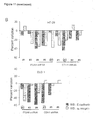
- Example 6 Coupling receptors and ligands (2): the ⁇ 6 integrin/E-cadherin complex and angiopoietin-like 6 mediate the adhesion of human metastatic CRC cells to the liver in vitro.
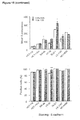
- NCI-H630 cells in which ITGA6 or both mRNAs were silenced lost the capacity to bind the CGIYRLRSC-phage. Consistently, these cells also exhibited an impaired adherence to microwells coated with the CGIYRLRSC-peptide; this effect was particularly pronounced when both ⁇ 6 integrin and E-cadherin were downmodulated.
- NCI-H630 cells in which both mRNAs were silenced exhibited significantly lower binding to microwells coated with recombinant angiopoietin-like 6 ( Figure 10C ).
- CGIYRLRSC-mimicked ligand proteins such as angiopoietin-like 6, can act as microenvironment addresses for metastatic cells that express ⁇ 6 integrin/E-cadherin receptor complex.
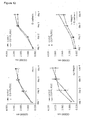
- Example 7 Coupling receptors and ligands (3): angiopoietin-like 6 mediates the attraction of cells expressing the ⁇ 6 integrin/E-cadherin complex.
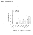
- angiopoietin-like 6 is a secreted factor whose chemotactic activity on endothelial cells has been reported ( Oike et al. 2004. Blood 103:3760-3765 ). Therefore, it was investigated if soluble angiopoietin-like 6 could affect the motility of cells expressing the ⁇ 6 integrin/E-cadherin receptor complex.
- U293 cells transduced with ⁇ 6 integrin, E-cadherin, or a combination of both were co-cultured with angiopoietin-like 6-producing cells in a transwell system and their migration toward the ligand gradient was evaluated after 48 hours.
- Co-cultures with mock-transfected U293 cells were exploited as a reference for basal cell motility.
- the presence of either ⁇ 6 integrin or E-cadherin slightly increased the capacity of U293 cells to migrate in basal conditions; however, this feature was not influenced by the presence of soluble angiopoietin-like 6.
- U293 cells expressing both ⁇ 6 integrin and E-cadherin showed a basal pro-migratory phenotype, which was significantly stimulated by angiopoietin-like 6 ( Figure 10D ).
- Example 8 Uncoupling ligands and receptors (1): CGIYRLRSC-peptide inhibits the adhesion and attraction of metastatic CRC cells to the liver in vitro.
- CGIYRLRSC specifically inhibits the adhesion of metastatic CRC cells to the liver, possibly through interference with the angiopoietin-like 6/ ⁇ 6 integrin/E-cadherin ligand/receptor system.
- the in vitro data indicated that two pivotal steps for the onset of liver metastasis, i.e. tumor/host tissue recognition and metastatic cell attraction, could be driven by the ⁇ 6 integrin/Ecadherin/angiopoietin-like 6 system. Accordingly, the interference with the described ligand/receptor pair was investigated to result in impaired liver colonization and homing in vivo.
- An animal model of hepatic colonization was established by direct injection of human CRC cells into the livers of CD-1 nude mice.
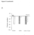
- LS-174T a cell line derived from a primary CRC that exhibits high expression of the complex proteins ( Figure 6A ) and an extremely aggressive behavior in vivo was used ( Price et al. 1989, Clin Exp Metastasis 7:55-68 ).
- animals were injected with LS-174T cells either in medium alone or in the presence of the soluble peptide. After 14 days, the livers were explanted for photographic documentation and tumor quantification. A significant reduction of liver tumors in mice injected with LS-174T cells in the presence of CGIYRLRSC was observed, although the overall morphology and the levels of receptor complex in the tumors were unchanged ( Figure 10H ).
- Example 10 Uncoupling ligands and receptors (3): interfering with liver homing for anti-metastatic therapy.
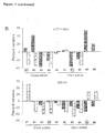
- ⁇ 6 integrin varied not only among different CRC cell lines (from ⁇ 2% in the non-metastatic HCT-116 cell line to >50% in the liver metastasis NCI-H630 cell line), but also as a consequence of different culture conditions, resulting in variable numbers of cell concomitantly expressing ⁇ 6 integrin and E-cadherin ( Figure 15 ). The results indicate that a clonal selection of cells expressing both partners of the complex is preferred.
- Example 11 The ⁇ 6 integrin/E-cadherin complex and angiopoietin-like 6 are correlated to the aggressiveness of human metastatic CRCs.
Landscapes
- Health & Medical Sciences (AREA)
- Life Sciences & Earth Sciences (AREA)
- Chemical & Material Sciences (AREA)
- Immunology (AREA)
- General Health & Medical Sciences (AREA)
- Engineering & Computer Science (AREA)
- Medicinal Chemistry (AREA)
- Molecular Biology (AREA)
- Organic Chemistry (AREA)
- Cell Biology (AREA)
- Biochemistry (AREA)
- Proteomics, Peptides & Aminoacids (AREA)
- Biomedical Technology (AREA)
- Zoology (AREA)
- Gastroenterology & Hepatology (AREA)
- Urology & Nephrology (AREA)
- Hematology (AREA)
- Biophysics (AREA)
- Genetics & Genomics (AREA)
- Pharmacology & Pharmacy (AREA)
- Animal Behavior & Ethology (AREA)
- Public Health (AREA)
- Veterinary Medicine (AREA)
- Epidemiology (AREA)
- Physics & Mathematics (AREA)
- Pathology (AREA)
- General Physics & Mathematics (AREA)
- Analytical Chemistry (AREA)
- Biotechnology (AREA)
- Toxicology (AREA)
- Microbiology (AREA)
- Bioinformatics & Cheminformatics (AREA)
- Food Science & Technology (AREA)
- Chemical Kinetics & Catalysis (AREA)
- Oncology (AREA)
- Nuclear Medicine, Radiotherapy & Molecular Imaging (AREA)
- General Chemical & Material Sciences (AREA)
- Pharmaceuticals Containing Other Organic And Inorganic Compounds (AREA)
- Medicines That Contain Protein Lipid Enzymes And Other Medicines (AREA)
Priority Applications (4)
| Application Number | Priority Date | Filing Date | Title |
|---|---|---|---|
| EP12182994.9A EP2703005A1 (fr) | 2012-09-04 | 2012-09-04 | Inhibiteurs d'intégrine alpha6/E-cadhérine complexe |
| US14/425,441 US20150218213A1 (en) | 2012-09-04 | 2013-09-03 | Inhibitors of alpha6 integrin/e-cadherin complex |
| EP13759481.8A EP2892545A1 (fr) | 2012-09-04 | 2013-09-03 | Inhibiteurs du complexe intégrine alpha6/e-cadhérine et leur utilisation thérapeutique et de diagnostic dans le cancer |
| PCT/EP2013/068150 WO2014037332A1 (fr) | 2012-09-04 | 2013-09-03 | Inhibiteurs du complexe intégrine alpha6/e-cadhérine et leur utilisation thérapeutique et de diagnostic dans le cancer |
Applications Claiming Priority (1)
| Application Number | Priority Date | Filing Date | Title |
|---|---|---|---|
| EP12182994.9A EP2703005A1 (fr) | 2012-09-04 | 2012-09-04 | Inhibiteurs d'intégrine alpha6/E-cadhérine complexe |
Publications (1)
| Publication Number | Publication Date |
|---|---|
| EP2703005A1 true EP2703005A1 (fr) | 2014-03-05 |
Family
ID=46826274
Family Applications (1)
| Application Number | Title | Priority Date | Filing Date |
|---|---|---|---|
| EP12182994.9A Withdrawn EP2703005A1 (fr) | 2012-09-04 | 2012-09-04 | Inhibiteurs d'intégrine alpha6/E-cadhérine complexe |
Country Status (1)
| Country | Link |
|---|---|
| EP (1) | EP2703005A1 (fr) |
Cited By (2)
| Publication number | Priority date | Publication date | Assignee | Title |
|---|---|---|---|---|
| WO2017118458A1 (fr) * | 2016-01-04 | 2017-07-13 | Universität Hamburg | Aptamère adn se liant à l'α6-intégrine |
| CN109797151A (zh) * | 2019-01-18 | 2019-05-24 | 中山大学附属第一医院 | Circ-CDH1抑制剂的应用 |
Citations (7)
| Publication number | Priority date | Publication date | Assignee | Title |
|---|---|---|---|---|
| EP0537654A2 (fr) * | 1991-10-18 | 1993-04-21 | F. Hoffmann-La Roche Ag | Anticorps contre l'intégrine alpha6 |
| WO2002030465A2 (fr) * | 2000-10-12 | 2002-04-18 | University Of Rochester | Compositions inhibant la proliferation de cellules cancereuses |
| WO2005046731A1 (fr) * | 2003-10-17 | 2005-05-26 | The Government Of The United States Of America, As Represented By The Secretary, Department Of Health And Human Services | Entrave de la fonction c-maf dans le myelome multiple |
| WO2006047878A1 (fr) * | 2004-11-03 | 2006-05-11 | British Columbia Cancer Agency Branch | Agents therapeutiques contre le cancer comprenant des domaines extracellulaires du recepteur notch humain et procedes d'utilisation correspondants |
| WO2008064910A2 (fr) | 2006-11-30 | 2008-06-05 | Università Degli Studi Di Torino | Peptides spécifiques des métastases et leurs applications à des fins de diagnostic et thérapeutiques |
| WO2009086189A2 (fr) * | 2007-12-21 | 2009-07-09 | Burnham Institute For Medical Research | Utilisation d'activation de facteur de transcription 2 (atf2) pour la détection du cancer de la peau |
| WO2010066046A1 (fr) * | 2008-12-10 | 2010-06-17 | Socpra Sciences Santé Et Humaines S.E.C. | Modulation associée à la prolifération d'épissage des isoformes intégrine alpha 6 |
-
2012
- 2012-09-04 EP EP12182994.9A patent/EP2703005A1/fr not_active Withdrawn
Patent Citations (7)
| Publication number | Priority date | Publication date | Assignee | Title |
|---|---|---|---|---|
| EP0537654A2 (fr) * | 1991-10-18 | 1993-04-21 | F. Hoffmann-La Roche Ag | Anticorps contre l'intégrine alpha6 |
| WO2002030465A2 (fr) * | 2000-10-12 | 2002-04-18 | University Of Rochester | Compositions inhibant la proliferation de cellules cancereuses |
| WO2005046731A1 (fr) * | 2003-10-17 | 2005-05-26 | The Government Of The United States Of America, As Represented By The Secretary, Department Of Health And Human Services | Entrave de la fonction c-maf dans le myelome multiple |
| WO2006047878A1 (fr) * | 2004-11-03 | 2006-05-11 | British Columbia Cancer Agency Branch | Agents therapeutiques contre le cancer comprenant des domaines extracellulaires du recepteur notch humain et procedes d'utilisation correspondants |
| WO2008064910A2 (fr) | 2006-11-30 | 2008-06-05 | Università Degli Studi Di Torino | Peptides spécifiques des métastases et leurs applications à des fins de diagnostic et thérapeutiques |
| WO2009086189A2 (fr) * | 2007-12-21 | 2009-07-09 | Burnham Institute For Medical Research | Utilisation d'activation de facteur de transcription 2 (atf2) pour la détection du cancer de la peau |
| WO2010066046A1 (fr) * | 2008-12-10 | 2010-06-17 | Socpra Sciences Santé Et Humaines S.E.C. | Modulation associée à la prolifération d'épissage des isoformes intégrine alpha 6 |
Non-Patent Citations (89)
| Title |
|---|
| ADAM ET AL., ANN SURG, vol. 240, 2004, pages 644 - 657 |
| ARAP ET AL., NAT MED, vol. 8, 2002, pages 121 - 127 |
| BARUGEL ET AL., EXPERT REV ANTICANCER THER, vol. 9, 2009, pages 1829 - 1847 |
| BERTOTTI ET AL., CANCER RES, vol. 65, 2005, pages 10674 - 10679 |
| BOS ET AL., NATURE, vol. 459, 2009, pages 1005 - 1009 |
| CESPEDES ET AL., AM J PATHOL, vol. 177, no. 4, 2010, pages 2067 - 79 |
| CHAMBERS ET AL., NAT REV CANCER, vol. 2, 2002, pages 563 - 572 |
| CHRISTOFORI, EMBO J, vol. 22, 2003, pages 2318 - 2323 |
| DAVIS ET AL., CELL GROWTH DIFFER., vol. 13, no. 3, 2002, pages 107 - 13 |
| DEMETRIOU ET AL., EXP CELL RES, vol. 294, no. 2, 2004, pages 550 - 8 |
| DEMETRIOU ET AL., OPEN CANCER J., vol. 2, 2008, pages 1 - 4 |
| DONG ET AL., J NATL CANCER INST, vol. 86, 1994, pages 913 - 920 |
| ELSTON ET AL., J CLIN ENDOCRINOL METAB, vol. 94, no. 4, 2009, pages 1436 - 42 |
| ELZAGHEID ET AL., WORLD J GASTROENTEROL, vol. 12, 2006, pages 4304 - 4309 |
| ENNS ANDREAS ET AL: "Integrins can directly mediate metastatic tumor cell adhesion within the liver sinusoids.", JOURNAL OF GASTROINTESTINAL SURGERY : OFFICIAL JOURNAL OF THE SOCIETY FOR SURGERY OF THE ALIMENTARY TRACT DEC 2004, vol. 8, no. 8, December 2004 (2004-12-01), pages 1049 - 1060, XP002688817, ISSN: 1091-255X * |
| ENNS ET AL., J GASTROINTEST SURG, vol. 8, 2004, pages 1049 - 1060 |
| FEARON ET AL., CELL, vol. 61, 1990, pages 759 - 767 |
| FERBER ET AL., J BIOL CHEM, vol. 283, no. 19, 2008, pages 12691 - 700 |
| GIANTONIO ET AL., J CLIN ONCOL, vol. 25, 2007, pages 1539 - 1544 |
| GONZALEZ ET AL., PROC NATL ACAD SCI U S A, vol. 99, 2002, pages 16075 - 16080 |
| GROTHEY ET AL., NAT REV CLIN ONCOL, vol. 6, 2009, pages 507 - 518 |
| HECHT ET AL., J CLIN ONCOL, vol. 27, 2009, pages 672 - 680 |
| HEMLER ET AL., J BIOL CHEM, vol. 263, 1988, pages 7660 - 7665 |
| HEMLER ET AL., J BIOL CHEM, vol. 264, 1988, pages 6529 - 6535 |
| HOCHSTER ET AL., J CLIN ONCOL, vol. 26, 2008, pages 3523 - 3529 |
| HOGERVORST ET AL., EUR J BIOCHEM, vol. 199, 1991, pages 425 - 433 |
| HOGERVORST ET AL., J CELL BIOL, vol. 121, 1993, pages 179 - 191 |
| HUANG ET AL., NAT PROTOC, vol. 4, 2009, pages 44 - 57 |
| HUANG ET AL., NAT PROTOC, vol. 4, 2009, pages 44 - 57, Retrieved from the Internet <URL:http://david.abcc.ncifcrf.gov> |
| HUANG ET AL., NUCLEIC ACIDS RES, vol. 37, 2009, pages 1 - 13 |
| HUGUENIN ET AL., PLOS ONE, vol. 3, no. 5, 2008, pages E2153 |
| HULME ET AL., BR J PHARMACOL., vol. 161, no. 6, 2010, pages 1219 - 37 |
| HUMPHRIES ET AL., J CELL SCI, vol. 119, 2006, pages 3901 - 3903 |
| HURWITZ ET AL., N ENGL J MED, vol. 350, 2004, pages 2335 - 2342 |
| ILYAS ET AL., GUT, vol. 40, 1997, pages 654 - 659 |
| JONKER ET AL., N ENGL J MED, vol. 357, 2007, pages 2040 - 2048 |
| KIM ET AL., BIOCHEM J, vol. 346, 2000, pages 603 - 610 |
| KOH ET AL., ONCOLOGY, vol. 75, 2008, pages 92 - 101 |
| KOPETZ ET AL., J CLIN ONCOL, vol. 28, pages 453 - 459 |
| KUO ET AL., PROC NATL ACAD SCI USA, vol. 92, 1995, pages 12085 - 12089 |
| KWAK ET AL., DIS COLON RECTUM, vol. 50, 2007, pages 1873 - 1880 |
| LAMPROPOULOS PAVLOS ET AL: "Prognostic significance of transforming growth factor beta (TGF-beta) signaling axis molecules and E-cadherin in colorectal cancer", TUMOR BIOLOGY, vol. 33, no. 4, August 2012 (2012-08-01), pages 1005 - 1014, XP002688818 * |
| LEE ET AL., EUR SURG RES, vol. 39, no. 4, 2007, pages 208 - 15 |
| LEE ET AL., J BIOL CHEM, vol. 281, 2006, pages 40450 - 40460 |
| LIN ET AL., ONCOL REP, vol. 17, 2007, pages 1541 - 1549 |
| LIPSCOMB ET AL., CANCER METASTASIS REV, vol. 24, 2005, pages 413 - 423 |
| LYNCH ET AL., J ONCOL, vol. 2010, 2010, pages 53074 - 5 |
| MARCHI6 ET AL., CANCER CELL, vol. 5, no. 2, 2004, pages 151 - 62 |
| MAYNARD ET AL., BIOTECHNOL J., vol. 4, no. 11, 2009, pages 1542 - 58 |
| MERIC ET AL., ANN SURG ONCOL, vol. 7, 2000, pages 490 - 495 |
| MINN ET AL., PROC NATL ACAD SCI USA, vol. 104, 2007, pages 6740 - 6745 |
| NAJY ET AL., J BIOL CHEM, vol. 283, no. 26, 2008, pages 18393 - 401 |
| NANNINI ET AL., CANCER TREAT REV, vol. 35, 2009, pages 201 - 209 |
| NATALWALA ET AL., WORLD J GASTROENTEROL, vol. 14, 2008, pages 3792 - 3797 |
| NIBBE ET AL., MOL CELL PROTEOMICS, vol. 8, 2009, pages 827 - 845 |
| NIE ET AL., CRIT REV BIOTECHNOL, vol. 27, 2007, pages 63 - 75 |
| OIKE ET AL., BLOOD, vol. 103, 2004, pages 3760 - 3765 |
| OIKE ET AL., PROC NATL ACAD SCI USA, vol. 100, 2003, pages 9494 - 9499 |
| PADUA ET AL., CELL, vol. 133, 2008, pages 66 - 77 |
| PAGET, LANCET, vol. 1, 1989, pages 571 - 573 |
| PARKIN ET AL., CA CANCER J CLIN, vol. 55, 2005, pages 74 - 108 |
| PAWAR ET AL., EXP CELL RES, vol. 313, no. 6, 2007, pages 1080 - 9 |
| PAWAR ET AL., INT J RADIAT BIOL., vol. 83, no. 11-12, 2007, pages 761 - 7 |
| PRICE ET AL., CLIN EXP METASTASIS, vol. 7, 1989, pages 55 - 68 |
| PRIMO ET AL., CANCER RES, vol. 70, 2010, pages 5759 - 5769 |
| RABINOVITZ ET AL., MOL BIOL CELL, vol. 12, 2001, pages 4030 - 4043 |
| ROBERTSON ET AL., CLIN EXP METASTASIS, vol. 26, 2009, pages 769 - 780 |
| ROBERTSON ET AL., CURR PHARM DES, vol. 14, 2008, pages 296 - 305 |
| SAHA ET AL., SCIENCE, vol. 294, 2001, pages 1343 - 1346 |
| SALTZ ET AL., NAT REV DRUG DISCOV, vol. 5, 2006, pages 987 - 988 |
| SALTZ, GASTROINTEST CANCER RES, vol. 2, 2008, pages 20 - 22 |
| SCOTT ET AL., SCIENCE, vol. 249, 1990, pages 386 - 390 |
| SMITH ET AL., METHODS ENZYMOL, vol. 217, 1993, pages 228 - 257 |
| SOLANAS ET AL., NAT CELL BIOL., 2011 |
| SROKA ET AL., CARCINOGENESIS, vol. 27, no. 9, 2006, pages 1748 - 57 |
| TAMURA ET AL., PROC NATL ACAD SCI USA, vol. 88, 1991, pages 10183 - 10187 |
| TAYLOR ET AL., CANCER RES, vol. 66, 2006, pages 5537 - 5539 |
| TIBBETTS ET AL., CANCER, vol. 71, 1993, pages 315 - 321 |
| TOMLINSON ET AL., J CLIN ONCOL, vol. 25, 2007, pages 4575 - 4580 |
| TRUANT ET AL., J SURG RES, vol. 150, 2008, pages 212 - 218 |
| URANO ET AL., ARTERIOSCLER THROMB VASC BIOL, vol. 28, 2008, pages 827 - 834 |
| VAN CUTSEM ET AL., J CLIN ONCOL, vol. 25, 2007, pages 1658 - 1664 |
| VARAMBALLY ET AL., CANCER CELL, vol. 8, 2005, pages 393 - 406 |
| VOGELSTEIN ET AL., N ENGL J MED, vol. 319, 1988, pages 525 - 532 |
| WAGNER ET AL., ANN SURG, vol. 199, 1984, pages 502 - 508 |
| WELLS ET AL., CLIN EXP METASTASIS, vol. 25, 2008, pages 621 - 628 |
| YOON ET AL., CANCER RES, vol. 66, 2006, pages 2732 - 2739 |
| ZEITOUN ET AL., ANTICANCER RES, vol. 28, 2008, pages 3609 - 3612 |
| ZHANG ET AL., BIOCHEM BIOPHYS RES COMMUN, vol. 347, 2006, pages 100 - 108 |
Cited By (3)
| Publication number | Priority date | Publication date | Assignee | Title |
|---|---|---|---|---|
| WO2017118458A1 (fr) * | 2016-01-04 | 2017-07-13 | Universität Hamburg | Aptamère adn se liant à l'α6-intégrine |
| CN109797151A (zh) * | 2019-01-18 | 2019-05-24 | 中山大学附属第一医院 | Circ-CDH1抑制剂的应用 |
| CN109797151B (zh) * | 2019-01-18 | 2023-03-31 | 中山大学附属第一医院 | Circ-CDH1抑制剂的应用 |
Similar Documents
| Publication | Publication Date | Title |
|---|---|---|
| Naba et al. | Extracellular matrix signatures of human mammary carcinoma identify novel metastasis promoters | |
| Bartolini et al. | BCAM and LAMA5 mediate the recognition between tumor cells and the endothelium in the metastatic spreading of KRAS-mutant colorectal cancer | |
| Marchio et al. | A complex of α6 integrin and E‐cadherin drives liver metastasis of colorectal cancer cells through hepatic angiopoietin‐like 6 | |
| US20150218213A1 (en) | Inhibitors of alpha6 integrin/e-cadherin complex | |
| ES2796111T3 (es) | Métodos terapéuticos relacionados con células madre cancerosas | |
| Gaiteiro et al. | Glycoproteogenomics characterizes the CD44 splicing code associated with bladder cancer invasion | |
| Wortmann et al. | The cell surface glycoprotein CDCP1 in cancer—insights, opportunities, and challenges | |
| CN107249636B (zh) | 以ckap4作为靶分子的抗肿瘤剂 | |
| Marcus et al. | Septin oligomerization regulates persistent expression of ErbB2/HER2 in gastric cancer cells | |
| Sun et al. | Cadmium promotes colorectal cancer metastasis through EGFR/Akt/mTOR signaling cascade and dynamics | |
| CN104411825B (zh) | 骨膜蛋白适配体及包含其的抗癌组合物 | |
| Mosig et al. | Application of RNA-Seq transcriptome analysis: CD151 is an Invasion/Migration target in all stages of epithelial ovarian cancer | |
| Zhi et al. | Podocalyxin‐like protein promotes gastric cancer progression through interacting with RUN and FYVE domain containing 1 protein | |
| D’Agostino et al. | The receptor protein tyrosine phosphatase PTPRJ negatively modulates the CD98hc oncoprotein in lung cancer cells | |
| Yang et al. | CD151 promotes Colorectal Cancer progression by a crosstalk involving CEACAM6, LGR5 and Wnt signaling via TGFβ1 | |
| Liu et al. | SPP1 Drives Colorectal Cancer Liver Metastasis and Immunotherapy Resistance by Stimulating CXCL12 Production in Cancer-Associated Fibroblasts | |
| US10359428B2 (en) | Tumor marker, monoclonal antibodies and methods of use thereof | |
| Langlois et al. | Inhibition of PANX1 channels reduces the malignant properties of human high-risk neuroblastoma | |
| WO2015098113A1 (fr) | Agent thérapeutique pour tumeur maligne | |
| KR101906558B1 (ko) | Tspan8에 특이적인 신규 항체 및 이의 용도 | |
| EP2703005A1 (fr) | Inhibiteurs d'intégrine alpha6/E-cadhérine complexe | |
| Liu et al. | Endogenous IL-33 inhibits apoptosis in non-small cell lung cancer cells by regulating BCL2/BAX via the ERK1/2 pathway | |
| WO2007061922A2 (fr) | Methodes de prediction et prevention de la resistance a des composes taxoides | |
| CN104225623B (zh) | 新的g蛋白偶联受体蛋白及其用途 | |
| EP3149483B1 (fr) | Outil diagnostique et thérapeutique pour le cancer |
Legal Events
| Date | Code | Title | Description |
|---|---|---|---|
| AK | Designated contracting states |
Kind code of ref document: A1 Designated state(s): AL AT BE BG CH CY CZ DE DK EE ES FI FR GB GR HR HU IE IS IT LI LT LU LV MC MK MT NL NO PL PT RO RS SE SI SK SM TR |
|
| AX | Request for extension of the european patent |
Extension state: BA ME |
|
| PUAI | Public reference made under article 153(3) epc to a published international application that has entered the european phase |
Free format text: ORIGINAL CODE: 0009012 |
|
| STAA | Information on the status of an ep patent application or granted ep patent |
Free format text: STATUS: THE APPLICATION IS DEEMED TO BE WITHDRAWN |
|
| 18D | Application deemed to be withdrawn |
Effective date: 20140906 |