EP2726877B1 - Verfahren zum spezifischen nachweis von ziel-biomolekülen - Google Patents
Verfahren zum spezifischen nachweis von ziel-biomolekülen Download PDFInfo
- Publication number
- EP2726877B1 EP2726877B1 EP12803774.4A EP12803774A EP2726877B1 EP 2726877 B1 EP2726877 B1 EP 2726877B1 EP 12803774 A EP12803774 A EP 12803774A EP 2726877 B1 EP2726877 B1 EP 2726877B1
- Authority
- EP
- European Patent Office
- Prior art keywords
- protein
- target
- overlapping
- sample
- antibodies
- Prior art date
- Legal status (The legal status is an assumption and is not a legal conclusion. Google has not performed a legal analysis and makes no representation as to the accuracy of the status listed.)
- Active
Links
Images


Classifications
-
- G—PHYSICS
- G01—MEASURING; TESTING
- G01N—INVESTIGATING OR ANALYSING MATERIALS BY DETERMINING THEIR CHEMICAL OR PHYSICAL PROPERTIES
- G01N33/00—Investigating or analysing materials by specific methods not covered by groups G01N1/00 - G01N31/00
- G01N33/48—Biological material, e.g. blood, urine; Haemocytometers
- G01N33/50—Chemical analysis of biological material, e.g. blood, urine; Testing involving biospecific ligand binding methods; Immunological testing
- G01N33/53—Immunoassay; Biospecific binding assay; Materials therefor
- G01N33/558—Immunoassay; Biospecific binding assay; Materials therefor using diffusion or migration of antigen or antibody
- G01N33/561—Immunoelectrophoresis
-
- G—PHYSICS
- G01—MEASURING; TESTING
- G01N—INVESTIGATING OR ANALYSING MATERIALS BY DETERMINING THEIR CHEMICAL OR PHYSICAL PROPERTIES
- G01N33/00—Investigating or analysing materials by specific methods not covered by groups G01N1/00 - G01N31/00
- G01N33/48—Biological material, e.g. blood, urine; Haemocytometers
- G01N33/50—Chemical analysis of biological material, e.g. blood, urine; Testing involving biospecific ligand binding methods; Immunological testing
- G01N33/53—Immunoassay; Biospecific binding assay; Materials therefor
- G01N33/543—Immunoassay; Biospecific binding assay; Materials therefor with an insoluble carrier for immobilising immunochemicals
-
- G—PHYSICS
- G01—MEASURING; TESTING
- G01N—INVESTIGATING OR ANALYSING MATERIALS BY DETERMINING THEIR CHEMICAL OR PHYSICAL PROPERTIES
- G01N27/00—Investigating or analysing materials by the use of electric, electrochemical, or magnetic means
- G01N27/26—Investigating or analysing materials by the use of electric, electrochemical, or magnetic means by investigating electrochemical variables; by using electrolysis or electrophoresis
- G01N27/416—Systems
- G01N27/447—Systems using electrophoresis
- G01N27/44704—Details; Accessories
- G01N27/44717—Arrangements for investigating the separated zones, e.g. localising zones
- G01N27/44721—Arrangements for investigating the separated zones, e.g. localising zones by optical means
- G01N27/44726—Arrangements for investigating the separated zones, e.g. localising zones by optical means using specific dyes, markers or binding molecules
Definitions
- the present invention relates to a method for specific identification of target biomolecules in a sample leading to improved specificity in especially multiplexed Western blotting assay formats.
- the method uses at least two probes directed against the same or different epitopes of the target biomolecule.
- signals from the true target biomolecule are enhanced while signals resulting from unspecific binding are diminished.
- Western blotting is an analytical technique used to detect specific proteins in a given sample of tissue homogenate, cell lysate or other protein containing samples. It uses gel electrophoresis to separate native or denatured proteins by the length of the polypeptide (denaturing conditions) or by the 3-D structure of the protein (native/ non-denaturing conditions). The proteins are then transferred to a membrane (typically nitrocellulose or PVDF), where they are probed (detected) using antibodies specific to the target protein. For certain applications, the proteins are probed in the gel without a transfer step. However, further description of western blot methodology is based on protein samples transferred to a membrane.
- the membrane is probed for the protein of interest by the use of an antibody specific for the protein of interest. Due to possibilities of increased signal amplification and to avoid negative effects on target specific affinity related to primary antibody conjugation, this traditionally takes place in a two-step process (using a primary target specific antibody and a secondary labeled antibody specific for the primary antibody), although there are now one-step detection methods available for certain applications.
- the one-step method allows the process to occur faster and with a lower amount of consumables, but sensitivity and specificity may be compromised.
- This requires a probe antibody which both recognizes the protein of interest and contains a detectable label, probes which are often available for known protein tags.
- the primary probe is incubated with the membrane in a manner similar to that for the primary antibody in a two-step process, and is then ready for direct detection after a series of wash steps.
- Preparing or raising antibodies against a partly purified target protein or proteins with weak immunogenic properties may result in low immuno-specificity and antibodies from suppliers are not always of sufficiently high quality.
- Affinity purification could be used to overcome the problem, but requires purified target protein and involves risks with sample handling including e.g. loss of antibody, activity and stability issues. Improper storage and aged antibodies can give similar reduced detection performance.
- a protein target mw size may be unknown
- the true immunogenic protein band corresponding to the target is very difficult to determine in a conventional Western blotting experiment. For many target proteins there is a limited availability of highly specific antibodies, resulting in a Western blot assay with low specificity.
- the present invention provides a method for identification of specific target biomolecules in a sample following a detection procedure (such as a Western blotting procedure, a biotin-streptavidin/avidin based assay, non-reducing gel electrophoresis, reducing electrophoresis, capillary electrophoresis, chip electrophoresis, isoelectric focusing, 2D electrophoresis or any detection procedure involving separation of biomolecules based on physical or chemical properties), wherein a gel or membrane is probed with at least two probes (such as antibodies, fragments thereof (Fab, F(ab') 2 or Fc fragment specifically binding to the target biomolecule), or other affinity binders, such as aptamers), directed against the same and/or different sites (such as epitopes when antibodies are used as probes) of the same target biomolecule, and wherein specific binding to the target biomolecule in a sample is differentiated from unspecific binding to the target biomolecule by comparing the resulting sample patterns (such as bands
- the detection procedure is a Western blotting procedure and the method comprises enhancing overlapping band features and sorting out non-overlapping features to produce image data with a pattern more specifically representing the target protein.
- the method is not limited to any special target biomolecule but it is preferably a protein, peptide or aggregated biomolecule, such as a fusion protein, or a modified protein, such as a phosphorylated, glycosylated, ubiquitinated, SUMOylated, or acetylated protein.
- the image data resulting from the method of the invention may be represented in the form of one or more intensity profiles or 3D surfaces.
- the final result may also be presented in the form of one or more images where optionally color coding has been used to highlight overlapping and/or non-overlapping features.
- the color coding may represent the result of applying an algorithm to image data.
- the detection procedure is Western blotting, preferably at least two primary antibodies are used as probes. These may be from different species or from the same species.
- the primary antibodies are differentially labelled.
- labelled secondary antibodies are used against said primary antibodies.
- the primary or secondary antibodies used in the method of the invention may be labelled with fluorescent dyes, preferably cyanine dyes.
- One or more of the antibodies may be detected using Enhanced chemiluminescence (ECL) to provide multiplexing. Multiplexing may also be provided by an antibody stripping and re-probing procedure.
- ECL Enhanced chemiluminescence
- one or more of the sample patterns represent detection using a specific or non-specific post stain.
- one or more of the sample patterns represent detection using specific or non-specific protein labeling.
- at least two sample patterns may represent different samples covalently labeled with different dyes and mixed together before Western blotting is carried out.
- At least two images may represent different pre-processing of sample aliquots covalently labeled with different dyes and mixed together before electrophoresis is carried out, wherein the pre-processing for example may involve affinity separation, for example using antibodies against the target protein.
- the resulting sample pattern may be quantitatively analyzed.
- the method of the invention may be implemented in a software package and/or in an instrument system, such as an instrument system for Western blotting analysis.
- Western blotting is one of the most widely used assays for identification of specific target proteins in a protein sample.
- the present invention provides not only improved quantitative comparison but also improves the specificity of the assay.
- the present invention uses several probes, preferably antibodies, directed against the same or different epitopes of the same target protein. These antibodies may be more or less specific. Since the method of the invention combines the signals from the different antibodies, specific signal can be discriminated from unspecific ones.
- the algorithm has been designed to allow also for quantitative comparison of target protein between samples.
- the algorithm can easily be extended to more than two antibodies.
- the algorithm can be adjusted to have higher discriminating power but probably on the expense of quantitative performance.
- the method of the invention is especially useful in the following situations:
- the electrophoresis MiniVE vertical Electrophoresis system, GE Healthcare was performed at 100 V until the bromophenol blue front reached the end of the gel.
- ECL Plex Fluorescent Rainbow Markers (GE Healthcare) was used to track the separation.
- the gel was equilibrated in transfer buffer (1 x Tris-glycine, 20 % Methanol) for 15 minutes before transfer onto a PVDF membrane (Hybond-LFP, GE Healthcare) using the TE 22 Mini tank transfer unit (GE Healthcare) at 25 V and 4°C for 2.5 h.
- the membrane was blocked in 2 % (w/v) ECL Advance blocking agent (GE Healthcare) for 1 h at room temperature. After blocking, the membrane was rinsed twice and washed for 2 x 5 min in PBS buffer with 0.1 % Tween20 (PBST).
- the membrane was incubated with monoclonal mouse anti tubulin (Sigma Aldrich) and rabbit anti tubulin, whole antiserum extract (Sigma Aldrich) primary antibodies at 1:2000 and 1:1000 dilution in PBST respectively over night at 4°C.
- the membrane was rinsed twice and then washed for 2 x 5 min in PBST and incubated with ECL Plex goat anti-mouse IgG, Cy5 (GE Healthcare) and ECL Plex goat anti-rabbit IgG, Cy3 (GE Healthcare) secondary antibodies dilutes of 1:2500 in PBST for 1 h at room temperature.
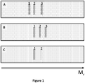
- Figure 1 illustrates the effect of combining the information in two schematic Western blotting membrane images A and B to produce a combined image C.
- Images A and B represent Western blot patterns from two different antibodies intended to target the same protein, detected in the same membrane using color multiplexing.
- Image C is formed by pixel wise multiplication of images A and B after background subtraction. It is obvious that bands representing non-specific binding and therefore not present in both images A and B, i.e. bands 1 and 3 in image A and bands 2 and 3 in image B are heavily suppressed in the resulting image C.
- the band representing the specific binding is present in both images A and B, i.e. band 2 in image A and band 1 in image B, and is therefore not suppressed in the resulting image 3 where it is labeled as band 1.
- Figure 2 shows the result of the color multiplexed Western experiment
- image A represents the Western signals from monoclonal mouse anti-tubulin (detected using a Cy5 conjugated anti-mouse IgG secondary antibody) and image B represents the Western signals from rabbit anti-tubulin whole antiserum (detected using a Cy3 conjugated anti-rabbit IgG secondary antibody).
- Image C is formed by pixel wise multiplication of images A and B after background subtraction. It is obvious that only band features prominent in both images A and B will become prominent also in image C. Many non-specific band features will therefore be suppressed in image C.
- the intensity profiles corresponding to the outlined lanes in images A-C (outlined by dashed lines) are presented in figure 3 . Relative migration positions (Mr) based on molecular weight standards are indicated.
- the images were analyzed to determine if the proposed algorithm can provide increased specificity by combining information from Cy3 and Cy5 images from the multiplexed membrane.
- the images were imported into ImageJ ( figures 2A and 2B ) and the background was subtracted using the rolling ball method with a radius of 200 pixels. After background subtraction pixel intensities in each image were multiplied with each other to generate a new image ( figure 2C ).
- Figure 3 shows the intensity profiles corresponding to the lanes outlined in figure 2 .
- Curve A represents the Western signal intensity profile from monoclonal mouse anti-tubulin (detected using a Cy5 conjugated anti-mouse IgG secondary antibody). Peak 1 represents the specific response from the antibody against tubulin (Mw of 55 kDa) while peaks 2-5 represent unspecifically detected proteins or artifacts.
- Curve B represents the Western signal intensity profile from rabbit anti-tubulin whole antiserum (detected using a Cy3 conjugated secondary anti-rabbit IgG antibody). Peak 3 represents the specific response against tubulin while peaks 1, 2 and 4-9 represent unspecifically detected proteins of artifacts.
- Curve C represents the corresponding intensity profile for image C.
- Peak 1 which represents the specific response to tubulin.
- Peaks 2-4 are strongly suppressed and represent peaks 2 and 4 in curve A and peak 9 in curve B respectively. Unspecific signals generated by either the antibody binding to other proteins than the protein of interest, contamination by the ladder or dust from the scanner were reduced in the resulting lane profile.
Landscapes
- Health & Medical Sciences (AREA)
- Life Sciences & Earth Sciences (AREA)
- Immunology (AREA)
- Chemical & Material Sciences (AREA)
- Molecular Biology (AREA)
- Engineering & Computer Science (AREA)
- Physics & Mathematics (AREA)
- Urology & Nephrology (AREA)
- Biomedical Technology (AREA)
- Hematology (AREA)
- Microbiology (AREA)
- General Physics & Mathematics (AREA)
- Pathology (AREA)
- Analytical Chemistry (AREA)
- Biochemistry (AREA)
- General Health & Medical Sciences (AREA)
- Chemical Kinetics & Catalysis (AREA)
- Electrochemistry (AREA)
- Cell Biology (AREA)
- Medicinal Chemistry (AREA)
- Food Science & Technology (AREA)
- Biotechnology (AREA)
- Spectroscopy & Molecular Physics (AREA)
- Peptides Or Proteins (AREA)
- Investigating Or Analysing Biological Materials (AREA)
Claims (7)
- Verfahren zur Identifikation spezifischer Zielbiomoleküle in einer Probe im Anschluss an ein Nachweisverfahren, wobei ein Gel oder eine Membran mit mindestens zwei Sonden, die gegen dieselben und/oder verschiedene Stellen desselben Zielbiomoleküls gerichtet sind, untersucht wird und wobei spezifische Bindung an das Zielbiomolekül in einer Probe von unspezifischer Bindung an das Zielbiomolekül differenziert wird, indem die entstandenen Probenmuster, wie zum Beispiel Banden oder Punktmuster, auf dem Gel oder der Membran miteinander verglichen werden, wobei die Probenmuster durch Überlappung verglichen werden, und wobei ein Algorithmus verwendet wird, um sich überlappende Bandmerkmale durch pixelweise Multiplikation zu verstärken, und die entstandenen Bilddaten nach der Verstärkung durch Anwendung einer A1/n-Funktion, bei der A der entstandene Pixelwert nach Verstärkung ist und n die Anzahl primärer Antikörper ist, skaliert werden.
- Verfahren nach Anspruch 1, wobei das Nachweisverfahren ein Western-Blot-Verfahren ist und das Verfahren das Verstärken der Überlappung von Bandmerkmalen und das Aussortieren sich nicht überlappender Merkmale zur Erzeugung von Bilddaten mit einem Muster, welches das Zielprotein spezifischer repräsentiert, umfasst und wobei das Zielbiomolekül ein Protein, Peptid oder aggregiertes Biomolekül, zum Beispiel ein Fusionsprotein, oder ein modifiziertes Protein, zum Beispiel ein phosphoryliertes, glycosyliertes, ubichinoliertes, SUMOyliertes oder acetyliertes Protein, ist.
- Verfahren nach den Ansprüchen 1 oder 2, wobei Bilddaten in Form einer oder mehrerer Intensitätsprofile oder 3D-Oberflächen repräsentiert werden.
- Verfahren nach einem oder mehreren der Ansprüche 1-2, wobei das Endergebnis in Form eines oder mehrerer Bilder präsentiert wird, bei denen Farbcodierung verwendet wurde, um sich überlappende und/oder sich nicht überlappende Merkmale hervorzuheben, und bei denen Farbcodierung das Ergebnis der Anwendung eines Algorithmus auf Bilddaten repräsentiert.
- Verfahren nach einem oder mehreren der vorstehenden Ansprüche, wobei mindestens zwei primäre Antikörper als Sonden verwendet werden.
- Verfahren nach einem oder mehreren der vorstehenden Ansprüche, wobei mindestens zwei Probenmuster verschiedene Proben repräsentieren, die mit verschiedenen Farbstoffen kovalent markiert und miteinander vermischt werden, bevor der Western Blot durchgeführt wird.
- Verfahren nach Anspruch 4, wobei mindestens zwei Bilder unterschiedliche Vorbearbeitung von Probenaliquoten, die mit verschiedenen Farbstoffen kovalent markiert und miteinander vermischt werden, bevor der Western Blot durchgeführt wird, repräsentieren, wobei die Vorbearbeitung zum Beispiel Affinitätstrennung, zum Beispiel unter Anwendung von Antikörpern gegen das Zielprotein, einschließen kann.
Applications Claiming Priority (2)
| Application Number | Priority Date | Filing Date | Title |
|---|---|---|---|
| SE1150599 | 2011-06-29 | ||
| PCT/SE2012/050728 WO2013002723A1 (en) | 2011-06-29 | 2012-06-27 | Method for specific identification of target biomolecules |
Publications (3)
| Publication Number | Publication Date |
|---|---|
| EP2726877A1 EP2726877A1 (de) | 2014-05-07 |
| EP2726877A4 EP2726877A4 (de) | 2015-04-08 |
| EP2726877B1 true EP2726877B1 (de) | 2018-10-17 |
Family
ID=47424395
Family Applications (1)
| Application Number | Title | Priority Date | Filing Date |
|---|---|---|---|
| EP12803774.4A Active EP2726877B1 (de) | 2011-06-29 | 2012-06-27 | Verfahren zum spezifischen nachweis von ziel-biomolekülen |
Country Status (3)
| Country | Link |
|---|---|
| US (1) | US10732176B2 (de) |
| EP (1) | EP2726877B1 (de) |
| WO (1) | WO2013002723A1 (de) |
Families Citing this family (1)
| Publication number | Priority date | Publication date | Assignee | Title |
|---|---|---|---|---|
| WO2019183334A1 (en) | 2018-03-21 | 2019-09-26 | Waters Technologies Corporation | Non-antibody high-affinity-based sample preparation, sorbents, devices and methods |
Citations (1)
| Publication number | Priority date | Publication date | Assignee | Title |
|---|---|---|---|---|
| US20080166030A1 (en) * | 2007-01-09 | 2008-07-10 | Morris Jeffrey S | Method and computer-program product for detecting and quantifying protein spots |
Family Cites Families (8)
| Publication number | Priority date | Publication date | Assignee | Title |
|---|---|---|---|---|
| SE9903988D0 (sv) * | 1999-11-03 | 1999-11-03 | Amersham Pharm Biotech Ab | Method of analysis |
| US7691645B2 (en) * | 2001-01-09 | 2010-04-06 | Agilent Technologies, Inc. | Immunosubtraction method |
| CA2525685A1 (en) * | 2003-05-28 | 2004-12-09 | Amersham Biosciences Uk Limited | Differential analysis of cell surface proteins on closed membrane structures by labelling with dyes in the presence of an internal standard |
| US20050186642A1 (en) * | 2004-02-24 | 2005-08-25 | Biocare Medical, Inc. | Immunoassay reagents and methods of use thereof |
| DE102006029454A1 (de) * | 2005-12-05 | 2007-06-06 | Dyomics Gmbh | Hydrophile Marker auf Basis von diasteromeren |
| US20080233660A1 (en) * | 2007-03-23 | 2008-09-25 | Mu Bioteknik Ab | Solid phase labeling method |
| WO2010010158A1 (en) | 2008-07-23 | 2010-01-28 | Leibniz-Institut Für Pflanzenbiochemie (Ipb) | Qualitative and/or quantitative determination of a proteinaceous molecule in a plurality of samples |
| WO2012057689A1 (en) | 2010-10-29 | 2012-05-03 | Ge Healthcare Bio-Sciences Ab | Proximity ligation technology for western blot applications |
-
2012
- 2012-06-27 EP EP12803774.4A patent/EP2726877B1/de active Active
- 2012-06-27 US US14/128,379 patent/US10732176B2/en active Active
- 2012-06-27 WO PCT/SE2012/050728 patent/WO2013002723A1/en not_active Ceased
Patent Citations (1)
| Publication number | Priority date | Publication date | Assignee | Title |
|---|---|---|---|---|
| US20080166030A1 (en) * | 2007-01-09 | 2008-07-10 | Morris Jeffrey S | Method and computer-program product for detecting and quantifying protein spots |
Non-Patent Citations (1)
| Title |
|---|
| MATTHIAS BERTH ET AL: "The state of the art in the analysis of two-dimensional gel electrophoresis images", APPLIED MICROBIOLOGY AND BIOTECHNOLOGY, SPRINGER, BERLIN, DE, vol. 76, no. 6, 23 August 2007 (2007-08-23), pages 1223 - 1243, XP019538783, ISSN: 1432-0614, DOI: 10.1007/S00253-007-1128-0 * |
Also Published As
| Publication number | Publication date |
|---|---|
| US20140134752A1 (en) | 2014-05-15 |
| EP2726877A4 (de) | 2015-04-08 |
| WO2013002723A1 (en) | 2013-01-03 |
| EP2726877A1 (de) | 2014-05-07 |
| US10732176B2 (en) | 2020-08-04 |
Similar Documents
| Publication | Publication Date | Title |
|---|---|---|
| Manning et al. | Benefits and pitfalls of secondary antibodies: why choosing the right secondary is of primary importance | |
| US8975373B2 (en) | Multi-protein quantitation standard | |
| Kubo et al. | Electrostatic interaction between polyglutamylated tubulin and the nexin–dynein regulatory complex regulates flagellar motility | |
| Tvrdonova et al. | Gold nanoparticles as labels for immunochemical analysis using laser ablation inductively coupled plasma mass spectrometry. | |
| Pretzer et al. | Saturation fluorescence labeling of proteins for proteomic analyses | |
| Litovchick | Immunoblotting | |
| Panfoli et al. | A blue dive: from ‘blue fingers’ to ‘blue silver’. A comparative overview of staining methods for in-gel proteomics | |
| Lausch et al. | Analysis of immunoglobulin G using a capillary electrophoretic affinity assay with protein A and laser‐induced fluorescence detection | |
| Eyford et al. | Identification of trypanosome proteins in plasma from African sleeping sickness patients infected with T. b. rhodesiense | |
| EP2726877B1 (de) | Verfahren zum spezifischen nachweis von ziel-biomolekülen | |
| Meier et al. | SDS-PAGE of proteins using a chameleon-type of fluorescent prestain | |
| EP3580563A1 (de) | Immunoassay zum nachweis von analyten | |
| Stasyk et al. | DIGE-Based Phosphoproteomic Analysis | |
| JP2024501572A (ja) | スイッチングペプチド及びそれを用いた免疫分析方法 | |
| JP2020506395A5 (de) | ||
| Wang et al. | Alternative visualization of SDS‐PAGE separated phosphoproteins by alizarin red S‐aluminum (III)‐appended complex | |
| EP1368632B1 (de) | Direkter nachweis von molekülen in polyacrylamidgel | |
| Kurien et al. | A brief review of other notable protein detection methods on blots | |
| Dutoit-Lefèvre et al. | An optimized fluorescence-based bidimensional immunoproteomic approach for accurate screening of autoantibodies | |
| Sroka-Bartnicka et al. | Particle-based N-linked glycan analysis of selected proteins from biological samples using nonglycosylated binders | |
| Giordano et al. | In-house CHO HCPs platform: A promising approach for HCPs ELISA monitoring | |
| Donoghue et al. | CyDye Immunoblotting for Proteomics: Co‐detection of specific immunoreactive and total protein profiles | |
| US10048270B2 (en) | HCP antiserum validation using a non-interfering protein stain | |
| Kurien et al. | Other notable methods of membrane protein detection: a brief review | |
| JP2006126166A (ja) | 測定感度を向上させるための組成物 |
Legal Events
| Date | Code | Title | Description |
|---|---|---|---|
| PUAI | Public reference made under article 153(3) epc to a published international application that has entered the european phase |
Free format text: ORIGINAL CODE: 0009012 |
|
| 17P | Request for examination filed |
Effective date: 20131209 |
|
| AK | Designated contracting states |
Kind code of ref document: A1 Designated state(s): AL AT BE BG CH CY CZ DE DK EE ES FI FR GB GR HR HU IE IS IT LI LT LU LV MC MK MT NL NO PL PT RO RS SE SI SK SM TR |
|
| DAX | Request for extension of the european patent (deleted) | ||
| RA4 | Supplementary search report drawn up and despatched (corrected) |
Effective date: 20150305 |
|
| RIC1 | Information provided on ipc code assigned before grant |
Ipc: G01N 33/543 20060101ALI20150227BHEP Ipc: G01N 33/561 20060101AFI20150227BHEP Ipc: G01N 27/447 20060101ALI20150227BHEP |
|
| 17Q | First examination report despatched |
Effective date: 20150519 |
|
| STAA | Information on the status of an ep patent application or granted ep patent |
Free format text: STATUS: EXAMINATION IS IN PROGRESS |
|
| GRAP | Despatch of communication of intention to grant a patent |
Free format text: ORIGINAL CODE: EPIDOSNIGR1 |
|
| STAA | Information on the status of an ep patent application or granted ep patent |
Free format text: STATUS: GRANT OF PATENT IS INTENDED |
|
| RIC1 | Information provided on ipc code assigned before grant |
Ipc: G01N 33/561 20060101AFI20180406BHEP Ipc: G01N 27/447 20060101ALI20180406BHEP |
|
| INTG | Intention to grant announced |
Effective date: 20180508 |
|
| GRAS | Grant fee paid |
Free format text: ORIGINAL CODE: EPIDOSNIGR3 |
|
| GRAA | (expected) grant |
Free format text: ORIGINAL CODE: 0009210 |
|
| STAA | Information on the status of an ep patent application or granted ep patent |
Free format text: STATUS: THE PATENT HAS BEEN GRANTED |
|
| AK | Designated contracting states |
Kind code of ref document: B1 Designated state(s): AL AT BE BG CH CY CZ DE DK EE ES FI FR GB GR HR HU IE IS IT LI LT LU LV MC MK MT NL NO PL PT RO RS SE SI SK SM TR |
|
| REG | Reference to a national code |
Ref country code: GB Ref legal event code: FG4D |
|
| REG | Reference to a national code |
Ref country code: CH Ref legal event code: EP |
|
| REG | Reference to a national code |
Ref country code: IE Ref legal event code: FG4D |
|
| REG | Reference to a national code |
Ref country code: DE Ref legal event code: R096 Ref document number: 602012052381 Country of ref document: DE Ref country code: AT Ref legal event code: REF Ref document number: 1054658 Country of ref document: AT Kind code of ref document: T Effective date: 20181115 |
|
| REG | Reference to a national code |
Ref country code: NL Ref legal event code: MP Effective date: 20181017 |
|
| REG | Reference to a national code |
Ref country code: LT Ref legal event code: MG4D |
|
| REG | Reference to a national code |
Ref country code: AT Ref legal event code: MK05 Ref document number: 1054658 Country of ref document: AT Kind code of ref document: T Effective date: 20181017 |
|
| PG25 | Lapsed in a contracting state [announced via postgrant information from national office to epo] |
Ref country code: NL Free format text: LAPSE BECAUSE OF FAILURE TO SUBMIT A TRANSLATION OF THE DESCRIPTION OR TO PAY THE FEE WITHIN THE PRESCRIBED TIME-LIMIT Effective date: 20181017 |
|
| PG25 | Lapsed in a contracting state [announced via postgrant information from national office to epo] |
Ref country code: NO Free format text: LAPSE BECAUSE OF FAILURE TO SUBMIT A TRANSLATION OF THE DESCRIPTION OR TO PAY THE FEE WITHIN THE PRESCRIBED TIME-LIMIT Effective date: 20190117 Ref country code: HR Free format text: LAPSE BECAUSE OF FAILURE TO SUBMIT A TRANSLATION OF THE DESCRIPTION OR TO PAY THE FEE WITHIN THE PRESCRIBED TIME-LIMIT Effective date: 20181017 Ref country code: LT Free format text: LAPSE BECAUSE OF FAILURE TO SUBMIT A TRANSLATION OF THE DESCRIPTION OR TO PAY THE FEE WITHIN THE PRESCRIBED TIME-LIMIT Effective date: 20181017 Ref country code: IS Free format text: LAPSE BECAUSE OF FAILURE TO SUBMIT A TRANSLATION OF THE DESCRIPTION OR TO PAY THE FEE WITHIN THE PRESCRIBED TIME-LIMIT Effective date: 20190217 Ref country code: LV Free format text: LAPSE BECAUSE OF FAILURE TO SUBMIT A TRANSLATION OF THE DESCRIPTION OR TO PAY THE FEE WITHIN THE PRESCRIBED TIME-LIMIT Effective date: 20181017 Ref country code: ES Free format text: LAPSE BECAUSE OF FAILURE TO SUBMIT A TRANSLATION OF THE DESCRIPTION OR TO PAY THE FEE WITHIN THE PRESCRIBED TIME-LIMIT Effective date: 20181017 Ref country code: PL Free format text: LAPSE BECAUSE OF FAILURE TO SUBMIT A TRANSLATION OF THE DESCRIPTION OR TO PAY THE FEE WITHIN THE PRESCRIBED TIME-LIMIT Effective date: 20181017 Ref country code: BG Free format text: LAPSE BECAUSE OF FAILURE TO SUBMIT A TRANSLATION OF THE DESCRIPTION OR TO PAY THE FEE WITHIN THE PRESCRIBED TIME-LIMIT Effective date: 20190117 Ref country code: AT Free format text: LAPSE BECAUSE OF FAILURE TO SUBMIT A TRANSLATION OF THE DESCRIPTION OR TO PAY THE FEE WITHIN THE PRESCRIBED TIME-LIMIT Effective date: 20181017 Ref country code: FI Free format text: LAPSE BECAUSE OF FAILURE TO SUBMIT A TRANSLATION OF THE DESCRIPTION OR TO PAY THE FEE WITHIN THE PRESCRIBED TIME-LIMIT Effective date: 20181017 |
|
| PG25 | Lapsed in a contracting state [announced via postgrant information from national office to epo] |
Ref country code: GR Free format text: LAPSE BECAUSE OF FAILURE TO SUBMIT A TRANSLATION OF THE DESCRIPTION OR TO PAY THE FEE WITHIN THE PRESCRIBED TIME-LIMIT Effective date: 20190118 Ref country code: RS Free format text: LAPSE BECAUSE OF FAILURE TO SUBMIT A TRANSLATION OF THE DESCRIPTION OR TO PAY THE FEE WITHIN THE PRESCRIBED TIME-LIMIT Effective date: 20181017 Ref country code: AL Free format text: LAPSE BECAUSE OF FAILURE TO SUBMIT A TRANSLATION OF THE DESCRIPTION OR TO PAY THE FEE WITHIN THE PRESCRIBED TIME-LIMIT Effective date: 20181017 Ref country code: SE Free format text: LAPSE BECAUSE OF FAILURE TO SUBMIT A TRANSLATION OF THE DESCRIPTION OR TO PAY THE FEE WITHIN THE PRESCRIBED TIME-LIMIT Effective date: 20181017 Ref country code: PT Free format text: LAPSE BECAUSE OF FAILURE TO SUBMIT A TRANSLATION OF THE DESCRIPTION OR TO PAY THE FEE WITHIN THE PRESCRIBED TIME-LIMIT Effective date: 20190217 |
|
| REG | Reference to a national code |
Ref country code: DE Ref legal event code: R097 Ref document number: 602012052381 Country of ref document: DE |
|
| PG25 | Lapsed in a contracting state [announced via postgrant information from national office to epo] |
Ref country code: IT Free format text: LAPSE BECAUSE OF FAILURE TO SUBMIT A TRANSLATION OF THE DESCRIPTION OR TO PAY THE FEE WITHIN THE PRESCRIBED TIME-LIMIT Effective date: 20181017 Ref country code: DK Free format text: LAPSE BECAUSE OF FAILURE TO SUBMIT A TRANSLATION OF THE DESCRIPTION OR TO PAY THE FEE WITHIN THE PRESCRIBED TIME-LIMIT Effective date: 20181017 Ref country code: CZ Free format text: LAPSE BECAUSE OF FAILURE TO SUBMIT A TRANSLATION OF THE DESCRIPTION OR TO PAY THE FEE WITHIN THE PRESCRIBED TIME-LIMIT Effective date: 20181017 |
|
| PLBE | No opposition filed within time limit |
Free format text: ORIGINAL CODE: 0009261 |
|
| STAA | Information on the status of an ep patent application or granted ep patent |
Free format text: STATUS: NO OPPOSITION FILED WITHIN TIME LIMIT |
|
| PG25 | Lapsed in a contracting state [announced via postgrant information from national office to epo] |
Ref country code: SK Free format text: LAPSE BECAUSE OF FAILURE TO SUBMIT A TRANSLATION OF THE DESCRIPTION OR TO PAY THE FEE WITHIN THE PRESCRIBED TIME-LIMIT Effective date: 20181017 Ref country code: RO Free format text: LAPSE BECAUSE OF FAILURE TO SUBMIT A TRANSLATION OF THE DESCRIPTION OR TO PAY THE FEE WITHIN THE PRESCRIBED TIME-LIMIT Effective date: 20181017 Ref country code: EE Free format text: LAPSE BECAUSE OF FAILURE TO SUBMIT A TRANSLATION OF THE DESCRIPTION OR TO PAY THE FEE WITHIN THE PRESCRIBED TIME-LIMIT Effective date: 20181017 Ref country code: SM Free format text: LAPSE BECAUSE OF FAILURE TO SUBMIT A TRANSLATION OF THE DESCRIPTION OR TO PAY THE FEE WITHIN THE PRESCRIBED TIME-LIMIT Effective date: 20181017 |
|
| 26N | No opposition filed |
Effective date: 20190718 |
|
| PG25 | Lapsed in a contracting state [announced via postgrant information from national office to epo] |
Ref country code: SI Free format text: LAPSE BECAUSE OF FAILURE TO SUBMIT A TRANSLATION OF THE DESCRIPTION OR TO PAY THE FEE WITHIN THE PRESCRIBED TIME-LIMIT Effective date: 20181017 |
|
| PG25 | Lapsed in a contracting state [announced via postgrant information from national office to epo] |
Ref country code: MC Free format text: LAPSE BECAUSE OF FAILURE TO SUBMIT A TRANSLATION OF THE DESCRIPTION OR TO PAY THE FEE WITHIN THE PRESCRIBED TIME-LIMIT Effective date: 20181017 |
|
| REG | Reference to a national code |
Ref country code: CH Ref legal event code: PL |
|
| REG | Reference to a national code |
Ref country code: BE Ref legal event code: MM Effective date: 20190630 |
|
| PG25 | Lapsed in a contracting state [announced via postgrant information from national office to epo] |
Ref country code: TR Free format text: LAPSE BECAUSE OF FAILURE TO SUBMIT A TRANSLATION OF THE DESCRIPTION OR TO PAY THE FEE WITHIN THE PRESCRIBED TIME-LIMIT Effective date: 20181017 |
|
| PG25 | Lapsed in a contracting state [announced via postgrant information from national office to epo] |
Ref country code: IE Free format text: LAPSE BECAUSE OF NON-PAYMENT OF DUE FEES Effective date: 20190627 |
|
| PG25 | Lapsed in a contracting state [announced via postgrant information from national office to epo] |
Ref country code: LI Free format text: LAPSE BECAUSE OF NON-PAYMENT OF DUE FEES Effective date: 20190630 Ref country code: BE Free format text: LAPSE BECAUSE OF NON-PAYMENT OF DUE FEES Effective date: 20190630 Ref country code: CH Free format text: LAPSE BECAUSE OF NON-PAYMENT OF DUE FEES Effective date: 20190630 Ref country code: LU Free format text: LAPSE BECAUSE OF NON-PAYMENT OF DUE FEES Effective date: 20190627 |
|
| REG | Reference to a national code |
Ref country code: DE Ref legal event code: R081 Ref document number: 602012052381 Country of ref document: DE Owner name: CYTIVA SWEDEN AB, SE Free format text: FORMER OWNER: GE HEALTHCARE BIO-SCIENCES AB, UPPSALA, SE |
|
| PG25 | Lapsed in a contracting state [announced via postgrant information from national office to epo] |
Ref country code: CY Free format text: LAPSE BECAUSE OF FAILURE TO SUBMIT A TRANSLATION OF THE DESCRIPTION OR TO PAY THE FEE WITHIN THE PRESCRIBED TIME-LIMIT Effective date: 20181017 |
|
| PG25 | Lapsed in a contracting state [announced via postgrant information from national office to epo] |
Ref country code: HU Free format text: LAPSE BECAUSE OF FAILURE TO SUBMIT A TRANSLATION OF THE DESCRIPTION OR TO PAY THE FEE WITHIN THE PRESCRIBED TIME-LIMIT; INVALID AB INITIO Effective date: 20120627 Ref country code: MT Free format text: LAPSE BECAUSE OF FAILURE TO SUBMIT A TRANSLATION OF THE DESCRIPTION OR TO PAY THE FEE WITHIN THE PRESCRIBED TIME-LIMIT Effective date: 20181017 |
|
| PG25 | Lapsed in a contracting state [announced via postgrant information from national office to epo] |
Ref country code: MK Free format text: LAPSE BECAUSE OF FAILURE TO SUBMIT A TRANSLATION OF THE DESCRIPTION OR TO PAY THE FEE WITHIN THE PRESCRIBED TIME-LIMIT Effective date: 20181017 |
|
| P01 | Opt-out of the competence of the unified patent court (upc) registered |
Effective date: 20230526 |
|
| PGFP | Annual fee paid to national office [announced via postgrant information from national office to epo] |
Ref country code: DE Payment date: 20250626 Year of fee payment: 14 |
|
| PGFP | Annual fee paid to national office [announced via postgrant information from national office to epo] |
Ref country code: GB Payment date: 20250617 Year of fee payment: 14 |
|
| PGFP | Annual fee paid to national office [announced via postgrant information from national office to epo] |
Ref country code: FR Payment date: 20250624 Year of fee payment: 14 |