EP2768945B1 - Medien zur stammzellenproliferation und -induzierung - Google Patents
Medien zur stammzellenproliferation und -induzierung Download PDFInfo
- Publication number
- EP2768945B1 EP2768945B1 EP12841648.4A EP12841648A EP2768945B1 EP 2768945 B1 EP2768945 B1 EP 2768945B1 EP 12841648 A EP12841648 A EP 12841648A EP 2768945 B1 EP2768945 B1 EP 2768945B1
- Authority
- EP
- European Patent Office
- Prior art keywords
- cells
- nme7
- media
- cell
- human
- Prior art date
- Legal status (The legal status is an assumption and is not a legal conclusion. Google has not performed a legal analysis and makes no representation as to the accuracy of the status listed.)
- Active
Links
Images



























Classifications
-
- C—CHEMISTRY; METALLURGY
- C12—BIOCHEMISTRY; BEER; SPIRITS; WINE; VINEGAR; MICROBIOLOGY; ENZYMOLOGY; MUTATION OR GENETIC ENGINEERING
- C12N—MICROORGANISMS OR ENZYMES; COMPOSITIONS THEREOF; PROPAGATING, PRESERVING, OR MAINTAINING MICROORGANISMS; MUTATION OR GENETIC ENGINEERING; CULTURE MEDIA
- C12N5/00—Undifferentiated human, animal or plant cells, e.g. cell lines; Tissues; Cultivation or maintenance thereof; Culture media therefor
- C12N5/06—Animal cells or tissues; Human cells or tissues
- C12N5/0602—Vertebrate cells
- C12N5/0607—Non-embryonic pluripotent stem cells, e.g. MASC
-
- C—CHEMISTRY; METALLURGY
- C12—BIOCHEMISTRY; BEER; SPIRITS; WINE; VINEGAR; MICROBIOLOGY; ENZYMOLOGY; MUTATION OR GENETIC ENGINEERING
- C12N—MICROORGANISMS OR ENZYMES; COMPOSITIONS THEREOF; PROPAGATING, PRESERVING, OR MAINTAINING MICROORGANISMS; MUTATION OR GENETIC ENGINEERING; CULTURE MEDIA
- C12N5/00—Undifferentiated human, animal or plant cells, e.g. cell lines; Tissues; Cultivation or maintenance thereof; Culture media therefor
- C12N5/06—Animal cells or tissues; Human cells or tissues
- C12N5/0602—Vertebrate cells
- C12N5/0603—Embryonic cells ; Embryoid bodies
- C12N5/0606—Pluripotent embryonic cells, e.g. embryonic stem cells [ES]
-
- C—CHEMISTRY; METALLURGY
- C12—BIOCHEMISTRY; BEER; SPIRITS; WINE; VINEGAR; MICROBIOLOGY; ENZYMOLOGY; MUTATION OR GENETIC ENGINEERING
- C12N—MICROORGANISMS OR ENZYMES; COMPOSITIONS THEREOF; PROPAGATING, PRESERVING, OR MAINTAINING MICROORGANISMS; MUTATION OR GENETIC ENGINEERING; CULTURE MEDIA
- C12N5/00—Undifferentiated human, animal or plant cells, e.g. cell lines; Tissues; Cultivation or maintenance thereof; Culture media therefor
- C12N5/0018—Culture media for cell or tissue culture
- C12N5/0037—Serum-free medium, which may still contain naturally-sourced components
-
- C—CHEMISTRY; METALLURGY
- C12—BIOCHEMISTRY; BEER; SPIRITS; WINE; VINEGAR; MICROBIOLOGY; ENZYMOLOGY; MUTATION OR GENETIC ENGINEERING
- C12N—MICROORGANISMS OR ENZYMES; COMPOSITIONS THEREOF; PROPAGATING, PRESERVING, OR MAINTAINING MICROORGANISMS; MUTATION OR GENETIC ENGINEERING; CULTURE MEDIA
- C12N5/00—Undifferentiated human, animal or plant cells, e.g. cell lines; Tissues; Cultivation or maintenance thereof; Culture media therefor
- C12N5/0018—Culture media for cell or tissue culture
- C12N5/0056—Xeno-free medium
-
- C—CHEMISTRY; METALLURGY
- C12—BIOCHEMISTRY; BEER; SPIRITS; WINE; VINEGAR; MICROBIOLOGY; ENZYMOLOGY; MUTATION OR GENETIC ENGINEERING
- C12N—MICROORGANISMS OR ENZYMES; COMPOSITIONS THEREOF; PROPAGATING, PRESERVING, OR MAINTAINING MICROORGANISMS; MUTATION OR GENETIC ENGINEERING; CULTURE MEDIA
- C12N2500/00—Specific components of cell culture medium
- C12N2500/90—Serum-free medium, which may still contain naturally-sourced components
-
- C—CHEMISTRY; METALLURGY
- C12—BIOCHEMISTRY; BEER; SPIRITS; WINE; VINEGAR; MICROBIOLOGY; ENZYMOLOGY; MUTATION OR GENETIC ENGINEERING
- C12N—MICROORGANISMS OR ENZYMES; COMPOSITIONS THEREOF; PROPAGATING, PRESERVING, OR MAINTAINING MICROORGANISMS; MUTATION OR GENETIC ENGINEERING; CULTURE MEDIA
- C12N2501/00—Active agents used in cell culture processes, e.g. differentation
-
- C—CHEMISTRY; METALLURGY
- C12—BIOCHEMISTRY; BEER; SPIRITS; WINE; VINEGAR; MICROBIOLOGY; ENZYMOLOGY; MUTATION OR GENETIC ENGINEERING
- C12N—MICROORGANISMS OR ENZYMES; COMPOSITIONS THEREOF; PROPAGATING, PRESERVING, OR MAINTAINING MICROORGANISMS; MUTATION OR GENETIC ENGINEERING; CULTURE MEDIA
- C12N2501/00—Active agents used in cell culture processes, e.g. differentation
- C12N2501/10—Growth factors
-
- C—CHEMISTRY; METALLURGY
- C12—BIOCHEMISTRY; BEER; SPIRITS; WINE; VINEGAR; MICROBIOLOGY; ENZYMOLOGY; MUTATION OR GENETIC ENGINEERING
- C12N—MICROORGANISMS OR ENZYMES; COMPOSITIONS THEREOF; PROPAGATING, PRESERVING, OR MAINTAINING MICROORGANISMS; MUTATION OR GENETIC ENGINEERING; CULTURE MEDIA
- C12N2501/00—Active agents used in cell culture processes, e.g. differentation
- C12N2501/10—Growth factors
- C12N2501/115—Basic fibroblast growth factor (bFGF, FGF-2)
-
- C—CHEMISTRY; METALLURGY
- C12—BIOCHEMISTRY; BEER; SPIRITS; WINE; VINEGAR; MICROBIOLOGY; ENZYMOLOGY; MUTATION OR GENETIC ENGINEERING
- C12N—MICROORGANISMS OR ENZYMES; COMPOSITIONS THEREOF; PROPAGATING, PRESERVING, OR MAINTAINING MICROORGANISMS; MUTATION OR GENETIC ENGINEERING; CULTURE MEDIA
- C12N2501/00—Active agents used in cell culture processes, e.g. differentation
- C12N2501/10—Growth factors
- C12N2501/15—Transforming growth factor beta (TGF-β)
-
- C—CHEMISTRY; METALLURGY
- C12—BIOCHEMISTRY; BEER; SPIRITS; WINE; VINEGAR; MICROBIOLOGY; ENZYMOLOGY; MUTATION OR GENETIC ENGINEERING
- C12N—MICROORGANISMS OR ENZYMES; COMPOSITIONS THEREOF; PROPAGATING, PRESERVING, OR MAINTAINING MICROORGANISMS; MUTATION OR GENETIC ENGINEERING; CULTURE MEDIA
- C12N2501/00—Active agents used in cell culture processes, e.g. differentation
- C12N2501/70—Enzymes
- C12N2501/72—Transferases [EC 2.]
- C12N2501/727—Kinases (EC 2.7.)
-
- C—CHEMISTRY; METALLURGY
- C12—BIOCHEMISTRY; BEER; SPIRITS; WINE; VINEGAR; MICROBIOLOGY; ENZYMOLOGY; MUTATION OR GENETIC ENGINEERING
- C12N—MICROORGANISMS OR ENZYMES; COMPOSITIONS THEREOF; PROPAGATING, PRESERVING, OR MAINTAINING MICROORGANISMS; MUTATION OR GENETIC ENGINEERING; CULTURE MEDIA
- C12N2501/00—Active agents used in cell culture processes, e.g. differentation
- C12N2501/90—Polysaccharides
Definitions
- the present application relates to stem cell growth media.
- the present application also relates to media used to induce mature or somatic cells to revert to less mature state.
- Stem cell therapy holds the promise of being able to not only treat but cure many acquired diseases, heritable conditions and consequences of traumatic injury.
- the first problem involves regulatory issues. FDA will impose guidelines to ensure patient safety and product reproducibility, which are expected to be modeled after those required for traditional drugs. This would mean that any therapeutic cell that began as a stem cell would need to be generated, from day-1, using defined, quantifiable reagents and under reproducible conditions. In the case of stem cell derived cells for therapy, this has not been possible.
- ES human embryonic stem
- iPS human induced pluripotent stem
- stem cells In an attempt to get away from the use of feeder cells, stem cells have been grown over a layer of Matrigel, which is a mixture of undefined and unquantifiable factors derived from mouse sarcoma cells. Stem cells can be grown over a layer of Matrigel, however, only if conditioned media from the feeder cells is added. Thus, the Matrigel method does not provide defined conditions for generating cells.
- bFGF basic fibroblast growth factor
- FGF-2 basic fibroblast growth factor
- FGF signaling inhibits neural induction in human embryonic stem cells.
- FGF FGF signaling inhibits neural induction in human embryonic stem cells.
- FGF grown stem cells can not differentiate into all types of neuronal cells required for treatment for neurodegenerative diseases like Parkinson's or Alzheimer's or for traumatic spinal cord injury.
- FGF is not the natural growth factor that makes the true pluripotent human stem cells grow.
- human stem cells that are in the true pluripotent state ground state or naive
- FGF the natural growth factor that makes the true pluripotent human stem cells grow.
- human stem cells that are in the true pluripotent state ground state or naive
- the true growth factor for naive stem cells should be able to work in a variety of media and culture conditions, including those expected to be required by drug regulatory agencies.
- the present invention is directed to a cell culture media for growth, maintenance and induction of reversion to a less mature state of a cell comprising a MUC1 ⁇ activating ligand.
- the cell may be stem or progenitor cell in the case where the cell is desired to be proliferated without differentiation occurring, or the cell may be somatic, mature or progenitor cell in which the mature or progenitor cell is desired to be induced to be a pluripotent cell.
- the cell may be human cell.
- the media may be free of bFGF, TGF-beta or both. Further, in another aspect, the media may be free of serum.
- the media may further include insulin, selenium, transferrin, 1-ascorbic acid or non-essential amino acids.
- the MUC1 ⁇ ligand may be an NME family protein.
- the NME family protein may be NME1.
- the NME1 may exist as two monomers dimerized or engineered as a single chain having two subunits.
- NME family protein may be NME7 or NME6.
- the cell culture media may include an inhibitor of a rho associated kinase.
- the cell culture media may include an inhibitor of a guanine exchange factor.
- the inhibitor of the guanine exchange factor may be NME1 in hexamer form or a peptide derived from NME1.
- the cell culture media may further include other growth factors, such as without limitation FGF-2 or TGF-beta.
- the invention is directed to a method that includes contacting cells with cell culture media of NME family protein to stimulate growth of stem or progenitor cells or to induce cells to revert to a less mature state.
- the cells may be stem or progenitor cells in the case where the cells are desired to be proliferated without differentiation occurring, or the cells may be somatic, mature or progenitor cells in which the mature or progenitor cells are desired to be induced to be pluripotent.
- the cells may be human cells.
- the media may be free of bFGF, TGF-beta or both. Further, in another aspect, the media may be free of serum.
- the media may further include insulin, selenium, transferrin, 1-ascorbic acid or non-essential amino acids.
- the NME family protein may be NME1.
- the NME1 may exist as two monomers dimerized or engineered as a single chain having two subunits.
- NME family protein may be NME7 or NME6.
- the cell culture media may include an inhibitor of a rho associated kinase.
- the cell culture media may include an inhibitor of a guanine exchange factor.
- the inhibitor of the guanine exchange factor may be NME1 in hexamer form or a peptide derived from NME1.
- the cell culture media may further include other growth factors, such as without limitation FGF-2 or TGF-beta.
- the invention is directed to a cell culture media of NME family protein, plus a base media and non-essential amino acids for the growth or maintenance of stem cells or induction to pluripotency of mature cells.
- the invention is directed to a method that includes contacting cells with cell culture media of NME family protein in serum-free minimal media to stimulate growth of stem or progenitor cells or to induce cells to revert to a less mature state.
- the invention is directed to a composition that includes a stem cell population, in which the composition further inlcudes a serum-free culture media that includes NME family of proteins.
- the invention is directed to a method of growing stem cells on a surface on which are ligands that bind to the progenitor or stem cells, comprising contacting the cells with media that includes NME family of proteins.
- the present invention is directed to a method of making a pure population of naive stem cells, comprising: (i) contacting cells with cell culture media comprising NME family protein so as to obtain a single colony of naive stem cells; (ii) isolating the single colony of naive stem cells; and (iii) contacting the colony with cell culture media comprising NME family protein to obtain a pure population of naive stem cells.
- step (i) above about 25 to 60%, or 30 to 50% of the cells in naive state may be obtained.
- the purity of the population of naive stem cell may be at least about 80%, 90%, 95%, 99%, or 100%.
- the invention is directed to a method of making a pure population of naive stem cells from induced pluripotent stem cells, comprising: (i) contacting mature or progenitor cells with cell culture media comprising NME family protein so as to obtain a single colony of naive stem cells; (ii) isolating the single colony of naive stem cells; and (iii) contacting the colony with cell culture media comprising NME family protein to obtain a pure population of naive stem cells.
- the mature cells may be transfected with pluripotency genes in step (i).
- the mature or progenitor cells may be somatic cells, dermablasts or fibroblasts.
- step (i) above about 25 to 60%, or 30 to 50% of the cells in naive state may be obtained.
- the purity of the population of naive stem cell may be at least about 80%, 90%, 95%, 99%, or 100%.
- the cell media and the method of using the cell media indicated above may include cell culture media that includes NME7, which has molecular weight of approximately 25 kDa or approximately 30 kDa.
- the NME7 protein may be NME7-AB.
- the cell culture media may include an inhibitor of a rho associated kinase, or an activator of signaling proteins in the PI3K or RAC pathway preferably in the absence of a rho kinase inhibitor.
- the cell culture media may further comprise nucleic acids that suppress expression of NME1 or NME2.
- the present invention is directed to a method of generating human embryonic stem cell lines comprising: (i) contacting cells derived from a blastocyst with cell culture media comprising NME family protein; (ii) isolating outgrowths having stem-like morphology; (iii) contacting the isolated outgrowths with cell culture media comprising NME family protein; and (iv) and isolating clones that have the desired karyotype and express levels of pluripotency and naive genes that indicate the cells are pluripotent.
- the NME family member may be NME7.
- the NME7 may be NME7-AB.
- the media containing the NME family member preferably may not contain FGF.
- the above method may include the step of suppressing NME1 and NME2 in the cells.
- the invention is directed to a method of generating human induced pluripotent stem cell lines comprising: (i) contacting cells derived from a donor or patient with cell culture media comprising NME family protein; (ii) contacting the cells with agents that induce expression of pluripotency genes OCT4, SOX2, NANOG, KLF4, c-Myc or LIN28; (iii) isolating cells having stem cell-like morphology; (iv) contacting the isolated cells with cell culture media comprising NME family protein; (v) isolating clones that have the desired karyotype and express levels of pluripotency genes that indicate the cells are pluripotent; and (vi) and propagating clones in a media comprising an NME family member.
- the NME family member may be NME7.
- the NME7 may be NME7-AB.
- the media containing the NME family member preferably may not contain FGF.
- the above method may include the step of suppressing NME1 and NME2 in the cells.
- the MUC1 ⁇ extra cellular domain is defined primarily by the PSMGFR sequence (GTINVHDVETQFNQYKTEAASRYNLTISDVSVSDVPFPFSAQSGA (SEQ ID NO:6)). Because the exact site of MUC1 cleavage depends on the enzyme that clips it, and that the cleavage enzyme varies depending on cell type, tissue type or the time in the evolution of the cell, the exact sequence of the MUC1 ⁇ extra cellular domain may vary at the N-terminus.
- NME family proteins are proteins grouped together because they all have at least one NDPK (nucleotide diphosphate kinase) domain. In some cases, the NDPK domain is not functional in terms of being able to catalyze the conversion of ATP to ADP.
- NME proteins were formally known as NM23 proteins, numbered H1, H2 and so on. Herein, the terms NM23 and NME are interchangeable.
- NME1, NME2, NME6 and NME7 are used to refer to the native protein as well as NME variants. In some cases these variants are more soluble, express better in E. coli or are more soluble than the native sequence protein.
- NME7 as used in the specification can mean the native protein or a variant, such as NME7-AB that has superior commercial applicability because variations allow high yield expression of the soluble, properly folded protein in E. coli.
- NME1 as referred to herein is interchangeable with "NM23-H1". It is also intended that the invention not be limited by the exact sequence of the NME proteins.
- the mutant NME1-S120G, also called NM23-S120G, are used interchangeably throughout the application. The S120G mutants and the P96S mutant are preferred because of their preference for dimer formation, but may be referred to herein as NM23 dimers or NME1 dimers.
- NME7 as referred to herein is intended to mean native NME7, or variants that increase yield, solubility or other characteristics that make the NME7 more effective or commercially more viable.
- FGF fibroblast growth factor
- Rho associated kinase inhibitors may be small molecules, peptides or proteins ( Rath N, Olson MF. Rho-associated kinases in tumorigenesis: re-considering ROCK inhibition for cancer therapy. EMBO Rep. 2012;13(10):900-8 ).
- Examples of rho kinase inhibitors are Y27632, HA-1077, also called Fasudil , H-1152 and thiazovivin ( Olson MF. Applications for ROCK kinase inhibition. Curr Opin Cell Biol. 2008;20(2):242-8 ; Watanabe K, Ueno M, Kamiya D, et al.
- a ROCK inhibitor permits survival of dissociated human embryonic stem cells.
- guanine exchange factors are upstream of Rho kinase.
- the GEFs activate the Rho kinases. Therefore, instead of using rho kinase inhibitors, the invention envisions using GEF inhibitors.
- any agent that increases the amount of GDP present in a cell such as RAD, GEM,and RhoE as well as others, can be used in place of rho kinase inhibitors to aid in stem cell growth, survival and attachment to surfaces ( Riento K, Guasch RM, Garg R, Jin B, Ridley AJ (2003) RhoE binds to ROCKI and inhibits downstream signaling. Mol Cell Biol 23: 4219-4229 ; Komander D, Garg R, Wan PT, Ridley AJ, Barford D (2008) Mechanism of multi-site phosphorylation from a ROCK-I:RhoE complex structure.
- Blebbistatin is a myosin inhibitor and can be used in place of any rho kinase inhibitor used according to methods of the invention ( Ohgushi M, Matsumura M, Eiraku M, Murakami K, Aramaki T, Nishiyama A, et al. Molecular pathway and cell state responsible for dissociation-induced apoptosis in human pluripotent stem cells. Cell Stem Cell 2010;7:225-39 ; Ohata H, Ishiguro T, Aihara Y, et al. Induction of the Stem-like Cell Regulator CD44 by Rho Kinase Inhibition Contributes to the Maintenance of Colon Cancer-Initiating Cells. Cancer Res. 2012;72(19):5101-10 ).
- Rho kinase inhibitors are abbreviated here and elsewhere as ROCi or ROCKi.
- ROCi Rho kinase inhibitors
- ROCKi ROCKi
- nucleotide symbols other than a, g, c, t they follow the convention set forth in WIPO Standard ST.25, Appendix 2, Table 1, wherein k represents t or g; n represents a, c, t or g; m represents a or c; r represents a or g; s represents c or g; w represents a or t and y represents c or t.
- ASANL SEQ ID NO:1 describes full-length MUC1 Receptor (Mucin 1 precursor, Genbank Accession number: P15941).
- SEQ ID NOS:2, 3 and 4 describe N-terminal MUC-1 signaling sequence for directing MUC1 receptor and truncated isoforms to cell membrane surface. Up to 3 amino acid residues may be absent at C-terminal end as indicated by variants in SEQ ID NOS:2, 3 and 4.
- GTINVHDVETQFNQYKTEAASRYNLTISDVSVSDVPFPFSAQSGAGVPGW GIALLVLVCVLVALAIVYLIALAVCQCRRKNYGQLDIFPARDTYHPMSEYPTYHTHG RYVPPSSTDRSPYEKVSAGNGGSSLSYTNPAVAAASANL (SEQ ID NO:5) describes a truncated MUC1 receptor isoform having nat-PSMGFR at its N-terminus and including the transmembrane and cytoplasmic sequences of a full-length MUC1 receptor.
- GTINVHDVETQFNQYKTEAASRYNLTISDVSVSDVPFPFSAQSGA (SEQ ID NO:6) describes Native Primary Sequence of the MUC1 Growth Factor Receptor (nat-PSMGFR - an example of "PSMGFR"):
- TINVHDVETQFNQYKTEAASRYNLTISDVSVSDVPFPFSAQSGA (SEQ ID NO:7) describes Native Primary Sequence of the MUC1 Growth Factor Receptor (nat-PSMGFR - An example of "PSMGFR"), having a single amino acid deletion at the N-terminus of SEQ ID NO:6).
- GTINVHDVETQFNQYKTEAASPYNLTISDVSVSDVPFPFSAQSGA (SEQ ID NO:8) describes "SPY” functional variant of the native Primary Sequence of the MUC1 Growth Factor Receptor having enhanced stability (var-PSMGFR - An example of "PSMGFR").
- TINVHDVETQFNQYKTEAASPYNLTISDVSVSDVPFPFSAQSGA (SEQ ID NO:9) describes "SPY” functional variant of the native Primary Sequence of the MUC1 Growth Factor Receptor having enhanced stability (var-PSMGFR - An example of "PSMGFR"), having a single amino acid deletion at the C-terminus of SEQ ID NO:8).
- CQCRRKNYGQLDIFPARDTYHPMSEYPTYHTHGRYVPPSSTDRSPYEKVS AGNGGSSLSYTNPAVAAASANL (SEQ ID NO:11) describes MUC1 cytoplasmic domain amino acid sequence.
- NM23-H1 describes amino acid sequence (NM23-H1: GENBANK ACCESSION AF487339).
- MANLERTFIAIKPDGVQRGLVGEIIKRFEQKGFRLVAMKFLRASEEHLKQH YIDLKDRPFFPGLVKYMNSGPVVAMVWEGLNVVKTGRVMLGETNPADSKPGTIRG DFCIQVGRNIIHGSDSVKSAEKEISLWFKPEELVDYKSCAHDWVYE (SEQ ID NO:19) describes NM23-H2 amino acid sequence (NM23-H2: GENBANK ACCESSION AK313448).
- NME family proteins are ligands of MUC1 ⁇ .
- NME proteins are grouped together because they all have an NDPK (nucleotide diphosphate kinase) domain.
- NME1 or NME2 expressed or purified such that a significant population exists as a dimer, support the growth of embryonic or induced pluripotent stem cells in the absence of feeder cells, their extracts, serum or any other cytokine.
- NME7 is also a ligand of MUC1 ⁇ and also supports the growth of embryonic or induced pluripotent stem cells in the absence of feeder cells, their extracts, serum or any other cytokine.
- NME6 NME1 and NME2 all have a single NDPK domain and are of similar molecular weight. NME7 is twice their molecular weight and contains two NDPK domains. NME6 can exist as a dimer, like NME1.
- NME1, NME2 and NME6 in dimeric form and NME7 are preferred NME family proteins for the growth of stem or progenitor cells, which included but are not limited to mesenchymal stem cells, hematopoietic stem cells, pluripotent stem cells and naive state stem cells, as well as for the maintenance or induction of pluripotency.
- stem or progenitor cells which included but are not limited to mesenchymal stem cells, hematopoietic stem cells, pluripotent stem cells and naive state stem cells, as well as for the maintenance or induction of pluripotency.
- NME1 and NME7 are particularly preferred.
- NME1 in dimeric form and NME7 are preferred. It is not intended that the invention be restricted to the natural forms of the proteins.
- the proteins may be made as recombinant proteins that may bear mutations, truncations or additional sequences.
- this NME7 variant is referred to simply as NME7 since the invention is not meant to be limited by specific variants that may increase the yield of expressed recombinant protein, solubility and the like. Further, the invention envisions the use of single chain variants of NME1 or NME6, wherein the single chain protein is comprised of two connected monomers, wherein each monomer may consist of a single NDPK A or B domain.
- NM23 (also known as NME) was previously identified, by the present inventor, as the activating ligand of MUC1 ⁇ (Mahanta et al., 2008).
- NME7 is an activating ligand of MUC1 ⁇ .
- OCT4 and SOX2 both bind to the promoter for MMP16 which we disclose herein is a cleavage enzyme of MUC1.
- OCT4, SOX2 or NANOG also bind to promoter sites for cleavage enzymes MMP2, MMP9, MMP10, ADAM TSL-1, ADAM TS-4, ADAM-17 (a MUC1 cleavage enzyme), ADAM-TS16, ADAM-19 and ADAM-28.
- Some or all of these cleavage enzymes may be upregulated to enhance the cleavage of MUC1 to the MUC1 ⁇ form to induce pluripotency or maintain it (Boyer et al, 2005).
- MUC1 ⁇ (cleaved form) and its ligand NME7 are activators of pluripotency, since the key pluripotency proteins, SOX2 and NANOG bind to the NME7 promoter and OCT4 and SOX2 bind to the MUC1 promoter, and turn on expression of these genes.
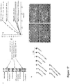
- FIG. 17A shows photographs of non-reducing gels of NM23-WT, NM23-S120G-mixed, NM23-S120G-hexamer and NM23-S120G-dimer, which show the multimerization state of the wild type protein and the three different preparations of the S120G mutant.
- Figure 17B shows an overlay of Surface Plasmon Resonance (SPR) measurements showing the ability of the four different NM23s to bind to a MUC1 ⁇ extra cellular domain peptide (PSMGFR) attached to the SPR chip surface.
- SPR Surface Plasmon Resonance
- Results show that the amount of binding of NM23 to its cognate receptor, MUC1 ⁇ , is a function of how much dimer is present in the sample.
- SPR measures protein mass at the chip-solution interface, so if the hexamer bound to the MUC1 ⁇ peptide surface, it would yield an SPR signal 3-times greater than if a dimer bound.
- Figure 17C shows photograph of a nanoparticle experiment that shows that only NM23 dimers bind to the cognate receptor MUC1 ⁇ .
- MUC1 ⁇ extra cellular domain peptide was immobilized onto gold nanoparticles.
- NM23-WT NM23-S120G-dimer
- NM23-S120G-hexamer was added to each aliquot of nanoparticles. If the NM23 bound to the nanoparticle immobilized MUC1 ⁇ peptide, it would cause the nanoparticles to become drawn close together which causes the solution to change from pink to blue.
- the experiment shows that only the NM23-S120G-dimer bound to the MUC1 ⁇ peptide.
- the addition of an anti-MUC1 ⁇ Fab competitively inhibited binding of NM23-S120G-dimers in solution to the MUC1 ⁇ peptide on the nanoparticles.
- Figures 17D-G show different NM23 multimers tested for their ability to support pluripotent stem cell growth.
- Human ES (embryonic stem) cells were cultured in either Figure 17D - NM23-S120G-dimer, Figure 17E - NM23-S120G-hexamer, Figure 17F - NM23-WT, or Figure 17G - NM23-S120G-dimer plus the MUC1 ⁇ extra cellular domain peptide (PSMGFR) to competitively inhibit binding of the NM23 dimer to the MUC1 ⁇ receptor on the stem cell surface.
- PSMGFR extra cellular domain peptide
- Figure 18 shows a native, non-denaturing gel that shows the multimerization state of NM23-WT versus three different preparations of recombinant NM23-S120G.
- Figure 19 shows SPR measurements of A) NM23 wild type (WT) and B) a preparation of NM23-S120G-"mixed” that produced 60% dimer. Protein was injected at five different concentrations. Results show that 8-times more NM23-S120G-mixed protein bound to a MUC1 ⁇ extra cellular domain peptide surface than NM23-WT. Because the wild type protein is a hexamer, the number of RUs must be divided by 3 to compare to the amount of dimer that bound. Although both wild type and S120G-dimer show a concentration dependence in binding, the amount of wild type hexamer that bound is so small that it may still be within the noise range of the system.
- MUC1 ⁇ is a significant "sternness" factor.
- OCT4 and SOX2 bind to the MUC1 gene promoter and also to the promoter of its cleavage enzymes.
- SOX2 and NANOG bind to the NM23 (NME7) promoter. Since blocking the extracellular domain of MUC1 ⁇ is lethal to hES cells, it follows that the pluripotency genes, OCT4, SOX2, and NANOG, bind to the promoter sites of MUC1, its cleavage enzymes and its activating ligand NME7 and induce their expression of MUC1.
- One or more of the genes or gene products that have already been shown to induce pluripotency can be replaced by transfecting the gene or introducing the gene product for MUC1 ⁇ alone or in addition to its cleavage enzymes and/or activating ligands, NME7, NME-H1, NME-H2 or an antibody that dimerizes the PSMGFR epitope of MUC1 or MUC1 ⁇ .
- the conventionally used standard protocol is to first plate dermablasts or fibroblasts (human foreskin fibroblast-neonatal, "hFFn”: #PC501A-hFF, System Biosciences, Mountain View, CA) on plastic and culture them in fibroblast media (FM), changed every 24 hours. After 5 days, the cells are transferred to a surface coated with inactivated fibroblast feeder cells, which can be mouse (MEFs) or human (HS27). For the next 2 days, cells remain in FM. On Day 7 the media is changed to bFGF-based media and media is changed every 24 hours.
- fibroblast media which can be mouse (MEFs) or human (HS27).
- colonies (clones) that have embryonic stem (ES) cell-like morphology are selected and individually plated into new wells coated with inactivated feeder cells (MEFs or HS27s) and sequentially passaged every 3-4 days.
- MEFs or HS27s inactivated feeder cells
- NM23 (NM23-H1, NM23-H2, or NME7) is introduced to cells, as the gene that encodes it, as the protein itself or as a protein bearing a leader sequence such as a poly-arginine tract, to facilitate entry into the cell, to aid in the induction or maintenance of pluripotency.
- the inventors recently showed that when NM23 is secreted by pluripotent stem cells (and cancer cells), it is an activating ligand of the cleaved form of MUC1 - MUC1 ⁇ - and triggers the MAP kinase proliferation pathway.
- NM23 stimulation of MUC1 ⁇ was shown to promote the growth of pluripotent hESCs and inhibited their differentiation (Hikita et al., 2008). NM23 also induces the transcription of c-MYC (Dexheimer at al., 2009) and replaces the need for c-MYC.
- NM23 is added exogenously either in its native state to activate the MUC1 ⁇ growth factor receptor or with a poly arginine tract to facilitate entry into the cell and nucleus where it induces C-MYC expression.
- NM23 (NME) may be added as the encoding nucleic acid, or as the expressed protein with or without a modification that facilitates entry into the cell.
- NME1or NME2 can be used in their native state or in mutant forms that favor the dimeric state, such as the S120G mutation or NME7.
- a bivalent antibody that binds to the extracellular domain of MUC1 ⁇ (PSMGFR) or a dimeric MUC1 ⁇ ligand, such as NM23, or genes encoding them are added to MUC1 ⁇ -expressing cells to induce pluripotency, increase the efficiency of the induction of pluripotency, to maintain pluripotency or to inhibit differentiation.
- the cells to which these MUC1 or MUC1 ⁇ interacting proteins are added may be naturally occurring cells or those into which genes to induce stem cell-like characteristics have been added, or have already entered the differentiation process or may be stem cells.
- Genes for inducing pluripotency may be introduced on the same or different plasmids, which may be lenti viral vector driven or adenovirus vectors or any integrating or non-integrating viral or non-viral vector, or any other system that facilitates introduction of these genes into the desired cells.
- the invention encompasses genes disclosed here for the induction of stem-like characteristics or pluripotency that can be replaced by the gene products, the proteins, either in their native state or modified with leader sequences such as poly-arginine tracts to allow entry into the cells.
- the products of these genes, i.e. proteins, or other proteins which interact with one or more of the products of the transfected genes are introduced to cells to induce or maintain pluripotency or other stem-cell like characteristics.
- NM23 protein such as, but not limited to, NME-H1, NME-H2, NME6 or NME-7 enhances the induction or maintenance of pluripotency.
- NM23 is introduced along with one or more of the previously identified pluripotency factors, including but not limited to OCT4, SOX2, KLF4, as well as others disclosed herein.
- NM23 exists as a family of proteins wherein the commonality among these proteins is the presence of a nucleoside diphosphate kinase (NDPK) domain that catalyzes the conversion of ATP to ADP.
- NDPK nucleoside diphosphate kinase
- Tumor Metastasis Factor With the recent identification of ten NM23 family members, they are now also known as NME proteins 1-10 ( Boissan et al., Mol Cell Biochem (2009) 329:51-62, "The mammalian Nm23/NDPK family: from metastasis control to cilia movement, ").
- Leukemia cells are blood cells that are blocked from terminal differentiation. Interestingly, the ability to inhibit differentiation of leukemia cells was shown to be independent of its catalytic domain. Mutations in the NDPK domain that abrogated its enzymatic activity had no effect on the protein's ability to block differentiation of some types of leukemia cells. However, the scientific literature of the following decades paints a picture of total confusion as to whether NM23 inhibits differentiation, accelerates differentiation or has no effect at all.
- NM23 inhibited erythroid differentiation of leukemia cell lines HEL, KU812, and K562 but not monocyte or granulocyte differentiation of progenitors HL60, U937, or HEL/S cells.
- Venturelli et al 1995 reported that NM23 overexpression inhibited G-CSF dependent granulocyte differentiation of human hematopoietic progenitors.
- Willems et al 1998 found that NM23 expression decreases as human CD34 + hematopoietic progenitors from the bone marrow cells differentiate.
- Willems et al showed that NM23 had no effect on cell proliferation, did not induce or inhibit differentiation but skewed differentiation of CD34+ cells toward the erythroid lineage.
- NME6 is roughly the same molecular weight as NME1 and in sea sponge ( Suberites domuncula ) it is reported to exist as a dimer ( Perina et al, 2011, "Characterization of Nme6-like gene/protein from marine sponge Suberites domuncula” Naunyn-Schmiedeberg's Arch Pharmacol, 384:451-460 ). Although NME7 (NM23-H7) is a monomeric protein, the inventors have discovered that it functions like a dimer.
- NME7-AB recombinant NME7
- NME7 was then added to the peptide surface to saturation.
- the second PSMGFR peptide bore either a histidine tag or a biotin molecule.
- HRP labeled anti-His-tag antibody or streptavidin was added, which showed robust and concentration dependent binding of the second MUC1 ⁇ peptide to the NME7.
- the results show that monomeric NME7 is able to dimerize MUC1 ⁇ on a cell surface.
- NME7 can also exist as a smaller protein of molecular weight -25 kDa. Pull down assays of stem cell lysates and supernatants were performed followed by mass spectrometry. The smaller molecular weight forms of NME7 all contained peptide sequences from its NDPK A domain, but not from the B domain. This may be an alternative splice isoform or a cleavage product. Like NME1, this smaller A domain NME7 may dimerize.
- NME family proteins are differentially expressed at different times of cell and tissue development. Whereas we detect NME6, NME7 and NME1 in embryonic stem cells, only NME1 and NME2 are routinely expressed in adult cells or adult stem cells. Because NME1 forms hexamers which induce rather than inhibit stem cell differentiation, it follows that NME6 and NME7 are expressed earlier in embryogenesis and in true pluripotent stem cells because they cannot form the hexamers.
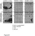
- Example 7 describes an experiment in which BGO1v human embryonic stem cells are grown in either NM23-S120G, which has been refolded and purified to exist primarily as a dimer ( Figs. 16-17 ), over a coating of anti-MUC1 ⁇ monoclonal antibody MN-C3 or cultured in bFGF over mouse fibroblast feeder cells.
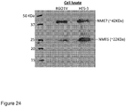
- Western blots of the resultant cells show that NME7 is highly expressed in stem cells that have been cultured in NM23-S120G dimers ( Figure 23 Part I. C - Lane 1) but only weakly expressed in stem cells cultured in bFGF (Lane 2).
- stem cells cultured in NM23 dimers revert to a more pluripotent state also called the naive state
- culturing stem cells in bFGF drives stem cells into a more differentiated state called the "primed" state.
- stem cells in the primed state are not capable of differentiating into any cell type the way true pluripotent stem cells should be able to.
- RT-PCR measurements were then performed to measure expression levels of the naive genes versus the primed genes for cells cultured in NME-based media compared to FGF-based media.
- NME-based media generated stem cells that were in a more naive state than cells cultured in FGF-based media.
- cells grown in FGF in minimal media (MM) grown over MEF feeder cells had higher expression of the primed markers and lower expression of the naive markers than the same source cells grown in NME1 dimers in minimal media (MM) wherein cells were plated over plasticware coated with an anti-MUC1 ⁇ antibody.
- FGF-based media fared even worse with very high levels of the primed genes and lower levels of the naive ones ( Figure 13 ).
- stem cells that had previously been cultured in FGF were transitioned to NME-based media, there was a trend toward expressing higher levels of the naive genes and lower levels of the primed genes with successive passage number. For one example of this trend, see Example 13C.
- Histone-3 is detected as a condensed dot in the nucleus of primed stem cells but not in the nucleus of naive stem cells.
- strategies that increase expression of NME7 in a cell for example via introduction of nucleic acids capable of causing expression of NME7 or methods that add NME7 protein, or NME1 mutants or variants that prefer dimer formation are strategies that maintain pluripotency, maintain the na ⁇ ve stem cell state in embryonic or induced pluripotent stem cells, and/or induce pluripotency in more mature cell types, including somatic cells, dermablasts and fibroblasts. These strategies may include ectopic expression of one or more of the pluripotency genes Oct4, Sox2, Nanog, Klf4 or c-myc in addition to NME7 or NME1 dimer forming or dimer mimicking variants.
- NME7 exists as a single protein but structurally is comprised of two monomers and so functions as a dimer. NME7 contains two NDPK domains, portions of which bind to the MUC1 ⁇ growth factor receptor.
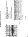
- Example 7 also describes a binding experiment called a pull-down assay. In this experiment, the MUC1 ⁇ extra cellular domain peptide was attached to beads which were then incubated with lysates from BGO1v human embryonic stem cells. After wash steps and release from the beads, species capture by interaction with the MUC1 ⁇ peptide were separated on an SDS-PAGE gel then probed with an anti-NME7 antibody.
- Figure 23 Part II. F) - Lane 1 shows that NME7 binds to the MUC1 ⁇ extra cellular domain. Portions of the double NDPK domains in NME7 bind to MUC1 ⁇ growth factor receptor and dimerize it which activates pathways that maintain pluripotency, induce pluripotency and inhibit differentiation of stem and progenitor cells.
- NME6 exists as a dimer, and resists formation of higher order multimers. NME6 dimers bind to MUC1 ⁇ growth factor receptor and dimerize it which activates pathways that maintain pluripotency, induce pluripotency and inhibit differentiation of stem and progenitor cells. Like NME1 mutants and variants that prefer dimer formation, both NME6 and NME7 are capable of maintaining and inducing pluripotency and inhibiting differentiation of stem and progenitor cells, including iPS cells.
- NME6 and NME7 can be added exogenously to stem cells (embryonic or induced pluripotent) or progenitor cells to induce growth, maintain them in an undifferentiated state or inhibit their differentiation. NME6 and NME7 can be added exogenously to stem or progenitor cells to induce pluripotency.
- nucleic acids encoding NME1 mutants and variants that prefer dimer formation, NME6 and/or NME7, or variants thereof, including single chain variants that behave as dimers can be introduced into cells to induce the cells to revert to a less differentiated state or to maintain cells in a less mature state.
- NME is highly conserved among all species, the methods described herein are not intended to be limited to human NME species nor limited to use with human cells.
- NME7 is a monomeric protein that has two NDPK domains, A and B. Until now, its function has not been elucidated. It was previously only known that NME7 was expressed in testes, ovary and brain. NME7 is suspected to be involved in the motility of flagella and is thought not to have NDPK activity because of the lack of certain key residues.
- NME7 expressed in E. coli and secreted as a soluble protein was added to human stem cells in culture in a minimal stem cell media devoid of any other growth factor, feeder cells or their conditioned media.
- the stem cells were adsorbed onto the cell culture plate by adhering to a layer of an antibody that recognizes a stem cell surface antigen. In this way, interference by growth factors and other cytokines present in surface coatings such as Matrigel were avoided.
- stem cells from the same source were cultured side-by-side but instead of adding exogenous recombinant NME7, recombinant NM23-H1 (also known as NME1) that had been refolded and purified as a population of dimers was added (See Examples 9 and 10 and Figures 25-30 ).
- NME7 recombinant NM23-H1
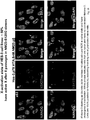
- Figures 27-30 show that the cells have stem cell morphology in that they are growing as a single layer of cells that have a high nucleus to cytoplasm ratio.
- immunocytochemistry and quantitative PCR showed that the resultant cells both expressed pluripotency markers but did not express differentiation markers such as FOXa2 and miR-145.
- NME7 stem cells cultured in NME7 produced 1.4-times more cells than those cultured in NM23-H1 dimers. This represents a significant improvement over the state of the art because the NM23-H1 isoform can exist as a dimer, tetramer or hexamer, wherein only the dimer is the active form and the hexamer induces differentiation.
- NME form that only activates such as NME7. From a commercial standpoint, it is more cost-effective to produce NME7 since it is active as a monomer, expressed in E. coli and secreted as the soluble protein.
- NME7 can also be added exogenously to induce cells to revert to a less mature state in vitro or in vivo or to induce pluripotency in somatic cells.
- NME1 NM23-H1
- siRNA siRNA
- anti-sense nucleic acids or any other method for suppressing gene expression
- NM23-H1 (NME1) forms tetramers and hexamers as its concentration increases, which then induce differentiation.
- NME1 hexamers are added exogenously or NME1 is genetically induced to be expressed within a stem cell, when it is desired to have a cell differentiate to a more mature state.
- NME7 constructs were made and tested. Although it has been previously reported that NME7 can be expressed in in vitro expression systems using wheat germ cell extracts (MW 36.52kDa) and in human HEK293 cells (MW 38kDa), we found that a smaller construct containing essentially just the NDPK A and B domains can be expressed in E. coli as a soluble protein with very high yields.
- NME7-AB with either a histidine tag or a strep tag, however it can be made without an affinity tag or with any other affinity tag.
- NME7-AB promoted pluripotency and inhibited differentiation of human stem cells as well as or better than NME1 (NM23-H1) dimers that had previously been shown by us to fully support stem cell growth, maintenance of pluripotency, able to induce pluripotency and inhibit differentiation.
- NME7 can substitute for NME1 dimers without the threat of hexamer formation over time and in a more cost-effective and commercially applicable way.
- NME family proteins are highly conserved across all species, including plants. Therefore, sequences of NME family members, isoforms and variants of the invention, especially NME1, NME6 and NME7 constructs, may be comprised of sequences from any species or combinations of sequences from different species, with mammalian species being preferred, mouse species still more preferred, and human species most preferred.
- Such NME family members, isoforms and variants can be used for the propagation of, self-renewal, maintenance of pluripotency, induction of pluripotency or inhibition of differentiation in any species, wherein mammalian species are preferred, mouse species more preferred and human species most preferred.
- NME1 in dimeric form or NME7 can also be used for the maintenance of naive human stem cells or for the induction of naive stem cells from stem cells in the primed state or for making naive stem cells from somatic cells in the process of generating iPS cells.
- Stem cells in the naive state are characterized by high expression levels of the pluripotency genes OCT4, KLF2 and NANOG or KLF4 and low expression levels of FOXa2, OTX, LHX and XIST compared to stem cells grown in bFGF-containing media.
- An even more stringent indicator of the naive state is that the cells have not yet undergone X-inactivation.
- High levels of XIST are indicators of inactivation of one of the X chromosomes, but a more definite determinant is immunocytochemical staining of Histone 3. If X-inactivation has occurred, Histone 3 is condensed and can be visualized as a discrete mass in the cell's nucleus. If X has not yet been inactivated, Histone 3 is either undetected or is dispersed throughout the nucleus, sometimes referred to as a "cloud.”
- Another indicator of stem cells being in the naive state is that the cells have a higher cloning efficiency, which is calculated as the number of discrete colonies that can be counted per a specified number of cells plated.
- the cloning efficiency of mouse stem cells which unlike human stem cells, are easily maintained in the naive state by culturing in media containing mLIF (murine leukemia inducing factor), is ⁇ 30%.
- the cloning efficiency of human stem cells cultured in FGF-based media is -1%.
- ES cells cultured in NME1 dimers had a cloning efficiency of 18% and the same cells cultured in NME7 had a cloning efficiency of 23%.
- the invention also envisions the use of NME1 or NME7 to generate populations of stem cells that are essentially 100% pluripotent and in the naive state.
- populations of cells are generated that are about 50% in the completely naive state.
- cloning techniques known to those skilled in the art, such as limiting dilution, on mixed population of naive and primed stem cells, populations of stem cells that are all in the naive state are generated.
- ES cell line generation has been described in the art using previously described techniques ( Embryonic Stem Cell Lines Derived from Human Blastocysts. James A.
- cells are cultured in media containing NME7 or NME1 wherein at least some of the NME1 protein is in the dimeric state, more preferably wherein at least 25% of the protein is in the dimeric state and still more preferably wherein at least 50% of the protein is a dimer.
- cells are derived from the inner cell mass of a non-human blastcyst and cultured in a media containing NME7 or NME1 dimers.
- somatic cells such as dermablasts or fibroblasts are induced to revert to a less mature state by culturing in a media containing NME7 or NME1 dimers.
- iPS cell lines are generated by transducing somatic cells, dermablasts or fibroblasts with one or more of the pluripotency genes Oct4, Sox2, Klf4, Nanog, c-Myc, or LIN28 or by treating the cells with the products of those genes, i.e., the proteins or with agents that cause the proteins to be produced and doing so in the presence of a media that contains NME1 dimers or NME7.
- the invention envisions generating new iPS cell lines, which are produced with greater efficiency or are in a more naive state than iPS cells generated by traditional methods that include the use of FGF, using any method that induces a cell to revert to a less mature state, including a pluripotent state, by culturing the cells in the presence of NME1 dimers or NME7.
- the use of FGF is eliminated.
- cells undergoing the process of generating iPS cells are not transferred onto fibroblast feeder cells.
- any suitable surface for stem cell growth or induction of pluripotency can be used with media containing NME1, NME6, or NME7.
- Such surfaces include but are not limited to feeder cells, Matrigel, hydrogels, integrins and integrin derivatives, E-cadherin and E-cadherin derivatives, vitronectin, antibodies to cell surface receptors, antibodies that recognize the MUC1 ⁇ extra cellular domain, antibodies that recognize the PSMGFR sequence, Vita TM brand plates (ThermoFisher), VITA TM plates coated with an anti-MUC1 ⁇ antibody and the like.
- NME1 mutants and variants that can form stable dimers were added to minimal stem cell media (MM) and used to propagate stem cells that were growing over a Matrigel surface or an anti-MUC1 ⁇ antibody coated surface.
- MM minimal stem cell media
- the growth of the stem cells and their ability to resist spontaneous differentiation was essentially equal in all three media and on both surfaces ( Figure 20 , Figure 21 ). Some surfaces negatively impact the quality of the stem cells grown on them.
- the surface which may be a plastic, biodegradable, mesh or solid, planar or particle-like, is coated with an antibody that recognizes the MUC1 ⁇ extra cellular domain, consisting primarily of the PSMGFR sequence.
- the surface is a VITA TM brand plate coated with an antibody that recognizes the PSMGFR peptide.
- the anti-MUC1 ⁇ antibody recognizes the N-terminal portion of the PSMGFR peptide, including the N-terminal 15 amino acids.
- a rho kinase inhibitor is used to enhance adhesion of the cells to the surface.
- Many rho kinase inhibitors including but not limited to HA100, Y27632, and thiozivincan be used to aid in enhancing adhesion of stem cells to surfaces, particularly to the surfaces that are plastic coated with anti-MUC1 ⁇ antibodies.
- a rho kinase inhibitor is added for the first 24-48 hours only.
- inhibitors of guanine exchange factors are added to the media instead of rho kinase inhibitor for at least a portion of the culture period, to enhance adhesion to surfaces that are not cells or comprised of complex cell mixtures such as Matrigel.
- Inhibitors of GEFs are used in some cases instead of rho kinase inhibitors to enhance the attachment of stem cells to surfaces that are coated with anti-MUC1 ⁇ antibodies.
- the guanine exchange factor inhibitor is NME1 in hexameric form.
- the guanine exchange factor inhibitor is a peptide that is derived from NME1.
- Inhibitors of rho kinase or guanine exchange factor may be required for maximal adhesion of primed stem cells or mixed populations of primed and naive stem cells to certain surfaces, such as plates coated with anti-MUC1 ⁇ antibodies.
- pure populations of naive stem cells do not require the use of rho kinase inhibitors or guanine exchange factor inhibitors.
- pure populations of naive stem cells are cultured in the absence of FGF or a rho kinase inhibitor.
- Rho kinase inhibitors are agents that inhibit rho kinase I or II. They may be small molecules, peptides or proteins Rath N, Olson MF. Rho-associated kinases in tumorigenesis: re-considering ROCK inhibition for cancer therapy. EMBO Rep. 2012;13(10):900-8 .
- Examples of rho kinase inhibitors are Y27632, HA-1077, also called Fasudil, H-1152, and thiazovivin ( Olson MF. Applications for ROCK kinase inhibition. Curr Opin Cell Biol. 2008;20(2):242-8 ; Watanabe K, Ueno M, Kamiya D, et al.
- a ROCK inhibitor permits survival of dissociated human embryonic stem cells. Nat Biotechnol. 2007;25(6):681-6 ; Breednlechner C, Gassel M, Hidaka H, et al. Protein kinase A in complex with Rho-kinase inhibitors Y-27632, Fasudil, and H-1152P: structural basis of selectivity. Structure. 2003;11(12):1595-607 ; Lin T, Ambasudhan R, Yuan X, et al. A chemical platform for improved induction of human iPSCs. Nat Methods. 2009;6(11):805-8 ).
- Rho kinase inhibitors In addition to Rho kinase inhibitors, the invention envisions using inhibitors of related pathways in place of the Rho kinase inhibitors. For example, in the same pathway, guanine exchange factors (GEFs) are upstream of Rho kinase. The GEFs activate the Rho kinases. Therefore instead of using rho kinase inhibitors, the invention envisions using GEF inhibitors.
- GEFs guanine exchange factors
- any agent that increases the amount of GDP present in a cell such as RAD, GEM,and RhoE as well as others, can be used in place of rho kinase inhibitors to aid in stem cell growth, survival and attachment to surfaces ( Riento K, Guasch RM, Garg R, Jin B, Ridley AJ (2003) RhoE binds to ROCKI and inhibits downstream signaling. Mol Cell Biol 23: 4219-4229 ; Komander D, Garg R, Wan PT, Ridley AJ, Barford D (2008) Mechanism of multi-site phosphorylation from a ROCK-I:RhoE complex structure.
- Blebbistatin is a myosin inhibitor and can be used in place of any rho kinase inhibitor used according to methods of the invention ( Ohgushi M, Matsumura M, Eiraku M, Murakami K, Aramaki T, Nishiyama A, et al. Molecular pathway and cell state responsible for dissociation-induced apoptosis in human pluripotent stem cells. Cell Stem Cell 2010;7:225-39 ; Ohata H, Ishiguro T, Aihara Y, et al. Induction of the Stem-like Cell Regulator CD44 by Rho Kinase Inhibition Contributes to the Maintenance of Colon Cancer-Initiating Cells. Cancer Res.
- rho associated kinase inhibitor Another alternative to using a rho associated kinase inhibitor, is to activate or inhibit any target that results in increased cell migration.
- Activation of modulators of the RAC pathway, that increases cell migration include PI3K, CDC42, PAK, N-WASP as well as RAC1, and lamellipodium.
- Agents that enhance their expression levels in the cell or increase their activity are used to enhance stem cell survival and attachment to surfaces.
- RAC1 GTPase expression decreases as stem cells dissociate and conversely Rho associated kinases increase, which leads to loss of cell migration and apoptosis ( Ohgushi M, Matsumura M, Eiraku M, et al.
- inhibitors of the PI3k or RAC pathway should be suppressed, or activators of RAC pathway increased in order to enhance stem cell survival and attachment to surfaces, such as to antibody coatings wherein the antibody binds to a stem cell surface protein, such as MUC1 ⁇ .
- NME1 in hexamer form and NME2 likely also in hexamer form, bind to GEF Tiam1 and Dbl-1 to inhibit GTPases RAC1 and CDC42 GTPase, respectively (( Ohgushi M, Matsumura M, Eiraku M, et al.
- NME1 and NME2 hexamers decrease cell migration and limit stem cell attachment and survival.
- NME1 and NME2 in embryonic and induced pluripotent stem cells in culture in NME7 media and observe no ill effects.
- Suppression of NME1 and NME2 in stem and progenitor cells that are cultured in NME6 or NME7 containing media are preferred methods for enhancing stem and progenitor cell survival and attachment to surfaces for in vitro culture.
- embryonic stem cells or iPS cells are cultured in a media that contains NME7 and siRNA to suppress NME1 and NME2.
- nucleic acids that suppress NME1 and NME2 are siRNA molecules derivatized with cholesterol moieties to enable entry into the cells (Darmicon).
- stem cells are not contacted with a rho kinase (ROCK1) inhibitor such as Y27632.
- ROCK1 rho kinase
- stem cells cultured in media containing NME1, NME6 or NME7 are grown in suspension.
- a number of techniques for cell growth in suspension are known to those skilled in the art. Wave bags, roller bottles, and the like can be used in association with NME-containing media for stem cell growth or for the induction of pluripotency in more mature cells such as somatic cells, dermablast or fibroblasts.
- the NME protein is NME1 in dimer form. In a still more preferred embodiment the NME protein is NME7.
- NME family proteins can be added to any media suitable for stem or progenitor cell growth or any media suitable for the induction of pluripotency.
- a base media such as DMEM, DMEM/F12, or similar further containing knockout serum replacement or similar and non-essential amino acids are preferred.
- the base media contains 60-80% DMEM-like base and 20-40% knockout serum replacement or similar, plus concentrated non-essential amino acids.
- the base media contains a DMEM-like base plus insulin, selenium, and transferrin.
- the preferred species of the protein components is human.
- MUC1 ⁇ growth factor receptor in particular NME family proteins, promote the growth of, and maintenance of pluripotency of stem and progenitor cells.
- MUC1 ⁇ ligands in a media also support the process of making induced pluripotent stem (iPS) cells and support the process of inducing cells to revert to a less mature state.
- iPS induced pluripotent stem
- MUC1 ⁇ ligands themselves induce cells to revert to a less mature state in the absence of other factors such as transfection or transduction of pluripotency genes including OCT4, SOX2, KLF4, NANOG or c-Myc.
- ligands of the MUC1 ⁇ receptor can function as growth factors. Ligands that dimerize the MUC1 ⁇ extra cellular domain were shown to increase the growth of MUC1-positive cancer cells and also stem and progenitor cells that expressed MUC1 ⁇ .
- the present application discloses that NME7 is also a ligand of the MUC1 ⁇ growth factor receptor. NME7 is secreted by stem cells. NME7 in a base media devoid of any other cytokines fully supports the growth of embryonic or induced pluripotent stem cells. NME7 functions to promote growth of and maintain pluripotency of stem cells in a manner very similar to NME1 dimers.
- NME1 expressed or purified such that a significant population exists as a dimer, fully supports the growth of embryonic or induced pluripotent stem cells and does not require serum or any other cytokines.
- NME1 in dimeric form and NME7 also induce pluripotency in cells that are in a more mature state than the naive or ground state stem cells.
- growth of human stem cells in FGF containing media produce stem cells in the "primed” state
- growth of human stem cells in NME1 dimers or NME7 induces those cells to revert to an earlier state called the "naive" or “ground” state.
- Recent research provides evidence that stem cells in the primed state cannot develop into all the cell types in the human body the way that naive stem cells can.
- stem cells cultured in NME1 or NME7 that are in a more naive state are ideally suited for use in human therapies because the naive state is the natural state of human stem cells and it is those naive stem cells that are able to develop into any cell in the human body.
- stem cells cultured in NME1 or NME7 are used for any and all human stem cell therapies.
- NM23 as a component of a defined, xeno-free media supports stem cell growth, including both embryonic and induced pluripotent and further can be used as the media in which to reprogram somatic, progenitor, or somewhat mature cells such that they become pluripotent stem cells, which are also referred to as iPS cells.
- NM23 to base media DMEM/F12 (or similar) plus insulin (human preferred), selenium, transferrin, 1-ascorbic acid, with pH adjusted using NaHCO 3 fully supported ES and iPS cell growth when cells were attached to Vitronectin-coated surfaces or anti-MUC1 ⁇ coated surfaces.
- insulin human preferred
- Rho kinase inhibitors HA100 or Y27632 for at least the first 24 hours greatly improved stem cell surface attachment.
- NM23 and NM23 variants that can be expressed and/or isolated as dimers.
- Other growth factors such as FGF-2 or TGF-beta may optionally be added to NM23 in this media.
- cells may be grown under hypoxic conditions for improved performance.
- NM23 to base media DMEM/F12 (or similar) plus insulin (human preferred), selenium, transferrin, 1-ascorbic acid, with pH adjusted using NaHC0 3 fully supported ES and iPS cell growth when cells were attached to Vitronectin-coated surfaces, anti-MUC1 ⁇ coated surfaces, Matrigel and other surfaces to which stem cells attach.
- insulin human preferred
- Rho kinase inhibitors HA100 or Y27632 for at least the first 24 hours greatly improved stem cell surface attachment.
- Addition of any Rho kinase inhibitor changes cell shape and attachment properties and can be used in media containing NM23 to enhance the attachment of stem cells, progenitor cells and other non-adherent cells to surfaces.
- Rho kinase inhibitors In the presence of a rho kinase inhibitor, cells transition within 24 to 48 hours from a rounded shape with weak surface attachments to a more flattened shape with many surface attachments. Although the use of Rho kinase inhibitors has been previously described for use with stem cells to enhance survival, Rho kinase inhibitors do not increase survival in NME based media. For that reason, it is only used to make cells flatten out and form greater attachments to surface.
- rho kinase inhibitors have been previously described for use with bFGF-based media, also to increase survival.
- Rho kinase inhibitors to aid in attachment to surfaces when NME1, NME6 or NME7 are used in a media.
- NM23 and NM23 variants that can be expressed and/or isolated as dimers as well as NME7, which is a monomer that has two NDPK domains.
- Figure 14 shows that in a pull-down assay, NME7 from human stem cells bound to a synthetic peptide having the sequence of the PSMGFR peptide.
- the inventors have shown that: 1) only NME1 dimers (not hexamers) support pluripotent stem cell growth; 2) NME1 dimers (not hexamers) bind to two or more MUC1 ⁇ extra cellular domain peptides in a nanoparticle assay; and 3) MUC1 ⁇ 's pluripotency activity and growth factor receptor activity occurs after ligand-induced dimerization of its extra cellular domain; a bivalent anti-MUC1 ⁇ antibody results in a bell-shaped curve of growth as a function of antibody concentration which shows that it is dimerization of the extra cellular domain that causes the growth factor receptor function.
- NME7 functions similarly to NME1 dimers in their ability to support pluripotent stem cell growth and inhibit differentiation.
- NME7 via NDPK domains binds to the MUC1 ⁇ receptor's extra cellular domain to promote pluripotent, naive stem cell growth and inhibits differentiation.
- NME7 binds to the PSMGFR peptide which is a part of the MUC1 ⁇ extra cellular domain.
- NM23-H1 which is also known as NME1
- NME1 is active as it is a stem or progenitor cell growth factor only when in dimer form.
- NME1 can exist as a dimer, tetramer or hexamer as a function of concentration as well as its sequence. Mutations such as the S120G or P96S with or without C-terminus deletions prefer dimer formation and are somewhat resistant to hexamer formation so are preferred as stem or progenitor cell growth factors.
- NME6 exists as a dimer and is also preferred for use as a growth factor, like NME1 dimers, for the maintenance and induction of pluripotency and for inhibition of differentiation.
- NME7 is a monomer but may bind like a dimer with its two NDPK domains.
- NME7 binds to MUC1 ⁇ extra cellular domain peptide, indicating that portions of at least one NDPK domain binds to the MUC1 ⁇ receptor, and like NME1 dimers, results in the maintenance or induction of pluripotency while inhibiting differentiation.
- NME7 was shown to simultaneously bind to two MUC1 ⁇ extra cellular domain peptides (PSMGFR), indicating that the NME7-AB protein can dimerize MUC1 ⁇ on the cell surface.
- PSMGFR MUC1 ⁇ extra cellular domain peptides
- NME1 also binds to the MUC1 ⁇ extra cellular domain peptide and the MUC1 ⁇ receptor on cells.
- NME1 hexamers do not bind to the MUC1 ⁇ receptor and do not maintain or induce pluripotency.
- NME1 dimers, NME6 dimers, or NME7 for the growth, maintenance, induction of pluripotency and inhibition of differentiation can be used in many different base media that are suitable for culture of stem or progenitor cells.
- the composition of compatible base media can vary.
- other growth factors or cytokines may be added into the media along with NME protein.
- Other growth factors such as FGF-2 or TGF-beta may optionally be added to NM23 in this media.
- NME1 dimers, NME6 dimers or NME7 is added to a base media that does not contain other growth factors or cytokines. Cells may be grown under hypoxic conditions for improved performance.
- an NME family protein is encoded in a nucleic acid sequence that may be contained within a plasmid that is introduced into a cell.
- the nucleic acid sequence that encodes the NME family protein can be part of a plasmid, or expression vector that also carries the sequence of a gene whose expression is desirable.
- the gene may be a corrected gene or gene to be expressed.
- the invention also envisions introducing a nucleic acid encoding an NME family member into a stem cell such that the cell constitutively expresses its own growth factor, simplifying culturing because it would only require a minimal solution for growth.
- the nucleic acid is an expression plasmid, which may have an inducible or controllable promoter.
- the NME family member is NME1, NME6 or NME7.
- the NME family member is NME7.
- the NME family is NME7-AB.
- the invention also discloses the use of these methods, in patients, in cells destined for transplant in patients.
- the invention additionally envisions the use of NME-based media for the generation of new stem cell lines that may be embryonic in origin or induced from more mature cells.
- the NME-based media is a minimal media containing recombinant NME7.
- Yet more preferred is a media containing NME7-AB.
- New stem cell lines are established as follows: cells are harvested from the inner mass of a non-human blastocyst or the entire non-human blastocyst can be used. The cells of the blastocyst are maintained and cultured in a media containing NME7. In a preferred embodiment, the media is free of other growth factors or cytokines. The harvested cells may be plated over a layer of feeder cells, which may be fibroblasts or may be MUC1-positive cancer cells, or over an antibody layer. In a preferred embodiment, the antibody is an anti-MUC 1 ⁇ antibody. Still more preferred is an antibody that binds to the N-10 PSMGFR peptide. Alternatively the cells may be cultured or maintained in suspension in an NME7 containing media.
- stem-like cells which can then be cloned to obtain clones that have the desired karyotype, and the desired gene expression profile.
- clones are isolated and proliferated that have gene expression indicative of the naive state and not the primed state and further have not undergone X-inactivation if the cells are female.
- the non-human blastocyst or cells from the non-human blastocyst are plated onto a surface and cultured for some period of time until blast outgrowths are observed that appear stem-like. These are harvested, grown and clones having the desired characteristics are isolated and maintained.
- Standard cloning techniques are performed to isolate clones that are positive for pluripotency markers as well as naive markers and having low or no expression of primed markers such as FOXA2, OTX, LHX or sometimes XIST or lack of condensed Histone-3 in the nucleus.
- Clones are maintained and propagated in minimal media containing NME7 and optionally maintained in the naive state by growing the cells over a layer of anti-MUC1 ⁇ antibodies.
- the cells are cultured in suspension.
- a Rho kinase inhibitor may be added to the NME7 containing media.
- the media is free of FGF.
- somatic cells can be reprogrammed by ectopic expression of transcription factors (Lowry et al., 2008; Maherali et al., 2007; Nakagawa et al., 2008; Okita et al., 2007; Park et al., 2008; Takahashi et al., 2006; Takahashi and Yamanaka, 2006; Wernig et al., 2007; Yu et al., 2006) to become pluripotent.
- transcription factors Lowry et al., 2008; Maherali et al., 2007; Nakagawa et al., 2008; Okita et al., 2007; Park et al., 2008; Takahashi et al., 2006; Takahashi and Yamanaka, 2006; Wernig et al., 2007; Yu et al., 2006
- iPS induced pluripotent stem
- iPS cell lines as follows: Somatic cells, dermablasts or fibroblasts which may be from a donor or a patient are cultured in a media containing an NME family protein.
- the NME family protein is NME1 dimer, NME6, or NME7.
- the NME family member is NME7, wherein NME7-AB is especially preferred.
- the cells can be treated with nucleic acids, small molecules, or proteins that result in an increase in levels of pluripotency genes or proteins in the cells, wherein the pluripotency genes or gene products are chosen from OCT4, NANOG, KLF4, c-Myc, LIN28, and NME7.
- the starting cells are transfected with Oct4, SOX2 and Klf4.
- the starting cells that are somatic cells, dermablasts, fibroblasts or other cell are initially plated onto plastic multi-well plates in a minimal media containing roughly 2-50 nM NME7, more preferred is 8-16 nM. Media is changed every 24-72 hours as is typical.
- cells begin to lose their adhesion to the plastic, they can be moved to a surface to which they will adhere, such as a surface of inactivated fibroblasts, inactivated cancer cells or a surface coated with an antibody to a stem cell surface protein, wherein an anti-MUC1 ⁇ antibody is preferred.
- a rho kinase inhibitor may be added to the media in addition to the NME family member. After approximately 2-3 weeks, stem-like cells will have emerged and can be isolated and cloned. Clones with desired karyotype and gene expression profiles are selected and proliferated as stable self-renewing cell lines. In apreferred embodiment, clones that are naive are selected. Human cells are preferred for these methods of generating new stem cell lines.
- stem or progenitor cell for which the methods of the invention are useful.
- any stem or progenitor cells that expresses the cleaved for of MUC1, i.e. MUC1 ⁇ can be used along with media and methods of the invention.
- NME1 dimers and NME7 fully support pluripotent, naive stem cell growth and inhibit differentiation and these functions are independent of base media components. Virtually any base media that is suitable for stem cell growth will suffice. Preferred are base media that do not contain other growth factors or cytokines.
- E8 that includes DMEM/F12, insulin, selenium, transferrin, l-ascorbic acid, and NaHCO 3 for pH adjustment, plus bFGF and TGF-beta as the growth factors, which is a defined and xeno-free media for uman stem cell growth ( Chen G, Gulbranson D, Hou Z et al, "Chemically defined conditions for human iPSC derivation and culture” Nature Methods, Vol. 8. No. 5, 2011 pgs 424-431 ).
- NME1 or NME6 dimers or NME7 that we called “MN6”
- MN6 MN2 media
- NME1 or NME6 dimers, or NME7 in MN2 media which is a base media and non-essential amino acids only (see Example 1), somewhat supported pluripotent stem cell growth.
- media suitable for use with NME1 dimers and NME7 is comprised of a base media such as DMEM/F12 or DME/F12/Glutamax I, L-ascorbic acid, sodium selenium, insulin, transferrin and an agent to adjust pH such as sodium bicarbonate.
- the base media to which NME1 dimers or NME7 is added is comprised of DME/F12/Glutamax I, a serum replacement such as Knockout serum replacement (LifeTechnologies/Invitrogen), amino acids, such as non-essential amino acid solution (LifeTechnologies/Invitrogen), and optionally betamercaptoethanol.
- Media containing components from each of these base media are also envisioned.
- any base media suitable for stem or progenitor cell growth can be used with NME1 dimers and NME7 for the growth of stem or progenitor cells, to inhibit differentiation even to the primed state, and to induce pluripotency in more mature cells, which may be somatic cells, dermablasts, fibroblasts or progenitor cells.
- NME1 dimers, NME6 dimers, or NME7 can vary. In one aspect the NME concentration is between 1nM and 100nM (based on the molecular weight of the monomer). In a preferred embodiment, the concentration of NME1 dimers, NME6 dimers, or NME7 is from 2nM to 64nM. In a still more preferred embodiment, the concentration of NME1 dimers, NME6 dimers, or NME7 is 4nM to 32nM.
- Minimal Media that when mixed with NME1 dimers or NME7, at 8nM - 16nM, fully supported human stem cell growth and mouse stem cell growth and made human primed stem cells revert to the naive state was comprised of:
- MN6 Another media that worked similarly when NME1 dimers or NME7 was added at 8nM - 16nM was "MN6", comprised of:
- MN2 Another media that was tested with NME1 dimers or NME7 was added at 8nM - 16nM was "MN2" comprised of:
- NME1 dimers and NME7 in “MM” or “MN6” support human stem cell growth as well as or better than the FGF-containing media "E8" or mTeSR.
- the experiments shown in Figures 1-10 were carried out by plating stem cells on a layer of Vitronectin while the experiments of Figures 11 and 12 were carried out by plating stem cells onto a layer of anti-MUC1 ⁇ antibody.
- NME1 dimers or NME7 in any minimal or defined media promotes pluripotency and inhibits differentiation. Any surface or base media that is suitable for stem cell growth are compatible with growth in NME1, NME6, or NME7. The methods of the invention are not limited to use with embryonic stem cells.
- NME1 dimers or NME7 also fully supported the growth of human ES cells and iPS cells as well as mouse ES and iPS cells.
- NME1 dimers or NME7 supported the induction of pluripotency in somatic cells and in fact increased the efficiency of iPS generation.
- NM23 in this defined and xeno free media for the growth and maintenance of ES and iPS cells, it is also used in the process of making iPS cells from progenitors or mature somatic cells such as dermablasts.
- MUC1 ⁇ ligand any cell culture may be used so long as a MUC1 ⁇ ligand is either added to the media or is expressed by the cell.
- minimal media is preferred that includes MUC1 ⁇ ligand such as NM23 family of proteins added to the media or expressed by the cell.
- a minimal media was made that included only DMEM/F12, insulin (human preferred), selenium, transferrin, l-ascorbic acid, with pH adjusted using NaHCO 3 was made; we called this media "MN6", see detailed formula below.
- MN2 MEM Non-essential Amino Acid Solution
- NME7 8 nM - 16 nM NME7, NME6 or NME1 (NM23-S120G), wherein NME1 was refolded and purified such that the population was essentially all dimers.
- a Rho kinase inhibitor such as Y27632 was added.
- Example 1.1 Components of Minimal Stem Cell Media (“MM”) (500mls)
- Example 1.3 Components of MN7 is E8 media, minus the bFGF
- Examples 1-3 and figs 1-14 are from 70124
- Rho kinase inhibitor Y27632
- FIGS. 1-10 are photographic images of the stem cells in culture Day 4 post-plating.
- Figures 11 and 12 show human ES cells (H9s) plated onto a layer of anti-MUC1 ⁇ antibody (MN-C3).
- the source cells had previously been cultured according to standard protocols in bFGF at 4ng/mL on mouse feeder cells (MEFs) for 55 passages.
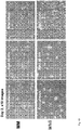
- Figure 11 shows the stem cells on the first passage (PI) onto the anti-MUC1 ⁇ antibody surface on Day 4 post-plating (D4), wherein A, C have been cultured in NM23 dimers at 8nM in Minimal Stem Cell media, "MM", and B, D have been cultured in NM23 dimers at 8nM in MN6 fully defined and xeno-free media.
- Figure 12 shows the same source stem cells after two passages (P2) on Day 2 post-plating (D2), wherein A, C were cultured in mTeSR media and B, D were cultured in MN6 media plus 8nM dimeric NM23.
- NM23 in MN6 media performed as well or better than mTeSR in terms of confluency and pluripotency of the resultant stem cells based on different base media. All media were supplemented with a Rho kinase inhibitor, Y27632 at 10uM for the first 48 hours only, for each passage.
- Figure 13 compares growth rates for the same cells on anti-MUC1 ⁇ antibody surface or on Vitronectin but cultured in different media.
- NM23-S120G In addition to the NM23-S120G refolded and purified as dimers, a single chain NM23 construct that included two NM23 monomer sub-units connected by a flexible linker of G 4 S 1 repeated 2-times - NM23-GS2 and a single chain NM23 construct that included 2 NM23 monomers connected by a longer linker - NM23-X4 were tested and performed as well as the naturally dimerized NM23-S120G.
- Example 2 Induction of pluripotency using NME-based media in the absence of FGF.
- the conventionally used standard protocol is to first plate dermablasts or fibroblasts (human foreskin fibroblast-neonatal, "hFFn”: #PC501A-hFF, System Biosciences, Mountain View, CA) on plastic and culture them in fibroblast media (FM), changed every 24 hours. After 5 days, the cells are transferred to a surface coated with inactivated fibroblast feeder cells, which can be mouse (MEFs) or human (HS27). For the next 2 days, cells remain in FM. On Day 7 the media is changed to bFGF-based media and media is changed every 24 hours.
- fibroblast media which can be mouse (MEFs) or human (HS27).
- colonies (clones) that have embryonic stem (ES) celllike morphology are selected and individually plated into new wells coated with inactivated feeder cells (MEFs or HS27s) and sequentially passaged every 3-4 days.
- MEFs or HS27s inactivated feeder cells
- NME7 is expressed to a greater degree in stem cells cultured in NME1 dimers, which we have shown express markers indicating that they are in a more naive state than cells cultured in bFGF, means that NME7 is expressed at a higher level in naive cells, compared to its expression in primed cells.
- NME7 also functions as a growth factor with MUC1 ⁇ as its target receptor
- pull-down assays a synthetic MUC1 ⁇ extra cellular domain peptide (His-tagged PSMGFR sequence) was immobilized on NTA-Ni magnetic beads. These beads were incubated with the cell lysates of BGO1v human embryonic stem cells that had been cultured in NME1 dimers over a surface coated with anti-MUC1 ⁇ antibodies (Lane 1), or cultured in bFGF over MEFs (Lane 2) or T47D human breast cancer cell lysates (Lane 3). Beads were rinsed and captured proteins were released by addition of imidazole.
- NME7 binds to the MUC1 ⁇ extra cellular domain peptide. This means that in stem cells and cancer cells, NME7 via its portions of its two NDPK domains, activates pluripotency pathways by dimerizing the MUC1 ⁇ extra cellular domain.
- NM23wt was amplified by polymerase chain reaction (PCR) using the following primers:
- the fragment was then purified, digested (NdeI, XhoI) and cloned between NdeI and XhoI restriction sites of the expression vector pET21b.
- NM23-H1 mutant S120G (serine #120 mutated to a glycine) was made using the GeneTailor TM Site-directed mutagenesis system (Life Technologies) following the manufacturer instructions using the following primers: 5'-gcaggaacattatacatggcggtgattctg-3' (SEQ ID NO:88) and 5'-gccatgtataatgttcctgccaacttgtat-3' (SEQ ID NO:89).
- Figure 16 shows overlay of FPLC traces comparing multimerization state of the wild type protein to the nonrefolded S120G mutant and the refolded S120G.
- Figures 17-19 show that only the dimeric form of the protein binds to MUC1 ⁇ (not the hexamer) and only the dimer is able to support pluripotent stem cell growth.
- Figure 21 shows non-reducing SDS-PAGE characterization and corresponding FPLC trace for the expressed and refolded protein as well as photographs of human stem cells, showing the NM23-S120G ability to support pluripotent stem cell growth.
- NM23 P96S ⁇ C1 was amplified using the following primers: 5'- atcgatcatatggccaactgtgagcgtaccttc -3' (SEQ ID NO:92) and 5'-gtggtgaccggtatagatccagttctgagcaca-3' (SEQ ID NO:93).
- NM23 P96S ⁇ C2 was amplified using the following primers: 5'-atcgatcatatggccaactgtgagcgtaccttc-3' (SEQ ID NO:94) and 5'-gtggtgaccggtgatccagttctgagcacagct-3' (SEQ ID NO:95).
- NM23 P96S ⁇ C6 was amplified using the following primers: 5'-atcgatcatatggccaactgtgagcgtaccttc-3' (SEQ ID NO:96) and 5'-gtggtgaccggtagcacagctcgtgtaatctacca-3' (SEQ ID NO:97).
- the resulting fragments were purified, digested (NdeI, AgeI) and cloned between NdeI and AgeI restriction sites of the expression vector pET21b.
- the pET21b was previously modified by replacing the XhoI restriction by AgeI using an overlap PCR method.
- LB broth Lia-Bertani broth
- OD600 OD600 reached -0.5.
- recombinant protein expression was induced with 0.4mM Isopropyl- ⁇ -D-thio-galactoside (IPTG, Gold Biotechnology) and culture was stopped after 5h. After harvesting the cells by centrifugation (6000 rpm for 10 min at 4°C), cell pellet was resuspended with running buffer: PBS pH7.4, 360 mM NaCl and 80 mM imidazole.
- lysozyme (1 mg/mL, Sigma), MgCl 2 (0.5mM) and DNAse (0.5 ug/mL, Sigma) was added.
- Cell suspension was incubated on a rotating platform (275 rpm) for 30 min at 37°C and sonicated on ice for 5 min.
- Insoluble cell debris was removed by centrifugation (20000 rpm for 30 min at 4°c).
- the cleared lysate was then applied to a Ni-NTA column (Qiagen) equilibrated with the running buffer. The column was washed (8CV) before eluting the protein off the column with the running buffer (6CV) supplemented with 420 mM imidazole.
- Example 5.2 Optional protein denaturation for subsequent refolding.
- the elution fractions were pooled and denatured by adding 1vol of 100mM Tris pH 8.0 + 8M urea, the solution was concentrated by half and another vol of 100mM Tris pH 8.0 + 8M urea was added. This cycle was repeated until final urea concentration was ⁇ 7M. The protein was then refolded by dialysis.
- NM23 supports proliferation of mouse ES cells with pluripotent colony morphology.
- Mouse ES cells (129/S6, EMD Millipore, Billerica, MA) were cultured on inactivated MEF feeder cell layers for two days in mouse ES cell minimal medium (mESC-MM) supplemented with either 1,000 U/mL recombinant mLIF (a, c) (EMD Millipore) or 16 nM NM23-S120G-RS (b, d), and photographed at low magnification under phase-contrast illumination. Size bars indicate 500 microns. In both cases, single cells and colonies consisting of just a few cells on day 1 give rise to larger multicellular oval colonies with bright, defined edges typical of pluripotent mouse ES cells.
- mESC-MM consists of KnockOut D-MEM basal medium, 15% KnockOut Serum Replacement, IX GlutaMax I, IX OptiMEM non-essential amino acids, 0.1 mM B-ME (Life Technologies, Carlsbad, CA), and IX Penicillin/Streptomycin (Lonza, Allendale, NJ).
- Results are shown in Figure 22 and demonstrate that mouse stem cells grow equally well in NM23 (human) as they do in mouse stem cell media with mouse LIF as the growth factor. Therefore, NM23 variants described herein can be used in mouse cell systems and mouse NM23 and NM23 variants can be used in human cell systems.
- Human embryonic stem cell line BGO1v cells were cultured either in a) NM23-S120G in dimer form only on a cell culture plate coated with anti-MUC1 ⁇ monoclonal antibody MN-C3; or b) bFGF at 4 ng/mL on mouse feeder cells (MEFs). After 3 days in culture, the stem cells were harvested and lysed, then analyzed by Western blot using antibodies to probe for the presence of NME1, NME6 and NME7.
- NM23-H1 wild type (NM23-wt) protein was loaded onto the gel and also probed with antibodies that recognize the 3 different NMEs.
- the gel is a denaturing gel so that the apparent molecular weight of the NM23-S120G dimer and the wild type hexamer will both appear to be the weight of a monomer.
- the antibodies used to probe the gel were: for NME1: nm23-H1 (C-20); NME6: nm23-H6 (L-17) and NME7: nm23-H7 (B9) (all purchased from Santa Cruz Biotechnology, Inc):
- Figure 23 shows photos of 6 Western blot gels. Part I.
- A-C shows the Western blots wherein the cell lysate was separated by gel electrophoresis and then probed with antibodies for: A) NME1, B) NME6 and C) NME7.
- Lane 1 corresponds to BGO1v stem cells cultured in NM23-S120G (in dimer form) on a cell culture plate coated with anti-MUC1 ⁇ monoclonal antibody MN-C3
- Lane 2 corresponds to BGO1v stem cells cultured in bFGF on MEFs
- Lane 3 corresponds to T47D breast cancer cells
- Lane 4 corresponds to purified recombinant NM23-H1 wild type (NM23-wt).
- Figure 23(A) shows that NME1 is present in BGO1v human embryonic stem cells, whether cultured in NM23 in dimer form on an anti-MUC1 ⁇ antibody surface (Lane 1) or cultured in bFGF on a surface of mouse feeder cells (MEFs) (Lane 2). NME1 is also present in human breast cancer cells (Lane 3). And the positive control, Lane 4, shows that the antibody used does in fact recognize NME1 purified protein.
- Figure 23(B) shows that NME6 is not present in any of the samples tested, using these antibodies.
- Figure 23(C) shows that NME7 is strongly expressed in human stem cells if they are cultured in NM23 (dimers) on an anti-MUC1 ⁇ surface (Lane 1) but only weakly expressed in stem cells cultured in bFGF on MEF feeder cells (Lane 2). NME7 is also strongly expressed human breast cancer cells (Lane 3), but is not recognized by the C-20 antibody purportedly specific for the H1 isoform.
- NME7 is an earlier form of NM23 that is expressed in a more naive stem cell.
- NM23 in dimer form induces stem cells to revert to a more pluripotent state often called the naive state.
- Our experiments and those of others have shown that culturing stem cells in bFGF or culturing stem cells over a layer of mouse fibroblast feeder cells (MEFs) drives or maintains stem cells in the less pluripotent state called the "primed" state.
- MEFs mouse fibroblast feeder cells
- the naive stem cells are the desired cells for research as well as for therapeutic use.
- strategies that involve inducing expression of NME7 are desired to obtain cells for therapeutic uses.
- strategies that decrease expression of NME7 in cancers would be anti-cancer therapies.
- Figures 23 show photos of Western blots of pull-down assays to determine which NMEs bound to the MUC1 ⁇ extra cellular domain peptide.
- a histidine-tagged MUC1 ⁇ extra cellular domain peptide (GTINVHDVETQFNQYKTEAASRYNLTISDVSVSDVPFPFSAQSGA-HHHHHH (SEQ ID NO:98)) was attached to NTA-Ni agarose beads and then incubated with the same cell lysates as in Part I of Figure 23 . After 1 hour incubation at 4 degrees C, beads were centrifuged for 5 minutes at 15000 RPMs.
- Figure 23(D) shows that NME1 in stem cells, whether cultured in NM23 (dimers) (Lane 1) or in bFGF (Lane 2), binds to MUC1 ⁇ extra cellular domain peptide, as the inventor has previously shown.
- Lane 3 shows that NME1 in breast cancer cell lysates also binds to MUC1 ⁇ extra cellular domain peptide and Lane 4 shows that the C-20 NME1 specific antibody binds to the purified recombinant wild type NME1.
- E This gel shows that NME6, which in Part I of Figure 23 was shown not to be in these cell lysates, was also not pulled down by the MUC1 ⁇ peptide.
- Figure 23(F) importantly shows that NME7 binds to the MUC1 ⁇ extra cellular domain peptide.
- NME7 was shown to bind to MUC1 ⁇ peptide and was pulled down in the assay by that interaction (Lane 1). However, NME7 does not appear in Lane 2, which is likely due to the reduced expression in cells cultured in bFGF. Lane 3 shows that NME7 expressed in breast cancer cells binds to MUC1 ⁇ and Lane 4 shows no protein because the NME7 antibody does not recognize the NME1 isotype. NME7 likely binds to two MUC1 ⁇ peptides to dimerize MUC1 ⁇ receptors on cells, thus stimulating pluripotency, growth and inhibiting differentiation.
- NME7 Generating recombinant NME7 - First constructs were made to make a recombinant NME7 that could be expressed efficiently and in soluble form. The first approach was to make a construct that would encode the native NME7 (-1) or an alternative splice variant NME7 (-2), which has an N-terminal deletion. In some cases, the constructs carried a histidine tag or a strep tag to aid in purification. NME7-1 expressed poorly in E . coli ( Figs. 25A , 25.1A , 25.2A ) and NME7-2 did not express at all in E. coli ( Figs. 25B , 25.1B , 25.2B ).
- NME7-AB was first purified over an NTA-Ni column ( Fig.27 A) and then further purified by size exclusion chromatography (FPLC) over a Sephadex 200 column ( Fig. 27 B) .
- the purified NME7-AB protein was then tested for its ability to promote pluripotency and inhibit differentiation of stem cells.
- NME7-AB A soluble variant of NME7, NME7-AB, was generated and purified as described in Example 9.
- Human stem cells iPS cat# SC101a-1, System Biosciences
- SC101a-1 Human stem cells
- mouse fibroblast feeder cells for four passages.
- These source stem cells were then plated into 6-well cell culture plates (Vita TM , Thermo Fisher) that had been coated with 12.5 ug/well of a monoclonal anti-MUC1 ⁇ antibody, MN-C3. Cells were plated at a density of 300,000 cells per well.
- the base media was Minimal Stem Cell Media consisting of: 400 ml DME/F12/GlutaMAX I (Invitrogen# 10565-018), 100 ml Knockout Serum Replacement (KO-SR, Invitrogen# 10828-028), 5 ml 100x MEM Non-essential Amino Acid Solution (Invitrogen# 11140-050) and 0.9 ml (0.1mM) ⁇ -mercaptoethanol (55mM stock, Invitrogen# 21985-023).
- the base media can be any media. In a preferred embodiment, the base media is free of other growth factors and cytokines.
- NME7-AB 8nM NM23-H1 refolded and purified as stable dimers.
- Media was changed every 48 hours and due to accelerated growth had to be harvested and passaged at Day 3 post-plating.
- Figures 27-30 document the day by day comparison of growth in NM23-H1 dimers to growth in NME7 monomers.
- NME7 and NM23-H1 (NME1) dimers both grew pluripotently and had no differentiation even when 100% confluent. As can be seen in the photos, NME7 cells grew faster than the cells grown in NM23-H1 dimers. Cell counts at the first harvest verified that culture in NME7 produced 1.4-times more cells than culture in NM23-H1 dimers.
- NME6 and NME7 as well as novel variants may be expressed with any affinity tag but were expressed with the following tags:
- NME7 wt-cDNA codon optimized for expression in E. coli was generated per our request by Genscript (NJ).
- NME7-1 was amplified by polymerase chain reaction (PCR) using the following primers:
- the fragment was then purified, digested (NdeI, XhoI) and cloned between NdeI and XhoI restriction sites of the expression vector pET21b.
- NME7-2 was amplified by polymerase chain reaction (PCR) using the following primers:
- the fragment was then purified, digested (NdeI, XhoI) and cloned between NdeI and XhoI restriction sites of the expression vector pET21b.
- NME7-A was amplified by polymerase chain reaction (PCR) using the following primers:
- the fragment was then purified, digested (NdeI, XhoI) and cloned between NdeI and XhoI restriction sites of the expression vector pET21b.
- NME7-AB was amplified by polymerase chain reaction (PCR) using the following primers:
- the fragment was then purified, digested (NdeI, XhoI) and cloned between NdeI and XhoI restriction sites of the expression vector pET21b.
- the protein is expressed with a C-Term His Tag.
- NME7-AB was amplified by polymerase chain reaction (PCR) using the following primers:
- the fragment was then purified, digested (NdeI, Agel) and cloned between NdeI and AgeI restriction sites of the expression vector pET21b where XhoI was replaced by Agel followed by the Strep Tag II and two stop codon before the His Tag .
- the protein is expressed with a C-Term Strep Tag II.
- NME6 was amplified by polymerase chain reaction (PCR) using the following primers:
- the fragment was then purified, digested (NdeI, XhoI) and cloned between NdeI and XhoI restriction sites of the expression vector pET21b.
- the protein is expressed with a C-Term His Tag.
- NME6 was amplified by polymerase chain reaction (PCR) using the following primers:
- the fragment was then purified, digested (NdeI, AgeI) and cloned between NdeI and AgeI restriction sites of the expression vector pET21b where XhoI was replaced by AgeI followed by the Strep Tag II and two stop codon before the His Tag .
- the protein is expressed with a C-Term Strep Tag II.
- LB broth (Luria-Bertani broth) is inoculated with 1/10 of an overnight culture and cultured at 37°C until OD600 reached -0.5. At this point, recombinant protein expression is induced with 0.4mM Isopropyl- ⁇ -D-thio-galactoside (IPTG, Gold Biotechnology) and culture is stopped after 5h. After harvesting the cells by centrifugation (6000 rpm for 10 min at 4°C), cell pellet is resuspended with running buffer: PBS pH7.4, 360 mM NaCl and 80 mM imidazole.
- running buffer PBS pH7.4, 360 mM NaCl and 80 mM imidazole.
- lysozyme (1 mg/mL, Sigma), MgCl 2 (0.5mM) and DNAse (0.5 ug/mL, Sigma) is added.
- Cell suspension is incubated on a rotating platform (275 rpm) for 30 min at 37°C and sonicated on ice for 5 min.
- Insoluble cell debris are removed by centrifugation (20000 rpm for 30 min at 4°c).
- the cleared lysate is then applied to a Ni-NTA column (Qiagen) equilibrated with the running buffer.
- NME7-AB is further purified by size exclusion chromatography (Superdex 200) "FPLC".
- LB broth (Luria-Bertani broth) is inoculated with 1/10 of an overnight culture and cultured at 37°C until OD600 reached -0.5. At this point, recombinant protein expression is induced with 0.4mM Isopropyl- ⁇ -D-thio-galactoside (IPTG, Gold Biotechnology) and culture is stopped after 5h. After harvesting the cells by centrifugation (6000 rpm for 10 min at 4°C), cell pellet is resuspended with running buffer: PBS pH7.4, 360 mM NaCl and 80 mM imidazole.
- running buffer PBS pH7.4, 360 mM NaCl and 80 mM imidazole.
- lysozyme (1 mg/mL, Sigma), MgCl 2 (0.5mM) and DNAse (0.5 ug/mL, Sigma) is added.
- Cell suspension is incubated on a rotating platform (275 rpm) for 30 min at 37°C and sonicated on ice for 5 min.
- Insoluble cell debris are removed by centrifugation (20000 rpm for 30 min at 4°c).
- the cleared lysate is then applied to a Ni-NTA column (Qiagen) equilibrated with the running buffer. The column is washed (8CV) before eluting the protein off the column with the running buffer (6CV) supplemented with 420 mM imidazole.
- NME6 is further purified by size exclusion chromatography (Superdex 200) "FPLC".
- Example 13 Quantitative PCR analysis of Na ⁇ ve and Primed genes in stem cells cultured in NME-base media compared to bFGF-based media.
- Human ES H9 cells were cultured for the indicated number of passages in either 4 ng/mL bFGF (Peprotech) over MEF feeder cells, or in mTeSR (Stem Cell Technologies) over Matrigel, or in 8 nM NM23 (NME1 S120G dimers) in minimal media "MM” wherein cells were plated onto a VITA TM plate (ThermoFisher) that was coated with 12.5 ug/mL C3 anti-MUC1 ⁇ mab, abbreviated in Figure 13 as "NM23/MUC1 ⁇ Ab”.
- H9 cells are cultured in 8 nM NM23 (HI S120G dimers) over a layer of anti-MUC1 ⁇ C3 mab for the indicated number of passages and gene expression is measured as described above. Data are expressed as a fold change relative to the bFGF/MEF control.
- D) H9 cells that had previously been cultured in bFGF over MEFs were cultured for a single passage in either NM23, bFGF or mTeSR as described above except that the cells were plated over a layer of human Vitronectin. For comparison, cells were cultured in NM23 over anti-MUC1 ⁇ antibody. Gene expression is measured as described above. Data are expressed as a fold change relative to the bFGF/MEF control.
- Panel A shows that most of the pluripotency genes (OCT4, NANOG, KLF4 and KLF2) are expressed at higher levels when cells are cultured in NM23 over a layer of MUC1 ⁇ antibody, compared to culture in either the standard FGF-based media or in another FGF-based media mTeSR. Most strikingly, the primed markers FOXA2 and XIST are significantly higher when stem cells are cultured in FGF-based media (standard or mTeSR). Panel B shows that as a function of passage number, growth in mTeSR may trend toward an increase in the already elevated expression levels of the primed genes.
- Panel C shows the opposite, that as a function of passage number, growth in NM23 over MUC1 ⁇ antibody trends toward decreasing expression of the primed (undesirable) genes.
- Panel D shows that growth over a Vitronectin surface negatively impacts the gene expression signature wherein high OCT4, NANOG, KLF4 and KLF2 along with low expression of FOXA2 and XIST are desirable and considered characteristic of the naive state.
- Example 14 - A MUC1 pull down assay shows that NME1, NME6 and NME7 bind to a MUC1 species protein.
- a pull down assay using an antibody to the MUC1 ⁇ cytoplasmic tail (Ab-5) was performed on a panel of cells.
- the proteins pulled down by the MUC1 antibody were separated by SDS-PAGE then probed with antibodies specific for NME1, NME6 and NME7, using Western blot technique.
- MUC1 ⁇ -positive breast cancer cell line T47D cells ATCC
- human embryonic stem cell line BGO1v BGO1v
- human ES cells HES-3, BioTime Inc.
- human iPS cells SC101A-1, System Biosciences Inc.
- All stem cells were cultured in minimal stem cell media "MM" with 8nM NM23-RS (recombinant NME1 S120G dimers).
- Stem cells were grown on plasticware coated with 12.5 ug/mL anti-MUC1 ⁇ C3 mab. Cells were lysed with 200uL RIPA buffer for 10 min on ice. After removal of cell debris by centrifugation, the supernatant was used in a coimmunoprecipitation assay.
- MUC1 ⁇ was pulled down using the Ab-5 antibody (anti-MUC-1 Ab-5, Thermo Scientific), which recognizes the MUC1 cytoplasmic tail, coupled to Dynabeads protein G (Life Technologies).
- Example 15 Recombinant NM23 (S120G mutant H1 dimers), NME7-AB, as well as native NME7 bind to the MUC1 ⁇ extra cellular domain peptide and can induce receptor dimerization.
- Gold nanoparticles of a diameter of 30.0 nm were coated with an NTA-SAM surface according to Thompson et al. (ACS Appl. Mater. Interfaces, 2011, 3 (8), pp 2979-2987 ).
- the NTA-SAM coated gold nanoparticles were then activated with an equal volume of 180 uM NiSO 4, incubated for 10 min at room temperature, washed, and resuspended in a 10mM phosphate buffer (pH 7.4).
- the gold nanoparticles were then loaded with PSMGFR N-10 peptide (QFNQYKTEAASRYNLTISDVSVSDVPFPPSAQSGAHHHHHH (SEQ ID NO:112)) at 0.5 uM final concentration, and incubated at room temperature for 10 min.
- Recombinant NME7-AB protein expressed and purified from E . coli was added free in solution at the concentrations indicated.
- particle-immobilized proteins bind to each other, or simultaneously bind to two different peptides on two different particles, the particle solution color changes from pink/red to purple/blue. If the protein added free in solution causes particle aggregation, it is strong evidence that the free protein dimerizes the cognate peptide, since binding to a single peptide would not induce two or more particles to be brought into close proximity to each other.
- Figure 33(A) shows NTA-Ni-SAM coated nanoparticles loaded with the PSMGFR N-10 peptide.
- the NME7-AB is added free in solution at the concentrations indicated. Solution color change from pink to purple/blue from particle aggregation indicates binding between the MUC1 ⁇ peptide on the particles and NME7 free in solution. This result shows that NME7 in solution has two binding sites for the MUC1 ⁇ peptide.
- the Fab of the anti-MUC1 ⁇ antibody fully inhibits the binding, showing that particle aggregation is due to the specific interaction of MUC1 ⁇ peptide and NME7.
- (B) shows NME7-AB added free in solution over a wider range of concentrations.
- NME7-AB turned purple almost immediately.
- NM23-RS H1 dimer
- the T47D breast cancer cell line Lysate, which contains native NME7 turns noticeably purple also.
- Example 16 - Human ES and iPS cells cultured in NME1 dimers or NME7 are in the naive state as evidenced by lack of condensed Histone-3 in the nucleus which would have indicated X-inactivation, a hallmark of the primed state.
- HES Human ES
- iPS SC101A-Ipsc, System Biosciences
- MM Minimal Media
- NME1 dimers NM23-RS
- NME7 NME7-AB construct
- the cells were plated onto a Vita TM plate (ThermoFisher) that had been coated with 12.5 ug/mL of an anti-MUC1 ⁇ monoclonal antibody (MN-C3) that binds to the distal portion of the PSMGFR sequence of the MUC1 ⁇ receptor.
- MN-C3 anti-MUC1 ⁇ monoclonal antibody
- samples of the stem cells were assayed by immunocytochemistry (ICC) and analyzed on a confocal microscope (Zeiss LSM 510 confocal microscope) to determine the cellular localization of Histone-3. If Histone-3 is condensed in the nucleus (appears as single dot), then a copy of the X chromosome has been inactivated and the cells are no longer in the pure ground state or naive state. If the stem cells have reverted from the primed state (all commercially available stem cells have been driven to the primed state by culturing in FGF) to the naive state, then Histone-3 will be seen as a "cloud,” speckled throughout or not detectable.
- ICC immunocytochemistry
- Figure 34 F shows the control cells, from the same source except that they have been grown in FGF on MEFs according to standard protocols, all show Histone-3 (H3K27me3) condensed in the nucleus, confirming that they are all 100% in the primed state and not in the naive state.
- Histone-3 H3K27me3
- the same source cells that were cultured in NME7 for 10 passages have several fields of stem cells that do not have condensed Histone-3, indicating that they are pre-X-inactivation and in the true naive state (see Figure 34 D,E , white arrows point to cells negative for condensed Histone-3). Approximately 50% or more of these cells are in the naive state as evidenced by the lack of condensed Histone-3 in the nucleus.
- Figure 36 A-H shows that the same cells cultured in NM23-S120G dimers at passage 6 were only about 25-30% naive.
- the "clouds" of Histone-3 staining can be seen in Figure 36 B,F , while C,G show white arrows pointing to the cells that lack condensed Histone-3.
- Confocal ICC images of HES-3 cells cultured in NM23-S120G dimers over anti-MUC1 ⁇ antibody surface for 8 passages ( Figure 37-39 ) are about 50% in the naive state and devoid of Histone-3 staining in the nucleus.
- Example 17 The confocal images of Histone-3 staining obtained in Example 16 were quantified.
- a semi-quantitative analysis of the X-activation status of HES-3 stem cells was performed using immunofluorescence images collected on a confocal microscope (Zeiss LSM 510) comparing cells grown on MEFs with 4ng/ml FGF (p72) versus 8 nM NME1 (p6, p8, p10) versus 8nM NME7 (p10). 5 - 6 images of random fields of view at x20 magnification were collected for each growth factor. Pluripotent stem cells were identified using Oct 3/4 (# sc-5279, Santa Cruz) and/or Nanog (# D73G4, Cell Signaling) staining.
- Pluripotent stem cells positive for H3K27me3 antibody (marker for Xist; #C36B11, Cell Signaling) were identified by a bright focused spot in the nucleus. Cells negative for H3K27me3 staining either had faint diffuse non-focused staining or it was absent. Total number of cells per image (positive and negative for H3K27me3 staining) were counted using the Cell Counter macro in Image J and data are expressed as a percentage of cells negative for H3K27me3 antibody (i.e. X-activated cells).
- Example 18 Measuring the cloning efficiency of human ES and iPS cells cultured in either NME1-dimers (8nM) or NME7 (8nM) compared to traditional culture in FGF media over MEF feeder cells.
- Human ES cells HES-3, BioTime Inc.
- human iPS cells SC101A-1, System Biosciences Inc.
- MM minimal media
- NME1-dimers 8nM
- NME7 8nM
- Cells were plated onto VITA TM 6-well plates (ThermoFisher) that had been coated with 12.5 ⁇ g/mL of a monoclonal anti-MUC1 ⁇ antibody called "C3" or "MN-C3”. Cells were plated at the following densities: 1,000 cells, 3,000 cells, and 5,000 cells per well.
- the same number of cells from the same parent cell line was plated over a layer of MEF feeder cells and cultured in 4ng/mL bFGF. After 4 to 6 days, the cells were stained with alkaline phosphatase (Leukocyte Alkaline Phosphatase Kit, Sigma-Aldrich) according to package instructions and the number of discrete colonies that arose for each condition was counted. See Figure 41 A . The number of colonies per a number of cells plated and the calculated percentage efficiency is shown in Figure 41 B . Whether ES or iPS cells, the cloning efficiency of stem cells cultured in NME1 or NME7 was about 20%.
- alkaline phosphatase Leukocyte Alkaline Phosphatase Kit, Sigma-Aldrich
- Histone-3 ICC analysis of these cell populations showed that by passage 10, only -50% of the cells were in the pre-X-inactivation state, which is the true naive state. Therefore, the actual cloning efficiency of the naive cells is at least double and is ⁇ 40%.
- the cloning efficiency of mouse naive stem cells is -30%.
- control cloning efficiency experiments were done in which the same cells were cultured in 4 ng/mL bFGF on MEF feeder cells and yielded cloning efficiencies of 1 % .
- Example 19 Human iPS and ES cells cultured in either minimal media (MM) or E8 media, minus FGF and TGF-beta, "MN6" plus either recombinant human NM23-RS (NME1 S120G dimers) or NME7-AB.
- Example 20 - RT-PCR quantified expression of naive and primed genes for human iPS and ES cells on Matrigel, cultured in either minimal media (MM) or E8 media, minus FGF and TGF-beta, "MN6," plus either recombinant human NM23-RS (NME1 S120G dimers) or NME7-AB.
- Human ES cells (HES-3, BioTime Inc.) plated onto Matrigel were cultured in either MN6 media or MM media supplemented with 8 nM NME7, plus/minus a rho kinase inhibitor (ROCi). For comparison, cells were also cultured in bFGF-containing E8 media. RT-PCR was performed as described above in Example 13 and normalized to the E8 control. Markers of the undesirable primed state are FOXA2, OTX, LHX, and XIST. Markers of the desirable naive state are OCT4, NANOG, KLF4 and KLF2. Figure 51 shows that cells cultured in MN6/NME7 had increased expression of some of the pluripotency genes compared to E8 and reduced expression of primed gene FOXA2.
- Human ES cells (HES-3, BioTime Inc.) were plated onto a VITA TM plate (ThermoFisher) coated with anti-MUC1 ⁇ antibody (C3) and cultured in either recombinant human NM23 (NME1 S120G dimers) or NME7-AB in minimal media (MM).
- RT-PCR performed as described in Example 13.
- Markers of the undesirable primed state are FOXA2, OTX, LHX, and XIST.
- Markers of the desirable naive state are OCT4, NANOG, KLF4 and KLF2. As Figure 52 shows, there is essentially no difference between cells cultured in NM23 and NME7.
- Example 22 RT-PCR analysis of human ES cells cultured in NME proteins versus FGF and over a variety of surfaces in the presence or absence of a rho kinase inhibitor.
- Human ES cells HES-3, BioTime Inc.
- VITA TM plate ThermoFisher coated with anti-MUC1 ⁇ antibody (C3), MEF feeder cells, or Matrigel.
- Cells on the VITA/C3 antibody surface were cultured for 3 passages in either Minimal Media (MM) plus NM23-H1 S120G dimers (NME1 dimers) or in NME7-AB.
- MM Minimal Media
- NME1 dimers NM23-H1 S120G dimers
- NME7-AB NME7-AB
- FIG. 53 shows that some of the naive markers are lower (KLF2/4) and some of the primed markers higher (FOXA2) compared to NME grown cells. Growth in NME7 over Matrigel adversely affects the signature of gene expression with a decrease in naive genes and an increase in primed genes compared to growth over a surface of MUC1 ⁇ antibody.
- Human ES cells (BGO1v and HES-3) as well as iPS cells (SC101-A1) were cultured in NME-based media wherein cells were plated over a layer of anti-MUC1 ⁇ antibody.
- NME7 species cells were harvested and lysed with RIPA buffer (Pierce), supplemented with protease inhibitor (Pierce).
- Cell lysates (20 uL) were separated by electrophoresis on a 12% SDS-PAGE reducing gel and transferred to a PVDF membrane (GE Healthcare). The blot was blocked with PBS-T containing 3% milk and then incubated with primary antibody (anti NM23-H7 clone B-9, Santa Cruz Biotechnology) at 4°C overnight.
- NME7 exist as -40 kDa species as well as a lower molecular weight NME7 species of -25-30 kDa, which may be an alternative splice isoform or a post translational modification such as cleavage.
- human iPS cells (SC101-A1) were cultured as described above or in the presence of siRNA that suppressed OCT4, NME1, NME6 or NME7.
- the Western blot of Figure 56 shows that when OCT4, NME6 or NME7 is suppressed, the high molecular weight band (-40 kDa) disappears. However, it did not disappear when NME1 was suppressed.
- NME6, NME7 and OCT4 are critical pluripotency genes.
- the results are also consistent with the idea that the high molecular weight form is the expressed form (would disappear before a cleavage product) in response to suppression of related genes that regulate its expression.
- iPS Conditioned media (20 uL) was separated by electrophoresis on either a 12% SDS-PAGE reducing gel and transferred to a PVDF membrane (GE Healthcare). The blot was blocked with PBS-T containing 3% milk and then incubated with primary antibody (anti NM23-H7 clone B-9, Santa Cruz Biotechnology) at 4°C overnight. After washing with PBS-T, the membrane was incubated with horseradish peroxidase (HRP)-conjugated secondary antibody (goat anti mouse, Pierce) for 1 hr at room temperature. Signals were detected with Immun-Star Chemiluminescence kit (Bio-Rad).
- HRP horseradish peroxidase
- a pull down assay was performed using an NME7 specific antibody (NM23 H7 B9, Santa Cruz) on a panel of MUC1 ⁇ -positive cells.
- Breast cancer cells (T47D) as well as human ES (BGO1v and HES-3) and iPS (SC101-A1) cells were cultured according to standard protocol (T47D) or cultured in NME-based media over a surface of anti-MUC1 ⁇ antibody.
- Cells were lysed with RIPA buffer (Pierce), supplemented with protease inhibitor (Pierce).
- Cell lysates were supplemented with 10ug of recombinant NME7-AB incubated at 4°C for 2h.
- NME7 was immuno-precipitated at 4°C overnight with anti NM23-H7 (B-9, Santa Cruz Biotechnology) coupled to Dynabeads protein G (Life technologies). Beads were washed twice with PBS and immuno-precipitated proteins were separated by electrophoresis on a 12% SDS-PAGE reducing gel. Proteins were detected by silver staining (Pierce). The -23 kDa bands of proteins that co-immunoprecipitated along with NME7, from the T47D sample and the BGO1v cells, were excised and analyzed by mass spec (Taplin Mass Spectrometry Facility, Harvard Medical School). Mass spec analysis showed that the protein bands that were excised all contained sequences from the NME7 NDPK A domain as shown below. The underlined sequences in the A domain of NME7 were identified by mass spec.
- the higher molecular weight protein bands, -30 kDa, that immunoprecipitated with NME7 were not analyzed by mass spec and may correspond to either an endogenous NME7 protein that may be a cleavage product or an alternative splice isoform or alternatively could be NME7-AB -33 kDa that was added to the cell lysates.
- Example 24 - ELISA assay showing NME7-AB simultaneously binds to two MUC1 ⁇ extra cellular domain peptides
- PSMGFR-Cys The PSMGFR peptide bearing a C-terminal Cysteine (PSMGFR-Cys) was covalently coupled to BSA using Imject Maleimide activated BSA kit (Thermo Fisher). PSMGFR-Cys coupled BSA was diluted to 10ug/mL in 0.1M carbonate/bicarbonate buffer pH 9.6 and 50uL was added to each well of a 96 well plate. After overnight incubation at 4°C, the plate was wash twice with PBS-T and a 3% BSA solution was added to block remaining binding site on the well. After 1h at RT the plate was washed twice with PBS-T and NME7, diluted in PBS-T + 1 % BSA, was added at different concentrations.
- ELISA MUC1 ⁇ dimerization The protocol for NME7 binding was used and NME7 was used at 11.6ug/mL.
- WT NME6 cDNA codon optimized for expression in E. coli was synthesized by our request by Genscript, NJ.
- the WT NME6 cDNA was then amplified by polymerase chain reaction (PCR) using the following primer: 5'-atcgacatatgacgcaaaatctgggctcggaaatg-3' (SEQ ID NO:114) and 5'-actgcctcgagtgccggacccagaccacccgtgc-3' (SEQ ID NO:115).
- PCR polymerase chain reaction
- LB broth Lia-Bertani broth
- OD600 OD600 reached -0.5.
- recombinant protein expression was induced with 0.4mM Isopropyl- ⁇ -D-thio-galactoside (IPTG, Gold Biotechnology) and culture was stopped after 5h. After harvesting the cells by centrifugation (6000 rpm for 10 min at 4°C), cell pellet was resupended with running buffer: PBS pH7.4, 360 mM NaCl, 10 mM imidazole and 8M urea.
- the protein was further purified by size exclusion chromatography (Superdex 200).
Landscapes
- Health & Medical Sciences (AREA)
- Life Sciences & Earth Sciences (AREA)
- Engineering & Computer Science (AREA)
- Biomedical Technology (AREA)
- Biotechnology (AREA)
- Wood Science & Technology (AREA)
- Zoology (AREA)
- Bioinformatics & Cheminformatics (AREA)
- Genetics & Genomics (AREA)
- Organic Chemistry (AREA)
- Chemical & Material Sciences (AREA)
- Microbiology (AREA)
- Biochemistry (AREA)
- General Engineering & Computer Science (AREA)
- General Health & Medical Sciences (AREA)
- Cell Biology (AREA)
- Developmental Biology & Embryology (AREA)
- Gynecology & Obstetrics (AREA)
- Reproductive Health (AREA)
- Micro-Organisms Or Cultivation Processes Thereof (AREA)
- Peptides Or Proteins (AREA)
Claims (18)
- Zellkulturmedium für Wachstum, Aufrechterhaltung und Induktion von Reversion zu einem weniger reifen Zustand einer Zelle, wobei das Zellkulturmedium einen MUC1∗-aktivierenden Liganden umfasst, wobei die Zelle eine reife, somatische Stamm- oder Vorläuferzelle ist, und wobei der MUC1∗-aktivierende Ligand ein Protein der NME-Familie ist, und wobei das Protein der NME-Familie NME7-AB oder NME6 in dimerer Form ist.
- Zellkulturmedium nach Anspruch 1, wobei die Zelle menschlich ist.
- Zellkulturmedium nach Anspruch 1, wobei das Medium:(a) frei von bFGF, TGF-beta oder beidem ist;(b) frei von Serum ist; oder(c) ferner Insulin, Selen, Transferrin, L-Ascorbinsäure oder nicht-essentielle Aminosäuren umfasst.
- Zellkulturmedium nach Anspruch 1, ferner umfassend:(a) einen Inhibitor einer Rho-assoziierten Kinase;(b) einen Aktivator von Signalproteinen in dem PI3K- oder RAC-Weg und in der Abwesenheit eines Rho-Kinase-Inhibitors;(c) Nukleinsäuren, die Expression von NME1 oder NME2 unterdrücken; oder(d) andere Wachstumsfaktoren, wobei die anderen Wachstumsfaktoren bevorzugt FGF-2 und/oder TGF-beta sind.
- Zellkulturmedium nach Anspruch 1, umfassend ein Basismedium und nicht-essentielle Aminosäuren.
- In-vitro-Verfahren, umfassend das Kultivieren von Stamm-, Vorläufer- oder somatischen Zellen mit Zellkulturmedien, die das Protein der NME-Familie umfassen, um Wachstum von Stamm- oder Vorläuferzellen zu stimulieren oder um Zellen zu veranlassen, in einen weniger reifen Zustand zurückzukehren, wobei das Protein der NME-Familie NME7-AB oder NME6 in dimerer Form ist.
- Verfahren nach Anspruch 6, wobei die Zellen menschlich sind.
- Verfahren nach Anspruch 6, wobei das Medium:(a) frei von bFGF, TGF-beta oder beidem ist;(b) frei von Serum ist; oder(c) ferner Insulin, Selen, Transferrin, L-Ascorbinsäure oder nicht-essentielle Aminosäuren umfasst.
- Verfahren nach Anspruch 6, wobei das Medium ferner Folgendes umfasst:(a) Insulin, Selen, Transferrin, L-Ascorbinsäure, wobei der pH unter Verwendung von NaHCO3 eingestellt wird;(b) einen Inhibitor einer Rho-Kinase;(c) einen Aktivator von Signalproteinen in dem PI3K- oder RAC-Weg und in der Abwesenheit eines Rho-Kinase-Inhibitors;(d) Nukleinsäuren, die Expression von NME1 oder NME2 unterdrücken;(e) einen Inhibitor eines Guanin-Austauschfaktors, wobei der Inhibitor des Guanin-Austauschfaktors bevorzugt:(i) NME1 in Hexamerform ist; oder(ii) ein Peptid ist, das von NME1 abgeleitet ist; oder(f) andere Wachstumsfaktoren, wobei die anderen Wachstumsfaktoren bevorzugt FGF-2 und/oder TGF-beta sind.
- Verfahren nach Anspruch 6, wobei die Stamm- oder Vorläuferzellen auf einer Oberfläche proliferiert werden, auf der sich Liganden befinden, die an die Vorläufer- oder Stammzellen binden.
- Zusammensetzung, umfassend eine Stammzellpopulation, wobei die Zusammensetzung ferner ein serumfreies Kulturmedium nach Anspruch 3 umfasst, das eines oder beide von NME7-AB oder NME6 in dimerer Form umfasst.
- In-vitro-Verfahren zur Herstellung einer reinen Population aus naiven Stammzellen, umfassend:(i) Kultivieren von Zellen mit Zellkulturmedien, die das Protein der NME-Familie umfassen, um eine einzelne Kolonie aus naiven Stammzellen zu erhalten;(ii) Isolieren der einzelnen Kolonie aus naiven Stammzellen; und(iii) Kontaktieren der Kolonie mit Zellkulturmedium, welches das Protein der NME-Familie umfasst, um eine reine Population aus naiven Stammzellen zu erhalten, wobei das Protein der NME-Familie NME7-AB oder NME6 in dimerer Form ist.
- Verfahren nach Anspruch 12, wobei:(a) in Schritt (i) etwa 25 bis 60 %, bevorzugt 30 bis 50 % der Zellen in naivem Zustand erhalten werden; oder(b) in Schritt (iii) die Reinheit der Population aus naiven Stammzellen zumindest etwa 80%, 90%, 95 %, vorzugsweise 99% ist.
- Verfahren nach Anspruch 12, wobei die reine Population aus naiven Stammzellen aus induzierten pluripotenten Stammzellen hergestellt wird, wobei die Zellen, die mit Zellkulturmedien kultiviert werden, die Protein der NME-Familie umfassen, um eine einzelne Kolonie aus naiven Stammzellen in Schritt (i) zu erhalten, reife oder Vorläuferzellen sind.
- Verfahren nach Anspruch 14, wobei:(a) die reifen Zellen in Schritt (i) mit Pluripotenzgenen transfiziert werden;(b) die reifen oder Vorläuferzellen somatische Zellen, Dermablasten oder Fibroblasten sind;(c) in Schritt (i) etwa 25 bis 60 %, bevorzugt 30 bis 50 % der Zellen in naivem Zustand erhalten werden; oder(d) in Schritt (iii) die Reinheit der Population aus naiven Stammzellen zumindest etwa 80 %, 90 %, 95 %, bevorzugt 99 % ist.
- In-vitro-Verfahren zum:(a) Erzeugen von humanen embryonalen Stammzelllinien, umfassend:(i) Kultivieren von embryonalen Stammzellen mit Zellkulturmedien, umfassend Protein der NME-Familie;(ii) Isolieren von Auswüchsen, die stielähnliche Morphologie aufweisen;(iii) Kontaktieren der isolierten Auswüchse mit Zellkulturmedien, die Protein der NME-Familie umfassen; und(iv) und Isolieren von Klonen, die den gewünschten Karyotyp aufweisen und Pluripotenzniveaus und naive Gene exprimieren, die angeben, dass die Zellen pluripotent sind, wobei das Protein der NME-Familie NME7-AB ist; oder(b) Erzeugen von humanen induzierten pluripotenten Stammzelllinien, umfassend:(i) Kultivieren von Zellen, die von einem Spender oder Patienten stammen, mit Zellkulturmedien, die Protein der NME-Familie umfassen;(ii) Kontaktieren der Zellen mit Mitteln, die Expression der Pluripotenzgene OCT4, SOX2, NANOG, KLF4, c-Myc oder LIN28 induzieren;(iii) Isolieren von Zellen, die stammzellähnliche Morphologie aufweisen;(iv) Kontaktieren der isolierten Zellen mit Zellkulturmedium, welches das Protein der NME-Familie umfasst;(v) Isolieren von Klonen, die den gewünschten Karyotyp aufweisen und Niveaus von Pluripotenzgenen exprimieren, die angeben, dass die Zellen pluripotent sind; und(vi) und Propagieren von Klonen in einem Medium, das ein Mitglied der NME-Familie umfasst, wobei das Protein der NME-Familie NME7-AB ist.
- Verfahren nach Anspruch 16, wobei das Medium, welches das Mitglied der NME-Familie enthält, kein FGF enthält.
- Verfahren nach Anspruch 16, wobei das Verfahren das Unterdrücken von NME1 und NME2 in den Zellen umfasst.
Applications Claiming Priority (15)
| Application Number | Priority Date | Filing Date | Title |
|---|---|---|---|
| US201161548199P | 2011-10-17 | 2011-10-17 | |
| US201261618578P | 2012-03-30 | 2012-03-30 | |
| US201261622422P | 2012-04-10 | 2012-04-10 | |
| PCT/US2012/036975 WO2012154759A2 (en) | 2011-05-09 | 2012-05-08 | Genetically engineered growth factor variants |
| US201261671588P | 2012-07-13 | 2012-07-13 | |
| US201261671558P | 2012-07-13 | 2012-07-13 | |
| US201261673617P | 2012-07-19 | 2012-07-19 | |
| US201261675264P | 2012-07-24 | 2012-07-24 | |
| US201261675292P | 2012-07-24 | 2012-07-24 | |
| US201261677442P | 2012-07-30 | 2012-07-30 | |
| US201261679021P | 2012-08-02 | 2012-08-02 | |
| US201261683155P | 2012-08-14 | 2012-08-14 | |
| US201261684654P | 2012-08-17 | 2012-08-17 | |
| US201261693712P | 2012-08-27 | 2012-08-27 | |
| PCT/US2012/060684 WO2013059373A2 (en) | 2011-10-17 | 2012-10-17 | Media for stem cell proliferation and induction |
Publications (3)
| Publication Number | Publication Date |
|---|---|
| EP2768945A2 EP2768945A2 (de) | 2014-08-27 |
| EP2768945A4 EP2768945A4 (de) | 2015-07-29 |
| EP2768945B1 true EP2768945B1 (de) | 2022-01-05 |
Family
ID=48141618
Family Applications (1)
| Application Number | Title | Priority Date | Filing Date |
|---|---|---|---|
| EP12841648.4A Active EP2768945B1 (de) | 2011-10-17 | 2012-10-17 | Medien zur stammzellenproliferation und -induzierung |
Country Status (8)
| Country | Link |
|---|---|
| US (2) | US11591565B2 (de) |
| EP (1) | EP2768945B1 (de) |
| JP (5) | JP6466170B2 (de) |
| CN (2) | CN103998598A (de) |
| AU (1) | AU2012326137B2 (de) |
| CA (1) | CA2852244C (de) |
| IL (1) | IL232107B (de) |
| WO (1) | WO2013059373A2 (de) |
Families Citing this family (17)
| Publication number | Priority date | Publication date | Assignee | Title |
|---|---|---|---|---|
| EP2686418A4 (de) | 2011-03-17 | 2015-04-22 | Minerva Biotechnologies Corp | Verfahren zur herstellung pluripotenter stammzellen |
| EP2768945B1 (de) | 2011-10-17 | 2022-01-05 | Minerva Biotechnologies Corporation | Medien zur stammzellenproliferation und -induzierung |
| JP6748430B2 (ja) * | 2012-07-13 | 2020-09-02 | ミネルバ バイオテクノロジーズ コーポレーション | より未分化状態への細胞の誘導方法 |
| TWI659210B (zh) * | 2013-08-12 | 2019-05-11 | Minerva Biotechnologies Corporation | 加強腫瘤生長的方法 |
| CN106414726A (zh) * | 2014-04-07 | 2017-02-15 | 米纳瓦生物技术公司 | 抗nme抗体 |
| CA2976089C (en) | 2015-02-10 | 2026-01-13 | Minerva Biotechnologies Corporation | Humanized anti-muc1* antibodies |
| AU2016287603A1 (en) * | 2015-07-01 | 2018-01-25 | Minerva Biotechnologies Corporation | Method of stem cell-based organ and tissue generation |
| JP2019515643A (ja) * | 2015-09-23 | 2019-06-13 | ミネルバ バイオテクノロジーズ コーポレーション | 幹細胞分化剤のスクリーニング方法 |
| US20190194621A1 (en) * | 2016-09-08 | 2019-06-27 | Kyowa Hakko Bio Co., Ltd. | Medium for culturing stem cells, method for culturing stem cells, growth promoter for stem cells, and cell composition and method for producing same |
| JP2020500031A (ja) * | 2016-10-11 | 2020-01-09 | ミネルバ バイオテクノロジーズ コーポレーション | ヒト化抗muc1*抗体及び開裂酵素の使用 |
| KR102121042B1 (ko) * | 2017-12-06 | 2020-06-09 | 이정복 | 조류의 에그로부터 분리한 세포인 배반엽 전분화능세포, 신경줄기세포, 배아섬유아세포 컨디션드 배지를 유효성분으로 하는 세포 증식, 피부재생 및 주름 개선용 배지 조성물 |
| EP3920693A4 (de) | 2019-02-04 | 2022-10-05 | Minerva Biotechnologies Corporation | Anti-nme-antikörper und verfahren zur behandlung von krebs oder krebsmetastasen |
| JP2022548776A (ja) * | 2019-09-19 | 2022-11-21 | ノースウェスタン ユニバーシティ | ヒト人工多能性幹細胞のための費用効果の高い培地およびプロトコル |
| CN116367857A (zh) | 2020-06-08 | 2023-06-30 | 米纳瓦生物技术公司 | 抗nme抗体及治疗癌症或癌症转移的方法 |
| KR20230028796A (ko) | 2020-06-26 | 2023-03-02 | 미네르바 바이오테크놀로지 코포레이션 | 항-nme 항체 및 암 또는 암 전이의 치료 방법 |
| JP7581015B2 (ja) | 2020-10-30 | 2024-11-12 | 株式会社小松製作所 | データ送信システム、作業機械、および作業機械のデータ送信方法 |
| KR20250009592A (ko) | 2022-04-12 | 2025-01-17 | 미네르바 바이오테크놀로지 코포레이션 | 항-가변 muc1* 항체 및 그의 용도 |
Family Cites Families (54)
| Publication number | Priority date | Publication date | Assignee | Title |
|---|---|---|---|---|
| US3854480A (en) | 1969-04-01 | 1974-12-17 | Alza Corp | Drug-delivery system |
| US4675189A (en) | 1980-11-18 | 1987-06-23 | Syntex (U.S.A.) Inc. | Microencapsulation of water soluble active polypeptides |
| US4452775A (en) | 1982-12-03 | 1984-06-05 | Syntex (U.S.A.) Inc. | Cholesterol matrix delivery system for sustained release of macromolecules |
| US4816567A (en) | 1983-04-08 | 1989-03-28 | Genentech, Inc. | Recombinant immunoglobin preparations |
| GB8607679D0 (en) | 1986-03-27 | 1986-04-30 | Winter G P | Recombinant dna product |
| US5225539A (en) | 1986-03-27 | 1993-07-06 | Medical Research Council | Recombinant altered antibodies and methods of making altered antibodies |
| US5075109A (en) | 1986-10-24 | 1991-12-24 | Southern Research Institute | Method of potentiating an immune response |
| ATE243754T1 (de) | 1987-05-21 | 2003-07-15 | Micromet Ag | Multifunktionelle proteine mit vorbestimmter zielsetzung |
| WO1989009622A1 (en) | 1988-04-15 | 1989-10-19 | Protein Design Labs, Inc. | Il-2 receptor-specific chimeric antibodies |
| US5530101A (en) | 1988-12-28 | 1996-06-25 | Protein Design Labs, Inc. | Humanized immunoglobulins |
| IL162181A (en) | 1988-12-28 | 2006-04-10 | Pdl Biopharma Inc | A method of producing humanized immunoglubulin, and polynucleotides encoding the same |
| US5133974A (en) | 1989-05-05 | 1992-07-28 | Kv Pharmaceutical Company | Extended release pharmaceutical formulations |
| US5859205A (en) | 1989-12-21 | 1999-01-12 | Celltech Limited | Humanised antibodies |
| EP1690934A3 (de) | 1990-01-12 | 2008-07-30 | Abgenix, Inc. | Bildung von xenogenen Antikörpern |
| US5407686A (en) | 1991-11-27 | 1995-04-18 | Sidmak Laboratories, Inc. | Sustained release composition for oral administration of active ingredient |
| NZ255101A (en) | 1992-07-24 | 1997-08-22 | Cell Genesys Inc | A yeast artificial chromosome (yac) vector containing an hprt minigene expressible in murine stem cells and genetically modified rodent therefor |
| EP0804249A2 (de) | 1994-03-15 | 1997-11-05 | Brown University Research Foundation | Polymeres system zur freisetzung von genen |
| CA2219361C (en) | 1995-04-27 | 2012-02-28 | Abgenix, Inc. | Human antibodies derived from immunized xenomice |
| EP0823941A4 (de) | 1995-04-28 | 2001-09-19 | Abgenix Inc | Menschliche antikörper aus xenomäusen |
| US5736152A (en) | 1995-10-27 | 1998-04-07 | Atrix Laboratories, Inc. | Non-polymeric sustained release delivery system |
| EP2322929B1 (de) | 2000-11-27 | 2016-04-06 | Minerva Biotechnologies Corporation | Diagnostika, Drogenscreening und Behandlung für Krebs |
| JP2005507876A (ja) | 2001-09-05 | 2005-03-24 | ミナーバ バイオテクノロジーズ コーポレイション | 癌を治療する組成物および方法 |
| EP1375661A1 (de) | 2002-06-17 | 2004-01-02 | Academisch Ziekenhuis Bij De Universiteit Van Amsterdam | Die Rolle der N-myc - nm23H1/nm23H2 - cdc42 Kaskade in der Proliferation, der Differenzierung und der Behandlung von Krebs |
| CA2537263C (en) | 2002-11-27 | 2017-05-30 | Minerva Biotechnologies Corporation | Techniques and compositions for the diagnosis and treatment of cancer (muc1) |
| GB0328245D0 (en) * | 2003-12-05 | 2004-01-07 | Univ Birmingham | Therapeutic use of biological cells |
| US20080064680A1 (en) | 2004-09-14 | 2008-03-13 | Bamdad Cynthia C | Methods for Diagnosis and Treatment of Cancer |
| CA2610292C (en) * | 2005-03-30 | 2015-06-02 | Minerva Biotechnologies Corporation | Proliferation of muc1 expressing cells |
| CA2650841A1 (en) | 2005-06-06 | 2006-12-14 | Suomen Punainen Risti, Veripalvelu | Method of profiling a cell population |
| CN101273060A (zh) | 2005-06-24 | 2008-09-24 | 美国政府(由卫生和人类服务部、国立卫生研究院的部长所代表) | 通过靶向肿瘤坏死因子受体的前配体装配域(plad)来缓解炎性关节炎 |
| JP2008099662A (ja) | 2006-09-22 | 2008-05-01 | Institute Of Physical & Chemical Research | 幹細胞の培養方法 |
| WO2008067065A2 (en) | 2006-10-19 | 2008-06-05 | Shiv Srivastava | Methods, kits, and systems for diagnosing and prognosing prostate cancer using secreted biomarkers |
| WO2008070082A2 (en) | 2006-12-04 | 2008-06-12 | The Johns Hopkins University | Stem-progenitor cell specific micro-ribonucleic acids and uses thereof |
| CN101652469B (zh) * | 2006-12-06 | 2014-04-16 | 米纳瓦生物技术公司 | 用于鉴定和操作细胞的方法 |
| WO2009012173A2 (en) | 2007-07-13 | 2009-01-22 | Dharmacon, Inc. | Enhanced biotherapeutic production using inhibitory rna |
| WO2009075787A1 (en) | 2007-12-05 | 2009-06-18 | The Johns Hopkins University | Compositions and methods of treating neoplasia |
| US20090148535A1 (en) * | 2007-12-06 | 2009-06-11 | Minerva Biotechnologies Corporation | Method for treating cancer using interference rna |
| US20100278788A1 (en) * | 2008-01-11 | 2010-11-04 | Bone Therapeutics, S.A. | Osteogenic Differentiation Of Bone Marrow Stem Cells And Mesenchymal Stem Cells Using A Combination Of Growth Factors |
| US20110014164A1 (en) * | 2008-02-15 | 2011-01-20 | President And Fellows Of Harvard College | Efficient induction of pluripotent stem cells using small molecule compounds |
| JP5464443B2 (ja) | 2008-03-31 | 2014-04-09 | オリエンタル酵母工業株式会社 | 多能性幹細胞を増殖させる方法 |
| EP3279314A1 (de) | 2008-06-04 | 2018-02-07 | Cellular Dynamics International, Inc. | Verfahren zur herstellung von ips-zellen unter verwendung eines nicht-viralen ansatzes |
| US9144584B2 (en) | 2008-06-11 | 2015-09-29 | Cell4Vet Corporation | Adipose tissue-derived stem cells for veterinary use |
| WO2010042891A2 (en) * | 2008-10-09 | 2010-04-15 | Minerva Biotechnologies Corporation | Method for inducing pluripotency in cells |
| WO2010042981A1 (en) | 2008-10-14 | 2010-04-22 | Shaun Joseph Cunningham | Photo-voltaic device |
| CA2741090C (en) | 2008-10-24 | 2018-10-16 | Wisconsin Alumni Research Foundation | Pluripotent stem cells obtained by non-viral reprogramming |
| KR20110134939A (ko) | 2009-04-03 | 2011-12-15 | 더 맥클린 하스피털 코퍼레이션 | 유도된 다능성 줄기 세포 |
| US20120115225A1 (en) | 2009-04-23 | 2012-05-10 | Xu C W | Reprogramming of somatic cells with purified proteins |
| SG176801A1 (en) * | 2009-06-11 | 2012-01-30 | Minerva Biotechnologies Corp | Methods for culturing stem and progenitor cells |
| EP3633025B1 (de) * | 2009-11-12 | 2022-09-14 | Technion Research & Development Foundation Ltd. | Kulturmedien, zellkulturen und verfahren zur kultivierung pluripotenter stammzellen in undifferenziertem zustand |
| EP3228629A1 (de) * | 2010-06-16 | 2017-10-11 | Minerva Biotechnologies Corporation | Neuprogrammierung von krebszellen |
| CN103189511B (zh) | 2010-07-12 | 2016-10-12 | 国立大学法人鸟取大学 | 利用siRNA导入的新型hiPSC制作法 |
| WO2012051515A2 (en) | 2010-10-14 | 2012-04-19 | University Of Central Florida Research Foundation, Inc. | Cardiac induced pluripotent stem cells and methods of use in repair and regeneration of myocardium |
| EP2768945B1 (de) | 2011-10-17 | 2022-01-05 | Minerva Biotechnologies Corporation | Medien zur stammzellenproliferation und -induzierung |
| JP6748430B2 (ja) | 2012-07-13 | 2020-09-02 | ミネルバ バイオテクノロジーズ コーポレーション | より未分化状態への細胞の誘導方法 |
| CN111849874A (zh) | 2012-07-24 | 2020-10-30 | 米纳瓦生物技术公司 | Nme变体物种表达和抑制 |
-
2012
- 2012-10-17 EP EP12841648.4A patent/EP2768945B1/de active Active
- 2012-10-17 AU AU2012326137A patent/AU2012326137B2/en active Active
- 2012-10-17 WO PCT/US2012/060684 patent/WO2013059373A2/en not_active Ceased
- 2012-10-17 JP JP2014537191A patent/JP6466170B2/ja not_active Expired - Fee Related
- 2012-10-17 CN CN201280062509.8A patent/CN103998598A/zh active Pending
- 2012-10-17 CN CN201811288611.5A patent/CN109456932A/zh active Pending
- 2012-10-17 CA CA2852244A patent/CA2852244C/en active Active
-
2014
- 2014-04-13 IL IL232107A patent/IL232107B/en active IP Right Grant
- 2014-04-16 US US14/254,749 patent/US11591565B2/en active Active
-
2016
- 2016-11-29 JP JP2016231442A patent/JP2017074054A/ja active Pending
-
2020
- 2020-07-17 JP JP2020122592A patent/JP2020182483A/ja active Pending
-
2021
- 2021-12-24 JP JP2021211485A patent/JP2022046658A/ja active Pending
-
2023
- 2023-01-12 US US18/153,974 patent/US11976295B2/en active Active
- 2023-12-21 JP JP2023216113A patent/JP2024038024A/ja active Pending
Non-Patent Citations (1)
| Title |
|---|
| A. J. LILLY ET AL: "Nm23-H1 Indirectly Promotes the Survival of Acute Myeloid Leukemia Blast Cells by Binding to More Mature Components of the Leukemic Clone", CANCER RESEARCH, vol. 71, no. 3, 1 February 2011 (2011-02-01), US, pages 1177 - 1186, XP055236075, ISSN: 0008-5472, DOI: 10.1158/0008-5472.CAN-10-1704 * |
Also Published As
| Publication number | Publication date |
|---|---|
| IL232107A0 (en) | 2014-05-28 |
| JP2024038024A (ja) | 2024-03-19 |
| WO2013059373A3 (en) | 2013-06-20 |
| EP2768945A2 (de) | 2014-08-27 |
| JP2020182483A (ja) | 2020-11-12 |
| CA2852244A1 (en) | 2013-04-25 |
| US20150017726A1 (en) | 2015-01-15 |
| US11976295B2 (en) | 2024-05-07 |
| CN103998598A (zh) | 2014-08-20 |
| AU2012326137B2 (en) | 2018-11-29 |
| US11591565B2 (en) | 2023-02-28 |
| WO2013059373A2 (en) | 2013-04-25 |
| US20230287338A1 (en) | 2023-09-14 |
| JP2017074054A (ja) | 2017-04-20 |
| IL232107B (en) | 2020-01-30 |
| JP2022046658A (ja) | 2022-03-23 |
| CN109456932A (zh) | 2019-03-12 |
| AU2012326137A1 (en) | 2014-05-01 |
| JP2014530025A (ja) | 2014-11-17 |
| EP2768945A4 (de) | 2015-07-29 |
| CA2852244C (en) | 2023-10-17 |
| JP6466170B2 (ja) | 2019-02-06 |
Similar Documents
| Publication | Publication Date | Title |
|---|---|---|
| US11976295B2 (en) | Media for stem cell proliferation and induction | |
| US20240247229A1 (en) | Media for stem cell proliferation and induction | |
| US11898160B2 (en) | Method for maintaining pluripotency in cells | |
| US20210054358A1 (en) | Method for making pluripotent stem cells | |
| US20230049461A1 (en) | Nme variant species expression and suppression | |
| EP2872636B1 (de) | Verfahren zur induzierung eines minderen zellreifezustands | |
| AU2014218872A1 (en) | NME inhibitors and methods of using NME inhibitors | |
| US20180235193A1 (en) | Method of stem cell-based organ and tissue generation | |
| WO2009055868A1 (en) | Process and compositions for culturing cells |
Legal Events
| Date | Code | Title | Description |
|---|---|---|---|
| PUAI | Public reference made under article 153(3) epc to a published international application that has entered the european phase |
Free format text: ORIGINAL CODE: 0009012 |
|
| 17P | Request for examination filed |
Effective date: 20140417 |
|
| AK | Designated contracting states |
Kind code of ref document: A2 Designated state(s): AL AT BE BG CH CY CZ DE DK EE ES FI FR GB GR HR HU IE IS IT LI LT LU LV MC MK MT NL NO PL PT RO RS SE SI SK SM TR |
|
| DAX | Request for extension of the european patent (deleted) | ||
| A4 | Supplementary search report drawn up and despatched |
Effective date: 20150629 |
|
| RIC1 | Information provided on ipc code assigned before grant |
Ipc: C12N 5/074 20100101ALI20150623BHEP Ipc: C12N 5/0735 20100101ALI20150623BHEP Ipc: C12N 5/00 20060101AFI20150623BHEP Ipc: C12N 5/02 20060101ALI20150623BHEP |
|
| 17Q | First examination report despatched |
Effective date: 20160713 |
|
| STAA | Information on the status of an ep patent application or granted ep patent |
Free format text: STATUS: EXAMINATION IS IN PROGRESS |
|
| GRAP | Despatch of communication of intention to grant a patent |
Free format text: ORIGINAL CODE: EPIDOSNIGR1 |
|
| STAA | Information on the status of an ep patent application or granted ep patent |
Free format text: STATUS: GRANT OF PATENT IS INTENDED |
|
| INTG | Intention to grant announced |
Effective date: 20210521 |
|
| GRAS | Grant fee paid |
Free format text: ORIGINAL CODE: EPIDOSNIGR3 |
|
| GRAA | (expected) grant |
Free format text: ORIGINAL CODE: 0009210 |
|
| STAA | Information on the status of an ep patent application or granted ep patent |
Free format text: STATUS: THE PATENT HAS BEEN GRANTED |
|
| AK | Designated contracting states |
Kind code of ref document: B1 Designated state(s): AL AT BE BG CH CY CZ DE DK EE ES FI FR GB GR HR HU IE IS IT LI LT LU LV MC MK MT NL NO PL PT RO RS SE SI SK SM TR |
|
| REG | Reference to a national code |
Ref country code: GB Ref legal event code: FG4D |
|
| REG | Reference to a national code |
Ref country code: CH Ref legal event code: EP |
|
| REG | Reference to a national code |
Ref country code: AT Ref legal event code: REF Ref document number: 1460614 Country of ref document: AT Kind code of ref document: T Effective date: 20220115 |
|
| REG | Reference to a national code |
Ref country code: DE Ref legal event code: R096 Ref document number: 602012077495 Country of ref document: DE |
|
| REG | Reference to a national code |
Ref country code: IE Ref legal event code: FG4D |
|
| REG | Reference to a national code |
Ref country code: SE Ref legal event code: TRGR |
|
| REG | Reference to a national code |
Ref country code: NL Ref legal event code: FP |
|
| REG | Reference to a national code |
Ref country code: LT Ref legal event code: MG9D |
|
| REG | Reference to a national code |
Ref country code: AT Ref legal event code: MK05 Ref document number: 1460614 Country of ref document: AT Kind code of ref document: T Effective date: 20220105 |
|
| PG25 | Lapsed in a contracting state [announced via postgrant information from national office to epo] |
Ref country code: RS Free format text: LAPSE BECAUSE OF FAILURE TO SUBMIT A TRANSLATION OF THE DESCRIPTION OR TO PAY THE FEE WITHIN THE PRESCRIBED TIME-LIMIT Effective date: 20220105 Ref country code: PT Free format text: LAPSE BECAUSE OF FAILURE TO SUBMIT A TRANSLATION OF THE DESCRIPTION OR TO PAY THE FEE WITHIN THE PRESCRIBED TIME-LIMIT Effective date: 20220505 Ref country code: NO Free format text: LAPSE BECAUSE OF FAILURE TO SUBMIT A TRANSLATION OF THE DESCRIPTION OR TO PAY THE FEE WITHIN THE PRESCRIBED TIME-LIMIT Effective date: 20220405 Ref country code: LT Free format text: LAPSE BECAUSE OF FAILURE TO SUBMIT A TRANSLATION OF THE DESCRIPTION OR TO PAY THE FEE WITHIN THE PRESCRIBED TIME-LIMIT Effective date: 20220105 Ref country code: HR Free format text: LAPSE BECAUSE OF FAILURE TO SUBMIT A TRANSLATION OF THE DESCRIPTION OR TO PAY THE FEE WITHIN THE PRESCRIBED TIME-LIMIT Effective date: 20220105 Ref country code: ES Free format text: LAPSE BECAUSE OF FAILURE TO SUBMIT A TRANSLATION OF THE DESCRIPTION OR TO PAY THE FEE WITHIN THE PRESCRIBED TIME-LIMIT Effective date: 20220105 Ref country code: BG Free format text: LAPSE BECAUSE OF FAILURE TO SUBMIT A TRANSLATION OF THE DESCRIPTION OR TO PAY THE FEE WITHIN THE PRESCRIBED TIME-LIMIT Effective date: 20220405 |
|
| PG25 | Lapsed in a contracting state [announced via postgrant information from national office to epo] |
Ref country code: PL Free format text: LAPSE BECAUSE OF FAILURE TO SUBMIT A TRANSLATION OF THE DESCRIPTION OR TO PAY THE FEE WITHIN THE PRESCRIBED TIME-LIMIT Effective date: 20220105 Ref country code: LV Free format text: LAPSE BECAUSE OF FAILURE TO SUBMIT A TRANSLATION OF THE DESCRIPTION OR TO PAY THE FEE WITHIN THE PRESCRIBED TIME-LIMIT Effective date: 20220105 Ref country code: GR Free format text: LAPSE BECAUSE OF FAILURE TO SUBMIT A TRANSLATION OF THE DESCRIPTION OR TO PAY THE FEE WITHIN THE PRESCRIBED TIME-LIMIT Effective date: 20220406 Ref country code: FI Free format text: LAPSE BECAUSE OF FAILURE TO SUBMIT A TRANSLATION OF THE DESCRIPTION OR TO PAY THE FEE WITHIN THE PRESCRIBED TIME-LIMIT Effective date: 20220105 Ref country code: AT Free format text: LAPSE BECAUSE OF FAILURE TO SUBMIT A TRANSLATION OF THE DESCRIPTION OR TO PAY THE FEE WITHIN THE PRESCRIBED TIME-LIMIT Effective date: 20220105 |
|
| PG25 | Lapsed in a contracting state [announced via postgrant information from national office to epo] |
Ref country code: IS Free format text: LAPSE BECAUSE OF FAILURE TO SUBMIT A TRANSLATION OF THE DESCRIPTION OR TO PAY THE FEE WITHIN THE PRESCRIBED TIME-LIMIT Effective date: 20220505 |
|
| REG | Reference to a national code |
Ref country code: DE Ref legal event code: R097 Ref document number: 602012077495 Country of ref document: DE |
|
| PG25 | Lapsed in a contracting state [announced via postgrant information from national office to epo] |
Ref country code: SM Free format text: LAPSE BECAUSE OF FAILURE TO SUBMIT A TRANSLATION OF THE DESCRIPTION OR TO PAY THE FEE WITHIN THE PRESCRIBED TIME-LIMIT Effective date: 20220105 Ref country code: SK Free format text: LAPSE BECAUSE OF FAILURE TO SUBMIT A TRANSLATION OF THE DESCRIPTION OR TO PAY THE FEE WITHIN THE PRESCRIBED TIME-LIMIT Effective date: 20220105 Ref country code: RO Free format text: LAPSE BECAUSE OF FAILURE TO SUBMIT A TRANSLATION OF THE DESCRIPTION OR TO PAY THE FEE WITHIN THE PRESCRIBED TIME-LIMIT Effective date: 20220105 Ref country code: EE Free format text: LAPSE BECAUSE OF FAILURE TO SUBMIT A TRANSLATION OF THE DESCRIPTION OR TO PAY THE FEE WITHIN THE PRESCRIBED TIME-LIMIT Effective date: 20220105 Ref country code: DK Free format text: LAPSE BECAUSE OF FAILURE TO SUBMIT A TRANSLATION OF THE DESCRIPTION OR TO PAY THE FEE WITHIN THE PRESCRIBED TIME-LIMIT Effective date: 20220105 Ref country code: CZ Free format text: LAPSE BECAUSE OF FAILURE TO SUBMIT A TRANSLATION OF THE DESCRIPTION OR TO PAY THE FEE WITHIN THE PRESCRIBED TIME-LIMIT Effective date: 20220105 |
|
| PLBE | No opposition filed within time limit |
Free format text: ORIGINAL CODE: 0009261 |
|
| STAA | Information on the status of an ep patent application or granted ep patent |
Free format text: STATUS: NO OPPOSITION FILED WITHIN TIME LIMIT |
|
| PG25 | Lapsed in a contracting state [announced via postgrant information from national office to epo] |
Ref country code: AL Free format text: LAPSE BECAUSE OF FAILURE TO SUBMIT A TRANSLATION OF THE DESCRIPTION OR TO PAY THE FEE WITHIN THE PRESCRIBED TIME-LIMIT Effective date: 20220105 |
|
| 26N | No opposition filed |
Effective date: 20221006 |
|
| PG25 | Lapsed in a contracting state [announced via postgrant information from national office to epo] |
Ref country code: SI Free format text: LAPSE BECAUSE OF FAILURE TO SUBMIT A TRANSLATION OF THE DESCRIPTION OR TO PAY THE FEE WITHIN THE PRESCRIBED TIME-LIMIT Effective date: 20220105 |
|
| PG25 | Lapsed in a contracting state [announced via postgrant information from national office to epo] |
Ref country code: MC Free format text: LAPSE BECAUSE OF FAILURE TO SUBMIT A TRANSLATION OF THE DESCRIPTION OR TO PAY THE FEE WITHIN THE PRESCRIBED TIME-LIMIT Effective date: 20220105 |
|
| REG | Reference to a national code |
Ref country code: BE Ref legal event code: MM Effective date: 20221031 |
|
| PG25 | Lapsed in a contracting state [announced via postgrant information from national office to epo] |
Ref country code: LU Free format text: LAPSE BECAUSE OF NON-PAYMENT OF DUE FEES Effective date: 20221017 |
|
| P01 | Opt-out of the competence of the unified patent court (upc) registered |
Effective date: 20230527 |
|
| PG25 | Lapsed in a contracting state [announced via postgrant information from national office to epo] |
Ref country code: IT Free format text: LAPSE BECAUSE OF FAILURE TO SUBMIT A TRANSLATION OF THE DESCRIPTION OR TO PAY THE FEE WITHIN THE PRESCRIBED TIME-LIMIT Effective date: 20220105 |
|
| PG25 | Lapsed in a contracting state [announced via postgrant information from national office to epo] |
Ref country code: BE Free format text: LAPSE BECAUSE OF NON-PAYMENT OF DUE FEES Effective date: 20221031 |
|
| PG25 | Lapsed in a contracting state [announced via postgrant information from national office to epo] |
Ref country code: HU Free format text: LAPSE BECAUSE OF FAILURE TO SUBMIT A TRANSLATION OF THE DESCRIPTION OR TO PAY THE FEE WITHIN THE PRESCRIBED TIME-LIMIT; INVALID AB INITIO Effective date: 20121017 |
|
| PG25 | Lapsed in a contracting state [announced via postgrant information from national office to epo] |
Ref country code: CY Free format text: LAPSE BECAUSE OF FAILURE TO SUBMIT A TRANSLATION OF THE DESCRIPTION OR TO PAY THE FEE WITHIN THE PRESCRIBED TIME-LIMIT Effective date: 20220105 |
|
| PG25 | Lapsed in a contracting state [announced via postgrant information from national office to epo] |
Ref country code: MK Free format text: LAPSE BECAUSE OF FAILURE TO SUBMIT A TRANSLATION OF THE DESCRIPTION OR TO PAY THE FEE WITHIN THE PRESCRIBED TIME-LIMIT Effective date: 20220105 |
|
| PG25 | Lapsed in a contracting state [announced via postgrant information from national office to epo] |
Ref country code: TR Free format text: LAPSE BECAUSE OF FAILURE TO SUBMIT A TRANSLATION OF THE DESCRIPTION OR TO PAY THE FEE WITHIN THE PRESCRIBED TIME-LIMIT Effective date: 20220105 |
|
| PG25 | Lapsed in a contracting state [announced via postgrant information from national office to epo] |
Ref country code: MT Free format text: LAPSE BECAUSE OF FAILURE TO SUBMIT A TRANSLATION OF THE DESCRIPTION OR TO PAY THE FEE WITHIN THE PRESCRIBED TIME-LIMIT Effective date: 20220105 |
|
| REG | Reference to a national code |
Ref country code: CH Ref legal event code: U11 Free format text: ST27 STATUS EVENT CODE: U-0-0-U10-U11 (AS PROVIDED BY THE NATIONAL OFFICE) Effective date: 20251101 |
|
| PGFP | Annual fee paid to national office [announced via postgrant information from national office to epo] |
Ref country code: NL Payment date: 20251026 Year of fee payment: 14 |
|
| PGFP | Annual fee paid to national office [announced via postgrant information from national office to epo] |
Ref country code: DE Payment date: 20251029 Year of fee payment: 14 |
|
| PGFP | Annual fee paid to national office [announced via postgrant information from national office to epo] |
Ref country code: GB Payment date: 20251027 Year of fee payment: 14 |
|
| PGFP | Annual fee paid to national office [announced via postgrant information from national office to epo] |
Ref country code: FR Payment date: 20251027 Year of fee payment: 14 |
|
| PGFP | Annual fee paid to national office [announced via postgrant information from national office to epo] |
Ref country code: SE Payment date: 20251027 Year of fee payment: 14 Ref country code: CH Payment date: 20251101 Year of fee payment: 14 |
|
| PGFP | Annual fee paid to national office [announced via postgrant information from national office to epo] |
Ref country code: IE Payment date: 20251027 Year of fee payment: 14 |