EP2807979A1 - Dispositif de détection destiné à améliorer in vivo et/ou in vitro un matériau d'échantillon - Google Patents
Dispositif de détection destiné à améliorer in vivo et/ou in vitro un matériau d'échantillon Download PDFInfo
- Publication number
- EP2807979A1 EP2807979A1 EP20140167170 EP14167170A EP2807979A1 EP 2807979 A1 EP2807979 A1 EP 2807979A1 EP 20140167170 EP20140167170 EP 20140167170 EP 14167170 A EP14167170 A EP 14167170A EP 2807979 A1 EP2807979 A1 EP 2807979A1
- Authority
- EP
- European Patent Office
- Prior art keywords
- functional
- guide element
- guide
- stabilizing
- detection device
- Prior art date
- Legal status (The legal status is an assumption and is not a legal conclusion. Google has not performed a legal analysis and makes no representation as to the accuracy of the status listed.)
- Granted
Links
Images

Classifications
-
- A—HUMAN NECESSITIES
- A61—MEDICAL OR VETERINARY SCIENCE; HYGIENE
- A61B—DIAGNOSIS; SURGERY; IDENTIFICATION
- A61B5/00—Measuring for diagnostic purposes; Identification of persons
- A61B5/145—Measuring characteristics of blood in vivo, e.g. gas concentration or pH-value ; Measuring characteristics of body fluids or tissues, e.g. interstitial fluid or cerebral tissue
- A61B5/14503—Measuring characteristics of blood in vivo, e.g. gas concentration or pH-value ; Measuring characteristics of body fluids or tissues, e.g. interstitial fluid or cerebral tissue invasive, e.g. introduced into the body by a catheter or needle or using implanted sensors
-
- A—HUMAN NECESSITIES
- A61—MEDICAL OR VETERINARY SCIENCE; HYGIENE
- A61B—DIAGNOSIS; SURGERY; IDENTIFICATION
- A61B10/00—Instruments for taking body samples for diagnostic purposes; Other methods or instruments for diagnosis, e.g. for vaccination diagnosis, sex determination or ovulation-period determination; Throat striking implements
- A61B10/0045—Devices for taking samples of body liquids
-
- A—HUMAN NECESSITIES
- A61—MEDICAL OR VETERINARY SCIENCE; HYGIENE
- A61B—DIAGNOSIS; SURGERY; IDENTIFICATION
- A61B10/00—Instruments for taking body samples for diagnostic purposes; Other methods or instruments for diagnosis, e.g. for vaccination diagnosis, sex determination or ovulation-period determination; Throat striking implements
- A61B10/02—Instruments for taking cell samples or for biopsy
-
- G—PHYSICS
- G01—MEASURING; TESTING
- G01N—INVESTIGATING OR ANALYSING MATERIALS BY DETERMINING THEIR CHEMICAL OR PHYSICAL PROPERTIES
- G01N33/00—Investigating or analysing materials by specific methods not covered by groups G01N1/00 - G01N31/00
- G01N33/48—Biological material, e.g. blood, urine; Haemocytometers
- G01N33/50—Chemical analysis of biological material, e.g. blood, urine; Testing involving biospecific ligand binding methods; Immunological testing
- G01N33/53—Immunoassay; Biospecific binding assay; Materials therefor
- G01N33/543—Immunoassay; Biospecific binding assay; Materials therefor with an insoluble carrier for immobilising immunochemicals
- G01N33/54366—Apparatus specially adapted for solid-phase testing
-
- A—HUMAN NECESSITIES
- A61—MEDICAL OR VETERINARY SCIENCE; HYGIENE
- A61B—DIAGNOSIS; SURGERY; IDENTIFICATION
- A61B10/00—Instruments for taking body samples for diagnostic purposes; Other methods or instruments for diagnosis, e.g. for vaccination diagnosis, sex determination or ovulation-period determination; Throat striking implements
- A61B10/02—Instruments for taking cell samples or for biopsy
- A61B2010/0216—Sampling brushes
Definitions
- the invention relates to a detection device for in vivo and / or in vitro enrichment of sample material comprising a functional surface equipped with detection receptors.
- the limiting factor of this method is the sample volume gained when applying a detection device for the accumulation of sample material within the body of a patient (in vivo), e.g. B.
- a functionalized catheter is many times higher.
- Vascular catheters for the application of medical interventions are usually cylindrical.
- the advantage of this form is the relatively low frictional resistance.
- a detection device for in vivo and / or in vitro enrichment of sample material in which a equipped with detection receptors functional surface has a three-dimensional structure with mutually facing functional sections that form enforceable by a sample liquid spaces, whereby the risk of thrombosis is reduced.
- the individual functional sections of the functional area can be equipped with chemically identical or different chemical detection receptors. When equipped with different detection receptors such a detection device can be used for example for the diagnosis of different diseases.
- the invention has for its object to provide a detection device of the type mentioned, by means of the diagnosis of different diseases can be done with reduced effort and improved diagnostic precision.
- a detection device for in vivo and / or in vitro enrichment of sample material which has at least one guide element for guiding at least one functional element and at least two arranged on the guide element functional elements, to each of which formed with detection receptors functional surface is formed, wherein the functional elements are detachable from each other and / or individually detachable from the guide element.
- the functional surface of a functional element can be equipped with chemically identical or chemically different detection receptors.
- different ligands can be enriched on a functional surface.
- the functional surface of a functional element is equipped with detection receptors or a combination of detection receptors which differ from the detection receptors or the combination of detection receptors on the functional surface of the respective other functional element.
- detection receptors all structures, in particular receptors or ligands which are suitable for capturing target molecules and target cells, are referred to as detection receptors. Furthermore, all target molecules and target cells that can dock at the detection receptors are referred to simply as ligands.
- sample fluid refers to a sample in liquid form.
- the term should refer proximally to a direction, an end or an end portion which is assigned to a potential operator of the detection device.
- the term should refer distally to a direction, an end or an end portion, which is assigned to a potential patient or a sample material to be examined.
- the functional sections are considered to be facing each other when they enclose an angle of less than 180 degrees, so they can "see” each other.
- the detection device has a larger functional surface than a detection device with a smooth or cylindrical functional surface.
- the sample liquid in the intermediate spaces can be conducted ideally over the functional area and enriched at the detection receptors.
- the docking at the detection receptors ligands can be held in the interstices and thus better protected from abrasion.
- the functional surface can also store a certain volume of sample liquid.
- the functional area of at least one functional element is structured three-dimensionally in the macroscopic and / or microscopic range.
- a functional surface which is structured three-dimensionally in the macroscopic or visible region and which is determined, for example, by the visible geometry of the detection device, the sample liquid can be advantageously guided over the functional surface.
- the flow velocity be reduced in the area of the boundary layer.
- the interspaces are preferably at least dimensioned such that specific ligands can dock to the detection receptors.
- the intermediate spaces are preferably dimensioned such that approximately the number of specific ligands corresponding to the number of detection receptors on the mutually facing functional sections can be accommodated and arranged.
- the functional area is used in an ideal way.
- the ligands can be better protected against abrasion.
- the intermediate space is at least partially channel-shaped, wherein a plurality of intermediate spaces preferably form a branched network of channels.
- the channel can extend over the entire length of the functional surface. This allows sample fluid to be routed ideally to the detection receptors. By designing the size of the interstices, a flow velocity and flow direction of the sample liquid can be influenced.
- the channel extends at least in sections in the longitudinal direction of the detection device, so that through the channel a natural flow direction of the sample liquid is impaired as little as possible.
- the functional surface of at least one functional element with elevations, depressions and / or branches is formed, and / or at least partially a helical, helical, helical, wavy, helical, filamentous, brush-shaped, comb-shaped, reticulate, porous, spongy or the like structure.
- Such molds have large surfaces and are well suited for use in the detection device according to the invention.
- Helical, helical, helical, wavy, helical structures have i.d.R. a low frictional resistance. As a result, the flow of the sample liquid, for example blood in a blood vessel, is little impaired, even if parts of the functional surface lie against the vessel walls.
- Filamentous, brush-shaped, comb-shaped, reticulated, porous and sponge-shaped structures slow down the natural flow of the sample fluid and are outstandingly suitable as a liquid reservoir, which additionally favors the enrichment of the ligands at the detection receptors.
- the detection receptors comprise antibodies, antibody fragments, amino acid structures, nucleic acid structures, inorganic materials and / or synthetic structures with specific affinity to cell surfaces, preferably monoclonal antibodies of murine origin, chimeric antibodies or humanized antibodies, preferably HLA -G and / or EpCAM antibodies.
- the functional surface of at least one functional element comprises saturated atomic groups and covalently bonded ligands and receptors to prevent undesired interactions with blood components and the attachment of nonspecific cells and molecules.
- the handling of the detection device can be further facilitated by the fact that the guide element, the at least two functional elements, the stabilizing element and / or the cover device are assembled into a stiletto.
- the guide element, the stabilization element and / or the covering device has a mark for application control.
- the detection device can be better controlled in use.
- the detection receptors are preferably operatively linked via linker or organic functional groups to the functional surface, preferably to the carrier and / or the biocompatible polymer, preferably by chemical bonding, preferably by covalent bonding. As a result, unspecific adsorption on the functional surface can be largely prevented.
- the detection device may have any of the aforementioned features.
- Yet another independent aspect of the invention relates to the use of a detection device according to one of the preceding embodiments for the enrichment of samples, preferably from the blood vessel system, pancreatic ducts, lacrimal glands, parotid glands, mucous gland ducts, mixed glands, from skin and skin glands, from mammary glands, from spinal canals, brain chamber systems, from peridural spaces, from gallbladder and their draining anatomical structures, from draining urinary or lymphatic systems, from body cavities of the abdomen, the thorax, the uterus, the genitourinary apparatus, a joint or the gastrointestinal tract.
- the detection device can be introduced via a vascular access into a vascular system, regardless of the intended use.
- a detection device can be used for endoscopic applications.
- one aspect of the invention relates to the use of a detection device according to one of the preceding embodiments for the invasive enrichment of sample material, for the elimination of drugs, for the elimination of radioactive tracers, for the elimination of magnetobeads, for the recovery of tumor or biomarkers and / or for the elimination of Toxins, or for the accumulation of cells, comprising embryonic trophoblasts, disseminated tumor cells, in particular hematogenous metastatic tumors.
- a detection device for the invasive enrichment of sample material, for the elimination of drugs, for the elimination of radioactive tracers, for the elimination of magnetobeads, for the recovery of tumor or biomarkers and / or for the elimination of toxins, or for the enrichment of cells, including embryonic trophoblasts, disseminated tumor cells, in particular hematogenous metastatic tumors.
- the invention also relates to the use of a detection device according to one of the preceding embodiments for the enrichment of samples for diagnosis, in particular prenatal diagnosis, cancer diagnosis and therapy monitoring and for the diagnosis of a disease selected from the group consisting of ErbBFen, proliferative diseases, inflammatory Diseases, autoimmune diseases, infectious diseases, hormonal diseases, diseases of the blood and the blood-forming organs, diseases of the digestive tract, the liver, bile, pancreas, diseases of the genitourinary tract and kidney, diseases of the heart, pathological changes of the blood vessel system and the lymphatic system, diseases of the lungs, diseases of the central or peripheral nervous system as well as electrical stimulation conduction and / or neurodegenerative diseases.
- a disease selected from the group consisting of ErbBFen, proliferative diseases, inflammatory Diseases, autoimmune diseases, infectious diseases, hormonal diseases, diseases of the blood and the blood-forming organs, diseases of the digestive tract, the liver, bile, pancreas, diseases of the genitourinary tract and
- the prenatal diagnosis may include the diagnosis of gene mutations, chromosome mutations and chromosome aberrations from the group of a deletion, an inversion, a duplication, a translocation of ring chromosomes, a gene transcription disorder, gene translation, mRNA stability, a splice Variants, a disruption of mRNA transport into the cytoplasm, protein biosynthesis and / or an epigenetic factor.
- the diagnosis of cancer may include primary diagnosis, diagnosis of tumor spread and / or tumor grading.
- the course of therapy monitoring may include the monitoring of a tumor treatment, the monitoring of an autologous, syngeneic, allogenic, xenogeneic or alloplastic transplantation, the monitoring of an inflammatory disease, the monitoring of an infectious disease, the monitoring of a hormonal disorder, the monitoring of a psychiatric disorder and / or include the monitoring of a neuro-degenerative disease.
- ErbBFen for whose diagnosis a detection device according to one of the preceding embodiments can be used, can be selected from the group comprising autosomal recessive, autosomal dominant, gonosomal, mitochondrial and / or extrachromosomal Erbfen and / or diseases that are attributed to a genetic disposition ,
- proliferative diseases which are diagnosed using a detection device can be tumors, precanceroses, dysplasias, neuroendocrine tumors, endometriosis and / or metaplasia.
- the invention also extends to the use of a detection device according to one of the preceding embodiments for the enrichment of samples for the diagnosis of an autoimmune disease, wherein the autoimmune disease is selected from the group comprising rheumatoid arthritis, inflammatory bowel disease, osteoarthritis, neuropathic Pain, alopecia areata, psoriasis, psoriatic arthritis, acute pancreatitis, allograft rejection, allergies, allergic inflammation of the lung, multiple sclerosis, Alzheimer's, Crohn's disease and / or systemic lupus erythematous.
- the autoimmune disease is selected from the group comprising rheumatoid arthritis, inflammatory bowel disease, osteoarthritis, neuropathic Pain, alopecia areata, psoriasis, psoriatic arthritis, acute pancreatitis, allograft rejection, allergies, allergic inflammation of the lung, multiple sclerosis, Alzheimer's, Crohn's disease and / or system
- a detection device can be used for the enrichment of samples for the diagnosis of an infectious disease, wherein the infectious disease is selected from the group comprising parasitic diseases, bacterial diseases and / or viral diseases.
- Hormonal diseases that may be diagnosed using a detection device according to any one of the preceding embodiments may be selected from the group consisting of a disease of sugar metabolism, lipid metabolism, protein metabolism, sexual development and reproduction, water-salt balance, growth and growth / or cell formation.
- the functional elements can be solved in an advantageous manner from each other and / or individually detached from the guide element and then be provided independently of each other for a respectively suitable diagnostic method.
- the detection device may have any of the aforementioned features.
- FIG. 1 is a schematic representation of a detection catheter 1a according to a first embodiment.
- the inventive detection device 1a according to the first exemplary embodiment comprises a flexible medical guide wire 40a, which has a thread structure, in particular an external thread 42a, on its outer circumference.
- the guidewire 40a further includes a distal end 44a and a proximal end 46a. At the distal end 44a of the guide wire 40a, a conical boundary 48a is formed.
- the detection device 1a comprises a stabilizing element 60a, which is arranged to stabilize and fix the guide wire 40a.
- the stabilizing element can be embodied as a cylindrical sleeve 60a, on the inner circumferential surface of which at least sections an internal thread 62a is formed.
- the guide wire 40a can be screwed into the cylindrical sleeve 60a via its distal end 46a.
- the detection device 1a further comprises at least two functional elements 50a, which are screwed onto the guide wire 40a.
- the functional elements are prismatic or cylindrical ring segments 50a, on whose inner peripheral surface an internal thread 52a is formed, as in FIG Fig. 2 shown.
- the outer peripheral surface 54a of a functional element 50a may be provided with eight surface portions and the surface portions bounding edges 56a, so that the respective functional element 50a handled in the manner of a nut and thereby can be screwed onto the guide wire 40a with little effort.
- the stabilizing element 60a To screw the functional elements 50a onto the guide wire 40a, the stabilizing element 60a must be away from the guide wire 40a.
- the functional elements 50a can accordingly be screwed onto the guide wire from the proximal end 46a thereof.
- the first screwed on functional element 50a comes to the conical boundary 48a at the distal end 44a of the guide wire to the plant.
- Each further functional element 50a, which is screwed onto the guide wire 40a can come to rest on the respectively previously screwed-on functional element 50a.
- the guide wire After arranging the respectively required number of functional element 50a, the guide wire can be screwed via its proximal end 46a into the stabilizing element designed as a cylindrical sleeve.
- the guide wire 40a is first unscrewed from the stabilization element 60a, so that subsequently the functional elements 50a are separated from the guide wire 40a can be unscrewed.
- Each of the functional elements 50a has a functional surface 10 equipped with detection receptors 12, wherein the functional surface 10 can be equipped with a variety of detection receptors 12 or with a combination of detection receptors 12. It may be advantageous if the functional surface 10 of a functional element 50a with detection receptors 12th or a combination of detection receptors 12 which differs from the detection receptors 12 or the combination of detection receptors 12 of the at least one other functional element 50a. In this way, the detection device 1a can be specifically configured for the different deployment or diagnostic purposes with functional elements 50a having different detection receptors 12 or different combinations of detection receptors 12.
- the different functional elements 50a are arranged separable from each other or are arranged individually detachable from the guide wire 40a, the different functional elements can be selectively supplied to different diagnostic methods, which is followed by the respective in vivo and / or in vitro enrichment of sample material.
- FIG. 3 is a schematic representation of a detection catheter 1b according to a second embodiment.
- the detection device 1b according to the second embodiment of the invention is a biofunctionalized, medical detection catheter for the invasive (in vivo) enrichment of rare cells, biomolecules or drugs.
- a detection catheter or detector is also referred to as a medical nano-catheter (MN-K) or medical nano-catheter (MN-C).
- the detection device 1b comprises a guide element 40b.
- the guide element 40b can be designed as a flexible, medical guide wire, for example made of a metallic material, or as a plastic thread.
- the guide member 40b has a distal end 44b and a proximal end 46b. At the distal end 44b, a thickening 48b is formed as a boundary.
- the detection device 1b comprises a stabilizing element 60b, which is arranged to stabilize and fix the guide element 40b.
- the stabilizing element may comprise a sleeve 61 b, through which the guide member 40b is passed.
- the sleeve 61b may also be connected via its proximal end 64b to a push-pull device 68b.
- the guide member 40b may be connected via the proximal end 46b to the push-pull device 68b, so as to be moved by means of the push-pull device 68b relative to the sleeve 61b.
- the sleeve 61 b has at its distal end 66b further on a Luer-lock thread for connection with indwelling cannulas.
- the detection device 1b further comprises at least two functional elements 50b, which are pushed onto the guide element 40b or plugged.
- the functional elements may be ring segments 50b, which may, for example, be spherical or bone-shaped. In the in Fig. 3 shown longitudinal cross section, the functional elements have a bone shape. In a state in which the functional elements 50 b are pushed or attached to the guide member 40 b, this undergoes a stabilization, without the flexibility is completely lost.
- the functional elements 50b on the guide member 40b this must be separated from the push-pull device 68b and located outside of the sleeve 61 b.
- the functional elements 50b can accordingly be pushed or pushed onto the latter from the proximal end 46b of the guide element 40b.
- the first deferred functional element 50b abuts the boundary 48b at the distal end 44b of the guide element 40b.
- Each further functional element 50b, which is pushed onto the functional element 40b can come to rest on the respective previously suspended functional element 50b.
- the guide element 40b After arranging the respectively required number of functional elements 50b, the guide element 40b can be pushed into the sleeve 61b via its proximal end 46b.
- the proximal end 46b of the guide member 40b may be led out of the sleeve 61b by the proximal end 64b of the sleeve 61b to be connected to the push-pull device 68b. This can subsequently be connected to the proximal end 64b of the sleeve 61b.
- the functional elements 50b can be stored within the sleeve 61b.
- an indwelling cannula connected to the distal end 66b of the sleeve 61b is introduced into a human body.
- the guide element 40b together with functional elements 50b can now be applied from the sleeve 61b into the respective body opening and subsequently moved back into the sleeve 61b.
- the push-pull device 68b is released from the sleeve 61b, then the guide element of FIG the push-pull device 68b removed and from the sleeve 61 b pulled out.
- the functional elements 50b can then be removed individually from the guide element 40b.
- Each of the functional elements 50b has a functional surface 10 equipped with detection receptors 12, reference being made to the preceding description of the first exemplary embodiment.
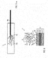
- FIG. 5a is a schematic representation of a detection catheter 1c according to a third embodiment.
- the detection device 1c according to the invention according to the second embodiment is a biofunktionalformaten, medical detection catheter for the invasive (in vivo) enrichment of rare cells, biomolecules or drugs.
- a detection catheter or detector is also referred to as a medical nano-catheter (MN-K) or medical nano-catheter (MN-C).
- the inventive detection device 1c according to the third embodiment comprises a flexible medical guide wire 40c, with a distal end 44c and a proximal end.
- the detection device 1c according to the third embodiment may further be equipped with a stabilizing element in the form of a sleeve (not shown here) and a pulling / pushing device (not shown here).
- a stabilizing element in the form of a sleeve (not shown here) and a pulling / pushing device (not shown here).
- the detection device 1c further comprises at least two functional elements 50c, which are formed as threads.
- the threads 50c are fixedly connected to the free end 44c of the guide wire 40c according to the third embodiment.
- the threads 50c have a smaller thickness than the guide wire 40c.
- the thickness of the threads 50c should be selected such that no thromboses are caused during use.
- the thickness of the threads should be chosen so that they do not stick together, but spread evenly in the bloodstream.
- the flexibility of the threads 50c can ensure, in use of a detection device 1c, that the entire vein cross-section is covered and, moreover, there is freedom of movement in the longitudinal direction and transversely to the longitudinal direction of the respective vein.
- the threads 50c thus form a structure in the manner of a "tentacle” in use.
- the coverage achieved thereby of the entire vein cross-section ensure good collection of sample material from the blood.
- the threads 50c can be individually cut or torn off the guide wire 40c.
- Each of the functional elements 50c has a functional surface 10 equipped with detection receptors 12, reference being made to the preceding description of the first or second exemplary embodiment.
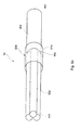
- FIG. 5b is a schematic representation of a detection catheter 1d according to a fourth embodiment.
- the detection device 1c according to the invention according to the second embodiment is a biofunktionalformaten, medical detection catheter for the invasive (in vivo) enrichment of rare cells, biomolecules or drugs.
- a detection catheter or detector is also referred to as a medical nano-catheter (MN-K) or medical nano-catheter (MN-C).
- the inventive detection device 1d according to the fourth embodiment comprises a flexible, medical guide wire 40d, with a distal end 44d and a proximal end 46d.
- the detection device 1d according to the fourth embodiment may further be equipped with a pulling / pushing device (not shown here).
- a pulling / pushing device not shown here.
- the detection device 1 d further comprises at least two functional elements 50d, preferably more than two functional elements 50d, which are formed as wires.
- the wires 50d are firmly connected to the free distal end 44d of the guide wire 40d according to the fourth embodiment, in particular welded.
- the detection device 1d can furthermore be equipped with a stabilizing element 60d in the form of a sleeve, which preferably covers, in particular surrounds, the connection point between the wires 50d and the distal end 44d of the guide wire 40d.
- the wires 50d are connected to each other at their distal ends 52d, in particular welded together, so that the wires 50d as a whole form a bundle of functional elements 50d.
- the wires 50d each have a smaller thickness than the guide wire 40d.
- the thickness of the wires 50d should in particular be chosen so that no thromboses are caused during use. At the same time, the thickness of the wires 50d should be chosen so that they can be guided well in the bundle through the guide wire 40d.
- the wires 50d can be individually cut or torn off the guide wire 40d.
- Each of the functional elements 50d has a functional surface 10 equipped with detection receptors 12, with reference being made to the preceding description of the first or second exemplary embodiment.
- the functional surface 10 may be formed on a carrier 2.
- the carrier 2 may be part of a functional element 50a, 50b or 50c.
- An exemplary layer structure of a carrier 2 is shown in FIG FIG. 6 shown.
- a substrate 21 made of medical grade stainless steel wire with a diameter of about 0.5 mm gives the support 2 its visible structure.
- the substrate 21 may include one or more coatings 22, 23.
- the substrate 21 is coated with a gold coating 22 having a thickness of 0.5 to 1.0 microns, which is applied by electroplating, ceramic methods, cementation or by vapor deposition.
- the substrate 21 may further be coated with a biocompatible dye to reduce intrinsic fluorescence of the base material during microscopic evaluation.
- the chemical activation of the carrier 2 is carried out via an affinity reaction usually by sulfur or nitrogen-containing compounds to which in turn directly or via polymer chains specific detection receptors 12 can be bound.
- a covalent secondary layer consisting of a functional biocompatible polymer 3 is applied to the carrier 2 by wet-chemical or physical methods.
- the layer thickness can be 1 to 2 microns.
- the carrier 2 is chemically activated.
- specific antibodies in particular monoclonal antibodies of murine origin, chimeric antibodies, humanized antibodies, or fragments of these antibodies or amino acid structures or nucleic acid structures or synthetic structures with specific affinity to cell surfaces or molecules can be covalently attached as detection receptors ,
- a complex sample fluid such as blood is not only a permanent connection of the detection receptors 12 while preserving the biological function, but also an efficient suppression of nonspecific adsorption processes for the selective attachment of ligands of crucial importance.
- An intermediate layer 23 here has the task of ensuring an effective shielding of the surface of the substrate 21 and at the same time to provide the functional groups for binding the biocompatible polymer layer 3 in sufficient density.
- the interlayer system accordingly forms an adhesion promoter between the gold coating 22 of the substrate 21 and the biocompatible polymer layer 3.
- the biocompatible polymer 3 is preferably a hydrogel with carbon-containing long branched macromolecules which have a high number of functional groups, for example carboxyl groups or polycarboxylates.
- the nature of the functional groups depends on the molecular properties of the specific detection receptors 12.
- the biocompatible hydrogel thereby ensures the permanent covalent binding of the detection receptors 12 while preserving the biological function and at the same time prevents the detection receptors 12 from being impaired in their recognition function by nonspecific adsorption phenomena.
- Hydrogels are three-dimensionally crosslinked hydrophilic polymers that absorb liquids, such as water, but are themselves insoluble therein. Main constituents of the hydrogel are polyacrylic acid (PAA) and polyethylene glycol (PEG).
- Property profiles can be tailor-made according to the desired requirements or areas of application via a suitable selection of the monomer building blocks, the degree of crosslinking and the crosslinking density.
- An essential property is the biocompatibility, ie the compatibility of the hydrogel with the living tissue.
- the branched polymer chains of the biocompatible polymer 3 also prevent the thrombogenic effect during invasive use.
- the functional groups receive an unbalanced molecular charge that allows electrostatically attracting and covalently binding dissolved detection receptors 12 from a solution.
- the detection receptors 12 permanently immobilized on the polymer layer 3 serve for the specific binding of the ligands or target molecules and target cells their surface antigens and thus enable the function of the detection device 1.
- chemically or enzymatically cleavable groups can be contained in this biocompatible polymer 3 in order to facilitate the quantitative recovery of bound target molecules or cells.
- the branched molecular structures of the biocompatible polymer 3 form a three-dimensionally structured functional area 10 with mutually facing functional sections 11 and interspaces 13 which can be penetrated by sample liquid.
- the surface of the carrier 2 (shown in the macroscopic or visible region) is structured in a three-dimensional manner.
- FIG. 6 the sample liquid in an advantageous manner via the respective functional element 50a, 50b, 50c passes, slows down the three-dimensionally structured in the microscopic area functional surface 10 of the biocompatible polymer 3 passes, slows down the three-dimensionally structured in the microscopic area functional surface 10 of the biocompatible polymer 3 (see FIG. FIG. 6 ), the flow of the sample liquid in the region of the boundary layer and favors the enrichment of the ligands at the detection receptors 12th
- a biocompatible protective layer (tertiary layer or stabilizer layer) 4 is applied over the biocompatible polymer 3.
- This protective layer 4 dries over the secondary layer and forms a dense network of crystalline structures and thus stabilizes and conserves the functional part 1a of the catheter 1.
- the protective layer 4 is not covalently bound. In the bloodstream, the protective layer 4 goes into solution and releases the functional surface 10 of the catheter. Alternatively, the protective layer 4 can be washed off with sterile water before use.
- the protective layer 4 may comprise high purity alginates, polyethylene glycols, cyclic and non-cyclic oligosaccharides and polysaccharides, antioxidant amino acids, proteins and vitamins.
- the protective layer 4 is preferably made of a biocompatible high-viscosity polysaccharide, which serves as a medium for added amino acids, proteins, vitamins and stabilizing polysaccharides.
- the high viscosity allows rapid wettability of the surface.
- the deposited protective layer 4 adheres to the secondary coating and prevents the ingress of foreign substances during storage.
- the added amino acids, proteins and vitamins are present in higher concentrations compared to the specific ligands and thereby able to control the likelihood of damage to the target molecules by radical molecules or charge carriers and to restore destroyed chemical bonds by recombination processes.
- the completed detection device 1a, 1b, 1c is packaged in a low-germ environment. Final sterilization is by gamma-irradiation at a radiation dose of 25 kGy.
- the detection device 1a, 1b, 1c is intended for single use.
- a preferred application of the detection device 1a, 1b, 1c is in prenatal and cancer diagnostics.
- the detection device 1a, 1b, 1c can be used, for example, for isolating circulating fetal cells or tumor cells in the bloodstream of pregnant women or cancer patients.
- the detection device 1a, 1b, 1c is introduced into the vein via a suitable commercially available brown-eye system and applied to the venous blood circulation.
- the dwell time in the vein can be about 30 minutes.
- the cells bound to the detection device 1a, 1b, 1c are further enriched by means of targeted laboratory diagnostics and characterized in terms of molecular biology and cell biology.
- the aim of the minimally invasive procedure to be performed is the selection of fetal or tumor cells from the blood. Due to the low cell concentration of the cells in the blood, a blood sample of about 0.5 I would be necessary to obtain the desired target cell count. However, this is excluded from a medical perspective.
- chromosome aberration eg trisomy 21 (Down syndrome)
- the Down syndrome is diagnosed prenatal so far only by invasive procedures, each having a Abortrisiko 1%: chorionic villus sampling between the 11th and 14th week of pregnancy and amniocentesis (amniocentesis) from the 15th week of pregnancy.
- the method according to the invention which will be usable from the 9th week of pregnancy, has no risk for the fetus and can be used in the first-trimester screening.
- the amniocentesis can be dispensed with.
- Fetal trophoblast cells from the placenta can be detected from the 6th week of pregnancy in the bloodstream of the mother. Only about 2 to 5 of these cells per ml of maternal blood are present. These trophoblast cells have a membrane-bound HLA-G complex (antigen) that binds to specific antibodies.
- a specific HLA-G antibody is used as the detection receptor 12, which reacts only with membrane-bound HLA-G (antigen) and thus to catch only the desired fetal cells from the mother's blood.
- Cancer tumor cells can be enriched with the EpCAM antibody (against the EpCAM antigen), which is humanized in its constant domains and covalently attached to the hydrogel.
Landscapes
- Health & Medical Sciences (AREA)
- Life Sciences & Earth Sciences (AREA)
- Engineering & Computer Science (AREA)
- Biomedical Technology (AREA)
- Molecular Biology (AREA)
- Immunology (AREA)
- General Health & Medical Sciences (AREA)
- Pathology (AREA)
- Hematology (AREA)
- Medical Informatics (AREA)
- Surgery (AREA)
- Public Health (AREA)
- Veterinary Medicine (AREA)
- Heart & Thoracic Surgery (AREA)
- Animal Behavior & Ethology (AREA)
- Physics & Mathematics (AREA)
- Urology & Nephrology (AREA)
- Chemical & Material Sciences (AREA)
- Food Science & Technology (AREA)
- Microbiology (AREA)
- Cell Biology (AREA)
- Medicinal Chemistry (AREA)
- Biotechnology (AREA)
- Analytical Chemistry (AREA)
- Biochemistry (AREA)
- General Physics & Mathematics (AREA)
- Optics & Photonics (AREA)
- Biophysics (AREA)
- Media Introduction/Drainage Providing Device (AREA)
- Apparatus Associated With Microorganisms And Enzymes (AREA)
- Measurement Of The Respiration, Hearing Ability, Form, And Blood Characteristics Of Living Organisms (AREA)
Applications Claiming Priority (1)
| Application Number | Priority Date | Filing Date | Title |
|---|---|---|---|
| DE201310210204 DE102013210204A1 (de) | 2013-05-31 | 2013-05-31 | Detektionsvorrichtung zur in vivo und/oder in vitro Anreicherung von Probenmaterial |
Publications (2)
| Publication Number | Publication Date |
|---|---|
| EP2807979A1 true EP2807979A1 (fr) | 2014-12-03 |
| EP2807979B1 EP2807979B1 (fr) | 2017-10-11 |
Family
ID=50842024
Family Applications (1)
| Application Number | Title | Priority Date | Filing Date |
|---|---|---|---|
| EP14167170.1A Active EP2807979B1 (fr) | 2013-05-31 | 2014-05-06 | Dispositif de détection destiné à améliorer in vivo et/ou in vitro un matériau d'échantillon |
Country Status (5)
| Country | Link |
|---|---|
| US (1) | US10178966B2 (fr) |
| EP (1) | EP2807979B1 (fr) |
| CN (1) | CN104274214A (fr) |
| CA (1) | CA2852065A1 (fr) |
| DE (1) | DE102013210204A1 (fr) |
Cited By (1)
| Publication number | Priority date | Publication date | Assignee | Title |
|---|---|---|---|---|
| WO2016116503A1 (fr) * | 2015-01-20 | 2016-07-28 | Gilupi Gmbh | Dispositif d'enrichissement, in-vivo et/ou in vitro, de structures cibles dans un échantillon liquide et procédé pour sa fabrication |
Families Citing this family (14)
| Publication number | Priority date | Publication date | Assignee | Title |
|---|---|---|---|---|
| US20050153309A1 (en) | 2003-12-22 | 2005-07-14 | David Hoon | Method and apparatus for in vivo surveillance of circulating biological components |
| FR3032106B1 (fr) * | 2015-02-02 | 2020-07-31 | Centre Nat Rech Scient | Microdispositif pour la capture in vivo de biomarqueurs cellulaires circulants. |
| CN105713829B (zh) * | 2016-03-18 | 2019-03-05 | 中国科学院长春应用化学研究所 | 一种基于多孔金属富集探针的单细胞取样装置及取样方法 |
| WO2017185086A1 (fr) * | 2016-04-22 | 2017-10-26 | Exosome Diagnostics, Inc. | Dispositifs et procédés pour la capture in vivo d'échantillons biologiques et d'acides nucléiques présents dans ces derniers |
| SG11201810969RA (en) | 2016-06-09 | 2019-01-30 | Haimachek Inc | Collector for detection and reversible capturing of cells from body fluids in vivo |
| KR102700936B1 (ko) * | 2016-06-17 | 2024-09-02 | 코웨이 주식회사 | 소변 분석 장치를 포함하는 비데 |
| CN106434285A (zh) * | 2016-10-31 | 2017-02-22 | 广州益养生物科技有限公司 | 一种用于血液液体活检的体内收集装置 |
| US9999899B2 (en) * | 2016-11-01 | 2018-06-19 | International Business Machines Corporation | Controlled exposure of in-vivo sensors |
| JPWO2018185939A1 (ja) * | 2017-04-07 | 2019-04-11 | エスゼット ディージェイアイ テクノロジー カンパニー リミテッドSz Dji Technology Co.,Ltd | 撮像制御装置、撮像装置、撮像システム、移動体、撮像制御方法、及びプログラム |
| CN108392232B (zh) * | 2018-04-11 | 2023-07-25 | 苏州大学 | 以功能化蛋白丝线为载体的体内细胞捕获器 |
| CN111122215B (zh) * | 2018-10-30 | 2023-01-03 | 哈尔滨工业大学 | 一种刷式小行星表面星壤采样装置 |
| DE102018220650A1 (de) * | 2018-11-30 | 2020-06-04 | Robert Bosch Gmbh | Biopsienadel zur in-vivo-Isolation von zirkulierenden Tumorzellen |
| CN113180745A (zh) * | 2021-04-14 | 2021-07-30 | 苏州新海生物科技股份有限公司 | 一种口腔采样拭子 |
| NL2030156B1 (en) * | 2021-12-16 | 2023-06-28 | Idris Oncology B V | Medical sampling device for capturing bioanalytes |
Citations (4)
| Publication number | Priority date | Publication date | Assignee | Title |
|---|---|---|---|---|
| WO2010025719A1 (fr) * | 2008-09-08 | 2010-03-11 | Ogeno Gmbh | Instrument de biopsie pour l’enrichissement d’un matériau échantillon |
| WO2010145824A1 (fr) | 2009-06-17 | 2010-12-23 | Gilupi Gmbh | Dispositif de détection pour enrichir in vivo et/ou in vitro un matériau d'échantillon |
| WO2011047671A1 (fr) * | 2009-10-20 | 2011-04-28 | Ogeno Gmbh | Instrument de biopsie comprenant un élément magnétique |
| WO2011113584A1 (fr) * | 2010-03-16 | 2011-09-22 | Gilupi Gmbh | Biodétecteur |
Family Cites Families (7)
| Publication number | Priority date | Publication date | Assignee | Title |
|---|---|---|---|---|
| US5814295A (en) | 1992-04-10 | 1998-09-29 | The Ohio State University Research Foundation | Determination of lymph nodes enriched in tumor reactive cells their proliferation and their use in adoptive cellular therapy |
| AU779835B2 (en) * | 1999-05-20 | 2005-02-10 | Illumina, Inc. | Method and apparatus for retaining and presenting at least one microsphere array to solutions and/or to optical imaging systems |
| US20080241262A1 (en) * | 2004-03-29 | 2008-10-02 | The University Of Houston System | Nanoshells and Discrete Polymer-Coated Nanoshells, Methods For Making and Using Same |
| US20080058676A1 (en) * | 2006-09-06 | 2008-03-06 | Yong Peter A K | Retractable segmented bio-molecular collector swab system |
| US8877518B2 (en) * | 2007-02-06 | 2014-11-04 | The Trustees Of The University Of Pennsylvania | Multiplexed nanoscale electrochemical sensors for multi-analyte detection |
| DE102009025511A1 (de) * | 2009-06-19 | 2010-12-23 | Qualimed Innovative Medizin-Produkte Gmbh | Implantat mit einem vom Körper resorbierbaren metallischen Werkstoff |
| CN101612035B (zh) * | 2009-07-17 | 2011-06-29 | 清华大学 | 微创多通道活体荧光信号实时检测系统及方法 |
-
2013
- 2013-05-31 DE DE201310210204 patent/DE102013210204A1/de active Pending
-
2014
- 2014-05-06 EP EP14167170.1A patent/EP2807979B1/fr active Active
- 2014-05-21 CA CA 2852065 patent/CA2852065A1/fr not_active Abandoned
- 2014-05-29 CN CN201410310252.4A patent/CN104274214A/zh active Pending
- 2014-05-29 US US14/290,180 patent/US10178966B2/en active Active
Patent Citations (4)
| Publication number | Priority date | Publication date | Assignee | Title |
|---|---|---|---|---|
| WO2010025719A1 (fr) * | 2008-09-08 | 2010-03-11 | Ogeno Gmbh | Instrument de biopsie pour l’enrichissement d’un matériau échantillon |
| WO2010145824A1 (fr) | 2009-06-17 | 2010-12-23 | Gilupi Gmbh | Dispositif de détection pour enrichir in vivo et/ou in vitro un matériau d'échantillon |
| WO2011047671A1 (fr) * | 2009-10-20 | 2011-04-28 | Ogeno Gmbh | Instrument de biopsie comprenant un élément magnétique |
| WO2011113584A1 (fr) * | 2010-03-16 | 2011-09-22 | Gilupi Gmbh | Biodétecteur |
Non-Patent Citations (2)
| Title |
|---|
| FLORES L M ET AL: "Improving the yield of circulating tumour cells facilitates molecular characterisation and recognition of discordant HER2 amplification in breast cancer", vol. 102, no. 10, 1 May 2010 (2010-05-01), pages 1495 - 1502, XP002671705, ISSN: 0007-0920, Retrieved from the Internet <URL:http://www.nature.com/bjc/journal/v102/n10/full/6605676a.html> [retrieved on 20100511], DOI: 10.1038/SJ.BJC.6605676 * |
| SAUCEDO-ZENI N ET AL: "A novel method for the in vivo isolation of circulating tumor cells from peripheral blood of cancer patients using a functionalized and structured medical wire", INTERNATIONAL JOURNAL OF ONCOLOGY, vol. 41, 16 July 2012 (2012-07-16), pages 1241 - 1250, XP002731287, DOI: 10.3892/ijo.2012.1557 * |
Cited By (2)
| Publication number | Priority date | Publication date | Assignee | Title |
|---|---|---|---|---|
| WO2016116503A1 (fr) * | 2015-01-20 | 2016-07-28 | Gilupi Gmbh | Dispositif d'enrichissement, in-vivo et/ou in vitro, de structures cibles dans un échantillon liquide et procédé pour sa fabrication |
| US10383605B2 (en) | 2015-01-20 | 2019-08-20 | Gilupi Gmbh | Device for the in-vivo and/or in-vitro enrichment of target structures in a sample liquid and method for the production thereof |
Also Published As
| Publication number | Publication date |
|---|---|
| US10178966B2 (en) | 2019-01-15 |
| US20140357967A1 (en) | 2014-12-04 |
| EP2807979B1 (fr) | 2017-10-11 |
| CA2852065A1 (fr) | 2014-11-30 |
| DE102013210204A1 (de) | 2014-12-18 |
| CN104274214A (zh) | 2015-01-14 |
Similar Documents
| Publication | Publication Date | Title |
|---|---|---|
| EP2807979B1 (fr) | Dispositif de détection destiné à améliorer in vivo et/ou in vitro un matériau d'échantillon | |
| EP2442726B1 (fr) | Dispositif de détection pour enrichir in vivo et/ou in vitro un matériau d'échantillon | |
| DE69534405T2 (de) | Vorrichtung zum gewinnen von autologen, mikrovaskulären endothelialen zellen enthaltendem fettgewebe | |
| DE60314413T2 (de) | Verfahren und vorrichtung zur isolierung von plättchen aus blut | |
| EP2346410B1 (fr) | Instrument de biopsie pour l enrichissement d un matériau échantillon | |
| EP2547250B1 (fr) | Biodétecteur | |
| DE10307487A1 (de) | Verfahren und Vorrichtungen zur verletzungsfreien Bewegung einer Sonde durch biologisches Zellmaterial | |
| DE69819689T2 (de) | Verfahren und gerät zum waschen von zellen | |
| EP3010399A1 (fr) | Cathéter comprenant un dispositif de détection pour déceler en temps réel une matière d'échantillon | |
| WO2006087220A2 (fr) | Récipient pour séparer des cellules tumorales | |
| EP3248009A1 (fr) | Dispositif d'enrichissement, in-vivo et/ou in vitro, de structures cibles dans un échantillon liquide et procédé pour sa fabrication | |
| DE10054632B4 (de) | Verfahren zur Isolierung von disseminierten Krebszellen aus einer zellhaltigen Körperflüssigkeit und Verwendung einer Filtervorrichtung in einem solchen Verfahren | |
| EP2240546A2 (fr) | Instruments revêtus destinés à la médecine invasive | |
| DE102014006906A1 (de) | Detektionsvorrichtung zur in vivo und/oder in vitro Anreicherung von Probenmaterial | |
| DE10316598B4 (de) | Verfahren und Zentrifuge zur Gewinnung von biologischen Flüssigkeiten | |
| DE102011088741B4 (de) | Verfahren und Anordnung zum Markieren und Separieren von Zellen aus einer Zellsuspension | |
| WO2009147081A1 (fr) | Dispositif d'intervention destiné à collecter un matériau biologique, et son procédé de fabrication | |
| DE10202982A1 (de) | Verfahren und Vorrichtung zum Einbringen von lebenden Zellen in eine biologische Körperflüssigkeit | |
| WO2005030287A1 (fr) | Prothese vacsulaire a liberation de substances actives | |
| DE102009008923B4 (de) | Zellbesiedelungskammer | |
| WO2019053193A1 (fr) | Dispositif, procédé et utilisation permettant le transfert de liquides organiques | |
| DE102018210696A1 (de) | Vorrichtung und Verfahren zur dielektrophoretischen Separierung von Zielpartikeln in einer Flüssigkeit | |
| DE8226034U1 (de) | Vorrichtung zur Implantation im menschlichen Körper | |
| DD286294A5 (de) | Vorrichtung und verfahren zur gewinnung von zellulaeren und humoralen immunstoffen |
Legal Events
| Date | Code | Title | Description |
|---|---|---|---|
| PUAI | Public reference made under article 153(3) epc to a published international application that has entered the european phase |
Free format text: ORIGINAL CODE: 0009012 |
|
| 17P | Request for examination filed |
Effective date: 20140506 |
|
| AK | Designated contracting states |
Kind code of ref document: A1 Designated state(s): AL AT BE BG CH CY CZ DE DK EE ES FI FR GB GR HR HU IE IS IT LI LT LU LV MC MK MT NL NO PL PT RO RS SE SI SK SM TR |
|
| AX | Request for extension of the european patent |
Extension state: BA ME |
|
| R17P | Request for examination filed (corrected) |
Effective date: 20150602 |
|
| RBV | Designated contracting states (corrected) |
Designated state(s): AL AT BE BG CH CY CZ DE DK EE ES FI FR GB GR HR HU IE IS IT LI LT LU LV MC MK MT NL NO PL PT RO RS SE SI SK SM TR |
|
| 17Q | First examination report despatched |
Effective date: 20150807 |
|
| RAP1 | Party data changed (applicant data changed or rights of an application transferred) |
Owner name: GILUPI GMBH |
|
| GRAP | Despatch of communication of intention to grant a patent |
Free format text: ORIGINAL CODE: EPIDOSNIGR1 |
|
| RIC1 | Information provided on ipc code assigned before grant |
Ipc: A61B 5/145 20060101ALI20170320BHEP Ipc: A61B 10/02 20060101ALI20170320BHEP Ipc: A61B 10/00 20060101AFI20170320BHEP Ipc: G01N 33/543 20060101ALI20170320BHEP |
|
| INTG | Intention to grant announced |
Effective date: 20170419 |
|
| GRAS | Grant fee paid |
Free format text: ORIGINAL CODE: EPIDOSNIGR3 |
|
| GRAA | (expected) grant |
Free format text: ORIGINAL CODE: 0009210 |
|
| AK | Designated contracting states |
Kind code of ref document: B1 Designated state(s): AL AT BE BG CH CY CZ DE DK EE ES FI FR GB GR HR HU IE IS IT LI LT LU LV MC MK MT NL NO PL PT RO RS SE SI SK SM TR |
|
| REG | Reference to a national code |
Ref country code: GB Ref legal event code: FG4D Free format text: NOT ENGLISH |
|
| REG | Reference to a national code |
Ref country code: CH Ref legal event code: EP |
|
| REG | Reference to a national code |
Ref country code: IE Ref legal event code: FG4D Free format text: LANGUAGE OF EP DOCUMENT: GERMAN |
|
| REG | Reference to a national code |
Ref country code: AT Ref legal event code: REF Ref document number: 935314 Country of ref document: AT Kind code of ref document: T Effective date: 20171115 |
|
| REG | Reference to a national code |
Ref country code: DE Ref legal event code: R096 Ref document number: 502014005736 Country of ref document: DE |
|
| REG | Reference to a national code |
Ref country code: CH Ref legal event code: NV Representative=s name: BOVARD AG PATENT- UND MARKENANWAELTE, CH |
|
| REG | Reference to a national code |
Ref country code: NL Ref legal event code: MP Effective date: 20171011 |
|
| REG | Reference to a national code |
Ref country code: LT Ref legal event code: MG4D |
|
| PG25 | Lapsed in a contracting state [announced via postgrant information from national office to epo] |
Ref country code: NL Free format text: LAPSE BECAUSE OF FAILURE TO SUBMIT A TRANSLATION OF THE DESCRIPTION OR TO PAY THE FEE WITHIN THE PRESCRIBED TIME-LIMIT Effective date: 20171011 |
|
| PG25 | Lapsed in a contracting state [announced via postgrant information from national office to epo] |
Ref country code: ES Free format text: LAPSE BECAUSE OF FAILURE TO SUBMIT A TRANSLATION OF THE DESCRIPTION OR TO PAY THE FEE WITHIN THE PRESCRIBED TIME-LIMIT Effective date: 20171011 Ref country code: FI Free format text: LAPSE BECAUSE OF FAILURE TO SUBMIT A TRANSLATION OF THE DESCRIPTION OR TO PAY THE FEE WITHIN THE PRESCRIBED TIME-LIMIT Effective date: 20171011 Ref country code: NO Free format text: LAPSE BECAUSE OF FAILURE TO SUBMIT A TRANSLATION OF THE DESCRIPTION OR TO PAY THE FEE WITHIN THE PRESCRIBED TIME-LIMIT Effective date: 20180111 Ref country code: SE Free format text: LAPSE BECAUSE OF FAILURE TO SUBMIT A TRANSLATION OF THE DESCRIPTION OR TO PAY THE FEE WITHIN THE PRESCRIBED TIME-LIMIT Effective date: 20171011 Ref country code: LT Free format text: LAPSE BECAUSE OF FAILURE TO SUBMIT A TRANSLATION OF THE DESCRIPTION OR TO PAY THE FEE WITHIN THE PRESCRIBED TIME-LIMIT Effective date: 20171011 |
|
| REG | Reference to a national code |
Ref country code: FR Ref legal event code: PLFP Year of fee payment: 5 |
|
| PG25 | Lapsed in a contracting state [announced via postgrant information from national office to epo] |
Ref country code: IS Free format text: LAPSE BECAUSE OF FAILURE TO SUBMIT A TRANSLATION OF THE DESCRIPTION OR TO PAY THE FEE WITHIN THE PRESCRIBED TIME-LIMIT Effective date: 20180211 Ref country code: RS Free format text: LAPSE BECAUSE OF FAILURE TO SUBMIT A TRANSLATION OF THE DESCRIPTION OR TO PAY THE FEE WITHIN THE PRESCRIBED TIME-LIMIT Effective date: 20171011 Ref country code: LV Free format text: LAPSE BECAUSE OF FAILURE TO SUBMIT A TRANSLATION OF THE DESCRIPTION OR TO PAY THE FEE WITHIN THE PRESCRIBED TIME-LIMIT Effective date: 20171011 Ref country code: GR Free format text: LAPSE BECAUSE OF FAILURE TO SUBMIT A TRANSLATION OF THE DESCRIPTION OR TO PAY THE FEE WITHIN THE PRESCRIBED TIME-LIMIT Effective date: 20180112 Ref country code: HR Free format text: LAPSE BECAUSE OF FAILURE TO SUBMIT A TRANSLATION OF THE DESCRIPTION OR TO PAY THE FEE WITHIN THE PRESCRIBED TIME-LIMIT Effective date: 20171011 Ref country code: BG Free format text: LAPSE BECAUSE OF FAILURE TO SUBMIT A TRANSLATION OF THE DESCRIPTION OR TO PAY THE FEE WITHIN THE PRESCRIBED TIME-LIMIT Effective date: 20180111 |
|
| REG | Reference to a national code |
Ref country code: DE Ref legal event code: R097 Ref document number: 502014005736 Country of ref document: DE |
|
| PG25 | Lapsed in a contracting state [announced via postgrant information from national office to epo] |
Ref country code: SK Free format text: LAPSE BECAUSE OF FAILURE TO SUBMIT A TRANSLATION OF THE DESCRIPTION OR TO PAY THE FEE WITHIN THE PRESCRIBED TIME-LIMIT Effective date: 20171011 Ref country code: DK Free format text: LAPSE BECAUSE OF FAILURE TO SUBMIT A TRANSLATION OF THE DESCRIPTION OR TO PAY THE FEE WITHIN THE PRESCRIBED TIME-LIMIT Effective date: 20171011 Ref country code: CZ Free format text: LAPSE BECAUSE OF FAILURE TO SUBMIT A TRANSLATION OF THE DESCRIPTION OR TO PAY THE FEE WITHIN THE PRESCRIBED TIME-LIMIT Effective date: 20171011 Ref country code: EE Free format text: LAPSE BECAUSE OF FAILURE TO SUBMIT A TRANSLATION OF THE DESCRIPTION OR TO PAY THE FEE WITHIN THE PRESCRIBED TIME-LIMIT Effective date: 20171011 |
|
| PLBE | No opposition filed within time limit |
Free format text: ORIGINAL CODE: 0009261 |
|
| STAA | Information on the status of an ep patent application or granted ep patent |
Free format text: STATUS: NO OPPOSITION FILED WITHIN TIME LIMIT |
|
| PG25 | Lapsed in a contracting state [announced via postgrant information from national office to epo] |
Ref country code: SM Free format text: LAPSE BECAUSE OF FAILURE TO SUBMIT A TRANSLATION OF THE DESCRIPTION OR TO PAY THE FEE WITHIN THE PRESCRIBED TIME-LIMIT Effective date: 20171011 Ref country code: PL Free format text: LAPSE BECAUSE OF FAILURE TO SUBMIT A TRANSLATION OF THE DESCRIPTION OR TO PAY THE FEE WITHIN THE PRESCRIBED TIME-LIMIT Effective date: 20171011 Ref country code: RO Free format text: LAPSE BECAUSE OF FAILURE TO SUBMIT A TRANSLATION OF THE DESCRIPTION OR TO PAY THE FEE WITHIN THE PRESCRIBED TIME-LIMIT Effective date: 20171011 |
|
| 26N | No opposition filed |
Effective date: 20180712 |
|
| PG25 | Lapsed in a contracting state [announced via postgrant information from national office to epo] |
Ref country code: MT Free format text: LAPSE BECAUSE OF FAILURE TO SUBMIT A TRANSLATION OF THE DESCRIPTION OR TO PAY THE FEE WITHIN THE PRESCRIBED TIME-LIMIT Effective date: 20171011 |
|
| PG25 | Lapsed in a contracting state [announced via postgrant information from national office to epo] |
Ref country code: SI Free format text: LAPSE BECAUSE OF FAILURE TO SUBMIT A TRANSLATION OF THE DESCRIPTION OR TO PAY THE FEE WITHIN THE PRESCRIBED TIME-LIMIT Effective date: 20171011 |
|
| REG | Reference to a national code |
Ref country code: BE Ref legal event code: MM Effective date: 20180531 |
|
| PG25 | Lapsed in a contracting state [announced via postgrant information from national office to epo] |
Ref country code: MC Free format text: LAPSE BECAUSE OF FAILURE TO SUBMIT A TRANSLATION OF THE DESCRIPTION OR TO PAY THE FEE WITHIN THE PRESCRIBED TIME-LIMIT Effective date: 20171011 |
|
| REG | Reference to a national code |
Ref country code: IE Ref legal event code: MM4A |
|
| PG25 | Lapsed in a contracting state [announced via postgrant information from national office to epo] |
Ref country code: LU Free format text: LAPSE BECAUSE OF NON-PAYMENT OF DUE FEES Effective date: 20180506 |
|
| PG25 | Lapsed in a contracting state [announced via postgrant information from national office to epo] |
Ref country code: IE Free format text: LAPSE BECAUSE OF NON-PAYMENT OF DUE FEES Effective date: 20180506 |
|
| PG25 | Lapsed in a contracting state [announced via postgrant information from national office to epo] |
Ref country code: BE Free format text: LAPSE BECAUSE OF NON-PAYMENT OF DUE FEES Effective date: 20180531 |
|
| PG25 | Lapsed in a contracting state [announced via postgrant information from national office to epo] |
Ref country code: TR Free format text: LAPSE BECAUSE OF FAILURE TO SUBMIT A TRANSLATION OF THE DESCRIPTION OR TO PAY THE FEE WITHIN THE PRESCRIBED TIME-LIMIT Effective date: 20171011 |
|
| PG25 | Lapsed in a contracting state [announced via postgrant information from national office to epo] |
Ref country code: HU Free format text: LAPSE BECAUSE OF FAILURE TO SUBMIT A TRANSLATION OF THE DESCRIPTION OR TO PAY THE FEE WITHIN THE PRESCRIBED TIME-LIMIT; INVALID AB INITIO Effective date: 20140506 Ref country code: PT Free format text: LAPSE BECAUSE OF FAILURE TO SUBMIT A TRANSLATION OF THE DESCRIPTION OR TO PAY THE FEE WITHIN THE PRESCRIBED TIME-LIMIT Effective date: 20171011 |
|
| PG25 | Lapsed in a contracting state [announced via postgrant information from national office to epo] |
Ref country code: CY Free format text: LAPSE BECAUSE OF FAILURE TO SUBMIT A TRANSLATION OF THE DESCRIPTION OR TO PAY THE FEE WITHIN THE PRESCRIBED TIME-LIMIT Effective date: 20171011 Ref country code: MK Free format text: LAPSE BECAUSE OF NON-PAYMENT OF DUE FEES Effective date: 20171011 |
|
| PG25 | Lapsed in a contracting state [announced via postgrant information from national office to epo] |
Ref country code: AL Free format text: LAPSE BECAUSE OF FAILURE TO SUBMIT A TRANSLATION OF THE DESCRIPTION OR TO PAY THE FEE WITHIN THE PRESCRIBED TIME-LIMIT Effective date: 20171011 |
|
| REG | Reference to a national code |
Ref country code: AT Ref legal event code: MM01 Ref document number: 935314 Country of ref document: AT Kind code of ref document: T Effective date: 20190506 |
|
| PG25 | Lapsed in a contracting state [announced via postgrant information from national office to epo] |
Ref country code: AT Free format text: LAPSE BECAUSE OF NON-PAYMENT OF DUE FEES Effective date: 20190506 |
|
| PGFP | Annual fee paid to national office [announced via postgrant information from national office to epo] |
Ref country code: IT Payment date: 20230727 Year of fee payment: 10 Ref country code: GB Payment date: 20230727 Year of fee payment: 10 Ref country code: CH Payment date: 20230801 Year of fee payment: 10 |
|
| REG | Reference to a national code |
Ref country code: CH Ref legal event code: PL |
|
| GBPC | Gb: european patent ceased through non-payment of renewal fee |
Effective date: 20240506 |
|
| PG25 | Lapsed in a contracting state [announced via postgrant information from national office to epo] |
Ref country code: CH Free format text: LAPSE BECAUSE OF NON-PAYMENT OF DUE FEES Effective date: 20240531 |
|
| PG25 | Lapsed in a contracting state [announced via postgrant information from national office to epo] |
Ref country code: GB Free format text: LAPSE BECAUSE OF NON-PAYMENT OF DUE FEES Effective date: 20240506 Ref country code: IT Free format text: LAPSE BECAUSE OF NON-PAYMENT OF DUE FEES Effective date: 20240506 |
|
| PGFP | Annual fee paid to national office [announced via postgrant information from national office to epo] |
Ref country code: DE Payment date: 20250528 Year of fee payment: 12 |
|
| PGFP | Annual fee paid to national office [announced via postgrant information from national office to epo] |
Ref country code: FR Payment date: 20250528 Year of fee payment: 12 |