EP2809810B1 - System und verfahren für den nachweis von durch lungenkrebs veränderten rnas im peripheren blut - Google Patents
System und verfahren für den nachweis von durch lungenkrebs veränderten rnas im peripheren blut Download PDFInfo
- Publication number
- EP2809810B1 EP2809810B1 EP13733697.0A EP13733697A EP2809810B1 EP 2809810 B1 EP2809810 B1 EP 2809810B1 EP 13733697 A EP13733697 A EP 13733697A EP 2809810 B1 EP2809810 B1 EP 2809810B1
- Authority
- EP
- European Patent Office
- Prior art keywords
- nucleic acids
- gene
- fpr1
- levels
- circulating free
- Prior art date
- Legal status (The legal status is an assumption and is not a legal conclusion. Google has not performed a legal analysis and makes no representation as to the accuracy of the status listed.)
- Active
Links
Images



Classifications
-
- C—CHEMISTRY; METALLURGY
- C12—BIOCHEMISTRY; BEER; SPIRITS; WINE; VINEGAR; MICROBIOLOGY; ENZYMOLOGY; MUTATION OR GENETIC ENGINEERING
- C12Q—MEASURING OR TESTING PROCESSES INVOLVING ENZYMES, NUCLEIC ACIDS OR MICROORGANISMS; COMPOSITIONS OR TEST PAPERS THEREFOR; PROCESSES OF PREPARING SUCH COMPOSITIONS; CONDITION-RESPONSIVE CONTROL IN MICROBIOLOGICAL OR ENZYMOLOGICAL PROCESSES
- C12Q1/00—Measuring or testing processes involving enzymes, nucleic acids or microorganisms; Compositions therefor; Processes of preparing such compositions
- C12Q1/68—Measuring or testing processes involving enzymes, nucleic acids or microorganisms; Compositions therefor; Processes of preparing such compositions involving nucleic acids
- C12Q1/6876—Nucleic acid products used in the analysis of nucleic acids, e.g. primers or probes
- C12Q1/6883—Nucleic acid products used in the analysis of nucleic acids, e.g. primers or probes for diseases caused by alterations of genetic material
- C12Q1/6886—Nucleic acid products used in the analysis of nucleic acids, e.g. primers or probes for diseases caused by alterations of genetic material for cancer
-
- C—CHEMISTRY; METALLURGY
- C12—BIOCHEMISTRY; BEER; SPIRITS; WINE; VINEGAR; MICROBIOLOGY; ENZYMOLOGY; MUTATION OR GENETIC ENGINEERING
- C12Q—MEASURING OR TESTING PROCESSES INVOLVING ENZYMES, NUCLEIC ACIDS OR MICROORGANISMS; COMPOSITIONS OR TEST PAPERS THEREFOR; PROCESSES OF PREPARING SUCH COMPOSITIONS; CONDITION-RESPONSIVE CONTROL IN MICROBIOLOGICAL OR ENZYMOLOGICAL PROCESSES
- C12Q1/00—Measuring or testing processes involving enzymes, nucleic acids or microorganisms; Compositions therefor; Processes of preparing such compositions
- C12Q1/68—Measuring or testing processes involving enzymes, nucleic acids or microorganisms; Compositions therefor; Processes of preparing such compositions involving nucleic acids
-
- C—CHEMISTRY; METALLURGY
- C12—BIOCHEMISTRY; BEER; SPIRITS; WINE; VINEGAR; MICROBIOLOGY; ENZYMOLOGY; MUTATION OR GENETIC ENGINEERING
- C12Q—MEASURING OR TESTING PROCESSES INVOLVING ENZYMES, NUCLEIC ACIDS OR MICROORGANISMS; COMPOSITIONS OR TEST PAPERS THEREFOR; PROCESSES OF PREPARING SUCH COMPOSITIONS; CONDITION-RESPONSIVE CONTROL IN MICROBIOLOGICAL OR ENZYMOLOGICAL PROCESSES
- C12Q2600/00—Oligonucleotides characterized by their use
- C12Q2600/158—Expression markers
Definitions
- cfRNA circulating free RNA
- Cancer is a major health risk in the United States and internationally. Treatments exist, but are often not administered to patients until the disease has progressed to a point at which treatment efficacy is compromised.
- a major challenge in cancer treatment is to identify patients early in the course of their disease. This is difficult under current methods because early cancerous or precancerous cell populations may be asymptomatic and may be located in regions which are difficult to access by biopsy. Thus a robust, minimally invasive assay that may be used to identify all stages of the disease, including early stages which may be asymptomatic, would be of substantial benefit for the treatment of cancer.
- WO03/074069 discloses FPR1 as a cancer marker and the analysis of FPR1 expression analysis in peripheral blood via PCT.
- WO03/074069 contains no disclosure of using free circulating nucleic acids of a FPR gene as a cancer marker, let alone a marker for detecting lung cancer.
- US2005/153918 discloses methods and materials designed to assay intracellular nucleic acids and polypeptides that are associated with specific tissues and cell types. US2005/153918 does not pertain to cell-free FPR1 transcript levels, let alone circulating free FPR1 transcript level.
- RNA molecules in samples taken from a patient.
- blood or blood component such as plasma is isolated from a patient suspected of having lung cancer or non-small cell lung cancer (NSCLC).
- the plasma is analyzed to measure the level of circulating free RNA (cfRNA) from one or more genes.
- cfRNA circulating free RNA
- the RNA to be measured is messenger ribonucleic acids (mRNA), such as mRNA from within the population of cfRNA in a patient's plasma.
- mRNA messenger ribonucleic acids
- ACTB circulating free actin beta
- the level of circulating free HNRNPA1 RNA is measured.
- the level of circulating free formylpeptide receptor gene (FPR1) RNA is measured.
- the level of cfRNA from FPR1 as compared with the level of cfRNA from ACTB and HNRNPA1 is compared, as discussed below, to determine if a patient is at risk for having lung cancer or NSCLC.
- the NSCLC assayed is Stage I NSCLC.
- RNAs are known to be present in plasma of cancer-free individuals in relatively consistent quantities, and is known to be present at a level different from this generally consistent level in individuals with cancer such as breast, colon or lung cancer.
- levels may be consistently lower in, for example patients having non-small cell lung cancer (NSCLC), such as stage I NSCLC.
- levels may be consistently higher in patients having cancer, for example patients having non-small cell lung cancer (NSCLC), such as stage I NSCLC.
- the lung cancer of said individuals does not show metastasis to the lymph nodes.
- the RNA(s) selected for subset #1 may be present in lower levels due to nuclease activity, and in some embodiments the RNA or region of the RNA assayed for may be highly sensitive to nucleases due to the presence of nuclease cleavage sites or the presence of secondary structures (i.e. double vs. single stranded) that are preferentially cleaved by RNases.
- the RNA selected for subset #1 may be represented in higher levels in certain cancer patients, such as lung cancer patients.
- the change in levels of RNA accumulation in subset #1 may be due to effects of molecules secreted by cancers or immune cells within close proximity to tumors on other non-malignant tissues.
- RNAs in subset #1 are released into the blood at increased levels in cancer patients due to one or more of the following: 1) preferential release from cells into the blood, 2) increased abundance in tumor cells, 3) increased abundance in cells near the tumor (i.e. reactive cells or stroma), 4) increased abundance from cells not near the tumor, mediated by secreted signals.
- RNAs in subset #2 may be known to be stably expressed in all individuals regardless of cancer status.
- the RNAs in subset #2 are less sensitive to ribonucleases.
- relative abundance of markers of subset #1 and subset #2 may be indicative of the presence of cancerous cells, tumors, or cells with a heightened potential to become cancerous, and in other embodiments, the accumulation levels of markers in subset #1 alone may be indicative of cancerous cells, tumors, or cells with a heightened potential to become cancerous.
- the RNA levels of a subset #2 member or members may vary inversely with the levels of a subset #1 member or member, such that the ratio of the levels is indicative of cancer status.
- digital PCR or real-time PCR is used as a detection method to determine the presence or accumulation levels of RNA markers.
- DNA sequencing may be used as a detection method to determine the quantity of RNA markers.
- detection may occur by molecular barcoding technologies such as NanoString or nCounter.
- the results of RNA and/or DNA copy number are analyzed to determine an assay outcome (i.e., positive or negative result) based at least in part on statistical distances between results.
- patients may be classified into different risk groups based at least in part on the analysis of the relative abundance of markers as disclosed herein.
- the cumulative distribution function of the normal, binomial and/or Poisson distribution or similar functions may be used to determine relative abundance of RNAs.
- the type of cancer present in a patient may be predicted at least in part on results of RNA expression.
- One embodiment relates systems and methods for determining whether a patient at risk for cancer may have the disease by analyzing circulating nucleic acids in the blood. Determination of patients that may have cancer may be done on blood-derived specimens to assay RNA accumulation levels, and such analysis may be conducted by expression microarray, sequencing, nCounter, or real-time PCR. In some embodiments, expression levels of first subset of control nucleic acids are compared to expression levels of a second subset of nucleic acids that are known to be increased in patients having cancer. The first subset of control nucleic acids may be found by analyzing plasma from many disease-free patients and selecting genes that are expressed at stable levels within those patients. Subsets may also be found by analyzing solid tissue specimens taken from multiple tissue types (i.e. colon, lung, kidney, liver, etc.), and selecting genes that are expressed as circulating free nucleic acids at stable levels in a patient's blood.
- tissue types i.e. colon, lung, kidney, liver, etc.
- Subset #1 can be selected by analyzing genes whose transcript accumulation levels increase in plasma or in solid tumor specimens.
- Subset #1 includes genes whose circulating free nucleic acid levels decrease in plasma or in solid tumor specimens taken from individuals suffering from cancer.
- subset #1 comprises genes whose transcript accumulation levels are unchanged in normal individuals as compared to cancer patients.
- Subset #2 is selected in combination with one or more genes of subset #1 whose accumulation levels increase in plasma or in solid tumors specimens.
- aspects of the invention relate to the discovery that circulating free RNA (cfRNA) levels of formylpeptide receptor gene (FPR1) RNA change in patients suffering from cancer.
- cfRNA levels of FPR1 were found to increase in patients having lung cancer, as described below.
- cfRNA levels of FPR1 were shown to increase in comparison to cfRNA levels of other genes, such as ACTB and HNRNPA1 or other transcripts listed in subset #2.
- Fig. 1A depicts transcript accumulation levels for the gene FPR1 in samples measured from plasma taken from patients classified as having a cancer status as either normal (i.e., putatively cancer free) and tumor cells (i.e., having known tumor cells).
- the y-axis logarithmic values indicate that the FPR1 transcript accumulates on average at about a 100-fold greater level in tumor cell patients as compared to normal patients.
- Fig. 1B and Fig. 1C depict transcript accumulation levels for the genes ACTB and HNRNPA1, respectively, in samples measured from plasma taken from patients classified as having a cancer status as either normal (i.e., putatively cancer free) and tumor cells (i.e., having known tumor cells).
- the y-axis logarithmic values indicate that the ACTB and HNRNPA1 transcripts accumulate on average at levels which are comparable in normal and in tumor cell patients.
- subset #2 can be selected by analyzing a large number of candidates from multiple specimens and selecting those for which the difference between subset #2 and subset #1 is largest in plasma from cancer patients.
- subset #2 can be selected by surveying transcript accumulation levels of many genes and finding which ones have the lowest variability.
- genes are selected not based on their individual accumulation levels but on the lack of change in their relative accumulation levels in cancer.
- Fig.s 1A-C indicate that FPR1 and ACTB, FPR1 and HNRNPA1, or FPR1 and both ACTB and HNRNPA1 are suitable combinations of subset#1 and subset #2 genes for the methods disclosed herein.
- subset #1 and subset #2 in some embodiments
- the expression profile can be measured in plasma taken from cancer patients and patients for which a cancer is to be assayed. Because plasma can be collected and prepared within many primary care physician offices without posing any more risk than a standard blood draw, relative cfRNA accumulation levels between subsets #1 and subset #2 in some embodiments may be a valuable cancer biomarker. Additionally, if subsets #1 and subset #2 in some embodiments may be assayed reliably, they may have a number of advantages over current cancer assays. For example, in some embodiments this method may detect cancer at an early stage of development, cancer that poses few symptoms, cancer that is difficult to distinguish from benign conditions or cancer that may be developing in an area of the body that may not be accessible to traditional biopsy assays.
- RNase activity is often present in tumors. This RNase activity may inhibit tumor growth, and may be part of the immune system's response to cancer. Cytotoxic T cells may lead to apoptosis of cancer cells via IFN- ⁇ , and this apoptosis may result in activation of RNases, such as RNase L. Death of cells via necrosis, which may be caused by hypoxia due to tumor growth, may also contribute to the release of RNases. It is known that plasma of lung cancer patients has increased RNase activity ( Marabella et al., (1976) "Serum ribonuclease in patients with lung carcinoma," Journal of Surgical Oncology, 8(6):501-505 ; Reddi et al.
- RNA detectable in plasma RNA preparations When higher levels of RNase are present in plasma, any free RNA is susceptible to more rapid degradation. Thus, there may be less RNA detectable in plasma RNA preparations. While all RNA may be present at decreased levels, it is only possible to detect this difference with any level of accuracy when the normal variability of a gene is low. For example, if the normal range of a gene's expression is between 10 and 100 units, it may be difficult to accurately detect a decrease of 1 unit. However, if a gene's expression is normally between 10 and 11 units, a decrease of 1 unit is readily detectable (i.e. any number under 10 units would indicate a decrease).
- FPR1 plays multiple roles in the lungs and cancer. FPR1 is expressed in lung fibroblasts ( VanCompernolle et al. (2003) J Immunol. 171(4):2050-6 ) and is necessary for wound repair in the lungs ( Shao (2011) Am J Respir Cell Mol Biol 44:264-269 ). It is known that fibroblasts are important in both attracting immune cells that fight the tumor ( Gemperle (2012) PLOSOne 7(11):1-7, e50195 ) and creation of stroma which protects the tumor ( Wang (2009) Clin Cancer Res 15(21) 6630-6638 ). FPR1 may also exacerbate the activity of other oncogenes in tumors ( Huang (2007) Cancer Res 67(12):5906-5913 ).
- FPR1 is known to be regulated by RNA stabilization ( Mandal (2007) J Immunol 178:2542-2548 , Mandal (2005) J Immunol 175:6085-6091 ). Given these roles, it is possible that FPR1 RNA is secreted deliberately by either tumor cells to enhance tumor growth (i.e. by activating wound-repair systems for growth or growing protective stroma) or immune cells to enhance the immune response (i.e. attracting additional immune cells).
- the method can begin by extracting cfRNA from a patient's sample and assaying the cfRNA extracted. See, e.g., O'Driscoll, L. et al. (2008) "Feasibility and relevance of global expression profiling of gene transcripts in serum from breast cancer patients using whole genome microarrays and quantitative RT-PCR.” Cancer Genomics Proteomics 5:94-104 .
- a consistent, repeatable method is used to isolate cfRNA from plasma or other source of RNA to ensure the reliability of the data. To obtain cfRNA from blood, one may use the protocol listed below although other methods are also contemplated.
- cfRNA molecules may be purified from plasma or other samples using, for example, Qiagen's QIAamp circulating nucleic acid kit.
- Qiagen's QIAamp circulating nucleic acid kit The protocol in this kit is described in the document " QIAamp Circulating Nucleic Acid Handbook", Second Edition, January 2011 .
- This protocol provides an embodiment of a method to purify circulating total nucleic acid from 1mL of plasma.
- lysis reagents and proteases are added along with inert carrier RNA.
- the total nucleic acid (DNA and RNA) is bound to a column, and the column is washed multiple times then eluted off the column.
- the protocol may be performed by executing the steps as follows. Pipet 100 ⁇ l, 200 ⁇ l, or 300 ⁇ l QIAGEN Proteinase K into a 50 ml centrifuge tube. Add 1 ml, 2 ml, or 3 ml of serum or plasma to the 50 ml tube. Add 0.8 ml, 1.6 ml, or 2.4 ml Buffer ACL (containing 1.0 ⁇ g carrier RNA). Close the cap and mix by pulse-vortexing for 30 s, making sure that a visible vortex forms in the tube. In order to ensure efficient lysis, mix the sample and Buffer ACL thoroughly to yield a homogeneous solution. The procedure should not be interrupted at this time.
- Elution volume is flexible and can be adapted according to the requirements of downstream applications.
- the recovered eluate volume will be up to 5 ⁇ l less than the elution volume applied to the QIAamp Mini column.
- the above example QIAamp Circulating Nucleic Acid Handbook 1/2011 is representative on knowledge of one of skill in the art and it illustrative rather than limiting.
- Samples produced by this method may be highly pure and free of PCR inhibitors, and may be suitable for qPCR as used in some embodiments to assay cfRNA relative expression as an assay of, for example, various types of cancer.
- the methods include performing PCR or qPCR in order to generate an amplicon.
- PCR and qPCR protocols are known in the art and exemplified herein below and can be directly applied or adapted for use using the presently described compositions for the detection and/or identification of
- Quantitative PCR also referred as real-time PCR.
- qPCR Quantitative PCR
- quantitative PCR refers to the direct monitoring of the progress of a PCR amplification as it is occurring without the need for repeated sampling of the reaction products.
- the reaction products may be monitored via a signaling mechanism (e.g., fluorescence) as they are generated and are tracked after the signal rises above a background level but before the reaction reaches a plateau.
- cycle threshold varies directly with the concentration of amplifiable targets at the beginning of the PCR process, enabling a measure of signal intensity to provide a measure of the amount of target nucleic acid in a sample in real time.
- the reaction mixture minimally comprises template nucleic acid (e.g., as present in test samples, except in the case of a negative control as described below) and oligonucleotide primers and/or probes in combination with suitable buffers, salts, and an appropriate concentration of a nucleic acid polymerase.
- template nucleic acid e.g., as present in test samples, except in the case of a negative control as described below
- oligonucleotide primers and/or probes in combination with suitable buffers, salts, and an appropriate concentration of a nucleic acid polymerase.
- nucleic acid polymerase refers to an enzyme that catalyzes the polymerization of nucleoside triphosphates. Generally, the enzyme will initiate synthesis at the 3'-end of the primer annealed to the target sequence, and will proceed in the 5'-3' direction along the template until synthesis terminates.
- DNA polymerases useful in the methods disclosed herein include, for example, E. coli DNA polymerase I, T7 DNA polymerase, Thermus thermophilus (Tth) DNA polymerase, Bacillus stearothermophilus DNA polymerase, Thermococcus litoralis DNA polymerase, Thermus aquaticus (Taq) DNA polymerase and Pyrococcus furiosus (Pfu) DNA polymerase, FASTSTARTTM Taq DNA polymerase, APTATAQTM DNA polymerase (Roche), KLENTAQ 1TM DNA polymerase (AB peptides Inc.), HOTGOLDSTARTM DNA polymerase (Eurogentec), KAPATAQTM HotStart DNA polymerase, KAPA2GTM Fast HotStart DNA polymerase (Kapa Biosystemss), PHUSIONTM Hot Start DNA Polymerase (Finnzymes), or the like.
- E. coli DNA polymerase I T7 DNA polymerase
- reaction mixture of the present methods includes primers, probes, and deoxyribonucleoside triphosphates (dNTPs).
- dNTPs deoxyribonucleoside triphosphates
- each dNTP will typically be present in an amount ranging from about 10 to 5000 ⁇ M, usually from about 20 to 1000 ⁇ M, about 100 to 800 ⁇ M, or about 300 to 600 ⁇ M.
- the reaction mixture can further include an aqueous buffer medium that includes a source of monovalent ions, a source of divalent cations, and a buffering agent.
- a source of monovalent ions such as potassium chloride, potassium acetate, ammonium acetate, potassium glutamate, ammonium chloride, ammonium sulfate
- the divalent cation may be magnesium, manganese, zinc, where the cation will typically be magnesium.
- Any convenient source of magnesium cation may be employed, including magnesium chloride, magnesium acetate.
- the amount of magnesium present in the buffer may range from 0.5 to 10 mM, and can range from about 1 to about 6 mM, or about 3 to about 5 mM.
- Representative buffering agents or salts that may be present in the buffer include Tris, Tricine, HEPES, MOPS, where the amount of buffering agent will typically range from about 5 to 150 mM, usually from about 10 to 100 mM, and more usually from about 20 to 50 mM, where the buffering agent will be present in an amount sufficient to provide a pH ranging from about 6.0 to 9.5, for example, about pH 6.0, 6.5, 7.0, 7.5, 8.0, 8.5, 9.0, or 9.5.
- Other agents that may be present in the buffer medium include chelating agents, such as EDTA, EGTA.
- the reaction mixture can include BSA, or the like.
- the reactions can include a cryoprotectant, such as trehalose, particularly when the reagents are provided as a master mix, which can be stored over time.
- the various constituent components may be combined in any convenient order.
- the buffer may be combined with primer, polymerase, and then template nucleic acid, or all of the various constituent components may be combined at the same time to produce the reaction mixture.
- premixed reagents can be utilized in the methods disclosed herein, according to the manufacturer's instructions, or modified to improve reaction conditions (e.g., modification of buffer concentration, cation concentration, or dNTP concentration, as necessary), including, for example, Quantifast PCR mixes (Qiagen), TAQMAN® Universal PCR Master Mix (Applied Biosystems), OMNIMIX® or SMARTMIX® (Cepheid), IQ™ Supermix (Bio-Rad Laboratories), LIGHTCYCLER® FastStart (Roche Applied Science, Indianapolis, IN), or BRILLIANT® QPCR Master Mix (Stratagene, La Jolla, CA).
- Quantifast PCR mixes Qiagen
- TAQMAN® Universal PCR Master Mix Applied Biosystems
- OMNIMIX® or SMARTMIX® Cepheid
- IQ™ Supermix
- Bio-Rad Laboratories LIGHTCYCLER® FastStart (Roc
- the reaction mixture can be subjected to primer extension reaction conditions ("conditions sufficient to provide polymerase-based nucleic acid amplification products”), i.e., conditions that permit for polymerase-mediated primer extension by addition of nucleotides to the end of the primer molecule using the template strand as a template.
- primer extension reaction conditions are amplification conditions, which conditions include a plurality of reaction cycles, where each reaction cycle comprises: (1) a denaturation step, (2) an annealing step, and (3) a polymerization step.
- the amplification protocol does not include a specific time dedicated to annealing, and instead comprises only specific times dedicated to denaturation and extension.
- the number of reaction cycles will vary depending on the application being performed, but will usually be at least 15, more usually at least 20, and may be as high as 60 or higher, where the number of different cycles will typically range from about 20 to 40. For methods where more than about 25, usually more than about 30 cycles are performed, it may be convenient or desirable to introduce additional polymerase into the reaction mixture such that conditions suitable for enzymatic primer extension are maintained.
- the denaturation step comprises heating the reaction mixture to an elevated temperature and maintaining the mixture at the elevated temperature for a period of time sufficient for any double-stranded or hybridized nucleic acid present in the reaction mixture to dissociate.
- the temperature of the reaction mixture will usually be raised to, and maintained at, a temperature ranging from about 85 to 100°C, usually from about 90 to 98°C, and more usually from about 93 to 96°C, for a period of time ranging from about 3 to 120 sec, usually from about 3 sec.
- the reaction mixture can be subjected to conditions sufficient for primer annealing to template nucleic acid present in the mixture (if present), and for polymerization of nucleotides to the primer ends in a manner such that the primer is extended in a 5' to 3' direction using the nucleic acid to which it is hybridized as a template, i.e., conditions sufficient for enzymatic production of primer extension product.
- the annealing and extension processes occur in the same step.
- the temperature to which the reaction mixture is lowered to achieve these conditions will usually be chosen to provide optimal efficiency and specificity, and will generally range from about 50 to 85°C, usually from about 55 to 70°C, and more usually from about 60 to 68°C.
- the annealing conditions can be maintained for a period of time ranging from about 15 sec to 30 min, usually from about 20 sec to 5 min, or about 30 sec to 1 minute, or about 30 seconds.
- This step can optionally comprise one of each of an annealing step and an extension step with variation and optimization of the temperature and length of time for each step.
- the annealing step is allowed to proceed as above.
- the reaction mixture will be further subjected to conditions sufficient to provide for polymerization of nucleotides to the primer ends as above.
- the temperature of the reaction mixture will typically be raised to or maintained at a temperature ranging from about 65 to 75°C, usually from about 67 to 73°C and maintained for a period of time ranging from about 15 sec to 20 min, usually from about 30 sec to 5 min.
- the methods disclosed herein do not include a separate annealing and extension step. Rather, the methods include denaturation and extension steps, without any step dedicated specifically to annealing.
- thermal cycler an automated device, typically known as a thermal cycler.
- Thermal cyclers that may be employed are described elsewhere herein as well as in U.S. Patent Nos. 5,612,473 ; 5,602,756 ; 5,538,871 ; and 5,475,610 .
- the methods described herein can also be used in non-PCR based applications to detect a target nucleic acid sequence, where such target may be immobilized on a solid support.
- Methods of immobilizing a nucleic acid sequence on a solid support are known in the art and are described in Ausubel et al, eds. (1995) Current Protocols in Molecular Biology (Greene Publishing and Wiley-Interscience, NY ), and in protocols provided by the manufacturers, e.g., for membranes: Pall Corporation, Schleicher & Schuell; for magnetic beads: Dynal; for culture plates: Costar, Nalgenunc; for bead array platforms: Luminex and Becton Dickinson; CPG, Inc.
- the subject qPCR detection has a sensitivity of detecting fewer than 50 copies (preferably fewer than 25 copies, more preferably fewer than 15 copies, still more preferably fewer than 10 copies, e.g. 5, 4, 3, 2, or 1 copy) of target nucleic acid in a sample.
- the method may involve PCR amplification of cfRNA template RNA.
- a DNase treatment may be conducted to remove DNA contamination from RNA samples.
- cfRNA may be converted to cDNA with a reverse transcriptase and this step may use one or more of the same primers used within a PCR reaction.
- Target cDNAs may be amplified by, for example, a consistent, repeatable method to amplify cDNA from plasma or other cDNA.
- one or more targets in cDNA may be amplified and quantified via Taqman chemistry. This protocol may not be the only suitable protocol to detect cfRNA quantity. However, it may be important to use a consistent protocol for cDNA synthesis and amplification, as variations in protocol may have a large effect on the eventual results.
- the method may involve an assay for non-small cell lung cancers (NSCLC).
- NSCLC non-small cell lung cancers
- an assay may involve one or more of the following genes to comprise subset #2: PLGLB2, GABARAP, HNRNPA1, NACA, EIF1, UBB, UBC, CD81, TMBIM6, MYL12B, ACTB, HSP90B1, CLDN18, RAMP2, MFAP4, FABP4, FABP4, MARCO, RGL1, ZBTB16, C10orf116, GRK5, AGER, SCGB1A1, HBB, TCF21, GMFG, HYAL1, TEK, GNG11, ADH1A, TGFBR3, INPP1, ADH1B; and one or more of the following genes to comprise subset #1: CTSS, FPR1, FPR2, FPRL1, FPRL2, CXCR2, NCF2.
- a proprietary R1b assay may be used.
- the assay may be a 3-plex qPCR assay that detects relative abundance of ACTB, HNRNPA1 and FPR1.
- ACTB and HNRNPA1 may fulfill the criteria for subset #2, and FPR1 may fulfill the criteria for subset #1.
- the subset #2 may consist of ACTB and subset #1 may consist of FPR1. In some embodiments the subset #2 may consist of HNRNPA1. In some embodiments the subset #2 may consist of ACTB and HNRNPA1. In some embodiments the subset #2 may comprise at least one of ACTB and HNRNPA1. In some embodiments subset #1 is FPR1 and Subset #2 is ACTB, or HNRNP1, or both ACTB and HNRNP1.
- Qiagen assay #QF00119602 may be used for the qPCR, using the primers/probes provided accorded to the manufacturer's protocol. Agilent's Universal RNA may be used as a standard in qPCR. In another embodiment, the R1b assay consisting of the following primer/probes may be as follows in Table 1. Table 1 Amplification Primers Gene Forward Primer Probe Reverse Primer FPR1 ACTB HNRNPA1
- RNA standard may be used to standardize result across multiple runs. This standard may be run at different dilutions.
- a synthetic standard may be used. For example, the normal ranges and cut-offs for one or more markers may be examined, and synthetic standards may be obtained and used directly, or diluted or combined such that they are at levels similar to predicted levels, such as predicted levels of the markers.
- the synthetic standards are present at levels that are at or within an order of magnitude of (i.e., 10-fold higher or 10-fold lower than) predicted levels in a patient sample.
- the synthetic standards are present at or within a difference of 5x (either 5-fold higher or five-fold lower) than levels predicted for a patient sample.
- the synthetic standards are present at or within a difference of 2x (either 2-fold higher or 2-fold lower) than levels predicted for a patient sample.
- Ct value or fitted value to a standard may be run on some number of samples representative of those and record the results (i.e. Ct value or fitted value to a standard). Each synthetic RNA may then be run on the same assay and the results may be measured on the same scale as the samples (i.e. Ct score or fitted value to a standard). Upon examination, one can determine which standards must be used. For example, 50 samples may be run and Ct scores ranging from 33-38 are obtained for a given gene. Standards of 10 7 , 10 6 , 10 5 , 10 4 , 10 3 , 10 2 copies per ⁇ L may yield Ct scores of 24, 28, 32, 36, 40, or 44.
- transcripts of widely ranging accumulation levels may be assayed with a reduced number of amplification reactions on standard templates.
- gene A may be in the range of 100 to 10,000 copies / ⁇ l and gene B to be in the range of 1,000,000 to 100,000,000 copies
- Using such a synthetic standard may in some embodiments dramatically reduce the number of standard or control samples that need to be run in a qPCR reaction plate to generate a standard curve that covers the expected ranges of both gene a and gene B. This method will also minimize risk of small errors introduced by pipetting from compounding during serial dilutions.
- the standard consists of RNA created from one or more cell lines.
- the standard may consist of synthetic RNAs. The number of fragments of each RNA within the standard may be known, and the standardized unit may be number of RNA molecules present for each target.
- the standard may consist of the following synthetic RNAs: Target Synthetic RNA sequence FPR1 ACTB HNRNPA1
- Assays may involve components of different sequence or with different detectable labels targeted to similar regions, components targeted to different regions of the same genes, or components targeting the regions of genes other than those listed in the R1a assay above.
- the results of an R1a test may be evaluated using the Decision Rules for Viomics' Test for cancer such as Viomics' NSCLC Test.
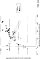
- a plot may be created where one axis is the ratio of FPR1 to ACTB, and the other axis is the ratio of FPR1 to HNRNPA1.
- An example of such a plot is indicated in Fig.s 2A-B .
- the plot in Fig. 2A is the initial data using a cell line control, and the plot in Fig. 2B is an independent data set that uses a synthetic standard.
- NSCLC and Normal Sample results are significantly different from one another. Despite the presence of some overlap, NSCLC samples consistently show ACTB to FPR1 ratios and HNRNPA1 to FPR1 ratios that are significantly greater than non-cancer samples when fit to a cell line control.
- RNA standard rather than a cell line control
- similar results are obtained but the degree of overlap is substantially decreased.
- This decreased overlap is due to decreased variability in the standards resulting from reduced numbers of serial dilutions (from 6 to 3). Each step of the serial dilution may introduce error.
- Fig. 2B a simple line can be drawn to separate all but one of the Normal synthetic standard result ratios from all of the NSCLC results.
- the results may also be interpreted as a single ratio between a linear combination of the type #1 markers and a linear combination of the type #2 markers.
- a decision rule may state that any score above a given threshold indicates cancer, while a score below the threshold indicates the lack of cancer.
- transcript accumulation values for genes selected from the lists above may be determined from a sample and compared to levels determined from a set of synthetic standards (i.e. in a serial dilution series) that span the range of values that are typically obtained.
- the transcript accumulation level determined from a patient sample is compared to the transcript accumulation level determined by performing a regression analysis on a synthetic standard template to fit the accumulation level values for each gene.
- the regression and fitted values are obtained for each gene individually. Additional analysis (i.e. calculating ratios) may be done once fitted values are obtained.
- scores may be compared to threshold values, such that scores above a threshold are indicative of a heightened risk of lung cancer as indicated by a patient sample.
- the correct concentrations for each standard, coefficients and threshold may be determined by collecting data on a small set of samples from both cancer and cancer-free patients, then using a linear model to separate them.
- the linear model may be generated via a statistical method such as logistic regression or support vector machines with a linear kernel function, or the linear model may be generated by inspection.
- Exclusionary criteria may be implemented, such that any sample that meets the exclusionary criteria has no result reported. These exclusionary criteria may include other test preformed before or after one of the described embodiments. The exclusionary criteria may also be based on results of the test itself. For example, in some embodiments very low quantities of the markers indicate a degraded sample, and an unexpectedly large ratio between two accumulation levels such as those of ACTB and HNRNP A1, for example, may indicate that there is contamination. In some embodiments a sample is excluded if the ratio of ACTB to HNRNPA1 differs by more than 10, 5, 4, 3, or 2-foled compared to the median ratio of the accumulation levels of the genes. One example of a plausible contamination source is that of lymphocytes in the plasma sample.
- the method may involve a Statistical Distance Determination. Because cfRNA from cancer cells may be highly diluted, a method may be required to determine significant changes in relative abundance. For this reason, in some embodiments, the method determines the assay outcome (i.e., positive or negative result) based on statistical distances between results as opposed to a fixed cutoff determined only through ROC curves.
- the results may be divided into groups (high confidence, low confidence, etc.). This number may also be transformed by some simple formula to create a numerical score for confidence.
- the method may involve Models and Derivations for predicting the type of cancer present in a patient based on results RNA expression in combination with demographic or lifestyle attribute(s).
- cfRNA levels may be assayed using sequencing technology.
- sequencing technology include one or more technologies such as pyrosequencing, e.g., 'the '454' method ( Margulies et al., (2005) Genome sequencing in microfabricated high-density picolitre reactors. Nature 437:376-380 ; Ronaghi, et al. (1996) Real-time DNA sequencing using detection of pyrophosphate release. Anal. Biochem. 242:84-89 ), 'Solexa' or Illumina-type sequencing ( Fedurco et al., (2006), BTA, a novel reagent for DNA attachment of glass and efficient generation of solid-phase amplified DNA colonies.
- the cfRNA levels may be assayed via hybridization to a microarray, nCounter or similar.
- a microarray nCounter or similar.
- one class of arrays commonly used in differential expression studies includes microarrays or oligonucleotide arrays. These arrays utilize a large number of probes that are synthesized directly on a substrate and are used to interrogate complex RNA or message populations based on the principle of complementary hybridization. Typically, these microarrays provide sets of 16 to 20 oligonucleotide probe pairs of relatively small length (20mers - 25mers) that span a selected region of a gene or nucleotide sequence of interest.
- the probe pairs used in the oligonucleotide array may also include perfect match and mismatch probes that are designed to hybridize to the same RNA or message strand.
- the perfect match probe contains a known sequence that is fully complementary to the message of interest while the mismatch probe is similar to the perfect match probe with respect to its sequence except that it contains at least one mismatch nucleotide which differs from the perfect match probe.
- the hybridization efficiency of messages from a sample nucleotide population are assessed with respect to the perfect match and mismatch probes in order to validate and quantitate the levels of expression for many messages simultaneously.
- an entire gene array is printed to a microarray.
- a subset of genes comprising FPR1 and at least one of ACTB and HNRNPA1 is included on a microarray.
- a microarray comprises at least FPR1, ACTB and HNRNPA1.
- An nCounter Analysis System is an integrated system comprising a fully automated prep station, a digital analyzer, the CodeSet (molecular barcodes) and all of the reagents and consumables needed to perform the analysis. Analysis on the nCounter system consists of in-solution hybridization, post-hybridization processing, digital data acquisition, and normalization in one simple workflow. In some embodiments the process is automated. In some embodiments custom or pre-designed sets of barcoded probes may be pre-mixed with a comprehensive set of system controls as part of said analysis.
- compositions and kits A number of methods of and devices for obtaining the cfRNA transcript accumulation level data necessary to perform the methods and for use with the compositions and kits is disclosed herein.
- the plasma was extracted with the QIAamp circulating nucleic acid kit (Qiagen #55114).
- the Quantifast Probe RT-PCR Plus Kit (Qiagen #204484) was used along with the previously described primers and probes for FPR1, ACTB, and HNRNPA1 to conduct quantitative Taqman PCR.
- Universal Human Reference RNA (Agilent #740000) was used as a standard, and regression was used to estimate quantities of each gene in the sample ( Fig. 2A ). Samples with a ratio of ACTB/HNRNPA1 above 75 were eliminated from the final results, and given a result of "no result". The following results were obtained. Note that the x-axis is the ratio of FPR1/ACTB and the Y-axis is the ratio of FPR1/HNRNPA1.
- FPR1 median accumulation levels normalized against accumulation levels measured in Universal Human Reference RNA differed by about 100-fold between Normal and Tumor patient populations.
- the upper limit of the 25% quartile of the Normal patent population was measurably below the lower limit 75% quartile of the measurements for the Tumor patient population.
- the highest, rarest extreme Normal measurements occasionally were as high as, but no higher than, the Tumor patient median values.
- ACTB median accumulation levels did not differ significantly between Normal and Tumor patient populations. There was also substantial overlap at the 25%-75% quartile range and among the extreme outliers in accumulation levels between Normal and Tumor patient populations.
- HNRNPA1 median accumulation levels did not differ significantly between Normal and Tumor patient populations. There was also substantial overlap at the 25%-75% quartile range and among the extreme outliers in accumulation levels between Normal and Tumor patient populations.
- This experiment shows that comparing the circulating free RNA levels of FPR1 to the circulating free RNA levels of HNRNPA1 or ACTB allows one to predict the presence of cancer in a patient.
- a second set of 6 NSCLC and 7 Normal patient samples were assayed using a protocol similar to that discussed in Example 1. Two technical modifications were made: plasma was extracted via the QIAsymphony DSP Virus/Pathogen Midi Kit (Qiagen 937055) and a synthetic standard as described above was used. Additional samples were excluded after a screen for obvious issues such as cloudiness or discoloration. The ratios of the fitted values for each gene were determined, and are shown in Fig. 2B . The X and Y axis are the same ratios as previously described.
- NSCLC and Normal Sample results were significantly different from one another. NSCLC samples consistently showed ACTB to FPR1 ratios and HNRNPA1 to FPR1 ratios that were significantly greater than the corresponding values obtained for plasma obtained from cancer-free individuals. A simple line can be drawn to separate all but one of the Normal synthetic standard result ratios from all of the NSCLC results.
- Plasma was collected from a patient suspected of having lung cancer.
- FPR1, ACTB and HNRNPA1 transcript accumulation levels in the cfRNA population of said patient were determined from the patient's plasma as described above.
- Patient FPR1 accumulation levels were compared to levels known to correspond with healthy individuals and with individuals that have cancer, for example, the accumulation levels indicated in Fig. 1A .
- Other reference measures of FPR1 accumulation in cfRNA of healthy and cancer-positive individuals could also have been used.
- An arbitrary value of 100 was assigned to the full concentration of the standard for each gene, with the two additional dilutions used to create the standard curve being 10 and 1 arbitrary units.
- Sample 171 was collected from a patient known to have NSCLC.
- Plasma was collected from patient 164 with no evidence of cancer. Transcript accumulation levels of FPR1, ACTB and HNRNPA1 were obtained as above. The values of 3.53, 12.2 and 2.87 were obtained for FPR1, ACTB and HNRNPA1 respectively. The score is calculated as 3.53/(12.2+2.87) 0.234 was obtained. Because this value is less than 0.5, a negative result for NSCLC was obtained.
- This experiment shows that determining the circulating free RNA levels of FPR1, ACTB and HNRNPA1 allows one to predict the presence of cancer, and particularly lung cancer, in a patient.
- Plasma is collected from a patient suspected of having lung cancer.
- FPR1 transcript accumulation levels in the cfRNA population of said patient are determined from the patient's plasma as described above.
- Patient FPR1 accumulation levels are compared to levels known to correspond with healthy individuals and with individuals that have cancer, for example, the accumulation levels indicated in Fig. 1A .
- Other reference measures of FPR1 accumulation in cfRNA of healthy and cancer-positive individuals may be used.
- Levels are found to be 60,000 FPR1 molecules per mL, which corresponds to the median accumulation level observed for Tumor patients but is at or above the extreme highest measured value for Normal patients.
- This FPR1 accumulation level indicates with a high degree of confidence that a NSCLC cell population is present in the patient.
- Plasma is collected from a second patient suspected of having lung cancer.
- FPR1 transcript accumulation levels in the cfRNA population of said patient are determined and compared as above. Levels are found to be 600 FPR1 molecules per mL, which corresponds to the median accumulation level observed for Normal patients but is at or below the extreme lowest measured value for Tumor patients. This FPR1 accumulation level indicates with a high degree of confidence that the patient is free of a cancerous or precancerous cell population detectable through this method.
- This experiment shows that determining the circulating free RNA levels of FPR1 allows one to predict the presence of cancer in a patient.
Landscapes
- Chemical & Material Sciences (AREA)
- Life Sciences & Earth Sciences (AREA)
- Health & Medical Sciences (AREA)
- Organic Chemistry (AREA)
- Proteomics, Peptides & Aminoacids (AREA)
- Engineering & Computer Science (AREA)
- Zoology (AREA)
- Wood Science & Technology (AREA)
- Immunology (AREA)
- Analytical Chemistry (AREA)
- Genetics & Genomics (AREA)
- Pathology (AREA)
- Physics & Mathematics (AREA)
- Biotechnology (AREA)
- Microbiology (AREA)
- Molecular Biology (AREA)
- Biophysics (AREA)
- Biochemistry (AREA)
- Bioinformatics & Cheminformatics (AREA)
- General Engineering & Computer Science (AREA)
- General Health & Medical Sciences (AREA)
- Hospice & Palliative Care (AREA)
- Oncology (AREA)
- Measuring Or Testing Involving Enzymes Or Micro-Organisms (AREA)
- Apparatus Associated With Microorganisms And Enzymes (AREA)
Claims (14)
- Verfahren zum Testen auf das Vorhandensein eines Lungenkrebses bei einer Person, umfassend:in einer biologischen Probe eines Patienten, bei dem der Verdacht auf Krebs besteht;Messen des Niveaus zirkulierender freier Nukleinsäuren, die von einem Ziel-Formylpeptidrezeptor-1-Gen (FPR1-Gen) abgeleitet sind, in der biologischen Probe; undVergleichen des gemessenen Niveaus zirkulierender freier Nukleinsäuren, die von dem Ziel-FPRl-Gen abgeleitet sind, mit bekannten Niveaus zirkulierender freier Nukleinsäuren, die von dem Gen gesunder Personen abgeleitet sind,wobei ein erhöhtes Niveau zirkulierender freier Nukleinsäuren, die von dem FPR1-Gen abgeleitet sind, das Vorhandensein eines Lungenkrebses anzeigt, der mit dem Gen im Patienten assoziiert ist.
- Verfahren nach Anspruch 1, wobei das Vergleichen das elektronische Vergleichen in einem Computer von Werten umfasst, die die Niveaus zirkulierender freier Nukleinsäuren widerspiegeln, die von dem Gen abgeleitet sind.
- Verfahren nach Anspruch 1, wobei die Probe Blutkomponenten umfasst.
- Verfahren nach Anspruch 1, wobei das Messen das Normalisieren der Niveaus auf einen Standard umfasst, gegebenenfalls wobei der Standard ein synthetischer Standard ist oder RNA umfasst, die aus einer Standardzelllinie extrahiert wurde.
- Verfahren nach Anspruch 1, wobei das Messen das Vergleichen der Niveaus zirkulierender freier Nukleinsäuren mit einem Niveau einer zweiten Population zirkulierender freier Nukleinsäuren umfasst, von denen bekannt ist, dass sie in stabilen Niveaus unabhängig vom Krebsstatus vorliegen.
- Verfahren nach Anspruch 1, wobei der Lungenkrebs nicht-kleinzelliger Lungenkrebs (NSCLC) ist.
- Verfahren nach Anspruch 5, wobei die zirkulierenden freien Nukleinsäuren zirkulierende freie Ribonukleinsäuren umfassen.
- Verwendung eines Kits in dem Verfahren nach Anspruch 1 zum Testen auf das Vorhandensein eines Lungenkrebses bei einem Patienten, umfassend:
einen Behälter, umfassend Amplifikationsprimer zum Amplifizieren zirkulierender freier Nukleinsäuren, die von einem Formylpeptidrezeptor-1-Gen (FPR1-Gen) abgeleitet sind, in einer biologischen Probe. - Verwendung eines Kits nach Anspruch 8, wobei die Amplifikationsprimer die Nukleinsäuresequenzen aufweisen:Vorwärtsprimer: 5' TGACGGTGAGAGGCATCA 3' (SEQ ID NO: 1)Rückwärtsprimer: 5' GGTGGCAATAAGCCCATAACTG 3' (SEQ ID NO: 3).
- Verfahren zum Testen auf das Vorhandensein von Lungenkrebs bei einer Person, umfassend die Schritte:in einer biologischen Probe, die Blut oder Blutkomponenten eines Patienten umfasst, bei dem der Verdacht auf Lungenkrebs besteht;Messen eines Niveaus zirkulierender freier Nukleinsäuren, die von einem Ziel-Formylpeptidrezeptor-1-Gen (FPR1-Gen) abgeleitet sind, in der biologischen Probe; undVergleichen des gemessenen Niveaus zirkulierender freier Nukleinsäuren, die von dem FPR1-Gen abgeleitet sind, mit wenigstens einem Referenzniveau zirkulierender freier Nukleinsäuren, die von einem zweiten Gen abgeleitet sind, wobei das Verhältnis des Niveaus zirkulierender freier Nukleinsäuren, die von dem Ziel-FPRl-Gen abgeleitet sind, zu dem Niveau zirkulierender freier Nukleinsäuren, die von einem zweiten Referenzgen abgeleitet sind, das Vorhandensein von Lungenkrebs bei der Person anzeigt.
- Verfahren nach Anspruch 10, wobei das Referenzniveau zirkulierender freier Nukleinsäuren, die von dem zweiten Gen abgeleitet sind, RNAs umfasst, die von Actin B (ACTB) oder HNRNPA1 oder einer Kombination davon abgeleitet sind.
- Verfahren nach Anspruch 4, wobei der Standard ein synthetischer Referenzstandard ist, der durch ein Verfahren hergestellt wird, das die folgenden Schritte umfasst:Auswählen einer ersten synthetischen RNA-Templatsequenz;Auswählen einer zweiten synthetischen RNA-Templatsequenz;Erhalten einer Schätzung einer erwarteten Konzentration von Transkripten, die die erste synthetische RNA-Templatsequenz umfassen, und von Transkripten, die die zweite synthetische RNA-Templatsequenz umfassen, in einer biologischen Probe; und
Kombinieren der ersten Templatsequenz und der zweiten Templatsequenz in einer Zusammensetzung, so dass die Konzentration jeder Templatsequenz der erwarteten Konzentration jeder Sequenz in einer unbekannten Probe entspricht. - Verfahren nach Anspruch 4, wobei der Standard ein synthetischer Referenzstandard ist, der Templatnukleinsäuren in Konzentrationen innerhalb einer Größenordnung der erwarteten Akkumulationsniveaus in einer Patientenprobe umfasst.
- Verfahren nach Anspruch 13, wobei der synthetische Referenzstandard Populationen von Templatnukleinsäuren umfasst, die wenigstens zwei zu testenden Transkripten in einer Patientenprobe entsprechen, und wobei jede der Referenznukleinsäurepopulationen in einer Konzentration innerhalb einer Größenordnung eines erwarteten Akkumulationsniveaus eines entsprechenden Transkripts in einer Patientenprobe vorliegt.
Priority Applications (1)
| Application Number | Priority Date | Filing Date | Title |
|---|---|---|---|
| PL13733697T PL2809810T3 (pl) | 2012-01-06 | 2013-01-04 | Układ i sposób wykrywania rna zmienionych przez raka płuca w krwi obwodowej |
Applications Claiming Priority (2)
| Application Number | Priority Date | Filing Date | Title |
|---|---|---|---|
| US201261584097P | 2012-01-06 | 2012-01-06 | |
| PCT/US2013/020383 WO2013103889A1 (en) | 2012-01-06 | 2013-01-04 | System and method of detecting rnas altered by cancer in peripheral blood |
Publications (3)
| Publication Number | Publication Date |
|---|---|
| EP2809810A1 EP2809810A1 (de) | 2014-12-10 |
| EP2809810A4 EP2809810A4 (de) | 2015-12-02 |
| EP2809810B1 true EP2809810B1 (de) | 2019-11-13 |
Family
ID=48745455
Family Applications (1)
| Application Number | Title | Priority Date | Filing Date |
|---|---|---|---|
| EP13733697.0A Active EP2809810B1 (de) | 2012-01-06 | 2013-01-04 | System und verfahren für den nachweis von durch lungenkrebs veränderten rnas im peripheren blut |
Country Status (12)
| Country | Link |
|---|---|
| US (2) | US9422592B2 (de) |
| EP (1) | EP2809810B1 (de) |
| JP (2) | JP2015503356A (de) |
| AU (1) | AU2013207385B2 (de) |
| CA (1) | CA2860338C (de) |
| DK (1) | DK2809810T3 (de) |
| ES (1) | ES2769795T3 (de) |
| HU (1) | HUE047739T2 (de) |
| IL (1) | IL233163B (de) |
| PL (1) | PL2809810T3 (de) |
| PT (1) | PT2809810T (de) |
| WO (1) | WO2013103889A1 (de) |
Families Citing this family (24)
| Publication number | Priority date | Publication date | Assignee | Title |
|---|---|---|---|---|
| EP2816122A1 (de) | 2008-02-15 | 2014-12-24 | Mayo Foundation For Medical Education And Research | Neoplasmanachweis von einer Stuhlprobe |
| US8361720B2 (en) | 2010-11-15 | 2013-01-29 | Exact Sciences Corporation | Real time cleavage assay |
| ES2859645T3 (es) | 2013-03-14 | 2021-10-04 | Mayo Found Medical Education & Res | Detección de neoplasia |
| CN106460046A (zh) | 2014-03-31 | 2017-02-22 | 梅奥医学教育和研究基金会 | 检测结直肠赘生物 |
| US10184154B2 (en) | 2014-09-26 | 2019-01-22 | Mayo Foundation For Medical Education And Research | Detecting cholangiocarcinoma |
| WO2016077709A1 (en) * | 2014-11-14 | 2016-05-19 | Liquid Genomics, Inc. | Use of circulating cell-free rna for diagnosis and/or monitoring cancer |
| WO2016160454A1 (en) | 2015-03-27 | 2016-10-06 | Exact Sciences Corporation | Detecting esophageal disorders |
| KR20250172724A (ko) | 2015-08-31 | 2025-12-09 | 메이오 파운데이션 포 메디칼 에쥬케이션 앤드 리써치 | 위 신생물 검출 방법 |
| KR20250073555A (ko) | 2015-10-30 | 2025-05-27 | 이그잭트 사이언시즈 코포레이션 | 복합 증폭 검출 분석 및 혈장으로부터 dna의 분리 및 검출 방법 |
| CN116064796A (zh) | 2016-05-05 | 2023-05-05 | 精密科学公司 | 通过分析甲基化dna来检测肺肿瘤 |
| JP7289264B2 (ja) | 2017-01-27 | 2023-06-09 | エグザクト サイエンシーズ コーポレーション | メチル化dnaの分析による結腸腫瘍の検出 |
| WO2018208892A1 (en) * | 2017-05-10 | 2018-11-15 | Nantomics, Llc | Circulating rna for detection, prediction, and monitoring of cancer |
| US11795197B2 (en) | 2017-06-07 | 2023-10-24 | Cedars-Sinai Medical Center | Peptide compositions for immuno-oncology molecular imaging and targeted drug delivery |
| CN111213209A (zh) | 2017-10-12 | 2020-05-29 | 南托米克斯有限责任公司 | 根据生物体液的评估用癌症评分和反应预测 |
| AU2018365883A1 (en) | 2017-11-07 | 2020-05-14 | Nanthealth Labs, Inc. | Targeted cell free nucleic acid analysis |
| JP7277460B2 (ja) | 2017-11-30 | 2023-05-19 | マヨ ファウンデーション フォア メディカル エデュケーション アンド リサーチ | 乳癌の検出 |
| US10648025B2 (en) | 2017-12-13 | 2020-05-12 | Exact Sciences Development Company, Llc | Multiplex amplification detection assay II |
| US11512349B2 (en) * | 2018-12-18 | 2022-11-29 | Grail, Llc | Methods for detecting disease using analysis of RNA |
| WO2020154665A1 (en) | 2019-01-24 | 2020-07-30 | Mayo Foundation For Medical Education And Research | Detecting endometrial cancer |
| WO2020236939A2 (en) | 2019-05-23 | 2020-11-26 | Paradigm Diagnostics | Tissue preparation using nuclease |
| JP2022546504A (ja) * | 2019-08-27 | 2022-11-04 | エグザクト サイエンシーズ コーポレーション | 肺新生物形成を有する疑いのある対象におけるメチル化されたdna、rna、及びタンパク質の特性決定 |
| US11702704B2 (en) | 2019-10-31 | 2023-07-18 | Mayo Foundation For Medical Education And Research | Detecting ovarian cancer |
| WO2021111476A1 (en) | 2019-12-05 | 2021-06-10 | Council Of Scientific And Industrial Research | Solvent assisted cavitation for waste water treatment |
| CN117947165B (zh) * | 2024-01-19 | 2024-10-08 | 西安交通大学医学院第二附属医院 | PCR检测肺腺癌癌前病变人群血浆lncRNA的检测方法 |
Family Cites Families (26)
| Publication number | Priority date | Publication date | Assignee | Title |
|---|---|---|---|---|
| KR100236506B1 (ko) | 1990-11-29 | 2000-01-15 | 퍼킨-엘머시터스인스트루먼츠 | 폴리머라제 연쇄 반응 수행 장치 |
| AU645915B2 (en) | 1991-07-23 | 1994-01-27 | F. Hoffmann-La Roche Ag | Improvements in the in situ PCR |
| KR0171154B1 (ko) | 1995-04-29 | 1999-03-20 | 배순훈 | 특징점 기반 움직임 추정을 이용하여 비디오 신호를 부호화하는 방법 및 장치 |
| US6040139A (en) | 1995-09-19 | 2000-03-21 | Bova; G. Steven | Laser cell purification system |
| US8043835B1 (en) | 1996-03-26 | 2011-10-25 | Oncomedx, Inc. | Methods for detecting and monitoring cancer using extracellular RNA |
| US6759217B2 (en) | 1996-03-26 | 2004-07-06 | Oncomedx, Inc. | Method enabling use of extracellular RNA extracted from plasma or serum to detect, monitor or evaluate cancer |
| US7785842B2 (en) | 1996-03-26 | 2010-08-31 | Oncomedx, Inc. | Comparative analysis of extracellular RNA species |
| ES2347854T5 (es) | 1996-03-26 | 2014-07-24 | Michael S. Kopreski | Método que permite el uso de RNA extracelular extraído de plasma o suero para detectar, monitorizar o evaluar un cáncer |
| US6100051A (en) | 1997-06-27 | 2000-08-08 | The United States Of America As Represented By The Department Of Health And Human Services | Method utilizing convex geometry for laser capture microdissection |
| US8163524B2 (en) | 1998-09-22 | 2012-04-24 | Oncomedx, Inc. | Comparative analysis of extracellular RNA species |
| US7767422B2 (en) | 1998-09-22 | 2010-08-03 | Oncomedx, Inc. | Detection of 5T4 RNA in plasma and serum |
| US20040085443A1 (en) | 2000-12-13 | 2004-05-06 | Kallioniemi Olli P | Method and system for processing regions of interest for objects comprising biological material |
| AU2002352799A1 (en) | 2001-11-20 | 2003-06-10 | Oncomedx, Inc. | Methods for evaluating drug-resistance gene expression in the cancer patient |
| AU2003214072A1 (en) * | 2002-03-07 | 2003-09-16 | Bayer Aktiengesellschaft | Diagnostics and therapeutics for diseases associated with the n-formyl peptide receptor 1 (fpr 1) |
| JP2006500916A (ja) | 2002-05-30 | 2006-01-12 | テロジェン インコーポレーテッド | hnRNPA1およびA2核酸分子と関連がある新形成を治療するための方法および組成物 |
| US7955846B2 (en) | 2004-05-17 | 2011-06-07 | The General Hospital Corporation | Compositions comprising female germline stem cells and methods of use thereof |
| US20060051736A1 (en) | 2004-08-19 | 2006-03-09 | Applied Imaging Corp. | Paraffin-control marker |
| EP2272983A1 (de) | 2005-02-01 | 2011-01-12 | AB Advanced Genetic Analysis Corporation | Reagentien, Verfahren und Bibliotheken zur Sequenzierung mit Kügelchen |
| US7405056B2 (en) | 2005-03-02 | 2008-07-29 | Edward Lam | Tissue punch and tissue sample labeling methods and devices for microarray preparation, archiving and documentation |
| EP2484782B1 (de) | 2006-01-05 | 2015-11-18 | The Ohio State University Research Foundation | Verfahren und Zusammensetzungen auf Mikro-RNA-Basis zur Diagnose und Behandlung von soliden Krebs |
| US8252538B2 (en) | 2006-11-01 | 2012-08-28 | The Ohio State University | MicroRNA expression signature for predicting survival and metastases in hepatocellular carcinoma |
| US8189882B2 (en) | 2008-01-30 | 2012-05-29 | Clarient, Inc. | Automated laser capture microdissection |
| US8880351B2 (en) | 2008-03-25 | 2014-11-04 | General Electric Company | Method and apparatus for analysis of tissue microarrays |
| CA2743211A1 (en) | 2008-11-12 | 2010-05-20 | Caris Life Sciences Luxembourg Holdings, S.A.R.L. | Methods and systems of using exosomes for determining phenotypes |
| US8765383B2 (en) * | 2009-04-07 | 2014-07-01 | Genomic Health, Inc. | Methods of predicting cancer risk using gene expression in premalignant tissue |
| WO2010123124A1 (ja) * | 2009-04-24 | 2010-10-28 | 国立大学法人九州大学 | 固形がんの再発および予後因子、およびその臨床利用 |
-
2013
- 2013-01-04 EP EP13733697.0A patent/EP2809810B1/de active Active
- 2013-01-04 DK DK13733697.0T patent/DK2809810T3/da active
- 2013-01-04 US US13/734,735 patent/US9422592B2/en active Active
- 2013-01-04 WO PCT/US2013/020383 patent/WO2013103889A1/en not_active Ceased
- 2013-01-04 PT PT137336970T patent/PT2809810T/pt unknown
- 2013-01-04 ES ES13733697T patent/ES2769795T3/es active Active
- 2013-01-04 CA CA2860338A patent/CA2860338C/en active Active
- 2013-01-04 JP JP2014551375A patent/JP2015503356A/ja active Pending
- 2013-01-04 HU HUE13733697A patent/HUE047739T2/hu unknown
- 2013-01-04 PL PL13733697T patent/PL2809810T3/pl unknown
- 2013-01-04 AU AU2013207385A patent/AU2013207385B2/en active Active
-
2014
- 2014-06-16 IL IL233163A patent/IL233163B/en active IP Right Grant
-
2016
- 2016-07-28 US US15/222,828 patent/US20160333424A1/en not_active Abandoned
-
2019
- 2019-03-13 JP JP2019045627A patent/JP2019162102A/ja active Pending
Non-Patent Citations (1)
| Title |
|---|
| None * |
Also Published As
| Publication number | Publication date |
|---|---|
| PL2809810T3 (pl) | 2020-07-27 |
| US9422592B2 (en) | 2016-08-23 |
| EP2809810A4 (de) | 2015-12-02 |
| WO2013103889A1 (en) | 2013-07-11 |
| EP2809810A1 (de) | 2014-12-10 |
| CA2860338A1 (en) | 2013-07-11 |
| ES2769795T3 (es) | 2020-06-29 |
| CA2860338C (en) | 2022-08-02 |
| US20130196332A1 (en) | 2013-08-01 |
| AU2013207385B2 (en) | 2018-09-13 |
| IL233163A0 (en) | 2014-07-31 |
| AU2013207385A1 (en) | 2014-07-10 |
| IL233163B (en) | 2019-02-28 |
| JP2019162102A (ja) | 2019-09-26 |
| JP2015503356A (ja) | 2015-02-02 |
| US20160333424A1 (en) | 2016-11-17 |
| HUE047739T2 (hu) | 2020-05-28 |
| DK2809810T3 (da) | 2020-02-03 |
| PT2809810T (pt) | 2020-02-14 |
Similar Documents
| Publication | Publication Date | Title |
|---|---|---|
| AU2013207385B2 (en) | System and method of detecting RNAs altered by cancer in peripheral blood | |
| AU2020336115B2 (en) | Characterizing methylated DNA, RNA, and proteins in subjects suspected of having lung neoplasia | |
| US12516372B2 (en) | Compositions and methods for screening mutations in thyroid cancer | |
| AU2017260630B2 (en) | Detection of lung neoplasia by analysis of methylated DNA | |
| CN113423410A (zh) | 在肺肿瘤检测中表征甲基化dna、rna和蛋白质 | |
| HK1250745A1 (zh) | 使用二代测序预测器官移植排斥的方法 | |
| WO2015183836A1 (en) | Compositions, methods, and uses related to ntrk2-tert fusions | |
| CN114672556A (zh) | 结直肠癌分子分型及生存风险基因群及诊断产品和应用 | |
| CN113621693B (zh) | 一种检测人类her2基因拷贝数变异的方法和试剂盒 | |
| AU2011209624B2 (en) | Methods and kits used in the detection of fungus | |
| CN109154026B (zh) | 用于将dlbcl分类的方法和组合物 | |
| CA2709106C (en) | Methods and reagents for the early detection of melanoma | |
| KR20210120737A (ko) | Pik3ca 돌연변이 검출용 조성물 및 이를 포함하는 키트 | |
| WO2017041084A2 (en) | Multivalent probes having single nucleotide resolution | |
| HK1152974A (en) | Methods and reagents for the early detection of melanoma | |
| HK1152974B (en) | Methods and reagents for the early detection of melanoma |
Legal Events
| Date | Code | Title | Description |
|---|---|---|---|
| PUAI | Public reference made under article 153(3) epc to a published international application that has entered the european phase |
Free format text: ORIGINAL CODE: 0009012 |
|
| 17P | Request for examination filed |
Effective date: 20140703 |
|
| AK | Designated contracting states |
Kind code of ref document: A1 Designated state(s): AL AT BE BG CH CY CZ DE DK EE ES FI FR GB GR HR HU IE IS IT LI LT LU LV MC MK MT NL NO PL PT RO RS SE SI SK SM TR |
|
| AX | Request for extension of the european patent |
Extension state: BA ME |
|
| DAX | Request for extension of the european patent (deleted) | ||
| RA4 | Supplementary search report drawn up and despatched (corrected) |
Effective date: 20151104 |
|
| RIC1 | Information provided on ipc code assigned before grant |
Ipc: C12Q 1/68 20060101AFI20151029BHEP |
|
| STAA | Information on the status of an ep patent application or granted ep patent |
Free format text: STATUS: EXAMINATION IS IN PROGRESS |
|
| 17Q | First examination report despatched |
Effective date: 20170808 |
|
| REG | Reference to a national code |
Ref country code: DE Ref legal event code: R079 Ref document number: 602013062852 Country of ref document: DE Free format text: PREVIOUS MAIN CLASS: C12Q0001680000 Ipc: C12Q0001688600 |
|
| GRAP | Despatch of communication of intention to grant a patent |
Free format text: ORIGINAL CODE: EPIDOSNIGR1 |
|
| STAA | Information on the status of an ep patent application or granted ep patent |
Free format text: STATUS: GRANT OF PATENT IS INTENDED |
|
| RIC1 | Information provided on ipc code assigned before grant |
Ipc: C12Q 1/6886 20180101AFI20190510BHEP |
|
| INTG | Intention to grant announced |
Effective date: 20190531 |
|
| GRAS | Grant fee paid |
Free format text: ORIGINAL CODE: EPIDOSNIGR3 |
|
| GRAA | (expected) grant |
Free format text: ORIGINAL CODE: 0009210 |
|
| STAA | Information on the status of an ep patent application or granted ep patent |
Free format text: STATUS: THE PATENT HAS BEEN GRANTED |
|
| AK | Designated contracting states |
Kind code of ref document: B1 Designated state(s): AL AT BE BG CH CY CZ DE DK EE ES FI FR GB GR HR HU IE IS IT LI LT LU LV MC MK MT NL NO PL PT RO RS SE SI SK SM TR |
|
| REG | Reference to a national code |
Ref country code: CH Ref legal event code: EP Ref country code: AT Ref legal event code: REF Ref document number: 1201703 Country of ref document: AT Kind code of ref document: T Effective date: 20191115 |
|
| REG | Reference to a national code |
Ref country code: DE Ref legal event code: R096 Ref document number: 602013062852 Country of ref document: DE |
|
| REG | Reference to a national code |
Ref country code: IE Ref legal event code: FG4D |
|
| REG | Reference to a national code |
Ref country code: DK Ref legal event code: T3 Effective date: 20200131 Ref country code: RO Ref legal event code: EPE |
|
| REG | Reference to a national code |
Ref country code: FI Ref legal event code: FGE |
|
| REG | Reference to a national code |
Ref country code: PT Ref legal event code: SC4A Ref document number: 2809810 Country of ref document: PT Date of ref document: 20200214 Kind code of ref document: T Free format text: AVAILABILITY OF NATIONAL TRANSLATION Effective date: 20200203 |
|
| REG | Reference to a national code |
Ref country code: NO Ref legal event code: T2 Effective date: 20191113 |
|
| REG | Reference to a national code |
Ref country code: SE Ref legal event code: TRGR |
|
| REG | Reference to a national code |
Ref country code: NL Ref legal event code: FP |
|
| REG | Reference to a national code |
Ref country code: LT Ref legal event code: MG4D |
|
| PG25 | Lapsed in a contracting state [announced via postgrant information from national office to epo] |
Ref country code: LT Free format text: LAPSE BECAUSE OF FAILURE TO SUBMIT A TRANSLATION OF THE DESCRIPTION OR TO PAY THE FEE WITHIN THE PRESCRIBED TIME-LIMIT Effective date: 20191113 Ref country code: BG Free format text: LAPSE BECAUSE OF FAILURE TO SUBMIT A TRANSLATION OF THE DESCRIPTION OR TO PAY THE FEE WITHIN THE PRESCRIBED TIME-LIMIT Effective date: 20200213 Ref country code: LV Free format text: LAPSE BECAUSE OF FAILURE TO SUBMIT A TRANSLATION OF THE DESCRIPTION OR TO PAY THE FEE WITHIN THE PRESCRIBED TIME-LIMIT Effective date: 20191113 |
|
| REG | Reference to a national code |
Ref country code: GR Ref legal event code: EP Ref document number: 20200400321 Country of ref document: GR Effective date: 20200511 |
|
| REG | Reference to a national code |
Ref country code: HU Ref legal event code: AG4A Ref document number: E047739 Country of ref document: HU |
|
| PG25 | Lapsed in a contracting state [announced via postgrant information from national office to epo] |
Ref country code: IS Free format text: LAPSE BECAUSE OF FAILURE TO SUBMIT A TRANSLATION OF THE DESCRIPTION OR TO PAY THE FEE WITHIN THE PRESCRIBED TIME-LIMIT Effective date: 20200313 Ref country code: RS Free format text: LAPSE BECAUSE OF FAILURE TO SUBMIT A TRANSLATION OF THE DESCRIPTION OR TO PAY THE FEE WITHIN THE PRESCRIBED TIME-LIMIT Effective date: 20191113 Ref country code: HR Free format text: LAPSE BECAUSE OF FAILURE TO SUBMIT A TRANSLATION OF THE DESCRIPTION OR TO PAY THE FEE WITHIN THE PRESCRIBED TIME-LIMIT Effective date: 20191113 |
|
| REG | Reference to a national code |
Ref country code: ES Ref legal event code: FG2A Ref document number: 2769795 Country of ref document: ES Kind code of ref document: T3 Effective date: 20200629 |
|
| PG25 | Lapsed in a contracting state [announced via postgrant information from national office to epo] |
Ref country code: AL Free format text: LAPSE BECAUSE OF FAILURE TO SUBMIT A TRANSLATION OF THE DESCRIPTION OR TO PAY THE FEE WITHIN THE PRESCRIBED TIME-LIMIT Effective date: 20191113 |
|
| PG25 | Lapsed in a contracting state [announced via postgrant information from national office to epo] |
Ref country code: CZ Free format text: LAPSE BECAUSE OF FAILURE TO SUBMIT A TRANSLATION OF THE DESCRIPTION OR TO PAY THE FEE WITHIN THE PRESCRIBED TIME-LIMIT Effective date: 20191113 Ref country code: EE Free format text: LAPSE BECAUSE OF FAILURE TO SUBMIT A TRANSLATION OF THE DESCRIPTION OR TO PAY THE FEE WITHIN THE PRESCRIBED TIME-LIMIT Effective date: 20191113 |
|
| REG | Reference to a national code |
Ref country code: DE Ref legal event code: R097 Ref document number: 602013062852 Country of ref document: DE |
|
| REG | Reference to a national code |
Ref country code: AT Ref legal event code: MK05 Ref document number: 1201703 Country of ref document: AT Kind code of ref document: T Effective date: 20191113 |
|
| PG25 | Lapsed in a contracting state [announced via postgrant information from national office to epo] |
Ref country code: MC Free format text: LAPSE BECAUSE OF FAILURE TO SUBMIT A TRANSLATION OF THE DESCRIPTION OR TO PAY THE FEE WITHIN THE PRESCRIBED TIME-LIMIT Effective date: 20191113 Ref country code: SK Free format text: LAPSE BECAUSE OF FAILURE TO SUBMIT A TRANSLATION OF THE DESCRIPTION OR TO PAY THE FEE WITHIN THE PRESCRIBED TIME-LIMIT Effective date: 20191113 Ref country code: SM Free format text: LAPSE BECAUSE OF FAILURE TO SUBMIT A TRANSLATION OF THE DESCRIPTION OR TO PAY THE FEE WITHIN THE PRESCRIBED TIME-LIMIT Effective date: 20191113 |
|
| REG | Reference to a national code |
Ref country code: CH Ref legal event code: PL |
|
| PLBE | No opposition filed within time limit |
Free format text: ORIGINAL CODE: 0009261 |
|
| STAA | Information on the status of an ep patent application or granted ep patent |
Free format text: STATUS: NO OPPOSITION FILED WITHIN TIME LIMIT |
|
| REG | Reference to a national code |
Ref country code: BE Ref legal event code: MM Effective date: 20200131 |
|
| 26N | No opposition filed |
Effective date: 20200814 |
|
| PG25 | Lapsed in a contracting state [announced via postgrant information from national office to epo] |
Ref country code: SI Free format text: LAPSE BECAUSE OF FAILURE TO SUBMIT A TRANSLATION OF THE DESCRIPTION OR TO PAY THE FEE WITHIN THE PRESCRIBED TIME-LIMIT Effective date: 20191113 Ref country code: CH Free format text: LAPSE BECAUSE OF NON-PAYMENT OF DUE FEES Effective date: 20200131 Ref country code: BE Free format text: LAPSE BECAUSE OF NON-PAYMENT OF DUE FEES Effective date: 20200131 Ref country code: AT Free format text: LAPSE BECAUSE OF FAILURE TO SUBMIT A TRANSLATION OF THE DESCRIPTION OR TO PAY THE FEE WITHIN THE PRESCRIBED TIME-LIMIT Effective date: 20191113 Ref country code: LI Free format text: LAPSE BECAUSE OF NON-PAYMENT OF DUE FEES Effective date: 20200131 |
|
| PG25 | Lapsed in a contracting state [announced via postgrant information from national office to epo] |
Ref country code: TR Free format text: LAPSE BECAUSE OF FAILURE TO SUBMIT A TRANSLATION OF THE DESCRIPTION OR TO PAY THE FEE WITHIN THE PRESCRIBED TIME-LIMIT Effective date: 20191113 Ref country code: MT Free format text: LAPSE BECAUSE OF FAILURE TO SUBMIT A TRANSLATION OF THE DESCRIPTION OR TO PAY THE FEE WITHIN THE PRESCRIBED TIME-LIMIT Effective date: 20191113 Ref country code: CY Free format text: LAPSE BECAUSE OF FAILURE TO SUBMIT A TRANSLATION OF THE DESCRIPTION OR TO PAY THE FEE WITHIN THE PRESCRIBED TIME-LIMIT Effective date: 20191113 |
|
| PG25 | Lapsed in a contracting state [announced via postgrant information from national office to epo] |
Ref country code: MK Free format text: LAPSE BECAUSE OF FAILURE TO SUBMIT A TRANSLATION OF THE DESCRIPTION OR TO PAY THE FEE WITHIN THE PRESCRIBED TIME-LIMIT Effective date: 20191113 |
|
| P01 | Opt-out of the competence of the unified patent court (upc) registered |
Effective date: 20230612 |
|
| REG | Reference to a national code |
Ref country code: DE Ref legal event code: R082 Ref document number: 602013062852 Country of ref document: DE Representative=s name: FORRESTERS IP LLP, DE |
|
| PGFP | Annual fee paid to national office [announced via postgrant information from national office to epo] |
Ref country code: PT Payment date: 20251218 Year of fee payment: 14 |
|
| PGFP | Annual fee paid to national office [announced via postgrant information from national office to epo] |
Ref country code: PL Payment date: 20251219 Year of fee payment: 14 |
|
| PGFP | Annual fee paid to national office [announced via postgrant information from national office to epo] |
Ref country code: LU Payment date: 20260127 Year of fee payment: 14 Ref country code: NL Payment date: 20260126 Year of fee payment: 14 |
|
| PGFP | Annual fee paid to national office [announced via postgrant information from national office to epo] |
Ref country code: HU Payment date: 20251223 Year of fee payment: 14 |
|
| PGFP | Annual fee paid to national office [announced via postgrant information from national office to epo] |
Ref country code: SE Payment date: 20260127 Year of fee payment: 14 |
|
| PGFP | Annual fee paid to national office [announced via postgrant information from national office to epo] |
Ref country code: GB Payment date: 20260127 Year of fee payment: 14 |
|
| PGFP | Annual fee paid to national office [announced via postgrant information from national office to epo] |
Ref country code: ES Payment date: 20260202 Year of fee payment: 14 |
|
| PGFP | Annual fee paid to national office [announced via postgrant information from national office to epo] |
Ref country code: NO Payment date: 20260128 Year of fee payment: 14 Ref country code: DE Payment date: 20260128 Year of fee payment: 14 Ref country code: DK Payment date: 20260126 Year of fee payment: 14 Ref country code: IE Payment date: 20260127 Year of fee payment: 14 |
|
| PGFP | Annual fee paid to national office [announced via postgrant information from national office to epo] |
Ref country code: RO Payment date: 20260105 Year of fee payment: 14 Ref country code: FI Payment date: 20260126 Year of fee payment: 14 Ref country code: IT Payment date: 20260121 Year of fee payment: 14 |
|
| PGFP | Annual fee paid to national office [announced via postgrant information from national office to epo] |
Ref country code: FR Payment date: 20260126 Year of fee payment: 14 |
|
| PGFP | Annual fee paid to national office [announced via postgrant information from national office to epo] |
Ref country code: GR Payment date: 20260127 Year of fee payment: 14 |