EP2816355A2 - Procédé de détection de bactéries, procédé de fabrication de protéines de fusion et protéine de fusion - Google Patents
Procédé de détection de bactéries, procédé de fabrication de protéines de fusion et protéine de fusion Download PDFInfo
- Publication number
- EP2816355A2 EP2816355A2 EP14173356.8A EP14173356A EP2816355A2 EP 2816355 A2 EP2816355 A2 EP 2816355A2 EP 14173356 A EP14173356 A EP 14173356A EP 2816355 A2 EP2816355 A2 EP 2816355A2
- Authority
- EP
- European Patent Office
- Prior art keywords
- protein
- bacteriophage
- detection
- tag
- proteins
- Prior art date
- Legal status (The legal status is an assumption and is not a legal conclusion. Google has not performed a legal analysis and makes no representation as to the accuracy of the status listed.)
- Granted
Links
Images


Classifications
-
- G—PHYSICS
- G01—MEASURING; TESTING
- G01N—INVESTIGATING OR ANALYSING MATERIALS BY DETERMINING THEIR CHEMICAL OR PHYSICAL PROPERTIES
- G01N33/00—Investigating or analysing materials by specific methods not covered by groups G01N1/00 - G01N31/00
- G01N33/48—Biological material, e.g. blood, urine; Haemocytometers
- G01N33/50—Chemical analysis of biological material, e.g. blood, urine; Testing involving biospecific ligand binding methods; Immunological testing
- G01N33/53—Immunoassay; Biospecific binding assay; Materials therefor
- G01N33/569—Immunoassay; Biospecific binding assay; Materials therefor for microorganisms, e.g. protozoa, bacteria, viruses
- G01N33/56911—Bacteria
-
- C—CHEMISTRY; METALLURGY
- C12—BIOCHEMISTRY; BEER; SPIRITS; WINE; VINEGAR; MICROBIOLOGY; ENZYMOLOGY; MUTATION OR GENETIC ENGINEERING
- C12N—MICROORGANISMS OR ENZYMES; COMPOSITIONS THEREOF; PROPAGATING, PRESERVING, OR MAINTAINING MICROORGANISMS; MUTATION OR GENETIC ENGINEERING; CULTURE MEDIA
- C12N15/00—Mutation or genetic engineering; DNA or RNA concerning genetic engineering, vectors, e.g. plasmids, or their isolation, preparation or purification; Use of hosts therefor
- C12N15/09—Recombinant DNA-technology
- C12N15/11—DNA or RNA fragments; Modified forms thereof; Non-coding nucleic acids having a biological activity
- C12N15/62—DNA sequences coding for fusion proteins
-
- G—PHYSICS
- G01—MEASURING; TESTING
- G01N—INVESTIGATING OR ANALYSING MATERIALS BY DETERMINING THEIR CHEMICAL OR PHYSICAL PROPERTIES
- G01N33/00—Investigating or analysing materials by specific methods not covered by groups G01N1/00 - G01N31/00
- G01N33/48—Biological material, e.g. blood, urine; Haemocytometers
- G01N33/50—Chemical analysis of biological material, e.g. blood, urine; Testing involving biospecific ligand binding methods; Immunological testing
- G01N33/53—Immunoassay; Biospecific binding assay; Materials therefor
- G01N33/566—Immunoassay; Biospecific binding assay; Materials therefor using specific carrier or receptor proteins as ligand binding reagents where possible specific carrier or receptor proteins are classified with their target compounds
-
- C—CHEMISTRY; METALLURGY
- C12—BIOCHEMISTRY; BEER; SPIRITS; WINE; VINEGAR; MICROBIOLOGY; ENZYMOLOGY; MUTATION OR GENETIC ENGINEERING
- C12N—MICROORGANISMS OR ENZYMES; COMPOSITIONS THEREOF; PROPAGATING, PRESERVING, OR MAINTAINING MICROORGANISMS; MUTATION OR GENETIC ENGINEERING; CULTURE MEDIA
- C12N2795/00—Bacteriophages
- C12N2795/00011—Details
- C12N2795/14011—Details ssDNA Bacteriophages
- C12N2795/14111—Inoviridae
- C12N2795/14131—Uses of virus other than therapeutic or vaccine, e.g. disinfectant
Definitions
- the invention relates to a detection method for the detection of bacteria, a method for the production of fusion proteins and a fusion protein obtainable by such a method.
- microbiological detection methods have certain disadvantages inherent in the underlying method. For example, established detection methods for microorganisms are often very time-consuming, since the microorganisms must first be transferred to culture media and incubated. After this pre-enrichment, a selective enrichment followed by a biochemical screening (API test).
- API test biochemical screening
- the object of the invention is therefore to propose an improved method for the detection of bacteria, which overcomes the disadvantages mentioned above.
- a free bacterial surface protein or a bound bacterial surface protein, in particular a bacterium bound is enriched.
- the enrichment structure is provided on an immobile or a mobile surface structure.
- the enrichment structure is particularly preferably immobilized on the surface structure via a linker structure, in particular via a biotin-streptavidin linker or a metal chelate-polyhistidine tag linker.
- magnetic beads As a surface structure, magnetic beads, more particularly Ni beads, non-magnetic beads, in particular Ni-NTA, are advantageous, Chromatography column materials, sepharoses, microtiter plates, plastics, filter surfaces, glass, metal, ceramic, wood, stone, fabric, nanoparticles and / or semiconductor materials used.
- the bacteriophage protein is provided as a free bacteriophage protein.
- the bacteriophage protein may also be provided as a bacteriophage protein bound to a bacteriophage.
- bacteriophage protein that is selective for a bacterium or bacterial family.
- both the enrichment and the detection reaction are performed by specific protein-protein interaction with a bacterial surface protein.
- enrichment and detection reaction take place on different epitopes of the bacterial surface protein.
- the bacteriophage protein is used as a detection protein and an antibody as an enrichment structure.
- an antibody can be used as a detection protein and the bacteriophage protein as an enrichment structure.
- bacteriophage protein both for enrichment and for the detection reaction.
- a marker molecule in particular an enzyme or a fluorescence molecule, is attached directly to a detection protein surface.
- the detection protein used is preferably a fusion protein which is produced by linking a bacteriophage protein with a marker protein via a linker protein.
- the fusion protein is coupled in particular via a protease interface with a cleaning tag.
- the bacteriophage protein is produced by recombinant expression.
- the fusion protein is also produced by recombinant expression.
- the bacteriophage proteins produced by expression are labeled and / or stabilized with a marker molecule.
- the fusion proteins formed by the expression are stabilized.
- the stabilization is advantageously carried out by vacuum drying, freeze drying, lyophilization, sublimation drying, PEGyliseren, protein engineering and / or by adding stabilizers.
- a bacteriophage having the expressed bacteriophage protein or the expressed fusion protein is advantageously enveloped with polyethylene glycol molecules.
- the pegylation of bacteriophages advantageously leaves the contractile sheath of the bacteriophage free.
- the fusion protein thus produced can be advantageously used in the above-described detection method for detecting bacteria.
- marker proteins which are selected from the group of enzymes, proteins, fluorescent proteins, human caspases, in particular caspase-3, bioluminescent enzymes, in particular luciferase, lytic proteins and / or polymerases, in particular tag polymerase, are preferably used as marker protein genes .
- the ligation in step A) is carried out serially.
- the bacteriophage protein gene in particular N-terminal
- the marker protein gene in particular C-terminal
- restriction enzymes for the bacteriophage protein gene and the marker protein gene are used for the ligation.
- BsmBl is used as the 5 'end and Acc65I as the 3' end for the bacteriophage protein gene and Sall as the 5 'end and NotI as the 3' end for the marker protein gene.
- linker protein is generated, which allows a free three-dimensional movement of the bacteriophage protein and the marker protein to each other.
- the generated linker protein has a length of nine codons and is more particularly free from stop codons.
- a preferred fusion protein is prepared by the method described above.
- microbiological detection methods have certain disadvantages that are based on the underlying method itself. For example, established detection methods for microorganisms are often very time-consuming, since the Microorganisms must first be transferred to culture media and incubated. After this pre-enrichment, selective enrichment takes place followed by biochemical screening (API test). Often, this classical detection process shows false-positive or false-negative results, and immunological (ELISA) and gene-based methods (PCR, ISH, and FISH) must be additionally included. These methods are very expensive in the routine and also show some uncertainty in terms of identification. For example, an ELISA based on a polyclonal antibody 14 often shows cross-reactions to other organisms. Another disadvantage of this immunological detection method is the finiteness of the resource, since a polyclonal antibody is produced by immunization of the animal (rabbit, mouse, goat, horse).
- bacteria 10 are detected by the following methods: Selective Nutrient Culture, Colorful Series (API Test, VITEK), Polymerase Chain Reaction (PCR), Enzyme Linked Immunosorbent Assay (ELISA), Fluorescence In Situ Hybridization (FISH), In -Situ hybridization (ISH), aptamers.
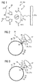
- Recombinant bacteriophage proteins 12 which allow the adsorption of a bacteriophage 16 to a bacterial surface 18, are now produced by protein expression. These bind specifically to the bacterial surface 18 via protein-protein interactions and can be used both as an enrichment structure 19 for enrichment and for detection of bacteria 10.
- Fig. 1 shows example of a coating via polyhistidine tag 28 as a linker structure 31 n (Source: http://www.chemgapedia.de)
- the bacteriophage proteins 12 After immobilization of the materials by the bacteriophage proteins 12, they may be used to specifically enrich the bacteria 10 for the materials described above. As already mentioned, the accumulation of the bacteria takes place via the specific protein-protein interaction between bacteriophage protein 12 on one side and bacterial surface protein 32 on the other side.
- Fig. 2 shows an enrichment via magnetic or non-magnetic metal chelate beads 34, in particular nickel or cobalt ions, via a magnetic or non-magnetic metal chelate bead 34, a metal chelate 36, a polyhistidine tag 28 (His tag or 6 ⁇ His- Tag) and a bacteriophage protein 12 a bacterium 10 or only a bacterial surface protein 32 of a bacterial family 37 is immobilized.
- a magnetic or non-magnetic metal chelate beads 34 in particular nickel or cobalt ions
- Fig. 3 shows an accumulation by means of magnetic 20 or non-magnetic beads 30 or sepharose beads 38 by biotin 22-streptavidin 24 interaction, wherein magnetic 20 or non-magnetic beads 30 or sepharose beads 38, streptavidin 24, biotin 22 and a bacteriophage protein 12 a bacterium 10 or only a bacterial surface protein 32 is immobilized.
- Fig. 4 shows an immobilization of the bacteriophage proteins 12 via biotin 22 - streptavidin 24 on material surfaces 40, in particular microtiter plates 31c, semiconductors 31 m, glass 31f, metal 31g, wherein on material surfaces 40 of all kinds on streptavidin 24, biotin 22 and a bacteriophage protein 12 a bacterium10 or only a bacterial surface protein 32 is immobilized.
- Fig. 5 shows an immobilization of the bacteriophage proteins 12 via metal chelates 36 to material surfaces 40, in particular to microtiter plates 31c, semiconductors 31 m, glass 31f or metal 31 g, wherein on material surfaces 40 of all kinds via a metal chelate 36, a polyhistidine tag 28 (His-tag or 6 x His-tag) and a bacteriophage protein 12 a bacterium 10 or only a bacterial surface protein 32 is immobilized.
- a polyhistidine tag 28 His-tag or 6 x His-tag
- the bacteriophage proteins 12 can be used in combination with an antibody 14.
- an antibody 14 it would be possible to use a bifurcated antibody 14 as detection protein 42 after the capture (capture), such as in Fig. 6 shown.
- An immobilized antibody 14 could act as a scavenger, while a (labeled) bacteriophage protein 12 labeled directly on the detection protein surface 43 would be used as a detection protein 42, such as in U.S. Pat Figs. 7 and 8 shown.
- Fig. 6 shows a schematic representation of such an immunological detection of a labeled antibody 14 in the form of a "hybrid phage immunoassay (HYPIA)" after an enrichment by bacteriophage proteins 12, wherein a material surface 40 of all kinds on streptavidin 24, biotin 22 and a Bacteriophage protein 12 is a bacterium 10 or bacterial
- Surface protein 32 was immobilized and a monoclonal or polyclonal antibody 14, which has on the surface protein of the bacterium 10 and a label 44 or a label in the form of a marker molecule 45, a detection by an enzyme or fluorescence (eg HRP or fluorescein) can be performed ,
- Figs. 7 and 8 12 show schematic representations of a detection or an assay via labeled (labeled) bacteriophage proteins 12 after enrichment by antibody 14 as a "hybrid phage immunoassay (HYPIA)", using material surfaces 40 of all kinds, via protein A 46 or protein G 48 or via streptavidin 24 and biotin 22, and an antibody 14 a bacterium 10 or only a bacterial surface protein 32 is immobilized and then a bacteriophage protein 12 and a label 44 or label of the bacteriophage protein 12, the detection of an enzyme or fluorescence (eg B HRP or fluorescein) can be performed as a fluorescent molecule 49.
- an enzyme or fluorescence eg B HRP or fluorescein
- the receptor proteins of bacteriophage 16 can be used not only as so-called “capture proteins” (capture protein), but also as detection proteins 42.
- the recombinant bacteriophage proteins 12 can after their expression and purification with a classical detection molecule or enzyme 52 (labeled with HRP, phosphatase, peroxidase, fluorophores such as CFDA).
- a classical detection molecule or enzyme 52 labeled with HRP, phosphatase, peroxidase, fluorophores such as CFDA.
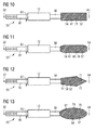
- Fig. 9 schematically shows representations of a fusion protein 50 consisting of bacteriophage protein 12 (capture protein, capture protein) and a Labeling protein or detector protein 54, with the following sequence from N-terminus 62 to C-terminus 64: Proetin tag, in particular a poly-histidine tag (His tag or 6 ⁇ His tag), protease recognition sequence 66, protease cleavage 60 , Bacteriophage protein 12 (capture protein or capture protein), linker 56 or loop, labeling protein or detector protein 54.
- Proetin tag in particular a poly-histidine tag (His tag or 6 ⁇ His tag)
- protease recognition sequence 66 protease cleavage 60
- Bacteriophage protein 12 capture protein or capture protein
- linker 56 or loop labeling protein or detector protein 54.
- bacteriophage protein 12 capture protein, capture protein
- any known bacteriophage 16 adsorption protein can be used.
- a bacteriophage protein 12 either a bacterium 10 can be detected extremely selectively (eg E. coli) or there is the possibility that a whole bacterial family 37 (eg coliforms) is sensitively detected.
- a known example is the bacteriophage M13 G3P protein which binds to tolA protein 84 of E. coli, as described in detail below.
- the following table shows the G3P protein with its closely related proteins, all of which could be used as a capture protein.
- Assessions no. protein name organism amino acids in length P69169 Attachment protein G3P (G3P) (Minor coat protein) Enterobacteria phage f1 (bacteriophage f1) 424 P03661 Attachment protein G3P (G3P) (Minor coat protein) Enterobacteria phage fd (bacteriophage fd) 424 P15415 Attachment protein G3P (G3P) (Minor coat protein) Enterobacteria phage 12-2 (bacteriophage 12-2) 434 080297 Attachment protein G3P (G3P) (Minor coat protein) Enterobacteria phage If1 (bacteriophage If1) 460 P03663 Attachment protein G3P (G3P) (Minor coat protein) Enterobacteria phage Ike (bacteriophage IKe) 434 P69168 Attachment protein G3P (
- Figs. 10 to 13 show schematic representations of fusion proteins 50 with different detector proteins 54, namely in Fig. 10 a fluorescent protein 72, in particular the green fluorescent protein (GFP), in Fig. 11 a human caspase 68, in particular caspase-3 70, in Fig. 12 a bioluminescence-based enzyme 52, especially luciferase 71, in Fig. 13 a polymerase (DNA and RNA) 74, in particular Taq polymerase 75.
- a fluorescent protein 72 in particular the green fluorescent protein (GFP)
- GFP green fluorescent protein
- Fig. 11 a human caspase 68
- caspase-3 70 in Fig. 12
- bioluminescence-based enzyme 52 especially luciferase 71
- Fig. 13 a polymerase (DNA and RNA) 74, in particular Taq polymerase 75.
- fusion proteins 50 can be very different assemble different assays.
- the assays are similar in structure to tests with conventionally labeled (labeled) bacteriophage proteins 12 in Figs. 7 and 8 ,
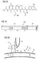
- Figs. 14 and 15 show schematic representations of a detection or an assay with a fusion protein 50 (bacteriophage protein 12 and detector protein 54) after an enrichment by antibodies 14, wherein on a material surface 40 of all kinds on protein A 46 or protein G 48 and an antibody 14, a bacterium 10 or only one bacterial surface protein 32 was immobilized.
- Various fusion proteins 50 are attached to the opposite side of the bacterium 10, having marker proteins 54 in the form of human caspases 68, in particular caspase-3 70, polymerases 74 (DNA and RNA), in particular tag polymerase 75, fluorescent proteins 72, in particular the green fluorescent protein (GFP) and bioluminescence-based enzymes 52, in particular luciferase 71.
- bacteriophage proteins 12 as fusion proteins 50 in combination with lytic proteins 76 will now be described.
- Fig. 16 shows a schematic representation of a fusion protein 50, consisting of a bacteriophage protein 12 (capture protein, capture protein) and a lytic protein 76 (eg., Lysozyme or lysostaphin) and a protein tag, in particular a poly-histidine tag (His-tag or 6 x His tag), a protease recognition sequence 66, a protease cleavage site 60, a linker 56 or loop, and a lytic protein 76.
- a bacteriophage protein 12 capture protein, capture protein
- a lytic protein 76 eg., Lysozyme or lysostaphin
- a protein tag in particular a poly-histidine tag (His-tag or 6 x His tag), a protease recognition sequence 66, a protease cleavage site 60, a linker 56 or loop, and a lytic protein 76.
- a polyclonal antibody 14 is a finite resource because the immunized animal dies at some point while the production of recombinant proteins is an endless resource.
- HYPIA hybrid phage immunoassay
- recombinant His tag protein provides an advantage in terms of assay development. Since the recombinant protein already on the beads (Ni-NTA 31, or magnetic ni-beads 26) is bound via the His tag, these can be used immediately for enrichment or assay development. It is therefore no longer necessary to elute the recombinant protein from the beads. Even with nickel ion coated microtiter plates 31 c or other vessels can be used for cleaning.
- fusion proteins 50 (bacteriophage protein 12 and detector protein 54, e.g., GFP)
- detector protein 54 e.g., GFP
- fluorophores are bound, respectively, which has a large impact on the sensitivity of the assay.
- conventional labeling is not 100% marking. Often only a fraction of the protein to be labeled is labeled (sometimes only 10%). This incomplete labeling can be virtually ruled out in the production of recombinant fusion proteins 50 (only one bacteriophage protein 12 and one reporter protein).
- human caspase-3 70 as a reporter enzyme has very great advantages since it is a purely human enzyme 52 and does not occur in the bacteria 10 to be detected.
- the enzymatic reaction is based on a proteolytic cleavage with the following amino acid recognition sequence: DEVDIX. This recognition sequence is extremely rare in bacterial proteins, so that their proteolytic cleavage is virtually eliminated.
- fluorescence substrates are N-acetyl-Asp-Glu-Vai-Asp-7-amido-4-methyl coumarin and N-acetyl-Asp-Gln-Met-Asp-7-amido-4-trifluoromethyl coumarin.
- tag polymerase 75 as a reporter enzyme in the form of a fusion protein 50 with a bacteriophage protein 12 as a "capture protein" results in the advantage of double specificity.
- the protein-protein interaction between bacteriophage protein 12 and bacterial surface protein 32 is highly specific, and second, the subsequent detection assay, in this case specific PCR, is also highly sensitive and specific detection. This dual specificity of identification enhances the safety of the assay so that false-positive results are virtually eliminated.
- This fusion protein 50 can also be used in a real-time PCR.
- the bacteriophage protein 12 is anyway a much more stable macromolecule than an antibody 14, which is composed of several fragments (light and heavy chain). Furthermore, it is possible to specifically modify the bacteriophage protein 12 by "protein engineering", so that the stability of the bacteriophage protein 12 can be significantly increased.
- the underlying step here is the adsorption of the bacteriophage 16 on the surface structure of the bacterium 10. This adsorption takes place over a very specific protein-protein interaction between virus and host cell. As a practical example here serves the bacteriophage M13 and the bacterium E. coli.
- the g3p gene is 1275 base pairs (bp) long and encodes a 424 amino acid (AS) protein.
- the G3P in the N-terminal region consists of four domains, which, as in Fig. 19 are respectively denoted by N1, L1, N2 and L2, while the C-terminal portion is characterized by a short transmembrane domain TD.
- the interaction of the bacteriophage 16 is well described in the literature and occurs first with the N2 domain which binds to the F pilus 82 of E.
- the N1 domain of bacteriophage 16 then interacts with the bacterial tolA protein 84 located in the periplasmic space 86 between the outer membrane 83 and the cytoplasmic membrane 88. After absorption via the two domains, the actual infection of the bacterium 10 by the bacteriophage 16 is then carried out by the virus injecting its genetic material (DNA) into the host cell.
- DNA genetic material
- Fig. 19 schematically shows the G3P protein with its N-terminal domains N1, L1, N2 and L2. C-terminal localized is the transmembrane domain TD.
- Fig. 20 shows schematically a representation of the absorption of bacteriophage M13 to the bacterium E. coli.
- the following describes the cloning strategy of g3p constructs into a T7-based expression vector.
- the PCR products were serially digested with the restriction enzymes Acc651 and BsmBI and ligated into a T7-based expression vector previously opened with the restriction enzymes Ncol and Acc65I, as described in U.S. Pat Fig. 26 shown.
- the transformation into E. coli NEB 5-alpha was carried out by electroporation, whereby the ligations were rendered salt-free by a butanol precipitation. There was a selection on LB-kanamycin culture media (final concentration of about 50 ug / ml kanamycin).
- the transformants were subjected to a DNA mini-plasmid preparation, after which the prepared plasmid DNA was checked by a restriction digest and by PCR for their accuracy with respect to the g3p-fragments.
- the following table shows the primers or oligonucleotides used with their restriction sites, which were used for the amplification of the five different gene fragments: fragment direction Surname sequence all fragments F g3p / Bsmbl / fw N1 R N1 / S / Ac / rv N1L1 R N1L1 / S / Ac / rv N1L1N2 R N1L1N2 / S / Ac / rv N1L2N2L2 R N1L1N2L2 / S / Ac / rv N1L2N2L2-C R g3p / OT / S / Ac / rv
- Fig. 27 shows the result of an electrophoresis of an agarose gel showing the PCR products as well as the cloning products of the g3p fragments N1, N1L1 and N1 L2N2.
- the first three columns from the left show the PCR products N1 (255bp), N1L1 (315bp) and N1 L2N2 (705bp), the fourth column from the left the positive control, the seventh to ninth columns from the left the alloy PCR products N1, N1L1s and N1 L2N2 with the T7-based expression vector and the eleventh column from the left the DNA marker M.
- the finished constructs (g3p domains in T7 expression vector) were transformed into calcium-competent E. coli BL21 (DE3) and then selected on LB-kanamycin culture media (final concentration of about 50 ⁇ g / ml medium). For the actual protein expression, more colonies of the transformants were taken from the selective plates and a preculture of 5-10 ml of LB and kanamycin 50 ⁇ g / ml inoculated. After incubation in the shaker (200 rpm) overnight at 37 ° C., the main culture (100 ml-1000 ml LB and kanamycin 50 ⁇ g / ml) was inoculated with the preculture.
- the main culture should have an optical density at 600 nm (OD 600) of about 0.05. There is then a logarithmic growth up to an OD 600 of 0.5-0.8. Upon reaching this final OD 600, cells 81 are induced with a 1 M isopropyl- ⁇ -D-thiogalactopyranoside (IPTG) solution at a dilution of 1: 1000. After induction, the temperature was immediately lowered from 37 ° C to 20-15 ° C and there was an expression time of 4 h to 24 h.
- IPTG isopropyl- ⁇ -D-thiogalactopyranoside
- a suitable lysis buffer (20 mM Tris HCl pH 8.0, 150 mM NaCl, 0.2% 10 mM NP-40 imidazole) and stored at -20 ° C to -80 ° C.
- a protease inhibitor eg Complete EDTA free
- whole cells 81 were lysed in SDS sample buffer before and after expression, and the lysate was electrophoresed in a denaturing 15% SDS gel (SDS-PAGE).
- Fig. 28 shows the verification of the expression of the G3P domains (N1, N1L1 and N1 L2N2) in a denaturing 15% SDS gel (SDS-PAGE), wherein - uninduced cells, + induced cells and M denotes the protein marker in kDa.
- the arrows indicate the recombinant protein of the G3P domains N1, N1L1 and N1 L2N2.
- the second to fourth columns from the right contain negative controls.
- the thus purified recombinant G3P domains are eluted with a single or 1 ⁇ 2-fold bed volume of elution buffer (20 mM Tris, HCl pH 8.0, 150 mM NaCl, 300 mM imidazole) from the nickel chelate column.
- elution buffer 20 mM Tris, HCl pH 8.0, 150 mM NaCl, 300 mM imidazole
- Purification of the recombinant His tag protein provides an advantage in terms of assay development. Since the recombinant protein is already bound to the beads (Ni-NTA 31, or magnetic Ni beads 26) via the His tag, they can be used immediately for enrichment or assay development. It is therefore no longer necessary to elute the recombinant protein from the beads.
- Fig. 29 shows the review of the purified recombinant G3P domains for their solubility and purity.
- Columns E1-E3 show the respective elution 1 to 3 of the nickel chelate.
- N1 is insoluble as the smallest G3P domain, no specific protein band is recognizable.
- N1L1 as a medium G3P domain is soluble, it is clearly a protein band at about 14.4 kDa recognizable.
- N1 L1 N2 as the largest G3P domain is soluble, it is clearly a protein band at about 35 kDa recognizable.
- the reporter genes were amplified from existing vectors using the PCR technique.
- the following primers were used for the amplification: reporter direction Surname sequence EmGFP F EGFP / Sall / fw EmGFP R EGFP / NotI / rv Caspase-3 F CASP3 / Sall / fw Caspase-3 R CASP3 / NotI / rv luciferase F Luci / Sall / fw luciferase R Luci / NotI / rv
- the cloning of the two genes into a fusion protein gene 91 was carried out serially, the g3p domains being ligated first as bacteriophage protein gene 92, followed by the reporter genes as marker protein gene 94.
- the g3p gene is N-terminal and the reporter gene C-terminal with a linker 54 as a linker protein gene 96 of nine codons 98 between both genes in a T7-based expression vector, as in Fig. 30 which shows a representation of the linker region between the N-terminal g3p domain N1L1N2 and the C-terminal reporter gene EmGFP. Between both genes is a linker 54 with nine codons 98. Shown with a vertical arrow is the start codon 100 (ATG) of the reporter gene EmGFP.
- Another aspect in the generation of a fusion protein 50 is the fact that both genes are preferably in the same reading frame and that preferably no stop codon is present in the linker 54 between the two genes.
- the entire construct is then transformed into E. coli NEB 5-alpha by electroporation after butanol precipitation, and cells 81 are selected on nutrient media containing 50 ⁇ g / ml kanamycin.
- the transformants obtained are examined for the existence of the g3p domains as well as those of the reporter genes after a DNA miniplasmid preparation. This analysis of the clones for correctness is carried out both with PCR and with a restriction digest.
- the expression of the fusion proteins 50 takes place under the same conditions as before the pure G3P domains.
- the EmGFP as a reporter protein, it is already possible to directly observe the success of the expression. Due to basal expression, the cells fluoresce 81 or colonies on the culture plates when exposed to UV light of 312 nm, as in Fig. 31 which shows a check of the expression of G3P-N1L1N2 and EmGFP as fusion protein 50 on a UV table at a wavelength of 312 nm.
- To the right are colonies and one negative control of a pure G3P domain (N1L1N2) without EmGFP, left colonies of basal expression of the fusion protein N1 L1 N2-EmGFP; the colonies appear bright.
- the following describes the solubility test and purification of the fusion protein 50 consisting of g3p domains and reporter enzymes EmGFP.
- the solubility test and the purification of the fusion proteins 50 were carried out according to the same protocol as the pure G3P domains.
- the solubility of the fusion protein 50 was analyzed in a denaturing 15% SDS gel (SDS-PAGE) as described in U.S. Pat FIGS. 33 and 34 shown.
- FIG. 33 Figure 1 shows the verification of solubility test and purification of the fusion protein N1L1N2-EmGFP in the 15% SDS gel (SDS-PAGE), where E denotes the elution of the fusion protein N1L1N2-EmGFP from the nickel chelate and M the protein marker in kDA 175 to 17 kDa.
- the arrow marks the protein band of the fusion protein N1 L1 N2-EmGFP with about 60 kDa.
- Fig. 34 shows the verification of solubility test and purification of the fusion proteins N1L1N2-caspase-3 and N1L1-caspase-3 in the 15% SDS gel (SDS-PAGE).
- E1 indicates the first elution of the fusion protein N1L1N2-caspase-3 from the nickel chelate
- E2 indicates the second elution of the fusion protein N1L1N2-caspase-3 from the nickel chelate
- E3 indicates the first elution of the fusion protein N1 L1-caspase-3 from the nickel chelate
- E4 denotes the second elution of the fusion protein N1 L1-caspase-3 from the nickel chelate
- M denotes the protein marker in kDA 175 to 17 kDa
- the arrows mark the protein bands of the fusion proteins N1 L1 N2 caspase3 (ca. 42kDa) and N1L1-Caspase3 (17kDa).
- the longest G3P domain N1L1N2L2-C was successfully expressed in E. coli BL21 (DE3). Based on this fact, this recombined protein was used for binding studies in the form of a pull-down or pull-down assay.
- the pull-down experiment is a test in which a binding between two proteins (protein-protein interaction) is detected. In the present results, the binding or the protein-protein interaction between the G3P protein (bacteriophage protein 12) and the tolA protein 84 (E. coli) is shown.
- the principle of the pull-down assay is based on the affinity of the bait molecule (E.
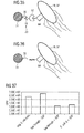
- Figs. 35 and 36 each show a schematic representation of a pull-down experiment to demonstrate the protein-protein interaction between the bacteriophage protein 12 G3P and the bacterial surface protein 32 tolA.
- Fig. 35 the binding between G3P and tolA is shown using Ni-NTA 31 as a matrix to which Ni chelate and Ni ions have the His tag of G3P, the bacteriophage protein 12 G3P or a G3P domain, via a protein Protein interaction between G3P and tolA is an E. coli surface protein tolA and thus an E. coli bound with peritrichic flagella.
- Fig. 36 shows a negative control without the bacteriophage protein 12 G3P, where no binding to the Ni-NTA matrix is observed.
- FIG. 37 Figure 4 shows a graph of the results of the pull-down experiment to demonstrate the protein-protein interaction between the bacteriophage protein 12 G3P and the bacterial protein tolA.
- the passage of a defined E. coli concentration of 2.4E + 06 was analyzed (first bar from the left).
- the second bar from the left shows a negative control, ie the passage of the bacterial concentration on the matrix without G3P (2,8E + 06).
- the third bar from the left shows the bacterial concentration after passing over the matrix with G3P (1,1E + 06).
- the fourth bar from the left shows the bacterial concentration that has adhered to the matrix (1.3E + 06).
Landscapes
- Health & Medical Sciences (AREA)
- Life Sciences & Earth Sciences (AREA)
- Engineering & Computer Science (AREA)
- Biomedical Technology (AREA)
- Chemical & Material Sciences (AREA)
- Immunology (AREA)
- Molecular Biology (AREA)
- Genetics & Genomics (AREA)
- Hematology (AREA)
- Biotechnology (AREA)
- Urology & Nephrology (AREA)
- Physics & Mathematics (AREA)
- Microbiology (AREA)
- General Health & Medical Sciences (AREA)
- Biochemistry (AREA)
- Cell Biology (AREA)
- General Engineering & Computer Science (AREA)
- Analytical Chemistry (AREA)
- Food Science & Technology (AREA)
- Organic Chemistry (AREA)
- General Physics & Mathematics (AREA)
- Pathology (AREA)
- Zoology (AREA)
- Wood Science & Technology (AREA)
- Medicinal Chemistry (AREA)
- Bioinformatics & Cheminformatics (AREA)
- Plant Pathology (AREA)
- Biophysics (AREA)
- Tropical Medicine & Parasitology (AREA)
- Virology (AREA)
- Measuring Or Testing Involving Enzymes Or Micro-Organisms (AREA)
- Peptides Or Proteins (AREA)
Applications Claiming Priority (1)
| Application Number | Priority Date | Filing Date | Title |
|---|---|---|---|
| DE102013106462.0A DE102013106462B3 (de) | 2013-06-20 | 2013-06-20 | Detektionsverfahren zur Detektion von Bakterien, Verfahren zur Herstellung von Fusionsproteinen und Fusionsprotein |
Publications (4)
| Publication Number | Publication Date |
|---|---|
| EP2816355A2 true EP2816355A2 (fr) | 2014-12-24 |
| EP2816355A3 EP2816355A3 (fr) | 2015-04-15 |
| EP2816355B1 EP2816355B1 (fr) | 2017-08-09 |
| EP2816355B8 EP2816355B8 (fr) | 2017-11-08 |
Family
ID=50976526
Family Applications (1)
| Application Number | Title | Priority Date | Filing Date |
|---|---|---|---|
| EP14173356.8A Active EP2816355B8 (fr) | 2013-06-20 | 2014-06-20 | Procédé de détection de bactéries, procédé de fabrication de protéines de fusion et protéine de fusion |
Country Status (2)
| Country | Link |
|---|---|
| EP (1) | EP2816355B8 (fr) |
| DE (1) | DE102013106462B3 (fr) |
Cited By (3)
| Publication number | Priority date | Publication date | Assignee | Title |
|---|---|---|---|---|
| CN110699241A (zh) * | 2019-10-28 | 2020-01-17 | 军事科学院系统工程研究院卫勤保障技术研究所 | 一种自动化消毒效果快速评价装置及方法 |
| CN114814200A (zh) * | 2022-05-09 | 2022-07-29 | 浙江大学 | 一种纳米颗粒-荧光标记融合蛋白复合体及其单链dna检测方法 |
| CN115282934A (zh) * | 2022-08-02 | 2022-11-04 | 杭州博岳生物技术有限公司 | 一种纳米抗体荧光亲和柱以及利用该亲和柱纯化HbeAg抗原的方法 |
Families Citing this family (2)
| Publication number | Priority date | Publication date | Assignee | Title |
|---|---|---|---|---|
| DE102015121034B4 (de) | 2015-12-03 | 2022-06-23 | Airbus Defence and Space GmbH | Verfahren und Vorrichtung zur Anreicherung von biologischen Partikeln |
| DE102015121035A1 (de) | 2015-12-03 | 2017-06-08 | Airbus Defence and Space GmbH | Verfahren zur Detektion coliformer Keime |
Family Cites Families (12)
| Publication number | Priority date | Publication date | Assignee | Title |
|---|---|---|---|---|
| ES2341926T3 (es) * | 1998-03-02 | 2010-06-29 | Massachusetts Institute Of Technology | Poliproteinas con dedos de cinc que tienen enlazadores mejorados. |
| DE19837751A1 (de) * | 1998-08-20 | 2000-02-24 | Siegfried Scherer | Markierung, Immobilisierung, Anreicherung, Reinigung und Nachweis von Zellen mittels der Verwendung spezifischer Zellwand-bindender Domänen (CBD) von Zellwand-bindenden Proteinen aus Viren, Bakterien oder eukaryontischen Zellen |
| WO2001002588A2 (fr) * | 1999-07-02 | 2001-01-11 | Morphosys Ag | Production de partenaires de liaison specifiques se liant a des (poly)peptides codes par des fragments d'adn genomiques ou est |
| CA2380480C (fr) * | 1999-07-30 | 2010-10-26 | Profos Ag | Mise en evidence et identification de souches bacteriennes |
| DE10129815A1 (de) * | 2001-06-24 | 2003-01-09 | Profos Ag | Verfahren zur Aufreinigung von Bakterienzellen und Zellbestandteilen |
| US20040197833A1 (en) * | 2003-04-03 | 2004-10-07 | Danisco A/S | Method for the enrichment of target cells by use of CBDs |
| US7329725B1 (en) * | 2003-10-29 | 2008-02-12 | Nastech Pharmaceutical Company Inc. | Phage displayed Trp cage ligands |
| CA2576195A1 (fr) * | 2004-08-05 | 2006-02-16 | Biosite Incorporated | Compositions et methodes d'expression a la surface des phages de polypeptides |
| DE102006009709A1 (de) * | 2006-03-02 | 2006-10-12 | Müller, Merold, Dr. | Mikromechanisches Nachweisverfahren für Mikroorganismen, Bakterien, Wirtszellen, Bakteriophagen oder Viren, geeignet auch für biologische Kampfstoffe und zur Abwehr bioterroristischer Gefährdung |
| JP5513398B2 (ja) * | 2007-11-02 | 2014-06-04 | ザ スクリプス リサーチ インスティチュート | 非天然アミノ酸を含有する蛋白質を使用する指向的進化 |
| US20100113304A1 (en) * | 2008-09-26 | 2010-05-06 | Wyeth | Compatible display vector systems |
| GB0903316D0 (en) * | 2009-02-26 | 2009-04-08 | Affitech As | Method |
-
2013
- 2013-06-20 DE DE102013106462.0A patent/DE102013106462B3/de not_active Expired - Fee Related
-
2014
- 2014-06-20 EP EP14173356.8A patent/EP2816355B8/fr active Active
Non-Patent Citations (1)
| Title |
|---|
| None |
Cited By (3)
| Publication number | Priority date | Publication date | Assignee | Title |
|---|---|---|---|---|
| CN110699241A (zh) * | 2019-10-28 | 2020-01-17 | 军事科学院系统工程研究院卫勤保障技术研究所 | 一种自动化消毒效果快速评价装置及方法 |
| CN114814200A (zh) * | 2022-05-09 | 2022-07-29 | 浙江大学 | 一种纳米颗粒-荧光标记融合蛋白复合体及其单链dna检测方法 |
| CN115282934A (zh) * | 2022-08-02 | 2022-11-04 | 杭州博岳生物技术有限公司 | 一种纳米抗体荧光亲和柱以及利用该亲和柱纯化HbeAg抗原的方法 |
Also Published As
| Publication number | Publication date |
|---|---|
| EP2816355B8 (fr) | 2017-11-08 |
| DE102013106462B3 (de) | 2014-10-09 |
| EP2816355A3 (fr) | 2015-04-15 |
| EP2816355B1 (fr) | 2017-08-09 |
Similar Documents
| Publication | Publication Date | Title |
|---|---|---|
| EP1675623B1 (fr) | Conjugues d'ubiquitine ou de gamma-crystalline et leur utilisation pour la therapie, le diagnostic et la chromatographie | |
| Tanaka et al. | Site‐specific protein modification on living cells catalyzed by sortase | |
| EP2816355B1 (fr) | Procédé de détection de bactéries, procédé de fabrication de protéines de fusion et protéine de fusion | |
| EP1844150B1 (fr) | Expression recombinee de proteines a forme bicatenaire a pont disulfure | |
| DE60305643T2 (de) | Protein-marker der eine biotinylierungsdomaine enthält und verwendung zur erhöhung der löslichkeit und zur ermittlung vom faltungsstatus | |
| DE112006003608T5 (de) | Verfahren zur Präsentation von Zielproteinen an der Zelloberfläche unter Verwendung von Bacillus Anthracis-Exosporium | |
| EP1356080B1 (fr) | Detection et identification de groupes de bacteries | |
| DE69333667T2 (de) | Kontrollierbare Zwischensequenzen enthaltende modifizierte Proteine und Verfahren zur deren Herstellung | |
| EP1619208B1 (fr) | Complexe de chaperonine/proteine cible, son procede de production, procede de stabilisation de proteine cible, procede d'immobilisation de proteine cible, procede d'analyse de la structure de proteine cible, preparation a liberation prolongee et procede de production d'anticorps contre une proteine | |
| Imani et al. | Recombinant production and affinity purification of the FraC pore forming toxin using hexa-His tag and pET expression cassette | |
| DE202019005825U1 (de) | Biologische Synthese von Aminosäureketten zur Herstellung von Peptiden und Proteinen | |
| EP0547200A1 (fr) | Anticorps recombinants a la surface d'e. coli | |
| CA2785359C (fr) | Mise en evidence de proteines | |
| EP1147419B1 (fr) | Marquage, immobilisation, enrichissement, purification et detection de cellules a l'aide de l'utilisation de domaines specifiques se liant a des parois cellulaires (cbd) de proteines se liant a des parois cellulaires tirees de virus, bacteries ou cellules eucaryotes | |
| Pinheiro et al. | Fusion proteins towards fungi and bacteria in plant protection | |
| Choudary et al. | Versatile substrates and probes for IgA1 protease activity | |
| Sudheer et al. | Cyclization tag for the detection and facile purification of backbone-cyclized proteins | |
| Hosseini et al. | Optimization and one-step purification of recombinant V antigen production from Yersinia pestis | |
| Walter et al. | Characteristics of the surface-located carbohydrate-binding protein CbpC from Streptomyces coelicolor A3 (2) | |
| DE69937453T2 (de) | Verfahren zur isolierung und charakterisierung potentieller funktionen ausgehend von einer biologischen probe, welche nukleinsäuren enthält | |
| AT511130A2 (de) | Polypeptidmaterial mit flexiblen Poreneigenschaften | |
| KR101896014B1 (ko) | 바실러스 세레우스 포자에 특이적인 결합을 보이는 박테리오파지 유래 포자 결합 도메인 및 이 도메인을 이용한 검출용 매개체 | |
| DE102013013609A1 (de) | Autodisplay einer aktiven Lipase aus Burkholderia cepacia auf Mikroorganismen | |
| EP1069136B1 (fr) | Procédé de production recombinante de ribonucléoproteins | |
| EP2847334B1 (fr) | Utilisation de corps à forisomes artificiels pour l'immobilisation d'enzymes ou la purification de protéines récombinantes |
Legal Events
| Date | Code | Title | Description |
|---|---|---|---|
| PUAI | Public reference made under article 153(3) epc to a published international application that has entered the european phase |
Free format text: ORIGINAL CODE: 0009012 |
|
| 17P | Request for examination filed |
Effective date: 20140620 |
|
| AK | Designated contracting states |
Kind code of ref document: A2 Designated state(s): AL AT BE BG CH CY CZ DE DK EE ES FI FR GB GR HR HU IE IS IT LI LT LU LV MC MK MT NL NO PL PT RO RS SE SI SK SM TR |
|
| AX | Request for extension of the european patent |
Extension state: BA ME |
|
| RIC1 | Information provided on ipc code assigned before grant |
Ipc: C07K 14/005 20060101ALI20141121BHEP Ipc: C07K 14/00 20060101ALI20141121BHEP Ipc: G01N 33/566 20060101ALI20141121BHEP Ipc: C12N 15/62 20060101ALI20141121BHEP Ipc: G01N 33/569 20060101AFI20141121BHEP |
|
| PUAL | Search report despatched |
Free format text: ORIGINAL CODE: 0009013 |
|
| AK | Designated contracting states |
Kind code of ref document: A3 Designated state(s): AL AT BE BG CH CY CZ DE DK EE ES FI FR GB GR HR HU IE IS IT LI LT LU LV MC MK MT NL NO PL PT RO RS SE SI SK SM TR |
|
| AX | Request for extension of the european patent |
Extension state: BA ME |
|
| RIC1 | Information provided on ipc code assigned before grant |
Ipc: C12N 15/62 20060101ALI20150312BHEP Ipc: G01N 33/569 20060101AFI20150312BHEP Ipc: G01N 33/566 20060101ALI20150312BHEP Ipc: C07K 14/005 20060101ALI20150312BHEP Ipc: C07K 14/00 20060101ALI20150312BHEP |
|
| R17P | Request for examination filed (corrected) |
Effective date: 20151009 |
|
| RBV | Designated contracting states (corrected) |
Designated state(s): AL AT BE BG CH CY CZ DE DK EE ES FI FR GB GR HR HU IE IS IT LI LT LU LV MC MK MT NL NO PL PT RO RS SE SI SK SM TR |
|
| 17Q | First examination report despatched |
Effective date: 20160408 |
|
| RIN1 | Information on inventor provided before grant (corrected) |
Inventor name: REIDT, ULRICH Inventor name: HUMMEL, THOMAS Inventor name: FRIEDBERGER, ALOIS |
|
| GRAP | Despatch of communication of intention to grant a patent |
Free format text: ORIGINAL CODE: EPIDOSNIGR1 |
|
| INTG | Intention to grant announced |
Effective date: 20170209 |
|
| GRAS | Grant fee paid |
Free format text: ORIGINAL CODE: EPIDOSNIGR3 |
|
| GRAA | (expected) grant |
Free format text: ORIGINAL CODE: 0009210 |
|
| AK | Designated contracting states |
Kind code of ref document: B1 Designated state(s): AL AT BE BG CH CY CZ DE DK EE ES FI FR GB GR HR HU IE IS IT LI LT LU LV MC MK MT NL NO PL PT RO RS SE SI SK SM TR |
|
| REG | Reference to a national code |
Ref country code: GB Ref legal event code: FG4D Free format text: NOT ENGLISH |
|
| REG | Reference to a national code |
Ref country code: CH Ref legal event code: EP Ref country code: AT Ref legal event code: REF Ref document number: 917407 Country of ref document: AT Kind code of ref document: T Effective date: 20170815 |
|
| REG | Reference to a national code |
Ref country code: IE Ref legal event code: FG4D Free format text: LANGUAGE OF EP DOCUMENT: GERMAN |
|
| REG | Reference to a national code |
Ref country code: DE Ref legal event code: R096 Ref document number: 502014004893 Country of ref document: DE |
|
| RAP2 | Party data changed (patent owner data changed or rights of a patent transferred) |
Owner name: AIRBUS DEFENCE AND SPACE GMBH |
|
| REG | Reference to a national code |
Ref country code: NL Ref legal event code: FP |
|
| REG | Reference to a national code |
Ref country code: LT Ref legal event code: MG4D |
|
| PG25 | Lapsed in a contracting state [announced via postgrant information from national office to epo] |
Ref country code: NO Free format text: LAPSE BECAUSE OF FAILURE TO SUBMIT A TRANSLATION OF THE DESCRIPTION OR TO PAY THE FEE WITHIN THE PRESCRIBED TIME-LIMIT Effective date: 20171109 Ref country code: HR Free format text: LAPSE BECAUSE OF FAILURE TO SUBMIT A TRANSLATION OF THE DESCRIPTION OR TO PAY THE FEE WITHIN THE PRESCRIBED TIME-LIMIT Effective date: 20170809 Ref country code: LT Free format text: LAPSE BECAUSE OF FAILURE TO SUBMIT A TRANSLATION OF THE DESCRIPTION OR TO PAY THE FEE WITHIN THE PRESCRIBED TIME-LIMIT Effective date: 20170809 Ref country code: FI Free format text: LAPSE BECAUSE OF FAILURE TO SUBMIT A TRANSLATION OF THE DESCRIPTION OR TO PAY THE FEE WITHIN THE PRESCRIBED TIME-LIMIT Effective date: 20170809 Ref country code: SE Free format text: LAPSE BECAUSE OF FAILURE TO SUBMIT A TRANSLATION OF THE DESCRIPTION OR TO PAY THE FEE WITHIN THE PRESCRIBED TIME-LIMIT Effective date: 20170809 |
|
| PG25 | Lapsed in a contracting state [announced via postgrant information from national office to epo] |
Ref country code: RS Free format text: LAPSE BECAUSE OF FAILURE TO SUBMIT A TRANSLATION OF THE DESCRIPTION OR TO PAY THE FEE WITHIN THE PRESCRIBED TIME-LIMIT Effective date: 20170809 Ref country code: IS Free format text: LAPSE BECAUSE OF FAILURE TO SUBMIT A TRANSLATION OF THE DESCRIPTION OR TO PAY THE FEE WITHIN THE PRESCRIBED TIME-LIMIT Effective date: 20171209 Ref country code: LV Free format text: LAPSE BECAUSE OF FAILURE TO SUBMIT A TRANSLATION OF THE DESCRIPTION OR TO PAY THE FEE WITHIN THE PRESCRIBED TIME-LIMIT Effective date: 20170809 Ref country code: BG Free format text: LAPSE BECAUSE OF FAILURE TO SUBMIT A TRANSLATION OF THE DESCRIPTION OR TO PAY THE FEE WITHIN THE PRESCRIBED TIME-LIMIT Effective date: 20171109 Ref country code: GR Free format text: LAPSE BECAUSE OF FAILURE TO SUBMIT A TRANSLATION OF THE DESCRIPTION OR TO PAY THE FEE WITHIN THE PRESCRIBED TIME-LIMIT Effective date: 20171110 Ref country code: PL Free format text: LAPSE BECAUSE OF FAILURE TO SUBMIT A TRANSLATION OF THE DESCRIPTION OR TO PAY THE FEE WITHIN THE PRESCRIBED TIME-LIMIT Effective date: 20170809 Ref country code: ES Free format text: LAPSE BECAUSE OF FAILURE TO SUBMIT A TRANSLATION OF THE DESCRIPTION OR TO PAY THE FEE WITHIN THE PRESCRIBED TIME-LIMIT Effective date: 20170809 |
|
| PG25 | Lapsed in a contracting state [announced via postgrant information from national office to epo] |
Ref country code: DK Free format text: LAPSE BECAUSE OF FAILURE TO SUBMIT A TRANSLATION OF THE DESCRIPTION OR TO PAY THE FEE WITHIN THE PRESCRIBED TIME-LIMIT Effective date: 20170809 Ref country code: CZ Free format text: LAPSE BECAUSE OF FAILURE TO SUBMIT A TRANSLATION OF THE DESCRIPTION OR TO PAY THE FEE WITHIN THE PRESCRIBED TIME-LIMIT Effective date: 20170809 Ref country code: RO Free format text: LAPSE BECAUSE OF FAILURE TO SUBMIT A TRANSLATION OF THE DESCRIPTION OR TO PAY THE FEE WITHIN THE PRESCRIBED TIME-LIMIT Effective date: 20170809 |
|
| REG | Reference to a national code |
Ref country code: DE Ref legal event code: R097 Ref document number: 502014004893 Country of ref document: DE |
|
| REG | Reference to a national code |
Ref country code: DE Ref legal event code: R082 Ref document number: 502014004893 Country of ref document: DE |
|
| PG25 | Lapsed in a contracting state [announced via postgrant information from national office to epo] |
Ref country code: SK Free format text: LAPSE BECAUSE OF FAILURE TO SUBMIT A TRANSLATION OF THE DESCRIPTION OR TO PAY THE FEE WITHIN THE PRESCRIBED TIME-LIMIT Effective date: 20170809 Ref country code: IT Free format text: LAPSE BECAUSE OF FAILURE TO SUBMIT A TRANSLATION OF THE DESCRIPTION OR TO PAY THE FEE WITHIN THE PRESCRIBED TIME-LIMIT Effective date: 20170809 Ref country code: EE Free format text: LAPSE BECAUSE OF FAILURE TO SUBMIT A TRANSLATION OF THE DESCRIPTION OR TO PAY THE FEE WITHIN THE PRESCRIBED TIME-LIMIT Effective date: 20170809 Ref country code: SM Free format text: LAPSE BECAUSE OF FAILURE TO SUBMIT A TRANSLATION OF THE DESCRIPTION OR TO PAY THE FEE WITHIN THE PRESCRIBED TIME-LIMIT Effective date: 20170809 |
|
| PLBE | No opposition filed within time limit |
Free format text: ORIGINAL CODE: 0009261 |
|
| STAA | Information on the status of an ep patent application or granted ep patent |
Free format text: STATUS: NO OPPOSITION FILED WITHIN TIME LIMIT |
|
| REG | Reference to a national code |
Ref country code: FR Ref legal event code: PLFP Year of fee payment: 5 |
|
| 26N | No opposition filed |
Effective date: 20180511 |
|
| PG25 | Lapsed in a contracting state [announced via postgrant information from national office to epo] |
Ref country code: SI Free format text: LAPSE BECAUSE OF FAILURE TO SUBMIT A TRANSLATION OF THE DESCRIPTION OR TO PAY THE FEE WITHIN THE PRESCRIBED TIME-LIMIT Effective date: 20170809 |
|
| PG25 | Lapsed in a contracting state [announced via postgrant information from national office to epo] |
Ref country code: MT Free format text: LAPSE BECAUSE OF FAILURE TO SUBMIT A TRANSLATION OF THE DESCRIPTION OR TO PAY THE FEE WITHIN THE PRESCRIBED TIME-LIMIT Effective date: 20170809 |
|
| REG | Reference to a national code |
Ref country code: CH Ref legal event code: PL |
|
| REG | Reference to a national code |
Ref country code: BE Ref legal event code: MM Effective date: 20180630 |
|
| REG | Reference to a national code |
Ref country code: IE Ref legal event code: MM4A |
|
| PG25 | Lapsed in a contracting state [announced via postgrant information from national office to epo] |
Ref country code: MC Free format text: LAPSE BECAUSE OF FAILURE TO SUBMIT A TRANSLATION OF THE DESCRIPTION OR TO PAY THE FEE WITHIN THE PRESCRIBED TIME-LIMIT Effective date: 20170809 Ref country code: LU Free format text: LAPSE BECAUSE OF NON-PAYMENT OF DUE FEES Effective date: 20180620 |
|
| PG25 | Lapsed in a contracting state [announced via postgrant information from national office to epo] |
Ref country code: IE Free format text: LAPSE BECAUSE OF NON-PAYMENT OF DUE FEES Effective date: 20180620 Ref country code: LI Free format text: LAPSE BECAUSE OF NON-PAYMENT OF DUE FEES Effective date: 20180630 Ref country code: CH Free format text: LAPSE BECAUSE OF NON-PAYMENT OF DUE FEES Effective date: 20180630 |
|
| PG25 | Lapsed in a contracting state [announced via postgrant information from national office to epo] |
Ref country code: BE Free format text: LAPSE BECAUSE OF NON-PAYMENT OF DUE FEES Effective date: 20180630 |
|
| PG25 | Lapsed in a contracting state [announced via postgrant information from national office to epo] |
Ref country code: TR Free format text: LAPSE BECAUSE OF FAILURE TO SUBMIT A TRANSLATION OF THE DESCRIPTION OR TO PAY THE FEE WITHIN THE PRESCRIBED TIME-LIMIT Effective date: 20170809 |
|
| PG25 | Lapsed in a contracting state [announced via postgrant information from national office to epo] |
Ref country code: HU Free format text: LAPSE BECAUSE OF FAILURE TO SUBMIT A TRANSLATION OF THE DESCRIPTION OR TO PAY THE FEE WITHIN THE PRESCRIBED TIME-LIMIT; INVALID AB INITIO Effective date: 20140620 Ref country code: PT Free format text: LAPSE BECAUSE OF FAILURE TO SUBMIT A TRANSLATION OF THE DESCRIPTION OR TO PAY THE FEE WITHIN THE PRESCRIBED TIME-LIMIT Effective date: 20170809 |
|
| PG25 | Lapsed in a contracting state [announced via postgrant information from national office to epo] |
Ref country code: CY Free format text: LAPSE BECAUSE OF FAILURE TO SUBMIT A TRANSLATION OF THE DESCRIPTION OR TO PAY THE FEE WITHIN THE PRESCRIBED TIME-LIMIT Effective date: 20170809 Ref country code: MK Free format text: LAPSE BECAUSE OF NON-PAYMENT OF DUE FEES Effective date: 20170809 |
|
| PG25 | Lapsed in a contracting state [announced via postgrant information from national office to epo] |
Ref country code: AL Free format text: LAPSE BECAUSE OF FAILURE TO SUBMIT A TRANSLATION OF THE DESCRIPTION OR TO PAY THE FEE WITHIN THE PRESCRIBED TIME-LIMIT Effective date: 20170809 |
|
| REG | Reference to a national code |
Ref country code: AT Ref legal event code: MM01 Ref document number: 917407 Country of ref document: AT Kind code of ref document: T Effective date: 20190620 |
|
| PG25 | Lapsed in a contracting state [announced via postgrant information from national office to epo] |
Ref country code: AT Free format text: LAPSE BECAUSE OF NON-PAYMENT OF DUE FEES Effective date: 20190620 |
|
| PGFP | Annual fee paid to national office [announced via postgrant information from national office to epo] |
Ref country code: DE Payment date: 20250618 Year of fee payment: 12 |
|
| PGFP | Annual fee paid to national office [announced via postgrant information from national office to epo] |
Ref country code: GB Payment date: 20250618 Year of fee payment: 12 |
|
| PGFP | Annual fee paid to national office [announced via postgrant information from national office to epo] |
Ref country code: NL Payment date: 20250618 Year of fee payment: 12 |
|
| PGFP | Annual fee paid to national office [announced via postgrant information from national office to epo] |
Ref country code: FR Payment date: 20250624 Year of fee payment: 12 |