EP2827149B1 - Antigènes de mycobactérium - Google Patents
Antigènes de mycobactérium Download PDFInfo
- Publication number
- EP2827149B1 EP2827149B1 EP14188545.9A EP14188545A EP2827149B1 EP 2827149 B1 EP2827149 B1 EP 2827149B1 EP 14188545 A EP14188545 A EP 14188545A EP 2827149 B1 EP2827149 B1 EP 2827149B1
- Authority
- EP
- European Patent Office
- Prior art keywords
- peptide
- seq
- bovis
- tuberculosis
- diagnostic reagent
- Prior art date
- Legal status (The legal status is an assumption and is not a legal conclusion. Google has not performed a legal analysis and makes no representation as to the accuracy of the status listed.)
- Active
Links
Images




Classifications
-
- G—PHYSICS
- G01—MEASURING; TESTING
- G01N—INVESTIGATING OR ANALYSING MATERIALS BY DETERMINING THEIR CHEMICAL OR PHYSICAL PROPERTIES
- G01N33/00—Investigating or analysing materials by specific methods not covered by groups G01N1/00 - G01N31/00
- G01N33/48—Biological material, e.g. blood, urine; Haemocytometers
- G01N33/50—Chemical analysis of biological material, e.g. blood, urine; Testing involving biospecific ligand binding methods; Immunological testing
- G01N33/53—Immunoassay; Biospecific binding assay; Materials therefor
- G01N33/569—Immunoassay; Biospecific binding assay; Materials therefor for microorganisms, e.g. protozoa, bacteria, viruses
- G01N33/56911—Bacteria
- G01N33/5695—Mycobacteria
-
- C—CHEMISTRY; METALLURGY
- C07—ORGANIC CHEMISTRY
- C07K—PEPTIDES
- C07K14/00—Peptides having more than 20 amino acids; Gastrins; Somatostatins; Melanotropins; Derivatives thereof
- C07K14/195—Peptides having more than 20 amino acids; Gastrins; Somatostatins; Melanotropins; Derivatives thereof from bacteria
- C07K14/35—Peptides having more than 20 amino acids; Gastrins; Somatostatins; Melanotropins; Derivatives thereof from bacteria from Mycobacteriaceae (F)
-
- C—CHEMISTRY; METALLURGY
- C07—ORGANIC CHEMISTRY
- C07K—PEPTIDES
- C07K7/00—Peptides having 5 to 20 amino acids in a fully defined sequence; Derivatives thereof
- C07K7/04—Linear peptides containing only normal peptide links
- C07K7/08—Linear peptides containing only normal peptide links having 12 to 20 amino acids
-
- A—HUMAN NECESSITIES
- A61—MEDICAL OR VETERINARY SCIENCE; HYGIENE
- A61K—PREPARATIONS FOR MEDICAL, DENTAL OR TOILETRY PURPOSES
- A61K39/00—Medicinal preparations containing antigens or antibodies
-
- A—HUMAN NECESSITIES
- A61—MEDICAL OR VETERINARY SCIENCE; HYGIENE
- A61K—PREPARATIONS FOR MEDICAL, DENTAL OR TOILETRY PURPOSES
- A61K39/00—Medicinal preparations containing antigens or antibodies
- A61K39/02—Bacterial antigens
-
- A—HUMAN NECESSITIES
- A61—MEDICAL OR VETERINARY SCIENCE; HYGIENE
- A61K—PREPARATIONS FOR MEDICAL, DENTAL OR TOILETRY PURPOSES
- A61K39/00—Medicinal preparations containing antigens or antibodies
- A61K39/02—Bacterial antigens
- A61K39/04—Mycobacterium, e.g. Mycobacterium tuberculosis
-
- G—PHYSICS
- G01—MEASURING; TESTING
- G01N—INVESTIGATING OR ANALYSING MATERIALS BY DETERMINING THEIR CHEMICAL OR PHYSICAL PROPERTIES
- G01N2333/00—Assays involving biological materials from specific organisms or of a specific nature
- G01N2333/195—Assays involving biological materials from specific organisms or of a specific nature from bacteria
- G01N2333/35—Assays involving biological materials from specific organisms or of a specific nature from bacteria from Mycobacteriaceae (F)
Definitions
- the present invention relates to antigens for use in the detection of Mycobacterium tuberculosis and M. bovis infection in mammals such as cattle.
- M. tuberculosis and M. bovis are important pathogens of man and animals. M. tuberculosis is thought to infect up to a third of the world's human population, remaining undetected during a latent phase of infection and reactivating to cause 10 million cases of tuberculosis and other diseases per year resulting in 2 million deaths (Corbett et al., 2003). M. bovis, which has more than 99.9% sequence identity with M. tuberculosis, is the causative agent of bovine tuberculosis (BTB) and also causes disease in human. BTB represents a significant economic burden to the agricultural industries of various countries including the United Kingdom (Krebs, 1997; DEFRA, 2006).
- BCG bovis bacillus Calmette-Guerin
- PPD purified protein derivative
- tuberculin a purified protein derivative harvested from mycobacterial cultures.
- the PPD skin test relies on a cellular immune response which is mounted in cattle with a mycobacterial infection.
- BTB control measures as applied for example in the United Kingdom and other European countries comprise a "test and slaughter" strategy where a positive result to a routine skin test with the single intradermal comparative tuberculin test (SICTT), leads to mandatory slaughter. In human populations the BCG vaccine has been used.
- BCG vaccination programs are hampered by widely differing rates of protection in different populations with efficacies that range from 0 to 80% (Colditz et al., 1994; Fine, 1995).
- vaccination sensitises individuals to tuberculin thereby interfering with diagnosis.
- IFN- ⁇ interferon gamma
- mice with a disrupted IFN- ⁇ gene and humans with mutated IFN- ⁇ receptor are highly susceptible to mycobacterial infections.
- specificity constraints are associated with the use of PPD in such assays. These arise due to the crude mixture of M. bovis proteins that PPD contains, many of which are cross-reactive with the BCG vaccine strain and environmental mycobacterial species such as M. avium and M. intracellulare.
- the ESAT-6/CFP-10 assay is reported to have a sensitivity of at least 77.9% in cattle with confirmed tuberculosis, and a specificity of 100% in BCG-vaccinated and non-vaccinated cattle (Vordermeier et al. 2001).
- the present invention accordingly addresses the problem of providing further discriminatory diagnostic reagents for the detection of mycobacterial infections.
- Camus et al. (Microbiology (2002) 148 2967-2973 ) and the associated NCBI Accession no. NP_218132 is a disclosure of the genome sequence of M. tuberculosis H37Rv, including the gene encoding Rv3615c. There is no suggestion of the use of the Rv3615c polypeptide or portions of it within a reagent for use in detection of M. bovis or M. tuberculosis infection in an animal.
- Garnier et al. Proc. Natl. Acad. Sci. U.S.A. (2003) 100 7877-7882 and the associated NCBI Accession no. NP_857284 is a disclosure of the genome sequence of M. bovis, including the gene encoding Mb3645c. There is no suggestion of the use of the Mb3645c polypeptide or portions of it within a reagent for use in detection of M. bovis or M. tuberculosis infection in an animal.
- US2003/0129601 discloses a comparison of the genome sequences of M. tuberculosis and M. leprae and reports a total of 644 common protein sequences. It is proposed that these sequence may have a variety of uses including potential as drug targets, diagnostic antigens or subunit vaccine compositions. The inventors for the present application have found that one of the sequences has particular efficacy in the diagnosis of M. bovis or M. tuberculosis infection.
- a diagnostic reagent for use in the detection of M. bovis or M. tuberculosis infection in a mammal comprising a peptide of up to 100 amino acids in length which has an epitope from the protein having the amino acid sequence SEQ ID NO: 1, the epitope consisting of consecutive amino acids from within SEQ ID NO: 1, wherein the peptide consists of consecutive amino acids from within SEQ ID NO: 1.
- the mammal may be a human being or a bovine species, for example a domestic cow. Alternatively, the mammal may be a badger.
- the detection may take place by analysis of a sample obtained from the animal, such as a blood, saliva, faecal or tissue sample.
- Detection of infection indicates that an animal which is infected with M. bovis or M. tuberculosis can be detected and, for example, may be distinguished from an animal which has been vaccinated against infection by one or both of these bacteria, for example, by use of the BCG vaccine.
- the ability to distinguish between these states using a peptide having an epitope from Mb3645c is surprising, in view of the presence of the nucleic acid sequence encoding this protein in all of M. bovis, M. tuberculosis and the live attenuated vaccine BCG.
- the known hypothetical protein Mb3645c comprises an epitope which can be used for diagnostic purposes, for example in the specific recognition of an M. bovis- or M. tuberculosis -infected mammal.
- Mb3645c comprises an epitope which can be used for diagnostic purposes, for example in the specific recognition of an M. bovis- or M. tuberculosis -infected mammal.
- the closest known protein to Mb3645c (other than Rv3615c) is a hypothetical protein MAP3219c from M. avium which shares 75% sequence identity with Mb3645c.
- the present invention excludes any epitope in MAP3219c which is not also found in Mb3645c.
- epitope refers to the amino acids (typically a group of around 5 or more amino acids) within a peptide sequence which are essential in the generation of an immune response and which can, therefore, be used in a diagnostic test.
- the immune response may be an antibody mediated immune response, but may also be a non-antibody mediated immune response, for example, an immune response which can be detected by means of a cell-mediated immunity (CMI) assay.
- CMI cell-mediated immunity
- the epitope may be one which is recognisable by a T cell, for example by binding of a T cell receptor to the epitope.
- Determination of the amino acids which form an epitope or part of an epitope can be undertaken using routine methods. For example, one of a series of small mutations such as point mutations may be made to a peptide and the mutated peptide assayed to determine whether the immunogenic or diagnostic activity has been retained. Where it has, then the variant retains the epitope. If activity has been lost, then the mutation has disrupted the epitope and so must be reversed.
- the diagnostic peptide has up to 75 amino acids, for example up to 50 amino acids, for example up to 25 amino acids, for example up to 20 amino acids. It may comprise a truncated form of Mb3645c.
- the diagnostic reagent peptide of the present disclosure may comprise an epitope from (i.e., contained in) one or more of the group of peptides consisting of SEQ ID NOs 2-13, which are defined as follows:
- the peptides of SEQ ID NO: 2-13 are overlapping 20-mer peptides which encompass the complete Mb3645c sequence of SEQ ID NO: 1. As demonstrated below, these peptides comprise epitopes which can be used for diagnostic purposes, for example in the specific recognition of an M. bovis- or M. tuberculosis -infected mammal.
- the diagnostic reagent comprises one or more peptides each selected from the group of peptides consisting of SEQ ID NOs: 2-13.
- the diagnostic reagent may comprise at least two, three, four, five, six, seven, eight, nine, ten or more peptides with an epitope from one or more peptides each selected from the group of peptides consisting of SEQ ID NOs: 2-13.
- the diagnostic reagent may comprise at least two, three, four, five, six, seven, eight, nine, ten or more peptides each defined by any of SEQ ID NOs: 2-13.
- the diagnostic reagent comprises a peptide having an epitope from the peptide of SEQ ID NO: 12 and/or 13.
- This diagnostic reagent may for example comprise the peptide of SEQ ID NO: 12 and/or 13.
- the diagnostic reagent comprises a peptide having one or more epitopes from one or more of the group of peptides consisting of SEQ ID NOs: 7, 8, 9, 10, 11, 12 and 13, or the group consisting of SEQ ID NOs: 9, 10, 11, 12 and 13 or the group consisting of SEQ ID NOs: 7, 8, 9, 10, 12 and 13.
- This diagnostic reagent may for example comprise any one or more peptides each selected from the group of peptides consisting of SEQ ID NOs: 9, 10, 11, 12 and 13.
- the diagnostic reagent comprises a peptide having one or more epitopes from one or more of the group of peptides consisting of SEQ ID NOs: 2 and 9-13.
- the diagnostic reagent may, for example, comprise a combination of epitopes derived from any one or more of the groups of peptides set out below: SEQ ID NOs SEQ ID NOs SEQ ID NOs SEQ ID NOs SEQ ID NOs 2, 9 2, 10 2,11 2, 12 2, 13 2, 9, 10 2, 9, 11 2, 9, 12 2, 9, 13 2, 10, 11 2, 10, 12 2, 10, 13 2,11,12 2, 11, 13 2, 12, 13 2,9, 10, 11 2,9, 10, 12 2,9, 10, 13 2,10,11,12 2,10,11,13 2,11,12,13 2, 9, 10, 11,12 2,9,10,11,13 2, 9, 10, 12, 13 9, 10 9,11 9, 12 9, 13 9, 10, 11 9, 10, 12 9, 10, 13 9,10,11,12 9,10,11,13 9, 10, 11 9, 10, 12 9, 10, 13 9,10,11,12 9,10,11,13 9, 10, 11, 10, 11 9, 10, 12 9, 10, 13 9,10,11,12 9,10,11,13 9, 10, 12, 13 10, 11 10, 12 10, 13, 10, 11, 12 10, 11,13 10, 12, 13 11, 12 11, 13 11, 12, 13
- the diagnostic reagent of the present disclosure may thus comprise any combination of peptides selected from those listed above, or any combination of the listed combinations.
- the diagnostic reagent of the present disclosure may comprise peptides having all of the epitopes from the group of peptides consisting of, for example, SEQ ID NOs: 12-13, or consisting of SEQ ID NOs: 11-13, or consisting of SEQ ID NOs: 10-13, or consisting of SEQ ID NOs: 9-13, or consisting of SEQ ID NOs: 8-13, or consisting of SEQ ID NOs: 7-13, or consisting of SEQ ID NOs: 2-13.
- the diagnostic reagent may comprise all of the peptides from the group of peptides consisting of, for example, SEQ ID NOs: 12-13, or consisting of SEQ ID NOs: 11-13, or consisting of SEQ ID NOs: 10-13, or consisting of SEQ ID NOs: 9-13, or consisting of SEQ ID NOs: 8-13, or consisting of SEQ ID NOs: 7-13, or consisting of SEQ ID NOs: 2-13.
- the disclosed diagnostic reagent of the present disclosure may also comprise a fusion peptide in which the peptides defined above have been joined.
- the diagnostic reagent Mb3645c-based peptides as defined herein may be used on their own or with one or more other peptides, for example to achieve greater sensitivity and/or specificity of a diagnostic test.
- the diagnostic reagent may in addition comprise one or more polypeptides or peptides derived from ESAT-6 (SEQ ID NO: 14) and/or the CFP-10 (SEQ ID NO: 15) polypeptides, in which ESAT-6 has the amino acid sequence: and in which CFP-10 has the amino acid sequence:
- the peptides derived from ESAT-6 may be the peptides of SEQ ID NO: 16-21, which are:
- the peptides derived from CFP-10 may be the peptides of SEQ ID NOs 21-25, which are:
- ESAT-6 and CFP-10 peptides of SEQ ID NOs 16-25 have been disclosed in Vordemeier et al. (2001) and provide a useful diagnostic for detection of M. bovis- and/or M. tuberculosis -infected animals. Used in combination with the Mb3645c-derived peptides, as defined here, a more sensitive diagnostic reagent is obtained.
- the diagnostic reagent may accordingly be specific for M. bovis and/or M. tuberculosis.
- the diagnostic reagent may be used in the detection of an M. bovis- and/or M. tuberculosis -infected mammal, for example an M. bovis -infected cow.
- a diagnostic kit comprising a diagnostic reagent as defined in any of claims 1-3.
- the diagnostic reagent is able to detect an M. bovis- or M. tuberculosis -infected mammal.
- the diagnostic reagent is able to differentiate between an M. bovis- and/or M. tuberculosis -infected mammal and a mammal vaccinated against M. bovis or M. tuberculosis (for example, a mammal vaccinated with the live attenuated vaccine BCG).
- the diagnostic kit is of particular use in the detection of a M. bovis- and/or M. tuberculosis -infected mammal which is not susceptible to diagnosis by the ESAT-6/CFP-10 assay as described in Vordemeier et al. (2001).
- the diagnostic kit is suitable for use in a cell-mediated immunity (CMI) assay.
- CMI cell-mediated immunity
- the CMI assay may use detection of interferon gamma (IFN- ⁇ ) as a readout system in either EIA (Wood & Jones, 2001) or ELISPOT format (Vordermeier et al., 2002).
- IFN- ⁇ interferon gamma
- EIA Wiod & Jones, 2001
- ELISPOT format Vordermeier et al., 2002.
- such assays do not depend on the detection of an antibody response but, instead, rely on recognition of an epitope by a T cell, for example via binding of a T cell receptor..
- an isolated peptide of between 5 to 100 amino acids in length, for example 8 to 100, 8 to 35, 8 to 25, 10 to 25 or 12-20 amino acids in length, in which the peptide has an epitope from M. bovis hypothetic protein Mb3645c (SEQ ID NO: 1) or from a polypeptide having at least 76% identity with SEQ ID NO: 1, and wherein the peptide has M. bovis- and/or M. tuberculosis -specific antigenic and/or immunogenic properties.
- the isolated peptide may be 5, 6, 7, 8, 9, 10, 11, 12, 13, 14, 15, 16, 17, 18, 19, 20, 21, 22, 23, 24 or 25 amino acids in length, for example 12 or 20 amino acids in length.
- the present invention provides an isolated peptide of between 8 to 35 amino acids in length, for example 10 to 25 or 12-20 amino acids in length, in which the peptide has an epitope from the protein having the amino acid sequence SEQ ID NO: 1, wherein the peptide consists of consecutive amino acids from within SEQ ID NO:1 and wherein the peptide has M. bovis- and/or M. tuberculosis -specific antigenic and/or immunogenic properties.
- the invention further provides a nucleic acid which encodes a diagnostic reagent peptide as defined in any of claims 1-3 or a peptide as defined in claim 6, a vector comprising the nucleic acid as defined in claim 6 and a cell transformed with the vector as defined in claim 8.
- M. bovis- and/or M. tuberculosis -specific antigenic and immunogenic properties indicates that the peptide according to this aspect of the disclosure is detectable by means of an immunogenic assay, preferably by means of a cell-mediated immunity (CMI) assay.
- CMI cell-mediated immunity
- the isolated peptide may have an amino acid sequence of any of SEQ ID NOs: 2-13, or may comprise a contiguous combination of one or more of these sequences (for example, any two of SEQ ID NOs: 2-13 joined together end-to-end).
- the isolated peptide may be a peptide in which, compared with the corresponding section of SEQ ID NO: 1, various amino acids have been deleted.
- the peptide may thus be restricted to comprise the minimum number of amino acids required to maintain specificity against M. bovis and/or M. tuberculosis.
- amino acid deletions may be acceptable provided that the tertirary structure of an epitope from SEQ ID NO:1 is maintained.
- a peptide modified in this way may be comprised within a fusion peptide.
- a variant means a peptide in which the amino acid sequence differs from the base sequence from which it is derived in that one or more amino acids within the sequence are substituted for other amino acids.
- the variant is a functional variant, in that the functional characteristics of the peptide from which the variant is derived are maintained. For example, a similar immune response is elicited by exposure of an animal, or a sample from an animal, to the variant polypeptide.
- any amino acid substitutions, additions or deletions must not alter or significantly alter the tertiary structure of one or more epitopes contained within the peptide from which the variant is derived.
- the skilled person is readily able to determine appropriate functional variants and to determine the tertiary structure of an epitope and any alterations thereof, without the application of inventive skill.
- Amino acid substitutions may be regarded as "conservative" where an amino acid is replaced with a different amino acid with broadly similar properties. Non-conservative substitutions are where amino acids are replaced with amino acids of a different type.
- conservative substitution is meant the substitution of an amino acid by another amino acid of the same class, in which the classes are defined as follows: Class Amino acid examples Nonpolar: Ala, Val, Leu, Ile, Pro, Met, Phe, Trp Uncharged polar: Gly, Ser, Thr, Cys, Tyr, Asn, Gln Acidic: Asp, Glu Basic: Lys, Arg, His.
- altering the primary structure of a peptide by a conservative substitution may not significantly alter the activity of that peptide because the side-chain of the amino acid which is inserted into the sequence may be able to form similar bonds and contacts as the side chain of the amino acid which has been substituted out. This is so even when the substitution is in a region which is critical in determining the peptide's conformation.
- non-conservative substitutions are possible provided that these do not disrupt the tertiary structure of an epitope within the peptide, for example, which do not interrupt the immunogenicity (for example, the antigenicity) of the peptide.
- variants may be at least 50% identical, 60% identical, for example at least 75% identical, such as at least 90% identical to the base sequence.
- nucleic acid which encodes a diagnostic reagent peptide, an isolated peptide, or variants thereof, as defined herein, but excluding the known ESAT-6 and CFP-10 polypeptides and peptides defined by SEQ ID NO. 14-25.
- a nucleic acid encoding an epitope or peptide may readily be conceived and manufactured by the skilled person.
- the nucleic acid may be DNA or RNA, and where it is a DNA molecule, it may comprise a cDNA or genomic DNA.
- the disclosure encompasses fragments and variants of the isolated nucleic acid, where each such fragment or variant encodes a peptide with antigenic properties as defined herein. Fragments may suitably comprise at least 15, for example at least 30, or at least 60 consecutive bases from the basic sequence.
- variant in relation to a nucleic acid sequences means any substitution of, variation of, modification of, replacement of deletion of, or addition of one or more nucleic acid(s) from or to a polynucleotide sequence providing the resultant peptide sequence encoded by the polynucleotide exhibits at least the same properties as the peptide encoded by the basic sequence.
- the properties to be conserved are the ability to form one or more epitopes such that an immune response is generated which is equivalent to that of the diagnostic reagent peptide or isolated peptide as defined herein.
- allelic variants includes allelic variants and also includes a polynucleotide which substantially hybridises to the polynucleotide sequence of the present disclosure.
- low stringency conditions can be defined a hybridisation in which the washing step takes place in a 0.330-0.825M NaCl buffer solution at a temperature of about 40-48°C below the calculated or actual melting temperature (T m ) of the probe sequence (for example, about ambient laboratory temperature to about 55°C), while high stringency conditions involve a wash in a 0.0165-0.0330M NaCl buffer solution at a temperature of about 5-10°C below the calculated or actual T m of the probe (for example, about 65°C).
- T m melting temperature
- the buffer solution may, for example, be SSC buffer (0.15M NaCl and 0.015M tri-sodium citrate), with the low stringency wash taking place in 3 x SSC buffer and the high stringency wash taking place in 0.1 x SSC buffer. Steps involved in hybridisation of nucleic acid sequences have been described for example in Sambrook et al. (1989; Molecular Cloning, Cold Spring Harbor Laboratory Press, Cold Spring Harb or).
- variants have 60% or more of the nucleotides in common with the nucleic acid sequence of the present disclosure, more typically 65%, 70%, 80%, 85%, or even 90%, 95%, 98% or 99% or greater sequence identity.
- Peptides and nucleic acids may be isolated from strains of M. bovis and M. tuberculosis. However, they may be prepared synthetically using conventional peptide synthesisers. Alternatively, they may be produced using recombinant DNA technology or isolated from natural sources followed by any chemical modification, if required. In these cases, a nucleic acid encoding the peptide is incorporated into suitable expression vector, which is then used to transform a suitable host cell, such as a prokaryotic cell such as E . coli. The transformed host cells are cultured and the peptide isolated therefrom. Vectors, cells and methods of this type form further aspects of the present disclosure.
- an in vitro method for diagnosing in a host an infection of, or exposure to, M. bovis and/or M. tuberculosis comprising the steps of:
- the diagnostic reagent based on Mb3645c may be contacted together or separately from the diagnostic reagent based on ESAT-6/CFP-10.
- the population of cells may include T-cells. Recognition of the diagnostic reagent by said cells may be by way of, for example, binding of a T cell receptor to the diagnostic reagent, for example, binding of the receptor to an epitope included within the diagnostic reagent.
- the method for diagnosing comprises a cell-mediated immunity (CMI) assay, for example a CMI assay which detects IFN- ⁇ as described herein.
- CMI cell-mediated immunity
- polypeptide as used herein includes long chain peptides, such as proteins and epitopic fragments thereof.
- peptide refers to smaller proteins, for example up to 100 amino acids in length.
- Sequence identity between nucleotide and amino acid sequences can be determined by comparing an alignment of the sequences. When an equivalent position in the compared sequences is occupied by the same amino acid or base, then the molecules are identical at that position. Scoring an alignment as a percentage of identity is a function of the number of identical amino acids or bases at positions shared by the compared sequences. When comparing sequences, optimal alignments may require gaps to be introduced into one or more of the sequences to take into consideration possible insertions and deletions in the sequences. Sequence comparison methods may employ gap penalties so that, for the same number of identical molecules in sequences being compared, a sequence alignment with as few gaps as possible, reflecting higher relatedness between the two compared sequences, will achieve a higher score than one with many gaps. Calculation of maximum percent identity involves the production of an optimal alignment, taking into consideration gap penalties.
- Suitable computer programs for carrying out sequence comparisons are widely available in the commercial and public sector. Examples include the Gap program ( Needleman & Wunsch, 1970, J. Mol. Biol. 48: 443-453 ) and the FASTA program ( Altschul et al., 1990, J. Mol. Biol. 215: 403-410 ). Gap and FASTA are available as part of the Accelrys GCG Package Version 11.1 (Accelrys, Cambridge, UK), formerly known as the GCG Wisconsin Package.
- the inventors used a quantitative microarray analysis to identify genes that are consistently highly expressed in both M. tuberculosis and M. bovis across a variety of growth conditions. Fourteen of these genes were then selected and screened for their potential as immunogens and diagnostic markers of infection using M. bovis infected cattle. No evidence was found to support a link between mRNA abundance and antigenicity. However, surprisingly, the inventors still identified one antigen that discriminated between infected and vaccinated cattle.
- RNA extraction and microarray hybridisations were performed as detailed in Bacon et al. (2004).
- the Perl computing language and the R statistical environment were used to perform all further data and statistical analysis.
- mRNA abundances were calculated as follows. Initially, all control spots on the array were removed from the dataset, including all representing ribosomal RNA. The local background noise, as determined by the image quantitation software, was subtracted from each spot. No data values were excluded from this study as it was reasoned that weak signals (after background subtraction) were reflective of low abundance transcripts.
- Probe normalsed intensity log e Rn i log e R i ⁇ intercept + slope * Probe Length i
- Candidate antigens were then selected based on their consistent high expression across all six of the datasets which come from a variety of experimental conditions: M . tuberculosis in aerobic and low oxygen chemostats, M. tuberculosis in batch culture, M. tuberculosis in macrophages, M. bovis in aerobic chemostats, and M. bovis in batch culture.
- genes were selected which were consistently amongst the top 15% of abundant mRNA transcripts across all conditions in either M. tuberculosis, M. bovis or both.
- Candidates were further selected based on close amino acid homology between M. tuberculosis and M. bovis and little significant homology to other closely related species. Further to this, all candidates which had been tested previously were excluded. A total of 14 candidates were screened using 20mer overlapping peptides in this study (Table 1).
- Blood samples were obtained from 30 naturally infected, tuberculin skin test positive reactors within herds known to have BTB. All animals were additionally screened for an in vitro IFN ⁇ response to PPD-B and the presence or absence of a response to ESAT6 and CFP10 was recorded. These animals were housed at VLA at the time of blood sampling. Infection was confirmed by necroscopy and/or M. bovis culture.
- Bovine tuberculin (PPD-B) and avian tuberculin (PPD-A) were supplied by the Tuberculin Production Unit at the Veterinary Laboratories Agency, Weybridge, Surrey UK and were used to stimulate whole blood at 10 ⁇ g.ml 1- .
- Staphylococcal enterotoxin B was included as a positive control at 5 ⁇ g.ml -1 .
- Peptides representing our candidates were pin synthesised as 20-mers spanning the length of all 14 proteins with each peptide overlapping its neighbour by 12 amino acid residues (Pepscan, Lelystad, Netherlands). These were dissolved in Hanks Balanced Salt Solution (Gibco) and 20% DMSO to 5 ⁇ g ml -1 and grouped by gene into 26 pools of 8 to 12 peptides, with some genes represented by more than one pool. Pools were used to stimulate whole-blood at a final concentration of 10 ⁇ g.ml -1 total peptide. Peptides from the ESAT-6 and CFP-10 proteins were synthesised, quality assessed and formulated into a peptide cocktail as previously described (Vordermeier et al., 2001).
- a positive result was defined as a PPD-B OD 450 minus PPD-A OD 450 of ⁇ 0.1 and a PPD-B OD 450 minus unstimulated OD 450 of ⁇ 0.1.
- PBMC Peripheral blood mononuclear cells
- IFN- ⁇ production by PBMC was analyzed using a Mabtech bovine IFN- ⁇ ELISPOT kit (Mabtech, Sweden).
- the ELISPOT plates (Multiscreen HTS-IP; Millipore) were coated at 4°C overnight with a bovine IFN- ⁇ -specific monoclonal antibody, after which the wells were blocked for 2 h using 10% fetal calf serum in RPMI 1640.
- the primary antibody and blocking buffer were removed from the plates, and PBMC suspended in tissue culture medium were then added (2 x 10 5 cells well -1 ) and incubated overnight at 37°C with 5% CO2 in the presence of the individual antigens.
- Stimulation was performed using the peptides at a concentration of 5 ⁇ g ml -1 or a pool of all 12 peptides containing 5 ⁇ g ml -1 of each peptide.
- the wells were washed using phosphate buffered saline plus 0.05% Tween 80.
- a secondary biotinylated antibody was used at a concentration of 0.025 ⁇ g ml -1 and this was followed by incubation with streptavidin-linked horseradish peroxidase. After a further wash, the spotforming cells were visualized using an AEC chromogen kit (Sigma). Spots were counted using an AID ELISPOT reader and EliSpot 4.0 software (Autoimmun Diagnostika, Germany).
- PBMC peripheral blood mononuclear cells
- brefeldin A (Sigma) was added at a concentration of 10 ⁇ g ml -1 , and the preparation was incubated for a further 4 h. The plate was centrifuged at 300 x g for 5 min, and the cells were resuspended in 250 ⁇ l (final volume) for transfer to a 96-well plate.
- Surface antibody staining was performed using Alexa Fluor 647-conjugated anti- CD4 (code MCA1653A627; Serotec) and fluorescein isothiocyanate-conjugated anti-CD8 (code MCA837F; Serotec) antibodies. Differential "live/dead" staining was performed using Vivid (Invitrogen).
- Overlapping 20-mer peptides were synthesised for the complete coding sequence of each gene and were grouped into 26 pools of 8 to 12 peptides, with some genes represented by more than one pool. These pools were then screened for their ability to stimulate an IFN ⁇ response in vitro using whole blood from 30 M. bovis infected (bovine tuberculin (PPD-B) positive) and 10 M. bovis naive (PPD-B negative) cattle.
- Table 2 Candidate antigens screened % aa seq homology to M.tuberculosis ("M.
- M. bovis infected cattle had positive responses to PPD-B and in addition 23 of the 30 infected cattle responded to an ESAT6/CFP10 peptide cocktail (Vordermeier et al ., 2001).
- the responder frequencies for all 14 candidate antigens in M. bovis infected and M. bovis naive cattle are shown in Figure 1 . Seven of the candidates failed to stimulate any significant IFN ⁇ response in either M. bovis infected or naive cattle. Four of the candidate antigens stimulated a positive response in 10% or more of the M. bovis naive animals. This suggested cross-reactivity with other environmental species even though the inventors had selected against significant homology in mycobacteria other than M. tuberculosis or M.
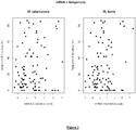
- Mb3645c was not recognised by any of the M. bovis naive cattle, whereas 11 of the 30 M. bovis infected animals (37%, p ⁇ 0.01, Figure 2 ) mounted a positive IFN ⁇ response when stimulated with this peptide pool.
- Rv3615c the M. tuberculosis equivalent to Mb3645c
- bovis infected animals were enriched in cattle that did not respond to ESAT6 or CFP10 (4 of 7, 57%, p ⁇ 0.05, Figure 2 ). This raises the possibility that these proteins could be used to increase the sensitivity of previously developed ESAT6/CFP10 based diagnostic tests (Vordermeier et al., 2001).
- the response to constituent peptides from the Rv3615c pool was determined using an IFN- ⁇ ELISPOT assay with PBMC isolated from M. bovis -infected cattle.
- Peptides SEQ ID NOs: 8-13 were recognized in at least two of the three cattle tested.
- Peptides SEQ ID NOs: 10-13 (spanning amino acids 57 to 103) from the C terminus of the protein were the most antigenic and were recognized by all three animals tested.
- Peptide SEQ ID NO: 13 (AKIYSEADEAWRKAIDGLFT), in particular, stimulated a response in all three animals, with an average of 509 spot-forming units (SFU) per 106 PBMC (standard deviation, 185.3 SFU per 106 PBMC), which is comparable to the results for the pool as a whole (414 SFU per 106 PBMC; standard deviation, 135.6 SFU per 106 PBMC) ( Figure 4 ).
- a FACS analysis was performed with PBMC isolated from the same M. bovis -infected cattle that were used for the ELISPOT analysis. Lymphocytes were analyzed for intracellular IFN- ⁇ production and the presence of CD4 and CD8 cell differentiation markers. It was found that, mirroring the ELISPOT data, peptides SEQ ID NO:2-8 stimulated little IFN- ⁇ production. Markedly higher levels of IFN- ⁇ were observed for the cells stimulated with peptides SEQ ID NOs: 9, 10, 12 and 13 ( Figure 5 ).
- a selection of consistently highly expressed genes were screened for their ability to stimulate IFN ⁇ responses from cattle infected with M. bovis. Fourteen candidates were selected based on their high expression in both M. tuberculosis and M. bovis across a variety of growth conditions, including in vitro chemostat and batch cultures, as well as from a macrophage infection and microaerophillic chemostat cultures. Proteins were excluded if they were known immunogens or had significant homology to proteins in other mycobacteria; hence the majority of the candidate antigens had no functional annotation. However, three were predicted membrane associated proteins, one an excisionase and one a PE family protein (Table 2).
- Rv3616c The product of the first gene in this operon, has also been shown to be a dominant mycobacterial antigen.
- Rv3616c is more frequently recognised in M. bovis infected cattle compared to Rv3615c: 84.6% versus 37% (Mustafa et al., 2006).
- Rv3616c is secreted in a mutually dependent manner with ESAT6 and CFP10 (Fortune et al., 2005), whereas Rv3615c appears to interact with other proteins of the secretion system (Macgurn et al., 2005) and may therefore remain within the bacterial cell, which could explain the difference in frequencies of response from M. bovis infected cattle.
- the ESAT6/CFP10 peptide cocktail had been developed as an alternative diagnostic reagent to PPD and differentiates infected and vaccinated individuals as these antigens are not present in M. bovis BCG (Vordermeier et al., 2001). The test is reported to have a sensitivity of around 77.9% in infected cattle. Rv3615c has been found not to be recognised by the immune systems of either M. bovis na ⁇ ve or BCG vaccinated animals, unlike Rv3616c to which 40% of vaccinated individuals respond (Mustafa et al., 2006), and is therefore highly specific. Furthermore, 57% of cattle infected with M.
Landscapes
- Health & Medical Sciences (AREA)
- Chemical & Material Sciences (AREA)
- Life Sciences & Earth Sciences (AREA)
- Molecular Biology (AREA)
- Organic Chemistry (AREA)
- Immunology (AREA)
- Engineering & Computer Science (AREA)
- General Health & Medical Sciences (AREA)
- Medicinal Chemistry (AREA)
- Biochemistry (AREA)
- Urology & Nephrology (AREA)
- Hematology (AREA)
- Biomedical Technology (AREA)
- Biophysics (AREA)
- Genetics & Genomics (AREA)
- Proteomics, Peptides & Aminoacids (AREA)
- Food Science & Technology (AREA)
- Cell Biology (AREA)
- Physics & Mathematics (AREA)
- Microbiology (AREA)
- General Physics & Mathematics (AREA)
- Pathology (AREA)
- Gastroenterology & Hepatology (AREA)
- Analytical Chemistry (AREA)
- Biotechnology (AREA)
- Virology (AREA)
- Tropical Medicine & Parasitology (AREA)
- Peptides Or Proteins (AREA)
- Medicines Containing Antibodies Or Antigens For Use As Internal Diagnostic Agents (AREA)
- Measuring Or Testing Involving Enzymes Or Micro-Organisms (AREA)
Claims (9)
- Réactif de diagnostic pour son utilisation dans la détection d'une infection par M. bovis ou M. tuberculosis chez un mammifère, comprenant un peptide ayant une longueur allant jusqu'à 100 acides aminés qui contient un épitope de lymphocyte T provenant de la protéine ayant la séquence d'acides aminés SEQ ID NO : 1, l'épitope consistant en des acides aminés consécutifs provenant de SEQ ID NO : 1, dans lequel le peptide consiste en des acides aminés consécutifs provenant de SEQ ID NO : 1, en outre dans lequel le réactif de diagnostic est un réactif de diagnostic de dosage de l'immunité à médiation cellulaire (IMC).
- Réactif de diagnostic selon la revendication 1, dans lequel le peptide contient jusqu'à 20 acides aminés.
- Réactif de diagnostic selon l'une quelconque des revendications précédentes, comprenant en outre un ou plusieurs polypeptides possédant chacun la séquence d'acides aminés de SEQ ID NO : 14 à 25.
- Kit de diagnostic comprenant un réactif de diagnostic tel que défini dans l'une quelconque des revendications 1 à 3, dans lequel le réactif de diagnostic est capable de détecter une infection par M. tuberculosis chez un mammifère et est capable de faire la différence entre un mammifère infecté par M. tuberculosis et un mammifère vacciné contre une infection par M. tuberculosis.
- Polypeptide isolé ayant une longueur située entre 8 et 35 acides aminés, par exemple une longueur de 10 à 25 ou de 12 à 20 acides aminés, dans lequel le peptide contient un épitope provenant de la protéine ayant la séquence d'acides aminés SEQ ID NO : 1, dans lequel l'épitope consistant en des acides aminés consécutifs provenant de SEQ ID NO : 1 et dans lequel le peptide possède des propriétés antigéniques et/ou immunogènes spécifiques de M. bovis et/ou de M. tuberculosis.
- Acide nucléique qui code pour un peptide de réactif de diagnostic tel que défini dans l'une quelconque des revendications 1 à 2 ou un peptide tel que défini dans la revendication 5.
- Vecteur approprié pour l'expression d'un peptide tel que défini dans l'une quelconque des revendications 1, 2 ou 5, le vecteur comprenant l'acide nucléique tel que défini dans la revendication 6.
- Cellule transformée avec le vecteur tel que défini dans la revendication 7.
- Méthode de diagnostic in vitro chez un hôte d'une infection par, ou d'une exposition à, M. bovis et/ou M. tuberculosis, comprenant les étapes de :i) contact d'une population de cellules provenant de l'hôte avec un réactif de diagnostic tel que défini dans l'une quelconque des revendications 1 à 3 ; etii) détermination de la reconnaissance ou non du réactif de diagnostic par les cellules de ladite population de cellules.
Applications Claiming Priority (3)
| Application Number | Priority Date | Filing Date | Title |
|---|---|---|---|
| GBGB0722105.4A GB0722105D0 (en) | 2007-11-10 | 2007-11-10 | Antigens |
| EP13182794.1A EP2672268B1 (fr) | 2007-11-10 | 2008-11-06 | Antigènes mycobactériens |
| EP08848298.9A EP2210105B1 (fr) | 2007-11-10 | 2008-11-06 | Antigènes mycobactériens |
Related Parent Applications (3)
| Application Number | Title | Priority Date | Filing Date |
|---|---|---|---|
| EP08848298.9A Division EP2210105B1 (fr) | 2007-11-10 | 2008-11-06 | Antigènes mycobactériens |
| EP13182794.1A Division-Into EP2672268B1 (fr) | 2007-11-10 | 2008-11-06 | Antigènes mycobactériens |
| EP13182794.1A Division EP2672268B1 (fr) | 2007-11-10 | 2008-11-06 | Antigènes mycobactériens |
Publications (2)
| Publication Number | Publication Date |
|---|---|
| EP2827149A1 EP2827149A1 (fr) | 2015-01-21 |
| EP2827149B1 true EP2827149B1 (fr) | 2017-01-04 |
Family
ID=38858506
Family Applications (3)
| Application Number | Title | Priority Date | Filing Date |
|---|---|---|---|
| EP08848298.9A Active EP2210105B1 (fr) | 2007-11-10 | 2008-11-06 | Antigènes mycobactériens |
| EP13182794.1A Active EP2672268B1 (fr) | 2007-11-10 | 2008-11-06 | Antigènes mycobactériens |
| EP14188545.9A Active EP2827149B1 (fr) | 2007-11-10 | 2008-11-06 | Antigènes de mycobactérium |
Family Applications Before (2)
| Application Number | Title | Priority Date | Filing Date |
|---|---|---|---|
| EP08848298.9A Active EP2210105B1 (fr) | 2007-11-10 | 2008-11-06 | Antigènes mycobactériens |
| EP13182794.1A Active EP2672268B1 (fr) | 2007-11-10 | 2008-11-06 | Antigènes mycobactériens |
Country Status (12)
| Country | Link |
|---|---|
| US (5) | US8697091B2 (fr) |
| EP (3) | EP2210105B1 (fr) |
| AU (2) | AU2008324031B2 (fr) |
| CA (1) | CA2703395C (fr) |
| DK (3) | DK2672268T3 (fr) |
| ES (3) | ES2564026T3 (fr) |
| GB (1) | GB0722105D0 (fr) |
| HR (1) | HRP20131015T1 (fr) |
| NZ (1) | NZ584481A (fr) |
| PL (1) | PL2210105T3 (fr) |
| PT (1) | PT2210105E (fr) |
| WO (1) | WO2009060184A1 (fr) |
Families Citing this family (14)
| Publication number | Priority date | Publication date | Assignee | Title |
|---|---|---|---|---|
| DK1144447T3 (da) * | 1998-11-04 | 2010-02-08 | Isis Innovation | Dianostisk tuberkulosetest |
| GB0618127D0 (en) * | 2006-09-14 | 2006-10-25 | Isis Innovation | Biomarker |
| GB0722105D0 (en) * | 2007-11-10 | 2007-12-19 | Sec Dep For Environment Food A | Antigens |
| GB0906215D0 (en) | 2009-04-09 | 2009-05-20 | Lalvani Ajit | Diagnostic test |
| GB201007075D0 (en) | 2010-04-28 | 2010-06-09 | Sec Dep For Environment Food A | Diagnostic reagents |
| GB201012072D0 (en) | 2010-07-19 | 2010-09-01 | Sec Dep For Environment Food A | Diagnostic reagents |
| ITRM20100411A1 (it) * | 2010-07-23 | 2012-01-24 | Massimo Amicosante | Uso di sequenze amminoacidiche da mycobacterium tuberculosis o dei loro corrispondenti acidi nucleici per la diagnosi e la prevenzione di infezione tubercolare, relativo kit diagnostico e vaccino. |
| CN103257236B (zh) * | 2012-02-20 | 2015-04-15 | 南京市疾病预防控制中心 | Hla-a24限制性结核分支杆菌特异性ctl细胞表位肽的应用 |
| TWI638829B (zh) * | 2012-07-10 | 2018-10-21 | 法商傳斯堅公司 | 分枝桿菌抗原疫苗 |
| US10684275B2 (en) | 2016-12-14 | 2020-06-16 | Becton, Dickinson And Company | Methods and compositions for obtaining a tuberculosis assessment in a subject |
| GB201909953D0 (en) | 2019-07-11 | 2019-08-28 | Sec Dep For Environment Food And Rural Affairs Acting Through The Animal And Plant Health Agency | Diagnostic reagents |
| BR112023006262A2 (pt) | 2020-10-06 | 2023-05-09 | Cepheid | Métodos de diagnóstico de tuberculose e de diferenciação entre tuberculose ativa e latente |
| CN115951052B (zh) * | 2022-12-09 | 2025-10-03 | 长江大学 | 一种快速检测鲟鱼感染海分枝杆菌的方法 |
| GB202318595D0 (en) | 2023-12-05 | 2024-01-17 | Sec Dep For Environment Food And Rural Affairs Acting Through Animal And Plant Health Agency | Diagnostic proteins and reagents |
Citations (2)
| Publication number | Priority date | Publication date | Assignee | Title |
|---|---|---|---|---|
| WO2004031211A2 (fr) * | 2002-10-03 | 2004-04-15 | Epimmune Inc. | Peptides de liaison hla et utilisations de ces derniers |
| US20100197607A1 (en) * | 2005-01-20 | 2010-08-05 | Indu Parikh | Mucin hypersecretion inhibitors and methods of use |
Family Cites Families (7)
| Publication number | Priority date | Publication date | Assignee | Title |
|---|---|---|---|---|
| DE10034826A1 (de) | 2000-07-18 | 2002-01-31 | Bosch Gmbh Robert | Baugruppe mit einem strukturierten Tärgerelement und einem mit diesem wirkverbundenen Substrat |
| US20040121322A9 (en) * | 2001-02-22 | 2004-06-24 | Cole Stewart T. | Comparative mycobacterial genomics as a tool for identifying targets for the diagnosis, prophylaxis or treatment of mycobacterioses |
| US7173112B2 (en) * | 2001-09-26 | 2007-02-06 | Takeda Pharmaceutical Company Limited | Antibody to GALP and uses thereof |
| DE602007013620D1 (de) | 2006-09-05 | 2011-05-12 | Hvidovre Hospital | Auf IP-10 basierende immunologische Überwachung |
| GB0716070D0 (en) | 2007-08-17 | 2007-09-26 | Fusion Antibodies Ltd | Diagnostic method and kit |
| GB0722105D0 (en) * | 2007-11-10 | 2007-12-19 | Sec Dep For Environment Food A | Antigens |
| GB0906215D0 (en) | 2009-04-09 | 2009-05-20 | Lalvani Ajit | Diagnostic test |
-
2007
- 2007-11-10 GB GBGB0722105.4A patent/GB0722105D0/en not_active Ceased
-
2008
- 2008-11-06 ES ES13182794.1T patent/ES2564026T3/es active Active
- 2008-11-06 PL PL08848298T patent/PL2210105T3/pl unknown
- 2008-11-06 DK DK13182794.1T patent/DK2672268T3/en active
- 2008-11-06 EP EP08848298.9A patent/EP2210105B1/fr active Active
- 2008-11-06 HR HRP20131015AT patent/HRP20131015T1/hr unknown
- 2008-11-06 ES ES14188545.9T patent/ES2618336T3/es active Active
- 2008-11-06 ES ES08848298T patent/ES2431891T3/es active Active
- 2008-11-06 US US12/742,223 patent/US8697091B2/en active Active
- 2008-11-06 DK DK08848298.9T patent/DK2210105T3/da active
- 2008-11-06 EP EP13182794.1A patent/EP2672268B1/fr active Active
- 2008-11-06 EP EP14188545.9A patent/EP2827149B1/fr active Active
- 2008-11-06 AU AU2008324031A patent/AU2008324031B2/en active Active
- 2008-11-06 NZ NZ584481A patent/NZ584481A/en unknown
- 2008-11-06 CA CA2703395A patent/CA2703395C/fr active Active
- 2008-11-06 DK DK14188545.9T patent/DK2827149T3/en active
- 2008-11-06 PT PT88482989T patent/PT2210105E/pt unknown
- 2008-11-06 WO PCT/GB2008/003724 patent/WO2009060184A1/fr not_active Ceased
-
2010
- 2010-04-30 AU AU2010100407A patent/AU2010100407A4/en not_active Expired
-
2014
- 2014-02-21 US US14/186,291 patent/US9285364B2/en active Active
-
2016
- 2016-02-05 US US15/016,859 patent/US9618509B2/en active Active
-
2017
- 2017-03-31 US US15/475,472 patent/US20170269079A1/en not_active Abandoned
-
2018
- 2018-07-03 US US16/026,590 patent/US10578617B2/en active Active
Patent Citations (2)
| Publication number | Priority date | Publication date | Assignee | Title |
|---|---|---|---|---|
| WO2004031211A2 (fr) * | 2002-10-03 | 2004-04-15 | Epimmune Inc. | Peptides de liaison hla et utilisations de ces derniers |
| US20100197607A1 (en) * | 2005-01-20 | 2010-08-05 | Indu Parikh | Mucin hypersecretion inhibitors and methods of use |
Non-Patent Citations (4)
| Title |
|---|
| BRANDT L ET AL: "Key epitopes on the ESAT-6 antigen recognized in mice during the recall of protective immunity to Mycobacterium tuberculosis.", JOURNAL OF IMMUNOLOGY (BALTIMORE, MD. : 1950) 15 OCT 1996, vol. 157, no. 8, 15 October 1996 (1996-10-15), pages 3527 - 3533, ISSN: 0022-1767 * |
| CHICZ R M ET AL: "Predominant naturally processed peptides bound to HLA-DR1 are derived from MHC-related molecules and are heterogeneous in size.", NATURE 27 AUG 1992, vol. 358, no. 6389, 27 August 1992 (1992-08-27), pages 764 - 768, ISSN: 0028-0836 * |
| RAVN P ET AL: "Human T cell responses to the ESAT-6 antigen from Mycobacterium tuberculosis.", THE JOURNAL OF INFECTIOUS DISEASES MAR 1999, vol. 179, no. 3, March 1999 (1999-03-01), pages 637 - 645, ISSN: 0022-1899 * |
| SKJOT RIKKE LOUISE VINTHER ET AL: "Epitope mapping of the immunodominant antigen TB10.4 and the two homologous proteins TB10.3 and TB12.9, which constitute a subfamily of the esat-6 gene family.", INFECTION AND IMMUNITY OCT 2002, vol. 70, no. 10, October 2002 (2002-10-01), pages 5446 - 5453, ISSN: 0019-9567 * |
Also Published As
| Publication number | Publication date |
|---|---|
| HRP20131015T1 (hr) | 2013-12-20 |
| AU2010100407A4 (en) | 2010-05-27 |
| ES2618336T3 (es) | 2017-06-21 |
| CA2703395A1 (fr) | 2009-05-14 |
| US9285364B2 (en) | 2016-03-15 |
| DK2672268T3 (en) | 2016-03-29 |
| EP2210105A1 (fr) | 2010-07-28 |
| AU2008324031B2 (en) | 2013-06-27 |
| GB0722105D0 (en) | 2007-12-19 |
| EP2672268B1 (fr) | 2016-02-10 |
| US9618509B2 (en) | 2017-04-11 |
| DK2827149T3 (en) | 2017-02-06 |
| US8697091B2 (en) | 2014-04-15 |
| CA2703395C (fr) | 2018-09-25 |
| PT2210105E (pt) | 2013-10-30 |
| DK2210105T3 (da) | 2013-11-04 |
| PL2210105T3 (pl) | 2014-01-31 |
| ES2431891T3 (es) | 2013-11-28 |
| US10578617B2 (en) | 2020-03-03 |
| US20190170746A1 (en) | 2019-06-06 |
| ES2564026T3 (es) | 2016-03-17 |
| EP2672268A1 (fr) | 2013-12-11 |
| US20140296091A1 (en) | 2014-10-02 |
| US20170269079A1 (en) | 2017-09-21 |
| US20100317036A1 (en) | 2010-12-16 |
| NZ584481A (en) | 2011-12-22 |
| EP2210105B1 (fr) | 2013-09-04 |
| WO2009060184A1 (fr) | 2009-05-14 |
| EP2827149A1 (fr) | 2015-01-21 |
| AU2008324031A1 (en) | 2009-05-14 |
| US20160153987A1 (en) | 2016-06-02 |
Similar Documents
| Publication | Publication Date | Title |
|---|---|---|
| US10578617B2 (en) | Mycobacterium antigens | |
| Sidders et al. | Screening of highly expressed mycobacterial genes identifies Rv3615c as a useful differential diagnostic antigen for the Mycobacterium tuberculosis complex | |
| US20120128708A1 (en) | Diagnostic mycobacterium tuberculosis test | |
| CA2685896C (fr) | Procede de diagnostic de la tuberculose par la detection d'une reponse immunitaire a ompatb | |
| KR102305770B1 (ko) | 결핵의 개선된 생체내 또는 시험관내 세포-매개 면역학적 진단을 위한 진단 시약 | |
| Dernivoix et al. | Field performance of six Mycobacterium avium subsp. paratuberculosis antigens in a 20 h interferon gamma release assay in Belgium | |
| EP1735623B1 (fr) | Test de diagnostic d'infection par mycobacterium tuberculosis | |
| Hewinson et al. | Screening of Highly Expressed | |
| Dernivoix et al. | Field performance of six Mycobacterium avium subsp. | |
| Gideon | Studies on in vitro human T cell reactivity to antigens of mycobacterium tuberculosis | |
| EA041840B1 (ru) | Диагностические реагенты для улучшенной in vivo или in vitro клеточно-опосредованной иммунологической диагностики туберкулеза |
Legal Events
| Date | Code | Title | Description |
|---|---|---|---|
| 17P | Request for examination filed |
Effective date: 20141105 |
|
| AC | Divisional application: reference to earlier application |
Ref document number: 2210105 Country of ref document: EP Kind code of ref document: P Ref document number: 2672268 Country of ref document: EP Kind code of ref document: P |
|
| AK | Designated contracting states |
Kind code of ref document: A1 Designated state(s): AT BE BG CH CY CZ DE DK EE ES FI FR GB GR HR HU IE IS IT LI LT LU LV MC MT NL NO PL PT RO SE SI SK TR |
|
| PUAI | Public reference made under article 153(3) epc to a published international application that has entered the european phase |
Free format text: ORIGINAL CODE: 0009012 |
|
| 17Q | First examination report despatched |
Effective date: 20150805 |
|
| GRAP | Despatch of communication of intention to grant a patent |
Free format text: ORIGINAL CODE: EPIDOSNIGR1 |
|
| INTG | Intention to grant announced |
Effective date: 20160629 |
|
| GRAS | Grant fee paid |
Free format text: ORIGINAL CODE: EPIDOSNIGR3 |
|
| GRAA | (expected) grant |
Free format text: ORIGINAL CODE: 0009210 |
|
| AC | Divisional application: reference to earlier application |
Ref document number: 2210105 Country of ref document: EP Kind code of ref document: P Ref document number: 2672268 Country of ref document: EP Kind code of ref document: P |
|
| AK | Designated contracting states |
Kind code of ref document: B1 Designated state(s): AT BE BG CH CY CZ DE DK EE ES FI FR GB GR HR HU IE IS IT LI LT LU LV MC MT NL NO PL PT RO SE SI SK TR |
|
| REG | Reference to a national code |
Ref country code: GB Ref legal event code: FG4D |
|
| REG | Reference to a national code |
Ref country code: CH Ref legal event code: EP |
|
| REG | Reference to a national code |
Ref country code: AT Ref legal event code: REF Ref document number: 859749 Country of ref document: AT Kind code of ref document: T Effective date: 20170115 |
|
| REG | Reference to a national code |
Ref country code: IE Ref legal event code: FG4D |
|
| REG | Reference to a national code |
Ref country code: NL Ref legal event code: FP |
|
| REG | Reference to a national code |
Ref country code: DK Ref legal event code: T3 Effective date: 20170131 |
|
| REG | Reference to a national code |
Ref country code: DE Ref legal event code: R096 Ref document number: 602008048319 Country of ref document: DE |
|
| REG | Reference to a national code |
Ref country code: LT Ref legal event code: MG4D |
|
| REG | Reference to a national code |
Ref country code: ES Ref legal event code: FG2A Ref document number: 2618336 Country of ref document: ES Kind code of ref document: T3 Effective date: 20170621 |
|
| PG25 | Lapsed in a contracting state [announced via postgrant information from national office to epo] |
Ref country code: NO Free format text: LAPSE BECAUSE OF FAILURE TO SUBMIT A TRANSLATION OF THE DESCRIPTION OR TO PAY THE FEE WITHIN THE PRESCRIBED TIME-LIMIT Effective date: 20170404 Ref country code: GR Free format text: LAPSE BECAUSE OF FAILURE TO SUBMIT A TRANSLATION OF THE DESCRIPTION OR TO PAY THE FEE WITHIN THE PRESCRIBED TIME-LIMIT Effective date: 20170405 Ref country code: FI Free format text: LAPSE BECAUSE OF FAILURE TO SUBMIT A TRANSLATION OF THE DESCRIPTION OR TO PAY THE FEE WITHIN THE PRESCRIBED TIME-LIMIT Effective date: 20170104 Ref country code: IS Free format text: LAPSE BECAUSE OF FAILURE TO SUBMIT A TRANSLATION OF THE DESCRIPTION OR TO PAY THE FEE WITHIN THE PRESCRIBED TIME-LIMIT Effective date: 20170504 Ref country code: LT Free format text: LAPSE BECAUSE OF FAILURE TO SUBMIT A TRANSLATION OF THE DESCRIPTION OR TO PAY THE FEE WITHIN THE PRESCRIBED TIME-LIMIT Effective date: 20170104 Ref country code: HR Free format text: LAPSE BECAUSE OF FAILURE TO SUBMIT A TRANSLATION OF THE DESCRIPTION OR TO PAY THE FEE WITHIN THE PRESCRIBED TIME-LIMIT Effective date: 20170104 |
|
| PG25 | Lapsed in a contracting state [announced via postgrant information from national office to epo] |
Ref country code: LV Free format text: LAPSE BECAUSE OF FAILURE TO SUBMIT A TRANSLATION OF THE DESCRIPTION OR TO PAY THE FEE WITHIN THE PRESCRIBED TIME-LIMIT Effective date: 20170104 Ref country code: SE Free format text: LAPSE BECAUSE OF FAILURE TO SUBMIT A TRANSLATION OF THE DESCRIPTION OR TO PAY THE FEE WITHIN THE PRESCRIBED TIME-LIMIT Effective date: 20170104 Ref country code: PL Free format text: LAPSE BECAUSE OF FAILURE TO SUBMIT A TRANSLATION OF THE DESCRIPTION OR TO PAY THE FEE WITHIN THE PRESCRIBED TIME-LIMIT Effective date: 20170104 Ref country code: PT Free format text: LAPSE BECAUSE OF FAILURE TO SUBMIT A TRANSLATION OF THE DESCRIPTION OR TO PAY THE FEE WITHIN THE PRESCRIBED TIME-LIMIT Effective date: 20170504 Ref country code: BG Free format text: LAPSE BECAUSE OF FAILURE TO SUBMIT A TRANSLATION OF THE DESCRIPTION OR TO PAY THE FEE WITHIN THE PRESCRIBED TIME-LIMIT Effective date: 20170404 |
|
| REG | Reference to a national code |
Ref country code: DE Ref legal event code: R097 Ref document number: 602008048319 Country of ref document: DE |
|
| PG25 | Lapsed in a contracting state [announced via postgrant information from national office to epo] |
Ref country code: CZ Free format text: LAPSE BECAUSE OF FAILURE TO SUBMIT A TRANSLATION OF THE DESCRIPTION OR TO PAY THE FEE WITHIN THE PRESCRIBED TIME-LIMIT Effective date: 20170104 Ref country code: SK Free format text: LAPSE BECAUSE OF FAILURE TO SUBMIT A TRANSLATION OF THE DESCRIPTION OR TO PAY THE FEE WITHIN THE PRESCRIBED TIME-LIMIT Effective date: 20170104 Ref country code: EE Free format text: LAPSE BECAUSE OF FAILURE TO SUBMIT A TRANSLATION OF THE DESCRIPTION OR TO PAY THE FEE WITHIN THE PRESCRIBED TIME-LIMIT Effective date: 20170104 Ref country code: RO Free format text: LAPSE BECAUSE OF FAILURE TO SUBMIT A TRANSLATION OF THE DESCRIPTION OR TO PAY THE FEE WITHIN THE PRESCRIBED TIME-LIMIT Effective date: 20170104 |
|
| PLBE | No opposition filed within time limit |
Free format text: ORIGINAL CODE: 0009261 |
|
| STAA | Information on the status of an ep patent application or granted ep patent |
Free format text: STATUS: NO OPPOSITION FILED WITHIN TIME LIMIT |
|
| REG | Reference to a national code |
Ref country code: FR Ref legal event code: PLFP Year of fee payment: 10 |
|
| PG25 | Lapsed in a contracting state [announced via postgrant information from national office to epo] |
Ref country code: SI Free format text: LAPSE BECAUSE OF FAILURE TO SUBMIT A TRANSLATION OF THE DESCRIPTION OR TO PAY THE FEE WITHIN THE PRESCRIBED TIME-LIMIT Effective date: 20170104 |
|
| PG25 | Lapsed in a contracting state [announced via postgrant information from national office to epo] |
Ref country code: MC Free format text: LAPSE BECAUSE OF FAILURE TO SUBMIT A TRANSLATION OF THE DESCRIPTION OR TO PAY THE FEE WITHIN THE PRESCRIBED TIME-LIMIT Effective date: 20170104 |
|
| PG25 | Lapsed in a contracting state [announced via postgrant information from national office to epo] |
Ref country code: LU Free format text: LAPSE BECAUSE OF NON-PAYMENT OF DUE FEES Effective date: 20171106 |
|
| REG | Reference to a national code |
Ref country code: IE Ref legal event code: MM4A |
|
| PG25 | Lapsed in a contracting state [announced via postgrant information from national office to epo] |
Ref country code: MT Free format text: LAPSE BECAUSE OF NON-PAYMENT OF DUE FEES Effective date: 20171106 |
|
| PG25 | Lapsed in a contracting state [announced via postgrant information from national office to epo] |
Ref country code: IE Free format text: LAPSE BECAUSE OF NON-PAYMENT OF DUE FEES Effective date: 20171106 |
|
| REG | Reference to a national code |
Ref country code: AT Ref legal event code: UEP Ref document number: 859749 Country of ref document: AT Kind code of ref document: T Effective date: 20170104 |
|
| PG25 | Lapsed in a contracting state [announced via postgrant information from national office to epo] |
Ref country code: HU Free format text: LAPSE BECAUSE OF FAILURE TO SUBMIT A TRANSLATION OF THE DESCRIPTION OR TO PAY THE FEE WITHIN THE PRESCRIBED TIME-LIMIT; INVALID AB INITIO Effective date: 20081106 |
|
| PG25 | Lapsed in a contracting state [announced via postgrant information from national office to epo] |
Ref country code: CY Free format text: LAPSE BECAUSE OF FAILURE TO SUBMIT A TRANSLATION OF THE DESCRIPTION OR TO PAY THE FEE WITHIN THE PRESCRIBED TIME-LIMIT Effective date: 20170104 |
|
| PG25 | Lapsed in a contracting state [announced via postgrant information from national office to epo] |
Ref country code: TR Free format text: LAPSE BECAUSE OF FAILURE TO SUBMIT A TRANSLATION OF THE DESCRIPTION OR TO PAY THE FEE WITHIN THE PRESCRIBED TIME-LIMIT Effective date: 20170104 |
|
| REG | Reference to a national code |
Ref country code: CH Ref legal event code: U11 Free format text: ST27 STATUS EVENT CODE: U-0-0-U10-U11 (AS PROVIDED BY THE NATIONAL OFFICE) Effective date: 20251201 |
|
| PGFP | Annual fee paid to national office [announced via postgrant information from national office to epo] |
Ref country code: NL Payment date: 20251119 Year of fee payment: 18 |
|
| PGFP | Annual fee paid to national office [announced via postgrant information from national office to epo] |
Ref country code: DE Payment date: 20251119 Year of fee payment: 18 |
|
| PGFP | Annual fee paid to national office [announced via postgrant information from national office to epo] |
Ref country code: GB Payment date: 20251121 Year of fee payment: 18 |
|
| PGFP | Annual fee paid to national office [announced via postgrant information from national office to epo] |
Ref country code: AT Payment date: 20251120 Year of fee payment: 18 |
|
| PGFP | Annual fee paid to national office [announced via postgrant information from national office to epo] |
Ref country code: IT Payment date: 20251121 Year of fee payment: 18 Ref country code: DK Payment date: 20251125 Year of fee payment: 18 |
|
| PGFP | Annual fee paid to national office [announced via postgrant information from national office to epo] |
Ref country code: FR Payment date: 20251125 Year of fee payment: 18 |
|
| PGFP | Annual fee paid to national office [announced via postgrant information from national office to epo] |
Ref country code: BE Payment date: 20251119 Year of fee payment: 18 |
|
| PGFP | Annual fee paid to national office [announced via postgrant information from national office to epo] |
Ref country code: CH Payment date: 20251201 Year of fee payment: 18 |
|
| PGFP | Annual fee paid to national office [announced via postgrant information from national office to epo] |
Ref country code: ES Payment date: 20251229 Year of fee payment: 18 |