EP2852403B1 - Verfahren zur modifizierung von gewebe - Google Patents
Verfahren zur modifizierung von gewebe Download PDFInfo
- Publication number
- EP2852403B1 EP2852403B1 EP13784517.8A EP13784517A EP2852403B1 EP 2852403 B1 EP2852403 B1 EP 2852403B1 EP 13784517 A EP13784517 A EP 13784517A EP 2852403 B1 EP2852403 B1 EP 2852403B1
- Authority
- EP
- European Patent Office
- Prior art keywords
- tissue
- tissues
- mmp
- treated
- stiffness
- Prior art date
- Legal status (The legal status is an assumption and is not a legal conclusion. Google has not performed a legal analysis and makes no representation as to the accuracy of the status listed.)
- Not-in-force
Links
- 230000004048 modification Effects 0.000 title claims description 31
- 238000012986 modification Methods 0.000 title claims description 31
- 238000000034 method Methods 0.000 title description 68
- 210000001519 tissue Anatomy 0.000 claims description 483
- 108010015302 Matrix metalloproteinase-9 Proteins 0.000 claims description 106
- 102100030412 Matrix metalloproteinase-9 Human genes 0.000 claims description 102
- 210000002808 connective tissue Anatomy 0.000 claims description 24
- 230000003628 erosive effect Effects 0.000 claims description 22
- RXSUFCOOZSGWSW-UHFFFAOYSA-M acetyloxy-(4-aminophenyl)mercury Chemical compound CC(=O)O[Hg]C1=CC=C(N)C=C1 RXSUFCOOZSGWSW-UHFFFAOYSA-M 0.000 claims description 20
- 210000000845 cartilage Anatomy 0.000 claims description 19
- 101000990902 Homo sapiens Matrix metalloproteinase-9 Proteins 0.000 claims description 10
- 230000004913 activation Effects 0.000 claims description 7
- 230000033001 locomotion Effects 0.000 claims description 5
- 230000003068 static effect Effects 0.000 claims 1
- 230000001965 increasing effect Effects 0.000 description 90
- 238000011282 treatment Methods 0.000 description 82
- 102000029816 Collagenase Human genes 0.000 description 71
- 108060005980 Collagenase Proteins 0.000 description 71
- 102000004190 Enzymes Human genes 0.000 description 71
- 108090000790 Enzymes Proteins 0.000 description 71
- 229940088598 enzyme Drugs 0.000 description 71
- 210000004027 cell Anatomy 0.000 description 70
- 210000002435 tendon Anatomy 0.000 description 70
- 239000000872 buffer Substances 0.000 description 57
- 230000035882 stress Effects 0.000 description 54
- 238000003556 assay Methods 0.000 description 53
- 230000000694 effects Effects 0.000 description 53
- 229960002424 collagenase Drugs 0.000 description 51
- 238000002054 transplantation Methods 0.000 description 51
- 102000010834 Extracellular Matrix Proteins Human genes 0.000 description 50
- 108010037362 Extracellular Matrix Proteins Proteins 0.000 description 50
- 210000002744 extracellular matrix Anatomy 0.000 description 50
- 210000000056 organ Anatomy 0.000 description 48
- 241000193159 Hathewaya histolytica Species 0.000 description 36
- 238000000338 in vitro Methods 0.000 description 35
- 230000002829 reductive effect Effects 0.000 description 35
- 210000003491 skin Anatomy 0.000 description 35
- 210000005036 nerve Anatomy 0.000 description 32
- 239000000243 solution Substances 0.000 description 31
- 210000003205 muscle Anatomy 0.000 description 29
- 230000006870 function Effects 0.000 description 28
- 102000002274 Matrix Metalloproteinases Human genes 0.000 description 26
- 108010000684 Matrix Metalloproteinases Proteins 0.000 description 26
- 230000035699 permeability Effects 0.000 description 26
- 239000003814 drug Substances 0.000 description 25
- 210000004153 islets of langerhan Anatomy 0.000 description 25
- 102000008186 Collagen Human genes 0.000 description 23
- 108010035532 Collagen Proteins 0.000 description 23
- 229920001436 collagen Polymers 0.000 description 23
- 210000000496 pancreas Anatomy 0.000 description 23
- 230000003247 decreasing effect Effects 0.000 description 22
- 210000004185 liver Anatomy 0.000 description 22
- 210000004207 dermis Anatomy 0.000 description 21
- 239000000463 material Substances 0.000 description 21
- NOESYZHRGYRDHS-UHFFFAOYSA-N insulin Chemical compound N1C(=O)C(NC(=O)C(CCC(N)=O)NC(=O)C(CCC(O)=O)NC(=O)C(C(C)C)NC(=O)C(NC(=O)CN)C(C)CC)CSSCC(C(NC(CO)C(=O)NC(CC(C)C)C(=O)NC(CC=2C=CC(O)=CC=2)C(=O)NC(CCC(N)=O)C(=O)NC(CC(C)C)C(=O)NC(CCC(O)=O)C(=O)NC(CC(N)=O)C(=O)NC(CC=2C=CC(O)=CC=2)C(=O)NC(CSSCC(NC(=O)C(C(C)C)NC(=O)C(CC(C)C)NC(=O)C(CC=2C=CC(O)=CC=2)NC(=O)C(CC(C)C)NC(=O)C(C)NC(=O)C(CCC(O)=O)NC(=O)C(C(C)C)NC(=O)C(CC(C)C)NC(=O)C(CC=2NC=NC=2)NC(=O)C(CO)NC(=O)CNC2=O)C(=O)NCC(=O)NC(CCC(O)=O)C(=O)NC(CCCNC(N)=N)C(=O)NCC(=O)NC(CC=3C=CC=CC=3)C(=O)NC(CC=3C=CC=CC=3)C(=O)NC(CC=3C=CC(O)=CC=3)C(=O)NC(C(C)O)C(=O)N3C(CCC3)C(=O)NC(CCCCN)C(=O)NC(C)C(O)=O)C(=O)NC(CC(N)=O)C(O)=O)=O)NC(=O)C(C(C)CC)NC(=O)C(CO)NC(=O)C(C(C)O)NC(=O)C1CSSCC2NC(=O)C(CC(C)C)NC(=O)C(NC(=O)C(CCC(N)=O)NC(=O)C(CC(N)=O)NC(=O)C(NC(=O)C(N)CC=1C=CC=CC=1)C(C)C)CC1=CN=CN1 NOESYZHRGYRDHS-UHFFFAOYSA-N 0.000 description 20
- 239000000203 mixture Substances 0.000 description 19
- 238000012205 qualitative assay Methods 0.000 description 19
- 208000027418 Wounds and injury Diseases 0.000 description 18
- 230000003902 lesion Effects 0.000 description 18
- 206010052428 Wound Diseases 0.000 description 17
- 238000002316 cosmetic surgery Methods 0.000 description 17
- 230000007838 tissue remodeling Effects 0.000 description 17
- 230000001070 adhesive effect Effects 0.000 description 16
- 230000008859 change Effects 0.000 description 16
- 239000002245 particle Substances 0.000 description 16
- 210000003899 penis Anatomy 0.000 description 16
- 238000002278 reconstructive surgery Methods 0.000 description 16
- 239000000853 adhesive Substances 0.000 description 15
- 230000001172 regenerating effect Effects 0.000 description 15
- 230000001580 bacterial effect Effects 0.000 description 14
- 239000007853 buffer solution Substances 0.000 description 14
- 238000007634 remodeling Methods 0.000 description 13
- 108010076557 Matrix Metalloproteinase 14 Proteins 0.000 description 12
- 230000008901 benefit Effects 0.000 description 12
- 201000010099 disease Diseases 0.000 description 12
- 208000037265 diseases, disorders, signs and symptoms Diseases 0.000 description 12
- 238000012423 maintenance Methods 0.000 description 12
- 231100000241 scar Toxicity 0.000 description 12
- 108090000712 Cathepsin B Proteins 0.000 description 11
- 102000004225 Cathepsin B Human genes 0.000 description 11
- 208000002193 Pain Diseases 0.000 description 11
- 102100030416 Stromelysin-1 Human genes 0.000 description 11
- 210000005228 liver tissue Anatomy 0.000 description 11
- 210000001087 myotubule Anatomy 0.000 description 11
- 230000000717 retained effect Effects 0.000 description 11
- 230000037390 scarring Effects 0.000 description 11
- 102000004877 Insulin Human genes 0.000 description 10
- 108090001061 Insulin Proteins 0.000 description 10
- 108090000855 Matrilysin Proteins 0.000 description 10
- 241001465754 Metazoa Species 0.000 description 10
- 102100033174 Neutrophil elastase Human genes 0.000 description 10
- 210000000227 basophil cell of anterior lobe of hypophysis Anatomy 0.000 description 10
- 230000003366 colagenolytic effect Effects 0.000 description 10
- 230000004927 fusion Effects 0.000 description 10
- 229940125396 insulin Drugs 0.000 description 10
- 238000002406 microsurgery Methods 0.000 description 10
- 235000015097 nutrients Nutrition 0.000 description 10
- 210000004872 soft tissue Anatomy 0.000 description 10
- 239000007787 solid Substances 0.000 description 10
- 230000004083 survival effect Effects 0.000 description 10
- 101710108790 Stromelysin-1 Proteins 0.000 description 9
- 230000001079 digestive effect Effects 0.000 description 9
- 238000011156 evaluation Methods 0.000 description 9
- 239000007789 gas Substances 0.000 description 9
- 231100000862 numbness Toxicity 0.000 description 9
- 238000001356 surgical procedure Methods 0.000 description 9
- 239000002699 waste material Substances 0.000 description 9
- VKUYLANQOAKALN-UHFFFAOYSA-N 2-[benzyl-(4-methoxyphenyl)sulfonylamino]-n-hydroxy-4-methylpentanamide Chemical compound C1=CC(OC)=CC=C1S(=O)(=O)N(C(CC(C)C)C(=O)NO)CC1=CC=CC=C1 VKUYLANQOAKALN-UHFFFAOYSA-N 0.000 description 8
- 102100030411 Neutrophil collagenase Human genes 0.000 description 8
- 230000009286 beneficial effect Effects 0.000 description 8
- 210000000988 bone and bone Anatomy 0.000 description 8
- 238000009472 formulation Methods 0.000 description 8
- 230000035755 proliferation Effects 0.000 description 8
- 230000002797 proteolythic effect Effects 0.000 description 8
- 102000011716 Matrix Metalloproteinase 14 Human genes 0.000 description 7
- 102100024130 Matrix metalloproteinase-23 Human genes 0.000 description 7
- 206010034464 Periarthritis Diseases 0.000 description 7
- 102100028848 Stromelysin-2 Human genes 0.000 description 7
- 208000002352 blister Diseases 0.000 description 7
- 230000017531 blood circulation Effects 0.000 description 7
- 208000019425 cirrhosis of liver Diseases 0.000 description 7
- 230000006378 damage Effects 0.000 description 7
- 206010012601 diabetes mellitus Diseases 0.000 description 7
- 230000004069 differentiation Effects 0.000 description 7
- 230000005489 elastic deformation Effects 0.000 description 7
- 210000002615 epidermis Anatomy 0.000 description 7
- 210000003953 foreskin Anatomy 0.000 description 7
- 208000028867 ischemia Diseases 0.000 description 7
- 210000001328 optic nerve Anatomy 0.000 description 7
- 210000001672 ovary Anatomy 0.000 description 7
- 238000011158 quantitative evaluation Methods 0.000 description 7
- 210000000130 stem cell Anatomy 0.000 description 7
- 238000007920 subcutaneous administration Methods 0.000 description 7
- 210000004291 uterus Anatomy 0.000 description 7
- 102000005600 Cathepsins Human genes 0.000 description 6
- 108010084457 Cathepsins Proteins 0.000 description 6
- 102000009123 Fibrin Human genes 0.000 description 6
- 108010073385 Fibrin Proteins 0.000 description 6
- BWGVNKXGVNDBDI-UHFFFAOYSA-N Fibrin monomer Chemical compound CNC(=O)CNC(=O)CN BWGVNKXGVNDBDI-UHFFFAOYSA-N 0.000 description 6
- 101000627851 Homo sapiens Matrix metalloproteinase-23 Proteins 0.000 description 6
- 102000000380 Matrix Metalloproteinase 1 Human genes 0.000 description 6
- 108010076497 Matrix Metalloproteinase 10 Proteins 0.000 description 6
- 101710118230 Neutrophil collagenase Proteins 0.000 description 6
- 230000004888 barrier function Effects 0.000 description 6
- 230000015556 catabolic process Effects 0.000 description 6
- 239000006059 cover glass Substances 0.000 description 6
- 238000006731 degradation reaction Methods 0.000 description 6
- 230000003203 everyday effect Effects 0.000 description 6
- 238000002474 experimental method Methods 0.000 description 6
- 229950003499 fibrin Drugs 0.000 description 6
- 239000000499 gel Substances 0.000 description 6
- 238000001727 in vivo Methods 0.000 description 6
- 230000007246 mechanism Effects 0.000 description 6
- 230000005012 migration Effects 0.000 description 6
- 238000013508 migration Methods 0.000 description 6
- 230000008520 organization Effects 0.000 description 6
- 210000004923 pancreatic tissue Anatomy 0.000 description 6
- 230000008569 process Effects 0.000 description 6
- 230000008707 rearrangement Effects 0.000 description 6
- 230000009467 reduction Effects 0.000 description 6
- 230000008439 repair process Effects 0.000 description 6
- 108090000624 Cathepsin L Proteins 0.000 description 5
- 102000004172 Cathepsin L Human genes 0.000 description 5
- 102100035654 Cathepsin S Human genes 0.000 description 5
- 108090000613 Cathepsin S Proteins 0.000 description 5
- 108010028275 Leukocyte Elastase Proteins 0.000 description 5
- 102000004318 Matrilysin Human genes 0.000 description 5
- 102100030417 Matrilysin Human genes 0.000 description 5
- 108010091175 Matriptase Proteins 0.000 description 5
- 108010016113 Matrix Metalloproteinase 1 Proteins 0.000 description 5
- 102100030216 Matrix metalloproteinase-14 Human genes 0.000 description 5
- 102100037942 Suppressor of tumorigenicity 14 protein Human genes 0.000 description 5
- 208000025865 Ulcer Diseases 0.000 description 5
- 230000008827 biological function Effects 0.000 description 5
- 238000004113 cell culture Methods 0.000 description 5
- 238000005119 centrifugation Methods 0.000 description 5
- 230000001419 dependent effect Effects 0.000 description 5
- 238000002224 dissection Methods 0.000 description 5
- 210000003414 extremity Anatomy 0.000 description 5
- 239000012530 fluid Substances 0.000 description 5
- 210000004209 hair Anatomy 0.000 description 5
- 230000007170 pathology Effects 0.000 description 5
- 206010034878 phimosis Diseases 0.000 description 5
- 210000003497 sciatic nerve Anatomy 0.000 description 5
- 241000894007 species Species 0.000 description 5
- 230000037303 wrinkles Effects 0.000 description 5
- 201000004384 Alopecia Diseases 0.000 description 4
- 108090000625 Cathepsin K Proteins 0.000 description 4
- 102000004171 Cathepsin K Human genes 0.000 description 4
- 208000032544 Cicatrix Diseases 0.000 description 4
- 241000238557 Decapoda Species 0.000 description 4
- 102100030386 Granzyme A Human genes 0.000 description 4
- 239000012981 Hank's balanced salt solution Substances 0.000 description 4
- 102000000424 Matrix Metalloproteinase 2 Human genes 0.000 description 4
- 108010016165 Matrix Metalloproteinase 2 Proteins 0.000 description 4
- 102000001776 Matrix metalloproteinase-9 Human genes 0.000 description 4
- 108090000265 Meprin A Proteins 0.000 description 4
- 108010067035 Pancrelipase Proteins 0.000 description 4
- 102000035195 Peptidases Human genes 0.000 description 4
- 108091005804 Peptidases Proteins 0.000 description 4
- 102100028847 Stromelysin-3 Human genes 0.000 description 4
- 231100000360 alopecia Toxicity 0.000 description 4
- 238000000502 dialysis Methods 0.000 description 4
- 238000010494 dissociation reaction Methods 0.000 description 4
- 230000005593 dissociations Effects 0.000 description 4
- 230000002255 enzymatic effect Effects 0.000 description 4
- 239000000835 fiber Substances 0.000 description 4
- 230000012010 growth Effects 0.000 description 4
- 230000035876 healing Effects 0.000 description 4
- 238000011065 in-situ storage Methods 0.000 description 4
- 238000011081 inoculation Methods 0.000 description 4
- 210000003734 kidney Anatomy 0.000 description 4
- 210000004072 lung Anatomy 0.000 description 4
- 238000001000 micrograph Methods 0.000 description 4
- 230000004899 motility Effects 0.000 description 4
- 230000001850 reproductive effect Effects 0.000 description 4
- 230000002441 reversible effect Effects 0.000 description 4
- 230000037387 scars Effects 0.000 description 4
- 238000002560 therapeutic procedure Methods 0.000 description 4
- 231100000397 ulcer Toxicity 0.000 description 4
- 210000003556 vascular endothelial cell Anatomy 0.000 description 4
- BHPQYMZQTOCNFJ-UHFFFAOYSA-N Calcium cation Chemical compound [Ca+2] BHPQYMZQTOCNFJ-UHFFFAOYSA-N 0.000 description 3
- 102100027995 Collagenase 3 Human genes 0.000 description 3
- 206010014989 Epidermolysis bullosa Diseases 0.000 description 3
- WQZGKKKJIJFFOK-GASJEMHNSA-N Glucose Natural products OC[C@H]1OC(O)[C@H](O)[C@@H](O)[C@@H]1O WQZGKKKJIJFFOK-GASJEMHNSA-N 0.000 description 3
- 101000851058 Homo sapiens Neutrophil elastase Proteins 0.000 description 3
- 102100022745 Laminin subunit alpha-2 Human genes 0.000 description 3
- 108010076502 Matrix Metalloproteinase 11 Proteins 0.000 description 3
- 102100030218 Matrix metalloproteinase-19 Human genes 0.000 description 3
- 206010028980 Neoplasm Diseases 0.000 description 3
- 102000056189 Neutrophil collagenases Human genes 0.000 description 3
- 108030001564 Neutrophil collagenases Proteins 0.000 description 3
- 206010033799 Paralysis Diseases 0.000 description 3
- 206010043248 Tendon rupture Diseases 0.000 description 3
- 101001011890 Xenopus laevis Matrix metalloproteinase-18 Proteins 0.000 description 3
- 229910001424 calcium ion Inorganic materials 0.000 description 3
- 230000006835 compression Effects 0.000 description 3
- 238000007906 compression Methods 0.000 description 3
- 238000012258 culturing Methods 0.000 description 3
- 238000001804 debridement Methods 0.000 description 3
- 238000009795 derivation Methods 0.000 description 3
- 238000011161 development Methods 0.000 description 3
- 230000018109 developmental process Effects 0.000 description 3
- VHJLVAABSRFDPM-QWWZWVQMSA-N dithiothreitol Chemical compound SC[C@@H](O)[C@H](O)CS VHJLVAABSRFDPM-QWWZWVQMSA-N 0.000 description 3
- 210000004177 elastic tissue Anatomy 0.000 description 3
- 230000002708 enhancing effect Effects 0.000 description 3
- 239000008103 glucose Substances 0.000 description 3
- 210000003494 hepatocyte Anatomy 0.000 description 3
- 239000007943 implant Substances 0.000 description 3
- 238000011534 incubation Methods 0.000 description 3
- 230000030214 innervation Effects 0.000 description 3
- 210000003141 lower extremity Anatomy 0.000 description 3
- 238000007500 overflow downdraw method Methods 0.000 description 3
- 206010033675 panniculitis Diseases 0.000 description 3
- 108090000623 proteins and genes Proteins 0.000 description 3
- 230000001953 sensory effect Effects 0.000 description 3
- 210000004304 subcutaneous tissue Anatomy 0.000 description 3
- 210000001550 testis Anatomy 0.000 description 3
- 230000002792 vascular Effects 0.000 description 3
- 230000035899 viability Effects 0.000 description 3
- 238000005406 washing Methods 0.000 description 3
- 102100026802 72 kDa type IV collagenase Human genes 0.000 description 2
- 208000002874 Acne Vulgaris Diseases 0.000 description 2
- 108010043324 Amyloid Precursor Protein Secretases Proteins 0.000 description 2
- IJGRMHOSHXDMSA-UHFFFAOYSA-N Atomic nitrogen Chemical compound N#N IJGRMHOSHXDMSA-UHFFFAOYSA-N 0.000 description 2
- 241000894006 Bacteria Species 0.000 description 2
- 102100021633 Cathepsin B Human genes 0.000 description 2
- 241000193403 Clostridium Species 0.000 description 2
- 244000247747 Coptis groenlandica Species 0.000 description 2
- 235000002991 Coptis groenlandica Nutrition 0.000 description 2
- 208000001708 Dupuytren contracture Diseases 0.000 description 2
- 102000005593 Endopeptidases Human genes 0.000 description 2
- 108010059378 Endopeptidases Proteins 0.000 description 2
- 102000001398 Granzyme Human genes 0.000 description 2
- 108060005986 Granzyme Proteins 0.000 description 2
- 101001009599 Homo sapiens Granzyme A Proteins 0.000 description 2
- 101000627854 Homo sapiens Matrix metalloproteinase-26 Proteins 0.000 description 2
- 101000990908 Homo sapiens Neutrophil collagenase Proteins 0.000 description 2
- 102100038298 Kallikrein-14 Human genes 0.000 description 2
- 101710115806 Kallikrein-14 Proteins 0.000 description 2
- 102100027998 Macrophage metalloelastase Human genes 0.000 description 2
- 108010076503 Matrix Metalloproteinase 13 Proteins 0.000 description 2
- 108090000609 Matrix metalloproteinase-20 Proteins 0.000 description 2
- 102100024131 Matrix metalloproteinase-25 Human genes 0.000 description 2
- 102100024128 Matrix metalloproteinase-26 Human genes 0.000 description 2
- 102100030882 Meprin A subunit alpha Human genes 0.000 description 2
- 206010028331 Muscle rupture Diseases 0.000 description 2
- 206010028851 Necrosis Diseases 0.000 description 2
- 206010034277 Pemphigoid Diseases 0.000 description 2
- 239000004365 Protease Substances 0.000 description 2
- 206010000496 acne Diseases 0.000 description 2
- 239000012190 activator Substances 0.000 description 2
- 230000001464 adherent effect Effects 0.000 description 2
- 210000003050 axon Anatomy 0.000 description 2
- 244000052616 bacterial pathogen Species 0.000 description 2
- 238000001574 biopsy Methods 0.000 description 2
- 210000004204 blood vessel Anatomy 0.000 description 2
- 208000000594 bullous pemphigoid Diseases 0.000 description 2
- 239000006285 cell suspension Substances 0.000 description 2
- 238000006243 chemical reaction Methods 0.000 description 2
- 230000007882 cirrhosis Effects 0.000 description 2
- 206010009259 cleft lip Diseases 0.000 description 2
- 201000006815 congenital muscular dystrophy Diseases 0.000 description 2
- 239000002537 cosmetic Substances 0.000 description 2
- 238000005520 cutting process Methods 0.000 description 2
- 230000002354 daily effect Effects 0.000 description 2
- 230000007547 defect Effects 0.000 description 2
- 230000008021 deposition Effects 0.000 description 2
- 230000029087 digestion Effects 0.000 description 2
- 238000007877 drug screening Methods 0.000 description 2
- 239000013013 elastic material Substances 0.000 description 2
- 210000003890 endocrine cell Anatomy 0.000 description 2
- 230000006355 external stress Effects 0.000 description 2
- 210000002950 fibroblast Anatomy 0.000 description 2
- 235000013305 food Nutrition 0.000 description 2
- 201000010603 frozen shoulder Diseases 0.000 description 2
- 230000005484 gravity Effects 0.000 description 2
- 210000005161 hepatic lobe Anatomy 0.000 description 2
- 210000002865 immune cell Anatomy 0.000 description 2
- 210000000987 immune system Anatomy 0.000 description 2
- 238000002513 implantation Methods 0.000 description 2
- 208000014674 injury Diseases 0.000 description 2
- 230000007774 longterm Effects 0.000 description 2
- 239000011159 matrix material Substances 0.000 description 2
- 230000005499 meniscus Effects 0.000 description 2
- 238000000386 microscopy Methods 0.000 description 2
- 230000035772 mutation Effects 0.000 description 2
- 230000017074 necrotic cell death Effects 0.000 description 2
- 230000001338 necrotic effect Effects 0.000 description 2
- 239000013642 negative control Substances 0.000 description 2
- 230000007935 neutral effect Effects 0.000 description 2
- 230000021368 organ growth Effects 0.000 description 2
- 238000012856 packing Methods 0.000 description 2
- 230000001575 pathological effect Effects 0.000 description 2
- 239000000843 powder Substances 0.000 description 2
- 238000002360 preparation method Methods 0.000 description 2
- 230000000750 progressive effect Effects 0.000 description 2
- 102000004169 proteins and genes Human genes 0.000 description 2
- 229940024999 proteolytic enzymes for treatment of wounds and ulcers Drugs 0.000 description 2
- 230000008929 regeneration Effects 0.000 description 2
- 238000011069 regeneration method Methods 0.000 description 2
- 238000012552 review Methods 0.000 description 2
- 238000002435 rhinoplasty Methods 0.000 description 2
- 238000012216 screening Methods 0.000 description 2
- 230000003248 secreting effect Effects 0.000 description 2
- 230000007480 spreading Effects 0.000 description 2
- 238000003892 spreading Methods 0.000 description 2
- 238000003860 storage Methods 0.000 description 2
- 239000000758 substrate Substances 0.000 description 2
- 238000012360 testing method Methods 0.000 description 2
- 230000036339 tooth positioning Effects 0.000 description 2
- 230000007306 turnover Effects 0.000 description 2
- 210000001835 viscera Anatomy 0.000 description 2
- 230000029663 wound healing Effects 0.000 description 2
- KIUKXJAPPMFGSW-DNGZLQJQSA-N (2S,3S,4S,5R,6R)-6-[(2S,3R,4R,5S,6R)-3-Acetamido-2-[(2S,3S,4R,5R,6R)-6-[(2R,3R,4R,5S,6R)-3-acetamido-2,5-dihydroxy-6-(hydroxymethyl)oxan-4-yl]oxy-2-carboxy-4,5-dihydroxyoxan-3-yl]oxy-5-hydroxy-6-(hydroxymethyl)oxan-4-yl]oxy-3,4,5-trihydroxyoxane-2-carboxylic acid Chemical compound CC(=O)N[C@H]1[C@H](O)O[C@H](CO)[C@@H](O)[C@@H]1O[C@H]1[C@H](O)[C@@H](O)[C@H](O[C@H]2[C@@H]([C@@H](O[C@H]3[C@@H]([C@@H](O)[C@H](O)[C@H](O3)C(O)=O)O)[C@H](O)[C@@H](CO)O2)NC(C)=O)[C@@H](C(O)=O)O1 KIUKXJAPPMFGSW-DNGZLQJQSA-N 0.000 description 1
- MGRVRXRGTBOSHW-UHFFFAOYSA-N (aminomethyl)phosphonic acid Chemical compound NCP(O)(O)=O MGRVRXRGTBOSHW-UHFFFAOYSA-N 0.000 description 1
- 101710151806 72 kDa type IV collagenase Proteins 0.000 description 1
- 102000029791 ADAM Human genes 0.000 description 1
- 108091022885 ADAM Proteins 0.000 description 1
- 102000029750 ADAMTS Human genes 0.000 description 1
- 108091022879 ADAMTS Proteins 0.000 description 1
- 241000251468 Actinopterygii Species 0.000 description 1
- 206010057573 Chronic hepatic failure Diseases 0.000 description 1
- 241001112695 Clostridiales Species 0.000 description 1
- 208000028006 Corneal injury Diseases 0.000 description 1
- 241000699802 Cricetulus griseus Species 0.000 description 1
- 108700033384 EC 3.4.24.24 Proteins 0.000 description 1
- 208000010334 End Stage Liver Disease Diseases 0.000 description 1
- 102000010911 Enzyme Precursors Human genes 0.000 description 1
- 108010062466 Enzyme Precursors Proteins 0.000 description 1
- 206010016654 Fibrosis Diseases 0.000 description 1
- 206010017711 Gangrene Diseases 0.000 description 1
- 201000000628 Gas Gangrene Diseases 0.000 description 1
- 101710113242 Granzyme A Proteins 0.000 description 1
- 102100030385 Granzyme B Human genes 0.000 description 1
- 102100038393 Granzyme H Human genes 0.000 description 1
- 101710113220 Granzyme H Proteins 0.000 description 1
- 208000005176 Hepatitis C Diseases 0.000 description 1
- 101001023261 Homo sapiens Laminin subunit gamma-3 Proteins 0.000 description 1
- 101000627852 Homo sapiens Matrix metalloproteinase-25 Proteins 0.000 description 1
- 101000627861 Homo sapiens Matrix metalloproteinase-28 Proteins 0.000 description 1
- 208000004044 Hypesthesia Diseases 0.000 description 1
- 108030004510 Interstitial collagenases Proteins 0.000 description 1
- 208000003618 Intervertebral Disc Displacement Diseases 0.000 description 1
- 206010061246 Intervertebral disc degeneration Diseases 0.000 description 1
- 206010050296 Intervertebral disc protrusion Diseases 0.000 description 1
- 108060005987 Kallikrein Proteins 0.000 description 1
- 102000001399 Kallikrein Human genes 0.000 description 1
- 108010085895 Laminin Proteins 0.000 description 1
- 102100022743 Laminin subunit alpha-4 Human genes 0.000 description 1
- 102100024629 Laminin subunit beta-3 Human genes 0.000 description 1
- 102100035158 Laminin subunit gamma-3 Human genes 0.000 description 1
- 108700018351 Major Histocompatibility Complex Proteins 0.000 description 1
- 108010076501 Matrix Metalloproteinase 12 Proteins 0.000 description 1
- 102000000422 Matrix Metalloproteinase 3 Human genes 0.000 description 1
- 108010016160 Matrix Metalloproteinase 3 Proteins 0.000 description 1
- 108090000560 Matrix metalloproteinase-15 Proteins 0.000 description 1
- 102100030201 Matrix metalloproteinase-15 Human genes 0.000 description 1
- 108090000561 Matrix metalloproteinase-16 Proteins 0.000 description 1
- 102100030200 Matrix metalloproteinase-16 Human genes 0.000 description 1
- 108090000585 Matrix metalloproteinase-17 Proteins 0.000 description 1
- 102100030219 Matrix metalloproteinase-17 Human genes 0.000 description 1
- 101710082398 Matrix metalloproteinase-18 Proteins 0.000 description 1
- 108090000587 Matrix metalloproteinase-19 Proteins 0.000 description 1
- 102100029693 Matrix metalloproteinase-20 Human genes 0.000 description 1
- 102000004159 Matrix metalloproteinase-20 Human genes 0.000 description 1
- 101710082411 Matrix metalloproteinase-21 Proteins 0.000 description 1
- 102100024129 Matrix metalloproteinase-24 Human genes 0.000 description 1
- 108050005214 Matrix metalloproteinase-24 Proteins 0.000 description 1
- 102100024132 Matrix metalloproteinase-27 Human genes 0.000 description 1
- 108050005201 Matrix metalloproteinase-27 Proteins 0.000 description 1
- 102100026799 Matrix metalloproteinase-28 Human genes 0.000 description 1
- 102000008887 Membrane-Associated Matrix Metalloproteinases Human genes 0.000 description 1
- 108010088571 Membrane-Associated Matrix Metalloproteinases Proteins 0.000 description 1
- 241001529936 Murinae Species 0.000 description 1
- 208000004362 Penile Induration Diseases 0.000 description 1
- 208000020758 Peyronie disease Diseases 0.000 description 1
- 102000016611 Proteoglycans Human genes 0.000 description 1
- 108010067787 Proteoglycans Proteins 0.000 description 1
- 206010072170 Skin wound Diseases 0.000 description 1
- 101710108792 Stromelysin-2 Proteins 0.000 description 1
- 206010053615 Thermal burn Diseases 0.000 description 1
- 208000007536 Thrombosis Diseases 0.000 description 1
- 206010052779 Transplant rejections Diseases 0.000 description 1
- 108090000631 Trypsin Proteins 0.000 description 1
- 102000004142 Trypsin Human genes 0.000 description 1
- 241000251539 Vertebrata <Metazoa> Species 0.000 description 1
- 241000700605 Viruses Species 0.000 description 1
- 241000269370 Xenopus <genus> Species 0.000 description 1
- 230000004308 accommodation Effects 0.000 description 1
- 239000002253 acid Substances 0.000 description 1
- 239000000654 additive Substances 0.000 description 1
- 210000000593 adipose tissue white Anatomy 0.000 description 1
- 210000004504 adult stem cell Anatomy 0.000 description 1
- 230000002411 adverse Effects 0.000 description 1
- 238000011316 allogeneic transplantation Methods 0.000 description 1
- 230000002424 anti-apoptotic effect Effects 0.000 description 1
- 229940127003 anti-diabetic drug Drugs 0.000 description 1
- 239000003146 anticoagulant agent Substances 0.000 description 1
- 239000003472 antidiabetic agent Substances 0.000 description 1
- 210000002469 basement membrane Anatomy 0.000 description 1
- 238000005452 bending Methods 0.000 description 1
- 230000000975 bioactive effect Effects 0.000 description 1
- 229960000074 biopharmaceutical Drugs 0.000 description 1
- 230000015572 biosynthetic process Effects 0.000 description 1
- -1 biotechnology Substances 0.000 description 1
- 210000004369 blood Anatomy 0.000 description 1
- 239000008280 blood Substances 0.000 description 1
- 230000023555 blood coagulation Effects 0.000 description 1
- 210000004556 brain Anatomy 0.000 description 1
- 210000000481 breast Anatomy 0.000 description 1
- 201000011510 cancer Diseases 0.000 description 1
- 239000002775 capsule Substances 0.000 description 1
- 239000006143 cell culture medium Substances 0.000 description 1
- 230000004663 cell proliferation Effects 0.000 description 1
- 230000001413 cellular effect Effects 0.000 description 1
- 210000000038 chest Anatomy 0.000 description 1
- 210000001612 chondrocyte Anatomy 0.000 description 1
- 208000011444 chronic liver failure Diseases 0.000 description 1
- 238000012937 correction Methods 0.000 description 1
- 238000004132 cross linking Methods 0.000 description 1
- 230000009089 cytolysis Effects 0.000 description 1
- 238000001446 dark-field microscopy Methods 0.000 description 1
- 230000007812 deficiency Effects 0.000 description 1
- 239000005548 dental material Substances 0.000 description 1
- 238000010790 dilution Methods 0.000 description 1
- 239000012895 dilution Substances 0.000 description 1
- 229940000406 drug candidate Drugs 0.000 description 1
- 210000004728 ear cartilage Anatomy 0.000 description 1
- 230000001700 effect on tissue Effects 0.000 description 1
- 230000032692 embryo implantation Effects 0.000 description 1
- 229940066758 endopeptidases Drugs 0.000 description 1
- 230000003511 endothelial effect Effects 0.000 description 1
- 238000004146 energy storage Methods 0.000 description 1
- 230000009144 enzymatic modification Effects 0.000 description 1
- 210000002907 exocrine cell Anatomy 0.000 description 1
- 102000036444 extracellular matrix enzymes Human genes 0.000 description 1
- 108091007167 extracellular matrix enzymes Proteins 0.000 description 1
- 230000001815 facial effect Effects 0.000 description 1
- 239000003527 fibrinolytic agent Substances 0.000 description 1
- 230000003480 fibrinolytic effect Effects 0.000 description 1
- 230000005057 finger movement Effects 0.000 description 1
- 230000008014 freezing Effects 0.000 description 1
- 238000007710 freezing Methods 0.000 description 1
- 238000007499 fusion processing Methods 0.000 description 1
- 238000007429 general method Methods 0.000 description 1
- 239000011521 glass Substances 0.000 description 1
- 230000013595 glycosylation Effects 0.000 description 1
- 238000006206 glycosylation reaction Methods 0.000 description 1
- 239000003102 growth factor Substances 0.000 description 1
- 239000001963 growth medium Substances 0.000 description 1
- 231100001261 hazardous Toxicity 0.000 description 1
- 210000002216 heart Anatomy 0.000 description 1
- 208000002672 hepatitis B Diseases 0.000 description 1
- 231100000844 hepatocellular carcinoma Toxicity 0.000 description 1
- 206010073071 hepatocellular carcinoma Diseases 0.000 description 1
- 210000000514 hepatopancreas Anatomy 0.000 description 1
- 229940088597 hormone Drugs 0.000 description 1
- 239000005556 hormone Substances 0.000 description 1
- 230000007366 host health Effects 0.000 description 1
- 210000005260 human cell Anatomy 0.000 description 1
- 229920002674 hyaluronan Polymers 0.000 description 1
- 229960003160 hyaluronic acid Drugs 0.000 description 1
- 208000034783 hypoesthesia Diseases 0.000 description 1
- 230000008105 immune reaction Effects 0.000 description 1
- 230000036039 immunity Effects 0.000 description 1
- 239000003018 immunosuppressive agent Substances 0.000 description 1
- 230000001771 impaired effect Effects 0.000 description 1
- 238000000099 in vitro assay Methods 0.000 description 1
- 238000005462 in vivo assay Methods 0.000 description 1
- 230000006698 induction Effects 0.000 description 1
- 208000015181 infectious disease Diseases 0.000 description 1
- 230000002401 inhibitory effect Effects 0.000 description 1
- 238000003780 insertion Methods 0.000 description 1
- 230000037431 insertion Effects 0.000 description 1
- 230000003914 insulin secretion Effects 0.000 description 1
- 230000010354 integration Effects 0.000 description 1
- 230000003993 interaction Effects 0.000 description 1
- 108010028309 kalinin Proteins 0.000 description 1
- 210000002510 keratinocyte Anatomy 0.000 description 1
- 210000003127 knee Anatomy 0.000 description 1
- 108010008094 laminin alpha 3 Proteins 0.000 description 1
- 210000003041 ligament Anatomy 0.000 description 1
- 210000000088 lip Anatomy 0.000 description 1
- 239000007788 liquid Substances 0.000 description 1
- 210000005229 liver cell Anatomy 0.000 description 1
- 210000005075 mammary gland Anatomy 0.000 description 1
- 230000027739 mammary gland involution Effects 0.000 description 1
- 108090000440 matrix metalloproteinase 25 Proteins 0.000 description 1
- 235000013372 meat Nutrition 0.000 description 1
- 108010017026 medullasin Proteins 0.000 description 1
- 210000004379 membrane Anatomy 0.000 description 1
- 239000012528 membrane Substances 0.000 description 1
- 230000001617 migratory effect Effects 0.000 description 1
- 238000012544 monitoring process Methods 0.000 description 1
- 210000002161 motor neuron Anatomy 0.000 description 1
- 230000004118 muscle contraction Effects 0.000 description 1
- 230000003387 muscular Effects 0.000 description 1
- 210000004126 nerve fiber Anatomy 0.000 description 1
- 230000001537 neural effect Effects 0.000 description 1
- 229910052757 nitrogen Inorganic materials 0.000 description 1
- 210000001331 nose Anatomy 0.000 description 1
- 230000000474 nursing effect Effects 0.000 description 1
- 210000004416 odontoblast Anatomy 0.000 description 1
- 229940099990 ogen Drugs 0.000 description 1
- 230000005789 organism growth Effects 0.000 description 1
- 230000008621 organismal health Effects 0.000 description 1
- 230000027758 ovulation cycle Effects 0.000 description 1
- 108090000155 pancreatic elastase II Proteins 0.000 description 1
- 230000036961 partial effect Effects 0.000 description 1
- 244000052769 pathogen Species 0.000 description 1
- 230000008506 pathogenesis Effects 0.000 description 1
- 230000001717 pathogenic effect Effects 0.000 description 1
- 230000035515 penetration Effects 0.000 description 1
- 238000002135 phase contrast microscopy Methods 0.000 description 1
- 230000000704 physical effect Effects 0.000 description 1
- 210000000557 podocyte Anatomy 0.000 description 1
- 229920001296 polysiloxane Polymers 0.000 description 1
- 210000003240 portal vein Anatomy 0.000 description 1
- 230000008092 positive effect Effects 0.000 description 1
- 230000035935 pregnancy Effects 0.000 description 1
- 238000012545 processing Methods 0.000 description 1
- 230000002062 proliferating effect Effects 0.000 description 1
- 208000005069 pulmonary fibrosis Diseases 0.000 description 1
- 238000012207 quantitative assay Methods 0.000 description 1
- 238000009877 rendering Methods 0.000 description 1
- 230000004044 response Effects 0.000 description 1
- 230000003938 response to stress Effects 0.000 description 1
- OARRHUQTFTUEOS-UHFFFAOYSA-N safranin Chemical compound [Cl-].C=12C=C(N)C(C)=CC2=NC2=CC(C)=C(N)C=C2[N+]=1C1=CC=CC=C1 OARRHUQTFTUEOS-UHFFFAOYSA-N 0.000 description 1
- 230000028327 secretion Effects 0.000 description 1
- 230000035945 sensitivity Effects 0.000 description 1
- 210000001044 sensory neuron Anatomy 0.000 description 1
- 239000011343 solid material Substances 0.000 description 1
- 238000005063 solubilization Methods 0.000 description 1
- 230000007928 solubilization Effects 0.000 description 1
- 238000010186 staining Methods 0.000 description 1
- 230000004936 stimulating effect Effects 0.000 description 1
- 108091007196 stromelysin Proteins 0.000 description 1
- 210000004003 subcutaneous fat Anatomy 0.000 description 1
- 230000009885 systemic effect Effects 0.000 description 1
- 230000002123 temporal effect Effects 0.000 description 1
- 238000010257 thawing Methods 0.000 description 1
- 230000002537 thrombolytic effect Effects 0.000 description 1
- 230000008354 tissue degradation Effects 0.000 description 1
- 230000030968 tissue homeostasis Effects 0.000 description 1
- 238000013519 translation Methods 0.000 description 1
- 230000008733 trauma Effects 0.000 description 1
- 239000012588 trypsin Substances 0.000 description 1
- 230000036269 ulceration Effects 0.000 description 1
- 210000005166 vasculature Anatomy 0.000 description 1
- XLYOFNOQVPJJNP-UHFFFAOYSA-N water Substances O XLYOFNOQVPJJNP-UHFFFAOYSA-N 0.000 description 1
Images









Classifications
-
- A—HUMAN NECESSITIES
- A61—MEDICAL OR VETERINARY SCIENCE; HYGIENE
- A61K—PREPARATIONS FOR MEDICAL, DENTAL OR TOILETRY PURPOSES
- A61K38/00—Medicinal preparations containing peptides
- A61K38/16—Peptides having more than 20 amino acids; Gastrins; Somatostatins; Melanotropins; Derivatives thereof
- A61K38/43—Enzymes; Proenzymes; Derivatives thereof
- A61K38/46—Hydrolases (3)
- A61K38/48—Hydrolases (3) acting on peptide bonds (3.4)
- A61K38/482—Serine endopeptidases (3.4.21)
-
- C—CHEMISTRY; METALLURGY
- C12—BIOCHEMISTRY; BEER; SPIRITS; WINE; VINEGAR; MICROBIOLOGY; ENZYMOLOGY; MUTATION OR GENETIC ENGINEERING
- C12N—MICROORGANISMS OR ENZYMES; COMPOSITIONS THEREOF; PROPAGATING, PRESERVING, OR MAINTAINING MICROORGANISMS; MUTATION OR GENETIC ENGINEERING; CULTURE MEDIA
- C12N9/00—Enzymes; Proenzymes; Compositions thereof; Processes for preparing, activating, inhibiting, separating or purifying enzymes
- C12N9/14—Hydrolases (3)
- C12N9/48—Hydrolases (3) acting on peptide bonds (3.4)
- C12N9/50—Proteinases, e.g. Endopeptidases (3.4.21-3.4.25)
- C12N9/64—Proteinases, e.g. Endopeptidases (3.4.21-3.4.25) derived from animal tissue
- C12N9/6421—Proteinases, e.g. Endopeptidases (3.4.21-3.4.25) derived from animal tissue from mammals
- C12N9/6489—Metalloendopeptidases (3.4.24)
-
- A—HUMAN NECESSITIES
- A61—MEDICAL OR VETERINARY SCIENCE; HYGIENE
- A61K—PREPARATIONS FOR MEDICAL, DENTAL OR TOILETRY PURPOSES
- A61K38/00—Medicinal preparations containing peptides
- A61K38/16—Peptides having more than 20 amino acids; Gastrins; Somatostatins; Melanotropins; Derivatives thereof
- A61K38/43—Enzymes; Proenzymes; Derivatives thereof
- A61K38/46—Hydrolases (3)
- A61K38/48—Hydrolases (3) acting on peptide bonds (3.4)
- A61K38/4873—Cysteine endopeptidases (3.4.22), e.g. stem bromelain, papain, ficin, cathepsin H
-
- A—HUMAN NECESSITIES
- A61—MEDICAL OR VETERINARY SCIENCE; HYGIENE
- A61K—PREPARATIONS FOR MEDICAL, DENTAL OR TOILETRY PURPOSES
- A61K38/00—Medicinal preparations containing peptides
- A61K38/16—Peptides having more than 20 amino acids; Gastrins; Somatostatins; Melanotropins; Derivatives thereof
- A61K38/43—Enzymes; Proenzymes; Derivatives thereof
- A61K38/46—Hydrolases (3)
- A61K38/48—Hydrolases (3) acting on peptide bonds (3.4)
- A61K38/4886—Metalloendopeptidases (3.4.24), e.g. collagenase
-
- C—CHEMISTRY; METALLURGY
- C12—BIOCHEMISTRY; BEER; SPIRITS; WINE; VINEGAR; MICROBIOLOGY; ENZYMOLOGY; MUTATION OR GENETIC ENGINEERING
- C12Y—ENZYMES
- C12Y304/00—Hydrolases acting on peptide bonds, i.e. peptidases (3.4)
- C12Y304/24—Metalloendopeptidases (3.4.24)
- C12Y304/24035—Gelatinase B (3.4.24.35), i.e. matrix metalloprotease 9 or MMP9
Definitions
- the present invention relates to the field of enzymatic modification of tissues, and especially to the use of eukaryotic endopeptidases for modification of mechanical properties or the shape of mammalian tissue.
- liver fibrosis (Wells 2005; Georges, Hui et al. 2007), liver cirrhosis (Dechene, Sowa et al. 2010), adhesive capsulitis , Dupuytren's contracture(Hurst, Badalêt et al. 2009), phimosis, aged eye lens stiffness (Hansen, Stitzel et al. 2003; Stitzel, Hansen et al. 2005).
- Stiffness of healthy organs sets limitations on (1) ability to reshape tissues, desired for plastic and reconstructive surgery, (2) ability of transplant tissue to adjust to shape of host tissue without gaps, (3) ability to increase amount of transplanted tissue by means of stretching, (4) ability for scarless healing of lesions and wounds, (5) ability to heal ruptured tissues.
- Stiffness of organs sets limitations on ability of organs to expand (grow). Notably, cell cultures, comprising the tissues, have high capacity for proliferation in vitro and in vivo, however, whole tissues and organ have far more limited ability to expand due to extracellular matrix stiffness.
- Stiffness of extracellular matrix is known to define cell behavior, such as:
- Proteolytical enzymes have been used for decades in medicine, biotechnology and industry in order to degrade extracellular matrix and thus affect properties of tissues.
- Collagenolytic enzymes of bacterial source Clostridium histolyticum
- crab digestive collagenases are used in medicine for decades. Those collagenases are highly efficient for tissues dissociation, effects being used for necrotic tissues debridement (Klasen 2000), scar removal, derivation of primary cell suspensions from tissues, islet derivation from pancreases (Li, Yuan et al. 2009).
- MMP's and other tissue-remodelling enzymes are related to their role in cancer and mostly cover their molecular structure, search for inhibititors or use as diagnostic markers.
- Hirayasu shows mammalian extracellular matrix -degrading enzyme Granzyme B use to detach cells in vitro.
- EP0637450 claims that protelytical enzymes, including those of MMP family, can be used "to provide partial degradation of either scar tissue or matrix component", being part of sofnening, expanding composition.
- Scar is a temporary tissue intended to interconnect gaps between ruptured permanent tissue, however, the rate of natural dissolving of scars is too low and enzymes are required to enhance the process.
- US patent application 20100247512 suggests that enhancing MT1-MMP secretion by host organism (patient's) own cells may enable fat deposition and inhibiting of MT1-MMP inhibits process of fat deposition.
- WO 2009111083 describes that cathepsin L, when injected subcutaneously, partially dissolves extracellular matrix of subcutaneous fat deposits (fibrous septae).
- MMPs namely, MMP-3 and MMP-7
- MMP-7 when applied to herniated intervertebrate cartilage discs can partially degrade it, both in vivo and in vitro.
- Authors discovered that (1) "using MMP-3, MMP-7 or both in combination significantly reduced the wet weight compared to the control” and (2) "resorption of herniated disc” that was treated by those MMPs was evaluated by histochemistry staining of MMP-treated tissues with Safranin O.
- the purpose of the method described by authors is reverse to effect of tissue maintenance and functional integrity that is the subject of the present invention.
- US patent application 20110044960 describes MMP-3 being delivered into tooth.
- Authors demonstrate proliferative, migrative, anti-apoptotic effect of MMP-3 on vascular endothelial cells in vitro and induction of odontoblasts by MMP-3 in vitro.
- Authors observed same benefectory effects when they applied MMP-3 to tooth in vivo, thus demonstrating stimulatory effect of MMP-3 on vascular endothelial and odonthoblast cells.
- authors did not demonstrate effects of changing mechanical or shape properties of actually tooth tissue (such as to correct positioning of tooth or to increase it's plasticity).
- the present invention relates to the use of pre-activated human MMP-9 to provide a modification of the mechanical properties of mammalian connective tissue while the tissue remains cohesive, wherein the MMP-9 has been pre-activated by 4-aminophenylmercuric acetate (APMA) activation and wherein the MMP-9 is incubated ex vivo with samples of mammalian connective tissue.
- APMA 4-aminophenylmercuric acetate
- the invention relates to a method for modification of mechanical properties of a mammalian tissue, excluding scar, blood clot, fat; comprising a step of contacting said tissue with an isolated eukaryotic endopeptidase in an amount sufficient to cause a modification of mechanical properties of said tissue, but insufficient to cause degradation of said tissue's into incohesive parts.
- pre-activated human MMP-9 is used ex vivo to modify mechanical properties of mammalian connective tissues while cohesion of treated tissues remain on level sufficient to preserve tissue function.
- the present description demonstrates effects of: reduced stiffness, reduced stress-strain ratio, increased plasticity, increased adhesiveness, reduced fragility on a wide range of mammalian tissues, after treatment by TREZ and having direct implementation for transplantation, plastic and reconstructive surgery.
- pre-activated human MMP-9 has the ability to modify mechanical properties and shape of permanent tissues while maintaining their functional integrity.
- tissue types of mammalian tissues skin, tendon, cartilage, liver, pancreas, ovaries, pancreatic islets of Langerhans, muscle, eye lens, connective tissues of penis, uterus, lip and other tissues. All the tissues maintained functional integrity while treated by such compositions of tissue-remodeling enzymes that enabled modification of tissue stiffness, plasticity, adhesivity and other mechanic properties. It is important that enzyme formulation, sufficient for plastic deformation of one tissue, would not rupture or damage integrity of adjacent tissues, such as nerve.
- tissue-remodeling enzyme for increased plasticity and adhesivity of tissues in transplantation
- Mechanical properties include the following and closely related (with synonyms) terms: elasticity, plasticity, fragility, viscoelasticity, adhesiveness, stiffness, integrity, stress-strain curve.
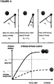
- the mechanical properties are used to describe the state of the tissues and their response to stress.
- the stress is a force, pressure or shear applied to the tissue.
- the response is measured as a strain or deformation of the tissue.
- the various stress loads and corresponding strain values can be plotted against each other and are referred here as "stress-strain curve".
- a solid body is characterized as stiff if the strain-stress curve is steep, i.e. little deformation is tolerated. If the stress is higher than rupture barrier then the solid body breaks (loses cohesiveness or integrity) and body can be described as fragile.
- a solid body is characterized as soft if the stress-strain curve is shallow, i.e. allows large strain (deformation).
- a solid body is characterized as elastic when the deformation is reversible if the stress is removed.
- the stress-strain curve in case of elastic bodies have little or no hysteresis.
- a solid body is characterized as plastic or gel-like when deformation is not reversible if the stress is removed.
- the stress-strain curve in case of plastic bodies have large hysteresis. Deformation of plastic (gel-like) bodies can also be tested in "creep test".
- Extracellular matrix plays an important part in providing a stiffness, shape, integrity, shape and architectural organization of mammalian tissues and consists of several proteins (collagens, laminins, perlecans, agrins etc) and proteoglycans (Aumailley et.al. 1998). Stiffness of extracellular matrix sets limitations upon organs ability to expand (e.g. cirrhotic liver), to move within certain range (e.g. adhesive capsulitis), to attain a different shape (e.g. wrinkles). Stiffness of extracellular matrix is capable of defining differentiation patterns of cells (Engler 2004, Cell), to stimulate or restrict migration of certain cell types.
- collagen scaffold which is formed by thick and dense cross-linked fibrils (Aumailley et.al. 1998). Crosslinking of collagen fibrils increases with age with tissues becoming more fragile and less elastic, as often seen in skin, eye lens, bones etc.
- Tendon is composed of different molecules assembled into different architecture compared to pancreatic islet or cartilage.
- Extracellular matrix provides not only durability and elasticity, but also architecture and compartmentalization of a complex tissue.
- Extracellular matrix and collagens are conservative between species. That is why animal collagens are used in medicine. They have reduced risk of immune reaction and rejection.
- Extracellular matrix and collagen scaffolds are extremely durable. Turnover of collagen fibrils in organism is extremely slow (years) compared to turnover rate of non-extracellular matrix molecules. Collagen scaffolds maintain their architecture, durability, shape and integrity for years, in living and non-living tissues.
- stress stress
- strain stress-strain curve
- Treatment with TREZ would allow to soften the tissues and allow their faster remodelling and healing, while preserving a functional integrity
- Stiffness of extracellular matrix is known to define cell behavior, such as: adhesion, migration, proliferation and even differentiation patterns. Modification of tissue stiffness in situ could be the key to regulate cell behavior, including differentiation, within natural environment of the cell (Discher, Janmey et al. 2005; Engler, Sen et al. 2006; Engler, Sweeney et al. 2007).
- Proteases are used in medicine, biotechnology, cosmetics, veterinary, food industry and other areas for decades ( US patent 3003917 by Beiler et.al., dated 1961) . Examples include wound debridement (Klasen 2000), pancreatic islets derivation (Li, Yuan et al. 2009), cell culture (trypsin), proteolytical enzymes are used for meat softening, hide processing and other industries.
- Collagenases are a subset of proteases that have collagen as as a preferred substrate. We classify collagenases into following three categories
- bacterial collagenases Biological function of bacterial collagenases is to allow pathogenic bacteria to invade and spread into host tissue. Expression of bacterial collagenases allows pathogenic bacteria to pass through mammalian protection barriers in the body. As Clostridium bacteria (including Clostridium histolyticum) cause gas gangrene, the bacteria making collagenase don't care about maintaining health of the host organism. In our experiments we see that collagenase from Clostridium histolyticum digests mammalian tissues into microscopic, non-cohesive parts or to cell suspension.
- proteolytical enzymes that can remodel mammalian (and other eukaryotic) tissues with high efficiency, however, being non-hazardous for the organism.
- Some of this enzymes are needed for organism growth (especially in embryonic and early postnatal age), some are needed for adult tissues remodeling (such as ovaries, uterus, mammary glands following onset of puberty, menstrual cycle, pregnancy, nursing following mammary gland involution), some are used by migratory immune cells to enable tissue-safe migration, some are overexpressed while organ repair after trauma (for example, skin healing or bone re-growth). All those natural processes require conditions named below:
- tissue-remodeling enzymes such as matrix metalloproteinases (MMPs)
- MMPs matrix metalloproteinases
- Tissue-Remodeling Enzymes would include a well-known family of matrix metalloproteinases (known as MMPs), and also less known families, such as: ADAMs, ADAMTSs, matriptases, collagenolytic cathepsins, collagenolytic members of kallikrein family and other endopeptidises, that possess protelytic activity towards extracellular matrix components that provide tissues stiffness.
- MMPs matrix metalloproteinases
- ADAMs ADAMTSs
- matriptases matriptases
- collagenolytic cathepsins collagenolytic members of kallikrein family
- endopeptidises that possess protelytic activity towards extracellular matrix components that provide tissues stiffness.
- Some of those enzymes are proteolytic towards interstitial collagens (they are named collagenases), some towards collagen IV, some towards other extracellular matrix components.
- native collagenases proteolytical enzymes that are naturally responsible for digesting collagen structures in tissues during remodeling
- Tissue-Remodeling Enzymes have evolved to remodel tissues without harming them they have evolutionary developed properties that provide advantage over aggressive collagenases in such applications that require to maintain functional integrity within treated tissues.
- Tissue of our organisms can be divided into permanent and temporary.
- Permanent tissues maintenance is essential for (1) organism health and (2) survival of species, to which organism belongs.
- Examples of permanent tissues are: liver, kidney, pancreas, heart, muscle, nerve system, circulatory system, skin, tendon, cartilage.
- Lack or deficiency of permanent tissues is a disease (examples: lack of skin is wound or ulcer, lack of vasculature causes ischemia followed by necrosis, lack of liver or kidney causes systemic failure).
- reproductive organs such as ovaries, uterus, testis, penis because they are necessary for survival of the species, therefore, are permanent part of healthy organism.
- Temporary tissues such as fibrin clot, scar tissue or fat, are, as the definition implies, created to serve a temporary function (wound healing, blood clotting, energy storage) and intended later to be degraded by organism itself.
- Functional integrity is level of tissue cohesiveness such that maintains the architecture integrity level sufficient for tissue to perform it's function.
- Permanent tissues evolved with a particular structure, tissue organization and compartmentalization to serve a biological purpose.
- liver's vascular system, hepatocytes and immune system cells are strictly compartmentalized.
- pancreatic islet insulin producing beta cells and vascular endothelial cells are proximal, but separated by a basement membrane. This arrangement is needed for a proper function of beta cells.
- tendon the collagen fibrils are strictly organized to provide high level of elasticity and durability. Cohesion between collagen fibrils is essential for tendon durability and stress-resistance.
- Blood-brain, blood-testis and blood-pancreatic barriers are examples of how compartmentalization is important. If these barriers fail, the immune system attacks brain, testis or pancreatic islets and destroys their function.
- Functional integrity refers not only to a cohesion within tissue, but also how it connects to other tissues.
- Figures 1 , 2 and Table 3 illustrate level required for functional integrity on example of muscle and skin. For example, if muscle maintains integrity and tendon maintains integrity, but muscle fails to connect tendon, then the whole muscle-tendon system fails to serve a biological function. If muscle fibers maintains integrity, but innervation within muscle is ruptured, the muscle will not able to react to neuronal signals and would fail the function. If skin dermis is detached from skin epidermis, it will cause blistering (example: epidermolysis bullosa, bullous pemphigoid, blistering)
- cell niche Many cell types can function properly only if they receive proper signals from local microenvironment (cell niche). It is important that cell keeps contact with particular neighboring cells (same type or different type), extracellular matrix around and maintains a proper focal orientation.
- Nerve-muscle-tendon system functional integrity
- Function of muscle is (1) to receive signal from motor neurons, (2) contract upon the signal, (3) pull the tendons, (4) the tendons would move the bones and the organism would move. Additionally, (5) the healthy, undamaged muscle should not send "pain" signals via sensory neurons.
- Desired outcome degradation or cohesive elastication of the tissue
- the desired outcome of enzymatic treatment of tissue can differ from complete tissue degradation to subtle change of its mechanical properties.
- Desired outcome is degradation of extracellular matrix with release of cells or complete destruction of the tissue
- Desired outcome to soften and elasticate the tissue while preserving a functional integrity of the treated tissue/organ and surrounding tissues/organ. Loss of functional integrity by aimed tissue or neighboring healthy tissues is considered a side effect.
- tissue samples treated by TREZ, remained in one piece, unlike samples treated by bacterial or crab collagenase which dissociated into incohesive parts.
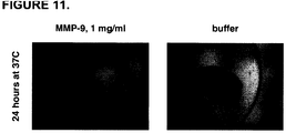
- MMP-9-treated tendon was split into individual, but interconnected fibers so that the tendon remains cohesive. Since the fibers remained intact and connected with each other and to the muscle, the functional integrity was preserved ( Figures 7 , 8 , 9 ). In other example pancreas treated by MMP split into smaller lobules that are interconnected and have intact acini and intralobular ducts ( Figures 10 , 11 ).
- MMP-9 treatment even enhanced cohesion for cells within liver sample; treatment with MMP-9 rendered liver tissues less prone to cell detachment and to microscopic ruptures.
- Tissues, treated by TREZ remained in one piece in the following conditions:
- Some permanent mammalian tissues can be described as stiff (bones) and most others as elastic (muscle, skin, tendons)
- Shape of tissues can be modified by scalpel or surgeon or by application of long-term pressure (such as dental braces to correct positioning of teeth).
- Plastic materials easily change shape and adjust shape to form to which they are placed, adhere to surfaces better.
- Creep assay is used to measure how stress causes gradual deformation.
- the assay is carried out as follow.
- the tissue sample is placed on sensitive scale.
- a depth micrometer (Starrett 445MBZ-150RL Vernier Depth Gauge) is rigged on a stand above the sample.
- Micrometer screw applies a defined deformation to a tissue sample.
- the resulting stress is measured on the scale readout. Change of stress in time can be measured and recorded.
- the best assay to evaluate stiffness vs stretch ability of tissues is building stress-strain curve. The curve allows to estimate
- Tissues fragility a risk for rupture and erosion
- TREZ-treatment allows to reduce tissues brittleness (fragility)
- MMP-9 treated and buffer-treated eye lens were subject to same force compression. While buffer-treated eye lens cracked, the MMP-9 treated eye lens resulted to external force with elastic deformation, but not with rupture.
- the best assay to evaluate stiffness vs stretch ability of tissues is building a stress-strain curve.
- the curve allows to estimate the limits of how far the tissue can be compressed (before and after TREZ treatment).
- Another assay is to apply same force to equivalent samples of tissue, treated by TREZ or by buffer (negative control). Start with low force and gradually increase, equal for both samples. As TREZ-treated sample reacts with deformation, the untreated sample (or one treated by buffer) would remain stiff until it suffers rupture at certain load.
- tissue samples (1) pancreatic islets, (2) eye lense (half), (3) eye lens (whole).
- tissues treated by TREZ become more gel-like and adhesive (1) to same type of tissue, (2) to different type of tissue, (3) to foreign surfaces.
- Adhesivity is a consequence of tissue becoming more plastic. Besides plasticity assays there are qualitative assays to specifically evaluate adhesivity:
- Possibility to change shape of permanent tissues would be highly desirable for (1) reconstructive surgery, (2) plastic surgery, (4) regenerative medicine, (5) transplantology and many other medical and veterinary applications (see Figure 12 for illustration).
- the natural stiffness of majority of permanent tissue allows them to maintain shape within range of physiological range for many years; however, for named medical applications it would be desirable to increase temporarily plasticity of tissue and mold it into desired shape.
- TREZ-treatment allows to modify shape and geometrical proportions of permanent tissues
- the effect can be quantitatively described by the (1) plasticity stress-strain characteristics (described in previous chapter) and also by plots of: (2) area increase after temporary stress applied and removed and (3) reduction of tissue sample thickness after temporary stress applied and remove.
- Permeability is a key property to tissues viability and ability to expand.
- Permeability is increased with (1) reduced thickness of individual components comprising tissue, (2) reduced stiffness and increased plasticity, that enables fluid flows within the tissue.
- tissue For example of tendon and pancreas we can see such remodeling of tissue that allows to reduce thickness of individual components comprising the tissue many times.
- TREZ-treatment allows to reduce thickness of the tissue sample significantly more, compared to tissues not treated by TREZ. Increased plasticity and decreased stiffness was quantitated and demonstrated for many permanent tissues.
- TREZ MMP-9 in our example
- tissues including skin dermis
- TREZ allows to soften, stretch, adjust shape, improve adherent abilities, while maintaining integrity.
- TREZ does not disrupt integrity of nerve bundle.
- Consequences for skin transplantation could be:
- Tissue fusion (cartilage and other soft tissues)
- ⁇ stem cells can undergo numerous divisions and maintain ability (1) to proliferate, (2) to differentiate into functional cell types. Not only stem cells, but also differentiated adult cells have high capacity for proliferation, for example: pancreatic insulin-producing beta cells, vascular endothelial cells, fibroblasts, keratinocytes, hepatocytes.
- Flap techniques allow to increase amount of autologous tissues for transplantation.
- the advantage of method is that tissue (while growing) is connected to blood circulation, thus reducing risks of ischemia.
- Living donor liver transplantation is a method to cure patients with end stage liver disease, such as cirrhosis and/or hepatocellular carcinoma (caused, for example, by hepatitis C or B infection).
- end stage liver disease such as cirrhosis and/or hepatocellular carcinoma (caused, for example, by hepatitis C or B infection).
- the liver is organ highly capable of regeneration; hepatocytes have high proliferation capacity.
- TREZ-softening of liver could be used to allow (1) the transplanted liver lobe and (2) the liver lobes left in the donor to expand to healthy amount of liver tissue and have no restriction to newly formed liver architecture caused by extracellular matrix stiffness.
- TREZ ability of TREZ to soften the tissues without loss of functional integrity allows to serve those needs and to reduce risks of (1) tissue blistering or disruption, (2) complete loss of structure within treated tissue or neighboring tissues, (3) damage the nerve structure (thus causing pain, numbness or loss of motility).
- Donor-derived insulin-producing beta islets are successfully used for treatment Diabetes type I. Edmonton Protocol, currently used for treatment of diabetes patients, involves isolating islets from a cadaveric donor pancreas using a bacterial collagenolytic enzymes and injecting them into patient's portal vein aiming for attachment and hosting within patients liver. Several cadaveric donors are required to treat one patient. Efficiency of transplanted islets are several years.
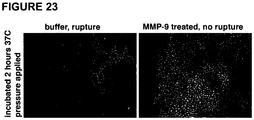
- pancreatic islets can allow to soften them and spread them thinner while maintaining functional integrity (without causing breakage (ruptures): see Figure 23 ). Also we have shown, that TREZ-treated islets become more adhesive: they can merge with tissues without gaps, hold attached and contact flat or complex surfaces without gaps. Such effect could be beneficial for the field of islet transplantation in following ways (but not limited to those):
- Human islets when derived from donor pancreases, suffer from insufficient exchange of gases, nutrients and waste products. In vivo the islets are surrounded by dense capsule, size of human islets varies within one to several hundred micrometers and mass exchange of endocrine cells within islets is enabled due to massive vascularization within the islet (a network of capillaries goes through each islet). After islets are detached from circulatory system, the inner capillaries of islet stop to serve function of mass exchange, and the only source of gases, nutrients and waste products is outside surrounding (in vitro culture medium or in vivo patient's bloodstream where the islets are injected).
- TREZ-softening is that modification of physical and geometrical properties of islet (softening, stretching, improved adhesivity, mechanically cut after softening) can be performed within few hours, before ischemia starts to affect the viability and healthy function of the islet cells.
- Islets after being transplanted into the patient, face the same problem of insufficient mass exchange rate. It takes over several days till islets may restore inner vascular flow connected to the patient blood circulation. In current islet transplantation protocols there is a problem, that part of islets fail to survive and function within the patient body.
- the islets can be frozen and stored in liquid nitrogen, however, size of islets sets limitations on the procedure (freezing and thawing whole islets is a more complicated compared to single cells). Reducing size of islets (or thickness) could reduce the risks and complications of islets banking.
- Islet size can be reduced by using TREZ as described in previous chapters.
- Improving mass exchange within derived islets may be beneficial also for long-term in vitro cultures, for example (1) for scientists, working to understand islet function, (2) R&D specialists, working on ability to expand islets in vitro, (3) in drug screening, islet culture being a target for anti-diabetes drug candidates, (4) in drug screening, islet culture used to study side effects of potential drug candidates
- pancreatic islets Possibility to expand insulin-producing pancreatic islets in vitro is a long-wanted need in islet transplantation and diabetes therapy. If islets could be expanded in vitro, (1) less of allogenic cadaveric donor material (pancreases) would be needed for transplatation, (2) it would be possible to arrange banking of all histocompatibility donor types, (3) it would allow to use a biopsy from living donor (for example: relative or histocompatible volunteer) to serve one patient instead of several whole cadaveric pancreases (as it is now).
- living donor for example: relative or histocompatible volunteer
- TREZ-treatment renders tissues more soft and gel-like, it would allow (1) to reduce local stress, (2) reduce risk of microscopic ruptures, (3) reduce risk of erosion for already occurring microscopic ruptures.
- pancreatic islets we demonstrate experimental MMP-9 effect of reducing brittleness and therefore reducing risk of rupture for (1) pancreatic islets, (2) eye lens: see Figures 23 and 24 .
- Plastic or reconstructive surgery of bones can benefit from use of TREZ as described above.
- Regenerative medicine offers opportunities of creating artificial tissues using (1a) autologous patient's cells (adult cells, stem cells, iPS cells or else) or (1b) allogenic donor compatible cells and (2a) artificial scaffolds, (2b) patient's own extracellular matrix, (2c) allogenic or xenogenic extracellular matrix.
- Another option is expanding tissues (autologous or donor) in vitro to increase amount of transplant material.
- TREZ for softening of allogenic or xenogenic extracellular matrix material, devoid of cells.
- Regenerative medicine offers opportunities of replacing a degenerated organ with a functional one, artificially engineered, or expanded from healthy tissue, or generated from human cells, untreated or modified, differentiated or stem cells in vitro. It would be highly desirable to expand small parts of healthy tissue, that can be taken as biopsy, into large ones for transplantation purposes. Possibility to expand in vitro whole tissues or organ parts instead of single cells could potentially allow to expand the functional organ of organized complexity (for example: podocyte cell culture expanded in vitro would not possess the functional organization of whole kidney). Therefore, methods to expand whole tissues would be highly desirable. However, the tissue proliferation ability is much lower compared to proliferation to single cell cultures comprising same tissue. Hence, stiffness of tissue and low plasticity of it set strict limitation on possibility fo tissue to expand (that is how mammalian organisms restrain size of internal organs).
- Proteolytical enzymes are traditionally used to treat scars caused by (1) acne, (2) dermatological pathologies, (3) fragility or sensitivity of skin. The enzymes are traditionally used to be applied to scars and dissolve them.
- TREZ can serve the same need, but in a different method.
- the TREZ can be applied to skin suffering fragility, rupture, erosion, lesion (caused by acne or other pathology) in order to (1) reduce risk of rupture and erosion, (2) reduce risk of scarring by the mechanisms we discussed in previous sections.
- Non-living tissues modifying physical properties
- TREZ-treatment of tissues can also be applied to non-living tissues.
- Extracellular matrix from allogenic or xenogenic donor can be transplanted and be inhabited by own cells of the patient or histocompatible donor (variants: stem cells, stem cells expanded in vitro, stem cells differentiated in vitro, adult cells, iPS cells, etc).
- Decellularized tissues and organs that are developed include:
- TREZ-treatment can be useful for extracellular matrix devoid of living cells.
- Whole extracellular matrix, devoid of cells, has same limitations for transplantation as a living transplant does.
- Treatment with TREZ prior to transplantation can improve following parameters:
- MMP matrix metalloproteinase
- Activation was performed according to standard protocols: 4-aminophenylmercuric acetate (APMA) activation (0.5-1 mM) at neutral pH (pH7.5-8.0) in presence of calcium ions (1-10 mM) for enzymes of MMP family and disulfide bonds reduction by 5mM DTT (dithiothreitol) at acid pH (pH 5.0) for the cathepsins, followed by removal of activator.
- APMA 4-aminophenylmercuric acetate
- Zymogens full-size, pre-activated forms of enzymes
- lyophilized form accordinging to producer's protocols
- transfer buffers by means of dialysis.
- Buffer replacement for example, to remove the activator
- membrane MWCO molecular weight cut-off
- HBSS Hank's Balanced Salt Solution
- Tissue samples were preliminary examined and photographed, prior to enzyme application.
- Tissue types used for our studies, were: skin, tendon (major limb tendons, including Achilles and minor finger tendons), cartilage (of nose and of ear), subcutaneous soft tissues of lip, penis foreskin, subcutaneous soft tissues of penis, liver, soft tissues of uterus wall, ovaries, pancreas, pancreatic insulin-producing islets of Langerhans, eye lens, nerve bundle (sciatic nerve, optic nerve), muscle, musculotendinous junction.
- tissue samples were generated, photographed and placed into plastic tubes, activated enzyme or control buffer solution being placed above. Tissue samples were gently rinsed by applied solution before incubation. Tissue samples were incubated at 37C at water bath (for non-living tissues) or cell culture incubator (for living tissues), time of incubation varying for each experiment (from 30 minutes to 4 days). During incubation tissue samples were gently rinsed by applied solution about 1-2 times per day. After treatment the samples were placed into HBSS solution devoid of enzymatic activity and subject to set of assays to qualify functional integrity maintenance, mechanoelastic and viscoelastic properties (such as: stiffness, plasticity, adhesivity, poroelasticity, fragility, cohesiveness, etc).
- mechanoelastic and viscoelastic properties such as: stiffness, plasticity, adhesivity, poroelasticity, fragility, cohesiveness, etc).
- In vivo assay thin segments of skin including dermis were generated in flap-like technique on anesthesized animals and activated enzyme vs control buffer were applied in form of solution were applied to each flap-like tissue segment in situ. The segments were rinsed by fresh solutions (of activated enzyme or buffer) several times per day for 2 days and solutions, after rinse, were kept applied to the treated segments. After treatment, the enzyme- and buffer-treated skin segments were excised and subject to set of assays to qualify functional integrity maintenance, mechanoelastic and viscoelastic properties (such as: stiffness, plasticity, adhesivity, poroelasticity, fragility, cohesiveness, etc).
- tissue-specific criteria of functional integrity are tissue-specific, such as: exocrine duct system integrity in pancreas, cohesion between muscle fibers in muscle, cohesiveness between two proximal ends of tendon. Tissue-specific criteria for functional integrity will be named for each tissue individually. However, general criteria are:
- phase contrast mode we variated settings for phase in order to distinguish between objects with similar transparity.
- Stress-strain characteristics of tissue are used in several assays and applications, such as: stiffness, creep assay, deformation under permanent load, residual deformation after temporary load.
- Method 1 Apply stress to the tissue specimen and measure relative deformation.
- the tissue specimen are placed in 24-well or 6-well tissue culture plate on coverglass and covered with HBSS buffer.
- the plate with specimen were placed under dissection microscope and photographed.
- Loads of progressive mass were applied to the specimen covered with coverglass; shape of load being thoroid in order to allow photograph the sample under the load.
- Coverglass placed upon the specimen were immersed in same buffer in order to avoid capillary effects that would distort the stress evaluation.
- the specimen were subject to increading loads of progressive mass. The specimen was photographed before and after the loads were applied; the surface area was measured by imaging program (in pixels, number being proportional to surface area).
- Strain was calculated as relative deformation of sample. Relative area is calculated difference in areas after load and prior to load, calibrated to initial area of specimen. Relative thickness of sample was estimated as reverse proportion of relative area of the specimen.
- Method 2 Apply deformation to the tissue specimen and monitor the resulting stress exhibited by specimen.
- the tissue specimen were placed sensitive scale.
- a depth micrometer (Starrett 445MBZ-150RL Vernier Depth Gauge) is rigged on a stand above the sample.
- Micrometer screw applied a defined permanent deformation to a tissue sample. The resulting stress is measured on the scale readout and the change of stress in time was observed.
- the assay measures relative residual deformation (strain) after temporary stress applied. Permanent stress was applied for 2 minutes, afterwards removed and the specimen was allowed to restore previous shape for another 2 minutes. Residual deformation was measured by technique described above.
- the assay measures relative deformation (strain) permanent stress applied as described before. Permanent load was applied to a tissue sample placed under dissection microscope, properties of load adjusted to allow photographing of the tissue sample under the load. Photographs were taken of tissue sample before loads, and following a series of increased loads. Stress and strain valued were calculated as described above.
- Tissue samples treated by enzymes or buffer, can be qualitatively evaluated: do they behave like elastic material (like they used to before treatment) or like gels.
- Permeability depends on thickness of individual elements within tissue sample. Effects of increased permeability of reduced thickness can be evaluated for MMP-9 treated tendon and MMP-9 treated pancreas; as seen on Figures 7-11 .
- Preparations of human recombinant proteinases MMP-1, MMP-8, MMP-9, Cathepsin B and Cathepsin S were activated and transferred into working buffer of appropriate pH (neutral for MMP's and pH5.0 for cathepsins) and ionic additives (calcium ions for MMP's in concentration from 1 to 10 mM).
- the enzymes were diluted to concentrations of 0.03 mg/ml and applied to lip connective tissue samples and several other tissue types.
- Collagenase of Clostridium histolyticum (Roche) and crab collagenase (YO Proteins) served as controls for aggressive collagenolysis.
- Buffers used for dilution of MMP's and Cathepsins served as negative controls. The samples were observed for several days and evaluated by qualitative assays to (1) functional integrity maintenance, (2) stiffness reduction, (3) plasticity enhancement, (4) viscous reaction to external stress/deformation, (5) ability for reshaping.
- Lip connective tissue reduced stiffness and increased plasticity after MMP-9 treatment
- Subcutaneous tissues of lip contain highly innervated muscular and connective tissues. They are highly sensitive, responsible for mimics and mouth function.
- Functional integrity requirement for such procedure would be (1) to maintain lip tissues cohesive (to avoid blisters and ulcers), (2) to avoid nerve rupture (such would cause pain, numbness, loss of motility)
- Tissue samples treated by MMP-9 (1) decreased stiffness, (2) increased plasticity, (3) improved adhesiveness while maintaining functional integrity.
- Tissue samples treated by collagenase of Clostridium histolyticum were dissociated into non-cohesive floating microscopic particles.
- Tissue samples treated by buffer (1) retained original stiffness and ability to maintain shape and (2) remained cohesive.
- Table 5 shows summary of qualitative assays.
- Figure 13 shows quantitative evaluation of stress-strain characteristics of lip tissue samples such as stiffness and plasticity, ability to increase area and to decrease thickness under stress.
- Figure 14 demonstrates effect of increased plascitity of MMP-9 treated lip tissue, compared to buffer-treated control.
- Figure 15 demonstrates effect of MMP-9 rendering lip tissue gel-like (adhesive, permeable, reshapable), compared to buffer-treated control.
- Lip connective tissue reduced stiffness and increased plasticity after Cathepsin B treatment
- Subcutaneous tissues of lip was treated by human recombinant Cathepsin B (concentration of 0.03 mg/ml; activated by 5 mM DTT at pH5.0 for 15 minutes at room temperature, which was subsequently removed by dialysis) vs buffer.
- the lip tissue was incubated with Cathepsin B vs buffer for 20 hours at 37C, being gently rinsed several times.
- Cathepsin B treated samples were of reduced stiffness and of increased plasticity compared to buffer-treated samples.
- Figure 16 shows quantitative evaluation of stress-strain characteristics of lip tissue samples such as stiffness and plasticity, ability to increase area and to decrease thickness under stress.
- Lip connective tissue reduced stiffness and increased plasticity after MMP-8 treatment
- Subcutaneous tissues of lip was treated by human recombinant MMP-8 (concentration of 0.03 mg/ml; activated by AMPA 0.5mM at pH8.0 at calcium ions concentration of 10mM for 2 hours at 37C) vs buffer.
- the lip tissue was incubated with MMP-8 vs buffer for three days at 37C, being gently rinsed 2 times per day.
- Qualitative evaluation shows that MMP-8 treated samples were less stiff and of increased plasticity compared to buffer-treated samples.
- Skin is a complex organ and it's proper function depends on cohesiveness within comprising tissue elements and between them (such as illustrated on Figure 2 and Table 3 ).
- Uniform samples of skin dermis were isolated by methods of microsurgery and were incubated for 2 days at 37°C in solution of: (1) MMP-9, 0.1 mg/ml, preactivated with 0.5mM APMA, (2) collagenase of Clostridium histolyticum, 0.1 mg/ml, (3) buffer solution. Samples were gently rinsed by applied solution every day and were subject to quantitative stress-strain and geometrical modification assessment, and for qualitative evaluation.
- Tissue samples treated by MMP-9 (1) decreased stiffness, (2) increased plasticity, (3) improved adhesiveness while maintaining functional integrity.
- Tissue samples treated by collagenase of Clostridium histolyticum were dissociated whole tissue into non-cohesive floating microscopic particles (hair remained intact, but detached).
- Tissue samples treated by buffer (1) retained original stiffness and ability to maintain shape and (2) remained cohesive.
- Table 5 shows summary of qualitative assays.
- Figure 17 shows quantitative evaluation of stress-strain characteristics skin dermis samples such as stiffness and plasticity, ability to increase area and to decrease thickness under stress.
- Tendons are highly durable and highly elastic tissues. Each tendon consists of interconnected fibrils, tightly packed in order to ensure high durability. There are medical and cosmetical needs for modification of mechanic and shape characteristics of tendon: (1) to decrease stiffness in aged or traumatized tendon, (2) to enable tendon expansion (in case of short tendon), (3) to expand tendon tissue in vitro for medical and veterinary purposes. Functional integrity requirement for such tendon modifications would be: (1) to maintain connection between individual fibrils comprising tendon, so that network of interconnected fibrils behaves as whole, (2) to avoid local ruptures
- Assay 1 Uniform samples of large limb tendon (Achilles or other) were isolated from mouse hind limbs by methods of microsurgery and were incubated for 1 day at 37°C in solution of: (1) MMP-9, 1 mg/ml, preactivated with 0.5mM APMA, (2) collagenase of Clostridium histolyticum, 1 mg/ml, (3) buffer solution. Samples were gently rinsed by applied solution.
- Assay 2 Uniform samples of large limb tendon (Achilles or other) were isolated from mouse hind limbs by methods of microsurgery and were incubated for 3 days at 37°C in solution of: (1) MMP-9, 0.1 mg/ml, preactivated with 0.5mM APMA, (2) collagenase of Clostridium histolyticum, 0.1 mg/ml, (3) buffer solution. Samples were gently rinsed by applied solution, daily.
- Assay 3 Uniform samples of finger tendon were isolated from mouse hind limbs by methods of microsurgery and were incubated for 1 day at 37°C in solution of: (1) MMP-9, 0.1 mg/ml, preactivated with 0.5mM APMA, (2) collagenase of Clostridium histolyticum, 0.1 mg/ml, (3) buffer solution. Samples were gently rinsed by applied solution.
- Table 5 shows summary of qualitative assays.
- Figures 8 , 9 shows difference in tendon dissociation pattern, when treated by MMP-9 (cohesive remodeling) vs collagenase of Clostridium histolyticum (non-cohesive digestion).
- Figure 7 illustrates the effect as a drawing.
- Cartilage (mouse and porcine): increased plasticity and reduced stiffness after MMP-9 treatment
- Cartilage is a flexible connective tissue comprising joints between bones, rib cage, ear, nose, bronchial tubes and intervertebral discs. Modification of cartilage mechanical and shape propertied would be desirable (1) in plastic and reconstructive surgery (example: rhinoplasty), (2) for treatment of herniated intervertebrate discs, however, on condition that treated cartilage does not degenerate, rupture or blister.
- Tissue samples treated by MMP-9 (1) decreased stiffness, (2) increased plasticity, (3) improved adhesiveness while maintaining functional integrity.
- Tissue samples treated by collagenase of Clostridium histolyticum were dissociated into non-cohesive floating microscopic particles.
- Tissue samples treated by buffer (1) retained original stiffness and ability to maintain shape and (2) remained cohesive.
- Table 5 shows summary of qualitative assays.
- Figure 18 shows quantitative evaluation of stress-strain characteristics of samples such as stiffness and plasticity, ability to increase area and to decrease thickness under stress.
- Penis foreskin and subcutaneous connective tissues: reduced stiffness and increased plasticity after MMP-9 treatment
- Tissue samples treated by MMP-9 (1) decreased stiffness, (2) increased plasticity, (3) improved adhesiveness while remaining cohesive.
- Tissue samples treated by collagenase of Clostridium histolyticum were dissociated into non-cohesive floating microscopic particles.
- Tissue samples treated by buffer (1) retained original stiffness and shape and (2) remained cohesive.
- Table 5 shows summary of qualitative assays.
- liver tissue stiffness would be desirable for expanding liver tissues for transplantation purposes, however, on condition that the treatment would maintain the complex composition of liver cells and compartments cohesive.
- Assay Uniform liver tissue samples incubated for 3 days at 37°C in solution of: (1) MMP-9, 0.1 mg/ml, preactivated with 0.5mM APMA, (2) collagenase of Clostridium histolyticum, 0.1 mg/ml, (3) buffer solution. Samples were gently rinsed by applied solution every day.
- Tissue samples treated by MMP-9 (1) decreased stiffness, (2) increased plasticity, (3) improved adhesiveness while maintaining functional integrity.
- Tissue samples treated by collagenase of Clostridium histolyticum were dissociated into non-cohesive floating microscopic particles.
- Tissue samples treated by buffer (1) retained original stiffness and ability to maintain shape and (2) remained cohesive.
- MMP-9 treated tissue allowed to maintain cells more cohesive within liver tissue sample, compared to buffer-treated tissue.
- Table 5 shows summary of qualitative assays.
- Figure 19 shows quantitative evaluation of stress-strain characteristics of samples such as stiffness and plasticity, ability to increase area and to decrease thickness under stress.
- Pancreas adhesive architecture remodeling improving permeability after MMP-9 treatment
- pancreas transplantation and (2) in vitro expansion of pancreatic tissue, however, on condition that tissue maintains functional integrity, which includes cohesion between exocrine cells and integrity of lumens and intralobular ducts, necessary for exocrine function.
- Tissue samples treated by MMP-9 have changed the architecture: solid piece of tissue separated into interconnected lobules in manner that maintained cohesion of cells within lobules and integrity of intralobular ducts. The lobules thickness was substantially lower compared to that of original tissue sample, thus, remodeling improved tissue permeability.
- Tissue samples treated by collagenase of Clostridium histolyticum were digested into non-cohesive floating microscopic particles.
- Table 5 shows summary of qualitative assays.
- Figure 11 shows difference in pancrea dissociation pattern, when treated by MMP-9 (cohesive remodeling) vs collagenase of Clostridium histolyticum (non-cohesive digestion).
- Figure 10 illustrates the effect as a drawing.
- Pancreatic insulin-producing islets of Langerhans reduced stress-strain ratio and increased adhesivity after MMP-9 treatment
- pancreatic islets ability to modify stiffness, plasticity, fragility, adhesivity and thickness of pancreatic islets would allow to solve a number of needs in islet transplantation for treatment of diabetes patients. However, it is highly desirable to maintain functional cohesion between cells within islet, as interaction between cells are important for proper islet function.
- Islets treated by MMP-9 (1) increased adhesivity, (2) increased plasticity and ability to reduce thickness, (3) increased cohesiveness between cells undergoing deformation.
- Tissue samples treated by collagenase of Clostridium histolyticum were dissociated into non-cohesive floating microscopic particles.
- Tissue samples treated by buffer (1) retained original stiffness and ability to maintain shape and (2) remained cohesive.
- MMP-9 treated islets could be deformed to high extent while cells remained cohesive and no ruptures occurred, compared to buffer-treated islets that ruptured under same deformation.
- Table 5 shows summary of qualitative assays.
- Figure 23 demonstrates effect of increased plasticity and increased cohesiveness within of MMP-9 treated pancreatic islets, compared to buffer-treated control that underwent rupture under same load.
- a method to reduce stiffness and reduce fragility of eye lens would be desirable in ophthalmology, however, that such method would not rupture the lens and optic nerve.
- Assay 1 Eye lenses were isolated from eyes of same healthy animal for MMP-9 and for buffer treatment to avoid potential differences in stiffness due to age and background. Eye lens for collagenase from Clostridium histoliticum was derived from animal of same age and condition. The eye lens samples were incubated for 4 days at 37°C in solution of: (1) MMP-9, 0.1 mg/ml, preactivated with 0.5mM APMA, (2) collagenase of Clostridium histolyticum, 0.1 mg/ml, (3) buffer solution. To assay compatibility with nerve integrity, uniform samples of optic nerve were isolated and were treated by same enzymes formulation at same conditions.
- Tissue samples treated by MMP-9 maintained functional integrity and were less less fragile: when pressure applied, the samples responded with elastic deformation, but not rupture.
- Tissue samples treated by buffer (1) retained original stiffness and shape and (2) responded to pressure by rupture, but not by elastic deformation.
- Tissue samples treated by collagenase of Clostridium histolyticum were dissociated into non-cohesive floating microscopic particles.
- Table 5 shows summary of qualitative assays.
- Figure 24 illustrates effect of MMP-9 treated eye lens is less prone to rupture, compared to buffer-treated control.
- Tissue samples treated by MMP-9 (1) maintained integrity of single nerves and of whole nerve bundle (no ruptures were observed, (2) integrity on nerves maintained also under deformation, (3) stiffness (stress-strain ratio) of MMP-9 treated sample reduced, thus allowing increased range of cohesive nerve deformation.
- Tissue samples treated by collagenase of Clostridium histolyticum were dissociated into non-cohesive floating microscopic particles.
- Tissue samples treated by buffer (1) retained original stiffness and ability to maintain shape and (2) remained cohesive.
- Table 5 shows summary of qualitative assays.
- MMP-9 reduces stiffness of muscle.
- Tissue samples (1) muscle samples including muscle fibers wrapped in endomysium and perimysium were subject to qualitative stress-strain assay (deformation under load); (2) muscle samples including myotendonous junction were used for qualitative assessment and to control integrity of myotendonous junction; (3) optic nerve and sciatic nerves were used as a control: being subject to enzyme of same formulation (concentration, activity, buffer composition, etc) in order to control integrity of nerves.
- Table 5 shows summary of qualitative assays.
- Granzyme A CTL tryptase Cytotoxic T-lymphocyte proteinase 1, Fragmentin-1, Granzyme-1, Hanukkah factor EC 3.4.21.78 P12544 (GRAA_HUMAN)
- Granzyme B EC 3.4.21.79
- Granzyme H P20718 GRAH_HUMAN
- TREZ 1 Producing organism aims to apply enzyme on it's own tissues
- NO NO YES 2 Producing organism aims to keep permanent mammalian tissues functional
- NO NO YES 3 Producing organism aims to preserve the functional integrity and architecture of aimed tissue NO NO YES 4
- Producing organism aims to preserve cohesive the aimed tissue NO NO YES 5
- Producing organism aims collagenolysis for healthy purposes for aimed tissue (like: regeneration, expansion, development)
- NO NO YES 6 Producing organism aims to preserve safe and functional the mammalian tissues neighboring to the aimed tissue
- NO NO YES 7 Producing organism aims to preserve safe the nerves (axons) within and around the aimed tissue NO NO NO YES
- Table 3 Summary of functional integrity of muscle and skin # Tissue unit
- Functional integrity of muscle and tendon 1 Nerves Numbness (sensory nerves), pain (sensory nerves), paralysis (motor nerves) 2 Muscle fiber wholeness Muscle rupture
- TREZ translation of mechanical and shape characteristics into medical and cosmetical applications.
- Diseases caused by pathological stiffness of organs and tissues can be treated by using TREZ to reduce stiffness (stress-strain ration) back to normal # Disease Evaluation criteria for disease and treatment efficacy Mechanoelastic properties of tissue resulting the disease How use of TREZ to modify the mechanoelastic characteristics can be used for disease treatment 1.
- Adhesive capsulitis (frozen shoulder), Range of shoulder motility is limited.
- Degree of pathology is evaluated as: (1) rotational range of motions (ROMs) and (2) glenohumeral (GH) stiffness Increased stiffness of connective tissues within shoulder, therefore,ROM is decreased in disease.
- TREZ treatment allows to decrease stress-strain ratio, therefore range of strain (range of shoulder movements) can be increased within physiological limits of stress (force that does not cause pain or damage). See Figure 4 .
- Dupuytren's contracture Angle of finger movement is limited by stiffened / shortened Dupuytren's cords. The range of finger bending angle is evaluation criteria (Hurst, Badalêt et al.
- Dupuytren's cords / shortened shape of Dupuytren's cords.
- TREZ treatment allows to decrease stress-strain characteristics of connective tissues; therefore, Dupuytren's cords can be either made more elastic, or be extended (shape modification). 3. Phimosis Inability of penis foreskin to extend enough to allow erection. Increased stiffness of penis foreskin TREZ treatment allows to reduce stiffness of penis tissues, therefore, healthy range of foreskin stretching can be restored. See Figure 5 .
- TREZ can be used to reduce stiffness (reduce stress-strain ration) for liver tissues, therefore, allow regenerative processes and restore function 7
- Increased stiffness of extracellular matrix within lungs TREZ can be used to reduce stress-strain ration, therefore, increase range of lung inflation
- Medical and cosmetical needs for increased plasticity can be treated by using TREZ to reduce stiffnes, increase plasticity and adhesivity # Medical procedure Evaluation criteria for disease and treatment efficacy Mechanoelastic properties of tissue resulting the disease How use of TREZ to modify the mechanoelastic characteristics can be used for disease treatment 8
- Plastic surgery rhinoplasty, otoplasty, mammoplasty, etc
- Shape or organ nose, ear, breasts, etc
- Natural stiffness (stress-strain characteristics) of tissues make tissues "remember" original shape.
- TREZ-treatment may be used to make tissues more plastic, therefore, more easy to attain desired shape.
- Use of temporary scaffold for intended shape would be beneficial (such as: dental braces for teeth or gold threads for facelift).
- 9 Reconstructive surgery for cleft lip. Extent of cleft (shape, depth of cleft) Stiffness and low adhesivity of untreated lip tissues TREZ-treated lip tissues could (1) expand, (2) merge to cover the cleft 10 Incorrect positioning of teeth Angle of incorrect tooth positioning Connective tissues around tooth are too stiff to allow easy change of tooth positioning Applying TREZ to connective tissues around tooth may allow to achieve better effect using dental braces. See Figure 6 .
- Islet transplantation increase survival rate Size of islet Untreated islets are too stiff to allow cohesive deformation into flat shape TREZ treatment may make make islets more plastic and allow cohesive reshaping into flat structures 12
- Transplantation adhesion of transplanted tissue to host tissue Gaps between transplanted tissue and host tissue Natural stiffness and low adhesiveness of tissues may cause gaps between transplant and host tissues TREZ treatment increases plasticity and adhesiveness of tissues. If transplant or host tissues are TREZ-treated, plasticity and adhesiveness would allow to reduce gaps. See Figure 12 .
Landscapes
- Health & Medical Sciences (AREA)
- Life Sciences & Earth Sciences (AREA)
- Chemical & Material Sciences (AREA)
- Engineering & Computer Science (AREA)
- Bioinformatics & Cheminformatics (AREA)
- Medicinal Chemistry (AREA)
- General Health & Medical Sciences (AREA)
- Immunology (AREA)
- Pharmacology & Pharmacy (AREA)
- Proteomics, Peptides & Aminoacids (AREA)
- Epidemiology (AREA)
- Animal Behavior & Ethology (AREA)
- Gastroenterology & Hepatology (AREA)
- Public Health (AREA)
- Veterinary Medicine (AREA)
- Wood Science & Technology (AREA)
- Genetics & Genomics (AREA)
- Biomedical Technology (AREA)
- Zoology (AREA)
- Organic Chemistry (AREA)
- Molecular Biology (AREA)
- Biotechnology (AREA)
- Microbiology (AREA)
- Biochemistry (AREA)
- General Engineering & Computer Science (AREA)
- Materials For Medical Uses (AREA)
Claims (7)
- Verwendung von voraktiviertem humanem MMP-9 zum Bereitstellen einer Modifizierung der mechanischen Eigenschaften von Säugetierbindegewebe, bei der das Gewebe kohäsiv bleibt, wobei das MMP-9 durch 4-Aminophenylquecksilberacetat-(APMA-)Aktivierung voraktiviert wurde und wobei das MMP-9 ex vivo mit Proben von Säugetierbindegewebe inkubiert wird.
- Verwendung von voraktiviertem humanem MMP-9 nach Anspruch 1, wobei das Bindegewebe Knorpelgewebe ist.
- Verwendung von voraktiviertem humanem MMP-9 nach einem der vorstehenden Ansprüche, wobei das Gewebe unter statischer oder dynamischer Belastung oder Deformierung Kohäsion bewahrt.
- Verwendung von voraktiviertem humanem MMP-9 nach einem der vorstehenden Ansprüche, wobei die mechanischen Eigenschaften eine oder mehrere Eigenschaften, ausgewählt aus der Gruppe, bestehend aus Steifigkeit, Plastizität, Elastizität, Brüchigkeit, Sprödigkeit, mechanische Elastizität, Viskoelastizität, Adhäsionsvermögen, Fähigkeit, das Rissrisiko zu reduzieren, Fähigkeit, das Erosionsrisiko zu reduzieren, oder beliebigen Belastungs-/Beanspruchungseigenschaften, sind.
- Verwendung von voraktiviertem humanem MMP-9 nach einem der vorstehenden Ansprüche, wobei die Modifizierung der mechanischen Eigenschaften eine Senkung der Steifigkeit (Belastungs-/Beanspruchungsverhältnis) des Gewebes, bevorzugt auf mehr als 1 %, mehr bevorzugt auf mehr als 10 % und am stärksten bevorzugt auf mehr als 30 %, in behandeltem Gewebe im Vergleich zu unbehandeltem Gewebe ist.
- Verwendung von voraktiviertem humanem MMP-9 nach einem der vorstehenden Ansprüche, wobei die Modifizierung der mechanischen Eigenschaften des Gewebes eine Erhöhung der Grenzen der kohäsiven Deformierung (Belastung) oder des Bewegungsbereichs (range of motion - ROM) des Gewebes, bevorzugt auf mehr als 1 %, mehr bevorzugt auf mehr als 10 % und am stärksten bevorzugt auf mehr als 30 %, in behandeltem Gewebe im Vergleich zu unbehandeltem Gewebe ist.
- Verwendung von voraktiviertem humanem MMP-9 nach einem der vorstehenden Ansprüche, wobei die Modifizierung der mechanischen Eigenschaften eine Erhöhung der Plastizität des Gewebes, bevorzugt auf mehr als 1 %, mehr bevorzugt auf mehr als 10 % und am stärksten bevorzugt auf mehr als 30 %, in behandeltem Gewebe im Vergleich zu unbehandeltem Gewebe ist.
Applications Claiming Priority (2)
| Application Number | Priority Date | Filing Date | Title |
|---|---|---|---|
| US201261640008P | 2012-04-30 | 2012-04-30 | |
| PCT/SE2013/050472 WO2013165304A1 (en) | 2012-04-30 | 2013-04-26 | Methods for modification of tissues |
Publications (3)
| Publication Number | Publication Date |
|---|---|
| EP2852403A1 EP2852403A1 (de) | 2015-04-01 |
| EP2852403A4 EP2852403A4 (de) | 2016-04-20 |
| EP2852403B1 true EP2852403B1 (de) | 2018-03-07 |
Family
ID=49514591
Family Applications (1)
| Application Number | Title | Priority Date | Filing Date |
|---|---|---|---|
| EP13784517.8A Not-in-force EP2852403B1 (de) | 2012-04-30 | 2013-04-26 | Verfahren zur modifizierung von gewebe |
Country Status (3)
| Country | Link |
|---|---|
| US (1) | US20150079069A1 (de) |
| EP (1) | EP2852403B1 (de) |
| WO (1) | WO2013165304A1 (de) |
Families Citing this family (10)
| Publication number | Priority date | Publication date | Assignee | Title |
|---|---|---|---|---|
| EP2844276B1 (de) * | 2012-05-01 | 2019-10-02 | Proteolease Ltd. | Verfahren zur extraktion eines zahns |
| WO2015038058A1 (en) * | 2013-09-16 | 2015-03-19 | Elastomics Ab | Modification of the mechanical properties of living tissue |
| WO2015140802A2 (en) | 2014-03-20 | 2015-09-24 | Technion Research & Development Foundation Limited | Delivery system comprising a proteolytic enzyme or effector thereof for use in a method for oral treatment and uses thereof |
| AU2016241901B2 (en) * | 2015-04-03 | 2021-10-28 | Nanocell Ltd. | Compositions for remodeling extracellular matrix and methods of use thereof |
| RU2687973C9 (ru) * | 2018-04-03 | 2020-01-23 | Федеральное государственное унитарное предприятие "Санкт-Петербургский научно-исследовательский институт вакцин и сывороток и предприятие по производству бактерийных препаратов" Федерального медико-биологического агентства России (ФГУП СПбНИИВС ФМБА России) | Способ получения лекарственного средства коллализин®, лиофилизата для приготовления раствора для местного и парентерального применения для лечения фибропролиферативных заболеваний |
| US20210298812A1 (en) * | 2018-08-02 | 2021-09-30 | Sofwave Medical Ltd. | Devices and methods for vaginal treatments |
| EP3829713B1 (de) | 2018-08-02 | 2025-10-01 | Sofwave Medical Ltd. | System zur behandlung von fettgewebe |
| US11253575B2 (en) * | 2020-04-24 | 2022-02-22 | Innomed Technologies, Inc. | Collagenase Clostridium Histolyticum injection therapies |
| CA3203037A1 (en) | 2020-12-31 | 2022-07-07 | Ariel Sverdlik | Cooling of ultrasound energizers mounted on printed circuit boards |
| CN114949357B (zh) * | 2022-05-20 | 2023-08-01 | 上海市第十人民医院 | 一种阴茎脱细胞支架及其制备方法和应用 |
Family Cites Families (19)
| Publication number | Priority date | Publication date | Assignee | Title |
|---|---|---|---|---|
| US3003917A (en) | 1959-01-14 | 1961-10-10 | Nat Drug Co | Wound healing composition |
| DE3719367A1 (de) * | 1987-06-10 | 1988-12-29 | Duncker Martin Dr | Verwendung von den stoffwechsel und/oder die struktureigenschaften von kollagen beeinflussenden wirksubstanzen in der kieferorthopaedie |
| US5260059A (en) * | 1989-04-14 | 1993-11-09 | The State Of Oregon Acting By And Through The State Board Of Higher Education On Behalf Of Oregon Health Sciences University | Treatment of open-angle glaucoma by modulation matrix metalloproteinases and their inhibitor |
| EP0637450A3 (de) | 1993-08-04 | 1995-04-05 | Collagen Corp | Verfahren und Zusammensetzung zur Wiederbelebung von Nerbgewebe. |
| US5641636A (en) * | 1994-05-20 | 1997-06-24 | University Of Pennsylvania | Method of predicting fetal membrane rupture based on matrix metalloproteinase-9 activity |
| US6194189B1 (en) * | 1994-12-16 | 2001-02-27 | Washington University | Catalytically-active gelatinase mutant |
| US5830468A (en) | 1995-05-17 | 1998-11-03 | The New York Blood Center, Inc. | Fibrin(ogen) degradation by fibrinolytic matrix metalloproteinase |
| US6086872A (en) * | 1997-03-27 | 2000-07-11 | Advance Biofactures Of Curacao, Nv | Amelioration of dupuytren's disease |
| AU1864999A (en) * | 1997-12-15 | 1999-07-05 | Universite Laval | Methods and compositions for improving the success of cell transplantation in a host |
| FR2798671A1 (fr) | 1999-09-16 | 2001-03-23 | Univ Paris Curie | Compositions de chondrocytes, preparation et utilisations |
| US7320783B2 (en) * | 2002-07-17 | 2008-01-22 | The Regents Of The University Of Michigan | Methods and compositions for the enhancement of wound healing |
| US7935337B2 (en) | 2002-12-25 | 2011-05-03 | Hiromichi Komori | Method of therapy for degenerative intervertebral discs |
| AU2004213050B2 (en) * | 2003-02-20 | 2010-09-09 | Proteon Therapeutics, Inc. | Methods for the treatment and prevention of diseases of biological conduits |
| WO2007117451A2 (en) | 2006-03-31 | 2007-10-18 | The Regents Of The University Of Michigan | A pericellular collagenase directs the 3-dimensional development of white adipose tissue |
| US8323642B2 (en) | 2006-12-13 | 2012-12-04 | Depuy Mitek, Inc. | Tissue fusion method using collagenase for repair of soft tissue |
| US8541370B2 (en) | 2007-02-26 | 2013-09-24 | Proyecto De Biomedicina Cima, S.L. | Use of matrix metalloproteinase-10 (MMP-10) for thrombolytic treatments |
| TWI395593B (zh) | 2008-03-06 | 2013-05-11 | Halozyme Inc | 可活化的基質降解酵素之活體內暫時性控制 |
| JP5572291B2 (ja) | 2008-04-07 | 2014-08-13 | 独立行政法人国立長寿医療研究センター | 歯髄及び/又は象牙質形成促進のための薬剤及びその利用 |
| EP2403523A1 (de) * | 2009-03-06 | 2012-01-11 | Halozyme, Inc. | Temperaturempfindliche mutanten von matrix metalloprotease 1 und ihre verwendungen |
-
2013
- 2013-04-26 EP EP13784517.8A patent/EP2852403B1/de not_active Not-in-force
- 2013-04-26 US US14/397,378 patent/US20150079069A1/en not_active Abandoned
- 2013-04-26 WO PCT/SE2013/050472 patent/WO2013165304A1/en not_active Ceased
Non-Patent Citations (1)
| Title |
|---|
| None * |
Also Published As
| Publication number | Publication date |
|---|---|
| WO2013165304A1 (en) | 2013-11-07 |
| EP2852403A4 (de) | 2016-04-20 |
| US20150079069A1 (en) | 2015-03-19 |
| EP2852403A1 (de) | 2015-04-01 |
Similar Documents
| Publication | Publication Date | Title |
|---|---|---|
| EP2852403B1 (de) | Verfahren zur modifizierung von gewebe | |
| Kawecki et al. | A review of decellurization methods caused by an urgent need for quality control of cell‐free extracellular matrix'scaffolds and their role in regenerative medicine | |
| JP5089006B2 (ja) | 軟骨および他の組織の修復および再生のための組成物および方法 | |
| Sargeant et al. | An in situ forming collagen–PEG hydrogel for tissue regeneration | |
| KR101495281B1 (ko) | 피부 재생 또는 상처 치유를 위한 중간엽 줄기세포-하이드로겔-생분해성 또는 중간엽 줄기세포-하이드로겔-비분해성 지지체 조성물 | |
| Ryzhuk et al. | Human amnion extracellular matrix derived bioactive hydrogel for cell delivery and tissue engineering | |
| Clark et al. | Tissue engineering for cutaneous wounds | |
| Jiang et al. | Extracellular matrix grafts: From preparation to application | |
| AU2001268882A1 (en) | Composition and method for the repair and regeneration of cartilage and other tissues | |
| JP2004528908A (ja) | 強化母材 | |
| JP2017514876A (ja) | 治療用胎盤組成物、その製造法及び使用法 | |
| JP2018523467A (ja) | 高純度コラーゲン粒子の製造及びその使用 | |
| CN102046194A (zh) | 药剂、牙科材料及筛选方法 | |
| CN103820412A (zh) | 用于临床手术创伤的快速止血产品及其制备方法和应用 | |
| Łabuś et al. | Tissue engineering in skin substitute | |
| JP7800856B2 (ja) | 脱細胞化腱マトリックス法及びその使用 | |
| Wang et al. | Differential MMP-2 activity induced by mechanical compression and inflammatory factors in human synoviocytes | |
| JP4044579B2 (ja) | 人工軟骨組織 | |
| AU2007266775B2 (en) | Chymotrypsin from Lucilia sericata larvae and its use for the treatment of wounds | |
| US20240368553A1 (en) | Porcine muscle extracellular matrix-derived hydrogel and uses thereof | |
| Hu et al. | Extracellular matrix and extracellular matrix-derived materials in reproductive medicine: Review of ECM in reproductive medicine | |
| CN101541829A (zh) | 来自丝光绿蝇的胰凝乳蛋白酶及其在治疗伤口中的用途 | |
| Ignatov et al. | Morphological evaluation of the amniotic membrane decellularization | |
| Tormos | Improved stem cell retention and mechanical stability in a chitosan-gelatin hydrogel | |
| de Sousa | Biological Characterization of a Decellularized Nucleus Pulposus (NP)-Based Hydrogel |
Legal Events
| Date | Code | Title | Description |
|---|---|---|---|
| PUAI | Public reference made under article 153(3) epc to a published international application that has entered the european phase |
Free format text: ORIGINAL CODE: 0009012 |
|
| 17P | Request for examination filed |
Effective date: 20141127 |
|
| AK | Designated contracting states |
Kind code of ref document: A1 Designated state(s): AL AT BE BG CH CY CZ DE DK EE ES FI FR GB GR HR HU IE IS IT LI LT LU LV MC MK MT NL NO PL PT RO RS SE SI SK SM TR |
|
| AX | Request for extension of the european patent |
Extension state: BA ME |
|
| DAX | Request for extension of the european patent (deleted) | ||
| RIC1 | Information provided on ipc code assigned before grant |
Ipc: A61K 38/48 20060101AFI20151118BHEP |
|
| RA4 | Supplementary search report drawn up and despatched (corrected) |
Effective date: 20160323 |
|
| RIC1 | Information provided on ipc code assigned before grant |
Ipc: A61K 38/48 20060101AFI20160317BHEP |
|
| STAA | Information on the status of an ep patent application or granted ep patent |
Free format text: STATUS: EXAMINATION IS IN PROGRESS |
|
| 17Q | First examination report despatched |
Effective date: 20170127 |
|
| GRAP | Despatch of communication of intention to grant a patent |
Free format text: ORIGINAL CODE: EPIDOSNIGR1 |
|
| STAA | Information on the status of an ep patent application or granted ep patent |
Free format text: STATUS: GRANT OF PATENT IS INTENDED |
|
| INTG | Intention to grant announced |
Effective date: 20171020 |
|
| GRAS | Grant fee paid |
Free format text: ORIGINAL CODE: EPIDOSNIGR3 |
|
| GRAA | (expected) grant |
Free format text: ORIGINAL CODE: 0009210 |
|
| STAA | Information on the status of an ep patent application or granted ep patent |
Free format text: STATUS: THE PATENT HAS BEEN GRANTED |
|
| AK | Designated contracting states |
Kind code of ref document: B1 Designated state(s): AL AT BE BG CH CY CZ DE DK EE ES FI FR GB GR HR HU IE IS IT LI LT LU LV MC MK MT NL NO PL PT RO RS SE SI SK SM TR |
|
| REG | Reference to a national code |
Ref country code: GB Ref legal event code: FG4D |
|
| REG | Reference to a national code |
Ref country code: CH Ref legal event code: EP Ref country code: AT Ref legal event code: REF Ref document number: 975835 Country of ref document: AT Kind code of ref document: T Effective date: 20180315 |
|
| REG | Reference to a national code |
Ref country code: IE Ref legal event code: FG4D |
|
| REG | Reference to a national code |
Ref country code: DE Ref legal event code: R096 Ref document number: 602013034110 Country of ref document: DE |
|
| REG | Reference to a national code |
Ref country code: NL Ref legal event code: MP Effective date: 20180307 |
|
| REG | Reference to a national code |
Ref country code: LT Ref legal event code: MG4D |
|
| PG25 | Lapsed in a contracting state [announced via postgrant information from national office to epo] |
Ref country code: NO Free format text: LAPSE BECAUSE OF FAILURE TO SUBMIT A TRANSLATION OF THE DESCRIPTION OR TO PAY THE FEE WITHIN THE PRESCRIBED TIME-LIMIT Effective date: 20180607 Ref country code: FI Free format text: LAPSE BECAUSE OF FAILURE TO SUBMIT A TRANSLATION OF THE DESCRIPTION OR TO PAY THE FEE WITHIN THE PRESCRIBED TIME-LIMIT Effective date: 20180307 Ref country code: CY Free format text: LAPSE BECAUSE OF FAILURE TO SUBMIT A TRANSLATION OF THE DESCRIPTION OR TO PAY THE FEE WITHIN THE PRESCRIBED TIME-LIMIT Effective date: 20180307 Ref country code: ES Free format text: LAPSE BECAUSE OF FAILURE TO SUBMIT A TRANSLATION OF THE DESCRIPTION OR TO PAY THE FEE WITHIN THE PRESCRIBED TIME-LIMIT Effective date: 20180307 Ref country code: LT Free format text: LAPSE BECAUSE OF FAILURE TO SUBMIT A TRANSLATION OF THE DESCRIPTION OR TO PAY THE FEE WITHIN THE PRESCRIBED TIME-LIMIT Effective date: 20180307 Ref country code: HR Free format text: LAPSE BECAUSE OF FAILURE TO SUBMIT A TRANSLATION OF THE DESCRIPTION OR TO PAY THE FEE WITHIN THE PRESCRIBED TIME-LIMIT Effective date: 20180307 |
|
| PGFP | Annual fee paid to national office [announced via postgrant information from national office to epo] |
Ref country code: DE Payment date: 20180619 Year of fee payment: 6 |
|
| REG | Reference to a national code |
Ref country code: AT Ref legal event code: MK05 Ref document number: 975835 Country of ref document: AT Kind code of ref document: T Effective date: 20180307 |
|
| PG25 | Lapsed in a contracting state [announced via postgrant information from national office to epo] |
Ref country code: GR Free format text: LAPSE BECAUSE OF FAILURE TO SUBMIT A TRANSLATION OF THE DESCRIPTION OR TO PAY THE FEE WITHIN THE PRESCRIBED TIME-LIMIT Effective date: 20180608 Ref country code: BG Free format text: LAPSE BECAUSE OF FAILURE TO SUBMIT A TRANSLATION OF THE DESCRIPTION OR TO PAY THE FEE WITHIN THE PRESCRIBED TIME-LIMIT Effective date: 20180607 Ref country code: RS Free format text: LAPSE BECAUSE OF FAILURE TO SUBMIT A TRANSLATION OF THE DESCRIPTION OR TO PAY THE FEE WITHIN THE PRESCRIBED TIME-LIMIT Effective date: 20180307 Ref country code: SE Free format text: LAPSE BECAUSE OF FAILURE TO SUBMIT A TRANSLATION OF THE DESCRIPTION OR TO PAY THE FEE WITHIN THE PRESCRIBED TIME-LIMIT Effective date: 20180307 Ref country code: LV Free format text: LAPSE BECAUSE OF FAILURE TO SUBMIT A TRANSLATION OF THE DESCRIPTION OR TO PAY THE FEE WITHIN THE PRESCRIBED TIME-LIMIT Effective date: 20180307 |
|
| PG25 | Lapsed in a contracting state [announced via postgrant information from national office to epo] |
Ref country code: NL Free format text: LAPSE BECAUSE OF FAILURE TO SUBMIT A TRANSLATION OF THE DESCRIPTION OR TO PAY THE FEE WITHIN THE PRESCRIBED TIME-LIMIT Effective date: 20180307 Ref country code: PL Free format text: LAPSE BECAUSE OF FAILURE TO SUBMIT A TRANSLATION OF THE DESCRIPTION OR TO PAY THE FEE WITHIN THE PRESCRIBED TIME-LIMIT Effective date: 20180307 Ref country code: EE Free format text: LAPSE BECAUSE OF FAILURE TO SUBMIT A TRANSLATION OF THE DESCRIPTION OR TO PAY THE FEE WITHIN THE PRESCRIBED TIME-LIMIT Effective date: 20180307 Ref country code: RO Free format text: LAPSE BECAUSE OF FAILURE TO SUBMIT A TRANSLATION OF THE DESCRIPTION OR TO PAY THE FEE WITHIN THE PRESCRIBED TIME-LIMIT Effective date: 20180307 Ref country code: IT Free format text: LAPSE BECAUSE OF FAILURE TO SUBMIT A TRANSLATION OF THE DESCRIPTION OR TO PAY THE FEE WITHIN THE PRESCRIBED TIME-LIMIT Effective date: 20180307 Ref country code: AL Free format text: LAPSE BECAUSE OF FAILURE TO SUBMIT A TRANSLATION OF THE DESCRIPTION OR TO PAY THE FEE WITHIN THE PRESCRIBED TIME-LIMIT Effective date: 20180307 |
|
| PGFP | Annual fee paid to national office [announced via postgrant information from national office to epo] |
Ref country code: GB Payment date: 20180618 Year of fee payment: 6 |
|
| PG25 | Lapsed in a contracting state [announced via postgrant information from national office to epo] |
Ref country code: SK Free format text: LAPSE BECAUSE OF FAILURE TO SUBMIT A TRANSLATION OF THE DESCRIPTION OR TO PAY THE FEE WITHIN THE PRESCRIBED TIME-LIMIT Effective date: 20180307 Ref country code: SM Free format text: LAPSE BECAUSE OF FAILURE TO SUBMIT A TRANSLATION OF THE DESCRIPTION OR TO PAY THE FEE WITHIN THE PRESCRIBED TIME-LIMIT Effective date: 20180307 Ref country code: AT Free format text: LAPSE BECAUSE OF FAILURE TO SUBMIT A TRANSLATION OF THE DESCRIPTION OR TO PAY THE FEE WITHIN THE PRESCRIBED TIME-LIMIT Effective date: 20180307 Ref country code: CZ Free format text: LAPSE BECAUSE OF FAILURE TO SUBMIT A TRANSLATION OF THE DESCRIPTION OR TO PAY THE FEE WITHIN THE PRESCRIBED TIME-LIMIT Effective date: 20180307 |
|
| REG | Reference to a national code |
Ref country code: CH Ref legal event code: PL |
|
| REG | Reference to a national code |
Ref country code: DE Ref legal event code: R097 Ref document number: 602013034110 Country of ref document: DE |
|
| REG | Reference to a national code |
Ref country code: BE Ref legal event code: MM Effective date: 20180430 |
|
| PG25 | Lapsed in a contracting state [announced via postgrant information from national office to epo] |
Ref country code: PT Free format text: LAPSE BECAUSE OF FAILURE TO SUBMIT A TRANSLATION OF THE DESCRIPTION OR TO PAY THE FEE WITHIN THE PRESCRIBED TIME-LIMIT Effective date: 20180709 |
|
| PLBE | No opposition filed within time limit |
Free format text: ORIGINAL CODE: 0009261 |
|
| STAA | Information on the status of an ep patent application or granted ep patent |
Free format text: STATUS: NO OPPOSITION FILED WITHIN TIME LIMIT |
|
| REG | Reference to a national code |
Ref country code: IE Ref legal event code: MM4A |
|
| PG25 | Lapsed in a contracting state [announced via postgrant information from national office to epo] |
Ref country code: DK Free format text: LAPSE BECAUSE OF FAILURE TO SUBMIT A TRANSLATION OF THE DESCRIPTION OR TO PAY THE FEE WITHIN THE PRESCRIBED TIME-LIMIT Effective date: 20180307 Ref country code: MC Free format text: LAPSE BECAUSE OF FAILURE TO SUBMIT A TRANSLATION OF THE DESCRIPTION OR TO PAY THE FEE WITHIN THE PRESCRIBED TIME-LIMIT Effective date: 20180307 Ref country code: LU Free format text: LAPSE BECAUSE OF NON-PAYMENT OF DUE FEES Effective date: 20180426 |
|
| 26N | No opposition filed |
Effective date: 20181210 |
|
| PG25 | Lapsed in a contracting state [announced via postgrant information from national office to epo] |
Ref country code: BE Free format text: LAPSE BECAUSE OF NON-PAYMENT OF DUE FEES Effective date: 20180430 Ref country code: SI Free format text: LAPSE BECAUSE OF FAILURE TO SUBMIT A TRANSLATION OF THE DESCRIPTION OR TO PAY THE FEE WITHIN THE PRESCRIBED TIME-LIMIT Effective date: 20180307 Ref country code: LI Free format text: LAPSE BECAUSE OF NON-PAYMENT OF DUE FEES Effective date: 20180430 Ref country code: CH Free format text: LAPSE BECAUSE OF NON-PAYMENT OF DUE FEES Effective date: 20180430 |
|
| PG25 | Lapsed in a contracting state [announced via postgrant information from national office to epo] |
Ref country code: FR Free format text: LAPSE BECAUSE OF NON-PAYMENT OF DUE FEES Effective date: 20180507 Ref country code: IE Free format text: LAPSE BECAUSE OF NON-PAYMENT OF DUE FEES Effective date: 20180426 |
|
| REG | Reference to a national code |
Ref country code: DE Ref legal event code: R119 Ref document number: 602013034110 Country of ref document: DE |
|
| GBPC | Gb: european patent ceased through non-payment of renewal fee |
Effective date: 20190426 |
|
| PG25 | Lapsed in a contracting state [announced via postgrant information from national office to epo] |
Ref country code: DE Free format text: LAPSE BECAUSE OF NON-PAYMENT OF DUE FEES Effective date: 20191101 Ref country code: MT Free format text: LAPSE BECAUSE OF NON-PAYMENT OF DUE FEES Effective date: 20180426 Ref country code: GB Free format text: LAPSE BECAUSE OF NON-PAYMENT OF DUE FEES Effective date: 20190426 |
|
| PG25 | Lapsed in a contracting state [announced via postgrant information from national office to epo] |
Ref country code: TR Free format text: LAPSE BECAUSE OF FAILURE TO SUBMIT A TRANSLATION OF THE DESCRIPTION OR TO PAY THE FEE WITHIN THE PRESCRIBED TIME-LIMIT Effective date: 20180307 |
|
| PG25 | Lapsed in a contracting state [announced via postgrant information from national office to epo] |
Ref country code: HU Free format text: LAPSE BECAUSE OF FAILURE TO SUBMIT A TRANSLATION OF THE DESCRIPTION OR TO PAY THE FEE WITHIN THE PRESCRIBED TIME-LIMIT; INVALID AB INITIO Effective date: 20130426 |
|
| PG25 | Lapsed in a contracting state [announced via postgrant information from national office to epo] |
Ref country code: MK Free format text: LAPSE BECAUSE OF NON-PAYMENT OF DUE FEES Effective date: 20180307 |
|
| PG25 | Lapsed in a contracting state [announced via postgrant information from national office to epo] |
Ref country code: IS Free format text: LAPSE BECAUSE OF FAILURE TO SUBMIT A TRANSLATION OF THE DESCRIPTION OR TO PAY THE FEE WITHIN THE PRESCRIBED TIME-LIMIT Effective date: 20180707 |