EP2857838A2 - Procédé de séparation immunomagnétique d'analytes à partir d'une matrice d'échantillons complexes - Google Patents
Procédé de séparation immunomagnétique d'analytes à partir d'une matrice d'échantillons complexes Download PDFInfo
- Publication number
- EP2857838A2 EP2857838A2 EP20140187534 EP14187534A EP2857838A2 EP 2857838 A2 EP2857838 A2 EP 2857838A2 EP 20140187534 EP20140187534 EP 20140187534 EP 14187534 A EP14187534 A EP 14187534A EP 2857838 A2 EP2857838 A2 EP 2857838A2
- Authority
- EP
- European Patent Office
- Prior art keywords
- particles
- analyte
- sample
- ferromagnetic
- magnetic
- Prior art date
- Legal status (The legal status is an assumption and is not a legal conclusion. Google has not performed a legal analysis and makes no representation as to the accuracy of the status listed.)
- Withdrawn
Links
Images


Classifications
-
- G—PHYSICS
- G01—MEASURING; TESTING
- G01N—INVESTIGATING OR ANALYSING MATERIALS BY DETERMINING THEIR CHEMICAL OR PHYSICAL PROPERTIES
- G01N33/00—Investigating or analysing materials by specific methods not covered by groups G01N1/00 - G01N31/00
- G01N33/48—Biological material, e.g. blood, urine; Haemocytometers
- G01N33/50—Chemical analysis of biological material, e.g. blood, urine; Testing involving biospecific ligand binding methods; Immunological testing
- G01N33/53—Immunoassay; Biospecific binding assay; Materials therefor
- G01N33/543—Immunoassay; Biospecific binding assay; Materials therefor with an insoluble carrier for immobilising immunochemicals
- G01N33/54313—Immunoassay; Biospecific binding assay; Materials therefor with an insoluble carrier for immobilising immunochemicals the carrier being characterised by its particulate form
- G01N33/54326—Magnetic particles
- G01N33/5434—Magnetic particles using magnetic particle immunoreagent carriers which constitute new materials per se
-
- G—PHYSICS
- G01—MEASURING; TESTING
- G01N—INVESTIGATING OR ANALYSING MATERIALS BY DETERMINING THEIR CHEMICAL OR PHYSICAL PROPERTIES
- G01N33/00—Investigating or analysing materials by specific methods not covered by groups G01N1/00 - G01N31/00
- G01N33/48—Biological material, e.g. blood, urine; Haemocytometers
- G01N33/50—Chemical analysis of biological material, e.g. blood, urine; Testing involving biospecific ligand binding methods; Immunological testing
- G01N33/53—Immunoassay; Biospecific binding assay; Materials therefor
- G01N33/543—Immunoassay; Biospecific binding assay; Materials therefor with an insoluble carrier for immobilising immunochemicals
- G01N33/54313—Immunoassay; Biospecific binding assay; Materials therefor with an insoluble carrier for immobilising immunochemicals the carrier being characterised by its particulate form
- G01N33/54326—Magnetic particles
-
- G—PHYSICS
- G01—MEASURING; TESTING
- G01N—INVESTIGATING OR ANALYSING MATERIALS BY DETERMINING THEIR CHEMICAL OR PHYSICAL PROPERTIES
- G01N33/00—Investigating or analysing materials by specific methods not covered by groups G01N1/00 - G01N31/00
- G01N33/48—Biological material, e.g. blood, urine; Haemocytometers
- G01N33/50—Chemical analysis of biological material, e.g. blood, urine; Testing involving biospecific ligand binding methods; Immunological testing
- G01N33/53—Immunoassay; Biospecific binding assay; Materials therefor
- G01N33/569—Immunoassay; Biospecific binding assay; Materials therefor for microorganisms, e.g. protozoa, bacteria, viruses
- G01N33/56911—Bacteria
- G01N33/56916—Enterobacteria, e.g. shigella, salmonella, klebsiella, serratia
-
- G—PHYSICS
- G01—MEASURING; TESTING
- G01N—INVESTIGATING OR ANALYSING MATERIALS BY DETERMINING THEIR CHEMICAL OR PHYSICAL PROPERTIES
- G01N2333/00—Assays involving biological materials from specific organisms or of a specific nature
- G01N2333/195—Assays involving biological materials from specific organisms or of a specific nature from bacteria
- G01N2333/24—Assays involving biological materials from specific organisms or of a specific nature from bacteria from Enterobacteriaceae (F), e.g. Citrobacter, Serratia, Proteus, Providencia, Morganella, Yersinia
- G01N2333/255—Salmonella (G)
Definitions
- the present invention is a method for the separation of analytes from a sample and an apparatus for carrying out the method.
- the analyte is present in a complex sample matrix and has to be separated in a sample extraction step, which precedes the actual analysis method.
- Typical cases are the extraction of microorganisms, viruses, toxins from clinical, environmental and food samples.
- liquid extractions solid phase extractions on columns and particle dispersions are used.
- a particularly advantageous form of particle extraction is magnetic particle extraction.
- ionic, hydrophobic or bioaffine ligands lectins, antibodies, receptors, enzyme substrates
- bioaffinity ligands which has the greatest application potential in the form of immunomagnetic separation (IMS) based on antibodies, the greatest selectivity.
- IMS immunomagnetic separation
- paramagnetic particles in the size range of 1-4 microns are available in which paramagnetic nanoparticles smaller than 30 nm in the polymer matrix (latex, silicates) are dispersed.
- the extraction of complex media such as food with these particles is disadvantageous because of poor separability and complex handling of these particles.
- the rather high price of these particles is responsible for the fact that separation processes are not economical. Therefore, the practical use is limited only to the research area and small sample volumes.
- the immunomagnetic separation was already used by Mattingly in the early 1980s to isolate Salmonella from food and feces 1 . Since then, a number of applications have been developed, which include the concentration of immune system cells, bacteria and viruses as well as the isolation of proteins and DNA 2,3,4,5,6 .
- the magnetic separation has established itself particularly in laboratory use as a suitable tool for the extraction of different cells from small volumes 7 .
- Currently There are only isolated extraction approaches, which provides a concentration of cells from large volumes. Accordingly, the commercially available extraction technique is designed almost exclusively for aqueous and low-viscosity starting mixtures of a few milliliters 8 .
- the application steps of an analysis using magnetic particles are easy to perform.
- the magnetic particles bind to the target (eg bacteria, viruses, toxins, nucleic acid).
- the target molecule or particle is specifically bound via the special coating of the magnetic particles.
- the particles are then fixed to the wall of the reaction vessel and the supernatant discarded.
- a washing of the bound particles may be carried out in order, for example, to remove inhibitors of a subsequent detection reaction.
- the resuspension is carried out in a new buffer, the bound particles can then be directly lysed eg for a PCR to release the DNA.
- the bound particles can then be directly lysed eg for a PCR to release the DNA.
- paramagnetic particles are used.
- the described paramagnetic particles are attracted by a magnetic field, but do not show permanent magnetization, so that they can be converted back into solution by simply shaking them off after switching off the field 13 .
- paramagnetic particles can often be separated only insufficiently due to insufficient magnetic forces
- paramagnetic particles to perform an immunomagnetic separation from food samples such as raw milk and mince-peptone water mixture from volumes of 50 ml to 250 ml have shown that paramagnetic particles are not or insufficiently suitable for extraction from a viscous and fatty matrix. Paramagnetic particles are enclosed by fat droplets (for example in the case of raw milk of the cream fraction or the fatty components of the minced meat) in such a way that recovery of the particles is impossible or only possible to a very limited extent.
- a problem underlying the invention is thus to overcome the stated disadvantages of the prior art and to provide a method which allows a simple separation of analytes from complex samples.
- the ferromagnetic particle which can be used in the method according to the invention is in particular monolithic, i. It consists of a uniform magnetic material and is not a paramagnetic particle dispersed in another solid phase.
- the particle of a ferromagnetic alloy is or comprises in particular manganese / zinc ferrite.
- particles of manganese / zinc ferrite exhibit improved magnetic properties in the process according to the invention, i. also in a matrix-rich medium, e.g. Fat, polysaccharides or proteins, the magnetic effect of the particles is not significantly limited.
- Comparable particles e.g. Ferromagnetic chromium dioxide, for example, shielded by high-fat media stronger and limited in their magnetism-based separation effect.
- the ferromagnetic particles which can be used in the method according to the invention can have a size d 50 of 0.3 to 30 ⁇ m, preferably 6-8 ⁇ m.
- the immobilized ligand binding the analyte can form a biospecific interaction with the analyte when brought into contact with the analyte.
- biospecific interaction will be apparent to those skilled in the art. Among other things, he understands an interaction, in particular sufficiently strong binding, as it takes place when contacting receptors and their ligands. These include, for example, affinity interactions such as receptor / ligand interactions, such as interactions between epitope and idiotype (antibody / antigen), lectin / glycoprotein, protein A / IgG, biotin / streptavidin, as well as enzyme / substrate interactions.
- the analyte binding immobilized biospecific ligand an oligo- or polypeptide, in particular an antibody, an antibody fragment, e.g. ScFab, a binding domain of an enzyme, a nucleic acid binding protein, or a lectin.
- the analyte-binding immobilized biospecific ligand may be an oligosaccharide or polysaccharide, an oligonucleotide, in particular a DNA probe. It is also possible to use an ionic group, in particular a carboxy, sulfone, sulfate amino group for binding an analyte, which forms an ionic interaction with this when brought into contact.
- the ligand may be bonded to the ferromagnetic particles in particular via an intermediate layer consisting of a crosslinked protein, a polycondensed siloxane or a polysaccharide, in particular an amino cellulose carbamate.
- the coating with amino cellulose carbamate is correspondingly particularly advantageous DE102013005184 entitled "Surface Functionalization Process" and the coupling of the ligands via a dicarboxylic acid linker.
- step f) is followed by a further step in which the analyte or parts of the analyte are dissociated from the particle into a separated fraction, and the separated fraction is used for detection.
- the analyte is detected on the ferromagnetic particle by addition of a detectable conjugate which forms a specific bond with the analyte.
- the analyte in the method according to the invention may be a microorganism, part of a microorganism, a virus or a toxin from a clinical sample, an environmental, food sample or a sample of a pharmaceutical raw material or active ingredient.
- the analyte may be a free RNA or DNA present in the sample.
- the invention also provides the method according to the invention, in which the separation of the analyte by means of a device having at least a first chamber 1 and at least one second chamber 2, wherein in which the first chamber 1, a complex sample 15, optionally in a liquid has been picked up, and the sample 15 has been brought into contact with ferromagnetic particles 10, on the surface of which the analyte-binding immobilized ligand 20 is arranged, with an average size of 0.3-30 ⁇ m,
- a liquid phase is present in the at least second chamber 2 and a magnetic field acts on the first chamber 1 through a magnet 3 immersed in the second chamber 2, whereby the ferromagnetic particles are separated by a porous partition 4 separating the first chamber 1 and second chamber 2 Having pores that holds back particles with a particle size> 100 microns, penetrate into the second chamber 2 and then further processed.
- solid ferromagnetic particles containing or consisting of a ferromagnetic alloy of manganese manganese / zinc ferrite in a size range between 0.3-50 ⁇ m (d99), preferably those whose average diameter (d50, Gaussian distribution) is 6-8 ⁇ m and at the surface of which an analyte-binding ligand or an ionic group is immobilized.
- ferromagnetic particles in the size range from 0.3 to 50 ⁇ m disperse very well by simple shaking in complex samples and can subsequently be separated off by simple magnets, preferably in the form of a simple coated magnetic bar, and further processed. It is also surprising to find that the permanent magnetization occurring does not substantially hinder the washing, elution or extraction or detection steps.
- the method according to the invention is also suitable for the use of very complex matrices.
- a "cladding effect" of fats or other matrices ingredients as described above as a disadvantage of using paramagnetic particles and raw milk, did not hinder separation from the sample.
- the present invention also provides a method in which the separation of the analyte by means of a device having at least a first chamber 1 and at least one second chamber 2, wherein in the first chamber 1, a complex sample 15, which optionally accommodated in a liquid was arranged, and the sample 15 with ferromagnetic particles 10 which contain or consist of a ferromagnetic alloy of manganese / zinc ferrite, on whose surface of the analyte binding immobilized ligand 20 is arranged, with an average size of 0.3-50 microns , in particular has been brought into contact, in the at least second chamber 2 is a liquid phase and a magnetic field by a submersed in the second chamber 2 magnet 3, in particular a magnet attached to a rod, acts on the first chamber 1, whereby the ferromagnetic Particles 10 through a first partition 1 and second chamber 2 separating porous partition 4, the pores of> 100 microns, penetrate into the second chamber 2 and then further processed. Ferromagnetic particles 10 bound to the magnets 3 can be
- the ferromagnetic particles according to the invention can be prepared easily by dispersing industrially produced ferromagnetic powders in a stirred tank and coating them with a ligand, optionally via an intermediate layer introduced in an upstream step.
- a ligand optionally via an intermediate layer introduced in an upstream step.
- nanostructured paramagnetic subparticles in an outer matrix is not necessary. This makes it possible to inexpensively produce large quantities of ferromagnetic particles and to use with correspondingly high particle density for the extraction of large volumes and thus to achieve detection limits that can be better by several orders of magnitude than with small-volume approaches. This advantage can also be found especially in detection of microorganisms used to significantly shorten necessary pre-incubation times before a PCR.
- the separation under Use a magnet eg neodymium magnet with approx.
- a magnet eg neodymium magnet with approx.
- the fleece can consist both of papierartigem fleece and be arranged laterally, as well as the full-page attached and made of plastic-like fleece.
- the magnetic particles are pulled through the web to the magnet.
- the magnet which may be in an enclosure, is placed in a vessel, e.g. B. with 50 ml capacity, in which there is a suitable amount of washing buffer (eg 5 ml PBS buffer). After removal of the magnet, the particles remain in the buffer. The magnet is removed from the enclosure and can now be held from the outside to the vessel to remove the wash buffer by simply pouring, the particles are held by the magnet located on the outer wall of the vessel to the inner wall of the vessel. After a second washing step, for example with 1 ml of PBS buffer, the analytes bound to the particles, z. B. Salmonella either for DNA extraction before PCR by means of per se known methods and work equipment, eg.
- the particles can be easily removed before the order on the silica column.
- the analyte bound to the particles e.g. As Salmonella
- Salmonella can also be lysed by a simple heat step (for example, in the case of salmonella detection 100 ° C / 10 min) and the DNA-containing supernatant can be used directly in the PCR.
- 100 mg of the particles are taken up in 1 ml of a 2% glutaraldehyde solution in PBS and shaken at RT for 3 h. Then it is washed with 3 ⁇ 5 ml of PBS and 1 mg of antibody in 1 ml of PBS is added. The dispersion is incubated with shaking overnight at 4 ° C and washed the next day with 3x5 ml PBS. Subsequently, the particles are taken up in 1 ml of storage buffer (10% dispersion).
- MnZn ferrite powder (MnZnP, d10 2 ⁇ m, d50 8 ⁇ m, d99 30 ⁇ m ground, Tridelta, Art. No. 2077) Ethylenediamine carbamate cellulose (Mirna Ltd.
- MnZn ferrite particles 1.0 g are weighed into a 2 ml eppendorf tube and treated for 15 min with 1 ml of freshly dissolved 0.1% EDA-carbamate-cellulose solution in DMF in an overhead shaker. Subsequently, the particles are washed five times with 1 ml DMF each.
- the modified particles are transferred to a new 2 ml Eppendorf tube. After adding 1 ml of 0.5M solution of succinic anhydride in DMF and 5 ⁇ l Pyridine, the particles are shaken overnight in the overhead shaker. The next morning is washed four times with water and twice with MES buffer.
- the particles are shaken for 15 min in 0.5 ml of 0.10 M solution of EDC in MES buffer before shaking after addition of 0.5 ml of 0.25 M solution of sulfo-NHS in MES buffer for a further 30 min , After washing the particles with 1 ml each of MES and PBS buffer, they are mixed with 1 ml of antibody solution (1.4 mg / ml in PBS buffer) and incubated for 2 h in an overhead shaker. It is then washed with 1 ml of PBS buffer. The particles are stored in 1 ml storage buffer.
- the antibody solution after the coupling process and the washing solution are examined by Bradford assay in a conventional manner for the presence of any unbound antibodies. Only small amounts of antibody could be detected, i. 1.35 mg of antibody were coupled to 1 g of MnZn ferrite particle.
- SiMAG carboxyl particles are washed twice with 1 ml of MES buffer. Between each washing step, the particles are retained with a magnet. After the second wash, the particles are resuspended in 0.25 ml MES buffer containing 10 mg EDC. After a 10 minute incubation with shaking at room temperature, 50 ⁇ g of monoclonal anti-Salmonella antibody are added and incubated for a further 2 h with shaking at room temperature. After washing the particles three times with 1 ml of PBS buffer, the particles are resuspended in blocking and storage buffer and stored at 4 ° C. until use.
- the particles are washed accordingly to remove the preservative.
- This dispersion is combined with the pre-incubated sample and incubated for 1 hour at 37 ° C with continuous, gentle agitation (shaking incubator).
- the separation takes place with the aid of a strong neodymium magnet (4 kg adhesive force).
- a magnet which is enclosed in a 15 ml tube and is located on a magnetic bar remover, is inserted behind the fleece of the Stomacher bag. This has the effect that, although the magnetic particles can pass through the fleece of the filter bag with the possibly bound target, coarse constituents of the food sample are retained.
- the fleece can consist both of paper-like fleece (ME001006a) and arranged laterally be, as well as mounted on all sides and consist of plastic-like fleece (ME001004).
- the magnetic particles on the outside of the tube bound by the magnet in the tube can be pulled out of the chamber of the Stomacher bag.
- the magnetic particles bound to the tube by the magnet in the tube are placed in a 50 ml tube containing 5 ml of PBS buffer. After the jerky withdrawal of the magnet from the 15 ml tube, the particles remain in the buffer. The empty 15 ml tube is removed, the particles are now in the PBS buffer in a 50 ml tube. The magnet will now be held on the outside of the 50 ml tube, whereby the magnetic particles are fixed to the wall to be able to remove the wash buffer by simply pouring. A second washing step is carried out with 1 ml PBS buffer.
- the Salmonella bound to the particles can be excluded in such a way (eg by a chemical or thermal lysis in a conventional manner) that either a direct PCR without further DNA extraction or DNA extraction by means of a suitable purification method can be performed before the PCR ,
- the particles are washed accordingly to remove the preservative.
- An overnight culture of Salmonella enterica subsp. enterica is quantified in a manner known per se and diluted logarithmically. Dilution levels of 1-10000 CFU / ml are delivered to 50-250 ml of fresh, uncultivated culture medium (eg buffered peptone water).
- uncultivated culture medium eg buffered peptone water
- the washed particles are added (100 ⁇ l of particles in 50 ml of medium) and incubated for 1 h at 37 ° C. in an overhead shaker at 15 rpm. After the incubation period, the particles are fixed with a bar magnet on the inner wall of the culture vessel and the supernatant poured out. The particles are washed with 2 x 1 ml PBS buffer. Thereafter, the Salmonella bound to the particles can be excluded in such a way (eg by a chemical or thermal lysis in a conventional manner) that either a direct PCR without further DNA extraction or DNA extraction by means of a suitable purification method before the PCR can be performed ,
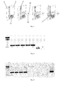
- Fig. 3 From 50 ml of culture medium supplemented with a defined amount of salmonella it was shown after 1 h of pre-incubation that the application of the ferromagnetic particles up to 10 CFU / 50 ml is positive. Direct detection of standard 1 ml was not possible.
- MnZn ferrite particles prepared as described in Example 2 Paramagnetic particles, prepared as described in Example 3 50 ml tubes (Greiner Bio One, Art. No .: 227261) Bar magnet ⁇ 10 mm, height 40 mm (Webcraft GmbH, S-10-40-N)
- Ferromagnetic particles stand out very clearly in the raw milk, because they are not colored like paramagnetic particles light brown, but a deep black Have color. The natural separation of the raw milk into cream and whey fraction did not in any way hinder the recovery of the ferromagnetic particles. The ferromagnetic particles partially sank to the bottom and were not retained in the cream fraction. If ferromagnetic particles were enclosed by fat components of the milk, they could still be held on the inner wall of the vessel due to the strong magnetic forces.
- An overnight culture of Salmonella enterica subsp. enterica is quantified in a manner known per se and diluted logarithmically. Dilution steps of 1-10000 cfu / ml are delivered to 250 ml of buffered peptone water. 25 g minced meat together with 250 ml artificially contaminated buffered peptone water are placed in a sterile sample bag, so-called Stomacher bag, and incubated for 4 - 7 h at 37 ° C (pre-enrichment).
- This washed magnetic particle dispersion is combined with the pre-incubated sample and incubated for 1 hour at 37 ° C with continuous, gentle agitation (shaking incubator).
- the separation takes place with the aid of a strong neodymium magnet (4 kg adhesive force).
- a magnet which is enclosed in a 15 ml tube and is located on a magnetic bar remover, is inserted behind the fleece of the Stomacher bag. This has the effect that, although the magnetic particles can pass through the fleece of the filter bag with the possibly bound target, coarse constituents of the food sample are retained.
- the fleece can consist both of paper-like fleece (ME001006a) and be arranged laterally, as well as be attached on all sides and consist of plastic-like fleece (ME001004).
- the paramagnetic particles are barely visible in the sample and are very difficult to separate from fat and coarse constituents of the sample. After the magnet had been behind the support fleece for a longer time (> 5 min) (for ferromagnetic particles 1 to 2 minutes are sufficient), a small amount of paramagnetic particles stuck to the magnet.
- the paramagnetic particles are washed, the possibly bound salmonella digested and fed to a suitable PCR reaction. Positive evidence of artificial contamination with 1000 CFU per sample was not possible.
- the magnetic particles are dispersed in a 2 ml reaction vessel with 1 ml PBS. Subsequently, 500 .mu.l color particle conjugate are added and it is incubated for 10 min at room temperature on a shaker. Subsequently, the reaction vessel is placed in a magnetic stand, the supernatant is decanted and washed three times with 1.50 ml of PBS buffer. Subsequently, the bound color particles are dissolved by adding 1 ml of ethanol, the solution is separated and the optical density (OD) is determined with a spectrophotometer at a wavelength of 525 nm and a layer thickness of 1 cm.
- OD optical density
- Example 1 MnZn ferrite particles prepared as described in Example 1 PBS buffer analogous to Example 1 Lysis Matrix H Zirconium Lysing Beads (MP Biomedicals Germany, 116917050) FastPrepInstrument (MP Biomedicals Germany, 116004500) 15 ml tube (Greiner Bio One, Art. No .: 188271)
- lymph node tissue was spiked with 9 ml PBS and homogenized using zirconium lysing beads in the FastPrep instrument (3x30 seconds at 6.5m / s).
- the 10% tissue slurry was spiked with 1x10 4 infectious units of adenovirus 5 and mixed on a laboratory shaker for 30 seconds in a 15 ml tube.
- To the mixture was added 200 ⁇ l of PBS-washed ferromagnetic particles. This corresponds to 20 ⁇ l of a 1 gram per 1 ml of stock particle solution.
- the washing of the particles was carried out analogously to Example 4.
- the mixture was mixed for 1 h at 37 ° C in a rotating and rocking incubator, with the magnetic particles constantly moving in the pulp and no visible sedimentation taking place.
- the ferromagnetic particles were then pulled from the outside to the wall of the tube by means of the permanent magnet and the tissue slurry was pipetted off.
- the tube was filled with 10 ml PBS and shaken on a laboratory shaker for 30 seconds to remove tissue remnants nonspecifically attached between the ferromagnetic particles (washing step). Subsequently, the ferromagnetic particles were pulled back to the tube wall and the wash buffer was pipetted off.
- the ferromagnetic particles were incubated for 1 h at 55 ° C. in 700 ⁇ l lysis buffer (NucleoSpin Tissue Kit) and the proteinase K contained therein was subsequently inactivated by heating at 98 ° C. for 20 minutes. From the lysate, the nucleic acid was separated by phenol extraction in a known manner and then purified the DNA from the aqueous phase with the NucleoSpinTissue kit according to the manufacturer's instructions and eluted in 50 ul H 2 O.
- 700 lysis buffer NucleoSpin Tissue Kit
- adenovirus-specific PCR 5 or 1 ⁇ l of the eluted DNA were used as template. 1 ⁇ l of template was sufficient to generate a specific product ( Fig. 4 , 2% agarose gel, staining with ethidium bromide). Since 1 ⁇ l template corresponds to one-fiftieth of the spike virus used, it can be assumed that the ferromagnetic particle slurry had bound at least 2000 PCR target equivalents from the tissue slurry at a detection limit of the PCR of 10 targets / reaction.
- the particles are used as a 2.5% solution and a 1-step EDC coupling is performed according to the manufacturer's instructions.
- 2 ml of phosphate buffer 0.1 M pH 7.4 in the case of SPHERO Amino Ferro-Magnetic Particles
- 2 ml of sodium acetate buffer 0.01 M, pH 5.0 when using the SPHERO Carboxyl Ferro-Magnetic Particles
- 0.5 mg of anti-Salmonella antibody 2 ml of 2.5% particle solution and 5 mg of EDC mixed together.
- the suspension is then incubated for 2 hours on an overhead shaker at room temperature. The supernatant is removed at the end of the incubation period and the particles are washed twice in 4 ml IBS. Finally, the particles are removed in 2 ml IBS and stored at 4 ° C until use.
- Fig. 5 Comparison of Magnetic Behavior of SPHERO Amino Ferro-Magnetic Particles 1% w / v 4.32 ⁇ m (Cat No. AFM-40-10) and SPHERO Carboxyl Ferro-Magnetic Particles 1% w / v 4.47 ⁇ m (Cat CFM-40-10) in aqueous solutions compared to ferromagnetic particles according to the invention as described in Example 4.
- SPHERO Amino Ferro-Magnetic Particles 1% w / v 4.32 ⁇ m (Cat No. AFM-40-10) and SPHERO Carboxyl Ferro-Magnetic Particles 1% w / v 4.47 ⁇ m as SPHERO-Tech Particles summarized, since they behave in aqueous solutions approximately equal.
- the SPHERO-Tech particles can hardly be attracted by a magnet. Fixing them to the bottom of a reaction vessel, for example to pour out the supernatant, is not possible. They are very easy to dissolve and sink very slowly to the ground.
- the magnetic Behavior was demonstrated in the ChipShop P device. Here, a mounted below the four-chamber chip magnet from left to right and back.
- the SPHERO-Tech particles can hardly be moved by the device.
- the magnetic force of the particles is not sufficient to move the particles back and forth as a clear band.
- the SPHERO-Tech particles form a diffuse band in the lower two chambers which only runs slightly back and forth.
- the particles according to the invention in the upper two chambers according to embodiment 4 form a discrete band, so that the magnetic properties of the particles are retained.
- Fig. 7 SPHERO-Tech particles according to embodiment 11 after 1 s, 3, s, 9 s, 25 s and 34 s
- the ferromagnetic particles according to the invention as described in Example 4 immediately strive for the magnet and are adhered within a few seconds.
- the SPHERO-Tech particles take over 30 seconds to collect there, when swiveling the stand the binding is lost immediately.
- the ferromagnetic MnZn ferrite particles of Embodiment 4 of the present invention allow better separation of the analyte attached to the particles from the surrounding solution.
- the ferromagnetic particles of the invention after 9 s show a virtually complete separation or segregation of the surrounding solution, while the SPHERO-Tech particles after 34 s still show no complete separation.
- the separation efficiency, measured as the time of separation / separation of the solution (dispersion) of the ferromagnetic particles according to the invention is thus greater by about a factor of 3-4 than in the case of the comparison particles with a chromium dioxide core.
- An overnight culture of Salmonella enterica subsp. enterica is quantified in a manner known per se and diluted logarithmically. Dilution steps of 1-10000 cfu / ml are delivered to 250 ml of buffered peptone water.
- This washed magnetic particle dispersion is combined with the pre-incubated sample and incubated for 1 hour at 37 ° C with continuous, gentle agitation (shaking incubator).
- the separation takes place with the aid of a strong neodymium magnet (4 kg adhesive force).
- a magnet which is enclosed in a 15 ml tube and is located on a magnetic bar remover, is inserted behind the fleece of the Stomacher bag. This has the effect that, although the magnetic particles can pass through the fleece of the filter bag with the possibly bound target, coarse constituents of the food sample are retained.
- the fleece can consist both of paper-like fleece (ME001006a) and be arranged laterally, as well as be attached on all sides and consist of plastic-like fleece (ME001004).
- the SPHERO Amino Ferro-Magnetic Particles or SPHERO Carboxyl Ferro-Magnetic Particles are hardly visible in the sample and are very difficult to separate from the fat and coarse components of the sample. After leaving the magnet behind the backing for a longer time (> 5 minutes) (for ferromagnetic particles 1-2 minutes), a small amount of SPHERO Amino Ferro-Magnetic Particles or SPHERO Carboxyl Ferro-Magnetic Particles stuck to the magnet.
- Example 4 the particles are washed, the possibly bound salmonella digested and fed to a suitable PCR reaction. Positive detection of artificial contamination at 100 CFU per sample was not possible compared to 100 CFU per sample when using the particles of Example 4.
Landscapes
- Health & Medical Sciences (AREA)
- Life Sciences & Earth Sciences (AREA)
- Immunology (AREA)
- Engineering & Computer Science (AREA)
- Molecular Biology (AREA)
- Biomedical Technology (AREA)
- Chemical & Material Sciences (AREA)
- Hematology (AREA)
- Urology & Nephrology (AREA)
- Food Science & Technology (AREA)
- Biochemistry (AREA)
- Cell Biology (AREA)
- Biotechnology (AREA)
- Medicinal Chemistry (AREA)
- Physics & Mathematics (AREA)
- Analytical Chemistry (AREA)
- Microbiology (AREA)
- General Health & Medical Sciences (AREA)
- General Physics & Mathematics (AREA)
- Pathology (AREA)
- Tropical Medicine & Parasitology (AREA)
- Virology (AREA)
- Measuring Or Testing Involving Enzymes Or Micro-Organisms (AREA)
- Investigating Or Analysing Biological Materials (AREA)
Priority Applications (1)
| Application Number | Priority Date | Filing Date | Title |
|---|---|---|---|
| EP14187534.4A EP2857838A3 (fr) | 2013-10-02 | 2014-10-02 | Procédé de séparation immunomagnétique d'analytes à partir d'une matrice d'échantillons complexes |
Applications Claiming Priority (2)
| Application Number | Priority Date | Filing Date | Title |
|---|---|---|---|
| EP13187037 | 2013-10-02 | ||
| EP14187534.4A EP2857838A3 (fr) | 2013-10-02 | 2014-10-02 | Procédé de séparation immunomagnétique d'analytes à partir d'une matrice d'échantillons complexes |
Publications (2)
| Publication Number | Publication Date |
|---|---|
| EP2857838A2 true EP2857838A2 (fr) | 2015-04-08 |
| EP2857838A3 EP2857838A3 (fr) | 2015-07-01 |
Family
ID=49274528
Family Applications (1)
| Application Number | Title | Priority Date | Filing Date |
|---|---|---|---|
| EP14187534.4A Withdrawn EP2857838A3 (fr) | 2013-10-02 | 2014-10-02 | Procédé de séparation immunomagnétique d'analytes à partir d'une matrice d'échantillons complexes |
Country Status (1)
| Country | Link |
|---|---|
| EP (1) | EP2857838A3 (fr) |
Citations (5)
| Publication number | Priority date | Publication date | Assignee | Title |
|---|---|---|---|---|
| US2885366A (en) | 1956-06-28 | 1959-05-05 | Du Pont | Product comprising a skin of dense, hydrated amorphous silica bound upon a core of another solid material and process of making same |
| US20040126902A1 (en) * | 2002-06-27 | 2004-07-01 | Toyo Boseki Kabushiki Kaisha | Magnetic carrier for biological substance, production method thereof and isolation method of biological substance using same |
| US20120061608A1 (en) * | 2010-09-10 | 2012-03-15 | Hitachi Maxell, Ltd. | Functional particle with rough-surfaced polymer coating |
| US20120220048A1 (en) * | 2010-06-03 | 2012-08-30 | Hitachi Maxell, Ltd. | Magnetic marker particle and method for producing the same |
| DE102013005184A1 (de) | 2013-03-20 | 2014-09-25 | fzmb GmbH, Forschungszentrum für Medizintechnik und Biotechnologie | Verfahren zur Funktionalisierung einer Oberfläche |
Family Cites Families (2)
| Publication number | Priority date | Publication date | Assignee | Title |
|---|---|---|---|---|
| JP3607320B2 (ja) * | 1994-09-02 | 2005-01-05 | 株式会社日立製作所 | 微粒子を用いた分析における固相の回収方法及び装置 |
| WO2010089098A1 (fr) * | 2009-02-05 | 2010-08-12 | Deklatec Gmbh | Procédés et agents pour le diagnostic de la tuberculose |
-
2014
- 2014-10-02 EP EP14187534.4A patent/EP2857838A3/fr not_active Withdrawn
Patent Citations (5)
| Publication number | Priority date | Publication date | Assignee | Title |
|---|---|---|---|---|
| US2885366A (en) | 1956-06-28 | 1959-05-05 | Du Pont | Product comprising a skin of dense, hydrated amorphous silica bound upon a core of another solid material and process of making same |
| US20040126902A1 (en) * | 2002-06-27 | 2004-07-01 | Toyo Boseki Kabushiki Kaisha | Magnetic carrier for biological substance, production method thereof and isolation method of biological substance using same |
| US20120220048A1 (en) * | 2010-06-03 | 2012-08-30 | Hitachi Maxell, Ltd. | Magnetic marker particle and method for producing the same |
| US20120061608A1 (en) * | 2010-09-10 | 2012-03-15 | Hitachi Maxell, Ltd. | Functional particle with rough-surfaced polymer coating |
| DE102013005184A1 (de) | 2013-03-20 | 2014-09-25 | fzmb GmbH, Forschungszentrum für Medizintechnik und Biotechnologie | Verfahren zur Funktionalisierung einer Oberfläche |
Non-Patent Citations (23)
| Title |
|---|
| BAUMANN S.; UTA CEGLAREK; GEORG MARTIN FIEDLER; JAN LEMBCKE; ALEXANDER LEICHTLE; JOACHIM THIERY: "Standardized Approach to Proteome Profiling of Human Serum Based on Magnetic Bead Separation and Matrix-Assisted Laser Desorption/Ionization Time-of-Flight Mass Spectrometry", CLINICAL CHEMISTRY, vol. 51, no. 6, 2005, pages 973 - 980 |
| BERENSMEIER S.: "Magnetic particles for the separation and purification of nucleic acids", APPL MICROBIOL BIOTECHNOL, vol. 73, 2006, pages 495 - 504 |
| BIRKMEYER ET AL., CLIN. CHEM., vol. 33, no. 9, 1987, pages 1543 - 1547 |
| BOSCHKE E.; STEINGROEWER J.; BLEY T.: "Einsatz der biomagnetischen Separation zur mikrobiologischen Qualitätskontrolle von Lebensmitteln", CHEMIE INGENIEUR TECHNIK, vol. 77, no. 7, 2005, pages 912 - 919 |
| DUODU ET AL., FOOD ANAL. METHODS, vol. 2, 2009, pages 23 - 29 |
| FERROMAGNETIC MICROPARTICLES AND NANOPARTICLES-SPHEROTECH, 12 March 2013 (2013-03-12), Retrieved from the Internet <URL:http://www.spherotech.com/fer par.htm> |
| FLUIT ET AL., APPLIED AND ENVIROMENTAL MICROBIOLOGY, May 1993 (1993-05-01), pages 1289 - 1293 |
| FLUIT ET AL., APPLIED AND ENVIROMENTAL MICROBIOLOGY, May 1993 (1993-05-01), pages 1342 - 1346 |
| HAUKANES, B. I.; KVAM, C.: "Application of magnetic beads in bioassays", BIOTECHNOLOGY, vol. 11, no. 1, 1993, pages 60 - 63, XP000609039, DOI: doi:10.1038/nbt0193-60 |
| LIU JING: "Magnetorheological Fluids: From Basic Physics to Application", JSME INTERNATIONAL JOURNAL SERIES B, vol. 45, no. 1, 2002, pages 55 - 60 |
| MATTINGLY, J.A.: "An enzyme immunoassay for the detection of all Salmonella using a combination of a myeloma protein and a hybridoma antibody", JOURNAL OF IMMUNOLOGICAL METHODS, vol. 73, 1984, pages 147 - 156, XP025425167, DOI: doi:10.1016/0022-1759(84)90040-1 |
| MERCANOGLU TABAN B; BEN U; AYTAC SA.: "Rapid detection of Salmonella in milk by combined immunomagnetic separation-polymerase chain reaction assay", J DAIRY SCI., vol. 92, 2009, pages 2382 - 8, XP026955428 |
| MULLANE NR; MURRAY J; DRUDY D; PRENTICE N; WHYTE P; WALL PG; PARTON A; FANNING S.: "Detection of Enterobacter sakazakii in dried infant milk formula by cationic-magnetic-bead capture", APPL. ENVIRON MICROBIOL., vol. 72, no. 9, 2006, pages 6325 - 30 |
| OLSVIK 0; POPOVIC T; SKJERVE E; CUDJOE KS; HORNES E; UGELSTAD J; UHLEN M: "Magnetic separation techniques in diagnostic microbiology", CLIN MICROBIOL REV, vol. 7, no. 1, 1994, pages 43 - 54, XP002996234 |
| OLSVIK, 0.; POPOVIC, T.; SKJERVE, E.; CUDJOE, K.; HOMES, E.; UGELSTAD, J.; UHLEN, M.: "Magnetic separation techniques in diagnostic microbiology", CLINICAL MICROBIOLOGY REVIEWS, vol. 7, 1994, pages 43 - 54, XP002996234 |
| PANNEERSEELAN L; MURIANA PM.: "An immunomagnetic PCR signal amplification assay for sensitive detection of Staphylococcus aureus enterotoxins in foods", J FOOD PROT., vol. 72, 2009, pages 2538 - 46 |
| SAFARIK, I.; SAFARIKOVA, M.; FORSYTHE, S.J.: "The application of magnetic separations in applied microbiology", THE JOURNAL OF APPLIED BACTERIOLOGY, vol. 78, 1995, pages 575 - 585, XP002096125 |
| SIEBEN S.; BERGEMANN C.; LÜBBE A.S.; BROCKMANN B.; RESCHELEIT D.: "Comparison of different particles and methods for magnetic isolation of circulating tumor cells", J. OF MAGNETISM AND MAGNETIC MATERIALS, vol. 225, 2001, pages 175 - 179, XP004234942, DOI: doi:10.1016/S0304-8853(00)01248-8 |
| UGELSTAD, J.; STENSTAD, P.; KILAAS, L.; PRESTVIK, W.S.; HERJE, R.; BERGE, A.; HORNES, E.: "Monodisperse magnetic polymer particles. New biochemical and biomedical applications", BLOOD PURIFICATION, vol. 11, no. 6, 1993, pages 349 - 369, XP009016520 |
| VON SCHÖNFELD ET AL., BIOLOGY OF REPRODUCTION, vol. 61, 1999, pages 582 - 589 |
| WADUD S; LEON-VELARDE CG; LARSON N; ODUMERU JA: "Evaluation of immunomagnetic separation in combination with ALOA Listeria chromogenic agar for the isolation and identification of Listeria monocytogenes in ready-to-eat foods", J MICROBIOL METHODS., vol. 6, 2010 |
| WIDJOJOATMODJO ET AL., EUR. J. CLIN. MICROBIOL. INFECT. DIS., November 1991 (1991-11-01), pages 935 - 938 |
| WIDJOJOATMODJO ET AL., JOURNAL OF CLINICAL MICROBIOLOGY, December 1992 (1992-12-01), pages 3195 - 3199 |
Also Published As
| Publication number | Publication date |
|---|---|
| EP2857838A3 (fr) | 2015-07-01 |
Similar Documents
| Publication | Publication Date | Title |
|---|---|---|
| DE69632785T2 (de) | Verbessertes verfahren zur herstellung magnetisch empfindlicher teilchen | |
| US12461095B2 (en) | Stable nanomagnetic particle dispersions | |
| DE19520398B4 (de) | Magnetisches Pigment | |
| DE60122138T2 (de) | Isolierung von nukleinsäuren | |
| DE69817484T2 (de) | Verfahren zur isolierung bestimmter biologischer stoffe mittels magnetischer silika-partikel | |
| DE69126541T2 (de) | Vorrichtung und verfahren für die magnetische trennung | |
| DE69222299T2 (de) | Vorrichtung und verfahren zur magnetischen abtrennung unter verwendung externer magnetischer mittel | |
| DE3689618T2 (de) | Magnetische polymerteilchen. | |
| DE69322073T2 (de) | Analyseverfahren | |
| DE69614205T2 (de) | Verfahren und vorrichtung zur bestimmung eines analyten, insbesondere eines bakteriums, auf magnetischem wege | |
| DE69029908T2 (de) | Magnetische und fluoreszierende polymerteilchen und ihre anwendung | |
| DE60026729T2 (de) | Verfahren zur modifikation von ausgewählten zellen in einer magnetischen zelltrennungsäule | |
| EP1274745B1 (fr) | Materiaux supports magnetiques et silanises, a base d'alcool polyvinylique | |
| WO2012007581A1 (fr) | Procédé pour enrichir des bactéries, des virus et des cellules, et pour isoler ensuite leurs acides nucléiques | |
| KR20180072014A (ko) | 항체가 고정된 자성 나노입자 클러스터를 포함하는 진단용 소재 및 그의 제조방법 | |
| WO2010089098A1 (fr) | Procédés et agents pour le diagnostic de la tuberculose | |
| EP2857838A2 (fr) | Procédé de séparation immunomagnétique d'analytes à partir d'une matrice d'échantillons complexes | |
| EP1766401A1 (fr) | Procédé de détection simple et rapide de cellules et de biomolécules à l'aide de particules paramagnétiques | |
| DE602004003163T2 (de) | Verfahren zur Bereitstellung von Proben zum Nachweis | |
| DE69622877T2 (de) | Fangverfahren und testmethoden | |
| AT501194A1 (de) | Verfahren zur isolierung von zellen und viren | |
| WO2020069385A1 (fr) | Isolement de l'adn et de l'arn d'un seul et même échantillon | |
| DE102005031755A1 (de) | Salmonella spp. bindende Peptide, deren Verwendung und Verfahren und Kits zur Anreicherung, Immobilisierung und zum Nachweis von Salmonella spp. | |
| DE112009000338T5 (de) | Verbesserte Verfahren für eine Gas- und/oder Dampfphasenanalyse biologischer Assays | |
| DE19549875B4 (de) | Verwendung von magnetischen Partikeln zur Isolierung von Nukleinsäuren |
Legal Events
| Date | Code | Title | Description |
|---|---|---|---|
| PUAI | Public reference made under article 153(3) epc to a published international application that has entered the european phase |
Free format text: ORIGINAL CODE: 0009012 |
|
| 17P | Request for examination filed |
Effective date: 20141002 |
|
| AK | Designated contracting states |
Kind code of ref document: A2 Designated state(s): AL AT BE BG CH CY CZ DE DK EE ES FI FR GB GR HR HU IE IS IT LI LT LU LV MC MK MT NL NO PL PT RO RS SE SI SK SM TR |
|
| AX | Request for extension of the european patent |
Extension state: BA ME |
|
| PUAL | Search report despatched |
Free format text: ORIGINAL CODE: 0009013 |
|
| AK | Designated contracting states |
Kind code of ref document: A3 Designated state(s): AL AT BE BG CH CY CZ DE DK EE ES FI FR GB GR HR HU IE IS IT LI LT LU LV MC MK MT NL NO PL PT RO RS SE SI SK SM TR |
|
| AX | Request for extension of the european patent |
Extension state: BA ME |
|
| RIC1 | Information provided on ipc code assigned before grant |
Ipc: G01N 33/543 20060101AFI20150528BHEP |
|
| R17P | Request for examination filed (corrected) |
Effective date: 20151222 |
|
| RBV | Designated contracting states (corrected) |
Designated state(s): AL AT BE BG CH CY CZ DE DK EE ES FI FR GB GR HR HU IE IS IT LI LT LU LV MC MK MT NL NO PL PT RO RS SE SI SK SM TR |
|
| STAA | Information on the status of an ep patent application or granted ep patent |
Free format text: STATUS: EXAMINATION IS IN PROGRESS |
|
| 17Q | First examination report despatched |
Effective date: 20170714 |
|
| STAA | Information on the status of an ep patent application or granted ep patent |
Free format text: STATUS: THE APPLICATION IS DEEMED TO BE WITHDRAWN |
|
| 18D | Application deemed to be withdrawn |
Effective date: 20180125 |