EP2872877B1 - Système et méthode de surveillance et de traitement de surface de patient - Google Patents
Système et méthode de surveillance et de traitement de surface de patient Download PDFInfo
- Publication number
- EP2872877B1 EP2872877B1 EP13817635.9A EP13817635A EP2872877B1 EP 2872877 B1 EP2872877 B1 EP 2872877B1 EP 13817635 A EP13817635 A EP 13817635A EP 2872877 B1 EP2872877 B1 EP 2872877B1
- Authority
- EP
- European Patent Office
- Prior art keywords
- therapeutic
- sensor
- matrix
- dressing
- release
- Prior art date
- Legal status (The legal status is an assumption and is not a legal conclusion. Google has not performed a legal analysis and makes no representation as to the accuracy of the status listed.)
- Active
Links
Images







Classifications
-
- A—HUMAN NECESSITIES
- A61—MEDICAL OR VETERINARY SCIENCE; HYGIENE
- A61K—PREPARATIONS FOR MEDICAL, DENTAL OR TOILETRY PURPOSES
- A61K49/00—Preparations for testing in vivo
- A61K49/001—Preparation for luminescence or biological staining
- A61K49/0013—Luminescence
- A61K49/0015—Phosphorescence
-
- A—HUMAN NECESSITIES
- A61—MEDICAL OR VETERINARY SCIENCE; HYGIENE
- A61B—DIAGNOSIS; SURGERY; IDENTIFICATION
- A61B5/00—Measuring for diagnostic purposes; Identification of persons
- A61B5/145—Measuring characteristics of blood in vivo, e.g. gas concentration or pH-value ; Measuring characteristics of body fluids or tissues, e.g. interstitial fluid or cerebral tissue
- A61B5/1455—Measuring characteristics of blood in vivo, e.g. gas concentration or pH-value ; Measuring characteristics of body fluids or tissues, e.g. interstitial fluid or cerebral tissue using optical sensors, e.g. spectral photometrical oximeters
- A61B5/14551—Measuring characteristics of blood in vivo, e.g. gas concentration or pH-value ; Measuring characteristics of body fluids or tissues, e.g. interstitial fluid or cerebral tissue using optical sensors, e.g. spectral photometrical oximeters for measuring blood gases
-
- A—HUMAN NECESSITIES
- A61—MEDICAL OR VETERINARY SCIENCE; HYGIENE
- A61B—DIAGNOSIS; SURGERY; IDENTIFICATION
- A61B5/00—Measuring for diagnostic purposes; Identification of persons
- A61B5/145—Measuring characteristics of blood in vivo, e.g. gas concentration or pH-value ; Measuring characteristics of body fluids or tissues, e.g. interstitial fluid or cerebral tissue
- A61B5/1455—Measuring characteristics of blood in vivo, e.g. gas concentration or pH-value ; Measuring characteristics of body fluids or tissues, e.g. interstitial fluid or cerebral tissue using optical sensors, e.g. spectral photometrical oximeters
- A61B5/14551—Measuring characteristics of blood in vivo, e.g. gas concentration or pH-value ; Measuring characteristics of body fluids or tissues, e.g. interstitial fluid or cerebral tissue using optical sensors, e.g. spectral photometrical oximeters for measuring blood gases
- A61B5/14552—Details of sensors specially adapted therefor
-
- A—HUMAN NECESSITIES
- A61—MEDICAL OR VETERINARY SCIENCE; HYGIENE
- A61B—DIAGNOSIS; SURGERY; IDENTIFICATION
- A61B5/00—Measuring for diagnostic purposes; Identification of persons
- A61B5/145—Measuring characteristics of blood in vivo, e.g. gas concentration or pH-value ; Measuring characteristics of body fluids or tissues, e.g. interstitial fluid or cerebral tissue
- A61B5/1455—Measuring characteristics of blood in vivo, e.g. gas concentration or pH-value ; Measuring characteristics of body fluids or tissues, e.g. interstitial fluid or cerebral tissue using optical sensors, e.g. spectral photometrical oximeters
- A61B5/14551—Measuring characteristics of blood in vivo, e.g. gas concentration or pH-value ; Measuring characteristics of body fluids or tissues, e.g. interstitial fluid or cerebral tissue using optical sensors, e.g. spectral photometrical oximeters for measuring blood gases
- A61B5/14556—Measuring characteristics of blood in vivo, e.g. gas concentration or pH-value ; Measuring characteristics of body fluids or tissues, e.g. interstitial fluid or cerebral tissue using optical sensors, e.g. spectral photometrical oximeters for measuring blood gases by fluorescence
-
- A—HUMAN NECESSITIES
- A61—MEDICAL OR VETERINARY SCIENCE; HYGIENE
- A61B—DIAGNOSIS; SURGERY; IDENTIFICATION
- A61B5/00—Measuring for diagnostic purposes; Identification of persons
- A61B5/44—Detecting, measuring or recording for evaluating the integumentary system, e.g. skin, hair or nails
- A61B5/441—Skin evaluation, e.g. for skin disorder diagnosis
- A61B5/443—Evaluating skin constituents, e.g. elastin, melanin, water
-
- A—HUMAN NECESSITIES
- A61—MEDICAL OR VETERINARY SCIENCE; HYGIENE
- A61B—DIAGNOSIS; SURGERY; IDENTIFICATION
- A61B5/00—Measuring for diagnostic purposes; Identification of persons
- A61B5/68—Arrangements of detecting, measuring or recording means, e.g. sensors, in relation to patient
- A61B5/6801—Arrangements of detecting, measuring or recording means, e.g. sensors, in relation to patient specially adapted to be attached to or worn on the body surface
- A61B5/683—Means for maintaining contact with the body
- A61B5/6832—Means for maintaining contact with the body using adhesives
- A61B5/6833—Adhesive patches
-
- A—HUMAN NECESSITIES
- A61—MEDICAL OR VETERINARY SCIENCE; HYGIENE
- A61B—DIAGNOSIS; SURGERY; IDENTIFICATION
- A61B90/00—Instruments, implements or accessories specially adapted for surgery or diagnosis and not covered by any of the groups A61B1/00 - A61B50/00, e.g. for luxation treatment or for protecting wound edges
- A61B90/39—Markers, e.g. radio-opaque or breast lesions markers
-
- C—CHEMISTRY; METALLURGY
- C07—ORGANIC CHEMISTRY
- C07D—HETEROCYCLIC COMPOUNDS
- C07D487/00—Heterocyclic compounds containing nitrogen atoms as the only ring hetero atoms in the condensed system, not provided for by groups C07D451/00 - C07D477/00
- C07D487/22—Heterocyclic compounds containing nitrogen atoms as the only ring hetero atoms in the condensed system, not provided for by groups C07D451/00 - C07D477/00 in which the condensed system contains four or more hetero rings
-
- A—HUMAN NECESSITIES
- A61—MEDICAL OR VETERINARY SCIENCE; HYGIENE
- A61B—DIAGNOSIS; SURGERY; IDENTIFICATION
- A61B2562/00—Details of sensors; Constructional details of sensor housings or probes; Accessories for sensors
- A61B2562/12—Manufacturing methods specially adapted for producing sensors for in-vivo measurements
-
- A—HUMAN NECESSITIES
- A61—MEDICAL OR VETERINARY SCIENCE; HYGIENE
- A61B—DIAGNOSIS; SURGERY; IDENTIFICATION
- A61B5/00—Measuring for diagnostic purposes; Identification of persons
- A61B5/0002—Remote monitoring of patients using telemetry, e.g. transmission of vital signals via a communication network
- A61B5/0015—Remote monitoring of patients using telemetry, e.g. transmission of vital signals via a communication network characterised by features of the telemetry system
- A61B5/0022—Monitoring a patient using a global network, e.g. telephone networks, internet
-
- A—HUMAN NECESSITIES
- A61—MEDICAL OR VETERINARY SCIENCE; HYGIENE
- A61B—DIAGNOSIS; SURGERY; IDENTIFICATION
- A61B5/00—Measuring for diagnostic purposes; Identification of persons
- A61B5/48—Other medical applications
- A61B5/4836—Diagnosis combined with treatment in closed-loop systems or methods
- A61B5/4839—Diagnosis combined with treatment in closed-loop systems or methods combined with drug delivery
-
- A—HUMAN NECESSITIES
- A61—MEDICAL OR VETERINARY SCIENCE; HYGIENE
- A61B—DIAGNOSIS; SURGERY; IDENTIFICATION
- A61B5/00—Measuring for diagnostic purposes; Identification of persons
- A61B5/74—Details of notification to user or communication with user or patient; User input means
- A61B5/742—Details of notification to user or communication with user or patient; User input means using visual displays
- A61B5/743—Displaying an image simultaneously with additional graphical information, e.g. symbols, charts, function plots
-
- A—HUMAN NECESSITIES
- A61—MEDICAL OR VETERINARY SCIENCE; HYGIENE
- A61F—FILTERS IMPLANTABLE INTO BLOOD VESSELS; PROSTHESES; DEVICES PROVIDING PATENCY TO, OR PREVENTING COLLAPSING OF, TUBULAR STRUCTURES OF THE BODY, e.g. STENTS; ORTHOPAEDIC, NURSING OR CONTRACEPTIVE DEVICES; FOMENTATION; TREATMENT OR PROTECTION OF EYES OR EARS; BANDAGES, DRESSINGS OR ABSORBENT PADS; FIRST-AID KITS
- A61F13/00—Bandages or dressings; Absorbent pads
- A61F2013/00361—Plasters
- A61F2013/00902—Plasters containing means
- A61F2013/0094—Plasters containing means for sensing physical parameters
- A61F2013/0097—Plasters containing means for sensing physical parameters oxygen content
Definitions
- tissue-gas analyses are an essential part of modern patient care and are used in the diagnosis and treatment of a number of conditions.
- measurement of tissue oxygen concentration is heavily relied upon both for general monitoring of overall patient health and for treatment of specific conditions, such as ischemia, burns, and diabetic foot syndrome.
- a first apparatus known as a pulse oximeter
- a pulse oximeter is a basic, non-invasive instrument that detects hemoglobin saturation. Measuring the percentage of bound hemoglobin provides an estimate of arterial oxygenation.
- the device may be placed on a finger or another body part, and the measurement is accomplished by monitoring the reflectance or absorbance of incident light.
- pulse oximetry is a fast and simple technique
- the main drawback is that the measurement does not directly measure arterial oxygen concentrations. In particular, an inaccurate diagnosis may arise is situations where hemoglobin concentrations are low or when hemoglobin is bound to a species other than oxygen.
- a second, more direct measurement of tissue gas levels may be made using probe-based systems.
- Two existing methods based on transcutaneous oxygen (Tc p O 2 ) measurement involve either electrodes or an optical sensor foil- based patch.
- Tc p O 2 measurements can provide a direct indication of microvascular function as Tc p O 2 maps the actual oxygen supply available for the skin tissue cells.
- Tc p O 2 also responds to macrocirculatory events, such as a change in blood pressure.
- the Tcp0 2 monitor consists of a combined platinum and silver electrode covered by an oxygen- permeable hydrophobic membrane, with a reservoir of phosphate buffer and potassium chloride trapped inside the electrode. A small heating element is located inside the silver anode.
- the electrode is applied to an acceptable site on the skin and is heated to 44°C in order to provide a measurement.
- VisiSens TM Another commercial system known as the VisiSens TM system combines optical sensor foils with imaging technology. Fluorescent chemical optical sensor foils are attached to the sample surface and read out non-invasively using a microscope. Two-dimensional visualization of oxygen, pH, or C0 2 distributions over time can be performed with microscopic resolution.
- Tc p O 2 systems include the need for two- (or more) point calibrations with specially prepared, well-defined samples.
- the sensor must be in contact with the tissue through a contact liquid. If there is air between the tissue and the sensor, the values will be questionable.
- it may take 15-20 minutes after the probe has been placed on the skin for the Tc p O 2 reading to stabilize and it is recommended that calibrations be performed prior to each monitoring period, when changing measuring sites, every four hours, and/or every time an electrode has been remembraned.
- heating may affect the ability to acquire physiologically relevant measurements.
- a third approach to monitoring gas levels involves a blood test.
- the test is performed using a blood sample drawn from an artery.
- the machine used for analysis aspirates this blood from the syringe and measures the pH and the partial pressures of oxygen and carbon dioxide.
- the bicarbonate concentration can also be calculated.
- An advantage of the test is that results are usually available for interpretation within minutes.
- the test is invasive, requires a trained practitioner to accurately acquire a sample, and samples must be maintained at room temperature and analyzed quickly or results may be inaccurate.
- US 2003/0050543 A1 relates to method and devices for determining local distribution of a measuring parameter, such as oxygen.
- US 2009/0112295 A1 relates to devices and systems that deliver nitric oxide.
- the present invention is defined in the appended independent claims. It overcomes the above and other drawbacks by providing for a non-invasive, oxygen sensing dressing that may be applied to the surface of tissues such as skin in order to provide fast, accurate readout without the requirement of specialized training.
- the present disclosure provides an interactive, sensing, and therapeutic delivery device designed for the detection and monitoring of tissue properties that are used to inform a user of the specific need and spatial location for therapeutic intervention.
- the sensing and therapeutic delivery device can be triggered to spatially release therapeutics using an interacting portion. Following therapeutic release, the present invention can continue to monitor tissue properties in response to treatment.
- a device in accordance with one aspect of the invention, includes a sensor configured to detect a concentration of an analyte that is selected from the group consisting of molecular oxygen, carbon dioxide, nitric oxides, dissolved analytes in plasma, and hydrogen ions.
- the device also includes a matrix compatible with said sensor, at least a portion of said matrix accessible to and/or permeable to said analyte.
- the device includes a dressing comprising said matrix and said sensor, where said device emits a signal, such as a phosphorescent signal in response to the concentration of said analyte detected by said sensor.
- a method of manufacturing a device for detecting an analyte includes selecting a sensor configured to detect a concentration of an analyte including but not limited to molecular oxygen, carbon dioxide, nitric oxides, dissolved analytes in plasma or tissue, and hydrogen ions.
- the method also includes selecting a compatible matrix base on a chemical nature of said sensor and enmeshing said sensor in said compatible matrix, at least a portion of said matrix permeable to said analyte.
- the method further includes incorporating said matrix into a dressing, where said dressing is configured to emit a signal that varies in response to the concentration of said analyte detected by said sensor.
- a kit for detecting an analyte.
- the kit includes a dressing comprising a sensor configured to detect at least one analyte, and a matrix compatible with said sensor and a detector configured to actuate said sensor and measuring a signal as a function of a position on a surface of said dressing.
- the at least one analyte includes, but is not limited to molecular oxygen, carbon dioxide, nitric oxides, dissolved analytes in plasma, and hydrogen ions and the signal corresponds to the presence of said at least one analyte proximate said surface of said dressing.
- a dressing may include a covering for application to a surface and especially for application to a surface of a body part of a patient.
- the term dressing may also apply to other coverings, such as an ointment or gauze and may be a solid of liquid.
- the terms dressing, bandage, covering and related terms may be used throughout the disclosure to refer to a covering.
- the present invention is a bandage/wound dressing device 100 having one or more of the following three components: a sensing portion 16, a therapeutic portion 14, and an interacting portion 20, as illustrated in Fig. 1 .
- the device 100 is contactable and, preferably, removably adhereable to body surface, such as intact and/or open/damaged tissue.
- the present device 100 may be used by a "user", wherein the user may be, for example, a patient's caregiver, a physician, or even a patient.
- the device 100 includes a sensing/diagnostic portion 16.
- the diagnostic portion 16 can be combined with an interacting portion 20 (for example, a detector) to measure a signal from the diagnostic portion and provide an output to a user.
- the device 100 includes both a sensing portion 16 and a therapeutic portion 14.
- the device 100 may include two distinct but interdependent devices: a dressing 10 including both diagnostic and therapeutic portions as described above and an interacting portion 20.
- the present invention is capable of performing several functions.
- the present invention is capable of generating a two-dimensional map 30 of physiologically relevant parameters, where each point on the map corresponds to a scalar value corresponding to data relevant to one or multiple analytes in the underlying tissue, such as the concentration of the analyte or analytes.
- the map can be read either continuously or point-wise by the naked eye as a colorimetric sensor response, the quantitative signal can be stimulated and recorded using the interactive device, or the quantitative signal can be collected by an external digital recording device.
- the dark points 32 on the map are distinct from the white points 42 indicating a variation in the spatiospecific signal detected.
- the present invention is capable of administering doses of therapeutics, such as, but not limited to, antibiotics, anti-inflammatory agents, biologics, and pain medications. Also, the present invention is capable of administering a dose of biomarkers or positron emission tomography tracers along with or instead of the therapeutics. Even further, the present device is capable of administering these therapeutics in a controlled manner in response to a specific externally applied stimulus or stimuli. This release can be accomplished through stand-alone mechanisms independent of interacting portion, such as, but not limited to, mechanical pressure, heat, other energetic stimulus or through a stimulus generated by the interactive device such as, but not limited to, light or ultrasound.
- therapeutics such as, but not limited to, antibiotics, anti-inflammatory agents, biologics, and pain medications.
- the present invention is capable of administering a dose of biomarkers or positron emission tomography tracers along with or instead of the therapeutics.
- the present device is capable of administering these therapeutics in a controlled manner in response to a specific externally applied stimulus
- the present device is capable of monitoring the quantity of therapeutics and/or the delivery location on the device's surface which can be recorded and/or tracked using either a colorimetric change that occurs during or after the delivery process or monitored using the interacting portion.
- the present device also is capable of storage, processing, visualization, and/or transmission of data.
- Measurements of parameters are made using sensor elements that are capable of providing feedback to the user directly or indirectly. These sensor elements may not be in direct contact with the tissue. In one aspect, the sensor elements are contained and/or compartmentalized within the sensing portion, which is in physical contact with the underlying tissue. Readout and quantification of tissue parameters can be made through optical means for signal detection from species including, but not limited to, chromophores, fluorophores, or phosphors whose absorption or emission properties change based on their passive or active interaction with the tissue. The signal may be responsive to the analytes by modulation of inelastic scattering of an electromagnetic field, including such mechanisms as phosphorescence, fluorescence, absorption, and the like.
- Excited states of a molecule have intrinsic lifetimes during which they can be populated. These lifetimes are dependent on an array of parameters, including molecular structure, temperature, solvation condition, surrounding molecules, and chemical interactions to name a few. The lifetime of these states can be important in the development of optimal sensors, especially in the case of oxygen sensing.
- tissues in the body contain natural molecules that are fluorescent, such that exposure of tissue to certain wavelengths of light can lead to fluorescent emissions. The signal strength of these emissions can be larger or comparable to the emissions of some sensing molecules.
- the oxygen sensor Oxyphor R2 has a maximum phosphorescence lifetime of almost 1 millisecond, a lifetime that is one thousand times longer than the longest tissue fluorescence source.
- the sensing portion includes a foam, hydrogel, polymer or mixture of multiple ingredients of uniform or variable porosity and/or heterogeneous/asymmetric or homogeneous/symmetric dendrimeric structures or layers surrounding each individual sensing element.
- the sensor elements include the elements illustrated in Figs. 2C-E . For example, Fig.
- the active ingredients include benzethonium chloride and the inactive ingredients include amyl acetate, benzalkonium chloride, castor oil, oil of cloves (eugenol, carophyllene), ethanol, n-butyl acetate and nitrocellulose.
- a feature of the present invention is the containment of these sensing/reporting agents in a compartment or series of compartments separated from the tissue by a semi-permeable layer to prevent direct interaction of the tissue and/or biological fluids with the sensor elements, as well as with unreleased therapeutics.
- Integral aspects of the semi-permeable membrane include its ability to physically separate the sensing elements and therapeutics from the tissue, and selectively allow the physiological analyte or analytes of interest to pass through and interact with the sensor elements and/or the therapeutic-encapsulating matrix.
- the analyte or analytes may be any number of biologically relevant species, including but not limited to molecular oxygen, carbon dioxide, nitric oxide, dissolved analytes in plasma, and hydrogen ions.
- the invention's interactive features can be implemented through a single "sandwich" type construction, in which the invention's spatial resolution is determined by the natural diffusion radius of the analyte throughout the matrix, or through the formation of multiple compartments physically arrayed across the invention.
- one embodiment of the invention cab makes use of penetrative features that reside on or are arrayed across the bottommost surface of the invention, in contact with the user's tissue, such as needles or microneedles. These penetrative features, will either permeabilize or penetrate whatever occluding layers (for example, skin, burn eschar) that fall between the bottom layer of the invention and the analyte(s) of interest, thereby facilitating more accurate and more rapid measurements.
- penetrative features that reside on or are arrayed across the bottommost surface of the invention, in contact with the user's tissue, such as needles or microneedles.
- Another approach is to utilize fractionated laser therapy technology to create micrometer sized holes in the tissue that can enable the diffusion of analytes across the tissue surface.
- Such an approach can be deployed prior to the placement of the invention or during its use to facilitate to movement of analytes across normally impermeable tissues. Note that these are only two embodiments of this feature (that is, a methodology for bringing the analytes across a diffusion barrier and into contact with the sensor apparatus); many other embodiments and variations may also be used.
- the present invention is capable of delivering one or more therapeutic agents to surface in contact with the invention.
- a user can determine to release a therapeutic from the invention due to perceived need or based on feedback provided by the invention.
- Therapeutics can be embedded in a degradable matrix that is affixed to, or is part of the invention.
- Embedding of therapeutics can be accomplished through a number of means, such as physical encapsulation (for example, in poly(lactic-co-glycolic) acid (PLGA), polydimethylsiloxane (PDMS), or another polymer matrix) or covalent bonding via a reactive chemical linker that releases therapeutics in response to a physical, chemical, or other stimulus (for example, light, pressure, or thermal changes).
- the matrix in which the therapeutic is embedded hereafter referred to as the "therapeutic apparatus,” can include, but is not limited to, polymers, such as PLGA or polystyrene, a mixture of multiple polymers, or other material classes, such as dendrimers or hydrogels.
- the therapeutic apparatus may be applied to either specific regions of the invention or the entire invention to facilitate therapeutic release at user-determined spatial locations on the inventive device.
- the invention can also include multiple compartments, each of which contains a different therapeutic or therapeutic mixture, all of which can be released by the user at selected spatial locations in a dose-controlled manner through application of the aforementioned external stimulus.
- the invention can act to both store and deliver multiple therapeutics, thereby providing, in one embodiment, a novel multi-therapeutic delivery platform.
- the invention can be constructed in different geometrical formats to optimize both sensing and therapeutic delivery functionality, such as, but not limited to, (i) a vertically stacked structure as illustrated in Fig. 3A , in which the therapeutic apparatus 14 resides physically below the sensing apparatus 16; (ii) an interleaved structure as in Fig. 3 , where the sensing 16 and therapeutic 14 apparatus are patterned in the same layer; (iii) a mixed structure in which the sensing apparatus and therapeutic apparatus are physically combined; and (iv) combinations thereof or variations therefrom.
- the mixed structure allows for the therapeutic apparatus to share common components with the sensor apparatus, as well as for the two matrices overlapping and/or otherwise coinciding within the same structure.
- the device 10 can have a top layer 18, which can function as a barrier layer or an enhancing layer.
- a top layer 18 can function as a barrier layer or an enhancing layer.
- the device 10 is applied to a surface 12, which can be a surface of a body part such as skin tissue.
- one embodiment of the invention makes use of penetrative features, such as needles, that reside on or are arrayed across the bottommost surface of the invention, and are in contact with the user's tissue. These features, which may be microscopic, will permeate or penetrate whatever occluding layers reside between the bottom layer of the invention and the therapeutic target.
- Fractional laser therapy can additionally be used to create micrometer-sized holes in the tissue that can facilitate the motion of therapeutics across normally therapeutic-impermeable tissues.
- a chemical agent or combinations of multiple chemical agents that act to improve tissue permeability either directly or in response to physical stimuli (for example, light, pressure, electrical signals, or thermal changes) can be built in to the invention to facilitate the penetration of therapeutics.
- the invention can be manufactured to be degradable over time.
- Many dressings, sutures, and bandages can be configured or manufactured such that the degraded components are not toxic, are not harmful, and/or are safely absorbable by the body.
- Such dressings can be safely incorporated into a number of devices, implantable probes, or within closed wounds for long-term safe monitoring and/or therapeutic release
- an integral aspect of the therapeutic apparatus is its ability to release therapeutics in response to some external stimulus, including but not limited to light, pressure, mechanical force, or thermal changes.
- the present invention provides the ability of a user to interactively and spatially control the exact location of therapeutic release into tissue from the invention. Whereas existing devices release therapeutics across a bandage, dressing or patch, the release properties and kinetics of the therapeutic apparatus in this invention can be quantitatively controlled through careful modulation of the applied external stimulus at multiple arbitrary points on the bandage as selected by the user, such that modulation of intensity or duration of the external stimulus will result in delivery of a predetermined dose of therapeutics.
- a therapeutic apparatus in accordance with the present invention may include a polymer containing or composed of photoactive/photosensitive molecules that trigger or otherwise facilitate therapeutic release in response to applied light at a given bandage position.
- the release of different therapeutics may be triggered or potentiated for using different wavelengths of light, thereby creating a simple platform for simultaneous or sequential administration of multiple different therapeutics within the same apparatus.
- Such a wavelength-triggered therapeutic release could be activated by a user to deliver different therapeutics to different spatial locations across the apparatus.
- Light-based release may be restricted to unique devices such as the invention's integrative apparatus, so that only use of a specific source of light will result in therapeutic release. In this way, the release of controlled substances can be monitored and restricted.
- Such mechanisms can include optical and/or mechanical approaches.
- therapeutic release can be initiated only by a predetermined pulse of light that conforms to specific parameters that may include, for example predetermined intensity, duration, polarization, wavelength, and wavelength shifts thereof.
- therapeutics can also be released through mechanical stimulation (such as pressure applied at spatial locations by a user); this pressure can be directly applied to the invention or induced via other means (that is, pressure waves created by light, light pulses, electrical signals, or ultrasound). Such mechanisms can also be used to permeabilize the underlying tissue for facilitated penetration through intact tissue.
- Therapeutic release kinetics and spatial administration patterns can also be mediated through the construction of the invention itself.
- the bandage can be constructed to have a single therapeutic matrix in which the therapeutics are embedded.
- the invention may be composed of discrete regions, each of which contains a different therapeutic, therapeutic dose, or a combination of the two.
- each discrete region has different therapeutic release kinetics that may be determined by the chemical properties of the selected therapeutic matrix.
- each discrete region can be constructed to release a therapeutic singly or in combination with other therapeutics in response to a variety of user-provided exogenous signals, such as different wavelengths of light applied to spatial locations on the invention. In this manner, the invention can suit a wide variety of applications. For example, therapeutic release can be achieved with light of different color throughout the visible spectrum. Examples are blue light (450-495 nm), green light (495-570 nm) or orange light (590-620 nm).
- the same mechanisms that provide for the reporting of tissue parameters can additionally be used post-treatment to monitor therapeutic response. If the clinical response is not perceived to be adequate, additional amounts of therapeutic can be released from the invention via the user-controlled release mechanisms discussed above.
- the delivery agent or control scheme used to facilitate or trigger therapeutic release can be part of an interactive, programmable system for controlled, metered therapeutic release and reporting.
- An interacting apparatus configured to effect pressure, sonic, light, electric, or other energy-based therapeutic release can be programmed to release a given amount of therapeutic in a given time interval at a selected spatial location, and can be reprogrammed or updated "on the fly" by a caregiver, physician, or remote administrator.
- the therapeutic delivery system can be interactively programmed securely over a network, intranet, or over the internet through wired or wireless communication, or a networked docking station, all providing a link to a local computer or remote server/computer.
- This remote link can be used in monitoring the dose and frequency of therapeutic administration and to inform a physician or caregiver, and be remotely programmed to allow for changes in therapeutic dose or type.
- the delivery scheme can incorporate sensors that readout regions of the invention to inform a caregiver or clinician of relevant tissue parameters and tissue response. Tissue parameters, treatment response, and all acquired data can be stored, processed, and visualized, either locally or remotely. Data storage, processing, and visualization can be accomplished using the interacting apparatus itself, a docking station, or a computer or mobile device.
- the invention also provides a "point-of-care" technology that enables more effective utilization of in- and out-patient clinical resources. Such a technological approach may be compatible with efforts in point-of-care medicine, such as monitored home use and kiosk-based patient interactions.
- Figs. 2A-B illustrate one embodiment of the device 100 in which a dressing 10 has a sensing matrix layer that includes an oxygen-sensitive phosphor whose emission intensity is dependent on oxygen partial pressure.
- An interacting device 20 includes an actuating portion 22 and a detecting portion 28. The actuating portion generates an actuating signal 24 and the detecting portion 28 detects a response signal 26.
- the oxygen sensitive phosphor within the dressing 10 can be actuated 24 and a corresponding measure of phosphorescence 26 can be translated into oxygen concentrations.
- the sensing matrix further includes an oxygen-permeable polymeric matrix that has the phosphor embedded into it and serves as a solid support for the sensor element.
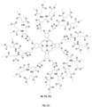
- the sensor element may be a construct in which a metallated porphyrin, such as illustrated in Fig. 2C , is covalently encapsulated inside a dendritic layer.
- the dendritic layer serves to attenuate oxygen diffusion towards the core, thus protecting the porphyrin from excessive quenching and increasing its sensitivity to small changes in oxygen pressures.
- Excitation and subsequent emission of the phosphor can be used to determine oxygen tension.
- tissue oxygen tension which is the partial pressure of oxygen (usually reported in mmHg or Torr) within the vapor that is in equilibrium with the tissue of interest, can be determined.
- tissue oxygen tension is proportional to the average oxygen concentration inside the cellular and extracellular components of the tissue and, accordingly, can be determined.
- the oxygen sensor can be excited using indirect excitation via energy transfer from a second fluorophore.
- the sensor is embedded into a polymeric matrix that allows oxygen to freely diffuse through it, and is solely used as a solid support for the sensing element.
- the sensing apparatus is disposed on the therapeutic release apparatus, and is separated from the therapeutic release apparatus via an oxygen-permeable membrane.
- the sensing apparatus is isolated from ambient oxygen pressure via an oxygen-impermeable membrane that comprises the top layer of the bandage apparatus as illustrated in Fig. 6A .
- a brightly emissive, custom built meso-unsubstituted platinum-porphyrin is encapsulated inside a second generation glutamic dendrimer, such as illustrated in Fig.2D .
- Glutamic dendrimers are known in the art, and have been successfully employed in increasing the sensitivity of various metallated porphyrins towards oxygen.
- the sensor is embedded into a layer of poly(lactic-co-glycolic acid) (PLGA) adhered to an oxygen-permeable plastic material that separates it from the release therapeutic apparatus.
- PLGA poly(lactic-co-glycolic acid)
- the porphyrin-dendrimer sensor element can be excited when illuminated at 532 nm, followed by emission of phosphorescence at 644 nm whose intensity can be used as an indicator for oxygen concentration.
- excitation and emission examples are likewise contemplated.
- excitation/emission examples for different sensors include (i) excitation at 546 nm and emission at 666 nm; and (ii) excitation at 595 nm and emission at 674 nm.
- a therapeutic agent is embedded in a light actuated polymer patterned on the proximal surface (that is, the surface closest to the body surface) of a patch (a supporting layer).
- the light actuated polymer contains a photoactive molecule embedded in the polymer.
- the light actuated polymer contains a light-actuated moiety within the polymer chemical structure. Referring to Fig. 6B , this polymer layer can either be directly adhered to the proximal surface using an adhesive layer, or can be separated from the adhesive by a permeable membrane layer. When irradiated by light, the photoactive compound leads to structural degradation of the polymer, facilitating local therapeutic release.
- Photosensitizer molecules used in photodynamic therapy can be encapsulated in PLGA polymer nanoparticles for enhanced uptake and delivery to cells and biological tissues.
- the photosensitizer (and fluorophore) 5-ethylamino-9-diethylaminobenzophenothiazinium (EtNBS) was encapsulated in 120 nm diameter PLGA polymer nanoparticles and delivered to both cells and model ovarian cancer tumors.
- the close proximity of the photosensitizers acts to quench absorption and emission properties of the particles, such that when encapsulated, illuminated EtNBS-PLGA particles weakly emit fluorescence.
- the radical species generated by EtNBS act to physically degrade the PLGA polymer structure, leading to the release of the therapeutic from the nanoparticle.
- the effect is observed as photo-brightening in samples containing the nanoparticles, as release of EtNBS negates quenching and results in increased fluorescence emission intensity.
- a therapeutic release mechanism may involve the cleavage of chemical bonds within the therapeutic apparatus via ultrasound, leading to controlled therapeutic release due to enhanced or facilitated diffusion through the degraded therapeutic matrix material.
- the therapeutic is enmeshed in or chemically bonded to the therapeutic matrix material via chemical moieties known as triazole linkers.
- Each triazole linker is attached to a long (-100 kDa) polymer linker such as polymethacrylate (PMA), resulting in the formation of a polymer/triazole network, the porosity of which can be tuned chemically.
- PMA polymethacrylate
- the use of PMA is doubly advantageous, as PMA is known for its ability to absorb many times its own mass in fluid, making it an absorbent and effective bandage/dressing material.
- Many other approaches are possible which fall within the scope of the invention, including alternative encapsulation schemes using different polymers and/or linker groups as well as other schemes in which the therapeutic is attached to the matrix polymer directly, rather than enmeshed.
- Therapeutics can be released through micro- or macro-structural degradation of a therapeutic release matrix.
- micro-structural degradation is defined as structural alterations of a matrix leading to therapeutic release at the nanometer and single micrometer size scales. Macro-structural degradation instead indicates structural alterations that occur at size scales larger than 10 micrometers.
- Multiple therapeutics can be packaged within in each region for the simultaneous release of a complete therapeutic regimen.
- different therapeutics or therapeutic regimens can be spatially patterned to different regions in the therapeutic apparatus. These different therapeutics or therapeutic regimens may be color-coded, both for indication (for example, red, blue, or green on the distal patch side) and to differentiate between different released therapeutics.
- a therapeutic can be contained in a nanoparticle form that is released from a matrix, such as a polymer, gel, or hydrogel, upon light activation via any of the mechanisms previously described.
- spatioselective therapeutic release is accomplished using a bandage apparatus formulated as an adhesive patch that stays affixed to a patient.
- a user can trigger the release of therapeutics in a controlled manner through the use of an interacting apparatus delivering light when held against the bandage apparatus.
- the irradiated region of the patch can change color to indicate that therapeutic has been released.
- a single patch can hold a multitude of therapeutic releasing regions.
- embodiments of the invention can be combined with therapeutic eluting patch technologies currently in use.
- These drug-eluting patches include single and double layer drug-in-adhesive patches as well as reservoir, matrix, and vapor patch designs.
- single and multiple drug-in-adhesive patches could be readily be used in combination with the therapeutic release matrices described here.
- Reservoir, matrix and vapor patch technologies can be embedded into embodiments of the invention, including, but not limited to, regions/spaces not containing light-actuated therapeutic release matrices, layers within the embodiment, or into therapeutic release apparatus itself.
- the anti-inflammatory therapeutic Diclofenac (2-(2-(2,6-dichlorophenylamino)phenyl)acetic acid) is embedded with the photoactive molecule methylene blue into a layer of PLGA adhered to the proximal surface of a plastic backing material.
- the patch backing material is composed of a 0.3 cm thick layer of polyvinyl alcohol.
- the proximal layer of the patch contains an adhesive layer composed of methylcellulose gel.
- the therapeutic releasing polymer matrix is patterned over the patch proximal surface in 1 cm diameter circular regions each separated by 2 cm. Each circular region has a corresponding circle that appears on the distal patch surface to indicate the exact spatial location of the therapeutic releasing apparatus in the patch.
- the spatial location is marked with cis-azobenzene, which undergoes trans isomerization when illuminated with 635 nm light that results in a color change post-irradiation. This color change indicates that a specific region of the patch has been used.
- the therapeutic release matrix could be a bladder filled with a single or multiple therapeutics that is released upon its rupture. The flow of material from the bladder would facilitate delivery of the therapeutic(s) to the patient.
- the use of pressure could trigger a material to structurally expand, leading to the release of therapeutic.
- the interacting apparatus can take the shape of a hand-held device, such as a pen-shaped applicator, that can hold an internal power supply such as a battery.
- this power supply powers a bright light emitting diode (LED) affixed to one end.
- the LED would be selected to emit a specific color of light, for example red light at, for example, approximately 630 nm.
- This LED can be switched on through the press of a button on the pen or activated by pressing the interacting apparatus against a surface. Once activated, the LED would remain operational for a set period of time determined by an electronic circuit inside the pen.
- an ultrasonic emitter can be powered by the power source, turned on by the user, and controlled using an electronic circuit.
- An electronic circuit used with an energy device such as an LED or ultrasound emitter can interface with a computer, mobile platform, or network to receive programming instructions, including information regarding total administrative dose, and number of doses within a set period of time.
- This electronic circuit could also interface with said computer, mobile platform, or network to communicate treatment information including number of times used, number of doses received, the historical dose schedule, and the like.
- the electronic circuit can either contain a data storage mechanism, or can interface with a computer, mobile platform, or network to store said information.
- One embodiment of the invention is designed for use on patients with open wounds and is comprised of diagnostic (sensing), treatment (therapeutic), and interacting apparatuses. It is known in the art that regions of local inflammation in acute and chronic wounds display high relative oxygen levels in contrast to non-inflamed tissue. An embodiment of the invention that reports tissue oxygenation could be used to identify and spatially map regions of inflammation in wounds, providing detailed wound status information to a caregiver or physician. Once these specific sites of inflammation are recognized, they can be treated using the interactive technology in the invention through user-triggered release of anti-inflammatory therapeutics using the interacting apparatus.
- the oxygenation status of the wound can be monitored to follow the inflammation status of regions treated, either using the interacting apparatus or other recording devices, to provide real-time therapeutic response feedback.
- a caregiver or physician treating the wound using this invention could then use the displayed information to determine the next optimal course of treatment, all without removing the invention and compromising sterility.
- These oxygen reporting systems can utilize molecules (that is, fluorophores) whose emission properties are insensitive to oxygen along with molecules (that is, phosphors) whose emission properties are influenced by molecular oxygen concentration.
- emission from the fluorophore/phosphor probe can be used to measure oxygen tension in biological systems reversibly with high fidelity.
- a first curve (a) shows an initial emission spectrum from the probe in air
- a second curve (b) shows emission following equilibration of the probe in a substantially oxygen free environment (i.e., following a nitrogen purge).
- a third curve (c) shows emission after once again equilibrating the probe in air.
- curves (a) and (c) display a similar profile.
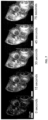
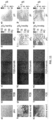
- Such probes can be calibrated so that the spectral ratio between fluorophore and phosphor emission correlate with oxygen concentration. Furthermore, this calibration can be used to read out a map of oxygen concentration in biological samples as shown in Figs. 10A-B . It is also possible to utilize molecules such as dyes whose light absorption properties (such as, absorption wavelength or absorption cross-section) can be modulated by the presence of analytes such as oxygen for light absorption-based colorimetric oxygen measurements.
- the present invention provides a system and method for imaging of multiple tissue parameters using a wound dressing.
- the invention provided here allows the selective, local release of therapeutics (as described previously and further detailed below) to any part of the tissue or wound covered by the dressing, as well as the subsequent monitoring of physiological parameters to assess therapeutic response.
- a conventional digital camera The feasibility of imaging tissue parameters (namely pO 2 and pH) using a conventional digital camera, has been demonstrated.
- the device displayed a wavelength dependent readout, with the data being stored in 3-color (red/green/blue) RGB channels.
- sensing elements were encapsulated in an analyte-permeable membrane, to form a skin patch for wound healing monitoring.
- the method was applied on intact skin as well as on a chronic wound.
- a homogeneous distribution of pO 2 and pH was observed.
- oxygen and pH values of a chronic wound indicated a sustained inflammatory phase.
- the exemplary method 120 includes, in step 121, a manufacturer or a physician determining whether it is desirable that a given device includes a sensing portion. If the individual determines that a sensing component is desired, in a next step 122, the individual identifies an analyte of interest to be sensed by the sensing portion. Once an analyte has been identified, in step 123, a sensor suitable for detecting the identified analyte is selected.
- a meso-unsubstituted metallated porphyrin molecule designed to be an effective sensor for oxygen may be selected.
- the porphyrin molecule can be excited with a specific wavelength of light and the presence of oxygen can be detected by measuring a corresponding phosphorescent emission signal.
- step 124 the designer determines whether or not a therapeutic portion is desired. If a therapeutic portion is to be included, a therapy of interest is identified in step 125.
- the identified therapy may include the release of pain medication such as an NSAID.
- the therapeutic(s), such as one or more NSAIDs is selected in step 126.
- the selection of a therapeutic portion can be done to ensure the compatibility of the sensor with the matrix, such as will be described below. That is, selection of a therapeutic portion may be done with consideration to the compatibility of the sensor and, as will be described, the matrix, such that selection of the therapeutic portion is based on a chemical property of one or both of the sensor and the matrix.
- one embodiment involves combining a singlet-oxygen degradable polymer, which can be the basic component of the therapeutic release matrix, with an oxygen-independent, free-base porphyrin core that could generate enough singlet oxygen to trigger the matrix degradation.
- a metallated, dendritic analogue of that porphyrin serves as the oxygen-reporting molecule, and is embedded in the sensing matrix selected at step 123. Having inherently different properties, these two molecules can be actuated using light of different wavelengths; thus allowing on-demand control of oxygen sensing separate from therapeutic release.
- dendritic encapsulation can efficiently minimize the amount of singlet oxygen that can reach the therapeutic matrix following light-activation of the oxygen-reporting molecule, thus maximizing selectivity in the use of the free-base porphyrin (or any photosensitizer) for light-activated therapeutic release.
- a suitable matrix material is selected.
- individual matrix materials can be selected for each of the sensing and therapeutic portions or a single matrix may be selected to support both of the sensing and therapeutic portions.
- the senor may be enmeshed in the matrix material.
- Mixing of the sensing matrix components with the sensor may involve the use an additional, silyl-based component (Triisopropylsilyl chloride or TIPS-CI) that is similar in structure with those found in the mixture that forms the matrix (Polydimethylsiloxane or PDMS).
- TIPS-CI Triisopropylsilyl chloride
- the oxygen-reporting molecule of the sensor may be enmeshed within the matrix.
- a matrix base may be selected based on a chemical nature of the sensor, such that the matrix base is ensured to be desirably compatible with the sensor and ensures "curing.”
- the compatibility between the sensor and its matrix is highly dependent on the structural similarity between the two components.
- the compatibility may be dependent upon hydrophobicity, polarity of surface functional groups, polarity of the entire molecule, type and number of charges, molecular weight, stability, reactivity, and other factors.
- sensor molecules modified with glutamic dendrimers carry highly polar surface functional groups, which is not compatible with the low-polarity hydrophobic PDMS matrix. If the sensor molecules were mixed into the PDMS curing mixture as a dichloromethane (DCM) solution, they would agglomerate and precipitate out of the curing mixture. When dimethylformamide (DMF) is used as the solvent for the sensor molecules, better mixing can be achieved with the curing mixture. However, the curing mixture does not polymerize and form a solid bandage in the presence of DMF.
- DCM dichloromethane
- DMF dimethylformamide
- TIPS-CI triisopropylsilyl chloride
- some general factors that may be considered while matching the sensor molecules with the bandage matrix include the chemical structures of the sensor and matrix, concentration of the sensor molecule inside the matrix, and potential changes in the matrix's optical, mechanical, and chemical properties upon mixing the two.
- step 127 stands in contrast to other approaches for coupling a sensor with another material, such as using solvents to solubilize an oxygen-reporting molecule in the sensing matrix mixture because such attempts have been shown to be incompatible as they prevent the polymerization process known as "curing", which is the basis for forming a solid and flexible sensing matrix from a viscous mixture of different components.
- step 128 the sensing and therapeutic portions and the selected matrix materials are assembled into a device, such as a dressing.
- a designer can choose to omit either of the sensing and therapeutic portions.
- step 121 if the designer determines that a sensing portion is not desirable, method 120 indicates that the designer should proceed directly to step 124. However, it is preferable that at least one of the sensing and therapeutic portions is included. Therefore, if in step 124, it is determined that a therapeutic portion is not desired, method 120 includes a step 130 in which if neither sensing nor therapeutic was selected for inclusion in the manufacture of the dressing, the method 120 proceeds to step 121. Alternatively, if at least one of a sensing and therapeutic portion is desired, method 120 indicates that the designer should proceed to step 127.
- the sensing portion can include a matrix composed of a highly breathable polydimethylsiloxane (PDMS) layer where the sensing molecules are enmeshed, and a polyvinyl chloride/polyvinylidene chloride (PVC/PVDC) gas-impermeable layer to block out room oxygen.
- PDMS polydimethylsiloxane
- PVC/PVDC polyvinyl chloride/polyvinylidene chloride
- Table 1 Oxygen Permeability of different polymeric materials (O 2 permeability has units of 10 -10 cm 2 s -1 cmHg -1 ).
- the amount (or thickness) of the material may also be considered.
- the commercial transparent film "Tegaderm” is mainly composed of polyurethane (PU) and acrylate polymers (for example, PMMA, PEMA). They are advertised as breathable and suitable for chronic wound dressing even through PU has a low permeability compared with PDMS.
- the gas permeability of the barrier materials can be tuned to adapt to different clinical applications.
- Higher breathability materials can be used in chronic wound dressings designed for long-term wearing, where sufficient oxygen exchange is essential for the healing of the wounds.
- the bandage is designed for acute wound management, where a quick oxygen reading and other properties of the bandage (exudate absorption, moisture keeping, infection control, and the like) are desired, less permeable materials can be used as the barrier layer.
- Traumatic injuries result in acute wounds and burns that often require skin grafts or flaps to salvage tissue and limbs to restore function and improve cosmetic outcome.
- Postsurgical assessment of perfusion and oxygenation in acute wounds and burns is typically qualitative and subjective, relying on episodic assessments such as wound color and temperature, capillary refill, Doppler ultrasound and touch ( Park et al., The Surgical Clinics of North America, 90(6): 1181-1194, 2010 ). These approaches require extensive training, are subject to operator experience, and can miss critical events due to their episodic nature, leading to poor surgical outcome.
- surgical rehabilitation procedures for wounded warriors can fail due to undetected anastomotic failure, causing loss of perfusion and subsequent ischemia, infarction and necrosis of the transplanted tissue ( Orgill et al., The New England Journal of Medicine, 360(9):893-901, 2009 ).
- This is also problematic when laying skin over thermal burns, where insufficient debridement results in the inability to detect adequate graft blood supply, leading to subsequent graft failure ( Meier et al., Angewandte Chemie International Edition, 50(46): 10893-10896, 2011 ).
- the invention provides a solution to these complex clinical problems.
- the invention can be built to display an active, real-time map of both oxygenation and pH across the entire wound or burn site for either colorimetric or augmented reality display.
- the oxygen and pH sensors themselves can be embedded in a bandage apparatus and separated from the wound or burn surface via selectively permeable membranes. This ensures readout of tissue properties without any risk of interaction with the bandage sensors. This readout is of great importance in the skin graft process for the treatment of burns, as the burn site must be adequately debrided for a graft to take. Inadequate debridement results in low perfusion and oxygenation that often results in graft failure.
- a bandage apparatus capable of visualizing perfusion and oxygenation across a burn site would save numerous surgical protocols and allow physicians to better plan their interventions to maximize graft success and minimize patient impact. Equally important, a bandage apparatus that can allow for the selective, spatial administration of therapeutics will allow caregivers to treat inflammation and infection without bandage removal and the accompanying disruption of the burn and loss of sterility.
- a separate portion of the invention contains therapeutics embedded in light-degraded polymers triggered by specific wavelengths of light.
- An interacting apparatus containing various colored LEDs is applied to the sensing and therapeutic bandage apparatus, with a specific color LED triggering the selective release of a specific therapeutic.
- regions of inflammation can be exposed to blue light to release an anti-inflammatory, while an infected region in the same wound sight can be treated with antibodies through red-light illumination.
- the invention-provided oxygenation and/or pH mapping allows for spatiotargeted delivery of needed therapeutics to affected tissue regions, without ever removing the bandage or compromising sterility.
- the same oxygen and pH mapping capability can track the recovery process, providing clinicians with real-time feedback for accurate wound recovery assessment.
- Chronic wound management is a challenging healthcare problem, with an estimated 2% of the world population suffering from chronic wounds or associated co-morbidities ( Gethin et al., Wounds UK, 3(3):52-55, 2007 ).
- Major sources of chronic wounds include venous and/or arterial ulcers, decubitus ulcers (bed/pressure sores), and diabetic ulcers.
- chronic wounds afflict patients from all demographics and age groups, the elderly and diabetics are particularly effected, accruing collective wound management costs in excess of $10 billion annually ( Snyder et al., Clinics in dermatology, 23(4):388-95, 2005 ).
- the present invention provides a platform that offers several applications in the management of chronic wounds.
- chronic wound patients report severe, persistent pain that adversely affects their activity level and quality of life ( Jorgensen et al., Official Publication of the Wound Healing Society [and] the European Tissue Repair Society, 14(3):233-9, 2006 ).
- oral pain medications are available for pain management, pain from chronic wounds such as venous leg ulcers is often undertreated due to patient frailty, contraindication or poor tolerance of systemic analgesics, or simple reluctance to take more medicine.
- an embodiment of the invention can function as a tiered chronic wound pain management platform, offering the ability treat pain on-demand spatiospecifically.
- the therapeutic apparatus within a bandage apparatus contains NSAIDs, which can either be constitutively eluted or eluted in response to a particular wavelength of light administered by the interacting apparatus (for example, green light).
- the therapeutic apparatus also contains an additional drug, representing a "step up" on the WHO pain ladder (for example, fentanyl, buprenorphine, or morphine), that is eluted from the therapeutic apparatus in response to a different wavelength of light (for example, blue light).
- a different wavelength of light for example, blue light
- the frequency of dressing removal and wound examination can be further reduced by the invention's capacity as a spatiospecific sensor of physiologically relevant parameters, such as pH.
- physiologically relevant parameters such as pH.
- pH can be correlated to the clinical stage of pressure ulcers ( Gethin et al., Wounds UK, 3(3):52-55, 2007 ) and could therefore be used to monitor wound healing as well as identify potential problems, such as infection, without the painful, labor-intensive, and psychologically problematic process of repeated dressing removal.
- early identification of wound regression would accelerate identification of wound pathologies, intervention, and treatment, thereby potentially preventing co-morbidities such as infection.
- pH also has been shown to correlate to oxygen tension, itself an important indicator of wound healing.
- a specially-tailored bandage apparatus can be additionally used to monitor and display lipopolysaccharides (LPS) as an indication of infections and release antibiotics on-demand.
- LPS lipopolysaccharides
- the management and surgery of chronic wounds is similar to what is found in the treatment of burn grafts.
- embodiments of the invention could be used in a similar fashion to verify adequate debridement and monitor wound beds post-operatively for infection without bandage removal.
- Another aspect of the present invention relates to pain management.
- the invention can be used to avoid the use of needles, catheters, infusion and syringe pumps while the associated interacting apparatus would allow remote telemetry and monitoring. Therefore, pain medication could be customized and calibrated in real-time to patient's pain intensity as measured by the pain scale. Accurate dosimetry and calibration has the potential of decreasing side effects.
- Another aspect of the present invention relates to accelerated on-demand therapeutic delivery.
- Many currently used therapeutic delivery patches and bandages use reservoirs saturated with a given drug that is separated from the patient's skin by a membrane.
- the membrane is chosen such that the rate of therapeutic diffusion through the membrane is lower than that of the protective skin layer called the stratum corneum. This ensures that the elution of the therapeutic through the membrane occurs at a rate allowing for transdermal delivery.
- Less expensive designs incorporate adhesive layers that accomplish the same effect, where the adhesive acts as both a reservoir and a diffusion-controlling matrix ( Prausnitz et al., Nature biotechnology, 26(11): 1261-8, November 2008 ).
- the delivery rate of the therapeutic is ultimately bound by the transdermal therapeutic transport rate. This can be acceptable for slow-therapeutic release systems or for marginally hydrophilic therapeutics.
- most existing drugs are significantly hydrophobic, and in many cases need to be delivered in a single dose instead of extended timed release.
- patches based on diffusion are environmentally sensitive: fentanyl patches accidentally worn in a hot shower, for example, lead to patient deaths due to increased diffusion at higher temperatures.
- a solution to these issues would be a mechanism that could temporarily disrupt the stratum corneum to facilitate increased therapeutic diffusion.
- One proposed mechanism would be to disrupt the stratum corneum using photodynamic therapy ( Dougherty et al., Journal of the National Cancer Institute, 90(12):889-905, 1998 ).
- a hydrophobic photosensitizer with limited dermal penetration would be taken up by the stratum corneum.
- reactive radical species released by the photosensitizer would react, opening up pores and fissures in the stratum corneum through which therapeutics could travel.
- Short-wavelength, blue light could additionally be used for photoactivation to limit the depth of reactive radical specie generation to only the stratum corneum.
- therapeutics in the invention could either (1) directly then flow into the tissue, (2) be released through a semi-permeable membrane, or (3) be triggered for release via a secondary membrane containing a different photosensitizer.
- the first case, direct flow of therapeutics would be useful for short-term, instant release of therapeutic agents not compatible with existing transdermal approaches. This case would also be useful for the delivery of therapeutics in special populations, such as children or the elderly, where standard administration approaches are either difficult or impossible to use.
- Another aspect of the present invention relates to vaccine delivery.
- the need for safe and efficient vaccine administration depends on engineering simple tools for transdermal delivery.
- One challenges to overcome is the limited penetration of materials through the stratum corneum.
- a number of different techniques that facilitate transcutaneous drug delivery are known in the art.
- Such drug delivery methods disrupt the stratum corneum, for example, through the use of microneedles ( Bariya et al., Journal of Pharmacy and Pharmacology, 64(1):11-29, 2012 ; Vrdoljak et al., Journal of Controlled Release, 159(1):34-42, 2012 ) and ablative fractional lasers (AFL) ( Chen et al., Journal of Controlled Release, 159(1):43-51, 2012 ), which are minimally invasive and not entirely painless, and/or require skin pretreatment.
- microneedles Bariya et al., Journal of Pharmacy and Pharmacology, 64(1):11-29, 2012 ; Vrdoljak et al., Journal of Controlled Release, 159(1):34-42, 2012
- AFL ablative fractional lasers
- Skin patches for transcutaneous vaccine delivery offer the advantages of being a noninvasive, painless and cost-effective method of immunization that does not require the assistance of highly trained health care personnel.
- a hydrogel patch is used in transcutaneous vaccination for tetanus and diphtheria ( Hirobe et al., Vaccine, 30(10): 1847-1854, 2012 ).
- Octyldodecyllactate which is an absorption enhancer that disrupts the lipid bilayer of the stratum corneum ( Hood et al., Food and Chemical Toxicology, 37(11): 1105-1111, 1999 ), was used in the preparation of the patch in order to promote the transmission of the vaccines through the skin.
- Embodiments of the invention that make use of light-activated skin penetration enhancement, as discussed above, could allow for the release of a vaccine from a skin patch apparatus for the controlled release of vaccine doses. In one embodiment of the invention, this could be achieved with polymer cages encapsulating vaccines that are degraded by applying light of a specific wavelength. Vaccine release occurs either simultaneously, or following light-activated skin penetration by using polymeric matrices with different degradation pathways.
- Vaccine administration using this embodiment of the invention is applicable in areas such as pediatrics, where having a painless method that requires minimal pretreatment is essential when dealing with young patients. Moreover, it can help fight the problem of disease outbreaks in developing countries, as a simple and inexpensive way to rapidly vaccinate large populations without the need of trained healthcare personnel.
- light-degradable polymeric matrices that are activatable by sunlight can be used, and vaccination can be simply accomplished by exposure to sunlight.
- Another advantage of the present invention is to promote new areas of investigation in the fields of pain release (postoperative, acute, chronic, and palliative) and vaccination (preventive, curative).
- Another advantage of the present invention is to provide customized therapeutic approaches in pediatric medicine, geriatrics, and post-trauma care.
- Yet another advantage of the present invention is contribute to the establishment of clinically powerful and user-friendly home care and telemonitoring programs.
Landscapes
- Health & Medical Sciences (AREA)
- Life Sciences & Earth Sciences (AREA)
- Physics & Mathematics (AREA)
- Surgery (AREA)
- Engineering & Computer Science (AREA)
- Veterinary Medicine (AREA)
- Public Health (AREA)
- General Health & Medical Sciences (AREA)
- Animal Behavior & Ethology (AREA)
- Biomedical Technology (AREA)
- Pathology (AREA)
- Molecular Biology (AREA)
- Heart & Thoracic Surgery (AREA)
- Medical Informatics (AREA)
- Biophysics (AREA)
- Chemical & Material Sciences (AREA)
- Spectroscopy & Molecular Physics (AREA)
- Optics & Photonics (AREA)
- Organic Chemistry (AREA)
- Nuclear Medicine, Radiotherapy & Molecular Imaging (AREA)
- Oral & Maxillofacial Surgery (AREA)
- Dermatology (AREA)
- Epidemiology (AREA)
- Measurement Of The Respiration, Hearing Ability, Form, And Blood Characteristics Of Living Organisms (AREA)
- Investigating, Analyzing Materials By Fluorescence Or Luminescence (AREA)
- Radiology & Medical Imaging (AREA)
- Computer Networks & Wireless Communication (AREA)
- Bioinformatics & Cheminformatics (AREA)
- Medicinal Chemistry (AREA)
- Pharmacology & Pharmacy (AREA)
- Investigating Materials By The Use Of Optical Means Adapted For Particular Applications (AREA)
Claims (14)
- Dispositif (100) comprenant :un capteur configuré pour détecter une concentration d'un analyte choisi dans le groupe constitué de l'oxygène moléculaire, le dioxyde de carbone, les oxydes nitriques, les analytes dissous dans le plasma et le tissu, et les ions d'hydrogène ;une matrice compatible avec ledit capteur, au moins une partie de ladite matrice est au moins l'une d'une accessible et perméable audit analyte ; etun pansement (10) comprenant une partie thérapeutique (14) et une partie de détection/diagnostic (16), ladite partie de détection/diagnostic présentant ladite matrice et ledit capteur ; etune seule partie à interaction (20) comportant une partie d'actionnement (22) pour générer un signal d'actionnement (24) sur le pansement (10) et une partie de détection (28) pour détecter un signal de réponse (26) provenant du pansement (10) ;dans lequel ledit dispositif est configuré pour émettre un signal en réponse à la concentration dudit analyte détecté par ledit capteur par modulation de la diffusion inélastique d'un champ électromagnétique, comportant au moins l'une d'une phosphorescence, fluorescence, et absorption,dans lequel ladite partie thérapeutique (14) est configurée pour administrer au moins l'un d'un agent thérapeutique, d'un biomarqueur, et d'un agent de diagnostic à une surface de tissu d'un patient ;dans lequel ladite partie thérapeutique comprend en outre un mécanisme de libération thérapeutique actionné configuré pour libérer au moins l'un de l'agent thérapeutique, du biomarqueur, et de l'agent de diagnostic sur des surfaces de tissu du patient, dans lequel ledit mécanisme de libération thérapeutique actionné comprend une matrice polymère actionnée par la lumière configurée pour être sensible audit signal.
- Dispositif selon la revendication 1, dans lequel ledit pansement (10) est configuré pour être fixé à une surface de tissu d'un patient.
- Dispositif selon la revendication 1, comprenant en outre un appareil à interaction, dans lequel un utilisateur peut déclencher la libération d'un agent thérapeutique à l'aide de l'appareil à interaction.
- Dispositif selon la revendication 3, dans lequel ledit appareil à interaction déclenche la libération de l'agent thérapeutique au moyen d'une lumière.
- Dispositif selon la revendication 1, dans lequel ledit capteur est configuré pour interagir avec une surface de tissu d'un patient.
- Dispositif selon la revendication 1, dans lequel ledit capteur comprend une porphyrine étalatée non méso-substituée.
- Dispositif selon la revendication 1, dans lequel la partie à interaction (14) est configurée pour actionner ladite partie thérapeutique ;
dans lequel ladite partie thérapeutique est apte à être en communication locale et/ou distante avec un système externe, réseau ou ordinateur. - Dispositif selon la revendication 1, comprenant en outre un mécanisme d'affichage configuré pour former une carte active en temps réel d'un paramètre associé audit analyte.
- Dispositif selon la revendication 1, dans lequel ladite partie de détection (28) est en outre configurée pour obtenir des signaux émis avec une résolution temporelle d'au moins un dudit pansement (10) et d'un environnement avoisinant.
- Dispositif selon la revendication 1, dans lequel ladite partie de détection (28) est en outre configurée pour rapporter des propriétés d'analyte comportant la concentration.
- Dispositif selon la revendication 1, dans lequel ladite partie de détection (28) est en outre configurée pour communiquer avec au moins l'un d'un réseau, d'un écran, et d'un système informatique.
- Dispositif selon la revendication 1, dans lequel le pansement (10) est configuré pour être appliqué sur une surface de tissu d'un patient en tant que liquide et former un solide sur la surface de tissu du patient.
- Procédé de fabrication d'un dispositif selon la revendication 1 pour détecter un analyte, le procédé comprenant les étapes consistant à :(a) sélectionner un capteur configuré pour détecter une concentration d'un analyte comportant au moins l'un de l'oxygène moléculaire, du dioxyde de carbone, des oxydes nitriques, des analytes dissous dans le plasma et le tissu, et des ions d'hydrogène ;(b) sélectionner une matrice compatible sur la base d'une nature chimique dudit capteur ;
intégrer ledit capteur dans ladite matrice compatible, au moins une partie de ladite matrice étant accessible audit analyte ;(d) fournir une partie thérapeutique (14) ;(e) incorporer ladite matrice dans un pansement (10) ; et(f) fournir une seule partie à interaction (20) comportant (i) une partie d'actionnement (22) pour diriger un signal d'actionnement au niveau du pansement et (ii) une partie de détection (28) pour détecter un signal de réponse (26) provenant du pansement,dans lequel ledit signal de réponse varie en réponse à la concentration dudit analyte détectée par ledit capteur, etdans lequel la partie thérapeutique est configurée pour administrer au moins l'un d'un agent thérapeutique, d'un biomarqueur, et d'un agent de diagnostic à une surface de tissu d'un patient ; etdans lequel ladite partie thérapeutique comprend en outre un mécanisme de libération thérapeutique actionné configuré pour libérer au moins l'un de l'agent thérapeutique, du biomarqueur, et de l'agent de diagnostic sur des surfaces de tissu du patient, dans lequel ledit mécanisme de libération thérapeutique actionné comprend une matrice polymère actionnée par la lumière configurée pour être sensible audit signal de réponse. - Utilisation du dispositif selon la revendication 1 pour surveiller l'état d'oxygénation d'une plaie à la suite de l'administration d'un agent thérapeutique.
Applications Claiming Priority (2)
| Application Number | Priority Date | Filing Date | Title |
|---|---|---|---|
| US201261669718P | 2012-07-10 | 2012-07-10 | |
| PCT/US2013/049847 WO2014011724A1 (fr) | 2012-07-10 | 2013-07-10 | Système et méthode de surveillance et de traitement de surface de patient |
Publications (3)
| Publication Number | Publication Date |
|---|---|
| EP2872877A1 EP2872877A1 (fr) | 2015-05-20 |
| EP2872877A4 EP2872877A4 (fr) | 2016-04-13 |
| EP2872877B1 true EP2872877B1 (fr) | 2023-05-24 |
Family
ID=49916522
Family Applications (1)
| Application Number | Title | Priority Date | Filing Date |
|---|---|---|---|
| EP13817635.9A Active EP2872877B1 (fr) | 2012-07-10 | 2013-07-10 | Système et méthode de surveillance et de traitement de surface de patient |
Country Status (7)
| Country | Link |
|---|---|
| US (6) | US10016164B2 (fr) |
| EP (1) | EP2872877B1 (fr) |
| JP (1) | JP6321004B2 (fr) |
| DK (1) | DK2872877T3 (fr) |
| ES (1) | ES2957540T3 (fr) |
| FI (1) | FI2872877T3 (fr) |
| WO (1) | WO2014011724A1 (fr) |
Families Citing this family (58)
| Publication number | Priority date | Publication date | Assignee | Title |
|---|---|---|---|---|
| EP2872877B1 (fr) | 2012-07-10 | 2023-05-24 | The General Hospital Corporation | Système et méthode de surveillance et de traitement de surface de patient |
| GB201317746D0 (en) | 2013-10-08 | 2013-11-20 | Smith & Nephew | PH indicator |
| US20190212311A1 (en) | 2013-01-11 | 2019-07-11 | Smith & Nephew Plc | Ph and moisture indicator devices and formulations |
| US9789206B2 (en) | 2013-07-10 | 2017-10-17 | The General Hospital Corporation | Compounds, systems, and methods for monitoring and treating a surface of a subject |
| JP6470748B2 (ja) * | 2013-07-10 | 2019-02-13 | ザ ジェネラル ホスピタル コーポレイション | 対象の表面をモニターし、処置するための化合物、システムおよび方法 |
| US10524707B2 (en) * | 2014-01-31 | 2020-01-07 | The General Hospital Corporation | System and method for photoluminescence detection |
| JP2016090312A (ja) * | 2014-10-31 | 2016-05-23 | 株式会社島津製作所 | 酸素濃度計測シール及びそれが用いられた燃料電池セル |
| JP6490399B2 (ja) * | 2014-11-10 | 2019-03-27 | 国立大学法人横浜国立大学 | センサー装置、生体分子視覚化装置 |
| GB201600747D0 (en) | 2016-01-14 | 2016-03-02 | Smith & Nephew | Improvements in and relating to devices |
| GB201600746D0 (en) | 2016-01-14 | 2016-03-02 | Smith & Nephew | Improvements in and relating to polymer materials |
| JP2017181211A (ja) * | 2016-03-29 | 2017-10-05 | 株式会社カテラ | 光学的経皮酸素センサ及びこれを用いた経皮酸素濃度測定装置 |
| EP3454809B8 (fr) * | 2016-05-10 | 2020-12-30 | 3M Innovative Properties Company | Moyen flexible pour déterminer l'étendue de débridement nécessaire pour enlever un tissu non viable |
| WO2017197385A1 (fr) | 2016-05-13 | 2017-11-16 | The General Hospital Corporation | Systèmes et procédés de surveillance optique de l'oxygénation transcutanée |
| CA3023772A1 (fr) | 2016-05-13 | 2017-11-16 | Smith & Nephew Plc | Appareil de surveillance et de therapie de plaie active par un capteur |
| JP7091356B2 (ja) | 2017-03-09 | 2022-06-27 | スミス アンド ネフュー ピーエルシー | 皮膚灌流圧を決定するデバイス、装置、および方法 |
| WO2018162736A1 (fr) | 2017-03-09 | 2018-09-13 | Smith & Nephew Plc | Pansement, élément de timbre et procédé de détection d'un ou plusieurs paramètres de plaie |
| WO2018162732A1 (fr) | 2017-03-09 | 2018-09-13 | Smith & Nephew Plc | Appareil et procédé d'imagerie du sang dans une région cible de tissu |
| CN110650713B (zh) | 2017-04-11 | 2022-06-24 | 史密夫及内修公开有限公司 | 用于传感器实现的伤口敷料的部件定位和应力释放 |
| CN110870021B (zh) | 2017-05-15 | 2024-12-20 | 史密夫及内修公开有限公司 | 使用欧拉视频放大的负压伤口治疗系统 |
| JP7272962B2 (ja) | 2017-05-15 | 2023-05-12 | スミス アンド ネフュー ピーエルシー | 創傷分析装置 |
| US11633153B2 (en) | 2017-06-23 | 2023-04-25 | Smith & Nephew Plc | Positioning of sensors for sensor enabled wound monitoring or therapy |
| GB201804502D0 (en) | 2018-03-21 | 2018-05-02 | Smith & Nephew | Biocompatible encapsulation and component stress relief for sensor enabled negative pressure wound therapy dressings |
| GB201809007D0 (en) | 2018-06-01 | 2018-07-18 | Smith & Nephew | Restriction of sensor-monitored region for sensor-enabled wound dressings |
| CA3072006A1 (fr) | 2017-08-10 | 2019-02-14 | Smith & Nephew Plc | Positionnement de capteurs pour la surveillance ou le traitement des plaies active(e) par capteurs |
| GB201718870D0 (en) | 2017-11-15 | 2017-12-27 | Smith & Nephew Inc | Sensor enabled wound therapy dressings and systems |
| GB201804971D0 (en) | 2018-03-28 | 2018-05-09 | Smith & Nephew | Electrostatic discharge protection for sensors in wound therapy |
| WO2019048624A1 (fr) | 2017-09-10 | 2019-03-14 | Smith & Nephew Plc | Systèmes et procédés d'inspection d'encapsulation et de composants dans des pansements équipés de capteurs |
| GB201718859D0 (en) | 2017-11-15 | 2017-12-27 | Smith & Nephew | Sensor positioning for sensor enabled wound therapy dressings and systems |
| EP3687380A1 (fr) | 2017-09-27 | 2020-08-05 | Smith & Nephew plc | Détection de ph pour appareils de surveillance et de thérapie de plaie à pression négative |
| WO2019072531A1 (fr) | 2017-09-28 | 2019-04-18 | Smith & Nephew Plc | Neurostimulation et surveillance à l'aide d'un appareil de surveillance et de traitement de plaie activé par un capteur |
| EP3709943B1 (fr) | 2017-11-15 | 2024-07-24 | Smith & Nephew PLC | Pansements et systèmes de surveillance et/ou de traitement de plaie activés à capteur intégré |
| CN108827480B (zh) * | 2018-04-11 | 2020-03-27 | 山东师范大学 | 一种基于磷光发射的抗氧干扰温度传感器 |
| GB201814011D0 (en) | 2018-08-29 | 2018-10-10 | Smith & Nephew | Componet positioning and encapsulation for sensor enabled wound dressings |
| WO2020053290A1 (fr) | 2018-09-12 | 2020-03-19 | Smith & Nephew Plc | Dispositif, appareil et procédé de détermination de pression de perfusion cutanée |
| WO2020064937A1 (fr) | 2018-09-28 | 2020-04-02 | T.J.Smith And Nephew,Limited | Fibres optiques permettant la détection optique à travers des pansements de plaie |
| GB201816838D0 (en) | 2018-10-16 | 2018-11-28 | Smith & Nephew | Systems and method for applying biocompatible encapsulation to sensor enabled wound monitoring and therapy dressings |
| JP2020071152A (ja) * | 2018-10-31 | 2020-05-07 | ソニー株式会社 | 免疫染色方法、免疫染色システム、および免疫染色キット |
| GB201820927D0 (en) | 2018-12-21 | 2019-02-06 | Smith & Nephew | Wound therapy systems and methods with supercapacitors |
| JP7528097B2 (ja) | 2019-01-30 | 2024-08-05 | スミス アンド ネフュー ピーエルシー | センサ一体型被覆材及びシステム |
| GB201901242D0 (en) | 2019-01-30 | 2019-03-20 | Smith & Nephew | Optical sensing systems and methods for sensing enabled wound dressings and systems |
| CN111557796A (zh) * | 2019-02-15 | 2020-08-21 | 华中科技大学同济医学院附属协和医院 | 一种用于肢体植皮术后伤口旷置器 |
| GB2597148B (en) | 2019-03-18 | 2022-12-21 | Smith & Nephew | Design rules for sensor integrated substrates |
| WO2020187643A1 (fr) | 2019-03-19 | 2020-09-24 | Smith & Nephew Plc | Systèmes et procédés pour la mesure de l'impédance d'un tissu |
| US20200392378A1 (en) * | 2019-06-17 | 2020-12-17 | Richard William Schofield | Adhesive tape with strip to help locate and lift the leading edge, and methods of manufacturing same |
| GB201914443D0 (en) | 2019-10-07 | 2019-11-20 | Smith & Nephew | Sensor enabled negative pressure wound monitoring apparatus with different impedances inks |
| US11726037B2 (en) * | 2019-10-17 | 2023-08-15 | C2Sense, Inc. | Luminescence imaging for sensing and/or authentication |
| KR20220098146A (ko) | 2019-10-17 | 2022-07-11 | 씨2센스, 인크. | 감지를 위한 발광 이미징 |
| GB201918856D0 (en) | 2019-12-19 | 2020-02-05 | Smith & Nephew | Sensor integrated dressings and systems |
| CN111135341B (zh) * | 2020-01-20 | 2021-10-29 | 浙江大学 | 一种用于感染创面预警和可控治疗的智能水凝胶敷料 |
| US11788925B1 (en) * | 2020-02-11 | 2023-10-17 | Ironhawk Industrial Distribution LLC | Monitoring system for a component in a cutting edge |
| GB202003203D0 (en) | 2020-03-05 | 2020-04-22 | Smith & Nephew | Sensor integrated dressings and systems |
| US20210285877A1 (en) * | 2020-03-10 | 2021-09-16 | SciLogica Corp. | Polymer matrix for sensing |
| JP7798783B2 (ja) | 2020-04-21 | 2026-01-14 | ティージェイ スミス アンド ネフュー リミテッド | 拡張現実オーバーレイを使用する創傷治療管理 |
| GB202007391D0 (en) | 2020-05-19 | 2020-07-01 | Smith & Nephew | Patient protection from unsafe electric current in sensor integrated dressings and systems |
| KR102405616B1 (ko) * | 2020-09-01 | 2022-06-08 | (주) 에이슨 | 유연 경피성 산소 분압센서 |
| WO2022051423A1 (fr) | 2020-09-01 | 2022-03-10 | The General Hospital Corporation | Systèmes et procédés de surveillance d'un analyte ou d'un paramètre relatif à un patient |
| US20220330886A1 (en) * | 2021-04-14 | 2022-10-20 | Nanowear Inc. | Smart wound management system |
| EP4356398A4 (fr) | 2021-06-14 | 2025-04-16 | Preh Holding, LLC | Module de soins de surface corporelle connecté |
Citations (1)
| Publication number | Priority date | Publication date | Assignee | Title |
|---|---|---|---|---|
| US20090112295A1 (en) * | 2007-10-30 | 2009-04-30 | Searete Llc, A Limited Liability Corporation Of The State Of Delaware | Devices and systems that deliver nitric oxide |
Family Cites Families (28)
| Publication number | Priority date | Publication date | Assignee | Title |
|---|---|---|---|---|
| KR910003821B1 (ko) * | 1987-06-01 | 1991-06-12 | 더 다우 케미칼 캄파니 | 하이드록시방향족 화합물의 프로파르길 에테르를 제조하는 방법 |
| US4840796A (en) * | 1988-04-22 | 1989-06-20 | Dow Corning Corporation | Block copolymer matrix for transdermal drug release |
| US6362175B1 (en) | 1991-09-20 | 2002-03-26 | The Trustees Of The University Of Pennsylvania | Porphyrin compounds for imaging tissue oxygen |
| EP0560014A1 (fr) * | 1992-03-12 | 1993-09-15 | Atrix Laboratories, Inc. | Pansement sous forme de film biodégradable et méthode pour sa fabrication |
| US5837865A (en) | 1993-10-15 | 1998-11-17 | Trustees Of The University Of Pennsylvania | Phosphorescent dendritic macromolecular compounds for imaging tissue oxygen |
| EP0862648B1 (fr) | 1995-11-22 | 2004-10-06 | Medtronic MiniMed, Inc. | Detection de molecules biologiques par amplification chimique et au moyen de detecteurs optiques |
| JP2000504239A (ja) * | 1995-12-19 | 2000-04-11 | アボツト・ラボラトリーズ | アナライトの検出および治療用物質の管理のための装置 |
| EP0915883B1 (fr) | 1996-07-22 | 2002-04-03 | Novartis AG | Procede de preparation de benzoporphyrines et de cyclohexenoporphyrines de platine ou de palladium, d'intermediaires et d'un detecteur d'oxygene comportant de la cyclohexenoporphyrine de platine ou de palladium |
| DE69725851T2 (de) * | 1996-07-22 | 2004-09-02 | Novartis Ag | Lichtempfindliche zusammensetzungen für sauerstoffoptroden |
| EP1040788B1 (fr) * | 1999-03-29 | 2001-12-19 | F.Hoffmann-La Roche Ag | Dispositif pour la détermination quantative de la distribution locale d'une grandeur a mesurer |
| AT409451B (de) * | 1999-12-14 | 2002-08-26 | Hoffmann La Roche | Vorrichtung zur bestimmung der örtlichen verteilung einer messgrösse |
| US20030203888A1 (en) | 2001-06-06 | 2003-10-30 | Boyle Ross William | Chromophores |
| US6664111B2 (en) | 2001-08-22 | 2003-12-16 | 3M Innovative Properties Company | Fluorescence based oxygen sensor systems |
| JP3931236B2 (ja) | 2003-07-09 | 2007-06-13 | 独立行政法人情報通信研究機構 | 基板上のポテンシャル特異点を用いたナノデバイスの製造方法 |
| KR20140134338A (ko) * | 2003-09-11 | 2014-11-21 | 테라노스, 인코포레이티드 | 피분석물의 모니터링 및 약물 전달을 위한 의료 기기 |
| WO2006015299A2 (fr) * | 2004-07-30 | 2006-02-09 | Microchips, Inc. | Dispositif multi-reservoir pour la delivrance de medicament transdermique et la detection |
| US20060173514A1 (en) * | 2005-02-02 | 2006-08-03 | Advanced Photodynamic Technologies, Inc. | Wound treatment device for photodynamic therapy and method of using same |
| JP2009506878A (ja) * | 2005-09-07 | 2009-02-19 | タイコ ヘルスケア グループ リミテッド パートナーシップ | マイクロポンプを有する自蔵創傷手当て |
| US7486977B2 (en) * | 2005-10-27 | 2009-02-03 | Smiths Medical Pm, Inc. | Single use pulse oximeter |
| US7499739B2 (en) * | 2005-10-27 | 2009-03-03 | Smiths Medical Pm, Inc. | Single use pulse oximeter |
| EP1973503B1 (fr) | 2006-01-04 | 2013-09-25 | The Trustees of the University of Pennsylvania | Capteur d'oxygene destine au controle interne d'oxygene tissulaire in vivo |
| JP2009162871A (ja) | 2007-12-28 | 2009-07-23 | Fujifilm Corp | 感光性組成物、フォトスペーサー及びフォトスペーサーの製造方法、保護膜、着色パターン、表示装置用基板、並びに表示装置 |
| KR101906132B1 (ko) * | 2010-07-17 | 2018-10-10 | 메르크 파텐트 게엠베하 | 침투 및 작용의 증진 |
| WO2012033916A1 (fr) | 2010-09-08 | 2012-03-15 | Southern Illinois University Edwardsville | Procédé de traitement de la douleur chronique |
| US9556213B2 (en) | 2012-02-27 | 2017-01-31 | Sergei A. Vinogradov | Phosphorescent meso-unsubstituted metallo-tetrabenzoporphyrin probe molecules for measuring oxygen and imaging methods |
| EP2872877B1 (fr) | 2012-07-10 | 2023-05-24 | The General Hospital Corporation | Système et méthode de surveillance et de traitement de surface de patient |
| EP3488871B1 (fr) * | 2013-03-14 | 2020-09-02 | Profusa, Inc. | Capteurs d'oxygène |
| JP6470748B2 (ja) | 2013-07-10 | 2019-02-13 | ザ ジェネラル ホスピタル コーポレイション | 対象の表面をモニターし、処置するための化合物、システムおよび方法 |
-
2013
- 2013-07-10 EP EP13817635.9A patent/EP2872877B1/fr active Active
- 2013-07-10 WO PCT/US2013/049847 patent/WO2014011724A1/fr not_active Ceased
- 2013-07-10 ES ES13817635T patent/ES2957540T3/es active Active
- 2013-07-10 DK DK13817635.9T patent/DK2872877T3/da active
- 2013-07-10 US US14/413,571 patent/US10016164B2/en active Active
- 2013-07-10 FI FIEP13817635.9T patent/FI2872877T3/fi active
- 2013-07-10 JP JP2015521766A patent/JP6321004B2/ja active Active
-
2017
- 2017-08-23 US US15/684,040 patent/US10039842B2/en active Active
-
2018
- 2018-08-03 US US16/054,028 patent/US10905780B2/en active Active
-
2020
- 2020-02-11 US US16/787,480 patent/US11253613B2/en active Active
-
2022
- 2022-01-25 US US17/583,997 patent/US12059481B2/en active Active
-
2024
- 2024-07-11 US US18/770,135 patent/US20240358860A1/en active Pending
Patent Citations (1)
| Publication number | Priority date | Publication date | Assignee | Title |
|---|---|---|---|---|
| US20090112295A1 (en) * | 2007-10-30 | 2009-04-30 | Searete Llc, A Limited Liability Corporation Of The State Of Delaware | Devices and systems that deliver nitric oxide |
Also Published As
| Publication number | Publication date |
|---|---|
| US11253613B2 (en) | 2022-02-22 |
| US20180344878A1 (en) | 2018-12-06 |
| DK2872877T3 (da) | 2023-08-21 |
| WO2014011724A1 (fr) | 2014-01-16 |
| EP2872877A1 (fr) | 2015-05-20 |
| US20200171172A1 (en) | 2020-06-04 |
| JP2015530888A (ja) | 2015-10-29 |
| US10905780B2 (en) | 2021-02-02 |
| EP2872877A4 (fr) | 2016-04-13 |
| US20150182166A1 (en) | 2015-07-02 |
| US10016164B2 (en) | 2018-07-10 |
| FI2872877T3 (fi) | 2023-08-18 |
| US20170348437A1 (en) | 2017-12-07 |
| US20240358860A1 (en) | 2024-10-31 |
| US20220257797A1 (en) | 2022-08-18 |
| US10039842B2 (en) | 2018-08-07 |
| JP6321004B2 (ja) | 2018-05-09 |
| ES2957540T3 (es) | 2024-01-22 |
| US12059481B2 (en) | 2024-08-13 |
Similar Documents
| Publication | Publication Date | Title |
|---|---|---|
| US12059481B2 (en) | Compounds, systems, and methods for monitoring and treating a surface of a subject | |
| Wang et al. | Advances in biomedical systems based on microneedles: design, fabrication, and application | |
| CN103260501B (zh) | 组织整合性传感器 | |
| JP6494044B2 (ja) | 生きた動物の媒体中の被検体を測定するセンサ | |
| US9789206B2 (en) | Compounds, systems, and methods for monitoring and treating a surface of a subject | |
| US20140357964A1 (en) | Tissue-integrating electronic apparatus | |
| EP3019854B1 (fr) | Composés, systèmes et procédés pour surveiller et traiter la surface d'un sujet | |
| JP2012505706A (ja) | 自己調整式光子治療介入の方法およびデバイス | |
| US12232870B2 (en) | Systems and methods of optical transcutaneous oxygenation monitoring | |
| Yang et al. | Engineering the functional expansion of microneedles | |
| JP2018020141A (ja) | 組織血液量の変化の影響を緩和して動物組織における浸潤又は遊出の診断を支援するシステム |
Legal Events
| Date | Code | Title | Description |
|---|---|---|---|
| PUAI | Public reference made under article 153(3) epc to a published international application that has entered the european phase |
Free format text: ORIGINAL CODE: 0009012 |
|
| 17P | Request for examination filed |
Effective date: 20150205 |
|
| AK | Designated contracting states |
Kind code of ref document: A1 Designated state(s): AL AT BE BG CH CY CZ DE DK EE ES FI FR GB GR HR HU IE IS IT LI LT LU LV MC MK MT NL NO PL PT RO RS SE SI SK SM TR |
|
| AX | Request for extension of the european patent |
Extension state: BA ME |
|
| DAX | Request for extension of the european patent (deleted) | ||
| RA4 | Supplementary search report drawn up and despatched (corrected) |
Effective date: 20160311 |
|
| RIC1 | Information provided on ipc code assigned before grant |
Ipc: G01N 21/65 20060101ALI20160307BHEP Ipc: A61B 5/1455 20060101ALI20160307BHEP Ipc: A61B 90/00 20160101ALI20160307BHEP Ipc: A61B 5/00 20060101ALI20160307BHEP Ipc: G01N 21/64 20060101AFI20160307BHEP Ipc: A61F 13/00 20060101ALI20160307BHEP |
|
| STAA | Information on the status of an ep patent application or granted ep patent |
Free format text: STATUS: EXAMINATION IS IN PROGRESS |
|
| 17Q | First examination report despatched |
Effective date: 20200131 |
|
| GRAP | Despatch of communication of intention to grant a patent |
Free format text: ORIGINAL CODE: EPIDOSNIGR1 |
|
| STAA | Information on the status of an ep patent application or granted ep patent |
Free format text: STATUS: GRANT OF PATENT IS INTENDED |
|
| INTG | Intention to grant announced |
Effective date: 20221125 |
|
| INTG | Intention to grant announced |
Effective date: 20221207 |
|
| GRAS | Grant fee paid |
Free format text: ORIGINAL CODE: EPIDOSNIGR3 |
|
| GRAA | (expected) grant |
Free format text: ORIGINAL CODE: 0009210 |
|
| STAA | Information on the status of an ep patent application or granted ep patent |
Free format text: STATUS: THE PATENT HAS BEEN GRANTED |
|
| AK | Designated contracting states |
Kind code of ref document: B1 Designated state(s): AL AT BE BG CH CY CZ DE DK EE ES FI FR GB GR HR HU IE IS IT LI LT LU LV MC MK MT NL NO PL PT RO RS SE SI SK SM TR |
|
| REG | Reference to a national code |
Ref country code: GB Ref legal event code: FG4D |
|
| REG | Reference to a national code |
Ref country code: CH Ref legal event code: EP |
|
| REG | Reference to a national code |
Ref country code: DE Ref legal event code: R096 Ref document number: 602013083843 Country of ref document: DE |
|
| REG | Reference to a national code |
Ref country code: AT Ref legal event code: REF Ref document number: 1569796 Country of ref document: AT Kind code of ref document: T Effective date: 20230615 |
|
| REG | Reference to a national code |
Ref country code: IE Ref legal event code: FG4D |
|
| P01 | Opt-out of the competence of the unified patent court (upc) registered |
Effective date: 20230528 |
|
| REG | Reference to a national code |
Ref country code: FI Ref legal event code: FGE |
|
| REG | Reference to a national code |
Ref country code: DK Ref legal event code: T3 Effective date: 20230814 |
|
| REG | Reference to a national code |
Ref country code: NL Ref legal event code: FP |
|
| REG | Reference to a national code |
Ref country code: SE Ref legal event code: TRGR |
|
| REG | Reference to a national code |
Ref country code: LT Ref legal event code: MG9D |
|
| REG | Reference to a national code |
Ref country code: NO Ref legal event code: T2 Effective date: 20230524 |
|
| REG | Reference to a national code |
Ref country code: AT Ref legal event code: MK05 Ref document number: 1569796 Country of ref document: AT Kind code of ref document: T Effective date: 20230524 |
|
| PG25 | Lapsed in a contracting state [announced via postgrant information from national office to epo] |
Ref country code: PT Free format text: LAPSE BECAUSE OF FAILURE TO SUBMIT A TRANSLATION OF THE DESCRIPTION OR TO PAY THE FEE WITHIN THE PRESCRIBED TIME-LIMIT Effective date: 20230925 Ref country code: AT Free format text: LAPSE BECAUSE OF FAILURE TO SUBMIT A TRANSLATION OF THE DESCRIPTION OR TO PAY THE FEE WITHIN THE PRESCRIBED TIME-LIMIT Effective date: 20230524 |
|
| PG25 | Lapsed in a contracting state [announced via postgrant information from national office to epo] |
Ref country code: RS Free format text: LAPSE BECAUSE OF FAILURE TO SUBMIT A TRANSLATION OF THE DESCRIPTION OR TO PAY THE FEE WITHIN THE PRESCRIBED TIME-LIMIT Effective date: 20230524 Ref country code: PL Free format text: LAPSE BECAUSE OF FAILURE TO SUBMIT A TRANSLATION OF THE DESCRIPTION OR TO PAY THE FEE WITHIN THE PRESCRIBED TIME-LIMIT Effective date: 20230524 Ref country code: LV Free format text: LAPSE BECAUSE OF FAILURE TO SUBMIT A TRANSLATION OF THE DESCRIPTION OR TO PAY THE FEE WITHIN THE PRESCRIBED TIME-LIMIT Effective date: 20230524 Ref country code: LT Free format text: LAPSE BECAUSE OF FAILURE TO SUBMIT A TRANSLATION OF THE DESCRIPTION OR TO PAY THE FEE WITHIN THE PRESCRIBED TIME-LIMIT Effective date: 20230524 Ref country code: IS Free format text: LAPSE BECAUSE OF FAILURE TO SUBMIT A TRANSLATION OF THE DESCRIPTION OR TO PAY THE FEE WITHIN THE PRESCRIBED TIME-LIMIT Effective date: 20230924 Ref country code: HR Free format text: LAPSE BECAUSE OF FAILURE TO SUBMIT A TRANSLATION OF THE DESCRIPTION OR TO PAY THE FEE WITHIN THE PRESCRIBED TIME-LIMIT Effective date: 20230524 Ref country code: GR Free format text: LAPSE BECAUSE OF FAILURE TO SUBMIT A TRANSLATION OF THE DESCRIPTION OR TO PAY THE FEE WITHIN THE PRESCRIBED TIME-LIMIT Effective date: 20230825 |
|
| PG25 | Lapsed in a contracting state [announced via postgrant information from national office to epo] |
Ref country code: SK Free format text: LAPSE BECAUSE OF FAILURE TO SUBMIT A TRANSLATION OF THE DESCRIPTION OR TO PAY THE FEE WITHIN THE PRESCRIBED TIME-LIMIT Effective date: 20230524 |
|
| REG | Reference to a national code |
Ref country code: ES Ref legal event code: FG2A Ref document number: 2957540 Country of ref document: ES Kind code of ref document: T3 Effective date: 20240122 |
|
| PG25 | Lapsed in a contracting state [announced via postgrant information from national office to epo] |
Ref country code: SM Free format text: LAPSE BECAUSE OF FAILURE TO SUBMIT A TRANSLATION OF THE DESCRIPTION OR TO PAY THE FEE WITHIN THE PRESCRIBED TIME-LIMIT Effective date: 20230524 Ref country code: SK Free format text: LAPSE BECAUSE OF FAILURE TO SUBMIT A TRANSLATION OF THE DESCRIPTION OR TO PAY THE FEE WITHIN THE PRESCRIBED TIME-LIMIT Effective date: 20230524 Ref country code: RO Free format text: LAPSE BECAUSE OF FAILURE TO SUBMIT A TRANSLATION OF THE DESCRIPTION OR TO PAY THE FEE WITHIN THE PRESCRIBED TIME-LIMIT Effective date: 20230524 Ref country code: EE Free format text: LAPSE BECAUSE OF FAILURE TO SUBMIT A TRANSLATION OF THE DESCRIPTION OR TO PAY THE FEE WITHIN THE PRESCRIBED TIME-LIMIT Effective date: 20230524 Ref country code: CZ Free format text: LAPSE BECAUSE OF FAILURE TO SUBMIT A TRANSLATION OF THE DESCRIPTION OR TO PAY THE FEE WITHIN THE PRESCRIBED TIME-LIMIT Effective date: 20230524 |
|
| REG | Reference to a national code |
Ref country code: DE Ref legal event code: R097 Ref document number: 602013083843 Country of ref document: DE |
|
| PG25 | Lapsed in a contracting state [announced via postgrant information from national office to epo] |
Ref country code: MC Free format text: LAPSE BECAUSE OF FAILURE TO SUBMIT A TRANSLATION OF THE DESCRIPTION OR TO PAY THE FEE WITHIN THE PRESCRIBED TIME-LIMIT Effective date: 20230524 |
|
| PG25 | Lapsed in a contracting state [announced via postgrant information from national office to epo] |
Ref country code: MC Free format text: LAPSE BECAUSE OF FAILURE TO SUBMIT A TRANSLATION OF THE DESCRIPTION OR TO PAY THE FEE WITHIN THE PRESCRIBED TIME-LIMIT Effective date: 20230524 |
|
| REG | Reference to a national code |
Ref country code: CH Ref legal event code: PL |
|
| REG | Reference to a national code |
Ref country code: NL Ref legal event code: MM Effective date: 20230801 |
|
| REG | Reference to a national code |
Ref country code: BE Ref legal event code: MM Effective date: 20230731 |
|
| PG25 | Lapsed in a contracting state [announced via postgrant information from national office to epo] |
Ref country code: LU Free format text: LAPSE BECAUSE OF NON-PAYMENT OF DUE FEES Effective date: 20230710 |
|
| REG | Reference to a national code |
Ref country code: DK Ref legal event code: EBP Effective date: 20230831 |
|
| PG25 | Lapsed in a contracting state [announced via postgrant information from national office to epo] |
Ref country code: LU Free format text: LAPSE BECAUSE OF NON-PAYMENT OF DUE FEES Effective date: 20230710 |
|
| PLBE | No opposition filed within time limit |
Free format text: ORIGINAL CODE: 0009261 |
|
| STAA | Information on the status of an ep patent application or granted ep patent |
Free format text: STATUS: NO OPPOSITION FILED WITHIN TIME LIMIT |
|
| PG25 | Lapsed in a contracting state [announced via postgrant information from national office to epo] |
Ref country code: NL Free format text: LAPSE BECAUSE OF NON-PAYMENT OF DUE FEES Effective date: 20230801 |
|
| REG | Reference to a national code |
Ref country code: IE Ref legal event code: MM4A |
|
| PG25 | Lapsed in a contracting state [announced via postgrant information from national office to epo] |
Ref country code: NL Free format text: LAPSE BECAUSE OF NON-PAYMENT OF DUE FEES Effective date: 20230801 Ref country code: FI Free format text: LAPSE BECAUSE OF NON-PAYMENT OF DUE FEES Effective date: 20230710 Ref country code: CH Free format text: LAPSE BECAUSE OF NON-PAYMENT OF DUE FEES Effective date: 20230731 |
|
| 26N | No opposition filed |
Effective date: 20240227 |
|
| PG25 | Lapsed in a contracting state [announced via postgrant information from national office to epo] |
Ref country code: SI Free format text: LAPSE BECAUSE OF FAILURE TO SUBMIT A TRANSLATION OF THE DESCRIPTION OR TO PAY THE FEE WITHIN THE PRESCRIBED TIME-LIMIT Effective date: 20230524 |
|
| PG25 | Lapsed in a contracting state [announced via postgrant information from national office to epo] |
Ref country code: SI Free format text: LAPSE BECAUSE OF FAILURE TO SUBMIT A TRANSLATION OF THE DESCRIPTION OR TO PAY THE FEE WITHIN THE PRESCRIBED TIME-LIMIT Effective date: 20230524 Ref country code: SE Free format text: LAPSE BECAUSE OF NON-PAYMENT OF DUE FEES Effective date: 20230711 Ref country code: NO Free format text: LAPSE BECAUSE OF NON-PAYMENT OF DUE FEES Effective date: 20230824 Ref country code: IT Free format text: LAPSE BECAUSE OF FAILURE TO SUBMIT A TRANSLATION OF THE DESCRIPTION OR TO PAY THE FEE WITHIN THE PRESCRIBED TIME-LIMIT Effective date: 20230524 Ref country code: BE Free format text: LAPSE BECAUSE OF NON-PAYMENT OF DUE FEES Effective date: 20230731 |
|
| PG25 | Lapsed in a contracting state [announced via postgrant information from national office to epo] |
Ref country code: IE Free format text: LAPSE BECAUSE OF NON-PAYMENT OF DUE FEES Effective date: 20230710 |
|
| PG25 | Lapsed in a contracting state [announced via postgrant information from national office to epo] |
Ref country code: DK Free format text: LAPSE BECAUSE OF NON-PAYMENT OF DUE FEES Effective date: 20230831 |
|
| PG25 | Lapsed in a contracting state [announced via postgrant information from national office to epo] |
Ref country code: IE Free format text: LAPSE BECAUSE OF NON-PAYMENT OF DUE FEES Effective date: 20230710 Ref country code: DK Free format text: LAPSE BECAUSE OF NON-PAYMENT OF DUE FEES Effective date: 20230831 |
|
| REG | Reference to a national code |
Ref country code: ES Ref legal event code: FD2A Effective date: 20240830 |
|
| PG25 | Lapsed in a contracting state [announced via postgrant information from national office to epo] |
Ref country code: ES Free format text: LAPSE BECAUSE OF NON-PAYMENT OF DUE FEES Effective date: 20230711 |
|
| PG25 | Lapsed in a contracting state [announced via postgrant information from national office to epo] |
Ref country code: ES Free format text: LAPSE BECAUSE OF NON-PAYMENT OF DUE FEES Effective date: 20230711 |
|
| PG25 | Lapsed in a contracting state [announced via postgrant information from national office to epo] |
Ref country code: BG Free format text: LAPSE BECAUSE OF FAILURE TO SUBMIT A TRANSLATION OF THE DESCRIPTION OR TO PAY THE FEE WITHIN THE PRESCRIBED TIME-LIMIT Effective date: 20230524 |
|
| PG25 | Lapsed in a contracting state [announced via postgrant information from national office to epo] |
Ref country code: BG Free format text: LAPSE BECAUSE OF FAILURE TO SUBMIT A TRANSLATION OF THE DESCRIPTION OR TO PAY THE FEE WITHIN THE PRESCRIBED TIME-LIMIT Effective date: 20230524 |
|
| PG25 | Lapsed in a contracting state [announced via postgrant information from national office to epo] |
Ref country code: CY Free format text: LAPSE BECAUSE OF FAILURE TO SUBMIT A TRANSLATION OF THE DESCRIPTION OR TO PAY THE FEE WITHIN THE PRESCRIBED TIME-LIMIT; INVALID AB INITIO Effective date: 20130710 |
|
| PG25 | Lapsed in a contracting state [announced via postgrant information from national office to epo] |
Ref country code: HU Free format text: LAPSE BECAUSE OF FAILURE TO SUBMIT A TRANSLATION OF THE DESCRIPTION OR TO PAY THE FEE WITHIN THE PRESCRIBED TIME-LIMIT; INVALID AB INITIO Effective date: 20130710 |
|
| PGFP | Annual fee paid to national office [announced via postgrant information from national office to epo] |
Ref country code: DE Payment date: 20250729 Year of fee payment: 13 |
|
| PGFP | Annual fee paid to national office [announced via postgrant information from national office to epo] |
Ref country code: GB Payment date: 20250728 Year of fee payment: 13 |
|
| PGFP | Annual fee paid to national office [announced via postgrant information from national office to epo] |
Ref country code: FR Payment date: 20250725 Year of fee payment: 13 |
|
| PG25 | Lapsed in a contracting state [announced via postgrant information from national office to epo] |
Ref country code: TR Free format text: LAPSE BECAUSE OF FAILURE TO SUBMIT A TRANSLATION OF THE DESCRIPTION OR TO PAY THE FEE WITHIN THE PRESCRIBED TIME-LIMIT Effective date: 20230524 |