EP2876489A1 - Lentille ophtalmique avec système de surveillance de vascularisation rétinienne - Google Patents
Lentille ophtalmique avec système de surveillance de vascularisation rétinienne Download PDFInfo
- Publication number
- EP2876489A1 EP2876489A1 EP14194393.6A EP14194393A EP2876489A1 EP 2876489 A1 EP2876489 A1 EP 2876489A1 EP 14194393 A EP14194393 A EP 14194393A EP 2876489 A1 EP2876489 A1 EP 2876489A1
- Authority
- EP
- European Patent Office
- Prior art keywords
- ophthalmic device
- retinal vascularization
- signal
- pulsating
- processor
- Prior art date
- Legal status (The legal status is an assumption and is not a legal conclusion. Google has not performed a legal analysis and makes no representation as to the accuracy of the status listed.)
- Withdrawn
Links
- 230000004276 retinal vascularization Effects 0.000 title claims abstract description 109
- 238000012544 monitoring process Methods 0.000 title claims abstract description 75
- 238000004891 communication Methods 0.000 claims abstract description 53
- 238000000034 method Methods 0.000 claims abstract description 34
- 230000000007 visual effect Effects 0.000 claims abstract description 11
- 230000000747 cardiac effect Effects 0.000 claims description 40
- 230000033764 rhythmic process Effects 0.000 claims description 32
- 230000008859 change Effects 0.000 claims description 20
- 238000003384 imaging method Methods 0.000 claims description 19
- 230000009471 action Effects 0.000 claims description 9
- 238000006073 displacement reaction Methods 0.000 claims description 9
- 239000003814 drug Substances 0.000 claims description 9
- 229940079593 drug Drugs 0.000 claims description 7
- 210000001525 retina Anatomy 0.000 claims description 7
- 239000013543 active substance Substances 0.000 claims description 4
- 238000012536 packaging technology Methods 0.000 claims description 3
- 230000001419 dependent effect Effects 0.000 claims 2
- 230000002159 abnormal effect Effects 0.000 abstract description 9
- 230000002123 temporal effect Effects 0.000 abstract description 3
- 206010019280 Heart failures Diseases 0.000 abstract 1
- 239000010410 layer Substances 0.000 description 51
- 230000006870 function Effects 0.000 description 16
- 238000005516 engineering process Methods 0.000 description 13
- 230000004438 eyesight Effects 0.000 description 13
- 230000003287 optical effect Effects 0.000 description 12
- 230000002207 retinal effect Effects 0.000 description 11
- 238000004458 analytical method Methods 0.000 description 10
- 239000000017 hydrogel Substances 0.000 description 9
- 239000000463 material Substances 0.000 description 7
- 210000004027 cell Anatomy 0.000 description 6
- 210000004087 cornea Anatomy 0.000 description 6
- 239000012530 fluid Substances 0.000 description 6
- 229920001296 polysiloxane Polymers 0.000 description 6
- 239000000758 substrate Substances 0.000 description 6
- 238000012937 correction Methods 0.000 description 5
- 238000013461 design Methods 0.000 description 5
- 238000001514 detection method Methods 0.000 description 5
- 230000007246 mechanism Effects 0.000 description 5
- 238000012545 processing Methods 0.000 description 5
- 239000002537 cosmetic Substances 0.000 description 4
- 238000010586 diagram Methods 0.000 description 4
- 238000005259 measurement Methods 0.000 description 4
- 230000000284 resting effect Effects 0.000 description 4
- 238000010276 construction Methods 0.000 description 3
- 230000002596 correlated effect Effects 0.000 description 3
- 230000000694 effects Effects 0.000 description 3
- 229910001416 lithium ion Inorganic materials 0.000 description 3
- 238000004377 microelectronic Methods 0.000 description 3
- 238000012216 screening Methods 0.000 description 3
- 230000001225 therapeutic effect Effects 0.000 description 3
- 238000002604 ultrasonography Methods 0.000 description 3
- WQZGKKKJIJFFOK-GASJEMHNSA-N Glucose Natural products OC[C@H]1OC(O)[C@H](O)[C@@H](O)[C@@H]1O WQZGKKKJIJFFOK-GASJEMHNSA-N 0.000 description 2
- HBBGRARXTFLTSG-UHFFFAOYSA-N Lithium ion Chemical compound [Li+] HBBGRARXTFLTSG-UHFFFAOYSA-N 0.000 description 2
- 210000001367 artery Anatomy 0.000 description 2
- 230000033228 biological regulation Effects 0.000 description 2
- 230000036772 blood pressure Effects 0.000 description 2
- 210000000988 bone and bone Anatomy 0.000 description 2
- 230000001276 controlling effect Effects 0.000 description 2
- 238000003745 diagnosis Methods 0.000 description 2
- 201000010099 disease Diseases 0.000 description 2
- 208000037265 diseases, disorders, signs and symptoms Diseases 0.000 description 2
- 238000012377 drug delivery Methods 0.000 description 2
- 239000010408 film Substances 0.000 description 2
- 238000001914 filtration Methods 0.000 description 2
- 125000000524 functional group Chemical group 0.000 description 2
- 239000008103 glucose Substances 0.000 description 2
- 238000010191 image analysis Methods 0.000 description 2
- 238000003709 image segmentation Methods 0.000 description 2
- 230000000670 limiting effect Effects 0.000 description 2
- 238000012986 modification Methods 0.000 description 2
- 230000004048 modification Effects 0.000 description 2
- 230000002911 mydriatic effect Effects 0.000 description 2
- 230000010349 pulsation Effects 0.000 description 2
- 239000004065 semiconductor Substances 0.000 description 2
- 210000003625 skull Anatomy 0.000 description 2
- 229910000679 solder Inorganic materials 0.000 description 2
- 230000005236 sound signal Effects 0.000 description 2
- 239000000126 substance Substances 0.000 description 2
- 239000010409 thin film Substances 0.000 description 2
- 238000012285 ultrasound imaging Methods 0.000 description 2
- 230000002792 vascular Effects 0.000 description 2
- 210000003462 vein Anatomy 0.000 description 2
- 229920002554 vinyl polymer Chemical group 0.000 description 2
- 238000012800 visualization Methods 0.000 description 2
- 239000011800 void material Substances 0.000 description 2
- HRPVXLWXLXDGHG-UHFFFAOYSA-N Acrylamide Chemical group NC(=O)C=C HRPVXLWXLXDGHG-UHFFFAOYSA-N 0.000 description 1
- 201000004569 Blindness Diseases 0.000 description 1
- 208000024172 Cardiovascular disease Diseases 0.000 description 1
- 206010020772 Hypertension Diseases 0.000 description 1
- 240000000015 Iris germanica Species 0.000 description 1
- 241001124569 Lycaenidae Species 0.000 description 1
- 206010025421 Macule Diseases 0.000 description 1
- CERQOIWHTDAKMF-UHFFFAOYSA-M Methacrylate Chemical group CC(=C)C([O-])=O CERQOIWHTDAKMF-UHFFFAOYSA-M 0.000 description 1
- 230000005856 abnormality Effects 0.000 description 1
- NIXOWILDQLNWCW-UHFFFAOYSA-M acrylate group Chemical group C(C=C)(=O)[O-] NIXOWILDQLNWCW-UHFFFAOYSA-M 0.000 description 1
- 230000004913 activation Effects 0.000 description 1
- 230000002411 adverse Effects 0.000 description 1
- -1 ambient gasses Substances 0.000 description 1
- 239000000560 biocompatible material Substances 0.000 description 1
- 230000005540 biological transmission Effects 0.000 description 1
- 239000006227 byproduct Substances 0.000 description 1
- 239000003990 capacitor Substances 0.000 description 1
- 238000006243 chemical reaction Methods 0.000 description 1
- 210000003477 cochlea Anatomy 0.000 description 1
- 230000003247 decreasing effect Effects 0.000 description 1
- 238000011161 development Methods 0.000 description 1
- 230000018109 developmental process Effects 0.000 description 1
- 206010012601 diabetes mellitus Diseases 0.000 description 1
- 238000002059 diagnostic imaging Methods 0.000 description 1
- 210000003027 ear inner Anatomy 0.000 description 1
- 238000005538 encapsulation Methods 0.000 description 1
- 238000004134 energy conservation Methods 0.000 description 1
- 238000005265 energy consumption Methods 0.000 description 1
- UYMKPFRHYYNDTL-UHFFFAOYSA-N ethenamine Chemical group NC=C UYMKPFRHYYNDTL-UHFFFAOYSA-N 0.000 description 1
- 230000001747 exhibiting effect Effects 0.000 description 1
- 210000003722 extracellular fluid Anatomy 0.000 description 1
- 230000004424 eye movement Effects 0.000 description 1
- 230000004384 eye physiology Effects 0.000 description 1
- 231100001261 hazardous Toxicity 0.000 description 1
- 230000036541 health Effects 0.000 description 1
- 230000002452 interceptive effect Effects 0.000 description 1
- 238000002955 isolation Methods 0.000 description 1
- 239000002346 layers by function Substances 0.000 description 1
- 239000007788 liquid Substances 0.000 description 1
- 238000007726 management method Methods 0.000 description 1
- 238000004519 manufacturing process Methods 0.000 description 1
- 230000005499 meniscus Effects 0.000 description 1
- FQPSGWSUVKBHSU-UHFFFAOYSA-N methacrylamide Chemical group CC(=C)C(N)=O FQPSGWSUVKBHSU-UHFFFAOYSA-N 0.000 description 1
- 239000000178 monomer Substances 0.000 description 1
- 230000007310 pathophysiology Effects 0.000 description 1
- 230000005043 peripheral vision Effects 0.000 description 1
- 230000000704 physical effect Effects 0.000 description 1
- 239000000049 pigment Substances 0.000 description 1
- 230000008569 process Effects 0.000 description 1
- 239000000047 product Substances 0.000 description 1
- 210000001747 pupil Anatomy 0.000 description 1
- 230000002829 reductive effect Effects 0.000 description 1
- 238000011160 research Methods 0.000 description 1
- 229920002379 silicone rubber Polymers 0.000 description 1
- 239000002356 single layer Substances 0.000 description 1
- 239000007787 solid Substances 0.000 description 1
- 125000005504 styryl group Chemical group 0.000 description 1
- 208000024891 symptom Diseases 0.000 description 1
- 210000001519 tissue Anatomy 0.000 description 1
- 230000001960 triggered effect Effects 0.000 description 1
- 125000000391 vinyl group Chemical group [H]C([*])=C([H])[H] 0.000 description 1
- XLYOFNOQVPJJNP-UHFFFAOYSA-N water Substances O XLYOFNOQVPJJNP-UHFFFAOYSA-N 0.000 description 1
Images


Classifications
-
- A—HUMAN NECESSITIES
- A61—MEDICAL OR VETERINARY SCIENCE; HYGIENE
- A61B—DIAGNOSIS; SURGERY; IDENTIFICATION
- A61B8/00—Diagnosis using ultrasonic, sonic or infrasonic waves
- A61B8/10—Eye inspection
-
- B—PERFORMING OPERATIONS; TRANSPORTING
- B29—WORKING OF PLASTICS; WORKING OF SUBSTANCES IN A PLASTIC STATE IN GENERAL
- B29D—PRODUCING PARTICULAR ARTICLES FROM PLASTICS OR FROM SUBSTANCES IN A PLASTIC STATE
- B29D11/00—Producing optical elements, e.g. lenses or prisms
- B29D11/0074—Production of other optical elements not provided for in B29D11/00009- B29D11/0073
- B29D11/00807—Producing lenses combined with electronics, e.g. chips
- B29D11/00817—Producing electro-active lenses or lenses with energy receptors, e.g. batteries or antennas
- B29D11/00826—Producing electro-active lenses or lenses with energy receptors, e.g. batteries or antennas with energy receptors for wireless energy transmission
-
- A—HUMAN NECESSITIES
- A61—MEDICAL OR VETERINARY SCIENCE; HYGIENE
- A61B—DIAGNOSIS; SURGERY; IDENTIFICATION
- A61B5/00—Measuring for diagnostic purposes; Identification of persons
- A61B5/02—Detecting, measuring or recording for evaluating the cardiovascular system, e.g. pulse, heart rate, blood pressure or blood flow
- A61B5/024—Measuring pulse rate or heart rate
- A61B5/0245—Measuring pulse rate or heart rate by using sensing means generating electric signals, i.e. ECG signals
-
- A—HUMAN NECESSITIES
- A61—MEDICAL OR VETERINARY SCIENCE; HYGIENE
- A61B—DIAGNOSIS; SURGERY; IDENTIFICATION
- A61B3/00—Apparatus for testing the eyes; Instruments for examining the eyes
- A61B3/10—Objective types, i.e. instruments for examining the eyes independent of the patients' perceptions or reactions
- A61B3/12—Objective types, i.e. instruments for examining the eyes independent of the patients' perceptions or reactions for looking at the eye fundus, e.g. ophthalmoscopes
- A61B3/1241—Objective types, i.e. instruments for examining the eyes independent of the patients' perceptions or reactions for looking at the eye fundus, e.g. ophthalmoscopes specially adapted for observation of ocular blood flow, e.g. by fluorescein angiography
-
- A—HUMAN NECESSITIES
- A61—MEDICAL OR VETERINARY SCIENCE; HYGIENE
- A61B—DIAGNOSIS; SURGERY; IDENTIFICATION
- A61B3/00—Apparatus for testing the eyes; Instruments for examining the eyes
- A61B3/10—Objective types, i.e. instruments for examining the eyes independent of the patients' perceptions or reactions
- A61B3/14—Arrangements specially adapted for eye photography
-
- A—HUMAN NECESSITIES
- A61—MEDICAL OR VETERINARY SCIENCE; HYGIENE
- A61B—DIAGNOSIS; SURGERY; IDENTIFICATION
- A61B5/00—Measuring for diagnostic purposes; Identification of persons
- A61B5/0033—Features or image-related aspects of imaging apparatus, e.g. for MRI, optical tomography or impedance tomography apparatus; Arrangements of imaging apparatus in a room
- A61B5/004—Features or image-related aspects of imaging apparatus, e.g. for MRI, optical tomography or impedance tomography apparatus; Arrangements of imaging apparatus in a room adapted for image acquisition of a particular organ or body part
-
- A—HUMAN NECESSITIES
- A61—MEDICAL OR VETERINARY SCIENCE; HYGIENE
- A61B—DIAGNOSIS; SURGERY; IDENTIFICATION
- A61B5/00—Measuring for diagnostic purposes; Identification of persons
- A61B5/68—Arrangements of detecting, measuring or recording means, e.g. sensors, in relation to patient
- A61B5/6801—Arrangements of detecting, measuring or recording means, e.g. sensors, in relation to patient specially adapted to be attached to or worn on the body surface
- A61B5/6802—Sensor mounted on worn items
- A61B5/6803—Head-worn items, e.g. helmets, masks, headphones or goggles
-
- A—HUMAN NECESSITIES
- A61—MEDICAL OR VETERINARY SCIENCE; HYGIENE
- A61B—DIAGNOSIS; SURGERY; IDENTIFICATION
- A61B8/00—Diagnosis using ultrasonic, sonic or infrasonic waves
- A61B8/02—Measuring pulse or heart rate
-
- A—HUMAN NECESSITIES
- A61—MEDICAL OR VETERINARY SCIENCE; HYGIENE
- A61B—DIAGNOSIS; SURGERY; IDENTIFICATION
- A61B8/00—Diagnosis using ultrasonic, sonic or infrasonic waves
- A61B8/52—Devices using data or image processing specially adapted for diagnosis using ultrasonic, sonic or infrasonic waves
- A61B8/5215—Devices using data or image processing specially adapted for diagnosis using ultrasonic, sonic or infrasonic waves involving processing of medical diagnostic data
- A61B8/5223—Devices using data or image processing specially adapted for diagnosis using ultrasonic, sonic or infrasonic waves involving processing of medical diagnostic data for extracting a diagnostic or physiological parameter from medical diagnostic data
-
- A—HUMAN NECESSITIES
- A61—MEDICAL OR VETERINARY SCIENCE; HYGIENE
- A61F—FILTERS IMPLANTABLE INTO BLOOD VESSELS; PROSTHESES; DEVICES PROVIDING PATENCY TO, OR PREVENTING COLLAPSING OF, TUBULAR STRUCTURES OF THE BODY, e.g. STENTS; ORTHOPAEDIC, NURSING OR CONTRACEPTIVE DEVICES; FOMENTATION; TREATMENT OR PROTECTION OF EYES OR EARS; BANDAGES, DRESSINGS OR ABSORBENT PADS; FIRST-AID KITS
- A61F2/00—Filters implantable into blood vessels; Prostheses, i.e. artificial substitutes or replacements for parts of the body; Appliances for connecting them with the body; Devices providing patency to, or preventing collapsing of, tubular structures of the body, e.g. stents
- A61F2/02—Prostheses implantable into the body
- A61F2/14—Eye parts, e.g. lenses or corneal implants; Artificial eyes
- A61F2/145—Corneal inlays, onlays, or lenses for refractive correction
- A61F2/1451—Inlays or onlays
-
- A—HUMAN NECESSITIES
- A61—MEDICAL OR VETERINARY SCIENCE; HYGIENE
- A61F—FILTERS IMPLANTABLE INTO BLOOD VESSELS; PROSTHESES; DEVICES PROVIDING PATENCY TO, OR PREVENTING COLLAPSING OF, TUBULAR STRUCTURES OF THE BODY, e.g. STENTS; ORTHOPAEDIC, NURSING OR CONTRACEPTIVE DEVICES; FOMENTATION; TREATMENT OR PROTECTION OF EYES OR EARS; BANDAGES, DRESSINGS OR ABSORBENT PADS; FIRST-AID KITS
- A61F2/00—Filters implantable into blood vessels; Prostheses, i.e. artificial substitutes or replacements for parts of the body; Appliances for connecting them with the body; Devices providing patency to, or preventing collapsing of, tubular structures of the body, e.g. stents
- A61F2/02—Prostheses implantable into the body
- A61F2/14—Eye parts, e.g. lenses or corneal implants; Artificial eyes
- A61F2/16—Intraocular lenses
-
- A—HUMAN NECESSITIES
- A61—MEDICAL OR VETERINARY SCIENCE; HYGIENE
- A61F—FILTERS IMPLANTABLE INTO BLOOD VESSELS; PROSTHESES; DEVICES PROVIDING PATENCY TO, OR PREVENTING COLLAPSING OF, TUBULAR STRUCTURES OF THE BODY, e.g. STENTS; ORTHOPAEDIC, NURSING OR CONTRACEPTIVE DEVICES; FOMENTATION; TREATMENT OR PROTECTION OF EYES OR EARS; BANDAGES, DRESSINGS OR ABSORBENT PADS; FIRST-AID KITS
- A61F9/00—Methods or devices for treatment of the eyes; Devices for putting in contact-lenses; Devices to correct squinting; Apparatus to guide the blind; Protective devices for the eyes, carried on the body or in the hand
- A61F9/0008—Introducing ophthalmic products into the ocular cavity or retaining products therein
- A61F9/0017—Introducing ophthalmic products into the ocular cavity or retaining products therein implantable in, or in contact with, the eye, e.g. ocular inserts
-
- G—PHYSICS
- G02—OPTICS
- G02C—SPECTACLES; SUNGLASSES OR GOGGLES INSOFAR AS THEY HAVE THE SAME FEATURES AS SPECTACLES; CONTACT LENSES
- G02C11/00—Non-optical adjuncts; Attachment thereof
- G02C11/10—Electronic devices other than hearing aids
-
- G—PHYSICS
- G02—OPTICS
- G02C—SPECTACLES; SUNGLASSES OR GOGGLES INSOFAR AS THEY HAVE THE SAME FEATURES AS SPECTACLES; CONTACT LENSES
- G02C7/00—Optical parts
- G02C7/02—Lenses; Lens systems ; Methods of designing lenses
- G02C7/024—Methods of designing ophthalmic lenses
- G02C7/027—Methods of designing ophthalmic lenses considering wearer's parameters
-
- G—PHYSICS
- G02—OPTICS
- G02C—SPECTACLES; SUNGLASSES OR GOGGLES INSOFAR AS THEY HAVE THE SAME FEATURES AS SPECTACLES; CONTACT LENSES
- G02C7/00—Optical parts
- G02C7/02—Lenses; Lens systems ; Methods of designing lenses
- G02C7/04—Contact lenses for the eyes
-
- G—PHYSICS
- G16—INFORMATION AND COMMUNICATION TECHNOLOGY [ICT] SPECIALLY ADAPTED FOR SPECIFIC APPLICATION FIELDS
- G16H—HEALTHCARE INFORMATICS, i.e. INFORMATION AND COMMUNICATION TECHNOLOGY [ICT] SPECIALLY ADAPTED FOR THE HANDLING OR PROCESSING OF MEDICAL OR HEALTHCARE DATA
- G16H50/00—ICT specially adapted for medical diagnosis, medical simulation or medical data mining; ICT specially adapted for detecting, monitoring or modelling epidemics or pandemics
- G16H50/30—ICT specially adapted for medical diagnosis, medical simulation or medical data mining; ICT specially adapted for detecting, monitoring or modelling epidemics or pandemics for calculating health indices; for individual health risk assessment
-
- A—HUMAN NECESSITIES
- A61—MEDICAL OR VETERINARY SCIENCE; HYGIENE
- A61B—DIAGNOSIS; SURGERY; IDENTIFICATION
- A61B8/00—Diagnosis using ultrasonic, sonic or infrasonic waves
- A61B8/42—Details of probe positioning or probe attachment to the patient
-
- A—HUMAN NECESSITIES
- A61—MEDICAL OR VETERINARY SCIENCE; HYGIENE
- A61B—DIAGNOSIS; SURGERY; IDENTIFICATION
- A61B8/00—Diagnosis using ultrasonic, sonic or infrasonic waves
- A61B8/44—Constructional features of the ultrasonic, sonic or infrasonic diagnostic device
- A61B8/4483—Constructional features of the ultrasonic, sonic or infrasonic diagnostic device characterised by features of the ultrasound transducer
Definitions
- the present invention relates to an energized ophthalmic device with a retinal vascularization monitoring system, and more specifically, the system used for the early detection of abnormal cardiac related conditions of a user.
- an ophthalmic device such as a contact lens, an intraocular lens, or a punctal plug, included a biocompatible device with a corrective, cosmetic, or therapeutic quality.
- a contact lens may provide one or more of vision correcting functionality, cosmetic enhancement, and therapeutic effects. Each function is provided by a physical characteristic of the lens.
- a design incorporating a refractive quality into a lens may provide a vision corrective function.
- a pigment incorporated into the lens may provide a cosmetic enhancement.
- An active agent incorporated into a lens may provide a therapeutic functionality. Such physical characteristics are accomplished without the lens entering into an energized state.
- An ophthalmic device has traditionally been a passive device.
- Novel ophthalmic devices based on energized ophthalmic inserts have recently been described. These devices may use the energization function to power active optical components.
- a wearable lens may incorporate a lens assembly having an electronically adjustable focus to augment or enhance performance of the eye, and/or embeddable microelectronic devices that can be useful for the diagnosis and treatment of various health conditions or diseases.
- Electrocardiogram also known as an ECG or EKG
- An electrocardiogram can be used to measure the rate and regularity of heartbeats, the size and position of the chambers, the presence of any damage to the heart, and the effects of drugs or devices used to regulate the heart by analysis of the electrical activity of the heart over a period of time, as detected by electrodes attached to the surface of the skin and recorded by a device external to the body.
- getting an electrocardiogram for many patients can not only be highly burdensome but is also not recommended for individuals absent additional symptoms or for patients who are at low risk.
- electrocardiogram screening results can be inconclusive.
- the microvasculature includes vessels between 100 ⁇ m and 300 ⁇ m making the study and analysis of these vessels difficult in part due to their size.
- the methods and procedures used to investigate the microvasculature have all been invasive and require highly specialized tools and settings. More recently however, with advances in photographic image techniques and computer-assisted image analysis techniques, alternative techniques that utilize non-invasive large complex cameras have been explored. These non-invasive techniques in turn can allow physicians and researchers to image and study the retinal vascularization of a patient.
- the foregoing needs are met, to a great extent, by the present invention, wherein in one aspect an energized ophthalmic device incorporating a retinal vascularization monitoring system is disclosed.
- the retinal vascularization monitoring system can include a micro-piezoelectric element with a feedback circuit that can be used for the ultrasound imaging of at least a portion of the retinal microvascularization.
- the image captured can be analyzed to measure and quantify changes without significant delay.
- the changes can be used to signal, to the patient or physician, or record abnormal cardiac pulse of a patient.
- a microsensor can be used to capture signals arising from a person's cardiac pulse.
- an energized ophthalmic device with a retinal vascularization monitoring system including: a media insert comprising a front curve arcuate surface and a back curve arcuate surface, wherein the front curve arcuate surface and the back curve arcuate surface form a cavity capable of containing an energy source dimensioned to conform to an area within the cavity, wherein the energy source is in electrical connection and capable of energizing a micro-piezoelectric element with an electronic feedback circuit and a controller, the controller comprising a computer processor in digital communication with a digital media storage device and wherein the digital media storage device stores software code; a transmitter in logical communication with the processor and also in logical communication with a communication network, wherein the software is executable upon demand and operative with the processor to: receive data descriptive of at least one identified portion of a pulsating vessel forming part of a retinal vascularization of an eye; cause the micro-piezoelectric element to output a signal towards the
- an associated method of monitoring the retinal vascularization of a patient's eye including: identifying at least one location of a pulsating vessel in the retinal vascularization of an eye; providing an ophthalmic device with a retinal vascularization monitoring system comprising an energy source in electrical connection and capable of energizing a micro-piezoelectric element with an electronic feedback circuit and a controller comprising a computer processor, a digital media storage device, a transmitter in logical communication with the processor and also in logical communication with a communication network; outputting a signal towards the at least one pulsating location identified using the micro-piezoelectric element with the electronic feedback circuit; receiving, using the feedback circuit, a return signal from the outputted signal; imaging the at least one pulsating location using the change in the outputted signal and the return signal; and monitoring changes of the retinal vascularization by comparing said at least one identified portion images with a previous image of the same said at least one identified portion over time.
- the method of monitoring the retinal vascularization of a patient's eye can alternatively include: identifying locations forming part of the retinal vascularization of the patient's eye including at least a portion of a pulsating vessel; providing an ophthalmic device with a retinal vascularization monitoring system comprising an energy source in electrical connection and capable of energizing a micro-piezoelectric element with an electronic feedback circuit and a controller comprising a computer processor, a digital media storage device, a transmitter in logical communication with the processor and also in logical communication with a communication network; detecting a change in a controlled signal outputted towards the at least said portion of the pulsating vessel identified; imaging the at least said portion of the retinal vascularization of the patient's eye using the detected change in said controlled signal; and recording the changes in the at least said portion of the retinal vascularization between a series of images over time.
- Coupled As used herein, the terms “coupled,” “sealed,” “attached,” and/or “joined” are used to indicate either a direct connection between two components or, where appropriate, an indirect connection to one another through intervening or intermediate components. In contrast, when a component is referred to as being “directly coupled,” “directly sealed,” “directly attached,” and/or “directly joined” to another component, there are no intervening elements present.
- exemplary means "serving as an example, instance, or illustration,” and should not necessarily be construed as preferred or advantageous over other embodiments disclosed herein.
- Energized refers to the state of being able to supply electrical current to or to have electrical energy stored within.
- Energy refers to the capacity of a physical system to do work. Many uses within this disclosure may relate to the said capacity being able to perform electrical actions in doing work.
- Energy Source refers to a device or layer that is capable of supplying energy or placing a logical or electrical device in an energized state.
- Energy Harvester refers to a device capable of extracting energy from the environment and converting it to electrical energy.
- Functionalized refers to making a layer or device able to perform a function including for example, energization, activation, or control.
- Ophthalmic Device refers to any device that resides in or on the eye. These devices may provide optical correction, may be cosmetic, or may provide functionality unrelated to vision.
- the term ophthalmic lens may refer to a contact lens, intraocular lens, overlay lens, ocular insert, optical insert, or other similar device through which vision can be corrected or modified, detection and treatment of a condition or through which eye physiology is cosmetically enhanced (e.g. iris color) without impeding vision.
- the ophthalmic lens may provide non-optic functions, for example, monitoring glucose levels, cardiac rhythm, recording a measurement, delivering sound signals, delivering visual signals, and/or administrating medicine.
- the preferred lenses of the invention are soft contact lenses are made from silicone elastomers or hydrogels, which include, for example, silicone hydrogels, and fluorohydrogels.
- Lithium Ion Cell refers to an electrochemical cell where Lithium ions move through the cell to generate electrical energy.
- This electrochemical cell typically called a battery, may be reenergized or recharged in its typical forms.
- Media Insert refers to an insert that that can form part of an energized ophthalmic device.
- the energization elements and circuitry may be incorporated in the media insert.
- the media insert can define the primary purpose of the energized ophthalmic device.
- the media insert may include energization elements that control a liquid meniscus portion in the optical zone of the ophthalmic device.
- a media insert may be annular so that the optical zone is void of material.
- the energized function of the lens may not be optic quality but may be, for example, monitoring glucose, sound/light delivery, and/or administering medicine.
- Micro-Acoustic Element(s) can refer to a micro acoustic electromechanical system and/or related components that can be used to conduct audible frequencies from the orb of the eye to the inner ear through the bones in the skull.
- the micro-acoustic elements can include, for example, a micro-electromechanical (MEMS) piezoelectric acoustic transducer and/or a condenser acoustic device, energized by an energy source contained in the ophthalmic device.
- MEMS micro-electromechanical
- Operating Mode refers to a high current draw state where the current over a circuit allows the device to perform its primary energized function.
- Optical Zone refers to an area of an ophthalmic device through which a wearer of the ophthalmic device sees.
- Power as used herein refers to work done or energy transferred per unit of time.
- Reenergize or Recharge refers to restoring to a state with higher capacity to do work. Many uses within this invention may relate to restoring a device to the capability to flow electrical current at a certain rate and for a certain reestablished period.
- Reference refers to a circuit which produces an, ideally, fixed and stable voltage or current output suitable for use in other circuits.
- a reference may be derived from a bandgap, may be compensated for temperature, supply, and process variation, and may be tailored specifically to a particular application-specific integrated circuit (ASIC).
- ASIC application-specific integrated circuit
- Reset Function refers to a self-triggering algorithmic mechanism to set a circuit to a specific predetermined state, including, for example, logic state or an energization state.
- a reset function may include, for example, a power-on reset circuit, which may work in conjunction with the switching mechanism to ensure proper bring-up of the chip, both on initial connection to the power source and on wakeup from storage mode.
- Retinal Vascularization Monitoring System refers an energized micro-piezoelectric element with a feedback circuit that can be configured to be included in an ophthalmic device and enable the visualization and detection a pulsating vessel forming part of the retinal microvascularization.
- the retinal vascularization monitoring system may focus on imaging one or more pre-identified specific areas of the retina in which changes of the vessels can be observed due to cardiac rhythm or an abnormal condition.
- the retinal vascularization monitoring system can include a microsensor that can be used to capture/hear frequency signals arising from a person's cardiac pulse.
- Sleep Mode or Standby Mode refers to a low current draw state of an energized device after the switching mechanism has been closed that allows for energy conservation when operating mode is not required.
- Stacked as used herein means to place at least two component layers in proximity to each other such that at least a portion of one surface of one of the layers contacts a first surface of a second layer.
- a film whether for adhesion or other functions may reside between the two layers that are in contact with each other through said film.
- Stacked Integrated Component Devices or SIC Devices refers to the products of packaging technologies that assemble thin layers of substrates that may contain electrical and electromechanical devices into operative-integrated devices by means of stacking at least a portion of each layer upon each other.
- the layers may comprise component devices of various types, materials, shapes, and sizes.
- the layers may be made of various device production technologies to fit and assume various contours.
- Storage Mode refers to a state of a system comprising electronic components where a power source is supplying or is required to supply a minimal designed load current. This term is not interchangeable with standby mode.
- Substrate Insert refers to a formable or rigid substrate capable of supporting an energy source within an ophthalmic device.
- the substrate insert also supports one or more functional electrical or electromecanical components.
- Switching Mechanism refers to a component integrated with the circuit providing various levels of resistance that may be responsive to an outside stimulus, which is independent of the ophthalmic device.
- the energized ophthalmic device can comprise the necessary elements to correct and/or enhance the vision of users using embedded micro-electronics. Additional functionality using micro-electronics can include, for example, variable vision correction, tear fluid analysis, audio, and/or visual feedback to the user.
- the present disclosure provides for an ophthalmic device that includes a retinal vascularization monitoring system.
- the retinal vascularization monitoring system can include an energized micro-piezoelectric element with a feedback circuit.
- the ophthalmic device can be in wireless communication with one or more wireless device(s) and transmit signal data that can be used for the determination of an abnormal condition and a correlated cause and/or the cardiac rhythm of a user.
- the wireless device(s) can include, for example, a smart phone device, a tablet, a personal computer, a FOB, a drug pump, an MP3 player, a PDA, and the like.
- an ophthalmic device can include a retinal vascularization monitoring system to enable the visualization and detection of pulses of vessels forming part of the retinal microvascularization.
- the retinal vascularization monitoring system may focus on imaging one or more specific areas of the retina in which greater changes of the vessels can be observed due to cardiac rhythm or an abnormal pre-identified condition.
- the retinal vascularization monitoring system can include a microsensor that can be used to capture/hear frequency signals arising from a person's cardiac pulse.
- the ophthalmic device 100 of the present disclosure may be a contact lens resting on the anterior surface of a patient's eye 110.
- the contact lens may be a soft hydrogel lens and can include a silicone containing component.
- a "silicone-containing component" is one that contains at least one [-Si-O-] unit in a monomer, macromer or prepolymer.
- the total Si and attached O are present in the silicone-containing component in an amount greater than about 20 weight percent, and more preferably greater than 30 weight percent of the total molecular weight of the silicone-containing component.
- Useful silicone-containing components preferably comprise polymerizable functional groups such as acrylate, methacrylate, acrylamide, methacrylamide, vinyl, N-vinyl lactam, N-vinylamide, and styryl functional groups.
- Embedded by the hydrogel portion partially or entirely, or in some embodiments placed onto the hydrogel portion can be a functionalized media insert 150.
- the media insert 150 can be used to encapsulate functionalized elements 105, including electronic and electromechanical elements, and in some embodiments one or more energy source (in section 140 magnified in Fig. 1B ).
- the functionalized elements 105 can preferably be located outside of the optical zone 175, such that the device does not interfere with the patient's sight.
- Functionalized elements 105 may be powered through an external means, energy harvesters, and/or energization elements contained in the ophthalmic device 100. For example, in some embodiments the power may be received using an antenna receiving RF signals that is in communication with the electronic elements 105.
- Fig. 1B an enlarged portion 140 of the cross section depicted in Fig. 1A showing aspects of the retinal vascularization monitoring system is depicted.
- the enlarged portion 140 illustrates a hydrogel portion 116 of the ophthalmic device 100 resting on ocular fluid 112 on the anterior surface of the eye 110.
- Ocular fluid 112 can include any one, or a combination of: tear fluid, aqueous humour, vitreous humour, and other interstitial fluids located in the eye.
- the hydrogel portion 116 may encapsulate the media insert 150 which in some embodiments can include energization elements 118, such as a battery and a load, along with components of the retinal vascularization monitoring system 126.
- the retinal vascularization monitoring system 126 can include a wireless communication element 120, such as a RF antenna in connection with a controller 122.
- the controller 122 can be used to control a piezoelectric transducer 130, a pick up 135, and an electronic feedback circuit including an amplifier 124 and a band-pass filter 126 which can all be powered through the energization elements 118 contained within the media insert 150.
- the piezoelectric transducer 130 and the pick-up 135 can resonate a signal and measure the change in the return signal to image one or more portions of the retinal vascularization.
- the piezo-electric transducer may be placed in contact with the retina.
- ultrasound pulses emanate from it for them to echo back to the surface and converted back to electrical pulses that can then be processed by the system and formed into an image.
- the images can be produced by surfaces or boundaries between two different types of tissues, such as the vessels forming part of the retina and the vitreous humour of the eye. Because the vitreous humour is a relatively homogenous gelatinous mass with insignificant amounts of solid matter, an identified portion of the retinal vascularization may be imaged by changing the focal depth from the transducer. The focal depth can be adjusted by changing the time delay between the electrical pulses.
- FIG. 2A a diagrammatic representation of the top view of a media insert 200 that may be included as part of another exemplary ophthalmic device 100 comprising both optics and the retinal vascularization monitoring system 205 is depicted.
- a top view of an exemplary media insert 200 for an energized ophthalmic device 250 (shown in Fig. 2B ) that can include retinal vascularization monitoring system 205 is illustrated.
- the media insert 200 may comprise an optical zone 220 that may or may not be functional to provide vision correction. Where the energized function of the ophthalmic device is unrelated to vision, the optic zone 220 of the media insert 200 may be void of material.
- the media insert 200 may include a portion not in the optical zone 220 comprising a substrate 215 incorporated with energization elements 210 and electronic components 205 which include retinal vascularization monitoring system elements.
- a power source 210 which may be, for example, a battery
- a load 205 which may be, for example, a semiconductor die
- Conductive traces 225 and 230 may electrically interconnect the electronic components 205 and the energization elements 210.
- the media insert 200 can be fully encapsulated to protect and contain the energization elements 210, traces 225 and 230, and electronic components 205.
- the encapsulating material may be semipermeable, for example, to prevent specific substances, such as water, from entering the media insert 200 and to allow specific substances, such as ambient gasses, fluid samples, and/or the byproducts of reactions within energization elements 210, to penetrate and/or escape from the media insert 200.
- FIG. 2B a diagrammatic representation of an isometric view of an ophthalmic device including the media insert depicted in Fig. 2A comprising both optics and the retinal vascularization monitoring system is depicted.
- the media insert 200 may be included in/or on an ophthalmic device 250, which may also comprise a polymeric biocompatible material.
- the ophthalmic device 250 may include a rigid center, soft skirt design wherein a central rigid optical element comprises the media insert 200.
- the media insert 200 may be in direct contact with the atmosphere and/or the corneal surface on respective anterior and posterior surfaces, or alternatively, the media insert 200 may be encapsulated in the ophthalmic device 250.
- the periphery 255 of the ophthalmic device 250 may be a soft skirt material, including, for example, a hydrogel material.
- the infrastructure of the media insert 200 and the ophthalmic device 250 can provide an environment to monitor the retinal microvascularization according to aspects of the present invention.
- micro-acoustic elements may be placed insider or on a surface of the media insert 200 to transmit audible signals through bone resonance through the skull and to the cochlea.
- the audible signals transmitted to the user using the micro-acoustic elements may be transmitted, for example, when the cardiac rhythm is determined to be outside a predetermined threshold based on monitored changes of the retinal vascularization.
- the audible signal may be a recommended action and/or warning based on cardiac rhythm or an abnormal condition.
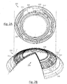
- FIG. 3 a diagrammatic representation of another exemplary energized ophthalmic device comprising both optics and the retinal vascularization monitoring system is depicted.
- a three dimensional cross section representation of an exemplary ophthalmic device 300 including a functionalized layer media insert 320 configured to include the retinal vascularization monitoring system on one or more of its layers 330, 331, 332 is illustrated.
- the media insert 320 can surround the entire periphery of the ophthalmic device 300.
- the actual media insert 320 may comprise a full annular ring or other shapes that still may reside inside or on the hydrogel portion of the ophthalmic device 300 and be within the size and geometry constraints presented by the ophthalmic environment of the user.
- Layers 330, 331 and 332 are meant to illustrate three of numerous layers that may be found in a media insert 320 formed as a stack of functional layers.
- a single layer may include one or more of: active and passive components and portions with structural, electrical or physical properties conducive to a particular purpose including the communication system functions described in the present disclosure.
- a layer 330 may include an energy source, such as, one or more of: a battery, a capacitor and a receiver within the layer 330.
- Item 331 then, in a non-limiting exemplary sense may comprise microcircuitry in a layer that detects actuation signals for the ophthalmic device 300.
- a power regulation layer 332 may be included that is capable of receiving power from external sources, charges the battery layer 330 and controls the use of battery power from layer 330 when the ophthalmic device 300 is not in a charging environment.
- the power regulation may also control signals to an exemplary active lens, demonstrated as item 310 in the center annular cutout of the media insert 320.
- An energized ophthalmic device 300 with an embedded media insert 320 may include an energy source, such as an electrochemical cell or battery (lithium ion cell) as the storage means for the energy and in some embodiments, encapsulation, and isolation of the materials comprising the energy source from an environment into which an ophthalmic device 300 is placed.
- an energy source such as an electrochemical cell or battery (lithium ion cell) as the storage means for the energy and in some embodiments, encapsulation, and isolation of the materials comprising the energy source from an environment into which an ophthalmic device 300 is placed.
- a media insert 320 can also include a pattern of circuitry, components, and energy sources.
- Various embodiments may include the media insert 320 locating the pattern of circuitry, components and energy sources around a periphery of an optic zone through which a wearer of an ophthalmic lens would see, while other embodiments may include a pattern of circuitry, components and energy sources which are small enough to not adversely affect the sight of the ophthalmic lens wearer and therefore the media insert 320 may locate them within, or exterior to, an optical zone without consequence.
- a single and/or multiple discrete electronic devices may be included as discrete chips, for example, in the ophthalmic media inserts.
- the energized electronic elements can be included in the media insert in the form of stacked integrated components.
- Fig. 4 a schematic diagram of an exemplary cross section of a stacked die integrated components implementing the retinal vascularization monitoring system 410 is depicted.
- the media insert may include numerous layers of different types which are encapsulated into contours consistent with the ophthalmic environment that they will occupy.
- these media inserts with stacked integrated component layers may assume the entire annular shape of the media insert.
- the media insert may be an annulus whereas the stacked integrated components may occupy just a portion of the volume within the entire shape.
- these thin film batteries 430 may comprise one or more of the layers that can be stacked upon each other with multiple components in the layers and interconnections therebetween.
- interconnections there may be additional interconnections between two layers that are stacked upon each other.
- interconnection may be made through solder ball interconnections between the layers. In some embodiments only these connections may be required; however, in other cases the solder balls may contact other interconnection elements, as for example with a component having through layer vias.
- a layer 425 may be dedicated for the interconnections two or more of the various components in the interconnect layers.
- the interconnect layer 425 may contain, vias and routing lines that can pass signals from various components to others.
- interconnect layer 425 may provide the various battery elements connections to a power management unit 420 that may be present in a technology layer 415.
- Other components in the technology layer 415 can include, for example, a transceiver 445, control components 450 and the like.
- the interconnect layer 425 may function to make connections between components in the technology layer 415 as well as components outside the technology layer 415; as may exist for example in the integrated passive device 455. There may be numerous manners for routing of electrical signals that may be supported by the presence of dedicated interconnect layers such as interconnect layer 425.
- the technology layer 415 may be included as multiple layers as these features represent a diversity of technology options that may be included in media inserts.
- one of the layers may include CMOS, BiCMOS, Bipolar, or memory based technologies whereas the other layer may include a different technology.
- the two layers may represent different technology families within a same overall family; as for example one layer may include electronic elements produced using a 0.5 micron CMOS technology and another layer may include elements produced using a 20 nanometer CMOS technology. It may be apparent that many other combinations of various electronic technology types would be consistent within the art described herein.
- the media insert may include locations for electrical interconnections to components outside the insert. In other examples, however, the media insert may also include an interconnection to external components in a wireless manner. In such cases, the use of antennas in an antenna layer 435 may provide exemplary manners of wireless communication. In many cases, such an antenna layer 435 may be located, for example, on the top or bottom of the stacked integrated component device within the media insert.
- the battery elements 430 may be included as elements in at least one of the stacked layers themselves. It may be noted as well that other embodiments may be possible where the battery elements 430 are located externally to the stacked integrated component layers. Still further diversity in embodiments may derive from the fact that a separate battery or other energization component may also exist within the media insert, or alternatively these separate energization components may also be located externally to the media insert.
- Components of the retinal vascularization monitoring system 410 may also be included in a stacked integrated component architecture.
- the retinal vascularization monitoring system 410 components may be attached as a portion of a layer.
- the entire retinal vascularization monitoring system 410 may also comprise a similarly shaped component as the other stacked integrated components.
- the controller 500 can include one or more processors 510, which may include one or more processor components coupled to a communication device 520.
- a controller 500 can be used to transmit energy to the energy source placed in the ophthalmic device.
- the processors 510 can be coupled to a communication device configured to communicate energy via a communication channel.
- the communication device may be used to electronically communicate with components within the media insert, for example.
- the communication device 520 may also be used to communicate, for example, with one or more controller apparatus or programming/interface device components.
- the processor 510 is also in communication with a storage device 530.
- the storage device 530 may comprise any appropriate information storage device, including combinations of magnetic storage devices, optical storage devices, and/or semiconductor memory devices such as Random Access Memory (RAM) devices and Read Only Memory (ROM) devices.
- RAM Random Access Memory
- ROM Read Only Memory
- the storage device 530 can store a program 540 for controlling the processor 510.
- the processor 510 performs instructions of a software program 540, and thereby operates in accordance with the present invention.
- the processor 510 may receive information descriptive of media insert placement, active target zones of the retinal vascularization, component placement, imaging resolution and/or frequency, and the like.
- the storage device 530 can also store other pre-determined ophthalmic related data in one or more databases 550 and 560.
- the database may include, for example, predetermined retinal zones exhibiting changes according to cardiac rhythm or an abnormal condition correlated with the retinal vascularization, measurement thresholds, metrology data, and specific control sequences for the system, flow of energy to and from a media insert, communication protocols, and the like.
- the database may also include parameters and controlling algorithms for the control of the retinal vascularization monitoring system that may reside in the ophthalmic device as well as data and/or feedback that can result from their action. In some embodiments, that data may be ultimately communicated to/from an external reception wireless device.
- FIG. 6A a representation of a region of the retinal vascularization of an eye is depicted.
- a series of vessels forming part of the retinal vascularization 600 are shown as they would be viewed from the anterior portion of the eye by the ultrasound imaging system.
- the vessels branch off each other forming a dense pattern that can be useful to study the vessel tortuosity, the angle and number of bifurcations, and the length to width ratio.
- Monitoring temporal changes of the vessels or their overall structures can be useful to predict risk to cardiac conditions including, for example, microvascular disease or diabetes, and cardiac rhythm.
- the region of the retinal vascularization of an eye of Fig. 6A is illustrated with a digital overlay useful for the analysis of changes of the retinal vascularization.
- the exemplary overlay can be useful to divide the observed region of the retinal vascularization into concentric grid sections that can be used for filtering and/or image segmentation.
- grid sections may take other forms such including line patterns and the like. Concentric grid sections however may be preferred for filtering and image segmentation due to the arcuate shape of the cornea conforming to the spherical shape of the eye.
- image processing techniques that include edge recognition can be used to connect the different segments allowing for the analysis of the portion of the retinal vascularization being monitored.
- changes of each vessel may be identified in relation to each of the particular concentric regions A, B, or C.
- the amount of image processing and analysis can be reduced thereby lowering power and processing requirements.
- the frequency in which each of the regions is imaged can be alternated depending on the changes in pulse detected.
- only a first region that includes a segment of a pulsating vessel may be monitored until a change that falls outside a predetermined threshold is detected.
- Average changes in vessel diameter during the cardiac cycle may be approximately 1.2 ⁇ m for arteries and 1.6 ⁇ m for veins.
- a signal may be outputted once a change in diameter of an identified artery is less than .8 ⁇ m and greater than 1.6 ⁇ m over a short period of time.
- a change in the diameter of a vein that is less than 1.2 ⁇ m or greater than 2.0 ⁇ m may trigger a signal.
- the signal may activate additional sensors, increase the rate of image capture, record the abnormality, and/or send a message to the user.
- the message can be sent to the user via an audio signal and/or a visual signal provided by the ophthalmic device itself or a device in wireless communication with the ophthalmic device.
- Point 605 may have been identified as a positive pulsating point of reference during an initial retinal examination using high definition imaging technique including mydriatic and/or nonmydriatic retinal screenings.
- point of reference 605 can be analyzed in conjunction with point of reference 610 pointing to the same vessel. Analysis at both points of reference may be used to provide the system with more measurements for the determination of blood pressure, for example.
- other reference points on pulsating/non-pulsating vessels in a different region may also be monitored, simultaneously or in alternating modes, to ensure that the change is uniform throughout the vascularization. For example, at additional point of reference 615 in region C.
- a side cross section representation of a patient's eye with an exemplary energized ophthalmic device is illustrated.
- an ophthalmic device 700 taking form of an energized contact lens is illustrated resting on the cornea 706 with ocular fluid in at least some portions between the ophthalmic device 700 and the cornea 706.
- the concave contour of the ophthalmic device 700 may be designed so that one or more piezoelectric transducers can rest directly on the cornea 706. Having the piezoelectric transducers resting directly on the cornea 706 can allow greater imaging detail as the ultrasonic pulses can travel directly towards the cornea 706 from focal points 702, 710.
- the piezoelectric transducer(s) are located on the periphery area of the energized contact lens and outside of the line of sight to prevent vision interference.
- the piezoelectric transducer may be located in the center region located in front of the pupil 704 also without significantly interfering with the vision of a user.
- the ultrasonic pulses may pass through the eye's lens 708 before passing through the vitreous humour 720 and reaching one or more retinal areas including pulsating vessels, e.g. 712 and 716.
- the retinal areas may be pre-determined areas near or that include ocular parts serving a specific function or that can be used as a predictor of a particular condition including, for example, the macula 714 which may be screened for the early detection of peripheral vision loss.
- regions/zones with retinal vascularization can be identified for imaging.
- the retinal vascularization in the identified region/zone includes portions of one or more pulsating vessels.
- These regions/zones can be identified using imaging systems that are capable of reproducing high definition images of the microvascularization in the retina. Imaging systems may include non-invasive OCT systems, or any other highly reliable mydriatic or nonmydriatic diagnostic imaging method used for geometrical measurements of retinal structures.
- Depth, shape, relative position, and structure of the vascularization may be pre-programmed into the ophthalmic device in order to lower processing/energy consumption requirements and reliably identify the target points in the areas monitored.
- target points can be easily identifiable image features, such as, edges, crossing, or line boundaries of vessels.
- an ophthalmic device including a retinal vascularization monitoring system is provided to a patient.
- the ophthalmic device may include one or two energized contact lenses configured to include a piezoelectric transducer with a feedback circuit used to provide an ultrasound pulse used to image the microvascularization of an eye.
- the energized contact lenses can additionally be capable of providing other functions including providing vision correction and/or enhancement via physical characteristics, wireless communication with other devices, and emitting visual and/or auditory signals to the user.
- the design of the ophthalmic device and, in particular, the location of the piezo-electric transducer forming part of the retinal vascularization monitoring system may be determined according to the identified region/zone of interest. In some embodiments however, guidelines for the imaging system to focus on the identified region/zone(s) may be programmed after the design of the ophthalmic device.
- the retinal vascularization monitoring system can monitor one or more pre-identified target points over pre-determined periods of time. Monitoring can include determining the cardiac rhythm from the rate of pulsations of a vessel over a particular period of time 815 and/or the displacement/changes ofvessel(s) 820 over time. The monitoring may be triggered, for example, based on a timer function, blink actuation, or upon receiving a signal from a wireless device in communication with the ophthalmic device.
- the wireless may serve as a user interface and may be a drug pump, smartphone, personal computer, tablet, and the such. Transmission of information with the wireless device can occur, for example, via a RF frequency, a local area network (LAN), and/or a private area network (PAN), depending on the communication device and functionality implemented by the ophthalmic device.
- LAN local area network
- PAN private area network
- an audio/visual signal alert may be sent to the user when either the determined cardiac rhythm and/or determined rate of vascular displacement are outside a predetermined threshold.
- a signal may be outputted when the system detects that the cardiac rhythm has increased/decreased to a hazardous level.
- the signal may be sent using a wireless device in communication with the ophthalmic device and/or through an audible signal using micro-acoustic elements included in the ophthalmic device.
- the signal may be a visual signal using micro-photonic elements that may also be included in the ophthalmic device.
- the audible signal may be played in conjunction with a visual signal, e.g., as part of a video clip with safety instructions to reduce the risk of a fatal heart condition.
- steps 835, 840, and 845 may occur depending on the particular embodiment implemented.
- an actuation signal may be sent to a drug delivery apparatus to deliver a drug/active agent.
- the drug delivery mechanism may include, for example, a drug pump in wireless communication with the ophthalmic device.
- the signal may be correlated with a specific event imputed by the patient using the wireless device as a user interface. For example, through a selection from a menu listing activities that can influence cardiac rhythm or blood pressure.
- the action and/or feedback from steps 810-840 can be recorded to improve future analysis, keep a medical record that can be accessed by an eye care practitioner, and/or tailor the retinal vascularization monitoring system to the particular patient.
- these recorded actions/records can also be sent/stored using the wireless device.
Landscapes
- Health & Medical Sciences (AREA)
- Life Sciences & Earth Sciences (AREA)
- Engineering & Computer Science (AREA)
- Physics & Mathematics (AREA)
- General Health & Medical Sciences (AREA)
- Public Health (AREA)
- Ophthalmology & Optometry (AREA)
- Biomedical Technology (AREA)
- Veterinary Medicine (AREA)
- Heart & Thoracic Surgery (AREA)
- Animal Behavior & Ethology (AREA)
- Medical Informatics (AREA)
- Biophysics (AREA)
- Molecular Biology (AREA)
- Surgery (AREA)
- Nuclear Medicine, Radiotherapy & Molecular Imaging (AREA)
- Radiology & Medical Imaging (AREA)
- Pathology (AREA)
- Cardiology (AREA)
- Vascular Medicine (AREA)
- General Physics & Mathematics (AREA)
- Optics & Photonics (AREA)
- Physiology (AREA)
- Computer Vision & Pattern Recognition (AREA)
- Transplantation (AREA)
- Oral & Maxillofacial Surgery (AREA)
- Hematology (AREA)
- Otolaryngology (AREA)
- Acoustics & Sound (AREA)
- Computer Networks & Wireless Communication (AREA)
- Databases & Information Systems (AREA)
- Data Mining & Analysis (AREA)
- Signal Processing (AREA)
- Epidemiology (AREA)
- Primary Health Care (AREA)
- Microelectronics & Electronic Packaging (AREA)
- Manufacturing & Machinery (AREA)
- Mechanical Engineering (AREA)
- Eye Examination Apparatus (AREA)
- Prostheses (AREA)
Applications Claiming Priority (1)
| Application Number | Priority Date | Filing Date | Title |
|---|---|---|---|
| US14/087,315 US9642525B2 (en) | 2013-11-22 | 2013-11-22 | Ophthalmic lens with retinal vascularization monitoring system |
Publications (1)
| Publication Number | Publication Date |
|---|---|
| EP2876489A1 true EP2876489A1 (fr) | 2015-05-27 |
Family
ID=51904880
Family Applications (1)
| Application Number | Title | Priority Date | Filing Date |
|---|---|---|---|
| EP14194393.6A Withdrawn EP2876489A1 (fr) | 2013-11-22 | 2014-11-21 | Lentille ophtalmique avec système de surveillance de vascularisation rétinienne |
Country Status (13)
| Country | Link |
|---|---|
| US (3) | US9642525B2 (fr) |
| EP (1) | EP2876489A1 (fr) |
| JP (1) | JP6517004B2 (fr) |
| KR (1) | KR20150059616A (fr) |
| CN (1) | CN104644149B (fr) |
| AU (1) | AU2014265094A1 (fr) |
| BR (1) | BR102014029034A2 (fr) |
| CA (1) | CA2871204A1 (fr) |
| HK (1) | HK1210277A1 (fr) |
| IL (1) | IL235400A0 (fr) |
| RU (1) | RU2604943C2 (fr) |
| SG (1) | SG10201407390RA (fr) |
| TW (1) | TWI627936B (fr) |
Cited By (2)
| Publication number | Priority date | Publication date | Assignee | Title |
|---|---|---|---|---|
| US10413182B2 (en) | 2015-07-24 | 2019-09-17 | Johnson & Johnson Vision Care, Inc. | Biomedical devices for biometric based information communication |
| US12032229B2 (en) | 2017-10-13 | 2024-07-09 | Sony Corporation | Contact lens |
Families Citing this family (15)
| Publication number | Priority date | Publication date | Assignee | Title |
|---|---|---|---|---|
| US10149958B1 (en) | 2015-07-17 | 2018-12-11 | Bao Tran | Systems and methods for computer assisted operation |
| US10335572B1 (en) | 2015-07-17 | 2019-07-02 | Naveen Kumar | Systems and methods for computer assisted operation |
| US10492981B1 (en) | 2015-07-17 | 2019-12-03 | Bao Tran | Systems and methods for computer assisted operation |
| US10685488B1 (en) | 2015-07-17 | 2020-06-16 | Naveen Kumar | Systems and methods for computer assisted operation |
| US10176642B2 (en) | 2015-07-17 | 2019-01-08 | Bao Tran | Systems and methods for computer assisted operation |
| US10046229B2 (en) | 2016-05-02 | 2018-08-14 | Bao Tran | Smart device |
| US10841724B1 (en) | 2017-01-24 | 2020-11-17 | Ha Tran | Enhanced hearing system |
| WO2018187693A1 (fr) | 2017-04-06 | 2018-10-11 | TearDX LLC | Dispositifs oculaires et leurs procédés d'utilisation |
| US11373450B2 (en) * | 2017-08-11 | 2022-06-28 | Tectus Corporation | Eye-mounted authentication system |
| JP7382955B2 (ja) * | 2017-12-21 | 2023-11-20 | ファイ・バイオメッド・インコーポレイテッド | 無線駆動スマートコンタクトレンズ |
| US20190346692A1 (en) * | 2018-05-09 | 2019-11-14 | Johnson & Johnson Vision Care, Inc. | Electronic ophthalmic lens for measuring distance using ultrasound time-of-flight |
| CN110367927B (zh) * | 2019-08-01 | 2022-04-08 | 中山大学 | 青光眼眼压连续检测系统及其检测方法 |
| US20220031256A1 (en) * | 2020-07-31 | 2022-02-03 | 3M Innovative Properties Company | Composite phonocardiogram visualization on an electronic stethoscope display |
| WO2023243164A1 (fr) * | 2022-06-17 | 2023-12-21 | 株式会社村田製作所 | Dispositif de médication en forme de capsule et système de chargement pour dispositif de médication en forme de capsule |
| TWI875647B (zh) * | 2023-05-25 | 2025-03-01 | 宏衍生物視覺股份有限公司 | 眼鏡以及應用於眼鏡的生理狀態偵測裝置與方法 |
Citations (4)
| Publication number | Priority date | Publication date | Assignee | Title |
|---|---|---|---|---|
| WO2000025662A1 (fr) * | 1998-11-02 | 2000-05-11 | Abreu Marcio Marc Aurelio Mart | Procede et appareil de transmission de signaux et de detection utilisant un dispositif de contact |
| WO2009100439A2 (fr) * | 2008-02-07 | 2009-08-13 | Ehrecke Timothy J | Moniteur de pression |
| GB2464981A (en) * | 2008-11-01 | 2010-05-05 | Univ Dundee | Pressure sensor for measuring intraocular pressure that can be worn on the eye. |
| WO2010107930A1 (fr) * | 2009-03-17 | 2010-09-23 | The Uwm Research Foundation, Inc. | Dispositif d'imagerie ultrasonore |
Family Cites Families (17)
| Publication number | Priority date | Publication date | Assignee | Title |
|---|---|---|---|---|
| JP2615519B2 (ja) | 1990-11-30 | 1997-05-28 | 松下電器産業株式会社 | 超音波ドプラ血流計 |
| US5830139A (en) * | 1996-09-04 | 1998-11-03 | Abreu; Marcio M. | Tonometer system for measuring intraocular pressure by applanation and/or indentation |
| US20030173870A1 (en) * | 2002-03-12 | 2003-09-18 | Shuh-Yueh Simon Hsu | Piezoelectric ultrasound transducer assembly having internal electrodes for bandwidth enhancement and mode suppression |
| GB0208945D0 (en) | 2002-04-19 | 2002-05-29 | Univ Belfast | Vascular impedance measurement apparatus |
| US7301212B1 (en) * | 2004-07-30 | 2007-11-27 | National Semiconductor Corporation | MEMS microphone |
| US8778022B2 (en) * | 2004-11-02 | 2014-07-15 | E-Vision Smart Optics Inc. | Electro-active intraocular lenses |
| WO2007050453A1 (fr) | 2005-10-28 | 2007-05-03 | Johnson & Johnson Vision Care, Inc. | Lentilles ophtalmiques servant à corriger une presbytie et comprenant une correction d'aberration de haut niveau |
| US7782464B2 (en) * | 2006-05-12 | 2010-08-24 | The General Hospital Corporation | Processes, arrangements and systems for providing a fiber layer thickness map based on optical coherence tomography images |
| AR064985A1 (es) | 2007-01-22 | 2009-05-06 | E Vision Llc | Lente electroactivo flexible |
| US9375886B2 (en) | 2008-10-31 | 2016-06-28 | Johnson & Johnson Vision Care Inc. | Ophthalmic device with embedded microcontroller |
| US9375885B2 (en) * | 2008-10-31 | 2016-06-28 | Johnson & Johnson Vision Care, Inc. | Processor controlled ophthalmic device |
| WO2010102310A2 (fr) | 2009-03-03 | 2010-09-10 | Mc10, Inc. | Systèmes, procédés et dispositifs ayant des éléments de circuits intégrés étirables pour détecter et administrer une thérapie |
| US8861811B2 (en) * | 2009-08-19 | 2014-10-14 | Merck Sharp & Dohme Corp. | System and method for segmenting M-mode ultrasound images showing blood vessel wall motion over time |
| US20120212696A1 (en) | 2011-01-27 | 2012-08-23 | Pixeloptics, Inc. | Variable optical element comprising a liquid crystal alignment layer |
| US9233513B2 (en) * | 2011-03-18 | 2016-01-12 | Johnson & Johnson Vision Care, Inc. | Apparatus for manufacturing stacked integrated component media inserts for ophthalmic devices |
| AU2012258902A1 (en) * | 2011-05-20 | 2014-01-16 | Doheny Eye Institute | Ocular ultrasound probe |
| CN104204913A (zh) * | 2012-01-26 | 2014-12-10 | 庄臣及庄臣视力保护公司 | 包括堆叠式集成元件的通电眼科镜片 |
-
2013
- 2013-11-22 US US14/087,315 patent/US9642525B2/en not_active Expired - Fee Related
-
2014
- 2014-10-30 IL IL235400A patent/IL235400A0/en unknown
- 2014-11-10 SG SG10201407390RA patent/SG10201407390RA/en unknown
- 2014-11-17 CA CA2871204A patent/CA2871204A1/fr not_active Abandoned
- 2014-11-20 KR KR1020140162619A patent/KR20150059616A/ko not_active Ceased
- 2014-11-20 TW TW103140162A patent/TWI627936B/zh not_active IP Right Cessation
- 2014-11-21 AU AU2014265094A patent/AU2014265094A1/en not_active Abandoned
- 2014-11-21 CN CN201410674036.8A patent/CN104644149B/zh not_active Expired - Fee Related
- 2014-11-21 RU RU2014146988/14A patent/RU2604943C2/ru not_active IP Right Cessation
- 2014-11-21 EP EP14194393.6A patent/EP2876489A1/fr not_active Withdrawn
- 2014-11-21 BR BR102014029034A patent/BR102014029034A2/pt not_active IP Right Cessation
- 2014-11-21 JP JP2014236333A patent/JP6517004B2/ja not_active Expired - Fee Related
-
2015
- 2015-11-10 HK HK15111048.2A patent/HK1210277A1/en unknown
-
2017
- 2017-03-28 US US15/471,435 patent/US9901322B2/en not_active Expired - Fee Related
-
2018
- 2018-01-12 US US15/870,392 patent/US10159461B2/en active Active
Patent Citations (4)
| Publication number | Priority date | Publication date | Assignee | Title |
|---|---|---|---|---|
| WO2000025662A1 (fr) * | 1998-11-02 | 2000-05-11 | Abreu Marcio Marc Aurelio Mart | Procede et appareil de transmission de signaux et de detection utilisant un dispositif de contact |
| WO2009100439A2 (fr) * | 2008-02-07 | 2009-08-13 | Ehrecke Timothy J | Moniteur de pression |
| GB2464981A (en) * | 2008-11-01 | 2010-05-05 | Univ Dundee | Pressure sensor for measuring intraocular pressure that can be worn on the eye. |
| WO2010107930A1 (fr) * | 2009-03-17 | 2010-09-23 | The Uwm Research Foundation, Inc. | Dispositif d'imagerie ultrasonore |
Cited By (2)
| Publication number | Priority date | Publication date | Assignee | Title |
|---|---|---|---|---|
| US10413182B2 (en) | 2015-07-24 | 2019-09-17 | Johnson & Johnson Vision Care, Inc. | Biomedical devices for biometric based information communication |
| US12032229B2 (en) | 2017-10-13 | 2024-07-09 | Sony Corporation | Contact lens |
Also Published As
| Publication number | Publication date |
|---|---|
| CN104644149A (zh) | 2015-05-27 |
| BR102014029034A2 (pt) | 2015-09-15 |
| HK1210277A1 (en) | 2016-04-15 |
| JP6517004B2 (ja) | 2019-05-22 |
| US10159461B2 (en) | 2018-12-25 |
| IL235400A0 (en) | 2015-01-29 |
| KR20150059616A (ko) | 2015-06-01 |
| US20180132818A1 (en) | 2018-05-17 |
| JP2015107323A (ja) | 2015-06-11 |
| RU2604943C2 (ru) | 2016-12-20 |
| CA2871204A1 (fr) | 2015-05-22 |
| US9642525B2 (en) | 2017-05-09 |
| US20150148650A1 (en) | 2015-05-28 |
| TWI627936B (zh) | 2018-07-01 |
| US9901322B2 (en) | 2018-02-27 |
| US20170238900A1 (en) | 2017-08-24 |
| CN104644149B (zh) | 2019-10-18 |
| SG10201407390RA (en) | 2015-06-29 |
| TW201534266A (zh) | 2015-09-16 |
| AU2014265094A1 (en) | 2015-06-11 |
| RU2014146988A (ru) | 2016-06-10 |
Similar Documents
| Publication | Publication Date | Title |
|---|---|---|
| US10159461B2 (en) | Ophthalmic lens with retinal vascularization monitoring system | |
| US20180368682A1 (en) | Ophthalmic lens with intraocular pressure monitoring system | |
| TWI619470B (zh) | 具神經頻率偵測系統之眼用鏡片 | |
| US9241669B2 (en) | Neuromuscular sensing for variable-optic electronic ophthalmic lens | |
| JP2017219847A (ja) | 医学的監視機能を備えた電子眼用レンズ | |
| CN106714666A (zh) | 用于患者监测的具有无线接口的超低功率充电植入式传感器 | |
| US20180267338A1 (en) | Temperature-sensing ophthalmic device |
Legal Events
| Date | Code | Title | Description |
|---|---|---|---|
| PUAI | Public reference made under article 153(3) epc to a published international application that has entered the european phase |
Free format text: ORIGINAL CODE: 0009012 |
|
| 17P | Request for examination filed |
Effective date: 20141121 |
|
| AK | Designated contracting states |
Kind code of ref document: A1 Designated state(s): AL AT BE BG CH CY CZ DE DK EE ES FI FR GB GR HR HU IE IS IT LI LT LU LV MC MK MT NL NO PL PT RO RS SE SI SK SM TR |
|
| AX | Request for extension of the european patent |
Extension state: BA ME |
|
| R17P | Request for examination filed (corrected) |
Effective date: 20151127 |
|
| RBV | Designated contracting states (corrected) |
Designated state(s): AL AT BE BG CH CY CZ DE DK EE ES FI FR GB GR HR HU IE IS IT LI LT LU LV MC MK MT NL NO PL PT RO RS SE SI SK SM TR |
|
| REG | Reference to a national code |
Ref country code: HK Ref legal event code: DE Ref document number: 1210277 Country of ref document: HK |
|
| 17Q | First examination report despatched |
Effective date: 20190522 |
|
| STAA | Information on the status of an ep patent application or granted ep patent |
Free format text: STATUS: THE APPLICATION IS DEEMED TO BE WITHDRAWN |
|
| 18D | Application deemed to be withdrawn |
Effective date: 20191002 |
|
| REG | Reference to a national code |
Ref country code: HK Ref legal event code: WD Ref document number: 1210277 Country of ref document: HK |