EP2895100B1 - Attelle réalisée selon un procédé numérique - Google Patents
Attelle réalisée selon un procédé numérique Download PDFInfo
- Publication number
- EP2895100B1 EP2895100B1 EP13753108.3A EP13753108A EP2895100B1 EP 2895100 B1 EP2895100 B1 EP 2895100B1 EP 13753108 A EP13753108 A EP 13753108A EP 2895100 B1 EP2895100 B1 EP 2895100B1
- Authority
- EP
- European Patent Office
- Prior art keywords
- support structure
- structure model
- patient
- surface data
- model
- Prior art date
- Legal status (The legal status is an assumption and is not a legal conclusion. Google has not performed a legal analysis and makes no representation as to the accuracy of the status listed.)
- Active
Links
Images







Classifications
-
- A—HUMAN NECESSITIES
- A61—MEDICAL OR VETERINARY SCIENCE; HYGIENE
- A61C—DENTISTRY; APPARATUS OR METHODS FOR ORAL OR DENTAL HYGIENE
- A61C1/00—Dental machines for boring or cutting ; General features of dental machines or apparatus, e.g. hand-piece design
- A61C1/08—Machine parts specially adapted for dentistry
- A61C1/082—Positioning or guiding, e.g. of drills
- A61C1/084—Positioning or guiding, e.g. of drills of implanting tools
-
- A—HUMAN NECESSITIES
- A61—MEDICAL OR VETERINARY SCIENCE; HYGIENE
- A61B—DIAGNOSIS; SURGERY; IDENTIFICATION
- A61B17/00—Surgical instruments, devices or methods
- A61B17/16—Instruments for performing osteoclasis; Drills or chisels for bones; Trepans
- A61B17/17—Guides or aligning means for drills, mills, pins or wires
- A61B17/1739—Guides or aligning means for drills, mills, pins or wires specially adapted for particular parts of the body
- A61B17/176—Guides or aligning means for drills, mills, pins or wires specially adapted for particular parts of the body for the jaw
-
- A—HUMAN NECESSITIES
- A61—MEDICAL OR VETERINARY SCIENCE; HYGIENE
- A61C—DENTISTRY; APPARATUS OR METHODS FOR ORAL OR DENTAL HYGIENE
- A61C9/00—Impression cups, i.e. impression trays; Impression methods
- A61C9/004—Means or methods for taking digitized impressions
-
- A—HUMAN NECESSITIES
- A61—MEDICAL OR VETERINARY SCIENCE; HYGIENE
- A61F—FILTERS IMPLANTABLE INTO BLOOD VESSELS; PROSTHESES; DEVICES PROVIDING PATENCY TO, OR PREVENTING COLLAPSING OF, TUBULAR STRUCTURES OF THE BODY, e.g. STENTS; ORTHOPAEDIC, NURSING OR CONTRACEPTIVE DEVICES; FOMENTATION; TREATMENT OR PROTECTION OF EYES OR EARS; BANDAGES, DRESSINGS OR ABSORBENT PADS; FIRST-AID KITS
- A61F5/00—Orthopaedic methods or devices for non-surgical treatment of bones or joints; Nursing devices ; Anti-rape devices
- A61F5/56—Devices for preventing snoring
- A61F5/566—Intra-oral devices
-
- A—HUMAN NECESSITIES
- A63—SPORTS; GAMES; AMUSEMENTS
- A63B—APPARATUS FOR PHYSICAL TRAINING, GYMNASTICS, SWIMMING, CLIMBING, OR FENCING; BALL GAMES; TRAINING EQUIPMENT
- A63B71/00—Games or sports accessories not covered in groups A63B1/00 - A63B69/00
- A63B71/08—Body-protectors for players or sportsmen, i.e. body-protecting accessories affording protection of body parts against blows or collisions
- A63B71/085—Mouth or teeth protectors
-
- G—PHYSICS
- G05—CONTROLLING; REGULATING
- G05B—CONTROL OR REGULATING SYSTEMS IN GENERAL; FUNCTIONAL ELEMENTS OF SUCH SYSTEMS; MONITORING OR TESTING ARRANGEMENTS FOR SUCH SYSTEMS OR ELEMENTS
- G05B15/00—Systems controlled by a computer
- G05B15/02—Systems controlled by a computer electric
-
- A—HUMAN NECESSITIES
- A61—MEDICAL OR VETERINARY SCIENCE; HYGIENE
- A61B—DIAGNOSIS; SURGERY; IDENTIFICATION
- A61B17/00—Surgical instruments, devices or methods
- A61B2017/00526—Methods of manufacturing
Definitions
- This invention relates in general to the field of cranial surgery, including dentistry. More particularly the invention relates to a method and apparatus for producing dental splints or occlusal splint used to protect teeth, such as night guards or sports guards, or to be used by a surgeon to facilitate and guide the installation of oral implants or guide other oral surgeries, such as maxillofacial surgeries.
- a radiographic guide in order to produce the surgical template, a radiographic guide must be produced first.
- the production of a radiographic guide is usually not done by the surgeon himself, but by a lab specializing in the production of dental components. This makes the treatment workflow more complex and slower due to transportation.
- the production of the radiographic guide is also time consuming and expensive.
- the scan with the radiographic guide further raises the radiation dose of the patient. Furthermore, if the radiographic guide is not fitted correctly to the patient during the scanning procedure, a rescan must be performed, further increasing the radiation dose.
- EP1486900 discloses a computer implemented method for manufacturing a surgical guide.
- An embodiment of the invention provides a method of producing a dental splint comprising the steps as defined in claim 1.
- a preferred embodiment of the invention is a method of producing a surgical template without the use of a radiographic guide.
- the same technique can be used to produce a range of dental splint types. Firstly, a set of 3D surface data describing the surface of a patient's oral situation is obtained. Secondly, a virtual surgical template model is generated in dependence on the 3D surface data and the planned implant and anchor pin positions. Thirdly, a physical surgical template is produced from the virtual surgical template model.
- the surface of the patient's oral situation is typically the upper or lower occlusal dental arch of the patient, including the soft tissue and teeth on the buccal, occlusal and lingual sides.
- the 3D surface data of the patient's oral situation may be obtained using a number of known optical or radiographic imaging techniques.
- an intra-oral optical scanner used on the patient's mouth would produce 3D surface data suitable for use with this method.
- Alternative methods include: an optical scan of an impression of the patient's oral situation (processed as required to compensate for the impression being a negative imprint of the oral situation), an optical scan of a die-cast model of the patient's oral situation, a high-resolution 3D X-ray computed tomography (CT) or magnetic resonance imaging (MRI) image of the oral situation combined with a surface detection algorithm.
- CT 3D X-ray computed tomography
- MRI magnetic resonance imaging
- Both the intra-oral optical scan and the optical or tactile scan of an impression or die-cast model have the advantage of minimal radiation exposure for the patient and high resolution surface scanning.
- the CT and MRI scanners provide the advantage of visualizing the internal anatomical structures as well.
- the present invention can be used for guided implant placement using: a digital model based approach without CT data, a digital model based approach fused with CT data or just a CT/MRI image combined with a surface detection algorithm.
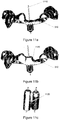
- the 3D surface data 110 of the patient's oral situation is preferable stored as vertices and triangles describing the triangulated surface shown in figures 1a and 1b .
- a virtual support structure model is used as an intermediate step in this process.
- a distance map imaging method is used to process the 3D surface data to form a virtual surgical template model.
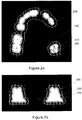
- the 3D surface data of the patient's oral situation is processed to form a 3D distance map image 200. This is done by starting from an empty voxel image having the same frame of reference as the 3D surface described by the 3D surface data. Each voxel of the distance map image is assigned a value corresponding to the minimum distance of the voxel to the 3D surface described by the 3D surface data.
- Figure 2a shows a horizontal slice of a distance map of the patient's oral situation and an outline of the 3D surface 210.
- Figure 2b shows a coronal slice of a distance map of the patient's oral situation and an outline of the 3D surface 210.
- voxels at the 3D surface or below are assigned a positive value.
- Voxels that are above the 3D surface are assigned a negative value. The further the voxel lies from the 3D surface, the greater the value (negative or positive) assigned to the voxel.
- Other embodiments may comprise alternative voxel value configurations.
- a support structure model comprising all the voxels having a value within a particular range of the distance map image.
- all voxels having a value (and thus a particular distance from the 3D surface) between a first value, representing a position close to the 3D surface, and a second value greater than the first value in magnitude, representing a position further away from the 3D surface are selected to form the support structure.
- the first value is chosen to select a distance from the 3D surface where the surface of the support structure model begins.
- the second value is chosen to define the thickness of the support structure, wherein the thickness is dependent on the difference between the first and second values.
- the resulting support structure model matches the 3D surface and will fit the patient's oral situation.
- a larger first value provides a larger tolerance between the 3D surface and the support structure.
- a small amount of play is provided by the tolerance.
- the tolerance is between 0.1mm and 0.5mm.
- Figure 2a and 2b show an outline for the first value 220 and the second value 230.
- the first and second values are chosen to generate a support structure model containing all voxels with a distance between 0.1 and 2.1 mm above the 3D surface.
- the resulting support structure model will have a consistent thickness of 2.0 mm.
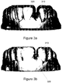
- Figures 3a and 3b show an embodiment of the support structure model 310 overlying the 3D surface 320 of the patient's oral situation.
- the support structure model 310 has an apical edge 330.
- the support structure model is shown partly transparent in order to see the 3D surface beneath.
- the support structure model should also be limited along the dental arc so that it does not stretch all the way up to the molar teeth at the back of the patient's mouth. Instead, a rearward limit is defined.
- limit line 420 shows the rearward or posterior limit of the support structure model along the dental arc.
- a subtractive technique is used to generate the virtual surgical template model from the 3D surface data of the patient's oral situation.
- a predefined shape having a shape approximating a dental splint is provided. Any shape larger than the oral situation of the patient and still small enough to be used as a support structure model would be suitable.
- the predefined shape is overlaid onto the 3D surface so that they overlap.
- a boolean operation is then performed to subtract the 3D surface of the 3D surface data from the predefined shape.
- the resulting shape has the same general shape as the predefined shape but with a surface matching the 3D surface. This resultant shape would be a suitable basis for a support structure.
- a dilation technique is used to generate the virtual surgical template model from the 3D surface data of the patient's oral situation. This comprises the step of creating a binary image of the 3D surface, wherein the voxels inside the 3D surface have a value of 1 and the voxels outside the 3D surface have a value of 0. The resultant image is then dilated to produce an enlarged binary image of the 3D surface. The original binary 3D surface is then subtracted from the enlarged image to form a resultant support structure, having a thickness dependent on the degree to which the dilated image was enlarged over the 3D surface.
- a z-transfer technique is used to generate the virtual surgical template model from the 3D surface data of the patient's oral situation. This comprises using a model of the 3D surface overlaid with a second model of the 3D surface data shifted in the Z-axis. The geometric space between the two 3D surfaces can then be used to form the support structure. This provides a support structure model with a surface matching the patient's oral situation and having a consistent thickness.
- the support structure model may be further refined to include specific characteristics or features.
- any changes to the characteristics of the support structure model will also result in changed characteristics of the final surgical template.
- the support structure model is configured to cover just a coronal portion of the patient's existing teeth or gum surfaces.
- a cut-off line limiting the support structure model in the apical direction is used. This is achieved in the following way:
- the buccal cut-off line is lowered by between 1mm and 4mm. It is understood that for an equivalent upper jaw, the buccal cut-off line is raised instead of lowered.
- two cut-off lines are used.
- a cut-off line as described above is used.
- the cut-off line 530 is a straight line at a fixed height. This straight line will result in a larger support structure model at the lingual side providing additional strength.
- an image is generated from the 3D surface wherein each voxel location is determined to be on the lingual or buccal side.
- the step of determining the lingual or buccal side aspect of the voxel is performed by determining at which side of the dental arc the voxel lies.
- the cut-off line on the buccal side is modified to provide additional material around the anchor pin sites. In one embodiment as shown in figure 7b , this is done by lowering the cut-off line to include an area defined by a circle 740 around the anchor pin location site 750 on the panoramic maximum intensity projection of the distance map image. This step is only performed at the side were the anchor pin is located, e.g. the buccal or lingual side.
- the cut-off line of figure 7b is smoothed according to the following process shown in figure 8 :
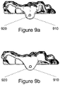
- a support structure model having a buccal edge based on the unsmoothed cut-off line is shown in figure 9a and a support structure model having a buccal edge based on the smoothed cut-off line is shown in figure 9b .
- the second one is clearly preferable for aesthetic and strength reasons and lowers the risk for hurting the patient.
- the support structure model is used to form a surgical template for guiding the drilling of bore holes to allow insertion of oral implants into the patient's jaw bone. Consequently, the support structure model is configured with guide holes.
- the guide holes may be fitted with matching guide sleeves which typically comprise a harder material and which serve the function of guiding the metal drill guide.
- the support structure model material around the guide holes will need to withstand a degree of force from the interaction with the surgeon's tools, the support structure model requires reinforcement for supporting the guide sleeves. The reinforcement is done by adding virtual material to the support structure.
- the tight fit of the support structure model to the patient's dentition means that the top surface of the support structure model is highly variable.
- guide sleeve support material is added to the support structure model by:
- step 5 or step 6 above if the combined distances of two points in the distance map images from their respective closest points on the respective surfaces is less than 2mm, additional support material is added.
- This technique for generating guide holes in the support structure model is also applicable to the anchor pin guide holes used by the anchor pins to fix a surgical template to the patient's jawbone.
- lateral openings are provided in the support structure.
- the corresponding opening(s) in the produced dental splint allows the lateral insertion of guide sleeves and/or guide tools (i.e. tools with a component which fits the guide hole opening and provides support for a drill bit or other tool during the surgical procedure) into the guide hole(s).
- the lateral openings are formed in the support structure model by performing a Boolean subtraction of a box shape from the support structure. The subtraction provides a continuous opening from the guide hole to the external surface of the support structure.
- the opening begins at the coronal edge of the support structure model and runs parallel to the opening with a height greater, equal to, or shorter than the length of the guide hole.
- the width in the distal-mesial direction is equal to or slightly smaller than the diameter of the guide hole to provide a retention fit for components in the guide hole.
- Lateral openings may be provided at either the buccal or lingual side of the support structure, although not both/
- the lateral openings are formed by adjusting the cut-off line to provide a gap in the support structure model at either the buccal or lingual side.
- the same dimensions as described above are used.
- the support structure model is modified to ensure that it can be easily fitted over the patient's dentition.
- the supporting tooth 1400 is wider at the crown 1405 of the tooth before narrowing towards the neck 1406 and root of the tooth.
- a support structure model 1410 formed according to the above methods will have a curving shape which matches the shape of the tooth. Consequently, the opening of the support structure model at the cut-off point 1420 could be narrower than the widest point of the tooth. This will make it difficult if not impossible for the dentist to fit a surgical template corresponding to the support structure model without damaging the surgical template.
- the support structure model is modified to remove any part 1420 of the support structure model which forms an undercut. In one embodiment, this is achieved by:
- a physical surgical template (or dental splint) is produced in dependence on the virtual surgical template model.
- the physical surgical template may be manufactured using an additive manufacturing technique.
- the advantage of this technique is the speed with which the physical surgical template can be produced.
- the physical surgical template is produced as a physical reproduction of the virtual surgical template model using stereolithography.
- Other additive manufacturing technologies include inkjet 3D printers or SLS printers.
- the physical reproduction of the virtual surgical template model may be milled from a block of material.
Landscapes
- Health & Medical Sciences (AREA)
- Oral & Maxillofacial Surgery (AREA)
- General Health & Medical Sciences (AREA)
- Life Sciences & Earth Sciences (AREA)
- Animal Behavior & Ethology (AREA)
- Public Health (AREA)
- Veterinary Medicine (AREA)
- Dentistry (AREA)
- Engineering & Computer Science (AREA)
- Epidemiology (AREA)
- Biomedical Technology (AREA)
- Heart & Thoracic Surgery (AREA)
- Orthopedic Medicine & Surgery (AREA)
- Surgery (AREA)
- Pulmonology (AREA)
- Vascular Medicine (AREA)
- Physical Education & Sports Medicine (AREA)
- Nursing (AREA)
- Otolaryngology (AREA)
- Nuclear Medicine, Radiotherapy & Molecular Imaging (AREA)
- Molecular Biology (AREA)
- Medical Informatics (AREA)
- General Engineering & Computer Science (AREA)
- Physics & Mathematics (AREA)
- General Physics & Mathematics (AREA)
- Automation & Control Theory (AREA)
- Dental Tools And Instruments Or Auxiliary Dental Instruments (AREA)
- Apparatus For Radiation Diagnosis (AREA)
Claims (9)
- Procédé de fabrication d'une attelle dentaire comprenant les étapes :- d'obtention d'un ensemble de données de surface 3D (110, 210), les données de surface 3D représentant une surface de la situation buccale d'un patient, et- de génération d'un modèle de structure de support virtuel (310) en fonction des données de surface 3D, et- de production de l'attelle dentaire en fonction du modèle de structure de support virtuel (310), dans lequel une image de carte de distance 3D (200) des données de surface 3D (110, 210) est utilisée pour générer le modèle de structure de support virtuel (310),
dans lequel la carte de distance 3D (200) est une image de voxel et dans lequel chaque voxel dans l'image de carte de distance 3D a une valeur correspondant à la distance depuis ledit voxel jusqu'au point le plus proche sur la surface décrite par les données de surface 3D (110, 210), et
dans lequel le modèle de structure de support comprend chaque voxel ayant une valeur entre une première (220) et une seconde (230) valeurs, dans lequel- les première et seconde valeurs correspondent à une première et une seconde distance jusqu'au point le plus proche sur les données de surface 3D (110, 210),- la première valeur (220) correspond à une distance à l'écart de la surface décrite par les données de surface 3D (210) et définit l'endroit où commence la surface du modèle de structure de support, de sorte qu'un jeu est prévu entre les données de surface 3D (210) et le modèle de structure de support virtuel (310), dans lequel la première valeur (220) correspond à une distance depuis la surface décrite par les données de surface 3D (210) comprise entre 0,1 mm et 0,5 mm,- la seconde valeur (230) est supérieure à la première valeur (220), et- la différence entre les première et seconde valeurs décrit l'épaisseur du modèle de structure de support (310). - Procédé selon la revendication 1, dans lequel la différence entre les première et seconde valeurs correspond à une épaisseur du modèle de structure de support comprise entre 1 mm et 4 mm.
- Procédé selon une quelconque revendication précédente, dans lequel la ou les extrémités postérieures ou distales du modèle de structure de support (310) sont limitées par une ligne de limite postérieure (420).
- Procédé selon une quelconque revendication précédente, dans lequel la structure de support est modifiée pour éliminer toute partie (1420) formant une contre-dépouille.
- Procédé selon une quelconque revendication précédente, dans lequel l'attelle dentaire est produite en utilisant la fabrication additive.
- Procédé selon une quelconque revendication précédente, dans lequel l'attelle dentaire est produite en utilisant une stéréolithographie.
- Procédé selon une quelconque revendication précédente, dans lequel l'attelle dentaire est un gabarit chirurgical.
- Procédé selon une quelconque revendication précédente, dans lequel l'attelle dentaire est une protection sportive ou une protection de nuit.
- Procédé selon une quelconque revendication précédente, dans lequel l'attelle dentaire est un gabarit chirurgical physique.
Applications Claiming Priority (2)
| Application Number | Priority Date | Filing Date | Title |
|---|---|---|---|
| GBGB1216214.5A GB201216214D0 (en) | 2012-09-12 | 2012-09-12 | A digital splint |
| PCT/EP2013/002557 WO2014040697A1 (fr) | 2012-09-12 | 2013-08-26 | Attelle réalisée selon un procédé numérique |
Publications (2)
| Publication Number | Publication Date |
|---|---|
| EP2895100A1 EP2895100A1 (fr) | 2015-07-22 |
| EP2895100B1 true EP2895100B1 (fr) | 2019-07-03 |
Family
ID=47137280
Family Applications (1)
| Application Number | Title | Priority Date | Filing Date |
|---|---|---|---|
| EP13753108.3A Active EP2895100B1 (fr) | 2012-09-12 | 2013-08-26 | Attelle réalisée selon un procédé numérique |
Country Status (13)
| Country | Link |
|---|---|
| US (1) | US9931177B2 (fr) |
| EP (1) | EP2895100B1 (fr) |
| JP (1) | JP6278965B2 (fr) |
| KR (1) | KR102093325B1 (fr) |
| CN (1) | CN104619282B (fr) |
| AU (1) | AU2013314690B2 (fr) |
| BR (1) | BR112015005524A2 (fr) |
| CA (1) | CA2883184C (fr) |
| DK (1) | DK2895100T3 (fr) |
| ES (1) | ES2740751T3 (fr) |
| GB (1) | GB201216214D0 (fr) |
| WO (1) | WO2014040697A1 (fr) |
| ZA (1) | ZA201501854B (fr) |
Families Citing this family (29)
| Publication number | Priority date | Publication date | Assignee | Title |
|---|---|---|---|---|
| MX2013013872A (es) | 2011-05-26 | 2014-05-28 | Viax Dental Technologies Llc | Herramienta dental y dispositivos de guia. |
| AU2012280708A1 (en) | 2011-07-06 | 2014-01-23 | Nobel Biocare Services Ag | Screw and driver tool |
| ITVI20110302A1 (it) * | 2011-11-23 | 2013-05-24 | Dws Srl | Oggetto tridimensionale perfezionato ottenuto tramite procedimento stereolitografico e metodo per la progettazione grafica computerizzata di detto oggetto |
| GB201216224D0 (en) | 2012-09-12 | 2012-10-24 | Nobel Biocare Services Ag | An improved virtual splint |
| GB201216230D0 (en) | 2012-09-12 | 2012-10-24 | Nobel Biocare Services Ag | An improved surgical template |
| GB2509739A (en) | 2013-01-11 | 2014-07-16 | Nobel Biocare Services Ag | Dental drill bit with spherical head and helical fluting |
| GB2518849A (en) | 2013-10-01 | 2015-04-08 | Nobel Biocare Services Ag | Dental Kit-of-parts and Method of assembling the same |
| US9827138B2 (en) | 2014-01-27 | 2017-11-28 | Bruce W. Hultgren | Fabrication of maxillofacial splints |
| CN105411717B (zh) * | 2014-09-19 | 2017-05-17 | 北京大学口腔医学院 | 一种计算机辅助设计的个体化牙弓夹板及其制作方法 |
| EP3374013B1 (fr) * | 2015-11-10 | 2024-01-10 | Koninklijke Philips N.V. | Détermination des informations concernant le visage d'un patient |
| EP3474959A4 (fr) | 2016-03-30 | 2020-03-25 | P3 Athletics Inc. | Procédé d'impression additive d'un embout buccal d'accroissement des voies aériennes et de l'oxygénation |
| DE102016109007A1 (de) * | 2016-05-17 | 2017-11-23 | Yong-min Jo | Retainer sowie Verfahren zu dessen Herstellung |
| US9808327B1 (en) * | 2016-07-22 | 2017-11-07 | ProSomnus Sleep Technologies, Inc. | Automated method of manufacturing oral appliances |
| WO2018017488A1 (fr) * | 2016-07-22 | 2018-01-25 | ProSomnus Sleep Technologies, Inc. | Matrice de conception assistée par ordinateur pour la fabrication de dispositifs dentaires |
| US11007035B2 (en) | 2017-03-16 | 2021-05-18 | Viax Dental Technologies Llc | System for preparing teeth for the placement of veneers |
| DE102017217558A1 (de) * | 2017-10-02 | 2019-04-04 | Sirona Dental Systems Gmbh | Verfahren zur Herstellung einer geführten Aufbissschiene und eine geführte Aufbissschiene |
| WO2019140531A1 (fr) * | 2018-01-18 | 2019-07-25 | The University Of British Columbia | Avancement mandibulaire supporté par un implant bimaxillaire (bima) |
| CN110270080A (zh) * | 2018-03-13 | 2019-09-24 | 北京大学口腔医学院 | 一种数字化设计的个性化运动护齿器 |
| CN109124830B (zh) * | 2018-06-22 | 2021-02-05 | 上海交通大学医学院附属第九人民医院 | 一种基于数字化设计和数控加工制作的赝复体制作方法 |
| CN110974451A (zh) * | 2019-04-27 | 2020-04-10 | 北京大学口腔医学院 | 一种牙周夹板及其制作方法 |
| US12558255B2 (en) * | 2019-05-30 | 2026-02-24 | Somnomed Limited | Method of manufacturing an intraoral device |
| TWI712396B (zh) * | 2020-01-16 | 2020-12-11 | 中國醫藥大學 | 口腔缺陷模型之修補方法及口腔缺陷模型之修補系統 |
| USD932626S1 (en) | 2020-05-13 | 2021-10-05 | ProSomnus Sleep Technologies, Inc. | Mandibular advancement device with comfort bumps |
| US20220087793A1 (en) * | 2020-09-20 | 2022-03-24 | Great Lakes Dental Technologies, Ltd. | Methods and systems for designing dental splints |
| JP7072920B1 (ja) * | 2020-11-30 | 2022-05-23 | 株式会社ホワイトライン | マウスガードの製造方法 |
| CN112716631B (zh) * | 2021-01-08 | 2021-10-08 | 四川大学 | 数字化小尺寸堆积式高固位力牙种植手术导板的制作方法 |
| CN114917569B (zh) * | 2022-06-21 | 2023-12-22 | 天津正丽科技有限公司 | 一种定制运动护齿套制作系统 |
| EP4555971A4 (fr) * | 2022-07-13 | 2026-03-04 | Medit Corp | Procédé de traitement d'image, dispositif électronique et support de stockage lisible par ordinateur |
| CN116549145A (zh) * | 2023-05-12 | 2023-08-08 | 上海市口腔医院(上海市口腔健康中心) | 一种前牙缺损的树脂修复导板及导板制作方法 |
Family Cites Families (62)
| Publication number | Priority date | Publication date | Assignee | Title |
|---|---|---|---|---|
| US5015183A (en) | 1989-08-07 | 1991-05-14 | Fenick Thomas J | Locating device and method of placing a tooth implant |
| US5133660A (en) | 1989-08-07 | 1992-07-28 | Fenick Thomas J | Device for locating the optimum position for a tooth implant |
| US5743916A (en) | 1990-07-13 | 1998-04-28 | Human Factors Industrial Design, Inc. | Drill guide with removable ferrules |
| US5320529A (en) | 1992-09-09 | 1994-06-14 | Howard C. Weitzman | Method and apparatus for locating an ideal site for a dental implant and for the precise surgical placement of that implant |
| GB2274062B (en) | 1993-01-07 | 1996-11-13 | Leeds Precision Components Ltd | Tooth securement |
| NL9301308A (nl) | 1993-07-26 | 1995-02-16 | Willem Frederick Van Nifterick | Werkwijze voor het vastzetten van een tandprothese op implantaten in het kaakbeen van een patiënt en middel te gebruiken daarbij. |
| SE502035C2 (sv) | 1993-12-06 | 1995-07-24 | Nobelpharma Ab | Metod och och anordning för framtagning av information för framställning av artifiella stödorgan eller ersättningsdelar till människokroppen |
| SE506279C2 (sv) | 1993-12-20 | 1997-12-01 | Nobel Biocare Ab | Skruvformat fästelement av titan för permanent förankring i benvävnad |
| BE1008372A3 (nl) * | 1994-04-19 | 1996-04-02 | Materialise Nv | Werkwijze voor het vervaardigen van een geperfektioneerd medisch model uitgaande van digitale beeldinformatie van een lichaamsdeel. |
| SE503073C2 (sv) | 1994-07-04 | 1996-03-18 | Nobelpharma Ab | Metod för framställning av långsträckt stöddel i ersättningsuppbyggnad samt sådan stöddel tillverkad med metoden |
| SE507336C2 (sv) | 1995-10-12 | 1998-05-18 | Nobel Biocare Ab | Hållarorgan för implantation i benvävnad |
| US5718579A (en) | 1995-12-05 | 1998-02-17 | Kennedy; Brent D. | Drill guide kit |
| IL118371A (en) | 1996-05-22 | 2000-06-29 | Conley Roy | Drill guide |
| US5725736A (en) | 1996-10-25 | 1998-03-10 | Kimberly-Clark Worldwide, Inc. | Tissue containing silicone betaines |
| US8496474B2 (en) * | 1997-06-20 | 2013-07-30 | Align Technology, Inc. | Computer automated development of an orthodontic treatment plan and appliance |
| US7247021B2 (en) | 1997-06-20 | 2007-07-24 | Align Technology, Inc. | Subdividing a digital dentition model |
| AU5472498A (en) | 1997-12-18 | 1999-07-12 | Michel Poirier | Manufacturing a dental implant drill guide and a dental implant superstructure |
| WO2000019929A1 (fr) * | 1998-10-08 | 2000-04-13 | Align Technology, Inc. | Developpement informatique automatise d'un plan de traitement orthodontique et d'un appareil prevu a cet effet |
| US6227850B1 (en) | 1999-05-13 | 2001-05-08 | Align Technology, Inc. | Teeth viewing system |
| US7160110B2 (en) | 1999-11-30 | 2007-01-09 | Orametrix, Inc. | Three-dimensional occlusal and interproximal contact detection and display using virtual tooth models |
| SE515632C2 (sv) | 2000-01-31 | 2001-09-17 | Nobel Biocare Ab | Förfarande och anordning vid superstruktur till implantat |
| SE522958C2 (sv) | 2000-12-29 | 2004-03-16 | Nobel Biocare Ab | Förfarande, arrangemang (anordning) och program vid eller för protetisk installation |
| US6671539B2 (en) * | 2001-10-03 | 2003-12-30 | Board Of Regents University Of Texas System | Method and apparatus for fabricating orthognathic surgical splints |
| SE520765C2 (sv) | 2001-12-28 | 2003-08-19 | Nobel Biocare Ab | Anordning och arrangemang för att medelst mall ta upp hål till implantat i ben, företrädesvis käkben |
| FR2836372B1 (fr) | 2002-02-28 | 2004-06-04 | Obl | Procede et dispositif pour la mise en place d'implants dentaires |
| EP1509124A2 (fr) * | 2002-05-22 | 2005-03-02 | Koninklijke Philips Electronics N.V. | Systeme de visualisation medical et traitement d'images pour la visualisation integree de donnnees medicales |
| EP1486900A1 (fr) | 2003-06-12 | 2004-12-15 | Materialise, Naamloze Vennootschap | Procédé et système de fabrication de guide chirurgical |
| US20050140670A1 (en) | 2003-11-20 | 2005-06-30 | Hong Wu | Photogrammetric reconstruction of free-form objects with curvilinear structures |
| GB0414277D0 (en) * | 2004-06-25 | 2004-07-28 | Leuven K U Res & Dev | Orthognatic surgery |
| SE527503C2 (sv) | 2004-08-05 | 2006-03-21 | Nobel Biocare Ab | Anordning och förfarande för att underlätta applicering till rätt läge av tand- eller tandrestmall |
| SE527504C2 (sv) | 2004-08-05 | 2006-03-21 | Nobel Biocare Ab | Styranordning samverkbar med ett antal i tandmall anordnade hylsor |
| SE530087C2 (sv) | 2004-09-01 | 2008-02-26 | Nobel Biocare Ab | Förfarande och anordning för att tillhandahålla dental produkt |
| GB0507204D0 (en) | 2005-04-08 | 2005-05-18 | Leuven K U Res & Dev | Maxillofacial and plastic surgery |
| SE528724C2 (sv) | 2005-06-03 | 2007-01-30 | Nobel Biocare Services Ag | Anordning vid i mun anordnad borrmall |
| US20070083210A1 (en) | 2005-09-16 | 2007-04-12 | Zimmer Spine, Inc. | Apparatus and method for minimally invasive spine surgery |
| FR2898485B1 (fr) | 2006-03-15 | 2008-05-30 | Obl Sa | Dispositif de guidage d'un foret implantaire |
| US20090325127A1 (en) | 2006-03-24 | 2009-12-31 | Sicat Gmbh & Co. Kg | Method and device for producing tooth prosthesis parts |
| ES2666492T3 (es) | 2006-05-04 | 2018-05-04 | Nobel Biocare Services Ag | Un dispositivo para fijar un implante dental en tejido óseo, un procedimiento para fabricar una plantilla quirúrgica y un procedimiento de fijación de un implante dental en tejido óseo |
| GB0609988D0 (en) * | 2006-05-19 | 2006-06-28 | Materialise Nv | Method for creating a personalized digital planning file for simulation of dental implant placement |
| US7835811B2 (en) | 2006-10-07 | 2010-11-16 | Voxelogix Corporation | Surgical guides and methods for positioning artificial teeth and dental implants |
| EP1982652A1 (fr) | 2007-04-20 | 2008-10-22 | Medicim NV | Procédé pour dériver des informations sur la forme |
| EP2156350A1 (fr) | 2007-05-25 | 2010-02-24 | Nobel Biocare Services AG | Procédé et système de planification dentaire |
| US9095377B2 (en) | 2007-10-25 | 2015-08-04 | Nobel Biocare Services Ag | Distraction device |
| JP2011509812A (ja) * | 2008-01-23 | 2011-03-31 | センサブル テクノロジーズ インコーポレイテッド | 触覚的に操作可能な歯科モデリングシステム |
| US8011927B2 (en) | 2008-04-16 | 2011-09-06 | Biomet 3I, Llc | Method for pre-operative visualization of instrumentation used with a surgical guide for dental implant placement |
| US7936911B2 (en) * | 2008-06-11 | 2011-05-03 | National Cheng Kung University | 3D planning and prediction method for optimizing facial skeleton symmetry in orthognathic surgery |
| CN101342096B (zh) * | 2008-09-04 | 2010-06-02 | 同济大学 | 牙科种植定位定向导板的制作方法 |
| DK2722818T3 (da) | 2009-09-04 | 2020-01-06 | Medicim Nv | Fremgangsmåde til digitalisering af dento-maxillofaciale objekter |
| CN201631403U (zh) * | 2010-01-29 | 2010-11-17 | 孙毅 | 一种手术导板 |
| GB201002778D0 (en) | 2010-02-18 | 2010-04-07 | Materialise Dental Nv | 3D digital endodontics |
| US20120046914A1 (en) * | 2010-08-20 | 2012-02-23 | Fei Gao | Hybrid method for dental implant treatment planning |
| TWI394560B (zh) * | 2010-10-20 | 2013-05-01 | Metal Ind Res & Dev Ct | 植牙手術導板及其製作方法 |
| US8371849B2 (en) | 2010-10-26 | 2013-02-12 | Fei Gao | Method and system of anatomy modeling for dental implant treatment planning |
| WO2012095851A2 (fr) | 2011-01-13 | 2012-07-19 | Cadent Ltd. | Procédés, systèmes et accessoires utiles pour des interventions relatives à des implants dentaires |
| US20120214121A1 (en) * | 2011-01-26 | 2012-08-23 | Greenberg Surgical Technologies, Llc | Orthodontic Treatment Integrating Optical Scanning and CT Scan Data |
| GB201216224D0 (en) | 2012-09-12 | 2012-10-24 | Nobel Biocare Services Ag | An improved virtual splint |
| GB201216230D0 (en) | 2012-09-12 | 2012-10-24 | Nobel Biocare Services Ag | An improved surgical template |
| GB2509739A (en) | 2013-01-11 | 2014-07-16 | Nobel Biocare Services Ag | Dental drill bit with spherical head and helical fluting |
| GB2511038A (en) | 2013-02-20 | 2014-08-27 | Nobel Biocare Services Ag | Surgical template arrangement and method |
| EP2842493B1 (fr) | 2013-08-30 | 2016-04-06 | Zfx GmbH | Corps de référence intra-orale |
| US9808326B2 (en) * | 2014-03-18 | 2017-11-07 | President And Fellows Of Harvard College | 3D dentofacial system and method |
| US9763757B2 (en) * | 2014-05-19 | 2017-09-19 | National Dentex, Llc | Method for creating a virtual oral-maxillofacial anatomical representation |
-
2012
- 2012-09-12 GB GBGB1216214.5A patent/GB201216214D0/en not_active Ceased
-
2013
- 2013-08-26 US US14/427,219 patent/US9931177B2/en active Active
- 2013-08-26 JP JP2015531482A patent/JP6278965B2/ja active Active
- 2013-08-26 ES ES13753108T patent/ES2740751T3/es active Active
- 2013-08-26 DK DK13753108.3T patent/DK2895100T3/da active
- 2013-08-26 WO PCT/EP2013/002557 patent/WO2014040697A1/fr not_active Ceased
- 2013-08-26 BR BR112015005524A patent/BR112015005524A2/pt not_active Application Discontinuation
- 2013-08-26 KR KR1020157009394A patent/KR102093325B1/ko active Active
- 2013-08-26 AU AU2013314690A patent/AU2013314690B2/en active Active
- 2013-08-26 EP EP13753108.3A patent/EP2895100B1/fr active Active
- 2013-08-26 CN CN201380047023.1A patent/CN104619282B/zh active Active
- 2013-08-26 CA CA2883184A patent/CA2883184C/fr active Active
-
2015
- 2015-03-18 ZA ZA2015/01854A patent/ZA201501854B/en unknown
Non-Patent Citations (1)
| Title |
|---|
| None * |
Also Published As
| Publication number | Publication date |
|---|---|
| ES2740751T3 (es) | 2020-02-06 |
| US9931177B2 (en) | 2018-04-03 |
| EP2895100A1 (fr) | 2015-07-22 |
| AU2013314690A1 (en) | 2015-03-19 |
| CN104619282B (zh) | 2018-02-13 |
| CA2883184A1 (fr) | 2014-03-20 |
| DK2895100T3 (da) | 2019-07-29 |
| CA2883184C (fr) | 2020-07-21 |
| KR102093325B1 (ko) | 2020-03-26 |
| BR112015005524A2 (pt) | 2018-05-22 |
| CN104619282A (zh) | 2015-05-13 |
| ZA201501854B (en) | 2016-10-26 |
| KR20150055015A (ko) | 2015-05-20 |
| JP2015531640A (ja) | 2015-11-05 |
| AU2013314690B2 (en) | 2016-12-08 |
| WO2014040697A1 (fr) | 2014-03-20 |
| JP6278965B2 (ja) | 2018-02-14 |
| GB201216214D0 (en) | 2012-10-24 |
| US20150245890A1 (en) | 2015-09-03 |
Similar Documents
| Publication | Publication Date | Title |
|---|---|---|
| US9931177B2 (en) | Digital splint | |
| EP2895102B1 (fr) | Attelle virtuelle améliorée | |
| US9839497B2 (en) | Surgical template | |
| US11690517B2 (en) | Systems for creating and interacting with three dimensional virtual models | |
| EP2877118B1 (fr) | Conception d'un gabarit de positionnement dentaire | |
| CN118634032B (zh) | 用于生成用于患者的目标拔除对象的虚拟拔除过程的模型的方法和系统 |
Legal Events
| Date | Code | Title | Description |
|---|---|---|---|
| PUAI | Public reference made under article 153(3) epc to a published international application that has entered the european phase |
Free format text: ORIGINAL CODE: 0009012 |
|
| 17P | Request for examination filed |
Effective date: 20150413 |
|
| AK | Designated contracting states |
Kind code of ref document: A1 Designated state(s): AL AT BE BG CH CY CZ DE DK EE ES FI FR GB GR HR HU IE IS IT LI LT LU LV MC MK MT NL NO PL PT RO RS SE SI SK SM TR |
|
| AX | Request for extension of the european patent |
Extension state: BA ME |
|
| DAX | Request for extension of the european patent (deleted) | ||
| STAA | Information on the status of an ep patent application or granted ep patent |
Free format text: STATUS: EXAMINATION IS IN PROGRESS |
|
| 17Q | First examination report despatched |
Effective date: 20180509 |
|
| GRAP | Despatch of communication of intention to grant a patent |
Free format text: ORIGINAL CODE: EPIDOSNIGR1 |
|
| STAA | Information on the status of an ep patent application or granted ep patent |
Free format text: STATUS: GRANT OF PATENT IS INTENDED |
|
| INTG | Intention to grant announced |
Effective date: 20190121 |
|
| GRAS | Grant fee paid |
Free format text: ORIGINAL CODE: EPIDOSNIGR3 |
|
| GRAA | (expected) grant |
Free format text: ORIGINAL CODE: 0009210 |
|
| STAA | Information on the status of an ep patent application or granted ep patent |
Free format text: STATUS: THE PATENT HAS BEEN GRANTED |
|
| AK | Designated contracting states |
Kind code of ref document: B1 Designated state(s): AL AT BE BG CH CY CZ DE DK EE ES FI FR GB GR HR HU IE IS IT LI LT LU LV MC MK MT NL NO PL PT RO RS SE SI SK SM TR |
|
| REG | Reference to a national code |
Ref country code: GB Ref legal event code: FG4D |
|
| REG | Reference to a national code |
Ref country code: CH Ref legal event code: EP Ref country code: AT Ref legal event code: REF Ref document number: 1150046 Country of ref document: AT Kind code of ref document: T Effective date: 20190715 |
|
| REG | Reference to a national code |
Ref country code: DE Ref legal event code: R096 Ref document number: 602013057402 Country of ref document: DE |
|
| REG | Reference to a national code |
Ref country code: IE Ref legal event code: FG4D |
|
| REG | Reference to a national code |
Ref country code: DK Ref legal event code: T3 Effective date: 20190724 |
|
| REG | Reference to a national code |
Ref country code: SE Ref legal event code: TRGR |
|
| REG | Reference to a national code |
Ref country code: NL Ref legal event code: MP Effective date: 20190703 |
|
| REG | Reference to a national code |
Ref country code: LT Ref legal event code: MG4D |
|
| REG | Reference to a national code |
Ref country code: AT Ref legal event code: MK05 Ref document number: 1150046 Country of ref document: AT Kind code of ref document: T Effective date: 20190703 |
|
| PG25 | Lapsed in a contracting state [announced via postgrant information from national office to epo] |
Ref country code: CZ Free format text: LAPSE BECAUSE OF FAILURE TO SUBMIT A TRANSLATION OF THE DESCRIPTION OR TO PAY THE FEE WITHIN THE PRESCRIBED TIME-LIMIT Effective date: 20190703 Ref country code: FI Free format text: LAPSE BECAUSE OF FAILURE TO SUBMIT A TRANSLATION OF THE DESCRIPTION OR TO PAY THE FEE WITHIN THE PRESCRIBED TIME-LIMIT Effective date: 20190703 Ref country code: AT Free format text: LAPSE BECAUSE OF FAILURE TO SUBMIT A TRANSLATION OF THE DESCRIPTION OR TO PAY THE FEE WITHIN THE PRESCRIBED TIME-LIMIT Effective date: 20190703 Ref country code: NO Free format text: LAPSE BECAUSE OF FAILURE TO SUBMIT A TRANSLATION OF THE DESCRIPTION OR TO PAY THE FEE WITHIN THE PRESCRIBED TIME-LIMIT Effective date: 20191003 Ref country code: LT Free format text: LAPSE BECAUSE OF FAILURE TO SUBMIT A TRANSLATION OF THE DESCRIPTION OR TO PAY THE FEE WITHIN THE PRESCRIBED TIME-LIMIT Effective date: 20190703 Ref country code: NL Free format text: LAPSE BECAUSE OF FAILURE TO SUBMIT A TRANSLATION OF THE DESCRIPTION OR TO PAY THE FEE WITHIN THE PRESCRIBED TIME-LIMIT Effective date: 20190703 Ref country code: PT Free format text: LAPSE BECAUSE OF FAILURE TO SUBMIT A TRANSLATION OF THE DESCRIPTION OR TO PAY THE FEE WITHIN THE PRESCRIBED TIME-LIMIT Effective date: 20191104 Ref country code: BG Free format text: LAPSE BECAUSE OF FAILURE TO SUBMIT A TRANSLATION OF THE DESCRIPTION OR TO PAY THE FEE WITHIN THE PRESCRIBED TIME-LIMIT Effective date: 20191003 Ref country code: HR Free format text: LAPSE BECAUSE OF FAILURE TO SUBMIT A TRANSLATION OF THE DESCRIPTION OR TO PAY THE FEE WITHIN THE PRESCRIBED TIME-LIMIT Effective date: 20190703 |
|
| REG | Reference to a national code |
Ref country code: ES Ref legal event code: FG2A Ref document number: 2740751 Country of ref document: ES Kind code of ref document: T3 Effective date: 20200206 |
|
| PG25 | Lapsed in a contracting state [announced via postgrant information from national office to epo] |
Ref country code: GR Free format text: LAPSE BECAUSE OF FAILURE TO SUBMIT A TRANSLATION OF THE DESCRIPTION OR TO PAY THE FEE WITHIN THE PRESCRIBED TIME-LIMIT Effective date: 20191004 Ref country code: LV Free format text: LAPSE BECAUSE OF FAILURE TO SUBMIT A TRANSLATION OF THE DESCRIPTION OR TO PAY THE FEE WITHIN THE PRESCRIBED TIME-LIMIT Effective date: 20190703 Ref country code: AL Free format text: LAPSE BECAUSE OF FAILURE TO SUBMIT A TRANSLATION OF THE DESCRIPTION OR TO PAY THE FEE WITHIN THE PRESCRIBED TIME-LIMIT Effective date: 20190703 Ref country code: RS Free format text: LAPSE BECAUSE OF FAILURE TO SUBMIT A TRANSLATION OF THE DESCRIPTION OR TO PAY THE FEE WITHIN THE PRESCRIBED TIME-LIMIT Effective date: 20190703 Ref country code: IS Free format text: LAPSE BECAUSE OF FAILURE TO SUBMIT A TRANSLATION OF THE DESCRIPTION OR TO PAY THE FEE WITHIN THE PRESCRIBED TIME-LIMIT Effective date: 20191103 |
|
| PG25 | Lapsed in a contracting state [announced via postgrant information from national office to epo] |
Ref country code: TR Free format text: LAPSE BECAUSE OF FAILURE TO SUBMIT A TRANSLATION OF THE DESCRIPTION OR TO PAY THE FEE WITHIN THE PRESCRIBED TIME-LIMIT Effective date: 20190703 |
|
| PG25 | Lapsed in a contracting state [announced via postgrant information from national office to epo] |
Ref country code: RO Free format text: LAPSE BECAUSE OF FAILURE TO SUBMIT A TRANSLATION OF THE DESCRIPTION OR TO PAY THE FEE WITHIN THE PRESCRIBED TIME-LIMIT Effective date: 20190703 Ref country code: PL Free format text: LAPSE BECAUSE OF FAILURE TO SUBMIT A TRANSLATION OF THE DESCRIPTION OR TO PAY THE FEE WITHIN THE PRESCRIBED TIME-LIMIT Effective date: 20190703 Ref country code: EE Free format text: LAPSE BECAUSE OF FAILURE TO SUBMIT A TRANSLATION OF THE DESCRIPTION OR TO PAY THE FEE WITHIN THE PRESCRIBED TIME-LIMIT Effective date: 20190703 |
|
| PG25 | Lapsed in a contracting state [announced via postgrant information from national office to epo] |
Ref country code: MC Free format text: LAPSE BECAUSE OF FAILURE TO SUBMIT A TRANSLATION OF THE DESCRIPTION OR TO PAY THE FEE WITHIN THE PRESCRIBED TIME-LIMIT Effective date: 20190703 Ref country code: IS Free format text: LAPSE BECAUSE OF FAILURE TO SUBMIT A TRANSLATION OF THE DESCRIPTION OR TO PAY THE FEE WITHIN THE PRESCRIBED TIME-LIMIT Effective date: 20200224 Ref country code: SM Free format text: LAPSE BECAUSE OF FAILURE TO SUBMIT A TRANSLATION OF THE DESCRIPTION OR TO PAY THE FEE WITHIN THE PRESCRIBED TIME-LIMIT Effective date: 20190703 Ref country code: SK Free format text: LAPSE BECAUSE OF FAILURE TO SUBMIT A TRANSLATION OF THE DESCRIPTION OR TO PAY THE FEE WITHIN THE PRESCRIBED TIME-LIMIT Effective date: 20190703 Ref country code: LU Free format text: LAPSE BECAUSE OF NON-PAYMENT OF DUE FEES Effective date: 20190826 |
|
| REG | Reference to a national code |
Ref country code: DE Ref legal event code: R097 Ref document number: 602013057402 Country of ref document: DE |
|
| PLBE | No opposition filed within time limit |
Free format text: ORIGINAL CODE: 0009261 |
|
| STAA | Information on the status of an ep patent application or granted ep patent |
Free format text: STATUS: NO OPPOSITION FILED WITHIN TIME LIMIT |
|
| PG2D | Information on lapse in contracting state deleted |
Ref country code: IS |
|
| PG25 | Lapsed in a contracting state [announced via postgrant information from national office to epo] |
Ref country code: IE Free format text: LAPSE BECAUSE OF NON-PAYMENT OF DUE FEES Effective date: 20190826 |
|
| 26N | No opposition filed |
Effective date: 20200603 |
|
| PG25 | Lapsed in a contracting state [announced via postgrant information from national office to epo] |
Ref country code: SI Free format text: LAPSE BECAUSE OF FAILURE TO SUBMIT A TRANSLATION OF THE DESCRIPTION OR TO PAY THE FEE WITHIN THE PRESCRIBED TIME-LIMIT Effective date: 20190703 |
|
| PG25 | Lapsed in a contracting state [announced via postgrant information from national office to epo] |
Ref country code: CY Free format text: LAPSE BECAUSE OF FAILURE TO SUBMIT A TRANSLATION OF THE DESCRIPTION OR TO PAY THE FEE WITHIN THE PRESCRIBED TIME-LIMIT Effective date: 20190703 |
|
| PG25 | Lapsed in a contracting state [announced via postgrant information from national office to epo] |
Ref country code: MT Free format text: LAPSE BECAUSE OF FAILURE TO SUBMIT A TRANSLATION OF THE DESCRIPTION OR TO PAY THE FEE WITHIN THE PRESCRIBED TIME-LIMIT Effective date: 20190703 Ref country code: HU Free format text: LAPSE BECAUSE OF FAILURE TO SUBMIT A TRANSLATION OF THE DESCRIPTION OR TO PAY THE FEE WITHIN THE PRESCRIBED TIME-LIMIT; INVALID AB INITIO Effective date: 20130826 |
|
| PG25 | Lapsed in a contracting state [announced via postgrant information from national office to epo] |
Ref country code: MK Free format text: LAPSE BECAUSE OF FAILURE TO SUBMIT A TRANSLATION OF THE DESCRIPTION OR TO PAY THE FEE WITHIN THE PRESCRIBED TIME-LIMIT Effective date: 20190703 |
|
| P01 | Opt-out of the competence of the unified patent court (upc) registered |
Effective date: 20230515 |
|
| PGFP | Annual fee paid to national office [announced via postgrant information from national office to epo] |
Ref country code: ES Payment date: 20250902 Year of fee payment: 13 |
|
| PGFP | Annual fee paid to national office [announced via postgrant information from national office to epo] |
Ref country code: DK Payment date: 20250814 Year of fee payment: 13 Ref country code: DE Payment date: 20250702 Year of fee payment: 13 |
|
| PGFP | Annual fee paid to national office [announced via postgrant information from national office to epo] |
Ref country code: IT Payment date: 20250722 Year of fee payment: 13 |
|
| PGFP | Annual fee paid to national office [announced via postgrant information from national office to epo] |
Ref country code: BE Payment date: 20250703 Year of fee payment: 13 Ref country code: GB Payment date: 20250703 Year of fee payment: 13 |
|
| PGFP | Annual fee paid to national office [announced via postgrant information from national office to epo] |
Ref country code: FR Payment date: 20250703 Year of fee payment: 13 |
|
| PGFP | Annual fee paid to national office [announced via postgrant information from national office to epo] |
Ref country code: CH Payment date: 20250901 Year of fee payment: 13 Ref country code: SE Payment date: 20250702 Year of fee payment: 13 |