EP2904103B1 - Verfahren zur herstellung induzierter pluripotenter stammzellen und deren anwendungen - Google Patents
Verfahren zur herstellung induzierter pluripotenter stammzellen und deren anwendungen Download PDFInfo
- Publication number
- EP2904103B1 EP2904103B1 EP13844080.5A EP13844080A EP2904103B1 EP 2904103 B1 EP2904103 B1 EP 2904103B1 EP 13844080 A EP13844080 A EP 13844080A EP 2904103 B1 EP2904103 B1 EP 2904103B1
- Authority
- EP
- European Patent Office
- Prior art keywords
- panel
- ipscs
- cells
- parp1
- ipsc
- Prior art date
- Legal status (The legal status is an assumption and is not a legal conclusion. Google has not performed a legal analysis and makes no representation as to the accuracy of the status listed.)
- Active
Links
Images


Classifications
-
- A—HUMAN NECESSITIES
- A61—MEDICAL OR VETERINARY SCIENCE; HYGIENE
- A61K—PREPARATIONS FOR MEDICAL, DENTAL OR TOILETRY PURPOSES
- A61K35/00—Medicinal preparations containing materials or reaction products thereof with undetermined constitution
- A61K35/12—Materials from mammals; Compositions comprising non-specified tissues or cells; Compositions comprising non-embryonic stem cells; Genetically modified cells
- A61K35/48—Reproductive organs
- A61K35/54—Ovaries; Ova; Ovules; Embryos; Foetal cells; Germ cells
- A61K35/545—Embryonic stem cells; Pluripotent stem cells; Induced pluripotent stem cells; Uncharacterised stem cells
-
- A—HUMAN NECESSITIES
- A61—MEDICAL OR VETERINARY SCIENCE; HYGIENE
- A61K—PREPARATIONS FOR MEDICAL, DENTAL OR TOILETRY PURPOSES
- A61K38/00—Medicinal preparations containing peptides
- A61K38/16—Peptides having more than 20 amino acids; Gastrins; Somatostatins; Melanotropins; Derivatives thereof
- A61K38/43—Enzymes; Proenzymes; Derivatives thereof
- A61K38/45—Transferases (2)
-
- A—HUMAN NECESSITIES
- A61—MEDICAL OR VETERINARY SCIENCE; HYGIENE
- A61K—PREPARATIONS FOR MEDICAL, DENTAL OR TOILETRY PURPOSES
- A61K38/00—Medicinal preparations containing peptides
- A61K38/16—Peptides having more than 20 amino acids; Gastrins; Somatostatins; Melanotropins; Derivatives thereof
- A61K38/43—Enzymes; Proenzymes; Derivatives thereof
- A61K38/46—Hydrolases (3)
-
- A—HUMAN NECESSITIES
- A61—MEDICAL OR VETERINARY SCIENCE; HYGIENE
- A61P—SPECIFIC THERAPEUTIC ACTIVITY OF CHEMICAL COMPOUNDS OR MEDICINAL PREPARATIONS
- A61P1/00—Drugs for disorders of the alimentary tract or the digestive system
- A61P1/16—Drugs for disorders of the alimentary tract or the digestive system for liver or gallbladder disorders, e.g. hepatoprotective agents, cholagogues, litholytics
-
- A—HUMAN NECESSITIES
- A61—MEDICAL OR VETERINARY SCIENCE; HYGIENE
- A61P—SPECIFIC THERAPEUTIC ACTIVITY OF CHEMICAL COMPOUNDS OR MEDICINAL PREPARATIONS
- A61P11/00—Drugs for disorders of the respiratory system
-
- C—CHEMISTRY; METALLURGY
- C12—BIOCHEMISTRY; BEER; SPIRITS; WINE; VINEGAR; MICROBIOLOGY; ENZYMOLOGY; MUTATION OR GENETIC ENGINEERING
- C12Y—ENZYMES
- C12Y204/00—Glycosyltransferases (2.4)
- C12Y204/02—Pentosyltransferases (2.4.2)
- C12Y204/0203—NAD+ ADP-ribosyltransferase (2.4.2.30), i.e. tankyrase or poly(ADP-ribose) polymerase
-
- C—CHEMISTRY; METALLURGY
- C12—BIOCHEMISTRY; BEER; SPIRITS; WINE; VINEGAR; MICROBIOLOGY; ENZYMOLOGY; MUTATION OR GENETIC ENGINEERING
- C12Y—ENZYMES
- C12Y306/00—Hydrolases acting on acid anhydrides (3.6)
- C12Y306/04—Hydrolases acting on acid anhydrides (3.6) acting on acid anhydrides; involved in cellular and subcellular movement (3.6.4)
- C12Y306/04012—DNA helicase (3.6.4.12)
Definitions
- the present invention relates generally to a method for preparing induced pluripotent stem cells.
- iPSCs induced pluripotent stem
- iPSCs Induced pluripotent stem cells
- Yamanaka et al. also provided the method for preparing an induced pluripotent stem cell (iPSC)by nuclear reprogramming of a somatic cell through introducing the genes Oct3/4, Klf4, c-Myc and Sox2in US Patent No. 8,058,065 (filed on June 9, 2009 and issued on November 15, 2011).
- the method for preparing somatic cells by inducing differentiation of the iPSC as obtained was disclosed in US Patent No. 8,129,187 (filed on February 18, 2010 and issued on March 6, 2012). Then, Yamanaka et al.
- iPSC induced pluripotent stem cell
- Irion provided a method for generating integration-free human induced pluripotent stem cells from blood cells using one or more DNA expression vectors encoding the reprogramming factors (a) Oct4, Sox2, Klf4, and c-Myc; (b) Oct4, Sox2, and Klf4; (c) Oct4, Sox2, Klf4, c-Myc, and Nanog; or (d) Oct 4, Sox2, Lin-28, and Nanog ( US Patent No. 8,048,675, filed on May 12, 2010 and issued on November 1, 2011).
- iPSCs Induced pluripotent stem cells
- iPSCs can be reprogrammed from adult somatic cells by the transduction of genes or chemical agents.
- iPSCs share the features of embryonic stem cells (ESCs) and are capable of self-renewal and tridermal differentiation, offering a resource for disease modeling and a potentially source for transplantation.
- ESCs embryonic stem cells
- ES embryonic stem
- human cystic fibrosis iPSCs were demonstrated to produce disease-specific lung progenitor cells and eventually form respiratory epithelium in immunodeficient mice ( Mou H, Zhao R, et al. Cell Stem Cell 2012;10:385-397 ).
- human iPSCs are capable of forming myogenic progenitors and neurons, leading to functional recovery after the transplantation into neuromuscular disorder or stroke disease models ( Darabi R et al. Cell Stem Cell 2012;10:610-619 ; and Oki K et al. Stem Cells 2012;30:1120-1133 ).
- the possible protective role of iPSCs and the underlying mechanisms remain unknown.
- iPSCs induced pluripotent stem cells
- IFN interferon
- IP-10 interferon- ⁇ inducible protein-10
- the present invention provides a method as defined in the claims for preparing induced pluripotent stem cells (iPSCs) from somatic cells without using c-Myc and/or Klf4.
- the method comprises (a) transfecting isolated somatic cells to express Oct3/4, Sox2 and Parp1; and (b) culturing the transfected somatic cells as obtained in step (a) under appropriate conditions, thereby converting the somatic cells into iPSCs and maintaining pluripotency and self-renewal ability.
- the present invention provides a method for preparing iPSCs from somatic cells, comprising: (a) contacting or exposing isolated somatic cells with/to Oct3/4, Sox2 and Parp1, and (b) culturing the somatic cells as obtained in step (a) under appropriate conditions, thereby converting, at least a subset of, the population of somatic cells into iPSCs and maintaining pluripotency and self-renewal ability.
- the present description also discloses a method of reprogramming adult cells comprising administering the cells with a protein that is/can be PARylated, or an enzyme that has PARylation activity.
- the present description also discloses a method of rejuvenating cells or senescent cells in a subject, comprising administering to the subject a protein that is able to be PARylated, or an enzyme that has PARylation activity.
- the present description also discloses a method for inducing the secretion of IP-10 which comprisesadministering to a subject in need thereof an effective amount of iPSCs or iPSC-CM.
- the present description also discloses a method for treatingtissue injuries which comprises administering to a subject in need thereof a therapeutically effective amount of iPSCs or iPSC-CM, wherein the therapeutically effective amount is an amount capable of inducing a sufficient level of IP-10 tosuppressinflammation responses.
- the present description also discloses a composition comprising iPSCs or iPSC-CM for inducing the secretion of IP-10 in a subject.
- the article “a” or “an” means one or more than one (that is, at least one) of the grammatical object of the article, unless otherwise made clear in the specific use of the article in only a singular sense.
- pluripotent refers to a cell with the capacity, under different conditions, to differentiate to more than one differentiated cell type, and preferably to differentiate to cell types characteristic of all three germ cell layers.
- iPSC induced pluripotent stem cell
- iPSC induced pluripotent stem cell
- non-pluripotent cell typically an adult somatic cell, for example, by inducing a forced expression of one or more genes.
- This invention is based on the finding that Parp1 can be used in place of c-Myc and Klf4 in the reprogramming process and the efficiency thereof was remarkablysimilar to those reprogrammed by c-Myc,Oct4 and Sox2 (OSM)in terms of production of iPSC. Moreover, several Parp1-associated and PARylation-interacted proteins in iPSCs and ESCs were also identified in the invention, including those involved in DNA repair and chromatin re-openning, thereby providing a useful tool for iPSC technology and regenerative medicine for aged cells or tissues.
- Parp1 Poly(ADP-ribose) polymerase 1 (Parp1), a member of the Parp family of proteins, is a highly conserved DNA-binding protein that is abundant in the nucleus. Parp1 is known as a key effector of several nuclear events such as DNA repair, replication, and transcription (Jagtap and Szabo, 2005; Kraus, 2008). It catalyzes a process called poly(ADP-ribosylation) (PARylation), in which NAD + is used as substrate to synthesize poly(ADP-ribose) polymers with sizes varying from 2 to 200 ADP-ribose units (Krishnakumar and Kraus, 2010). This Parp1-catalyzed PARylation has been implicated in several processes, including chromatin remodeling, enhancer binding, coregulation, and insulation (Kraus, 2008).
- the present invention provides a method as defined in the claims for preparing induced pluripotent stem cells (iPSCs) from somatic cells, comprising: (a) transfecting isolated somatic cells to express Oct3/4, Sox2, and Parp1; and (b) culturing the transfected somatic cells as obtained in step (a) under appropriate conditions, thereby converting the somatic cells into iPSCs and maintaining pluripotency and self-renewal ability.
- iPSCs induced pluripotent stem cells
- the isolated somatic cells are transfected with one or more plasmid or viral vectors comprising Oct3/4, Sox2, and Parploperably linked to a promoter.
- the present invention provides a method for preparing induced pluripotent stem cell (iPSCs) from somatic cells, comprising: (a) contacting or exposing isolated somatic cells with Oct3/4, Sox2, and Parp1, and (b) culturing the somatic cells as obtained in step (a) under appropriate conditions, thereby converting, at least a subset of, the population of somatic cells into iPSCs and maintaining pluripotency and self-renewal ability.
- the invention is characterized in that the method does not comprise a step of transfecting, contacting, or exposing the somatic cells to with/to c-Myc, K1f4, Nanog, Lin28, or any combination thereof, distinguish from the prior art.
- iPSC produced by the method described herein.
- Also provided in the invention is a method as defined in the claims of preparing iPSCs, which comprises (a) providing isolated somatic cells; (b) transfecting, contacting or exposing the isolated somatic cells with/to express Oct3/4, Sox2, and Parp1; and (c) culturing the somatic cells as obtained in step (b) under appropriate conditions, thereby converting the somatic cells into iPSCs and maintaining pluripotency and self-renewal ability.
- the present description also discloses a method of reprogramming adult cells comprising administering the cells with a protein that is/can be PARylated. Furthermore, it is believed that the cell rejuvenation can be performed by the reprogramming process. Accordingly, the present description discloses a method of rejuvenating cells or senescent cells in a subject, comprising administering the cells or the subject with a protein that is/can be PARylated.
- reprogramming refers to a process of erasure and remodeling of epigenetic marks, such as DNA methylation wherein the original DNA methylation patterns are erased and re-established.
- telomere size refers to a process for rendering the telomere size, gene expression profiles, oxidative stress, or mitochondrial metabolism of such cells indistinguishable from that of embryonic stem cells.
- the term "subject” refers to a human or a mammal, such as a patient, a companion animal (e.g., dog, cat, and the like), a farm animal (e.g., cow, sheep, pig, horse, and the like) or a laboratory animal (e.g., rat, mouse, guinea pig, and the like).
- a companion animal e.g., dog, cat, and the like
- a farm animal e.g., cow, sheep, pig, horse, and the like
- laboratory animal e.g., rat, mouse, guinea pig, and the like.
- the protein that can be PARylated or is related to PARylation can be one selected from the group consisting of the proteins listed in the table below and a combination thereof: no protein name 1 Poly [ADP-ribose] polymerase 1 2 FACT complex subunit SPT16 3 Poly [ADP-ribose] polymerase 2 4 Chromodomain-helicase-DNA-binding protein 1-like 5 DNA ligase 3 6 FACT complex subunit SSRP1 7 Leucine-rich repeat flightless-interacting protein 2 8 X-ray repair cross-complementing protein 6 9 X-ray repair cross-complementing protein 1 10 Splicing factor U2AF 35 kDa subunit 11 Protein timeless homolog 12 Nucleolar RNA helicase 2 13 U1 small nuclear ribonucleoprotein A 14 Tyrosyl-DNA phosphodiesterase 1 15 Aprataxin and PNK-like factor 16 Replication protein A 70 kDa DNA-binding subunit 17 Apoptotic chromatin condensation inducer in the nucleus 18 Heter
- the protein can be Chromodomain-helicase-DNA-binding protein 1-like (Chd1L).
- An enzyme that has PARylation activity can also be used to reprogramming or rejuvenating adult cells.
- the enzyme can be Parp 1.
- adult cell refers to a cell found throughout the body after embryonic development.
- the iPSCs or iPSC-CM thereof was demonstrated to have the efficacy to induce the production of IP-10 in injured tissues, such as injured lungs and livers ( Fig. 10 , 13 (panel A) and 17). Accordingly, the description discloses a method for inducing the secretion of IP-10 which comprisesadministering to a subject in need thereof an effective amount ofiPSCs oriPSC-CM.
- iPSCs or "iPS cells” refers to induced pluripotent stem cells which can be generated from an individual's somatic cells and are capable of self-renewal and differentiation into several different cell types after proper induction.
- the somatic cells as used herein may be from a human or a mammal.
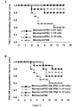
- IP-10-neutralizing antibodies increased neutrophil infiltration, impaired lung oxygenation and deteriorated the protective effects mediated by iPSC-CMin a ventilator-induced lung injury (VILI) model ( Fig. 10 ).
- VIP ventilator-induced lung injury
- IP-10 neutralization blocked the positive effects of iPSC-CM treatment or iPSCtransplantation on the survival rate of bleomycin-injected mice ( Fig. 15 ).
- the application of IP-10 neutralizing antibody attenuated the protective effects of iPSCs on a CCl4-injured liver ( Fig. 18 ).
- the secretion of IP-10 in a subject induced by the method provided herein is in a level effective to repair tissue injuries, including but not limited to lung injuries, liver injuries, and those injuries caused by corneal damages, limb disorders, kidney diseases, and ischemia.
- tissue injuries including but not limited to lung injuries, liver injuries, and those injuries caused by corneal damages, limb disorders, kidney diseases, and ischemia.
- ischemia are focal stoke and myocardial infarction.
- the description discloses a method for treating tissue injuries, for example lung or liver injuries, which comprises administering to a subject in need thereof a therapeutically effective amount of iPSCs or iPSC-CM, wherein the therapeutically effective amount is an amount capable of inducing a sufficient level of IP-10 to suppress inflammation responses.
- composition comprising iPSCs or iPSC-CM for inducing the secretion of IP-10 in a subject is also disclosed in the present description.
- Murine iPSCs were generated from mouse embryonic fibroblasts (MEFs) derived from 13.5-day-old embryos of C57/B6 mice.
- the iPSCs were reprogrammed by the transduction of retroviral vectors encoding four transcription factors (Oct4/Sox2/Klf4/c-Myc; OSKM), or three transcription factors (Oct4/Sox2/Klf4; OSK), as described previously (Li et al., 2011).
- Total of 12 clones (Re-1 to Re-12; OSKM) were selected and established.
- Re-7 iPSCs were selected and widely used in this study. Briefly, undifferentiated iPSCs were routinely cultured and expanded on mitotically-inactivated MEFs (50,000 cells/cm 2) in six-well culture plates (BD Technology) in the presence of 0.3% leukemia inhibitory factor in an iPSC medium consisting of Dulbecco's Modified Eagle's Medium (DMEM; Sigma) supplemented with 15% fetal bovine serum (FBS; Invitrogen), 100 mM minimal essential medium (MEM) nonessential amino acids (Sigma), 0.55 mM 2-mercaptoethanol (Gibco), and antibiotics (Invitrogen).
- DMEM Dulbecco's Modified Eagle's Medium
- iPSCs were dissociated into a single cell suspension by 0.25% trypsin-EDTA and plated onto non-adherent culture dishes in DMEM with 15% FBS, 100 mM MEM nonessential amino acids, 0.55 mM 2-mercaptoethanol, and antibiotics at a density of 2x10 6 cells/100 mm plate. After 4 days in floating culture, EBs were transferred onto gelatin-coated plates and maintained in the same medium for 24 h. EBs werethen assigned for in vitro differentiation into tridermal lineages as previously described (Li et al., 2011).
- cells were fixed with 80% alcohol, and then fixed cells were stained using the Vector Blue Alkaline Phosphatase Substrate Kit III (Vector Laboratories) according to the manufacturer's instructions. Alizarin red staining and PAS staining was performed as previously described (Li et al., 2011).
- LC-MS/MS analysis was performed through the application of LTQ Orbitrap (Thermo Fisher Scientific Inc., Bremen, Germany) as described. In breif, each sample of digested peptides was reconstituted to 20 ⁇ l of 0.1% formic acid (FA). Peptides were firstly injected in and separated by the nanoflow HPLC (Agilent 1100, Agilent Technologies, Santa Clara, CA, USA) with a C18 column (75 ⁇ m ID ⁇ 360 ⁇ m OD ⁇ 15 cm; Agilent Technologies, Santa Clara, CA, USA), and became ionized particles once passed through the succeeding nanospray tip (New Objective, Woburn, MA).
- nanoflow HPLC Agilent Technologies, Santa Clara, CA, USA
- the PAR affinity resin set (Tulip) conjugated Af1521 macrodomains was used to pulldown the parylated proteins.
- Protein was extracted from cells by lysis buffer (e.g.: 50mM Tris, pH 8, 200mM NaCl, 1mM EDTA, 1% Triton X-100, 10% glycerol, 1 mM DTT, 0.5% deoxycholate, and protease inhibitors) and incubated with resin overnight in 4°C. After incubation, resin was washed with lysis buffer three times followed by addition of IX SDS-sample buffer at 95°C for 10 min to dissociated proteins.
- lysis buffer e.g.: 50mM Tris, pH 8, 200mM NaCl, 1mM EDTA, 1% Triton X-100, 10% glycerol, 1 mM DTT, 0.5% deoxycholate, and protease inhibitors
- GO-Elite implements an over-representation statistical inference that can identify significantly enriched GO categories with nuclear proteins.
- Canonical pathway and gene interaction network analyses were conducted using Ingenuity Pathway Analysis web tool (IPA, http.//www.ingenuity.com/).
- IPA Ingenuity Pathways Knowledge Base
- Reverse transcription reactions were performed using SuperScript TM IIIReverse Transcriptase (Invitrogen). cDNA was used in the following quantitative PCR (qPCR) and RT-PCR. qPCR was performed with Power SYBR Green PCR Master Mix (Applied Biosystems) according to manufacturer's instructions. Signals were detected with 7900HT Fast Real-Time PCR System (Applied Biosystems).
- the stable ablation of Parp1 and Parp2 in MEFs was obtained using small hairpin RNA (shRNA) probes for the mouse gene Parp1 and Parp2.
- Control cells were allowed to stably express shLuc (pLKO.1-shLuc).
- Cells were infected with shRNA lentiviral-vector generated using a three-plasmid-based lentiviral system (all plasmids are available from the RNAi Consortium [TRC]).
- Lentivirus production was performed by transfection of 293T cells at 5x10 6 cells per 10 cm plate using Lipofectamine 2000 (LF2000, Invitrogen Life Technologies, Carlsbad, CA, USA). Supernatants were collected 48 h after transfection and then were filtered.
- Subconfluent cells were infected with lentiviral-vector in the presence of 8 mg/ml polybrene (Sigma). Infected cells were selected with puromycin (2 mg/ml) until control uninfected cells were completely dead. Immunoblotting was used to confirm the knockdown efficiency of shParp1 and shParp2 (Chen et al., 2011).
- STO cells were grown in 24-well tissue culture dishes to 70% confluence and then cotransfected with 0.2 ⁇ g of pMXs and pMXs-c-Myc in the presence of 0.2 ⁇ g of pGL3-PARP promoter firefly luciferase or PARP promoter mutants and 10 ng of SV40 Renilla luciferase plasmids (Promega). Twenty-four hours post-transfection, cells were harvested in 100 ⁇ lreporter lysis buffer and then subjected to a dual luciferase assay according to the manufacturer's protocol (Dual-Luciferase Reporter Assay System, Promega). Firefly luciferase activity was normalized to Renilla luciferase activity, and data are represented as the mean standard deviation of three independent experiments, each performed in triplicate.
- Chromatin Immunoprecipitation (ChIP) and site-directed mutagenesis of mouse PARP promoter mutants
- ChIP chromatin immunoprecipitation
- EZ ChIP kit Upstate
- anti-c-Myc sc-764, Santa Cruz
- the mouse PARP promoter with deletion or point mutation clones were created by site-directed mutagenesis according to the manufacturer's instructions (Phusion Site-Directed Mutagenesis Kit, Finnzymes), and all mutants were amplified by using PARP (-2000) as the template. Amplified fragments were further amplified with suitable forward primers with MluI cutting sequences and HIndIII cutting sequences in the reverse primers.
- mice iPS cells derived from C57BL/6J strain, black coat color
- mouse blastocystsfrom C57BL/6J-Tyrc2J strain albino
- the adult chimeras were confirmed by coat color, demonstrating that iPSCs were competent to produce adult chimeric mice. This study was assisted by Transgenic Mouse Model Core Facility, Academic Sinica, Taiwan.
- Results are reported as mean ⁇ SD. Statistical analysis was performed using Student's t test or a one-way or two-way analysis of variance (ANOVA) followed by Turkey's test, as appropriate. The survival rate analysis was performed using log-rank test. Results were considered statistically significant at P ⁇ 0.05.
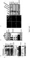
- nuclear protein extracts from MEFs and Re-7 iPSCs were prepared. These extracts were then separated into five fractions by SDS-PAGE ( Fig. 1 , panel A).
- ID LC-MS/MS nuclear protein extracts from MEFs and Re-7 iPSCs.
- the predominant processes upregulated in the nuclear protein profiles of iPSCs included those pertaining to RNA processing, chromatin packaging and remodeling, cell structure and motility, and protein biosynthesis, as well as those involved in mRNA transcription and DNA replication ( Fig. 1 , panel B).
- ID LC-MS/MS Based on Gene Ontology (GO) database analysis, the predominant processes upregulated in the nuclear protein profiles of iPSCs included those pertaining to RNA processing, chromatin packaging and remodeling, cell structure and motility, and protein biosynthesis, as well as those involved in mRNA transcription and DNA replication ( Fig. 1 , panel B).
- ID LC-MS/MS 2D-differential gel electrophoresis
- Parp1 is the posttranslational modification of target proteins by attaching a poly(ADP-ribose) chain (PARylation) (Krishnakumar and Kraus, 2010).
- PARylation poly(ADP-ribose) chain
- affinity resin to pull down the PARylated proteins, we further demonstrated that Parp1 is the most highly expressed PARylaed protein in iPSCs compared to MEFs (Table 1).
- Parp1 protein, as well as Oct4, Nanog, and c-Myc were upregulated in both whole-cell lysates and nuclear fractions of Re-7 iPSCs ( Fig. 1 , panel C).
- This upregulation of Parp1 accompanied by increased PARylation activity, was consistently observed in iPSCs generated with OSKM (Re-7 cells) or OSK, Dr. Yamanaka's iPSC clone (miPSCs) and ESCs ( Fig. 1 , panel C).
- iPSC-derived embryoid bodies EBs
- Fig. 1 panel E
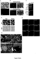
- Differentiation into different lineages was induced by specific protocols. Neuron-like, osteocyte-like (mesoderm), and hepatocyte-like (endoderm) cells were confirmed by immunofluorescence, Alizarin red and PAS staining, respectively ( Fig. 1 , panel F, left).
- Fig. 1 panel F
- western blotting showed that the Parp1 protein, as well as Parp2, topoisomerase II alpha, Klf4, Oct4, and Sox2, was substantially downregulated ( Fig.
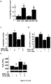
- Parp1 overexpression significantly enhanced the reprogramming efficiency in MEFs transfected with OSKM or OSK ( Fig 2 , panel F, left and middle, respectively).
- Parp2 overexpression also enhanced the reprogramming efficiency in MEFs transfected with OSK ( Fig. 2 , panel F, right), but the effect of Parp2 overexpression was significantly less than that of Parp1 overexpression.
- administration of various PARylation inhibitors consistently led to reduction in the efficiency of iPSC generation induced by OSKM at day 21 post-reprogramming (PJ-34: Fig.
- Parp1 knockdown by a lentivirus-delivered shRNA led to a significant inhibition of the efficiency of iPSC generation ( Fig. 2 , panel G, middle), and Parp2 knockdown also suppressed iPSC generation at a similar extent at day 21 post-reprogramming ( Fig. 2 , panel G, right).
- Fig. 2 , panel G, right shows that modulating Parp1 and PARylation activity influences the reprogramming efficiency and the pluripotent status of iPSCs, indicating that Parp1 and PARylation are crucial for nuclear reprogramming.
- c-Myc a proto-oncogene
- Parp1 can increase the efficiency of iPSC generation in the OSK-transfection protocol
- OSP iPSC-reprogramming efficiency of OS with Parp1
- OSM Fig. 3 , panel A
- iPSCs Yamanaka's iPSC clone (miPSCs), and iPSCs generated by transfection of either OSK or OSP (data not shown), as described previously (Fujii-Yamamoto et al., 2005). These data indicate that the Parp1 effect on reprogramming efficiency and iPSC generation is cell cycle-independent. Furthermore, OSP transfection activated the expression of Nanog-GFP during reprogramming in a Nanog-GFP reporter MEF clone ( Fig. 3 , panel C). The high passages of OSP-reprogrammed iPSCs were stably positive for markers of mouse ESCs, such as ALP activity ( Fig.
- c-Myc is a direct regulator of Parp1 and PARylation
- c-Myc regulates Parp1 expression by fusing the Parp1 promoter to a luciferase reporter plasmid and co-expressing the reporter with c-Myc.
- Three putative c-Myc-binding sites were identified in the proximal promoter region (-2000 to -100 base pairs) of Parp1 and deletion constructs were cloned in the luciferase reporter plasmid ( Fig. 4 , panel D).
- Cotransfection experiments showed that c-Myc activated the transcriptional activity of the Parp1 promoter containing three (-2000) or two proximal (-1100) c-Myc binding sites.
- Parp1 promoter deletion mutants lacking c-Myc-C1 and c-Myc-C2 suppressed c-Myc-activated Parp1 transcription ( Fig. 4 , panel E), indicating that C2 is an important site responding to c-MyC activity.
- the Parp1 promoter construct without all three c-Myc binding sites (-125) or with point mutations in C2 (c-Myc-C2 mt) could not be stimulated by c-Myc.
- chromatin immunoprecipitation (ChIP) assays were performed using C1, C2, and C3 primer sets ( Fig. 4 , panel F). The result showed that the endogenous c-Myc indeed only bound to the C2, but not C1 or C3, position of the Parp1 promoter. As a positive control, c-Myc bound to its reported target, cyclin D2 promoter. Panel Gof Fig. 4 shows the result of panel Fof Fig. 4 ChIP with quantitative PCR (qChIP).
- PARylation was previously considered the major catalytic function of Parp1; we therefore attempted to identify the proteins that are involved in Parp1-mediated PARylation in pluripotent stem cells.
- the PARylated proteins indentified by LC-MS/MS in iPSCs are listed in Table 1.
- Parp1, Chd1L, DNA ligase III, Ssrp1, Xrcc6, and Parp2 were identified by LC-MS/MS as having more than 3 peptides per protein.
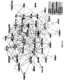
- Parp1 may be a key factor regulating the pathways related to DNA repair, chromatin modification, the polycomb complex, and histone modification.
- bioinformatic analysis revealed that Parp1-PARylated proteins interacted significantly with Oct4, Nanog, c-Myc, Klf4, CTNNB1, WDR5, SUZ12, EZH2, DNMT3A/B, and JARID2 in the core network of nuclear reprogramming and pluripotent status ( Fig. 6 ).
- Parp1 as a pivotal regulator of nuclear reprogramming and pluripotency.
- Our data demonstrated that the expression of Parp1 and PARylation increased during reprogramming and decreased upon differentiation.
- Parp1 replaced Klf4 or c-Myc in promoting iPSC production and generating chimeric mice with Oct4/Sox2-transfected cells ( Fig. 3 ).
- c-Myc directly binds to the Parp1 promoter to enhance its expression, resulting in increased PARylation activity.
- Acute lung injury (ALI) and acute respiratory distress syndrome (ARDS) are disordersof acute respiratory failure and manifest as non-cardiogenic pulmonary edema, respiratorydistress and hypoxemia.
- High tidal volume-induced mechanical ventilation in patients hasbeen shown to increase the risk of pathologic overdistention in the lungs, elicit the productionof inflammatory mediators, recruit inflammatory cells, and eventually induce a type of ALI, termed ventilator-induced lung injury (VILI).
- VILI ventilator-induced lung injury
- MSCs meenchymal stem cells
- Mouse embryonic fibroblasts were reprogrammed into iPSCs by ectopicallytransfection of Oct4/Sox2/Klf4 (OSK) as previously described ( Li HY et al. Biomaterials 2011;32:5994-6005 ).
- Undifferentiated iPSCs were cultured on inactivated MEF, and formed colonies very similar to ESCs.
- These iPSC clones were positive for alkaline phosphatase (AP) and SSEA-1, detected by AP and immunofluorescent staining, respectively.
- AP alkaline phosphatase
- SSEA-1 alkaline phosphatase
- the iPSCs were able to differentiate into chondrocyte-like cells, osteocyte-like cells, andhepatocyte-like cells and neuronal-lineaged cells (data not shown).
- RNA RNA was reverse transcribed by using a GeneAmp PCR system 9600 (PerkinElmer,Life Sciences, Inc., Boston, MA), as previously described ( Li LF et al. Respir Res 2011;12:90 ).
- the lungs were lavaged via tracheostomy with a 20-gauge angiocatheter (sham instillation) 3 times with 0.6 ml of 0.9% normal salineat the end of the study period.
- the effluents were pooled and centrifuged at 2,000 rpm for 10 min. Supernatants were frozen at -80°C for further analysis of the cytokine.
- PAI-1 with a lower detection limit of 0.02 ng/ml and HMGB1 with a lower detection limit of 1 ng/ml were measured in BAL fluid using a commercially available immunoassay kit containing antibodies that were cross-reactive with rat and mouse PAI-1 (Molecular Innovations, Inc., Southfield, MI) and HMGB1 (Shino-Test corporation, Kanagawa, Japan). Each sample was run in duplicate according to the manufacturer's instructions.
- the lungs were fixed in 3% glutaraldehyde in 0.1 M cacodylate buffer (pH 7.4) for 1 h at 4 °C. The lungs were then postfixed in 1% osmium tetroxide (pH 7.4), dehydrated in a graded series of ethanol, and embedded in EPON-812. Thin sections (70 nm) were cut, stained with uranyl acetate and lead citrate, and examined on a Hitachi H-7500 EM transmission electron microscope (Hitachi, Ltd., Tokyo, Japan).
- V T 30 high tidal volume
- V T 30 high tidal volume
- V T 6 low tidal volume
- AV T 30 also increased lung Evans blue dye (EBD) content, bronchoalveolar lavage (BAL) totalprotein, and the wet-to-dry ratio, indicating capillary leakage.
- EBD Evans blue dye
- BAL bronchoalveolar lavage
- a V T 6 showed noeffect on these parameters when compared with non-ventilated mice ( Fig.7 , panels B-D).
- the macroscopic lung congestion and elevation of capillary permeability induced by a V T 30 was not affected by MEF treatment, but was substantially suppressed by treatment with eitheriPSCs or iPSC-CM ( Fig. 7 , panels B-D).
- the PaO 2 /FiO 2 ratio an index of gasexchange
- a V T 30 when compared with non-ventilatedmice or mice receiving a V T 6 ( Fig.7 , panel E).
- the decreases in oxygenation with a V T 30 were significantly improved by the administration iPSCs or iPS-CM. Therefore, thesedata suggest that iPSCs or iPSC-CM improve microvascular leakage, lung edema, total lunginjury, and help to recover respiratory functions in a VILI model induced by a V T 30.
- the neutrophil counts and myeloperoxidase (MPO) assay revealedthat neutrophils migrated into the injured lung sites in mice after mechanical ventilation at V T 30 when compared with non-ventilated mice or mice receiving a V T 6 ( Fig.8 , panels C and D).Meanwhile, the HMGB1 and PAI-1 protein levels were elevated in response to V T 30 treatment ( Fig.8 , panels E and F), indicating an upregulation of chemoattractants for neutrophils inthis model. Significantly, iPSC or iPSC-CM ameliorated neutrophil migration and HMGB1 and PAI-1 protein elevation ( Fig.8 , panels C-F).
- V T 30 andiPSC-CM administration were investigated the effect of V T 30 andiPSC-CM administration on the expression of macrophage inflammatory protein-2 (MIP2),nitrate/nitrite, malondialdehyde (MDA) and total glutathione (GSH) from lung tissues inrecipients.
- MIP2 macrophage inflammatory protein-2
- MDA malondialdehyde
- GSH total glutathione
- iPSC-CM administration effectively inhibited the upregulation of MIP2, nitrate/nitrite, and the production of MDA, but elevated GSH production in recipients ( Fig.9 , panels B-E).
- cytokine array we found in a cytokine array that several cytokines, including uPA and TIMP-4, were also secreted by iPSCsinto the conditioned medium ( Fig.9 , panel F).
- IP-10 monokine induced by IFN- ⁇ (MIG) and the IFN- ⁇ inducible T-cell chemoattractant (iTAC) are three chemokines that bind to acommon receptor, CXCR3. These three chemokines can be induced by INF-y.
- INF-y acommon receptor
- IP-10 has exhibited protective ability against hepatitis,pulmonary fibrosis, and myocardial infarction and has been involved in tissuerepair and remodeling. Accordingly, we investigated whether IP-10 was involved in the reparative effect of iPSC-CM in the V T 30-induced VILI model.
- IP-10 nAb alone significantlyimpaired structural changes, lung injury scores, neutrophil infiltration, and the PaO2/FiO2 ratio in V T 30-treated mice. IP-10 nAb also substantially block the reparative effectproduced by iPSC-CM on these parameters ( Fig.10 , panels C-E). Taken together, these findings demonstrate that IP- 10 serves a pivotal role and is involved in the reparative effect of iPSC-CM on airway structuraldamage and oxygenation ability in VILI.
- ARDS acute respiratory distress syndrome
- IPF idiopathic pulmonary fibrosis
- Bleomycin has been used to model fibroticlung injury in animal studies, as the characteristics of bleomycin-induced lung damage in animals includeacute inflammatory injury of the alveolar epithelium followed by reversible fibrosis, which overlaps with the symptoms of ARDS and IPF. The extent of fibrosis is also proportional to the severity of the initialinjury.
- Intra-trachealadministration of bleomycin requires only a single dose to result in injury and fibrosis, with fibrosis starting after 14 days and maximal responses occurring after 21-28 days.
- Previousstudies have suggested that the early reduction of inflammation may result in the attenuation ofdownstream events leading to collagen deposition ( Moodley Y et al. Am J Pathol 2009; 175:303-313 ).
- Developing an effective strategy for amelioratingpulmonary damage during the early phases of pulmonary fibrosis pathogenesis is therefore a top priority.
- Mouse iPSCs which express a gene signature similar to that of ESCs (i.e., the expression of Oct4,Sox2, Nanog, Klf-4, Fbx5, Eras, Dppa5a, and Rex1 and a pluripotent status), were established by introducing three genes (Oct4/Sox2/Klf4) not including c-Myc.
- murine iPSCs were generatedfrom mouse embryonic fibroblasts (MEFs) derived from 13.5-day-old embryos of C57/B6 mice.
- TheiPSCs were reprogrammed via the transduction of pMX-based retroviral vectors encoding threetranscription factors, Oct-4, Sox2, and Klf4, according to the protocol described in previous studies with minor modifications( Takahashi K et al. Cell 2006; 126:663-676 ).
- Plat-E packing cells were incubated overnight at a density of 3.6x10 6 cells per 100-mm dish.
- pMX-based retroviral vectors encoding mouse complementary DNAs wereintroduced into the Plat-E cells using the Fugene 6 transfection reagent (Roche Applied Science,Indianapolis, IN). Forty-eight hours after transfection, virus-containing supernatants were collected for target cell infection.
- iPSC culture undifferentiated iPSCs were routinely cultured and expanded on mitotically inactivated MEFs(50,000 cells/cm 2 ) in six-well culture plates (BD Technology, Franklin Lakes, NJ) in the presence of 0.3% leukemia inhibitory factor (LIF) in an iPS medium consisting of Dulbecco's modified Eagle'smedium (DMEM; Sigma-Aldrich) supplemented with 15% fetal bovine serum (FBS; Invitrogen, Carlsbad,CA), 100 mM minimal essential medium nonessential amino acids (Sigma-Aldrich), 0.55 mM2-mercaptoethanol (Gibco, Gaithersburg, MD), and antibiotics (Invitrogen).
- DMEM Dulbecco's modified Eagle'smedium
- FBS Invitrogen, Carlsbad,CA
- mouse iPSCs were placed at 20,000 cells per cm 2 and incubated in a 10-ml volume of serum-freebasal medium (DMEM-high glucose (Gibco) with a 100 mM concentration of nonessential amino acids(Gibco), 0.3% LIF, and 1% penicillin-streptomycin) in a 10-cm dish (Corning Incorporated) for 48 h.Then, the trypsinized iPSCs in whole culture medium were collected and centrifuged for 10 min at 1500rpm to obtain the supernatant to be used as the conditioned medium in subsequent in vivo experiments.
- serum-freebasal medium DMEM-high glucose (Gibco) with a 100 mM concentration of nonessential amino acids(Gibco), 0.3% LIF, and 1% penicillin-streptomycin
- mice were intra-tracheally injected with 1.5 U/kg bleomycin sulphate (Merk, Darmstadt,Germany) in 50 ⁇ l PBS under light anesthesia to induce pulmonary fibrosis.
- mice received either iPSCs (2 ⁇ 106 cells in 200 ⁇ l of PBS; iPSC-treated mice) or PBS 200 ⁇ l(PBS-treated mice) via tail vein injection 24h after the induction of lung injury.
- Cell administration 24hours after lung injury was chosen to optimize cell incorporation into the lungs during early inflammation.
- mouse was given two doses (0.5 ⁇ g/dose, i.p.) of anti-IP-10 antibodies(Abcam, ab9938, Cambridge, UK) at 4 h before and 4 h after injury.
- Samples were collected from each mouse for the assessment of lung injury,which was performed according to the hydroxyproline assay, pulmonary physiology, histology,immunohistochemistry, and cytokine and myeloperoxidase (MPO) analysis.
- MPO myeloperoxidase
- Lungs from each group of animals were excised at 7, 14, and 21 days after bleomycin-induced lunginjury.
- the lung tissues were fixed in 4% paraformaldehyde, dehydrated using a graded ethanol series,embedded in paraffin blocks, cut into 3- ⁇ m sections, and stained with hematoxylin and eosin (H&E)using standard histologic techniques.
- H&E hematoxylin and eosin
- Lung fibrosis was scored on a scale of 0 to 8 according to the following criteria: grade 0, normal lung; grade 1-2, minimal fibrous thickening of alveolar or bronchiolar wall; grade 3-4, moderate thickening of walls without obvious damage to lung architecture; grade 5-6, increased fibrosis with definite damage to lungstructure; and grade 7-8, severe distortion of structure and large fibrous areas. Following the examinationof 30 randomly chosen regions in each sample at 100x magnification, the mean score of all the fields wastaken as the fibrosis score for each sample. The destruction of alveolar walls was quantified according to the destructive index value, which wasmeasured using a previously described technique ( Saetta M et al. Am Rev Respir Dis 1985; 131:764-769 ).
- Airspace size was quantified by measuring the meanlinear intercept (Lm) using a modified Thurlbeck method ( Thurlbeck WM. Am Rev Respir Dis 1967; 95:752-764 ). Briefly, 30 fields at 200x magnification wererandomly observed on two slides from each mouse, and point counting in each field was performed. Then, the total distance divided by the number of alveolar intercepts provided the value for Lm.
- Lm meanlinear intercept
- Immunohistochemical staining was performed using primary antibodies targeting Ly6C (Santa CruzBiotechnology, Santa Cruz, CA), collagen I (Abcam, Cambridge, UK), and alpha-smooth muscle actin( ⁇ -SMA) (Biomeda, Foster City, CA). Briefly, paraffin sections of the lungs were de-waxed and rehydrated. Lung sections were subjected to antigen retrieval and blocked with a peroxidase-blockingreagent. Sections were incubated with primary antibody overnight at 4°C. After washing, the lungsections were incubated with a SuperSensitiveTM system horseradish peroxidase-labeled polymer(BioGeneX, San Ramon, CA) for 1 hour at room temperature.
- Lung tissues for ELISA were homogenized in PBS with protease inhibitors containing 10 ⁇ g/mLAprotinin, 10 ⁇ g/mL Leupeptin, and 10 ⁇ g/mL Pepstatin. Triton X-100 was added to a final concentrationof 1%. Samples were frozen at ⁇ -70°C, thawed, and centrifuged at 10,000 g for 5 min to remove cellulardebris. Quantitation of sample protein concentrations in the supernatants was performed using a totalprotein assay. Cytokine levels were determined according to the manufacturer's instructions provided with commercial ELISA kits (R&D Systems, Minneapolis, MN). Cytokine array data on developed X-rayfilm was quantified by scanning the film with an ImageScanner III (GE Healthcare) and analyzing the array image file using ImageQuant software (GE Healthcare).
- the intra-tracheal injection of bleomycin resulted in the previously documented sequence of eventsleading to lung injury.
- the early phase of injury was characterized by neutrophilic alveolitis, whereas the late phase wascharacterized by patchy areas of fibrosis,asevidenced by the histopathological examination of bleomycin-injuredlungs.
- bleomycin-induced interstitial thickening, inflammation, and distortion of lung architecture were attenuated following the administration ofiPSCs or iPSC-CM at days 14 and 21 post-bleomycin treatment.
- the bleomycin-treatedrecipients of iPSCs also demonstrated significantly lower Ashcroft lung fibrosis scores than thoseof MEFs or PBS alone ( Fig.11 ,panel A).
- Restoration of bleomycin-impaired lung structure following treatment with iPSCs was also observed according to measurements of the destructive index ( Fig.11 ,panel B).
- Therestorative effect of iPSC-CM on Ashcroft lung fibrosis scores and the destructive index was slightly lessthan that of iPSCs.
- the alveolar mean linear intercept (Lm), a morphometric parameter of averagealveolus size also recovered to near-normal levels following the delivery of iPSCs or iPSC-CM( Fig.11 ,panel C).
- Pulmonary fibrosis is characterized by the accumulation of fibrillar collagens like type 1 collagen. Immuno-staining for collagen-1 demonstrated thick bands of fibrosis in PBS-treated mice, while the lungs of bleomycin-challenged recipients treated with either iPSCs or iPSC-CM were significantlyprotected from bleomycin-induced fibrosis ( Fig. 12 , panel A). The accumulation of myofibroblasts,which are considered important fibrogenic effector cells, was also examined.
- mice As indicated by the staining of lung sections for ⁇ -SMA at 14 and 21 days post-bleomycin treatment, recipients of iPSCs oriPSC-CM mice exhibited a significant decrease in myofibroblast accumulation in comparison toPBS-treated mice ( Fig.12 , panel B). Lung collagen content was further assessed by measuringhydroxyproline content. Values in the iPSC-treated group and iPSC-CM-treated group were significantlylower than those in the PBS-treated group at 14 days and 21 days, indicating that bleomycin-inducedcollagen synthesis was significantly suppressed in mice treated with iPSCs or iPSC-CM ( Fig. 12 , panel C).
- IPSCs Increased Production of Anti-Fibrotic Chemokine IP-10 in Injured Lungs
- cytokines and chemokines knownto mediate inflammation i.e., IL-1, IL-2, IL-10, TNF- ⁇ , and MCP-1
- fibrosis i.e., INF- ⁇ andMCP-5
- the ELISA data also showed that both iPSCs and iPSC-CM treatment stimulated the production of IP-10 ( Fig.13 ,panel A).
- Real-time PCRfurther indicated that administration of iPSCs or iPSC-CM significantly increased the mRNAexpression of IP-10 at 3 days post-bleomycin injury ( Fig. 13 ,panel B). In addition, these treatments reduced INF- ⁇ mRNA expression but showed no effect on MIG, iTAC, or CXCR3 mRNA expression ( Fig. 13 ,panels C-E).
- IPSCs and iPSC-CM rescued the survival of bleomycin-treated recipient in a celldose-dependent manner
- mice were firstintra-tracheally injected with 3.0 U/kg bleomycin sulphate, a dose sufficient to induce recipient lethalit.
- the intravenous delivery of iPSCs led to a dose-dependent improvement in recipientsurvival, and the maximal protective effect of iPSCs was observed at a dose of 2 ⁇ 10 6 cells.
- iPSC-CM Treatment with iPSC-CM (collected from iPSC culture at the indicated cell dosages) similarly improvedrecipient survival in a cell dose-dependent manner ( Fig. 14 ,panel B).
- IP-10 nAb IP-10-neutralizing antibody
- IP-10 nAb administration largelyblocked the effects of iPSC-CM on collagen deposition (positive staining for collagen-1; Fig.15 ,panel B),myofibroblast accumulation (positive staining for ⁇ -SMA; Fig.15 , panel C), inflammatory cell accumulation(positive staining for Ly6C; Fig.15 ,panel D), and pulmonary fibrosis ( Fig. 15 ,panel E).
- Administration of IP-10 nAb alsodeteriorated the reparative effect of iPSCs, identical to the observations in that of iPSC-CM (datanot shown).
- these findings demonstrate that IP-10 is the major contributor to the iPSC and iPSC-CM-mediated inhibition of collagen deposition and inflammation in pulmonaryfibrosis.
- Liver diseases and injuries are important medical problem worldwide. Liver transplantation iscurrently the most efficient therapy for liver failure and end-stage liver disease. However, it islimited by the scarcity of donor, expensive medical cost, surgical risk and requiring life-longimmunosuppressant agents.
- the development and application of hepatocytes transplantation hasbeen attempted to treat different forms of liver diseases. It has minimal invasive procedures and fewer surgical complications compared to the orthotopic liver transplantation. Stem celltransplantation has also gained considerable attention recently. Stem cells have the potential tosupportive tissue regeneration and to generates large amounts of donor cells ready for transplant ( Kakinuma S et al. J Gastroenterol 44: 167-172 ; Navarro-Alvarez N et al.
- iPS cells Mouse germline-competent iPS cells were provided by Kyoto University (Dr. Shinya Yamanaka)and RIKEN BRC, Japan ( Takahashi K et al.(2006) Cell 126:663-676 ).
- IPS cells were cultured as previously described ( Okita K et al. (2007) Nature 448: 313-317.9 ).
- the iPS cells weresuccessfully induced to differentiate into hepatocyte-like (iHL) cells with functions resemblingprimary hepatocytes.
- rIP-10 hepatocytes were seeded on 24-well plates at the same density.
- the rIP-10 (0.5ng or 5ng) wasgiven at 4 h post-injury.
- the viability of hepatocytes was evaluated at 24 h by methyl thiazoltetrazolium (MTT, Sigma) assay.
- MTT methyl thiazoltetrazolium
- mice C57/B6, 8 to 10 weeks were housed in cage and allowed free access to food and water.
- Mouse was given carbon tetrachloride (CCl4, Sigma) in mineral oil (0.35 ⁇ l/g, single dose, i.p.) to induce liver injury.
- mice were randomized to receive vehicle (PBS), iPS cells or iHL (2x10 6 cells/in 100 ⁇ l PBS) infusions via tail veins.
- PBS vehicle
- iPS cells iPS cells
- iHL 2x10 6 cells/in 100 ⁇ l PBS
- mice received blood withdrawal ( ⁇ 100 ⁇ l) from facial veins at scheduled time for liver biochemistry.
- blood was drawn from the heart and the liver was harvested and prepared for subsequent experiments including histochemistry, cytokine assay, protein and gene expression analysis.
- mice were given two doses (0.5 ⁇ g/dose, i.p.) of anti-IP-10 antibodies (Abcam, ab9938, Cambridge, UK) at 4 h before and 4 h after injury.
- mice received repetitive CCl4 injury at 0, 24 and 48 h.
- the iPS cells (2x10 6 cells/in 100 ⁇ l PBS) or recombinant IP10 (rIP10, 0.5ng) were given once at 4 h and the mortality rate of mice was observed until 72 hpost-injury. All animals received humane care according to the Guide for the Care and Use of Laboratory Animals prepared by the National Academy of Sciences (NIH publication no.
- Necrotic area were determined by measuring fiveindependent fields per liver using a computerized morphometry system (MicroCam, M&T OPTICS,Taiwan) and expressed as percentage of the filed area.
- mice were injected with 5-bromo-2'-deoxyuridine (BrdU, 50mg/kg, i.p.,Sigma).
- the peroxidase-coupled mouse monoclonal anti-BrdU (DAKO, M0744) and anti-Ki67(DAKO, M7249) were used in subsequent immunohistochemistry study for detecting proliferativehepatocytes.
- Ten pictures of the interested areas (different portal and central vein areas) per animal were photo-taken under microscopy at x200 magnification. The mean numbers of BrdU-positive orKi67-positive cells of per area per animal were used in statistical analysis.
- Tissue lysate were prepared in a buffer containing 50 mM Tris-HCl, pH 7.4, 150 mM NaCl, 0.25%deoxychoic acid, 1% NP40, 1 mM EDTA, 1 mM Na orthovanadate, 1 mM Na fluoride, 1 Mmphenylmethylsulfony fluoride, 1ug/ml aprotinin, 1ug/ml leupeptin and 1 ug/ml peptstain, on ice asdescribed before ( Kao CL et al. (2010) Stem Cells Dev 19: 247-2580 ). The concentrations of sample proteins were determined using the ProteinAssay kit (Bio-Rad, Hercules, CA).
- liver tissues were homogenized and prepared in PBS with protease inhibitors (proteaseinhibitors (10 ⁇ g/mL Aprotinin, 10 ⁇ g/mL Leupeptin, and 10 ⁇ g/mL Pepstatin) and 1% Triton X-100.
- protease inhibitors 10 ⁇ g/mL Aprotinin, 10 ⁇ g/mL Leupeptin, and 10 ⁇ g/mL Pepstatin
- Triton X-100 Triton X-100
- Theprotein concentrations were quantified (DC-Bradford protein assay, Bradford, Bio-Rad, Hercules,CA, USA) and 200 ⁇ g of proteins were used for the analysis of cytokines by the commercializedassay kits (Mouse cytokine array panel A and IP-10 Immunoassay, R&D, MN) according themanufacture's instruction.
- the expression of individual cytokines in cytokine array study wasquantified by densitometry and expressed as fold change relative to their expressions in the injuredliver without cell treatment.
- results are expressed as mean ⁇ SEM.
- Statistical analysis was performed by using anindependent Student t test and one- and two-way ANOVA with Tukey post hoc test whenappropriate.
- the survival analysis was performed by using logrank test.A p value ⁇ 0.05 wasconsidered statistically significant.
- hepatocyte proliferation which is a criticalsign of liver regeneration. BrdU incorporation was used to quantify hepatocytes in S phase. Positiveimmunostaining of Ki67 represents the hepatocytes in cell cycle progression.
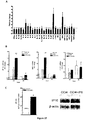
- iPS cells-induced changes of hepatic cytokines were evaluated by cytokine array ( Fig.17 ,panel A).
- IP-10 and MIG are upregulated by 7- and 6-folds respectively.
- the mRNA expression of IP-10 and MIG significantly increased at 24 hpost-injury ( Fig.17 ,panel B).
- the expression of iTAC which belong to the same cytokinefamily as IP-10 and MIG, decreased ( Fig.17 ,panel B).
- IP-10 could be an important hepatoprotective mediator.
- rIP-10 recombinant IP-10
Landscapes
- Health & Medical Sciences (AREA)
- Life Sciences & Earth Sciences (AREA)
- Chemical & Material Sciences (AREA)
- Engineering & Computer Science (AREA)
- General Health & Medical Sciences (AREA)
- Bioinformatics & Cheminformatics (AREA)
- Organic Chemistry (AREA)
- Animal Behavior & Ethology (AREA)
- Medicinal Chemistry (AREA)
- Pharmacology & Pharmacy (AREA)
- Public Health (AREA)
- Veterinary Medicine (AREA)
- Immunology (AREA)
- Epidemiology (AREA)
- Zoology (AREA)
- Cell Biology (AREA)
- Developmental Biology & Embryology (AREA)
- Gastroenterology & Hepatology (AREA)
- Proteomics, Peptides & Aminoacids (AREA)
- Reproductive Health (AREA)
- Genetics & Genomics (AREA)
- Wood Science & Technology (AREA)
- Biochemistry (AREA)
- General Engineering & Computer Science (AREA)
- Virology (AREA)
- Biotechnology (AREA)
- Biomedical Technology (AREA)
- Nuclear Medicine, Radiotherapy & Molecular Imaging (AREA)
- Gynecology & Obstetrics (AREA)
- Chemical Kinetics & Catalysis (AREA)
- General Chemical & Material Sciences (AREA)
- Pulmonology (AREA)
- Micro-Organisms Or Cultivation Processes Thereof (AREA)
- Medicines Containing Material From Animals Or Micro-Organisms (AREA)
Claims (4)
- Verfahren zur Herstellung von induzierter pluripotenter Stammzelle (iPSCs) aus somatischen Zellen, umfassend:(a) Inkontaktbringen von isolierten somatischen Zellen mit oder Einwirkenlassen auf diese von Oct3/4, Sox2, und Parp1, und(b) Kultivieren der somatischen Zellen wie in Schritt (a) erhalten unter geeigneten Bedingungen, damit sich mindestens eine Teilmenge der Population von somatischen Zellen in iPSCs umwandelt und Pluripotenz und Selbsterneuerungsfähigkeit erhalten bleiben.
- Verfahren zur Herstellung von induzierten pluripotenten Stammzellen (iPSCs) aus somatischen Zellen, umfassend:(a) Transfizieren von isolierten somatischen Zellen für Expression von Oct3/4, Sox2, und Parp1, wobei die isolierten somatischen Zellen mit einem oder mehr Plasmid- oder viralen Vektoren umfassend Oct3/4, Sox2, und Parp1 funktionsfähig verbunden mit einem Promotor transfiziert werden; und(b) Kultivieren der transfizierten somatischen Zellen wie in Schritt (a) erhalten unter geeigneten Bedingungen, damit sich die somatischen Zellen in iPSCs umwandeln und Pluripotenz und Selbsterneuerungsfähigkeit erhalten bleiben.
- Verfahren nach Anspruch 1, wobei das Verfahren keinen Schritt von Transfizieren der somatischen Zellen mit, Inkontaktbringen von diesen mit, oder Einwirkenlassen auf diese von c-Myc, Klf4, Nanog, Lin28, oder jedweder Kombination davon umfasst.
- Verfahren nach Anspruch 2, wobei das Verfahren keinen Schritt von Transfizieren der somatischen Zellen mit, Inkontaktbringen von diesen mit, oder Einwirkenlassen auf diese von c-Myc, Klf4, Nanog, Lin28, oder jedweder Kombination davon umfasst.
Applications Claiming Priority (3)
| Application Number | Priority Date | Filing Date | Title |
|---|---|---|---|
| US201261708128P | 2012-10-01 | 2012-10-01 | |
| US201261717871P | 2012-10-24 | 2012-10-24 | |
| PCT/CN2013/001187 WO2014053082A1 (en) | 2012-10-01 | 2013-09-30 | Method for preparing induced pluripotent stem cells and its applications |
Publications (4)
| Publication Number | Publication Date |
|---|---|
| EP2904103A1 EP2904103A1 (de) | 2015-08-12 |
| EP2904103A4 EP2904103A4 (de) | 2016-03-30 |
| EP2904103B1 true EP2904103B1 (de) | 2020-01-22 |
| EP2904103B8 EP2904103B8 (de) | 2020-03-11 |
Family
ID=50385434
Family Applications (1)
| Application Number | Title | Priority Date | Filing Date |
|---|---|---|---|
| EP13844080.5A Active EP2904103B8 (de) | 2012-10-01 | 2013-09-30 | Verfahren zur herstellung induzierter pluripotenter stammzellen und deren anwendungen |
Country Status (5)
| Country | Link |
|---|---|
| US (1) | US20140093486A1 (de) |
| EP (1) | EP2904103B8 (de) |
| CN (1) | CN104919048B (de) |
| TW (2) | TW201606083A (de) |
| WO (1) | WO2014053082A1 (de) |
Families Citing this family (22)
| Publication number | Priority date | Publication date | Assignee | Title |
|---|---|---|---|---|
| CN104919048B (zh) | 2012-10-01 | 2018-10-26 | 台北荣民总医院 | 制备诱导多能干细胞的方法及其应用 |
| WO2015140005A1 (en) * | 2014-03-19 | 2015-09-24 | Ifom Fondazione Istituto Firc Di Oncologia Molecolare | Method of generation of pluripotent cells |
| JP2016128396A (ja) * | 2015-01-09 | 2016-07-14 | 上田 実 | iPS細胞培養上清を含む医薬組成物およびその製造方法、化粧品およびその製造方法、抗加齢組成物、疾患発症抑制方法、疾患治療方法、組織異常治療方法ならびに美容方法 |
| EP4026563B1 (de) * | 2015-06-18 | 2025-10-29 | Yeda Research and Development Co. Ltd | Konditionierungsprotokolle und verwendung davon zur geweberegeneration |
| US10898522B2 (en) | 2015-08-19 | 2021-01-26 | Children's Research Institute, Children's National Medical Center | Compositions and methods for treating graft versus host disease |
| WO2017053963A1 (en) * | 2015-09-25 | 2017-03-30 | Cedars-Sinal Medical Center | A method effective to modulate expression of t-box protein 4 (tbx4) for reducing progression of lung fibrosis after a lung injury |
| US11525119B2 (en) | 2016-09-06 | 2022-12-13 | The Children's Medical Center Corporation | Immune cells derived from induced pluripotent stem cell |
| WO2018085419A1 (en) | 2016-11-01 | 2018-05-11 | Jian Feng | Method of producing naive pluripotent stem cells |
| CN110621323B (zh) * | 2017-05-02 | 2022-10-14 | 田边刚士 | 医药品组合物及化妆品组合物 |
| EP3621630A4 (de) | 2017-06-14 | 2021-03-10 | The Children's Medical Center | Hämatopoietische stamm- und vorläuferzellen aus hämogenen endothelzellen durch episomalen plasmid-gentransfer |
| US11821003B2 (en) | 2017-08-14 | 2023-11-21 | Sanford Burnham Prebys Medical Discovery Institute | Cardiogenic mesoderm formation regulators |
| US11725056B2 (en) | 2017-10-03 | 2023-08-15 | Cedars-Sinai Medical Center | Methods for targeting the immune checkpoint PD1 pathway for treating pulmonary fibrosis |
| WO2019099552A1 (en) * | 2017-11-14 | 2019-05-23 | The J. David Gladstone Institutes | Methods of generating retinal pigment epithelium (rpe) |
| CN109833358A (zh) * | 2017-11-28 | 2019-06-04 | 大江生医股份有限公司 | 刺梨萃取物用于制备细胞回春组合物的用途 |
| US12274733B2 (en) | 2018-09-28 | 2025-04-15 | President And Fellows Of Harvard College | Cellular reprogramming to reverse aging and promote organ and tissue regeneration |
| IL281470B2 (en) | 2018-09-28 | 2025-08-01 | Harvard College | Cellular reprogramming to reverse aging and promote organ and tissue regeneration |
| WO2021150919A1 (en) | 2020-01-23 | 2021-07-29 | The Children's Medical Center Corporation | Stroma-free t cell differentiation from human pluripotent stem cells |
| CN115698335A (zh) | 2020-05-22 | 2023-02-03 | 因斯特罗公司 | 使用机器学习模型预测疾病结果 |
| WO2022104237A1 (en) * | 2020-11-16 | 2022-05-19 | Duke University | Compositions for and methods of enhancing tissue regeneration |
| CN114591915B (zh) * | 2022-02-28 | 2023-11-21 | 集美大学 | 一种大黄鱼体外诱导多能性干细胞的方法 |
| US12252518B2 (en) | 2023-01-06 | 2025-03-18 | Life Biosciences, Inc. | Methods of treating non-arteritic anterior ischemic optic neuropathy |
| CN119265241A (zh) * | 2023-06-30 | 2025-01-07 | 深圳菁童生命科学有限公司 | 一种无相分离转录因子介导启始阶段重编程使细胞年轻化的方法 |
Citations (2)
| Publication number | Priority date | Publication date | Assignee | Title |
|---|---|---|---|---|
| US8048675B1 (en) * | 2010-05-12 | 2011-11-01 | Ipierian, Inc. | Integration-free human induced pluripotent stem cells from blood |
| US8058065B2 (en) * | 2005-12-13 | 2011-11-15 | Kyoto University | Oct3/4, Klf4, c-Myc and Sox2 produce induced pluripotent stem cells |
Family Cites Families (3)
| Publication number | Priority date | Publication date | Assignee | Title |
|---|---|---|---|---|
| US9410128B2 (en) * | 2011-02-22 | 2016-08-09 | Sanford-Burnham Medical Research Institute | Method and compounds for generation of iPSCs |
| CN102653774B (zh) * | 2011-03-04 | 2014-12-10 | 中国科学院上海生命科学研究院 | 山羊可诱导多能干细胞的制备方法 |
| CN104919048B (zh) | 2012-10-01 | 2018-10-26 | 台北荣民总医院 | 制备诱导多能干细胞的方法及其应用 |
-
2013
- 2013-09-30 CN CN201380051459.8A patent/CN104919048B/zh not_active Expired - Fee Related
- 2013-09-30 WO PCT/CN2013/001187 patent/WO2014053082A1/en not_active Ceased
- 2013-09-30 EP EP13844080.5A patent/EP2904103B8/de active Active
- 2013-10-01 US US14/043,096 patent/US20140093486A1/en not_active Abandoned
- 2013-10-01 TW TW104120990A patent/TW201606083A/zh unknown
- 2013-10-01 TW TW102135595A patent/TWI496889B/zh not_active IP Right Cessation
Patent Citations (2)
| Publication number | Priority date | Publication date | Assignee | Title |
|---|---|---|---|---|
| US8058065B2 (en) * | 2005-12-13 | 2011-11-15 | Kyoto University | Oct3/4, Klf4, c-Myc and Sox2 produce induced pluripotent stem cells |
| US8048675B1 (en) * | 2010-05-12 | 2011-11-01 | Ipierian, Inc. | Integration-free human induced pluripotent stem cells from blood |
Also Published As
| Publication number | Publication date |
|---|---|
| TW201606083A (zh) | 2016-02-16 |
| EP2904103A1 (de) | 2015-08-12 |
| US20140093486A1 (en) | 2014-04-03 |
| EP2904103B8 (de) | 2020-03-11 |
| CN104919048B (zh) | 2018-10-26 |
| EP2904103A4 (de) | 2016-03-30 |
| TWI496889B (zh) | 2015-08-21 |
| CN104919048A (zh) | 2015-09-16 |
| WO2014053082A1 (en) | 2014-04-10 |
| TW201430132A (zh) | 2014-08-01 |
Similar Documents
| Publication | Publication Date | Title |
|---|---|---|
| EP2904103B1 (de) | Verfahren zur herstellung induzierter pluripotenter stammzellen und deren anwendungen | |
| Chiou et al. | Poly (ADP-ribose) polymerase 1 regulates nuclear reprogramming and promotes iPSC generation without c-Myc | |
| JP6113160B2 (ja) | 軟骨損傷を修復するために遺伝子改変を伴わずに細胞を再プログラミングするための組成物および方法 | |
| Liang et al. | Mesenchymal stem cells attenuate sepsis-induced liver injury via inhibiting M1 polarization of Kupffer cells | |
| ES2695550T3 (es) | Método para rejuvenecer células | |
| Chen et al. | Heterochromatin loosening by the Oct4 linker region facilitates Klf4 binding and iPSC reprogramming | |
| Baker | Tight junctions in salivary epithelium | |
| US20120301446A1 (en) | Compositions and methods for re-programming cells without genetic modification for treatment of neurological disorders | |
| US20110258713A1 (en) | Compositions and methods for re-programming cells without genetic modification | |
| JP2013534525A (ja) | 心臓疾患の治療のために遺伝子改変を伴わずに細胞を再プログラミングするための組成物および方法 | |
| Ho et al. | Mechanistic insights into reprogramming to induced pluripotency | |
| Zhu et al. | The generation and functional characterization of induced pluripotent stem cells from human intervertebral disc nucleus pulposus cells | |
| Bekdash et al. | Developing inhibitory peptides against SARS-CoV-2 envelope protein | |
| Wu et al. | Polymorphic USP8 allele promotes Parkinson’s disease by inducing the accumulation of α-synuclein through deubiquitination | |
| Chen et al. | Organelle-tuning condition robustly fabricates energetic mitochondria for cartilage regeneration | |
| CN104781396A (zh) | 用于体细胞核重编程的先天免疫激活 | |
| JP5939985B2 (ja) | 多能性の増強方法 | |
| WO2011050334A1 (en) | Compositions and methods for re-programming cells without genetic modification for treatment of obesity and related diseases | |
| Zhan et al. | The RASSF1C-HIF-1α axis drives macrophage lipid metabolism to promote pancreatic cancer | |
| Athwal | Sox10 as a Regulator of Progenitor Plasticity in Exocrine Salivary Glands | |
| Zhang et al. | Human papillomavirus E7 inhibits immune responses in keratinocytes by activating HTRA1-mediated mitophagy | |
| Hardy | Investigating the Role of PAX8 in High Grade Serous Ovarian Cancer | |
| Song et al. | Generation of CD44 gene-deficient mouse derived induced pluripotent stem cells: CD44 gene-deficient iPSCs | |
| Sun | Understanding how Extracellular Matrix Stiffness Drives Mammary Epithelial Cell Transformation | |
| McBride | Chromatin Regulation by mSWI/SNF (BAF) Chromatin Remodeling Complexes in Synovial Sarcoma |
Legal Events
| Date | Code | Title | Description |
|---|---|---|---|
| PUAI | Public reference made under article 153(3) epc to a published international application that has entered the european phase |
Free format text: ORIGINAL CODE: 0009012 |
|
| 17P | Request for examination filed |
Effective date: 20150407 |
|
| AK | Designated contracting states |
Kind code of ref document: A1 Designated state(s): AL AT BE BG CH CY CZ DE DK EE ES FI FR GB GR HR HU IE IS IT LI LT LU LV MC MK MT NL NO PL PT RO RS SE SI SK SM TR |
|
| AX | Request for extension of the european patent |
Extension state: BA ME |
|
| RIN1 | Information on inventor provided before grant (corrected) |
Inventor name: YANG, YI-PING Inventor name: CHIEN, YUEH Inventor name: CHIOU, GUANG-YUH |
|
| DAX | Request for extension of the european patent (deleted) | ||
| RA4 | Supplementary search report drawn up and despatched (corrected) |
Effective date: 20160229 |
|
| RIC1 | Information provided on ipc code assigned before grant |
Ipc: A61K 38/45 20060101ALI20160223BHEP Ipc: A61K 35/545 20150101ALI20160223BHEP Ipc: A61K 38/46 20060101ALI20160223BHEP Ipc: A61P 11/00 20060101ALI20160223BHEP Ipc: C12N 15/867 20060101AFI20160223BHEP Ipc: C12N 5/10 20060101ALI20160223BHEP Ipc: A61P 1/16 20060101ALI20160223BHEP |
|
| STAA | Information on the status of an ep patent application or granted ep patent |
Free format text: STATUS: REQUEST FOR EXAMINATION WAS MADE |
|
| STAA | Information on the status of an ep patent application or granted ep patent |
Free format text: STATUS: EXAMINATION IS IN PROGRESS |
|
| 17Q | First examination report despatched |
Effective date: 20170731 |
|
| TPAC | Observations filed by third parties |
Free format text: ORIGINAL CODE: EPIDOSNTIPA |
|
| GRAP | Despatch of communication of intention to grant a patent |
Free format text: ORIGINAL CODE: EPIDOSNIGR1 |
|
| STAA | Information on the status of an ep patent application or granted ep patent |
Free format text: STATUS: GRANT OF PATENT IS INTENDED |
|
| INTG | Intention to grant announced |
Effective date: 20190819 |
|
| GRAS | Grant fee paid |
Free format text: ORIGINAL CODE: EPIDOSNIGR3 |
|
| GRAA | (expected) grant |
Free format text: ORIGINAL CODE: 0009210 |
|
| STAA | Information on the status of an ep patent application or granted ep patent |
Free format text: STATUS: THE PATENT HAS BEEN GRANTED |
|
| AK | Designated contracting states |
Kind code of ref document: B1 Designated state(s): AL AT BE BG CH CY CZ DE DK EE ES FI FR GB GR HR HU IE IS IT LI LT LU LV MC MK MT NL NO PL PT RO RS SE SI SK SM TR |
|
| REG | Reference to a national code |
Ref country code: GB Ref legal event code: FG4D |
|
| REG | Reference to a national code |
Ref country code: CH Ref legal event code: EP |
|
| REG | Reference to a national code |
Ref country code: DE Ref legal event code: R096 Ref document number: 602013065369 Country of ref document: DE |
|
| REG | Reference to a national code |
Ref country code: CH Ref legal event code: PK Free format text: BERICHTIGUNG B8 |
|
| REG | Reference to a national code |
Ref country code: AT Ref legal event code: REF Ref document number: 1226926 Country of ref document: AT Kind code of ref document: T Effective date: 20200215 |
|
| REG | Reference to a national code |
Ref country code: IE Ref legal event code: FG4D |
|
| RIN2 | Information on inventor provided after grant (corrected) |
Inventor name: CHIEN, YUEH Inventor name: CHIOU, GUANG-YUH Inventor name: CHIOU, SHIH-HWA Inventor name: YANG, YI-PING |
|
| REG | Reference to a national code |
Ref country code: CH Ref legal event code: PK Free format text: BERICHTIGUNGEN |
|
| REG | Reference to a national code |
Ref country code: NL Ref legal event code: MP Effective date: 20200122 |
|
| REG | Reference to a national code |
Ref country code: LT Ref legal event code: MG4D |
|
| PG25 | Lapsed in a contracting state [announced via postgrant information from national office to epo] |
Ref country code: PT Free format text: LAPSE BECAUSE OF FAILURE TO SUBMIT A TRANSLATION OF THE DESCRIPTION OR TO PAY THE FEE WITHIN THE PRESCRIBED TIME-LIMIT Effective date: 20200614 Ref country code: NO Free format text: LAPSE BECAUSE OF FAILURE TO SUBMIT A TRANSLATION OF THE DESCRIPTION OR TO PAY THE FEE WITHIN THE PRESCRIBED TIME-LIMIT Effective date: 20200422 Ref country code: FI Free format text: LAPSE BECAUSE OF FAILURE TO SUBMIT A TRANSLATION OF THE DESCRIPTION OR TO PAY THE FEE WITHIN THE PRESCRIBED TIME-LIMIT Effective date: 20200122 Ref country code: RS Free format text: LAPSE BECAUSE OF FAILURE TO SUBMIT A TRANSLATION OF THE DESCRIPTION OR TO PAY THE FEE WITHIN THE PRESCRIBED TIME-LIMIT Effective date: 20200122 Ref country code: NL Free format text: LAPSE BECAUSE OF FAILURE TO SUBMIT A TRANSLATION OF THE DESCRIPTION OR TO PAY THE FEE WITHIN THE PRESCRIBED TIME-LIMIT Effective date: 20200122 |
|
| PG25 | Lapsed in a contracting state [announced via postgrant information from national office to epo] |
Ref country code: BG Free format text: LAPSE BECAUSE OF FAILURE TO SUBMIT A TRANSLATION OF THE DESCRIPTION OR TO PAY THE FEE WITHIN THE PRESCRIBED TIME-LIMIT Effective date: 20200422 Ref country code: SE Free format text: LAPSE BECAUSE OF FAILURE TO SUBMIT A TRANSLATION OF THE DESCRIPTION OR TO PAY THE FEE WITHIN THE PRESCRIBED TIME-LIMIT Effective date: 20200122 Ref country code: LV Free format text: LAPSE BECAUSE OF FAILURE TO SUBMIT A TRANSLATION OF THE DESCRIPTION OR TO PAY THE FEE WITHIN THE PRESCRIBED TIME-LIMIT Effective date: 20200122 Ref country code: IS Free format text: LAPSE BECAUSE OF FAILURE TO SUBMIT A TRANSLATION OF THE DESCRIPTION OR TO PAY THE FEE WITHIN THE PRESCRIBED TIME-LIMIT Effective date: 20200522 Ref country code: GR Free format text: LAPSE BECAUSE OF FAILURE TO SUBMIT A TRANSLATION OF THE DESCRIPTION OR TO PAY THE FEE WITHIN THE PRESCRIBED TIME-LIMIT Effective date: 20200423 Ref country code: HR Free format text: LAPSE BECAUSE OF FAILURE TO SUBMIT A TRANSLATION OF THE DESCRIPTION OR TO PAY THE FEE WITHIN THE PRESCRIBED TIME-LIMIT Effective date: 20200122 |
|
| REG | Reference to a national code |
Ref country code: DE Ref legal event code: R097 Ref document number: 602013065369 Country of ref document: DE |
|
| PG25 | Lapsed in a contracting state [announced via postgrant information from national office to epo] |
Ref country code: CZ Free format text: LAPSE BECAUSE OF FAILURE TO SUBMIT A TRANSLATION OF THE DESCRIPTION OR TO PAY THE FEE WITHIN THE PRESCRIBED TIME-LIMIT Effective date: 20200122 Ref country code: RO Free format text: LAPSE BECAUSE OF FAILURE TO SUBMIT A TRANSLATION OF THE DESCRIPTION OR TO PAY THE FEE WITHIN THE PRESCRIBED TIME-LIMIT Effective date: 20200122 Ref country code: SK Free format text: LAPSE BECAUSE OF FAILURE TO SUBMIT A TRANSLATION OF THE DESCRIPTION OR TO PAY THE FEE WITHIN THE PRESCRIBED TIME-LIMIT Effective date: 20200122 Ref country code: ES Free format text: LAPSE BECAUSE OF FAILURE TO SUBMIT A TRANSLATION OF THE DESCRIPTION OR TO PAY THE FEE WITHIN THE PRESCRIBED TIME-LIMIT Effective date: 20200122 Ref country code: LT Free format text: LAPSE BECAUSE OF FAILURE TO SUBMIT A TRANSLATION OF THE DESCRIPTION OR TO PAY THE FEE WITHIN THE PRESCRIBED TIME-LIMIT Effective date: 20200122 Ref country code: EE Free format text: LAPSE BECAUSE OF FAILURE TO SUBMIT A TRANSLATION OF THE DESCRIPTION OR TO PAY THE FEE WITHIN THE PRESCRIBED TIME-LIMIT Effective date: 20200122 Ref country code: SM Free format text: LAPSE BECAUSE OF FAILURE TO SUBMIT A TRANSLATION OF THE DESCRIPTION OR TO PAY THE FEE WITHIN THE PRESCRIBED TIME-LIMIT Effective date: 20200122 Ref country code: DK Free format text: LAPSE BECAUSE OF FAILURE TO SUBMIT A TRANSLATION OF THE DESCRIPTION OR TO PAY THE FEE WITHIN THE PRESCRIBED TIME-LIMIT Effective date: 20200122 |
|
| REG | Reference to a national code |
Ref country code: AT Ref legal event code: MK05 Ref document number: 1226926 Country of ref document: AT Kind code of ref document: T Effective date: 20200122 |
|
| PLBE | No opposition filed within time limit |
Free format text: ORIGINAL CODE: 0009261 |
|
| STAA | Information on the status of an ep patent application or granted ep patent |
Free format text: STATUS: NO OPPOSITION FILED WITHIN TIME LIMIT |
|
| 26N | No opposition filed |
Effective date: 20201023 |
|
| PG25 | Lapsed in a contracting state [announced via postgrant information from national office to epo] |
Ref country code: AT Free format text: LAPSE BECAUSE OF FAILURE TO SUBMIT A TRANSLATION OF THE DESCRIPTION OR TO PAY THE FEE WITHIN THE PRESCRIBED TIME-LIMIT Effective date: 20200122 Ref country code: IT Free format text: LAPSE BECAUSE OF FAILURE TO SUBMIT A TRANSLATION OF THE DESCRIPTION OR TO PAY THE FEE WITHIN THE PRESCRIBED TIME-LIMIT Effective date: 20200122 |
|
| PG25 | Lapsed in a contracting state [announced via postgrant information from national office to epo] |
Ref country code: PL Free format text: LAPSE BECAUSE OF FAILURE TO SUBMIT A TRANSLATION OF THE DESCRIPTION OR TO PAY THE FEE WITHIN THE PRESCRIBED TIME-LIMIT Effective date: 20200122 Ref country code: SI Free format text: LAPSE BECAUSE OF FAILURE TO SUBMIT A TRANSLATION OF THE DESCRIPTION OR TO PAY THE FEE WITHIN THE PRESCRIBED TIME-LIMIT Effective date: 20200122 |
|
| PG25 | Lapsed in a contracting state [announced via postgrant information from national office to epo] |
Ref country code: MC Free format text: LAPSE BECAUSE OF FAILURE TO SUBMIT A TRANSLATION OF THE DESCRIPTION OR TO PAY THE FEE WITHIN THE PRESCRIBED TIME-LIMIT Effective date: 20200122 |
|
| REG | Reference to a national code |
Ref country code: CH Ref legal event code: PL |
|
| REG | Reference to a national code |
Ref country code: BE Ref legal event code: MM Effective date: 20200930 |
|
| PG25 | Lapsed in a contracting state [announced via postgrant information from national office to epo] |
Ref country code: LU Free format text: LAPSE BECAUSE OF NON-PAYMENT OF DUE FEES Effective date: 20200930 |
|
| PG25 | Lapsed in a contracting state [announced via postgrant information from national office to epo] |
Ref country code: LI Free format text: LAPSE BECAUSE OF NON-PAYMENT OF DUE FEES Effective date: 20200930 Ref country code: IE Free format text: LAPSE BECAUSE OF NON-PAYMENT OF DUE FEES Effective date: 20200930 Ref country code: CH Free format text: LAPSE BECAUSE OF NON-PAYMENT OF DUE FEES Effective date: 20200930 Ref country code: BE Free format text: LAPSE BECAUSE OF NON-PAYMENT OF DUE FEES Effective date: 20200930 |
|
| PG25 | Lapsed in a contracting state [announced via postgrant information from national office to epo] |
Ref country code: TR Free format text: LAPSE BECAUSE OF FAILURE TO SUBMIT A TRANSLATION OF THE DESCRIPTION OR TO PAY THE FEE WITHIN THE PRESCRIBED TIME-LIMIT Effective date: 20200122 Ref country code: MT Free format text: LAPSE BECAUSE OF FAILURE TO SUBMIT A TRANSLATION OF THE DESCRIPTION OR TO PAY THE FEE WITHIN THE PRESCRIBED TIME-LIMIT Effective date: 20200122 Ref country code: CY Free format text: LAPSE BECAUSE OF FAILURE TO SUBMIT A TRANSLATION OF THE DESCRIPTION OR TO PAY THE FEE WITHIN THE PRESCRIBED TIME-LIMIT Effective date: 20200122 |
|
| PG25 | Lapsed in a contracting state [announced via postgrant information from national office to epo] |
Ref country code: MK Free format text: LAPSE BECAUSE OF FAILURE TO SUBMIT A TRANSLATION OF THE DESCRIPTION OR TO PAY THE FEE WITHIN THE PRESCRIBED TIME-LIMIT Effective date: 20200122 Ref country code: AL Free format text: LAPSE BECAUSE OF FAILURE TO SUBMIT A TRANSLATION OF THE DESCRIPTION OR TO PAY THE FEE WITHIN THE PRESCRIBED TIME-LIMIT Effective date: 20200122 |
|
| PGFP | Annual fee paid to national office [announced via postgrant information from national office to epo] |
Ref country code: GB Payment date: 20230927 Year of fee payment: 11 |
|
| PGFP | Annual fee paid to national office [announced via postgrant information from national office to epo] |
Ref country code: FR Payment date: 20230925 Year of fee payment: 11 Ref country code: DE Payment date: 20230927 Year of fee payment: 11 |
|
| REG | Reference to a national code |
Ref country code: DE Ref legal event code: R119 Ref document number: 602013065369 Country of ref document: DE |
|
| GBPC | Gb: european patent ceased through non-payment of renewal fee |
Effective date: 20240930 |
|
| PG25 | Lapsed in a contracting state [announced via postgrant information from national office to epo] |
Ref country code: DE Free format text: LAPSE BECAUSE OF NON-PAYMENT OF DUE FEES Effective date: 20250401 |
|
| PG25 | Lapsed in a contracting state [announced via postgrant information from national office to epo] |
Ref country code: GB Free format text: LAPSE BECAUSE OF NON-PAYMENT OF DUE FEES Effective date: 20240930 |
|
| PG25 | Lapsed in a contracting state [announced via postgrant information from national office to epo] |
Ref country code: FR Free format text: LAPSE BECAUSE OF NON-PAYMENT OF DUE FEES Effective date: 20240930 |