EP2906953B1 - Nouveaux biomarqueurs pour le sous-typage d'un adénocarcinome canalaire du pancréas - Google Patents
Nouveaux biomarqueurs pour le sous-typage d'un adénocarcinome canalaire du pancréas Download PDFInfo
- Publication number
- EP2906953B1 EP2906953B1 EP13780067.8A EP13780067A EP2906953B1 EP 2906953 B1 EP2906953 B1 EP 2906953B1 EP 13780067 A EP13780067 A EP 13780067A EP 2906953 B1 EP2906953 B1 EP 2906953B1
- Authority
- EP
- European Patent Office
- Prior art keywords
- subtype
- pdac
- hnf
- cells
- antibody
- Prior art date
- Legal status (The legal status is an assumption and is not a legal conclusion. Google has not performed a legal analysis and makes no representation as to the accuracy of the status listed.)
- Active
Links
Images

Classifications
-
- G—PHYSICS
- G01—MEASURING; TESTING
- G01N—INVESTIGATING OR ANALYSING MATERIALS BY DETERMINING THEIR CHEMICAL OR PHYSICAL PROPERTIES
- G01N33/00—Investigating or analysing materials by specific methods not covered by groups G01N1/00 - G01N31/00
- G01N33/48—Biological material, e.g. blood, urine; Haemocytometers
- G01N33/50—Chemical analysis of biological material, e.g. blood, urine; Testing involving biospecific ligand binding methods; Immunological testing
- G01N33/53—Immunoassay; Biospecific binding assay; Materials therefor
- G01N33/575—Immunoassay; Biospecific binding assay; Materials therefor for cancer
- G01N33/57525—Immunoassay; Biospecific binding assay; Materials therefor for cancer of the liver or pancreas
-
- C—CHEMISTRY; METALLURGY
- C12—BIOCHEMISTRY; BEER; SPIRITS; WINE; VINEGAR; MICROBIOLOGY; ENZYMOLOGY; MUTATION OR GENETIC ENGINEERING
- C12Q—MEASURING OR TESTING PROCESSES INVOLVING ENZYMES, NUCLEIC ACIDS OR MICROORGANISMS; COMPOSITIONS OR TEST PAPERS THEREFOR; PROCESSES OF PREPARING SUCH COMPOSITIONS; CONDITION-RESPONSIVE CONTROL IN MICROBIOLOGICAL OR ENZYMOLOGICAL PROCESSES
- C12Q1/00—Measuring or testing processes involving enzymes, nucleic acids or microorganisms; Compositions therefor; Processes of preparing such compositions
- C12Q1/68—Measuring or testing processes involving enzymes, nucleic acids or microorganisms; Compositions therefor; Processes of preparing such compositions involving nucleic acids
- C12Q1/6876—Nucleic acid products used in the analysis of nucleic acids, e.g. primers or probes
- C12Q1/6883—Nucleic acid products used in the analysis of nucleic acids, e.g. primers or probes for diseases caused by alterations of genetic material
- C12Q1/6886—Nucleic acid products used in the analysis of nucleic acids, e.g. primers or probes for diseases caused by alterations of genetic material for cancer
-
- C—CHEMISTRY; METALLURGY
- C12—BIOCHEMISTRY; BEER; SPIRITS; WINE; VINEGAR; MICROBIOLOGY; ENZYMOLOGY; MUTATION OR GENETIC ENGINEERING
- C12Q—MEASURING OR TESTING PROCESSES INVOLVING ENZYMES, NUCLEIC ACIDS OR MICROORGANISMS; COMPOSITIONS OR TEST PAPERS THEREFOR; PROCESSES OF PREPARING SUCH COMPOSITIONS; CONDITION-RESPONSIVE CONTROL IN MICROBIOLOGICAL OR ENZYMOLOGICAL PROCESSES
- C12Q1/00—Measuring or testing processes involving enzymes, nucleic acids or microorganisms; Compositions therefor; Processes of preparing such compositions
- C12Q1/68—Measuring or testing processes involving enzymes, nucleic acids or microorganisms; Compositions therefor; Processes of preparing such compositions involving nucleic acids
- C12Q1/6876—Nucleic acid products used in the analysis of nucleic acids, e.g. primers or probes
- C12Q1/6888—Nucleic acid products used in the analysis of nucleic acids, e.g. primers or probes for detection or identification of organisms
-
- C—CHEMISTRY; METALLURGY
- C12—BIOCHEMISTRY; BEER; SPIRITS; WINE; VINEGAR; MICROBIOLOGY; ENZYMOLOGY; MUTATION OR GENETIC ENGINEERING
- C12Q—MEASURING OR TESTING PROCESSES INVOLVING ENZYMES, NUCLEIC ACIDS OR MICROORGANISMS; COMPOSITIONS OR TEST PAPERS THEREFOR; PROCESSES OF PREPARING SUCH COMPOSITIONS; CONDITION-RESPONSIVE CONTROL IN MICROBIOLOGICAL OR ENZYMOLOGICAL PROCESSES
- C12Q2600/00—Oligonucleotides characterized by their use
- C12Q2600/112—Disease subtyping, staging or classification
-
- C—CHEMISTRY; METALLURGY
- C12—BIOCHEMISTRY; BEER; SPIRITS; WINE; VINEGAR; MICROBIOLOGY; ENZYMOLOGY; MUTATION OR GENETIC ENGINEERING
- C12Q—MEASURING OR TESTING PROCESSES INVOLVING ENZYMES, NUCLEIC ACIDS OR MICROORGANISMS; COMPOSITIONS OR TEST PAPERS THEREFOR; PROCESSES OF PREPARING SUCH COMPOSITIONS; CONDITION-RESPONSIVE CONTROL IN MICROBIOLOGICAL OR ENZYMOLOGICAL PROCESSES
- C12Q2600/00—Oligonucleotides characterized by their use
- C12Q2600/158—Expression markers
-
- G—PHYSICS
- G01—MEASURING; TESTING
- G01N—INVESTIGATING OR ANALYSING MATERIALS BY DETERMINING THEIR CHEMICAL OR PHYSICAL PROPERTIES
- G01N2333/00—Assays involving biological materials from specific organisms or of a specific nature
- G01N2333/435—Assays involving biological materials from specific organisms or of a specific nature from animals; from humans
- G01N2333/46—Assays involving biological materials from specific organisms or of a specific nature from animals; from humans from vertebrates
- G01N2333/47—Assays involving proteins of known structure or function as defined in the subgroups
-
- G—PHYSICS
- G01—MEASURING; TESTING
- G01N—INVESTIGATING OR ANALYSING MATERIALS BY DETERMINING THEIR CHEMICAL OR PHYSICAL PROPERTIES
- G01N2333/00—Assays involving biological materials from specific organisms or of a specific nature
- G01N2333/435—Assays involving biological materials from specific organisms or of a specific nature from animals; from humans
- G01N2333/46—Assays involving biological materials from specific organisms or of a specific nature from animals; from humans from vertebrates
- G01N2333/47—Assays involving proteins of known structure or function as defined in the subgroups
- G01N2333/4701—Details
- G01N2333/4742—Keratin; Cytokeratin
-
- G—PHYSICS
- G01—MEASURING; TESTING
- G01N—INVESTIGATING OR ANALYSING MATERIALS BY DETERMINING THEIR CHEMICAL OR PHYSICAL PROPERTIES
- G01N2800/00—Detection or diagnosis of diseases
- G01N2800/52—Predicting or monitoring the response to treatment, e.g. for selection of therapy based on assay results in personalised medicine; Prognosis
-
- G—PHYSICS
- G01—MEASURING; TESTING
- G01N—INVESTIGATING OR ANALYSING MATERIALS BY DETERMINING THEIR CHEMICAL OR PHYSICAL PROPERTIES
- G01N2800/00—Detection or diagnosis of diseases
- G01N2800/56—Staging of a disease; Further complications associated with the disease
Definitions
- This invention relates to novel approaches for the identification and stratification of subtypes of pancreatic ductal adenocarcinoma (PDAC), in particular to novel PDAC subtype-specific markers, and to diagnostic kits comprising reagents for detecting said markers.
- PDAC pancreatic ductal adenocarcinoma
- Personalized oncology has the potential to revolutionize the way cancer patients will be treated in the future.
- Different entities of cancer can be divided into subclasses based on molecular differences, including the specific activation of signaling pathways that often determine therapy response and clinical outcome.
- the identification of such subtypes and the possibility to stratify patients into cohorts has already been translated into clinical practice to treat patients in a subtype-specific manner.
- PDAC pancreatic cancer
- PDAC pancreatic ductal adenocarcinoma and their differing responses to therapy. Nat Med 17, 500-U140 ). The identification of these subtypes was based on comparative gene expression analysis in micro-dissected epithelial cells form patient specimens.
- PDAC subtypes raises the possibility of inter-subtype specific differences regarding the sensitivity to therapeutic agents.
- the successful establishment of individualized therapy approaches for PDAC depends on novel biomarkers that allow for reliable and accurate attribution of PDAC specimen to a specific PDAC subtype. Such novel subtype-specific biomarkers would be useful for quick and reliable patient stratification for individualized therapy of PDAC.
- the great need for improved therapy approaches for PDAC based on patient stratification has not been met so far.
- novel biomarkers specific for subtypes of PDAC can be identified. Furthermore, it was surprisingly found that the novel PDAC subtype-specific biomarkers of the present invention allow for reliable and accurate attribution of PDAC specimen to a specific PDAC subtype.
- the novel subtype-specific biomarkers of the present invention might be useful to greatly improve prognostic evaluation of patients and PDAC treatment approaches by exploitation of subtype-specific drug vulnerabilities. Quick and reliable PDAC patient stratification has been rendered possible for the first time utilizing the novel biomarkers of the present invention.
- the present invention relates to an in vitro method for the subtype-specific detection of PDAC cells in a tumor sample characterized by the step of detecting expression of subtype-specific markers, either as proteins or as oligonucleotides encoding such proteins, wherein keratin 81 and vimentin are detected as markers for PDAC cells of the quasi-mesenchymal subtype, wherein (i) one or more transcription factors selected from HNF-1A, HNF-1B, FOXA2 (HNF3B), FOXA3 (HNF3G), HNF4G, and ONECUT1 (HNF6), (ii) one or more target genes regulated by HNF-1A, particularly the HNF-1A target genes listed in Table 2, and (iii) cadherin17 (CDH17) are detected as markers for cells of the exocrine-like subtype, and wherein PDAC cells of the classical subtype are characterized by the absence of (i) keratin 81 and/or vimentin, and (
- the disclosure relates to an in vitro method for the subtype-specific detection of PDAC cells in a tumor sample, characterized by the step of analyzing the Src inhibitor sensitivity signature in a gene expression profile.
- the present invention relates to a kit for the subtype-specific detection of PDAC cells comprising at least two reagents specific for PDAC subtype-specific markers selected from the list of: (i) keratin 81, (ii) vimentin, (iii) one or more transcription factors selected from HNF-1A, HNF-1B, FOXA2 (HNF3B), FOXA3 (HNF3G), HNF4G, and ONECUT1 (HNF6), (iv) one or more target genes regulated by HNF-1A, particularly the HNF-1A target genes listed in Table 2, and (v) cadherin17 (CDH17), wherein said reagents are selected from the list of: detection antibodies and oligonucleotide probes, and optionally further comprising at least one reagent for the detection of at least one of said reagents.
- the disclosure relates to a kit for the subtype-specific detection of Src inhibitor-sensitive PDAC cells comprising oligonucleotide probes for the identification of a Src inhibitor sensitivity signature.
- the present invention relates to a method of stratifying patients suffering from PDAC into cohorts, the method comprising the steps of: (a) in vitro determination of (i) the expression and/or the presence of (i) keratin 81, (ii) vimentin, (iii) one or more transcription factors selected from HNF-1A, HNF-1 B, FOXA2 (HNF3B), FOXA3 (HNF3G), HNF4G, and ONECUT1 (HNF6), (iv) one or more HNF-1A target genes, particularly the HNF-1A target genes listed in Table 2, and/or (v) cadherin17 (CDH17), (b) identifying the PDAC subtype based on the expression and/or the presence of the selected proteins, and (c) stratifying PDAC patients into prognostic and/or drug treatment cohorts based on the PDAC subtype they are suffering from.
- the disclosure relates to a method of stratifying patients suffering from PDAC into cohorts, the method comprising the steps of: (a) in vitro gene expression analysis for determination of the Src inhibitor sensitivity signature in cells of a patient sample containing pancreatic tissue, (b) identifying the PDAC subtype based on the gene expression analysis, and (c) stratifying PDAC patients into prognostic and/or drug treatment cohorts based on the PDAC subtype they are suffering from.
- the present invention relates to an in vitro method for the subtype-specific detection of PDAC cells in a tumor sample characterized by the step of detecting expression of subtype-specific markers, either as proteins or as oligonucleotides encoding such proteins, wherein keratin 81 and vimentin are detected as markers for PDAC cells of the quasi-mesenchymal subtype, wherein (i) one or more transcription factors selected from HNF-1A, HNF-1B, FOXA2 (HNF3B), FOXA3 (HNF3G), HNF4G, and ONECUT1 (HNF6), (ii) one or more target genes regulated by HNF-1A, particularly the HNF-1A target genes listed in Table 2, and (iii) cadherin17 (CDH17) are detected as markers for cells of the exocrine-like subtype, and wherein PDAC cells of the classical subtype are characterized by the absence of (i) keratin 81 and/or vimentin, and (ii)
- PDAC pancreatic ductal adenocarcinoma
- pancreatic cancer the most common type of pancreatic cancer, accounting for 95% of these tumors, arising within the exocrine component of the pancreas. It is typically characterized by moderately to poorly differentiated glandular structures on microscopic examination.
- pancreatic cancer refers to a cancer originating from transformed cells arising in tissues forming the pancreas.
- the terms “classical”, “quasi-mesenchymal”, and “exocrine-like subtype of PDAC” refer to the PDAC subtypes as identified by Collisson et al. (loc. cit.) based on their gene expression profiles. In this study, a 62-gene panel was devised that enables classification of tumor samples into one of the three subtypes.
- Tumors of the quasi-mesenchymal PDAC subtype give rise to poorly differentiated tumors characterized by abundant mitoses with abnormal spindle formation and cells with large cytoplasm and anisomorphic (i.e. of abnormal morphology) to pleomorphic (i.e. variable in shape) nuclei.
- the classical or the exocrine-like PDAC subtype gives rise to tumors with a more differentiated growth pattern of medium-sized neoplastic duct-structures with only moderate variation in nuclear size and chromatin structure.
- the present inventors surprisingly identified that cells of PDAC of the quasi-mesenchymal subtype show specific expression of the type-II keratin 81 and/or vimentin. While expression of keratin 81 is usually restricted to normal hair follicles, a correlation has been reported between keratin 81 expression and (a) pulmonary metastasis in breast cancer and (b) the risk of recurrence in lung cancer. Vimentin expression can be found in stroma cells, so that any marker analysis in accordance with the present invention has ensure that the cells to be tested are tumor cells, and do not comprise stroma cells.
- the term "specific expression” refers to the detection of a protein or a transcript in a sample compared to one or more comparator samples.
- the expression of an investigated marker is considered specific to a sample if of 500 analyzed tumor cells at least 1 tumor cell shows a signal above that observed with an unspecific control antibody and in the comparator sample or comparator samples no positive signal for the investigated marker can be detected.
- the term "specific expression” for analysis can also refer to the detection of the amount of a specific RNA-transcript in the total sample.
- the relative amount of the mRNA can be determined quantitatively (by e.g. qRT-PCR) by comparing it to one or more suited standards (e.g. the housekeeping genes beta-actin or GAPDH).
- the expression can be determined by comparison to other tumor samples or normal, non-cancerous, tissue. If the relative expression of the transcript is greater than a previously determined cutoff (e.g.1.5 fold, 2 fold, 5 fold or 10 fold) the expression is considered to be specific for the sample.
- the term "absence” refers to the percentage of marker-positive tumor cells in a sample (determined as described in Section [0026]). If in 500 analyzed tumor cells of a sample no cell with a signal for the investigated marker is detected, the marker is considered to be 'absent' in the sample. Alternatively, if the level of a transcript (determined as described in Section [0027]) is below a previously determined cut-off, the transcript is considered to be 'absent' from the sample.
- HNF-1A target genes particularly HNF-1A target genes listed in Table 2
- cadherin17 are specifically expressed in PDAC cells of the exocrine-like subtype.
- HNF-1A and HNF-1B are transcription factors, which are expressed in the liver and have previously been described in both endo- and exocrine cells of the developing pancreas, while they are not expressed in the adult normal pancreas. Further, expression of cadherin17 (CDH17) has also been found to be specific for the exocrine-like subtype.
- cells of the classical PDAC subtype are characterized by the absence of (i) keratin 81 and/or, vimentin, and (ii) HNF-1A and/or HNF-1B, and optionally, the additional absence of one or more of the transcription factors HNF-1A, HNF-1B, FOXA2 (HNF3B), FOXA3 (HNF3G), HNF4G, and ONECUT1 (HNF6), HNF-1A target genes, particularly HNF-1A target genes listed in Table 2, and cadherin17 (CDH17).
- PDAC of the classical subtype are identified by determining the absence of specific expression of the biomarkers keratin 81 and HNF-1A.
- PDAC of the classical subtype are identified by determining the absence of specific expression of one or more additional biomarkers selected from (i) transcription factors selected from HNF-1A, HNF-1B, FOXA2 (HNF3B), FOXA3 (HNF3G), HNF4G, and ONECUT1 (HNF6), (ii) one or more target genes regulated by HNF-1A, particularly the HNF-1A target genes listed in Table 2, and (iii) cadherin17 (CDH17).
- additional biomarkers selected from (i) transcription factors selected from HNF-1A, HNF-1B, FOXA2 (HNF3B), FOXA3 (HNF3G), HNF4G, and ONECUT1 (HNF6), (ii) one or more target genes regulated by HNF-1A, particularly the HNF-1A target genes listed in Table 2, and (iii) cadherin17 (CDH17).
- the present invention relates to an in vitro method for the subtype-specific detection of PDAC cells in a tumor sample characterized by the step of detecting expression of subtype-specific markers, either as proteins or as oligonucleotides encoding such proteins, wherein keratin 81 is the marker for PDAC cells of the quasi-mesenchymal subtype, wherein (i) HNF-1A is the marker for cells of the exocrine-like subtype, and wherein PDAC cells of the classical subtype are characterized by the absence of keratin 81 and HNF-1A.
- subtype-specific markers either as proteins or as oligonucleotides encoding such proteins
- said detection is based on binding of a detection antibody to an epitope of one of said subtype-specific markers.
- epitopope refers to the part of an antigen that is recognized by an antibody.
- the in vitro process for the subtype-specific detection of PDAC cells comprises the steps of (a) bringing said sample into contact with a detection antibody specific for one of said subtype-specific markers under conditions that allow binding of the detection antibody to said epitope, (b) removal of excess detection antibody molecules, and (c) detection of bound detection antibody in said sample.
- the term “comprises” or “comprising” means “including, but not limited to”.
- the term is intended to be open-ended, to specify the presence of any stated features, elements, integers, steps or components, but not to preclude the presence or addition of one or more other features, elements, integers, steps, components, or groups thereof.
- the term “comprising” thus includes the more restrictive terms “consisting of” and “consisting essentially of”.
- the detection antibody is a monoclonal antibody, a polyclonal antibody, a recombinantly produced antibody, including a recombinantly produced chimeric or humanized antibody, or a fully synthetic antibody.
- the antibody is a full immunoglobulin, including an IgA, IgD, Ige, IgG, and IgM, particularly an IgA or IgG.
- the antibody is a functional fragment of an antibody, including a Fab fragment, a F(ab')2 fragment, an Fv fragment, and a single chain Fv fragment (scFv).
- the term "monoclonal antibody” refers to a monospecific antibody that is produced by an immortalized unique parent immune cell.
- polyclonal antibodies refer to antibodies that are obtained from different B cell resources. They are a combination of immunoglobulin molecules secreted against a specific antigen, identifying different epitopes.
- chimeric antibody refers to an antibody made by combining genetic material from a non-human source with human genetic material, particular antibodies obtained by combining non-human variable regions from a target-specific antibody of a non-human species with human antibody constant domains. Such chimeric antibodies are generally around two thirds human, reducing the risk of a reaction to foreign antibodies from a non-human animal when they are used in therapeutic treatments.
- humanized antibody refers to a particular form of a chimeric antibody, wherein CDR regions from a target-specific antibody of a non-human species are inserted into a human antibody variable domain framework, particularly wherein certain residues of the human framework are back-mutated to residues resembling the non-human species in order to provide appropriate structural support for the three-dimensional arrangement of the binding CDR loops.
- CDR-grafted or humanized antibodies are thus composed to an even larger extent of human sequences than chimeric antibodies described above.
- the term "functional fragment of an antibody” refers to an antibody-based protein that retains the property of being able to specifically bind to an antigen.
- such fragment comprises at least the CDR1, CDR2 and CDR3 regions of a variable heavy chain (VH chain).
- VH chain variable heavy chain
- such fragment comprises a fully VH chain (CDR1, CDR2 and CDR3 regions plus framework regions FR1, FR2, FR3 and FR4).
- such fragment comprises at least the CDR1, CDR2 and CDR3 regions of a variable light chain (VL chain).
- such fragment comprises a fully VL chain (CDR1, CDR2 and CDR3 regions plus framework regions FR1, FR2, FR3 and FR4). In certain embodiments, such fragment comprises at least the CDR1, CDR2 and CDR3 regions of both VH and VL chain (VH chain). In certain embodiments, such fragment comprises both a full VH and a full VL chain.
- Fab fragment relates to functional fragment of an antibody that is composed of one constant and one variable domain of each of the heavy and the light chain.
- the two variable domains bind the epitope on their specific antigens.
- F(ab')2 fragment relates to two identical Fab fragments connected via a hinge region.
- single chain antibody or “single-chain variable fragment (scFv)” refers to a fusion protein of the variable regions of the heavy (VH) and light chains (VL) of immunoglobulins, connected with a short linker peptide of ten to about 25 amino acids.
- the linker is usually rich in glycine for flexibility, as well as serine or threonine for solubility, and can either connect the N-terminus of the VH with the C-terminus of the VL, or vice versa. This protein retains the specificity of the original immunoglobulin, despite removal of the constant regions and the introduction of the linker.
- detection antibody binding is detected by incubation with a secondary antibody, particularly a labeled secondary antibody.
- the secondary antibody is labeled with a marker molecule.
- the secondary antibody is labeled with a fluorescent dye, a biologically active enzymatic label, a radioactive label, a nuclear magnetic resonance active label, a luminescent label or a chromophoric label.
- the radioactive label is selected from the group of 32 P, 125 I 3 H, 14 C, 188 Rh, 131 I, 99 mTc and 111 In .
- antibody binding is detected by immunohistological staining.
- said detection is based on determining mRNA encoding one of said subtype-specific markers, particularly by quantitative real-time polymerase chain reaction (qRT-PCR).
- qRT-PCR quantitative real-time polymerase chain reaction
- the sample to be tested contains pancreatic tissue.
- said sample is obtained from a mammal, particularly a human.
- the disclosure relates to an in vitro method for the subtype-specific detection of PDAC cells in a tumor sample, characterized by the step of analyzing the Src inhibitor sensitivity signature in a gene expression profile.
- Src relates to a protein (also called c-Src for "cellular Src”), which is a tyrosine kinase encoded by the proto-oncogene SRC, which is frequently overexpressed and highly activated in malignancies.
- Src is a member of a kinase family (the so-called “Src family”). Additional members of that family are: Lyn, Fyn, Lck, Hck, Fgr, Blk, Yrk und c-Yes.
- PDAC cells can be separated into a Src inhibitor sensitivity predictor-positive PDAC subtype and a Src inhibitor sensitivity predictor-negative PDAC subtype.
- Src inhibitor sensitivity signature refers to the gene expression signature characteristic of cells which are sensitive to Src inhibitors, as established in accordance with Example 2.
- Src inhibitor sensitivity predictor-positive PDAC subtype refers to a subtype that is characterized by tumor cells scoring positive in a gene expression analysis using a list of gene-expression values as input for the GSEA algorithm ( Subramanian, A., Tamayo, P., Mootha, V. K., Mukherjee, S., Ebert, B. L., Gillette, M. A., Paulovich, A., Pomeroy, S. L., Golub, T. R., Lander, E. S., and Mesirov, J. P. (2005). Gene set enrichment analysis: a knowledge-based approach for interpreting genome-wide expression profiles.
- Scoring positive means having a false discovery rate ("FDR") of less than 0.200.
- Src inhibitor sensitivity predictor-negative PDAC subtype refers to a subtype that is characterized by tumor cells scoring negative in such gene expression analysis with an FDR of 0.200 or higher.
- the gene expression profile is based on the set of 30 genes as shown in Table 1, particularly wherein the gene expression profile is established as described in Example 2.
- the present inventors have found that cells of the classical PDAC subtype, and certain cells of the quasi-mesenchymal PDAC subtype belong to the Src inhibitor sensitivity predictor-positive PDAC subtype, and such cells are sensitive to Src inhibitors, while the remaining cells of the quasi-mesenchymal PDAC subtype as well as cells of the exocrine-like subtype belong to the Src inhibitor sensitivity predictor-negative PDAC subtype, and such cells are resistant to Src inhibitors.
- the present invention relates to a kit for the subtype-specific detection of PDAC cells comprising at least two reagents specific for PDAC subtype-specific markers selected from the list of: (i) keratin 81, (ii) vimentin, (iii) one or more transcription factors selected from HNF-1A, HNF-1B, FOXA2 (HNF3B), FOXA3 (HNF3G), HNF4G, and ONECUT1 (HNF6), (iv) one or more HNF-1A target genes, particularly HNF-1A target genes listed in Table 2, and (v) cadherin17 (CDH17), wherein said reagents are selected from the list of: detection antibodies and oligonucleotide probes, and optionally further comprising at least one reagent for the detection of at least one of said reagents.
- said kit comprises (i) at least one reagent specific for the quasi-mesenchymal PDAC subtype selected from the list of: keratin 81 and vimentin, particularly keratin 81, and (ii) at least one reagent specific for the exocrine-like PDAC subtype selected from the list of: HNF-1A and HNF-1B, particularly HNF-1A.
- the present disclosure relates to a kit for the subtype-specific detection of Src inhibitor-sensitive PDAC cells comprising oligonucleotide probes for the identification of a Src inhibitor sensitivity signature.
- the kit comprises oligonucleotide probes representing the 30 genes shown in Table 1.
- the oligonucleotides are present in an oligonucleotide array, particularly an array on a gene chip.
- the present invention relates to a method of stratifying patients suffering from PDAC into treatment cohorts, the method comprising the steps of: (a) in vitro determination of the expression and/or the presence of (i) keratin 81, (ii) vimentin, (iii) one or more transcription factors selected from HNF-1A, HNF-1B, FOXA2 (HNF3B), FOXA3 (HNF3G), HNF4G, and ONECUT1 (HNF6), (iv) one or more HNF-1A target genes, particularly HNF-1A target genes listed in Table 2, and (v) cadherin17 (CDH17), (b) identifying the PDAC subtype based on the expression and/or the presence of the selected proteins, and (c) stratifying PDAC patients into drug treatment cohorts based on the PDAC subtype they are suffering from.
- the method comprises the step of (a) in vitro determination of the expression and/or the presence of (i) keratin 81 or vimentin, particularly of keratin 81, and (ii) HNF-1A or HNF-1B, particularly HNF-1A,
- the present disclosure relates to a method of stratifying patients suffering from PDAC into cohorts, the method comprising the steps of: (a) establishing an in vitro gene expression profile for the determination of the Src inhibitor sensitivity signature in cells of a patient sample containing pancreatic tissue, (b) identifying the PDAC subtype based on the gene expression profile, and (c) stratifying PDAC patients into drug treatment cohorts based on the PDAC subtype they are suffering from.
- the present invention relates to a method of evaluating the prognosis of patients suffering from PDAC, the method comprising the steps of: (a) in vitro determination of (i) the expression and/or the presence of (i) keratin 81, (ii) vimentin, (iii) one or more transcription factors selected from HNF-1A, HNF-1B, FOXA2 (HNF3B), FOXA3 (HNF3G), HNF4G, and ONECUT1 (HNF6), (iv) one or more HNF-1A target genes, particularly HNF-1A target genes listed in Table 2, and (v) cadherin17 (CDH17), (b) identifying the PDAC subtype based on the expression and/or the presence of the selected proteins, and (c) stratifying PDAC patients into prognostic cohorts based on the PDAC subtype they are suffering from.
- the method comprises the step of (a) in vitro determination of the expression and/or the presence of (i) keratin 81 or vimentin, particularly of keratin 81, and (ii) HNF-1A or HNF-1B, particularly HNF-1A,
- the present invention relates to a method of evaluating the prognosis of patients suffering from PDAC, the method comprising the steps of: (a) establishing an in vitro gene expression profile for the determination of the Src inhibitor sensitivity signature in cells of a patient sample containing pancreatic tissue, (b) identifying the PDAC subtype based on the gene expression profile, and (c) stratifying PDAC patients into prognostic cohorts based on the PDAC subtype they are suffering from.
- the term “stratifying” or “stratification” relates to the identification of a group of patients with shared “biological” characteristics by using molecular and biochemical diagnostic testing to select the optimal management for the patients.
- the expression level is determined on mRNA level by quantitative real-time polymerase chain reaction (qRT-PCR).
- the expression level is determined on protein level based on detection of antibody binding to the selected proteins.
- said cells are obtained by purifying cancer cells from said patient sample, particularly wherein the purifying step comprises flow sorting or laser capture microdissection.
- said patient sample is a tumor biopsy.
- said tumor biopsy is obtained by fine needle aspiration.
- the patient sample is selected from blood, serum, and plasma.
- the patient sample is a collection of circulating tumor cells (CTCs), particularly isolated from the blood of a patient.
- CTCs are isolated by apheresis.
- the patient sample originates from a resectable PDAC.
- Subtype-specific gene expression analysis was performed using primary xenografts, proprietary stable PDAC cell lines that retain the gene-expression pattern associated with the original tumor subtype and thus allow for molecular characterization of the classical, quasi-mesenchymal, and exocrine-like PDAC subtype for the identification of subtype-specific biomarkers, and cell line-derived tumors.
- Genes showing strong (> 5 fold) differential subtype-specific expression in the gene expression analysis were included in the screen for subtype-specific marker proteins. Since immunohistochemical markers are most useful for clinical settings, this initial candidate list was refined using the Protein Atlas database. In addition a search for subtype-specific expression of transcription factors using the GSEA motif module was carried out.
- Nonspecific binding was blocked using the Linaris Avidin/Biotin blocking Kit (Vector Labs, Burlingame) according to the manufacturer's instructions. Slides were incubated with primary antibodies for 30 min, rinsed in PBS-T (PBS with 0.5% Tween-20), incubated for 20 min with the appropriate secondary antibody using the Dako REAL Detection System and rinsed in PBS-T. After blocking of endogenous peroxidase and incubation with Streptavidin HRP (20 min at RT), slides were developed with AEC (Dako) and counterstained with Hematoxylin.
- Candidate marker proteins were only selected, if the respective antibodies stained a specific subset of the PDAC specimens which therefore proved useful for the discrimination and classification of the three PDAC subtypes.
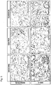
- the antibody against HNF-1A specifically stained the nuclei of tumor cells of the exocrine-like subtype.
- keratin 81 expression was exclusively detectable in tumors of the quasi-mesenchymal subtype. No specific marker could be identified for the classical type that could later be shown to be predictive when analyzing patient samples.
- PDACs of the classical subtype can be characterized by the absence of keratin 81 and HNF-1A expression.
- HNF-1A H-205, Santa Cruz
- keratin 81 36-Z, Santa Cruz
- Vimentin Vimentin
- SRC-SP sensitivity predictor signature
- a FDR cut-off value of 0.200 was determined to be optimal for accurate classification of samples into either Src-inhibitor sensitive or resistant. Samples resulting in a FDR ⁇ 0.200 are predicted to be sensitive, while a FDR > 0.200 predicts resistance.
- tumor cells Only tumor cells are considered in the evaluation of the staining for subtype assignment. 500 tumor cells are evaluated in each specimen. A signal is considered positive if the observed signal can be clearly distinguished from the background staining observed with an isotype control antibody on a comparable specimen. The specimen is considered positive for keratin 81 if least one tumor cell shows a clearly detectable intracellular signal. The specimen is considered positive for HNF-1A if at least one of the tumor cells shows a clearly detectable nuclear staining. The subtype is then determined as follows:
- Example 5 Correlation of marker expression with patient survival in a tissue microarray
- the tissue microarray was constructed from patients that received partial pancreatoduodenectomy for PDAC between 1991 and 2006 at the Charotti University Hospital Berlin.
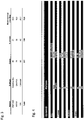
- the use of this tumor cohort for biomarker analysis has been approved by the Charotti University ethics committee (EA1/06/2004). Patient characteristics are summarized in Figure 4 .
- tissue microarrays Formalin-fixed and paraffin-embedded tissue samples were used to generate tissue microarrays as described previously ( Weichert, W., Roske, A., Gekeler, V., Beckers, T., Ebert, M. P., Pross, M., Dietel, M., Denkert, C., and Rocken, C. (2008). Association of patterns of class I histone deacetylase expression with patient prognosis in gastric cancer: a retrospective analysis. The Lancet Oncology 9, 139-148 ). Briefly, three morphologically representative regions of the paraffin 'donor' blocks were chosen.
- Table 1 List of genes contained in the SRC-SP predictor Gene-Symbol Refseq ID Definition IFI44L NM_006820.1 Homo sapiens interferon-induced protein 44-like (IFI44L), mRNA. IFI6 NM_022872.2 Homo sapiens interferon, alpha-inducible protein 6 (IFI6), transcript variant 2, mRNA.
- MX1 NM_002462.2 Homo sapiens myxovirus (influenza virus) resistance 1, interferon-inducible protein p78 (mouse) (MX1), mRNA.
- OAS2 NM_016817.2 Homo sapiens 2'-5'-oligoadenylate synthetase 2, 69/71 kDa (OAS2), transcript variant 1, mRNA.
- IFI44 NM_006417.3 Homo sapiens interferon-induced protein 44 (IF144), mRNA.
- IFIT1 NM_001548.3 Homo sapiens interferon-induced protein with tetratricopeptide repeats 1 (IFIT1), transcript variant 2, mRNA.
- IFIT3 NM_001031683.1 Homo sapiens interferon-induced protein with tetratricopeptide repeats 3 (IFIT3), mRNA.
- IFIT3 NM_001549.2 Homo sapiens interferon-induced protein with tetratricopeptide repeats 3 (IFIT3), mRNA.
- IFI6 NM_022873.2 Homo sapiens interferon, alpha-inducible protein 6 (IFI6), transcript variant 3, mRNA.
- ISG15 NM_005101.1 Homo sapiens ISG15 ubiquitin-like modifier (ISG15), mRNA.
- IFIT2 NM_001547.4 Homo sapiens interferon-induced protein with tetratricopeptide repeats 2 (IFIT2), mRNA.
- OASL NM_198213.1 Homo sapiens 2'-5'-oligoadenylate synthetase-like (OASL), transcript variant 2, mRNA.
- IFI27 NM_005532.3 Homo sapiens interferon, alpha-inducible protein 27 (IF127), transcript variant 2, mRNA.
- EPSTI1 epithelial stromal interaction 1
- APOBEC3G Homo sapiens apolipoprotein B mRNA editing enzyme, catalytic polypeptide-like 3G (APOBEC3G), mRNA.
- HERC5 NM_016323.2 Homo sapiens hect domain and RLD 5 (HERC5), mRNA.
- OAS3 NM_006187.2 Homo sapiens 2'-5'-oligoadenylate synthetase 3, 100kDa (OAS3), mRNA.
- APOBEC3G NM_021822.1 Homo sapiens apolipoprotein B mRNA editing enzyme, catalytic polypeptide-like 3G (APOBEC3G), mRNA.
- CXCL10 NM_001565.2 Homo sapiens chemokine (C-X-C motif) ligand 10 (CXCL10), mRNA.
- XAF1 NM_199139.1 Homo sapiens XIAP associated factor 1 (XAF1), transcript variant 2, mRNA.
- RSAD2 NM_080657.4 Homo sapiens radical S-adenosyl methionine domain containing 2 (RSAD2), mRNA.
- CDKN2B cyclin-dependent kinase inhibitor 2B
- IRF7 NM_004029.2 Homo sapiens interferon regulatory factor 7 (IRF7), transcript variant b, mRNA.
- ITGB2 NM_000211.1 Homo sapiens integrin, beta 2 (antigen CD18 (p95), lymphocyte function-associated antigen 1; macrophage antigen 1 (mac-1) beta subunit) (ITGB2), mRNA.
- SAMD9L NM_152703.2 Homo sapiens sterile alpha motif domain containing 9-like (SAMD9L), mRNA.
- ERAP2 NM_022350.2 Homo sapiens endoplasmic reticulum aminopeptidase 2 (ERAP2), mRNA.
- Table 2 List of HNF-1 target genes that are expressed in Exocrine-like PDAC SLC5A1 MSX2 TINAG FGFR4 MMP11 USP3 ANXA13 TLE4 HABP2 C11ORF9 SFRS7 CREB5 TM4SF4 SGK2 CLDN10 KIF12 GUCA2A PURA RNASE4 SERPINA4 PRODH2 UGT1A1 ALB GRB7 TCEA3 GJB1 AGT CDH17 SLC37A4 TTR SLC4A4 SPINK1 SERPING1 NR5A2 SLC12A2 MLL LGALS2 TBL1X THAP11 NR1H4 SLC1A1 UGT1A6 RBP4 GATA6 ARHGAP12 NPAS2 NDST2 NFE2L2 EML4 FAM20C NEO1 TBXAS1 THRA IGFBP1 PRDM1 CDAN1 LPP GUCA2B PLS3 SLC3A1 YES1 FOX
Landscapes
- Chemical & Material Sciences (AREA)
- Life Sciences & Earth Sciences (AREA)
- Health & Medical Sciences (AREA)
- Engineering & Computer Science (AREA)
- Immunology (AREA)
- Organic Chemistry (AREA)
- Proteomics, Peptides & Aminoacids (AREA)
- Analytical Chemistry (AREA)
- Molecular Biology (AREA)
- Wood Science & Technology (AREA)
- Zoology (AREA)
- Pathology (AREA)
- Genetics & Genomics (AREA)
- Biochemistry (AREA)
- Biotechnology (AREA)
- Physics & Mathematics (AREA)
- Microbiology (AREA)
- General Health & Medical Sciences (AREA)
- Urology & Nephrology (AREA)
- Hematology (AREA)
- Biomedical Technology (AREA)
- Biophysics (AREA)
- Bioinformatics & Cheminformatics (AREA)
- General Engineering & Computer Science (AREA)
- Oncology (AREA)
- Hospice & Palliative Care (AREA)
- Food Science & Technology (AREA)
- General Physics & Mathematics (AREA)
- Cell Biology (AREA)
- Medicinal Chemistry (AREA)
- Measuring Or Testing Involving Enzymes Or Micro-Organisms (AREA)
- Gastroenterology & Hepatology (AREA)
- Investigating Or Analysing Biological Materials (AREA)
Claims (16)
- Procédé in vitro pour la détection spécifique de sous-type de cellules d'adénocarcinome canalaire pancréatique dans un échantillon de tumeur caractérisé par l'étape de détection de l'expression des marqueurs spécifiques de sous-type kératine 81 et HNF-1A, sous forme de protéines ou sous forme d'oligonucléotides codant de telles protéines, où la kératine 81 est le marqueur pour les cellules de PDAC du sous-type quasi-mésenchymateux, où (i) HNF-1A est le marqueur pour les cellules du sous-type analogue à exocrine, et où les cellules de PDAC du sous-type classique sont caractérisées par l'absence de kératine 81 et de HNF-1A.
- Procédé in vitro selon la revendication 1, caractérisé par l'étape de détection de l'expression de marqueurs spécifiques de sous-type, sous forme de protéines ou sous forme d'oligonucléotides codant de telles protéines, où la vimentine est détectée comme marqueur pour les cellules de PDAC du sous-type quasi-mésenchymateux, où (i) un ou plusieurs facteurs de transcription choisis parmi HNF-1B, FOXA2 (HNF3B), FOXA3 (HNF3G), HNF4G et ONECUT1 (HNF6), (ii) un ou plusieurs gènes cibles régulés par HNF-1A choisis parmi SLC5A1, FGFR4, ANXA13, C11ORF9, TM4SF4, KIF12, RNASE4, UGT1A1, TCEA3, CDH17, SLC4A4, NR5A2, LGALS2, NR1H4, UGT1A6, GATA6, NPAS2, NFE2L2, FAM20C, TBXAS1, IGFBP1, CDAN1, GUCA2B, SLC3A1, FOXA2, STAT5B, MSH5, APOM, FXYD2, HPN, SEMA4G, ROBO3, SLC39A14, SLC7A9, NFIX, PDGFRA, RAB3IP, GC, ANPEP, CRB3, MSX2, MMP11, TLE4, SFRS7, SGK2, GUCA2A, SERPINA4, ALB, GJB1, SLC37A4, SPINK1, SLC12A2, TBL1X, SLC1A1, RBP4, ARHGAP12, NDST2, EML4, NEO1, THRA, PRDM1, LPP, PLS3, YES1, AFM, C14ORF138, BACE2, UBE3A, LRRC19, AQP4, LRRFIP2, BPHL, C5, CYFIP2, SLC39A5, AXIN2, TMEM27, SULF2, MIA2, STAG2, TINAG, USP3, HABP2, CREB5, CLDN10, PURA, PRODH2, GRB7, AGT, TTR, SERPING1, MLL et THAP11, et (iii) la cadhérine17 (CDH17) sont détectés comme marqueurs pour les cellules du sous-type analogue à exocrine, et où les cellules de PDAC du sous-type classique sont caractérisées par l'absence de (i) kératine 81 et éventuellement de vimentine, et (ii) de HNF-1A et éventuellement de HNF-1B.
- Procédé in vitro selon la revendication 1 ou 2, où les PDAC du sous-type classique sont identifiés en déterminant l'absence d'expression spécifique d'un ou plusieurs biomarqueurs supplémentaires choisis parmi (i) les facteurs de transcription choisis parmi HNF-1B, FOXA2 (HNF3B), FOXA3 (HNF 3G), HNF4G et ONECUT1 (HNF6), (ii) un ou plusieurs gènes cibles régulés par HNF-1A choisis parmi SLC5A1, FGFR4, ANXA13, C11ORF9, TM4SF4, KIF12, RNASE4, UGT1A1, TCEA3, CDH17, SLC4A4, NR5A2, LGALS2, NR1H4, UGT1A6, GATA6, NPAS2, NFE2L2, FAM20C, TBXAS1, IGFBP1, CDAN1, GUCA2B, SLC3A1, FOXA2, STAT5B, MSH5, APOM, FXYD2, HPN, SEMA4G, ROBO3, SLC39A14, SLC7A9, NFIX, PDGFRA, RAB3IP, GC, ANPEP, CRB3, MSX2, MMP11, TLE4, SFRS7, SGK2, GUCA2A, SERPINA4, ALB, GJB1, SLC37A4, SPINK1, SLC12A2, TBL1X, SLC1A1, RBP4, ARHGAP12, NDST2, EML4, NEO1, THRA, PRDM1, LPP, PLS3, YES1, AFM, C14ORF138, BACE2, UBE3A, LRRC19, AQP4, LRRFIP2, BPHL, C5, CYFIP2, SLC39A5, AXIN2, TMEM27, SULF2, MIA2, STAG2, TINAG, USP3, HABP2, CREB5, CLDN10, PURA, PRODH2, GRB7, AGT, TTR, SERPING1, MLL et THAP11, et (iii) la cadhérine 17 (CDH17).
- Procédé in vitro selon l'une quelconque des revendications 1 à 3, où ladite détection est basée sur la liaison d'un anticorps de détection à un épitope de l'un desdits marqueurs spécifiques de sous-type.
- Procédé in vitro selon la revendication 4, comprenant les étapes de :(a) mise en contact dudit échantillon avec un anticorps de détection spécifique de l'un desdits marqueurs spécifiques de sous-type dans des conditions qui permettent la liaison de l'anticorps de détection audit épitope ;(b) retrait des molécules d'anticorps de détection en excès ; et(c) détection d'anticorps de détection lié dans ledit échantillon.
- Procédé in vitro selon la revendication 4 ou 5, où l'anticorps de détection est un anticorps monoclonal, un anticorps polyclonal, un anticorps produit de manière recombinante, incluant un anticorps chimérique ou humanisé produit de manière recombinante, un anticorps totalement synthétique, ou un fragment fonctionnel de l'un desdits anticorps.
- Procédé in vitro selon l'une quelconque des revendications 4 à 6, où la liaison d'un anticorps de détection est détectée par incubation avec un anticorps secondaire, en particulier un anticorps secondaire étiqueté, plus particulièrement où l'anticorps secondaire est étiqueté avec un molécule marqueur, plus particulièrement où l'anticorps secondaire est étiqueté avec un colorant fluorescent, une étiquette enzymatique biologiquement active, une étiquette radioactive, une étiquette active en résonance magnétique nucléaire, une étiquette luminescente ou une étiquette chromophore, tout particulièrement où l'étiquette radioactive est choisie dans le groupe de 32P,125I, 3H, 14C, 188Rh, 131I 99mTc et 111In.
- Procédé in vitro selon l'une quelconque des revendications 4 à 7, où la liaison d'un anticorps est détectée par coloration immunohistologique.
- Procédé in vitro selon l'une quelconque des revendications 1 à 3, où ladite détection est basée sur la détermination d'ARNm codant l'un desdits marqueurs spécifiques de sous-type, en particulier par réaction en chaîne par polymérase en temps réel quantitative (qRT-PCR).
- Procédé in vitro selon l'une quelconque des revendications 1 à 9, où ledit échantillon contient du tissu pancréatique, en particulier où ledit échantillon est obtenu auprès d'un mammifère, en particulier d'un humain.
- Kit pour la détection spécifique de sous-type de cellules de PDAC comprenant au moins deux réactifs spécifiques des marqueurs spécifiques de sous-type kératine 81 et HNF-1A et éventuellement un ou plusieurs réactifs spécifiques de marqueurs spécifiques de sous-type de PDAC choisis dans la liste de: (i) la vimentine, (ii) un ou plusieurs facteurs de transcription choisis parmi HNF-1B, FOXA2 (HNF3B), FOXA3 (HNF3G), HNF4G et ONECUT1 (HNF6), (iii) un ou plusieurs gènes cibles régulés par HNF-1A choisis parmi SLC5A1, FGFR4, ANXA13, C11ORF9, TM4SF4, KIF12, RNASE4, UGT1A1, TCEA3, CDH17, SLC4A4, NR5A2, LGALS2, NR1H4, UGT1A6, GATA6, NPAS2, NFE2L2, FAM20C, TBXAS1, IGFBP1, CDAN1, GUCA2B, SLC3A1, FOXA2, STAT5B, MSH5, APOM, FXYD2, HPN, SEMA4G, ROBO3, SLC39A14, SLC7A9, NFIX, PDGFRA, RAB3IP, GC, ANPEP, CRB3, MSX2, MMP11, TLE4, SFRS7, SGK2, GUCA2A, SERPINA4, ALB, GJB1, SLC37A4, SPINK1, SLC12A2, TBL1X, SLC1A1, RBP4, ARHGAP12, NDST2, EML4, NEO1, THRA, PRDM1, LPP, PLS3, YES1, AFM, C14ORF138, BACE2, UBE3A, LRRC19, AQP4, LRRFIP2, BPHL, C5, CYFIP2, SLC39A5, AXIN2, TMEM27, SULF2, MIA2, STAG2, TINAG, USP3, HABP2, CREB5, CLDN10, PURA, PRODH2, GRB7, AGT, TTR, SERPING1, MLL et THAP11, et (iv) la cadhérine 17 (CDH17), où lesdits réactifs sont choisis dans la liste de : les anticorps de détection et les sondes oligonucléotidiques, et éventuellement comprenant en outre au moins un réactif pour la détection d'au moins l'un desdits réactifs.
- Procédé de stratification de patients souffrant de PDAC en cohortes de traitement, le procédé comprenant les étapes de :(a) détermination in vitro de l'expression et/ou de la présence de kératine 81 comme marqueur pour les cellules de PDAC du sous-type quasi-mésenchymateux et de HNF-1A comme marqueur pour les cellules de PDAC du sous-type analogue à exocrine, et éventuellement de vimentine comme marqueur pour les cellules de PDAC du sous-type quasi-mésenchymateux, et éventuellement d'un ou plusieurs marqueurs pour les cellules de PDAC du sous-type analogue à exocrine choisis parmi (i) un ou plusieurs facteurs de transcription choisis parmi HNF-1B, FOXA2 (HNF3B), FOXA3 (HNF3G), HNF4G et ONECUT1 (HNF6), (ii) un ou plusieurs gènes cibles régulés par HNF-1A choisis parmi SLC5A1, FGFR4, ANXA13, C11ORF9, TM4SF4, KIF12, RNASE4, UGT1A1, TCEA3, CDH17, SLC4A4, NR5A2, LGALS2, NR1H4, UGT1A6, GATA6, NPAS2, NFE2L2, FAM20C, TBXAS1, IGFBP1, CDAN1, GUCA2B, SLC3A1, FOXA2, STAT5B, MSH5, APOM, FXYD2, HPN, SEMA4G, ROBO3, SLC39A14, SLC7A9, NFIX, PDGFRA, RAB3IP, GC, ANPEP, CRB3, MSX2, MMP11, TLE4, SFRS7, SGK2, GUCA2A, SERPINA4, ALB, GJB1, SLC37A4, SPINK1, SLC12A2, TBL1X, SLC1A1, RBP4, ARHGAP12, NDST2, EML4, NEO1, THRA, PRDM1, LPP, PLS3, YES1, AFM, C14ORF138, BACE2, UBE3A, LRRC19, AQP4, LRRFIP2, BPHL, C5, CYFIP2, SLC39A5, AXIN2, TMEM27, SULF2, MIA2, STAG2, TINAG, USP3, HABP2, CREB5, CLDN10, PURA, PRODH2, GRB7, AGT, TTR, SERPING1, MLL et THAP11, et (iii) la cadhérine 17 (CDH17) ;(b) identification du sous-type de PDAC sur la base de l'expression et/ou de la présence des protéines sélectionnées, où les cellules de PDAC du sous-type classique sont caractérisées par l'absence de kératine 81 et de HNF-1A ;(c) stratification de patients atteints de PDAC en cohortes de traitement médicamenteux sur la base du sous-type de PDAC dont ils souffrent.
- Procédé d'évaluation du pronostic de patients souffrant de PDAC, le procédé comprenant les étapes de :(a) détermination in vitro de l'expression et/ou de la présence de kératine 81 comme marqueur pour les cellules de PDAC du sous-type quasi-mésenchymateux et de HNF-1A comme marqueur pour les cellules de PDAC du sous-type analogue à exocrine, et éventuellement de vimentine comme marqueur pour les cellules de PDAC du sous-type quasi-mésenchymateux et éventuellement d'un ou plusieurs marqueurs pour les cellules de PDAC du sous-type analogue à exocrine choisis parmi (i) un ou plusieurs facteurs de transcription choisis parmi HNF-1B, FOXA2 (HNF3B), FOXA3 (HNF3G), HNF4G et ONECUT1 (HNF6), (ii) un ou plusieurs gènes cibles régulés par HNF-1A choisis parmi SLC5A1, FGFR4, ANXA13, C11ORF9, TM4SF4, KIF12, RNASE4, UGT1A1, TCEA3, CDH17, SLC4A4, NR5A2, LGALS2, NR1H4, UGT1A6, GATA6, NPAS2, NFE2L2, FAM20C, TBXAS1, IGFBP1, CDAN1, GUCA2B, SLC3A1, FOXA2, STAT5B, MSH5, APOM, FXYD2, HPN, SEMA4G, ROBO3, SLC39A14, SLC7A9, NFIX, PDGFRA, RAB3IP, GC, ANPEP, CRB3, MSX2, MMP11, TLE4, SFRS7, SGK2, GUCA2A, SERPINA4, ALB, GJB1, SLC37A4, SPINK1, SLC12A2, TBL1X, SLC1A1, RBP4, ARHGAP12, NDST2, EML4, NEO1, THRA, PRDM1, LPP, PLS3, YES1, AFM, C14ORF138, BACE2, UBE3A, LRRC19, AQP4, LRRFIP2, BPHL, C5, CYFIP2, SLC39A5, AXIN2, TMEM27, SULF2, MIA2, STAG2, TINAG, USP3, HABP2, CREB5, CLDN10, PURA, PRODH2, GRB7, AGT, TTR, SERPING1, MLL et THAP11, et (iii) la cadhérine 17 (CDH 17) ;(b) identification du sous-type de PDAC sur la base de l'expression et/ou de la présence des protéines sélectionnées, où les cellules de PDAC du sous-type classique sont caractérisées par l'absence de kératine 81 et de HNF-1A ;(c) stratification de patients atteints de PDAC en cohortes de pronostic sur la base du sous-type de PDAC dont ils souffrent.
- Procédé selon la revendication 12 ou 13, où le niveau d'expression est déterminé au niveau de l'ARNm par réaction en chaîne par polymérase en temps réel quantitative (qRT-PCR), ou bien où le niveau d'expression est déterminé au niveau des protéines sur la base de la détection d'anticorps se liant aux protéines sélectionnées.
- Procédé selon l'une quelconque des revendications 12 à 14 où lesdites cellules sont obtenues par purification de cellules cancéreuses à partir dudit échantillon d'un patient, en particulier où l'étape de purification comprend le tri en flux ou la microdissection par capture laser.
- Procédé selon l'une quelconque des revendications 12 à 15, où ledit échantillon d'un patient est une biopsie de tumeur, en particulier où ladite biopsie de tumeur est obtenue par aspiration avec une aiguille fine.
Priority Applications (1)
| Application Number | Priority Date | Filing Date | Title |
|---|---|---|---|
| EP13780067.8A EP2906953B1 (fr) | 2012-10-12 | 2013-10-14 | Nouveaux biomarqueurs pour le sous-typage d'un adénocarcinome canalaire du pancréas |
Applications Claiming Priority (3)
| Application Number | Priority Date | Filing Date | Title |
|---|---|---|---|
| EP12007128 | 2012-10-12 | ||
| EP13780067.8A EP2906953B1 (fr) | 2012-10-12 | 2013-10-14 | Nouveaux biomarqueurs pour le sous-typage d'un adénocarcinome canalaire du pancréas |
| PCT/EP2013/003085 WO2014056626A1 (fr) | 2012-10-12 | 2013-10-14 | Nouveaux biomarqueurs pour le sous-typage d'un adénocarcinome canalaire du pancréas |
Publications (2)
| Publication Number | Publication Date |
|---|---|
| EP2906953A1 EP2906953A1 (fr) | 2015-08-19 |
| EP2906953B1 true EP2906953B1 (fr) | 2017-12-06 |
Family
ID=47071057
Family Applications (1)
| Application Number | Title | Priority Date | Filing Date |
|---|---|---|---|
| EP13780067.8A Active EP2906953B1 (fr) | 2012-10-12 | 2013-10-14 | Nouveaux biomarqueurs pour le sous-typage d'un adénocarcinome canalaire du pancréas |
Country Status (5)
| Country | Link |
|---|---|
| US (2) | US20150260721A1 (fr) |
| EP (1) | EP2906953B1 (fr) |
| JP (2) | JP2015534648A (fr) |
| CA (1) | CA2887920C (fr) |
| WO (1) | WO2014056626A1 (fr) |
Families Citing this family (5)
| Publication number | Priority date | Publication date | Assignee | Title |
|---|---|---|---|---|
| WO2016061252A1 (fr) * | 2014-10-14 | 2016-04-21 | The University Of North Carolina At Chapel Hill | Procédés et compositions pour le sous-typage pronostique et/ou diagnostique du cancer du pancréas |
| CA3135033A1 (fr) | 2019-04-01 | 2020-10-08 | The University Of North Carolina At Chapel Hill | Sous-typage independant de la purete de tumeurs (purist), plateforme et classificateur d'echantillon unique independant du type d'echantillon pour la prise de decision de traitement dans le cancer du pancreas |
| EP3730941A1 (fr) * | 2019-04-23 | 2020-10-28 | Institut Jean Paoli & Irène Calmettes | Procédé de détermination d'un gradient moléculaire d'agressivité tumorale de référence pour un adénocarcinome canalaire du pancréas |
| WO2022046576A1 (fr) * | 2020-08-22 | 2022-03-03 | The Broad Institute, Inc. | Signatures d'adénocarcinome canalaire pancréatique et leurs utilisations |
| KR20240026431A (ko) * | 2022-08-18 | 2024-02-28 | 주식회사 메드팩토 | 암의 진단, 전이 가능성 또는 예후 예측용 바이오 마커 및 이의 용도 |
Family Cites Families (10)
| Publication number | Priority date | Publication date | Assignee | Title |
|---|---|---|---|---|
| ATE304174T1 (de) * | 2000-06-14 | 2005-09-15 | Univ Texas | Systeme und verfahren zur zellteilbevölkerungsanalyse |
| AU2002342053A1 (en) * | 2001-10-11 | 2003-04-22 | The Johns Hopkins University | Pancreatic cancer diagnosis and therapies |
| EP1380644A1 (fr) * | 2002-07-08 | 2004-01-14 | Kylix B.V. | Utilisation de gènes cible spécifiques de TCF pour identifier des medicaments pour le traitement du cancer, en particulier le cancer colorectal, dans lequel TCF/beta-catenin/WNT signalisation joue un rôle central |
| US8178505B2 (en) * | 2005-01-05 | 2012-05-15 | Sloan-Kettering Institute For Cancer Research | Method of predicting and reducing risk of metastasis of breast cancer to lung |
| US20080026481A1 (en) * | 2006-06-16 | 2008-01-31 | Michael Mitas | Molecular based two-marker assays that predict outcome of adenocarcinoma patients |
| JP5240739B2 (ja) * | 2007-04-13 | 2013-07-17 | オーエスアイ・フアーマシユーテイカルズ・エル・エル・シー | キナーゼ阻害剤に対する抗癌応答を予測する生物学的マーカー |
| WO2010060920A1 (fr) * | 2008-11-27 | 2010-06-03 | INSERM (Institut National de la Santé et de la Recherche Médicale) | Cxcl4l1 en tant que biomarqueur du cancer du pancréas |
| CA2756493C (fr) * | 2009-03-24 | 2019-07-02 | Biocept, Inc. | Dispositifs et procedes de capture et d'analyse de cellules |
| JP2012524280A (ja) * | 2009-04-17 | 2012-10-11 | オーエスアイ・ファーマシューティカルズ,エルエルシー | 上皮成長因子受容体キナーゼ阻害剤に対する抗がん反応を予測するための生物学的マーカー |
| WO2012178087A1 (fr) * | 2011-06-22 | 2012-12-27 | Oncocyte Corporation | Méthodes et compositions pour traitement et diagnostique d'un cancer de la vessie |
-
2013
- 2013-10-14 EP EP13780067.8A patent/EP2906953B1/fr active Active
- 2013-10-14 CA CA2887920A patent/CA2887920C/fr active Active
- 2013-10-14 WO PCT/EP2013/003085 patent/WO2014056626A1/fr not_active Ceased
- 2013-10-14 JP JP2015536020A patent/JP2015534648A/ja active Pending
- 2013-10-14 US US14/434,036 patent/US20150260721A1/en not_active Abandoned
-
2018
- 2018-02-06 JP JP2018019195A patent/JP6683745B2/ja active Active
-
2019
- 2019-05-24 US US16/422,778 patent/US20190317097A1/en not_active Abandoned
Non-Patent Citations (2)
| Title |
|---|
| BIOLOGICAL ABSTRACTS, vol. unknown, 7 September 2011, Philadelphia, PA, US; abstract no. 274, EISEN: "An advanced culture system for pancreatic cancer reveals tumor heterogeneity and facilitates stratification of novel diagnostic and therapeutic targets" * |
| EISEN: "Subtype-specific Models of Pancreatic Ductal Adenocarcinoma Reveal Signaling Pathways That Can Be Exploited Therapeutically", EUROPEAN JOURNAL OF CANCER, vol. 48, no. Suppl. 5, July 2012 (2012-07-01), 22ND BIENNIAL CONGRESS OF THE EUROPEAN-ASSOCIATION-FOR-CANCER-RESEARCH; BARCELONA, SPAIN; JULY 07 -10, 2012, pages S75 * |
Also Published As
| Publication number | Publication date |
|---|---|
| HK1213640A1 (en) | 2016-07-08 |
| CA2887920C (fr) | 2021-03-23 |
| JP2018105876A (ja) | 2018-07-05 |
| WO2014056626A1 (fr) | 2014-04-17 |
| JP6683745B2 (ja) | 2020-04-22 |
| JP2015534648A (ja) | 2015-12-03 |
| US20150260721A1 (en) | 2015-09-17 |
| EP2906953A1 (fr) | 2015-08-19 |
| US20190317097A1 (en) | 2019-10-17 |
| CA2887920A1 (fr) | 2014-04-17 |
Similar Documents
| Publication | Publication Date | Title |
|---|---|---|
| US20190317097A1 (en) | Novel Biomarkers for Sub-Typing Pancreatic Ductal Adenocarcinoma | |
| KR101032607B1 (ko) | 간암 진단용 단백질성 마커 | |
| EP2591363B1 (fr) | Diagnostic et traitement du cancer du sein | |
| JP2016538545A (ja) | 肝臓癌の診断及び予後判定のための材料及び方法 | |
| WO2019010429A1 (fr) | Méthodes permettant de diagnostiquer un cancer du pancréas | |
| CN113683699B (zh) | 预测食管癌对抗erbb3抗体治疗的应答的方法和试剂盒 | |
| WO2010009171A2 (fr) | Activation de src pour déterminer le pronostic du cancer et en tant que cible pour la thérapie du cancer | |
| WO2013082249A2 (fr) | Compositions et procédés pour l'analyse du cancer de la prostate | |
| WO2011000573A1 (fr) | Méthode de diagnostic pour prédire le risque de récurrence d'un cancer sur la base d'isoformes de l'histone macroh2a | |
| JP2014519818A (ja) | 前立腺癌用の予測バイオマーカ | |
| EP2457089A1 (fr) | Méthode de diagnostic du cancer | |
| US20150290193A1 (en) | Novel approaches for individualized therapy of pancreatic ductal adenocarcinoma | |
| KR101200194B1 (ko) | 간암 진단용 마커로서의 socs6의 용도 | |
| JP7114112B2 (ja) | 肺癌の予後バイオマーカー | |
| US20230416836A1 (en) | Method for predicting responsiveness to plk1 inhibition | |
| JP2022502662A (ja) | レンバチニブ及びエベロリムスを含む併用療法のためのバイオマーカー | |
| Bai et al. | Expression profiles of stemness genes in gastrointestinal stromal tumor | |
| US20220235357A1 (en) | Compositions and methods for identifying and inhibiting a pan-cancer cellular transition of adipose-derived stromal cells | |
| HK1213640B (en) | Novel biomarkers for sub-typing pancreatic ductal adenocarcinoma | |
| US20090068171A1 (en) | Protein markers for the detection of thyroid cancer metastasis | |
| CA3214819A1 (fr) | Marqueurs proteiques pour un cancer du sein positif au recepteur des ?strogenes (er) de type luminal a (la) et de type luminal b1 (lb1) | |
| JP2017529074A (ja) | 治療剤に対する抵抗性の診断に使用するためのERCC1アイソフォーム3のmRNAおよび/またはタンパク質ならびにこのmRNAおよび/またはタンパク質を使用する治療剤に対する抵抗性を診断するための方法 | |
| KR102416614B1 (ko) | 방사선 저항성 지표 단백질 및 이의 검출방법 | |
| JP2024012078A (ja) | Her2陽性乳癌治療予後予測用新規バイオマーカーおよびその用途 | |
| CA3214821A1 (fr) | Marqueurs proteiques pour le cancer du sein du type positif aux recepteurs des oestrogenes (er) et du type negatif aux recepteurs des oestrogenes (er) |
Legal Events
| Date | Code | Title | Description |
|---|---|---|---|
| PUAI | Public reference made under article 153(3) epc to a published international application that has entered the european phase |
Free format text: ORIGINAL CODE: 0009012 |
|
| 17P | Request for examination filed |
Effective date: 20150505 |
|
| AK | Designated contracting states |
Kind code of ref document: A1 Designated state(s): AL AT BE BG CH CY CZ DE DK EE ES FI FR GB GR HR HU IE IS IT LI LT LU LV MC MK MT NL NO PL PT RO RS SE SI SK SM TR |
|
| AX | Request for extension of the european patent |
Extension state: BA ME |
|
| DAX | Request for extension of the european patent (deleted) | ||
| REG | Reference to a national code |
Ref country code: HK Ref legal event code: DE Ref document number: 1213640 Country of ref document: HK |
|
| GRAP | Despatch of communication of intention to grant a patent |
Free format text: ORIGINAL CODE: EPIDOSNIGR1 |
|
| INTG | Intention to grant announced |
Effective date: 20170607 |
|
| GRAS | Grant fee paid |
Free format text: ORIGINAL CODE: EPIDOSNIGR3 |
|
| GRAA | (expected) grant |
Free format text: ORIGINAL CODE: 0009210 |
|
| AK | Designated contracting states |
Kind code of ref document: B1 Designated state(s): AL AT BE BG CH CY CZ DE DK EE ES FI FR GB GR HR HU IE IS IT LI LT LU LV MC MK MT NL NO PL PT RO RS SE SI SK SM TR |
|
| REG | Reference to a national code |
Ref country code: GB Ref legal event code: FG4D |
|
| REG | Reference to a national code |
Ref country code: AT Ref legal event code: REF Ref document number: 952849 Country of ref document: AT Kind code of ref document: T Effective date: 20171215 Ref country code: CH Ref legal event code: EP |
|
| REG | Reference to a national code |
Ref country code: IE Ref legal event code: FG4D |
|
| REG | Reference to a national code |
Ref country code: DE Ref legal event code: R096 Ref document number: 602013030482 Country of ref document: DE |
|
| REG | Reference to a national code |
Ref country code: CH Ref legal event code: NV Representative=s name: CRONIN INTELLECTUAL PROPERTY, CH |
|
| REG | Reference to a national code |
Ref country code: NL Ref legal event code: MP Effective date: 20171206 |
|
| REG | Reference to a national code |
Ref country code: LT Ref legal event code: MG4D |
|
| PG25 | Lapsed in a contracting state [announced via postgrant information from national office to epo] |
Ref country code: ES Free format text: LAPSE BECAUSE OF FAILURE TO SUBMIT A TRANSLATION OF THE DESCRIPTION OR TO PAY THE FEE WITHIN THE PRESCRIBED TIME-LIMIT Effective date: 20171206 Ref country code: LT Free format text: LAPSE BECAUSE OF FAILURE TO SUBMIT A TRANSLATION OF THE DESCRIPTION OR TO PAY THE FEE WITHIN THE PRESCRIBED TIME-LIMIT Effective date: 20171206 Ref country code: FI Free format text: LAPSE BECAUSE OF FAILURE TO SUBMIT A TRANSLATION OF THE DESCRIPTION OR TO PAY THE FEE WITHIN THE PRESCRIBED TIME-LIMIT Effective date: 20171206 Ref country code: SE Free format text: LAPSE BECAUSE OF FAILURE TO SUBMIT A TRANSLATION OF THE DESCRIPTION OR TO PAY THE FEE WITHIN THE PRESCRIBED TIME-LIMIT Effective date: 20171206 Ref country code: NO Free format text: LAPSE BECAUSE OF FAILURE TO SUBMIT A TRANSLATION OF THE DESCRIPTION OR TO PAY THE FEE WITHIN THE PRESCRIBED TIME-LIMIT Effective date: 20180306 |
|
| REG | Reference to a national code |
Ref country code: AT Ref legal event code: MK05 Ref document number: 952849 Country of ref document: AT Kind code of ref document: T Effective date: 20171206 Ref country code: CH Ref legal event code: PCAR Free format text: NEW ADDRESS: CHEMIN DE LA VUARPILLIERE 29, 1260 NYON (CH) |
|
| PG25 | Lapsed in a contracting state [announced via postgrant information from national office to epo] |
Ref country code: BG Free format text: LAPSE BECAUSE OF FAILURE TO SUBMIT A TRANSLATION OF THE DESCRIPTION OR TO PAY THE FEE WITHIN THE PRESCRIBED TIME-LIMIT Effective date: 20180306 Ref country code: LV Free format text: LAPSE BECAUSE OF FAILURE TO SUBMIT A TRANSLATION OF THE DESCRIPTION OR TO PAY THE FEE WITHIN THE PRESCRIBED TIME-LIMIT Effective date: 20171206 Ref country code: GR Free format text: LAPSE BECAUSE OF FAILURE TO SUBMIT A TRANSLATION OF THE DESCRIPTION OR TO PAY THE FEE WITHIN THE PRESCRIBED TIME-LIMIT Effective date: 20180307 Ref country code: RS Free format text: LAPSE BECAUSE OF FAILURE TO SUBMIT A TRANSLATION OF THE DESCRIPTION OR TO PAY THE FEE WITHIN THE PRESCRIBED TIME-LIMIT Effective date: 20171206 Ref country code: HR Free format text: LAPSE BECAUSE OF FAILURE TO SUBMIT A TRANSLATION OF THE DESCRIPTION OR TO PAY THE FEE WITHIN THE PRESCRIBED TIME-LIMIT Effective date: 20171206 |
|
| REG | Reference to a national code |
Ref country code: HK Ref legal event code: GR Ref document number: 1213640 Country of ref document: HK |
|
| PG25 | Lapsed in a contracting state [announced via postgrant information from national office to epo] |
Ref country code: NL Free format text: LAPSE BECAUSE OF FAILURE TO SUBMIT A TRANSLATION OF THE DESCRIPTION OR TO PAY THE FEE WITHIN THE PRESCRIBED TIME-LIMIT Effective date: 20171206 |
|
| PG25 | Lapsed in a contracting state [announced via postgrant information from national office to epo] |
Ref country code: EE Free format text: LAPSE BECAUSE OF FAILURE TO SUBMIT A TRANSLATION OF THE DESCRIPTION OR TO PAY THE FEE WITHIN THE PRESCRIBED TIME-LIMIT Effective date: 20171206 Ref country code: CZ Free format text: LAPSE BECAUSE OF FAILURE TO SUBMIT A TRANSLATION OF THE DESCRIPTION OR TO PAY THE FEE WITHIN THE PRESCRIBED TIME-LIMIT Effective date: 20171206 Ref country code: SK Free format text: LAPSE BECAUSE OF FAILURE TO SUBMIT A TRANSLATION OF THE DESCRIPTION OR TO PAY THE FEE WITHIN THE PRESCRIBED TIME-LIMIT Effective date: 20171206 |
|
| PG25 | Lapsed in a contracting state [announced via postgrant information from national office to epo] |
Ref country code: AT Free format text: LAPSE BECAUSE OF FAILURE TO SUBMIT A TRANSLATION OF THE DESCRIPTION OR TO PAY THE FEE WITHIN THE PRESCRIBED TIME-LIMIT Effective date: 20171206 Ref country code: PL Free format text: LAPSE BECAUSE OF FAILURE TO SUBMIT A TRANSLATION OF THE DESCRIPTION OR TO PAY THE FEE WITHIN THE PRESCRIBED TIME-LIMIT Effective date: 20171206 Ref country code: SM Free format text: LAPSE BECAUSE OF FAILURE TO SUBMIT A TRANSLATION OF THE DESCRIPTION OR TO PAY THE FEE WITHIN THE PRESCRIBED TIME-LIMIT Effective date: 20171206 Ref country code: RO Free format text: LAPSE BECAUSE OF FAILURE TO SUBMIT A TRANSLATION OF THE DESCRIPTION OR TO PAY THE FEE WITHIN THE PRESCRIBED TIME-LIMIT Effective date: 20171206 Ref country code: IT Free format text: LAPSE BECAUSE OF FAILURE TO SUBMIT A TRANSLATION OF THE DESCRIPTION OR TO PAY THE FEE WITHIN THE PRESCRIBED TIME-LIMIT Effective date: 20171206 |
|
| REG | Reference to a national code |
Ref country code: DE Ref legal event code: R097 Ref document number: 602013030482 Country of ref document: DE |
|
| PLBE | No opposition filed within time limit |
Free format text: ORIGINAL CODE: 0009261 |
|
| STAA | Information on the status of an ep patent application or granted ep patent |
Free format text: STATUS: NO OPPOSITION FILED WITHIN TIME LIMIT |
|
| REG | Reference to a national code |
Ref country code: FR Ref legal event code: PLFP Year of fee payment: 6 |
|
| 26N | No opposition filed |
Effective date: 20180907 |
|
| PG25 | Lapsed in a contracting state [announced via postgrant information from national office to epo] |
Ref country code: SI Free format text: LAPSE BECAUSE OF FAILURE TO SUBMIT A TRANSLATION OF THE DESCRIPTION OR TO PAY THE FEE WITHIN THE PRESCRIBED TIME-LIMIT Effective date: 20171206 Ref country code: DK Free format text: LAPSE BECAUSE OF FAILURE TO SUBMIT A TRANSLATION OF THE DESCRIPTION OR TO PAY THE FEE WITHIN THE PRESCRIBED TIME-LIMIT Effective date: 20171206 |
|
| REG | Reference to a national code |
Ref country code: BE Ref legal event code: MM Effective date: 20181031 |
|
| PG25 | Lapsed in a contracting state [announced via postgrant information from national office to epo] |
Ref country code: MC Free format text: LAPSE BECAUSE OF FAILURE TO SUBMIT A TRANSLATION OF THE DESCRIPTION OR TO PAY THE FEE WITHIN THE PRESCRIBED TIME-LIMIT Effective date: 20171206 Ref country code: LU Free format text: LAPSE BECAUSE OF NON-PAYMENT OF DUE FEES Effective date: 20181014 |
|
| REG | Reference to a national code |
Ref country code: IE Ref legal event code: MM4A |
|
| PG25 | Lapsed in a contracting state [announced via postgrant information from national office to epo] |
Ref country code: BE Free format text: LAPSE BECAUSE OF NON-PAYMENT OF DUE FEES Effective date: 20181031 |
|
| PG25 | Lapsed in a contracting state [announced via postgrant information from national office to epo] |
Ref country code: IE Free format text: LAPSE BECAUSE OF NON-PAYMENT OF DUE FEES Effective date: 20181014 |
|
| PG25 | Lapsed in a contracting state [announced via postgrant information from national office to epo] |
Ref country code: MT Free format text: LAPSE BECAUSE OF NON-PAYMENT OF DUE FEES Effective date: 20181014 |
|
| PG25 | Lapsed in a contracting state [announced via postgrant information from national office to epo] |
Ref country code: TR Free format text: LAPSE BECAUSE OF FAILURE TO SUBMIT A TRANSLATION OF THE DESCRIPTION OR TO PAY THE FEE WITHIN THE PRESCRIBED TIME-LIMIT Effective date: 20171206 |
|
| PG25 | Lapsed in a contracting state [announced via postgrant information from national office to epo] |
Ref country code: PT Free format text: LAPSE BECAUSE OF FAILURE TO SUBMIT A TRANSLATION OF THE DESCRIPTION OR TO PAY THE FEE WITHIN THE PRESCRIBED TIME-LIMIT Effective date: 20171206 |
|
| PG25 | Lapsed in a contracting state [announced via postgrant information from national office to epo] |
Ref country code: HU Free format text: LAPSE BECAUSE OF FAILURE TO SUBMIT A TRANSLATION OF THE DESCRIPTION OR TO PAY THE FEE WITHIN THE PRESCRIBED TIME-LIMIT; INVALID AB INITIO Effective date: 20131014 Ref country code: MK Free format text: LAPSE BECAUSE OF NON-PAYMENT OF DUE FEES Effective date: 20171206 Ref country code: CY Free format text: LAPSE BECAUSE OF FAILURE TO SUBMIT A TRANSLATION OF THE DESCRIPTION OR TO PAY THE FEE WITHIN THE PRESCRIBED TIME-LIMIT Effective date: 20171206 |
|
| PG25 | Lapsed in a contracting state [announced via postgrant information from national office to epo] |
Ref country code: AL Free format text: LAPSE BECAUSE OF FAILURE TO SUBMIT A TRANSLATION OF THE DESCRIPTION OR TO PAY THE FEE WITHIN THE PRESCRIBED TIME-LIMIT Effective date: 20171206 Ref country code: IS Free format text: LAPSE BECAUSE OF FAILURE TO SUBMIT A TRANSLATION OF THE DESCRIPTION OR TO PAY THE FEE WITHIN THE PRESCRIBED TIME-LIMIT Effective date: 20180406 |
|
| REG | Reference to a national code |
Ref country code: CH Ref legal event code: U11 Free format text: ST27 STATUS EVENT CODE: U-0-0-U10-U11 (AS PROVIDED BY THE NATIONAL OFFICE) Effective date: 20251101 |
|
| REG | Reference to a national code |
Ref country code: DE Ref legal event code: R079 Ref document number: 602013030482 Country of ref document: DE Free format text: PREVIOUS MAIN CLASS: G01N0033574000 Ipc: G01N0033575000 |
|
| PGFP | Annual fee paid to national office [announced via postgrant information from national office to epo] |
Ref country code: DE Payment date: 20251020 Year of fee payment: 13 |
|
| PGFP | Annual fee paid to national office [announced via postgrant information from national office to epo] |
Ref country code: GB Payment date: 20251024 Year of fee payment: 13 |
|
| PGFP | Annual fee paid to national office [announced via postgrant information from national office to epo] |
Ref country code: FR Payment date: 20251029 Year of fee payment: 13 |
|
| PGFP | Annual fee paid to national office [announced via postgrant information from national office to epo] |
Ref country code: CH Payment date: 20251101 Year of fee payment: 13 |