EP2907452B1 - Fotosteuerungsvorrichtung, fotografisches system, fotosteuerungsverfahren und programm - Google Patents
Fotosteuerungsvorrichtung, fotografisches system, fotosteuerungsverfahren und programm Download PDFInfo
- Publication number
- EP2907452B1 EP2907452B1 EP13845852.6A EP13845852A EP2907452B1 EP 2907452 B1 EP2907452 B1 EP 2907452B1 EP 13845852 A EP13845852 A EP 13845852A EP 2907452 B1 EP2907452 B1 EP 2907452B1
- Authority
- EP
- European Patent Office
- Prior art keywords
- imager
- imaging
- imagers
- imaging timing
- section
- Prior art date
- Legal status (The legal status is an assumption and is not a legal conclusion. Google has not performed a legal analysis and makes no representation as to the accuracy of the status listed.)
- Active
Links
Images






Classifications
-
- A—HUMAN NECESSITIES
- A61—MEDICAL OR VETERINARY SCIENCE; HYGIENE
- A61B—DIAGNOSIS; SURGERY; IDENTIFICATION
- A61B6/00—Apparatus or devices for radiation diagnosis; Apparatus or devices for radiation diagnosis combined with radiation therapy equipment
- A61B6/02—Arrangements for diagnosis sequentially in different planes; Stereoscopic radiation diagnosis
- A61B6/03—Computed tomography [CT]
- A61B6/032—Transmission computed tomography [CT]
-
- A—HUMAN NECESSITIES
- A61—MEDICAL OR VETERINARY SCIENCE; HYGIENE
- A61B—DIAGNOSIS; SURGERY; IDENTIFICATION
- A61B6/00—Apparatus or devices for radiation diagnosis; Apparatus or devices for radiation diagnosis combined with radiation therapy equipment
- A61B6/40—Arrangements for generating radiation specially adapted for radiation diagnosis
- A61B6/4007—Arrangements for generating radiation specially adapted for radiation diagnosis characterised by using a plurality of source units
- A61B6/4014—Arrangements for generating radiation specially adapted for radiation diagnosis characterised by using a plurality of source units arranged in multiple source-detector units
-
- A—HUMAN NECESSITIES
- A61—MEDICAL OR VETERINARY SCIENCE; HYGIENE
- A61B—DIAGNOSIS; SURGERY; IDENTIFICATION
- A61B6/00—Apparatus or devices for radiation diagnosis; Apparatus or devices for radiation diagnosis combined with radiation therapy equipment
- A61B6/40—Arrangements for generating radiation specially adapted for radiation diagnosis
- A61B6/4064—Arrangements for generating radiation specially adapted for radiation diagnosis specially adapted for producing a particular type of beam
- A61B6/4085—Cone-beams
-
- A—HUMAN NECESSITIES
- A61—MEDICAL OR VETERINARY SCIENCE; HYGIENE
- A61B—DIAGNOSIS; SURGERY; IDENTIFICATION
- A61B6/00—Apparatus or devices for radiation diagnosis; Apparatus or devices for radiation diagnosis combined with radiation therapy equipment
- A61B6/54—Control of apparatus or devices for radiation diagnosis
- A61B6/547—Control of apparatus or devices for radiation diagnosis involving tracking of position of the device or parts of the device
-
- A—HUMAN NECESSITIES
- A61—MEDICAL OR VETERINARY SCIENCE; HYGIENE
- A61N—ELECTROTHERAPY; MAGNETOTHERAPY; RADIATION THERAPY; ULTRASOUND THERAPY
- A61N5/00—Radiation therapy
- A61N5/10—X-ray therapy; Gamma-ray therapy; Particle-irradiation therapy
- A61N5/1048—Monitoring, verifying, controlling systems and methods
- A61N5/1049—Monitoring, verifying, controlling systems and methods for verifying the position of the patient with respect to the radiation beam
-
- A—HUMAN NECESSITIES
- A61—MEDICAL OR VETERINARY SCIENCE; HYGIENE
- A61B—DIAGNOSIS; SURGERY; IDENTIFICATION
- A61B6/00—Apparatus or devices for radiation diagnosis; Apparatus or devices for radiation diagnosis combined with radiation therapy equipment
- A61B6/48—Diagnostic techniques
- A61B6/486—Diagnostic techniques involving generating temporal series of image data
-
- A—HUMAN NECESSITIES
- A61—MEDICAL OR VETERINARY SCIENCE; HYGIENE
- A61B—DIAGNOSIS; SURGERY; IDENTIFICATION
- A61B6/00—Apparatus or devices for radiation diagnosis; Apparatus or devices for radiation diagnosis combined with radiation therapy equipment
- A61B6/58—Testing, adjusting or calibrating thereof
- A61B6/582—Calibration
-
- A—HUMAN NECESSITIES
- A61—MEDICAL OR VETERINARY SCIENCE; HYGIENE
- A61N—ELECTROTHERAPY; MAGNETOTHERAPY; RADIATION THERAPY; ULTRASOUND THERAPY
- A61N5/00—Radiation therapy
- A61N5/10—X-ray therapy; Gamma-ray therapy; Particle-irradiation therapy
- A61N5/1048—Monitoring, verifying, controlling systems and methods
- A61N5/1049—Monitoring, verifying, controlling systems and methods for verifying the position of the patient with respect to the radiation beam
- A61N2005/1061—Monitoring, verifying, controlling systems and methods for verifying the position of the patient with respect to the radiation beam using an x-ray imaging system having a separate imaging source
-
- A—HUMAN NECESSITIES
- A61—MEDICAL OR VETERINARY SCIENCE; HYGIENE
- A61N—ELECTROTHERAPY; MAGNETOTHERAPY; RADIATION THERAPY; ULTRASOUND THERAPY
- A61N5/00—Radiation therapy
- A61N5/10—X-ray therapy; Gamma-ray therapy; Particle-irradiation therapy
- A61N5/1077—Beam delivery systems
- A61N5/1081—Rotating beam systems with a specific mechanical construction, e.g. gantries
Definitions
- the present invention relates to an imaging controller, an imaging system, an imaging control method, and a program.
- cone beam computed tomography In cone beam computed tomography (cone beam CT; CBCT), an imager in which an X-ray source and a two-dimensional detector form a pair captures a two-dimensional radiographic image by emitting cone-shaped X-rays from the X-ray source to the two-dimensional detector while rotating around an imaging target to change the irradiation angle.
- cone-shaped X-rays in the cone beam CT, it is possible to generate (reconstruct) a CT image (tomographic image) by capturing a two-dimensional radiographic image without the need to rotate the imaging target during imaging and without the need for multiple rotations.
- a CBCT for keeping the relatively small rotation angle of each imager by using a plurality of imagers, such as a dual-source CBCT using two pairs of imagers (for example, WO2010/073308 .
- the x-ray system comprises a control device, a data storage device and two x-ray units.
- Each x-ray unit is provided with an x-ray detector which can be pivoted around a pivot axis.
- the two x-ray sources are arranged opposite one another in relation to the pivot axis.
- An object can be arranged in the area of the pivot axis.
- the control device controls the x-ray units so that they are simultaneously pivoted around the pivot axis.
- the images of the object are recorded by means of the x-ray detectors at angular positions and conveyed to the data storage device.
- the pivot angle and the angular positions are determined such that, on the basis of the recorded images of the object, at least one 3D reconstruction of the object can be determined.
- US 2005/0286679 describes an x-ray diagnostic apparatus and x-ray imaging method.
- Projection data are obtained using a method for imaging by rotating the first imaging system and the second imaging system, both being provided with an x-ray generating unit.
- the obtained projection data are subjected to a reconstruction process to generate x-ray image data.
- a relative angle formed between the imaging systems is setup according to a palmic period of a subject and a rotation velocity of the imaging systems. In such a way, the projection data from more directions at a predetermined time-phase are collected without overlapping.
- US 2012/199760 A1 discloses a radiotherapy device controller comprising a visual line calculator configured to calculate a visual line based on a position on a transmissive image, imaged using a radiation penetrating a subject.
- a position calculator is configured to calculate a position of a specific part of the subject being shown at a position.
- a position calculator is configured to calculate a position of the specific part based on a position of a trajectory of the specific part and a position of the visual line.
- An emitter controls a radiation emitter which emits a therapeutic radiation, so that the therapeutic radiation penetrates at the position of the specific part.
- JP 2011050416 describes a radiation tomography device and method which represents the closest prior art.
- the radiation dose can be reduced in a CBCT using a plurality of imagers, such as a dual-source CBCT, it is possible to reduce the burden on the imaging target.
- the present invention provides an imaging controller, an imaging system, an imaging control method, and a program that can reduce the radiation dose.
- An imaging controller is an imaging controller of an imaging apparatus which includes first and second imagers for capturing a radiographic image by emitting a cone beam toward a rotation axis and a rotation mechanism for rotating the first and second imagers integrally around the rotation axis and in which an angle between an irradiation axis of the first imager and an irradiation axis of the second imager with the rotation axis as a reference is a predetermined angle.
- the imaging controller includes: an imager position determination section that determines whether or not the first imager is located in an overlapping region where a rotation range of the first imager and a rotation range of the second imager overlap each other when the rotation mechanism rotates the first and second imagers by an angle greater than the predetermined angle; and an imaging timing control section that causes one or both of the first and second imagers to perform imaging when arrival of an imaging timing is detected and that causes only the second imager to perform imaging in at least one imaging timing whose arrival is detected in a state where the imager position determination section determines that the first imager is located in the overlapping region.
- a position specifying section that specifies a three-dimensional position of a position specification target based on a radiographic image captured by the first imager and a radiographic image captured by the second imager is included.
- an imaging controller in the imaging controller described above, when the imaging timing control section detects arrival of an imaging timing and causes both of the first and second imagers to perform imaging and the position specifying section succeeds in specifying the three-dimensional position of the position specification target based on the obtained radiographic images, if arrival of a next imaging timing is detected in a state where the imager position determination section determines that the first imager is located in the overlapping region, the imaging timing control section causes only the second imager to perform imaging.
- a position specifying section that specifies a three-dimensional position of a position specification target based on a radiographic image captured by the first imager and a radiographic image captured by the second imager is included.
- the imaging timing control section detects arrival of an imaging timing and causes both of the first and second imagers to perform imaging and the position specifying section fails to detect an image of the position specification target in at least one of the obtained radiographic images, if arrival of a next imaging timing is detected in a state where the imager position determination section determines that the first imager is located in the overlapping region, the imaging timing control section causes only the second imager to perform imaging.
- the imaging timing control section causes only the second imager to perform imaging at all imaging timings whose arrival is detected in a state where the imager position determination section determines that the first imager is located in the overlapping region.
- an imaging system includes: an imaging apparatus which includes first and second imagers for capturing a radiographic image by emitting a cone beam toward a rotation axis and a rotation mechanism for rotating the first and second imagers integrally around the rotation axis and in which an angle between an irradiation axis of the first imager and an irradiation axis of the second imager with the rotation axis as a reference is a predetermined angle; and an imaging controller configured to include an imager position determination section that determines whether or not the first imager is located in an overlapping region where a rotation range of the first imager and a rotation range of the second imager overlap each other when the rotation mechanism rotates the first and second imagers by an angle greater than the predetermined angle and an imaging timing control section that causes one or both of the first and second imagers to perform imaging when arrival of an imaging timing is detected and that causes only the second imager to perform imaging in at least one imaging timing whose arrival is detected in a state
- an imaging control method is an imaging control method of an imaging controller of an imaging apparatus which includes first and second imagers for capturing a radiographic image by emitting a cone beam toward a rotation axis and a rotation mechanism for rotating the first and second imagers integrally around the rotation axis and in which an angle between an irradiation axis of the first imager and an irradiation axis of the second imager with the rotation axis as a reference is a predetermined angle.
- the imaging control method includes: an imager position determination step of determining whether or not the first imager is located in an overlapping region where a rotation range of the first imager and a rotation range of the second imager overlap each other when the rotation mechanism rotates the first and second imagers by an angle greater than the predetermined angle; and an imaging timing control step of causing one or both of the first and second imagers to perform imaging when arrival of an imaging timing is detected and causing only the second imager to perform imaging in at least one imaging timing whose arrival is detected in a state where it is determined that the first imager is located in the overlapping region in the imager position determination step.
- a program is a program causing a computer as an imaging controller of an imaging apparatus, which includes first and second imagers for capturing a radiographic image by emitting a cone beam toward a rotation axis and a rotation mechanism for rotating the first and second imagers integrally around the rotation axis and in which an angle between an irradiation axis of the first imager and an irradiation axis of the second imager with the rotation axis as a reference is a predetermined angle, to execute: an imager position determination step of determining whether or not the first imager is located in an overlapping region where a rotation range of the first imager and a rotation range of the second imager overlap each other when the rotation mechanism rotates the first and second imagers by an angle greater than the predetermined angle; and an imaging timing control step of causing one or both of the first and second imagers to perform imaging when arrival of an imaging timing is detected and causing only the second imager to perform imaging in at least one
- the imaging controller According to the imaging controller, the imaging system, the imaging control method, and the program described above, it is possible to reduce the radiation dose.
- Fig. 1 is a schematic block diagram showing the functional configuration of an imaging system in an embodiment of the present invention.
- a radiotherapy system 1 includes a radiotherapy apparatus controller 2 and a radiotherapy apparatus 3.
- the radiotherapy system 1 performs the emission (emission of radiation toward a certain target, such as an affected part) of radiation (may be a heavy particle beam) in radiation therapy or the generation of a CT image (tomographic image) for treatment planning.
- the radiotherapy system 1 corresponds to an example of an imaging system in the present embodiment.
- the radiotherapy apparatus 3 emits radiation to the affected part in radiation therapy.
- the radiotherapy apparatus 3 captures a radiographic image (X-ray radiographic image) in order to generate a CT image.
- the radiotherapy apparatus 3 corresponds to an example of an imaging apparatus in the present embodiment.
- the radiotherapy apparatus controller 2 controls the radiotherapy apparatus 3 to emit radiation and capture a radiographic image.
- the radiotherapy apparatus controller 2 generates a CT image based on the radiographic image captured by the radiotherapy apparatus 3.
- the radiotherapy apparatus controller 2 corresponds to an example of an imaging controller in the present embodiment.
- Fig. 2 is a schematic diagram showing the apparatus configuration of the radiotherapy apparatus 3.
- the radiotherapy apparatus 3 includes a rotary driving unit 311, an O ring 312, a traveling gantry 313, a swing mechanism 321, an irradiation unit 330, a sensor array 351, a sensor array 361, a sensor array 362, a couch 381, and a couch driving unit 382.
- the irradiation unit 330 includes a therapeutic radiation emission unit 331, a multi-leaf collimator (MLC) 332, a diagnostic X-ray source 341, and a diagnostic X-ray source 342.
- MLC multi-leaf collimator
- the diagnostic X-ray source 341 and diagnostic X-ray source 342 will be referred to collectively as a “diagnostic X-ray source 340" hereinafter.
- the sensor array 361 and the sensor array 362 will be referred to collectively as a “sensor array 360” hereinafter.
- the rotary driving unit 311 supports the O ring 312 on a base so as to be able to rotate around a rotation axis A11, and rotates the O ring 312 according to the control of the radiotherapy apparatus controller 2.
- the rotation axis A11 is an axis in a vertical direction.
- the O ring 312 is formed in a ring shape having a rotation axis A12 at the center, and supports the traveling gantry 313 so as to be able to rotate around the rotation axis A12.
- the rotation axis A12 is an axis in a horizontal axis (that is, an axis perpendicular to the vertical direction), and is perpendicular to the rotation axis A11 at the isocenter P11.
- the rotation axis A12 is fixed to the O ring 312. That is, the rotation axis A12 rotates around the rotation axis A11 with the rotation of the O ring 312.
- the traveling gantry 313 is formed in a ring shape having the rotation axis A12 at the center, and is disposed inside the O ring 312 so as to be concentric with the O ring 312.
- the radiotherapy apparatus 3 further includes a traveling driving device (not shown), and the traveling gantry 313 rotates around the rotation axis A12 with power from the traveling driving device.
- the traveling gantry 313 itself is rotated to integrally rotate each unit provided in the traveling gantry 313, such as the diagnostic X-ray source 341 and the sensor array 361 or the diagnostic X-ray source 342 and the sensor array 362.
- the traveling gantry 313 corresponds to an example of a rotation mechanism in the present embodiment.
- the swing mechanism 321 is fixed inside the ring of the traveling gantry 313, and supports the irradiation unit 330 on the traveling gantry 313.
- the swing mechanism 321 rotates the irradiation unit 330 around a pan axis A21 and rotates the irradiation unit 330 around a tilt axis A22 according to the control of the radiotherapy apparatus controller 2.
- the pan axis A21 is an axis parallel to the rotation axis A12, and is fixed to the traveling gantry 313.
- the swing mechanism 321 causes the irradiation unit 330 to perform a swinging operation to the left and right with respect to the rotation axis A12 (accordingly, to the left and right with respect to an imaging target T11 shown in Fig. 2 ) by rotating the irradiation unit 330 around the pan axis A21.
- the tilt axis A22 is an axis perpendicular to the pan axis A21, and is fixed to the traveling gantry 313.
- the swing mechanism 321 causes the irradiation unit 330 to perform a swinging operation in a direction of the rotation axis A12 (accordingly, up and down with respect to the imaging target T11) by rotating the irradiation unit 330 around the tilt axis A22.
- the irradiation unit 330 is disposed inside the traveling gantry 313 so as to be supported by the swing mechanism 321, and emits therapeutic radiation B11 or a diagnostic X-ray B21 or B22.
- the therapeutic radiation emission unit 331 emits the therapeutic radiation B11 according to the control of the radiotherapy apparatus controller 2.
- the therapeutic radiation emission unit 331 is supported by the traveling gantry 313 through the swing mechanism 321. For this reason, once the therapeutic radiation emission unit 331 is directed toward the isocenter P11 by adjustment of the swing mechanism 321, the therapeutic radiation B11 always passes through the isocenter P11 in general even if the O ring 312 is rotated by the rotary driving unit 311 and even if the traveling gantry 313 is rotated by the traveling driving device. Therefore, the therapeutic radiation emission unit 331 can emit the therapeutic radiation B11 toward the isocenter P11 from various directions by rotating around the rotation axis All or the rotation axis A12.
- the swing mechanism 321 corrects the direction of the therapeutic radiation emission unit 331. Accordingly, it is possible to perform high-accuracy positioning.
- the multi-leaf collimator 332 matches the shape of the radiation field when the therapeutic radiation B11 is emitted to the patient with the shape of the affected part by shielding a part of the therapeutic radiation B11 according to the control of the radiotherapy apparatus controller 2.
- the diagnostic X-ray source 341 emits the diagnostic X-ray B21 toward the isocenter P11 according to the control of the radiotherapy apparatus controller 2.
- the diagnostic X-ray B21 is a cone beam that is emitted from one point of the diagnostic X-ray source 341 and that has a conical shape, such as a cone or a pyramid, with the one point as an apex.
- the diagnostic X-ray source 342 emits the diagnostic X-ray B22 toward the isocenter P11 according to the control of the radiotherapy apparatus controller 2.
- the diagnostic X-ray B22 is a cone beam that is emitted from one point of the diagnostic X-ray source 342 and that has a conical shape, such as a cone or a pyramid, with the one point as an apex.
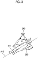
- Fig. 3 is an explanatory diagram showing an example of the cone beam emitted from the diagnostic X-ray source 340.
- the cone beam emitted from the diagnostic X-ray source 340 spreads in both of the direction perpendicular to the rotation axis A12 (left and right direction of the imaging target T11) and the direction of the rotation axis A12 (up and down direction of the imaging target T11).
- a fan angle which is an angle indicating the spread of a cone beam in a direction perpendicular to the rotation axis A12
- ⁇ a cone angle
- ⁇ which is an angle indicating the spread of a cone beam in a direction of the rotation axis A12
- both of the diagnostic X-rays B21 and B22 are shown as the irradiation axis.
- the irradiation axis of the cone beam referred to herein is the central axis of the cone formed by the cone beam (for example, when the cone beam has a conical shape, the rotation axis of the cone) .
- the diagnostic X-ray sources 341 and 342 are fixed to the irradiation unit 330 (for example, a housing of the multi-leaf collimator 332) in a direction perpendicular to the irradiation axis.
- the irradiation axis of the diagnostic X-ray source 341 and the irradiation axis of the diagnostic X-ray source 342 are perpendicular to each other at the isocenter P11.
- the sensor array 351 is disposed at a position hit by the therapeutic radiation B11 from the therapeutic radiation emission unit 331 so as to face the therapeutic radiation emission unit 331, and is fixed inside the ring of the traveling gantry 313.
- the sensor array 351 receives the therapeutic radiation B11, which is emitted from the therapeutic radiation emission unit 331 and is transmitted through the subject, such as the affected part, and generates (captures) a radiographic image (radiation projection image) of the subject.
- the radiographic image of the subject generated by the sensor array 351 is used when checking the radiation position of the therapeutic radiation B11, when recording the treatment, and the like. Reception referred to herein is receiving the radiation.
- the sensor array 351 it is possible to use various devices that can receive the therapeutic radiation B11 and generate a radiographic image.
- the sensor array 351 may be a flat panel detector (FPD), or may be an X-ray image intensifier (II).
- the sensor array 361 is disposed at a position hit by the diagnostic X-ray B21 from the diagnostic X-ray source 341 so as to face the diagnostic X-ray source 341, and is fixed inside the ring of the traveling gantry 313.
- the sensor array 361 receives the diagnostic X-ray B21, which is emitted from the diagnostic X-ray source 341 and is transmitted through the subject, such as the affected part, and generates a radiographic image of the subject.
- the radiographic image of the subject generated by the sensor array 361 and the radiographic image of the subject generated by the sensor array 362 are used when the radiotherapy apparatus controller 2 generates a CT image.
- the sensor array 361 it is possible to use various devices that can receive the diagnostic X-ray B21 and generate a radiographic image.
- the sensor array 361 may be an FPD, or may be an X-ray II.
- the sensor array 362 is disposed at a position hit by the diagnostic X-ray B22 from the diagnostic X-ray source 342 so as to face the diagnostic X-ray source 342, and is fixed inside the ring of the traveling gantry 313.
- the sensor array 362 receives the diagnostic X-ray B22, which is emitted from the diagnostic X-ray source 342 and is transmitted through the subject, such as the affected part, and generates a radiographic image of the subject.
- the radiographic image of the subject generated by the sensor array 362 and the radiographic image of the subject generated by the sensor array 361 are used when the radiotherapy apparatus controller 2 generates a CT image.
- the sensor array 362 it is possible to use various devices that can receive the diagnostic X-ray B22 and generate a radiographic image.
- the sensor array 362 may be an FPD, or may be an X-ray II.
- the diagnostic X-ray source 341 and the sensor array 361, the diagnostic X-ray source 342 and the sensor array 362, and the therapeutic radiation emission unit 331 and the sensor array 351 rotate around the rotation axis A12 passing through the isocenter P11 while maintaining the positional relationship therebetween.
- the combination of the diagnostic X-ray source 341 and the sensor array 361 is referred to as an "imager 371", and the combination of the diagnostic X-ray source 342 and the sensor array 362 is referred to as an "imager 372".
- Each of the imager 371 and the imager 372 corresponds to an example of an imager in the present embodiment. That is, the diagnostic X-ray source 341 emits the diagnostic X-ray B21 of a cone beam toward the rotation axis A12, and the sensor array 361 captures a radiographic image based on the diagnostic X-ray B21. In addition, the diagnostic X-ray source 342 emits the diagnostic X-ray B22 of a cone beam toward the rotation axis A12, and the sensor array 362 captures a radiographic image based on the diagnostic X-ray B22.
- emitting the cone beam toward the rotation axis is emitting the cone beam so that the cone beam and the rotation axis cross each other.
- the cone beam is emitted so that the irradiation axis of the cone beam and the rotation axis cross each other.
- the embodiment of the present invention is not limited to this.
- the imager 371 is an example of a first imager and the imager 372 is an example of a second imager will be described as an example.
- the embodiment of the present invention is not limited to this.
- the imager 372 may be an example of the first imager, and the imager 371 may be an example of the second imager.
- an angle between the irradiation axis of the imager 371 and the irradiation axis of the imager 372 is fixed at a predetermined angle with the rotation axis A12 as a reference.
- the irradiation axis of the imager referred to herein is the irradiation axis of the cone beam emitted from the imager.
- the swing mechanism 321 sets the direction of the irradiation unit 330 so that the therapeutic radiation B11 passes through the isocenter P11
- the irradiation axis of the diagnostic X-ray B21 and the irradiation axis of the diagnostic X-ray B22 are perpendicular to each other at the isocenter P11 of the rotation axis A12.
- the traveling gantry 313 rotates, the imager 371 (the diagnostic X-ray source 341 and the sensor array 361) and the imager 372 (the diagnostic X-ray source 342 and the sensor array 362) rotate while keeping the irradiation axes perpendicular to each other.
- the traveling gantry 313 rotates the imager 371 and the imager 372 integrally.
- the angle between the diagnostic X-ray B21 and the diagnostic X-ray B22 is not limited to 90°.
- the couch 381 is used for the lying down of the imaging target T11 who is a patient to be treated.
- the couch 381 includes a fixture (not shown). By fixing the imaging target T11 to the couch 381 using a fixed portion, it is possible to reduce subject blur (image blur due to subject movement) when the diagnostic X-ray source 340 emits a diagnostic X-ray to capture a radiographic image or the shift of the radiation position when the therapeutic radiation emission unit 331 emits the therapeutic radiation B11.
- the couch driving unit 382 supports the couch 381 on a base, and moves the couch 381 according to the control of the radiotherapy apparatus controller 2. By moving the couch 381 using the couch driving unit 382, it is possible to locate the affected part of the imaging target T11 at the isocenter P11 or near the isocenter P11.
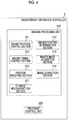
- Fig. 4 is a schematic block diagram showing the functional configuration of the radiotherapy apparatus controller 2.
- the radiotherapy apparatus controller 2 includes an imaging processing unit 210 and a treatment control unit 220.
- the imaging processing unit 210 includes an imager rotation control section 211, an imager position determination section 212, an imaging timing control section 213, an imager image acquisition section 214, a position specifying section 215, an image correction section 216, and a CT image reconstruction section 217.
- the imaging processing unit 210 controls the radiotherapy apparatus 3 ( Fig. 2 ) to acquire a radiographic image of the imaging target T11, and generates a CT image based on the acquired radiographic image.
- the CT image reconstruction section 217 generates (reconstructs) a CT image from the radiographic image that has been captured by the sensor array 360 and has an affected part position corrected by the image correction section 216 as will be described later.
- the affected part position for example, the center-of-gravity position corresponding to the shape (area) of the affected part image can be used.
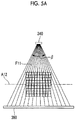
- Figs. 5A to 5C are explanatory diagrams of the reconstruction of a CT image that the CT image reconstruction section 217 performs.
- the cone beam emitted from the diagnostic X-ray source 340 has a spread of the fan angle ⁇ and the cone angle ⁇ . Therefore, the cone beam emitted from the diagnostic X-ray source 340 is projected onto the light receiving surface (surface that receives X-rays) of the sensor array 360 in a two-dimensional manner (for example, in a rectangular area of the light receiving surface of the sensor array 360).
- the light receiving surface of the sensor array 360 is divided into pixels, and the CT image reconstruction section 217 acquires the density information of each pixel as radiographic image data.
- the density information indicates the intensity of the X-ray received by each pixel of the sensor array 360 (value after the correction of the image correction section 216).
- the pixel of the light receiving surface of the sensor array 360 is referred to as a "pixel of the sensor array 360".
- the space of a target whose CT image is to be generated is classified into voxels corresponding to the pixels of the sensor array 360.
- the CT image reconstruction section 217 converts the X-ray intensity of each pixel into the total value of the X-ray transmittance in each voxel through which the X-rays pass.
- back projection the process of converting the X-ray intensity of each pixel into the total value of the X-ray transmittance in each voxel through which the X-rays pass.
- Fig. 5A shows X-rays, which are emitted to the row of pixels of the sensor array 360, as a surface, such as a surface F11.
- the row of pixels referred to herein is the arrangement of pixels in a direction perpendicular to the rotation axis A12 (in the left and right direction of the imaging target T11 shown in Fig. 2 ).
- Fig. 5A shows a state when viewed from the direction perpendicular to the rotation axis A12, each surface is shown by a line.
- Fig. 5B shows X-rays, which are emitted to the respective pixels of the sensor array 360, as lines, such as line L11, in relation to the surface F11.
- Fig. 5C shows voxels through which line L11 passes.
- Line L11 passes through voxels B11, B12, and B13, and the CT image reconstruction section 217 performs back projection of the X-ray intensity in a pixel corresponding to line L11 onto the total value of the X-ray transmittances of the voxels B11, B12, and B13.
- the CT image reconstruction section 217 acquires a radiographic image by projecting X-rays onto the subject (affected part of the imaging target T11) from various angles, and performs back projection for each pixel of the sensor array 360 for each radiographic image.
- the CT image reconstruction section 217 calculates the X-ray transmittance of each voxel based on the obtained total value of the X-ray transmittances from various angles, and generates a CT image based on the obtained X-ray transmittance of each voxel.
- each quantity or the positional relationship shown in Figs. 5A to 5C is an example for explanation, and the present invention is not limited thereto.
- the fan angle ⁇ and the cone angle ⁇ of the cone beam emitted from the diagnostic X-ray source 340, the number of pixels of the sensor array 360, the number of voxels set in the space of the target whose CT image is to be generated, or the positional relationship between the diagnostic X-ray source 340 or the sensor array 360 and the voxels is not limited to those shown in Figs. 5A to 5C .
- the imager rotation control section 211 controls the traveling driving device of the radiotherapy apparatus 3 to rotate the traveling gantry 313, thereby rotating the diagnostic X-ray source 341 and the sensor array 361 or the diagnostic X-ray source 342 and the sensor array 362.
- the imager rotation control section 211 rotates the imager 371 and the imager 372 to locate the subject at various angles.
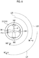
- Fig. 6 is an explanatory diagram showing an example of the angle by which the imager rotation control section 211 rotates an imager.
- Line L21 shown in this diagram indicates the rotation range of the imager 371
- line L22 indicates the rotation range of the imager 372.
- line L23 indicates a combined range of the rotation range of the imager 371 and the rotation range of the imager 372.
- the diagnostic X-ray source 341 and the diagnostic X-ray source 342 are disposed such that the irradiation axes thereof are perpendicular to each other.
- the imager rotation control section 211 controls the traveling driving device of the radiotherapy apparatus 3 to rotate the traveling gantry 313 by 90° or more
- the rotation range of the imager 371 and the rotation range of the imager 372 overlap each other. Therefore, if the rotation range of the imager 371 and the rotation range of the imager 372 are combined, it is possible to rotate the subject in the range of an angle of 180° or more with respect to the subject.
- the imaging time can be reduced to approximately half of that when there is one imager.
- the imager rotation control section 211 rotates the traveling gantry 313 (accordingly, the imager 371 and the imager 372) by 90° + fan angle ⁇ (by an angle obtained by adding the fan angle ⁇ to 90°). Therefore, the traveling gantry 313 rotates the imager 371 and the imager 372 by an angle greater than 90° (angle between the irradiation axis of the imager 371 and the irradiation axis of the imager 372 when the rotation axis A12 is a reference). This will be described with reference to Fig. 7 .
- Fig. 7 is an explanatory diagram showing the angle of an X-ray passing through the end of the fan angle of a cone beam when the rotation range of the imager is 180°.
- a point P21 shown in this diagram is located on line L31 at the end of the fan angle ⁇ of the cone beam, which is emitted from the diagnostic X-ray source 340, when the diagnostic X-ray source 340 starts to rotate.
- the point P21 is located on line L32 at the end of the fan angle ⁇ of the cone beam emitted from the diagnostic X-ray source 340.
- the direction of the X-ray passing through the point P21 changes in a range of an angle obtained by subtracting the fan angle ⁇ from 180°.
- the CT image reconstruction section 217 it is preferable to acquire information (density information of each pixel of the sensor array 360) by emitting X-rays to all voxels from the respective directions in the range of 180° or more.
- the imager rotation control section 211 controls the traveling driving device to rotate the traveling gantry 313 by 90° + fan angle ⁇ . Then, the combined rotation range of the rotation range of the imager 371 and the rotation range of the imager 372 becomes 180° + fan angle ⁇ . Accordingly, the CT image reconstruction section 217 can acquire the information by emitting X-rays to all voxels from the respective directions in the range of 180° or more.
- the imager position determination section 212 acquires the position information of the imager 371 (information indicating the amount of rotation of the imager 371), and determines whether or not the imager 371 is located in an overlapping region where the rotation range of the imager 371 and the rotation range of the imager 372 overlap each other (hereinafter, simply referred to as an "overlapping region").
- the imager position determination section 212 may acquire the control information of the traveling driving device from the imager rotation control section 211, and calculate the rotation angle from the reference position (for example, the position at the start of rotation) of the diagnostic X-ray source 340, as the position information of the imager 371, based on the control information.
- the reference position for example, the position at the start of rotation
- the traveling gantry 313 may measure the rotation angle, and the imager position determination section 212 may acquire the rotation angle as the position information of the imager 371 (rotation angle of the imager 371).
- the imager position determination section 212 determines whether or not the imager 371 is located in the overlapping region by determining whether or not the amount of rotation of the imager 371 is equal to or greater than 90° based on the position information of the imager 371. That is, when the amount of rotation of the imager 371 is equal to or greater than 90°, the imager 371 is located in the overlapping region.
- the imaging timing control section 213 causes one or both of the imager 371 and the imager 372 to perform imaging when detecting the arrival of imaging timing. Specifically, the imaging timing control section 213 outputs control information instructing one or both of the imager 371 and the imager 372 to perform imaging every time set in advance as an imaging period.
- the imaging timing control section 213 causes only the imager 372 to perform imaging in at least one imaging timing whose arrival is detected in a state where the imager position determination section 212 determines that the imager 371 is located in the overlapping region.
- the imaging timing control section 213 causes only the imager 372 to perform imaging at all of the imaging timings whose arrival is detected in a state where the imager position determination section 212 determines that the imager 371 is located in the overlapping region. That is, when the imager position determination section 212 determines that the amount of rotation of the imager 371 is equal to or greater than 90°, the imaging timing control section 213 outputs control information for the instruction of imaging only to the imager 372.
- Fig. 8 is an explanatory diagram of a first example of the control of the imaging timing.
- 13 imaging timings are set while the imager 371 or the imager 372 rotates by 90° + fan angle ⁇ .
- line L41 indicates the rotation range of the diagnostic X-ray source 341
- line L42 indicates the rotation range of the diagnostic X-ray source 342.
- the imaging timing control section 213 instructs the imager 371 and the imager 372 to perform simultaneous imaging.
- the imager 371 is located in the overlapping region at the tenth and subsequent imaging timings. Therefore, even if the imager 371 stops imaging, the CT image reconstruction section 217 can acquire a radiographic image for the 180° + fan angle ⁇ . As a result, it is possible to acquire the information by emitting X-rays to all voxels from the respective directions in the range of 180° or more.
- the imaging timing control section 213 instructs only the imager 372 to perform imaging (one-sided imaging).
- the imaging timing control section 213 instructs only the imager 372 to perform imaging (stops the imaging of the imager 371), thereby being able to reduce the radiation dose.
- the imaging timing control section 213 instructs only the imager 372 to perform imaging (stops the imaging of the imager 371), thereby being able to reduce the radiation dose.
- imaging timing shown in Fig. 8 is an example for explanation, and is not limited thereto.
- the number of imaging timings whose arrival is detected by the imaging timing control section 213 is not limited to 13 times.
- the imaging timing control section 213 may instruct the imager 371 and the imager 372 to perform simultaneous imaging at some imaging timings.
- Fig. 9 is an explanatory diagram of a second example of the control of the imaging timing. Similar to the case shown in Fig. 8 , in the example shown in Fig. 9 , 13 imaging timings are set while the imager 371 or the imager 372 rotates by 90° + fan angle ⁇ . In addition, similar to the case shown in Fig. 8 , line L41 indicates the rotation range of the diagnostic X-ray source 341, and line L42 indicates the rotation range of the diagnostic X-ray source 342.
- the imaging timing shown in Fig. 9 is an example for explanation, and is not limited thereto.
- the imaging timing control section 213 instructs the imager 371 and the imager 372 to perform simultaneous imaging.
- the imaging timing control section 213 instructs only the imager 372 to perform imaging (one-sided imaging).
- the imaging timing control section 213 instructs the imager 371 and the imager 372 to perform simultaneous imaging.
- the position specifying section 215 can specify the position of a position specification target (in the present embodiment, an affected part that is a target to which the therapeutic radiation B11 is to be emitted) in a three-dimensional manner due to the simultaneous imaging of the imager 371 and the imager 372. Therefore, as will be described later, the image correction section 216 can correct a radiographic image even if the position specification target moves in a direction of the diagnostic X-ray B21 or in a direction of the diagnostic X-ray B22.
- a position specification target in the present embodiment, an affected part that is a target to which the therapeutic radiation B11 is to be emitted
- a criterion for performing switching between one-sided imaging and simultaneous imaging by the imaging timing control section 213 in a state where the imager 371 is located in the overlapping region various criteria can be used.
- the image correction section 216 can correct a radiographic image for the subject blur in the diagnostic X-ray direction due to the breathing of the imaging target T11 even if the position specifying section 215 does not specify a three-dimensional position every imaging timing.
- the image correction section 216 may use the three-dimensional position.
- the position specifying section 215 may calculate the three-dimensional position of the position specification target every imaging timing.
- the imaging timing control section 213 may cause only the imager 372 to perform imaging (one-sided imaging) at a predetermined number (1 or more) of imaging timings after the position specifying section 215 succeeds in specifying the three-dimensional position of the position specification target.
- the imaging timing control section 213 detects the arrival of the imaging timing and causes both of the imager 371 and the imager 372 to perform imaging (simultaneous imaging) and the position specifying section 215 succeeds in specifying the three-dimensional position of the position specification target based on the obtained radiographic image
- the imaging timing control section 213 may cause only the imager 372 to perform imaging (one-sided imaging).
- the position specifying section 215 may continuously fail to specify a three-dimensional position.
- the imaging timing control section 213 may cause only the imager 372 to perform imaging (one-sided imaging) at a predetermined number (1 or more) of imaging timings after the position specifying section 215 fails to specify the three-dimensional position of the position specification target.
- the imaging timing control section 213 detects the arrival of the imaging timing and causes both of the imager 371 and the imager 372 to perform imaging (simultaneous imaging) and the position specifying section 215 fails to detect the image of the position specification target in at least one of the obtained radiographic images, if the arrival of the next imaging timing is detected in a state where the imager position determination section 212 determines that the imager 371 is located in the overlapping region, the imaging timing control section 213 may cause only the imager 372 to perform imaging (one-sided imaging).
- the imaging timing control section 213 may store an imaging plan determined by a doctor or the like in advance, and perform switching between simultaneous imaging and one-sided imaging according to the imaging plan when the imager 371 is located in the overlapping region.
- the imager image acquisition section 214 acquires a radiographic image (radiographic image data) captured by the imager 371 and a radiographic image (radiographic image data) captured by the imager 372 from the radiotherapy apparatus 3, and outputs the radiographic images to the position specifying section 215.
- the position specifying section 215 specifies the three-dimensional position of the position specification target (hereinafter, will be described as an affected part) based on the radiographic image captured by the imager 371 and the radiographic image captured by the imager 372. That is, it is not possible to specify the position of the affected part in a depth direction from only one radiographic image, but the position specifying section 215 can also detect the position of the affected part in the depth direction by using another radiographic image that has been simultaneously captured from another direction.
- the position specifying section 215 specifies the position of an affected part by image matching (template matching) for each radiographic image obtained by simultaneous imaging of the imager 371 and the imager 372.
- Information (template) of the shape of the affected part for image matching is generated, for example, by a doctor based on the radiographic image (Roentgen image) captured in advance, and is given to the position specifying section 215.
- the position specifying section 215 When the position specifying section 215 succeeds in specifying the position of the affected part in both of the radiographic image captured by the imager 371 and the radiographic image captured by the imager 372, the position specifying section 215 specifies the three-dimensional position of the affected part based on the position information of the affected part in both of the images.
- a method used when the position specifying section 215 specifies the three-dimensional position of the affected part it is possible to use known methods, such as a method disclosed in Japanese Patent No. 404952 .
- the image correction section 216 corrects the position of the affected part in the radiographic image captured by the imager 371 or the radiographic image captured by the imager 372.
- the position of the affected part may shift due to the breathing of the imaging target T11 or the like. Due to the shift of the affected part, there is a possibility that the accuracy of the CT image will be lowered. Therefore, the image correction section 216 corrects the position of the affected part (performs correction for acquiring a radiographic image close to a radiographic image when the affected part does not move), thereby reducing the lowering of accuracy of the CT image.
- the image correction section 216 moves the position of an image of the affected part in the radiographic image.
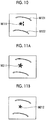
- Fig. 10 is a diagram showing an example of a radiographic image when the position of an affected part is shifted.
- An image M111 of an affected part and bone images M121 and M122 are included in the radiographic image shown in this diagram.
- the position of the affected part moves according to the breathing of the imaging target T11.
- the position of the bone shown in the image M121 or M122 does not move.
- the image correction section 216 moves the position of the affected part image while keeping the positions of the bone images M121 and M122 in the radiographic image, thereby reducing the influence of the shift of the affected part position on the CT image.
- a method used when the image correction section 216 moves the position of the affected part image it is possible to use known methods, such as a method using an optical flow shown in Japanese Unexamined Patent Application Publication 2001-259059 .
- the image correction section 216 translates the position of the affected part image to a shift vector and an inverse vector of the affected part shown in the optical flow.
- the image correction section 216 also corrects the radiographic image for the movement of the position of the affected part in the diagnostic X-ray direction.
- Figs. 11A and 11B are explanatory diagrams showing an example of the correction of a radiographic image for the movement of the position of the affected part in the diagnostic X-ray direction that is performed by the image correction section 216.
- Fig. 11A shows an example of a radiographic image before correction
- Fig. 11B shows an example of a radiographic image after correction.
- the affected part is shifted to the side of the diagnostic X-ray source 340.
- an image M211 of the affected part is relatively large. Therefore, the image correction section 216 performs correction for reducing the image of the affected part as an image M212 of the affected part in Fig. 11B , according to a case where the affected part is located closer to the sensor array 360 side.
- a method of correcting the radiographic image for the movement of the position of the affected part in the diagnostic X-ray direction that is performed by the image correction section 216 it is possible to use known methods, such as a method of calculating the optical flow in a three-dimensional manner.
- the image correction section 216 calculates the optical flow in a three-dimensional manner, it is possible to eliminate the influence of the shift of the imaging angle at each imaging timing.
- a shift in the imaging direction occurs between a radiographic image captured by the imager 371 at the first imaging timing and a radiographic image captured by the imager 371 at the second imaging timing.
- the shift in the imaging direction may become an error when calculating the two-dimensional optical flow.
- the position specifying section 215 specifies the position of the affected part with the three-dimensional coordinates fixed to the imaging target T11, the three-dimensional coordinates are fixed irrespective of the imaging direction. Accordingly, it is possible to eliminate the influence of the shift in the imaging direction when calculating the optical flow.
- the image correction section 216 may correct the direction of the rotation axis A12 (body axis direction).
- the image correction section 216 may acquire an image of the organ from another radiographic image and add the image to the radiographic image to be corrected.
- the treatment control unit 220 controls the radiotherapy apparatus 3 when emitting the therapeutic radiation B11.
- the treatment control unit 220 performs various kinds of control, such as the control of the position of the therapeutic radiation emission unit 331, the control of the emission time of the therapeutic radiation B11, or the control of the multi-leaf collimator 332, according to the treatment plan made by the doctor based on the CT image generated by the CT image reconstruction section 217.
- radiotherapy apparatus controller 2 for example, a computer can be used.
- functions of each unit of the radiotherapy apparatus controller 2 can be realized by causing a central processing unit (CPU) provided in the computer to read a program from a storage device provided in the computer and execute the program.
- CPU central processing unit
- each unit of the radiotherapy apparatus controller 2 can be formed by dedicated hardware.
- Fig. 12 is a flowchart showing the procedure of the process performed by the imaging processing unit 210.
- the imaging processing unit 210 starts the process shown in this diagram when the radiotherapy apparatus controller 2 receives a user operation for instructing the generation of a CT image.
- the imaging processing unit 210 performs a calibration first (step S101).
- the imager rotation control section 211 adjusts the position of each of the imager 371 and the imager 372 to the initial position, and adjusts the direction of the irradiation axis of each of the imager 371 and the imager 372 to a direction passing through the isocenter P11.
- the imaging timing control section 213 performs a response performance calibration for matching the response performance of the sensor array 361 and the sensor array 362.
- the imager rotation control section 211 controls the traveling driving device to rotate the traveling gantry 313 at a fixed speed (step S102).
- the imaging timing control section 213 determines whether or not the imaging timing has arrived (step S103). Specifically, the imaging timing control section 213 has a timer function, and detects the arrival of the imaging timing every predetermined time set in advance as an imaging period.
- the imaging timing control section 213 determines that the imaging timing has arrived in step S103 (step S103: YES). Specifically, as described above, when the imager position determination section 212 determines that the imager 371 is not located in the overlapping region, the imaging timing control section 213 instructs the imager 371 and the imager 372 to perform simultaneous imaging. On the other hand, when the imager position determination section 212 determines that the imager 371 is located in the overlapping region, the imaging timing control section 213 selects one-sided imaging or simultaneous imaging based on the imaging plan acquired in advance or a program set in advance, and gives an imaging instruction according to the selection.
- the imager image acquisition section 214 acquires the radiographic images (radiographic image data) (step S112).
- the position specifying section 215 calculates the three-dimensional position of the affected part as described above (step S113).
- the image correction section 216 corrects the position of the affected part as described above for the image captured by the imager 371 or the image captured by the imager 372. In particular, when the information of the three-dimensional position of the affected part is acquired from the position specifying section 215, the image correction section 216 also corrects the position of the affected part for the movement of the affected part in the diagnostic X-ray direction (step S114).
- the CT image reconstruction section 217 performs a calculation for reconstructing the CT image (step S115). Specifically, the CT image reconstruction section 217 performs the above-described back projection based on the projection image corrected by the image correction section 216.
- the imaging processing unit 210 determines whether or not the imaging at all angles has been completed (step S116). For example, the imager position determination section 212 acquires the amount of rotation of the traveling gantry 313, and determines whether or not the amount of rotation is equal to or greater than 90° + ⁇ .
- step S116: NO the process returns to step S102.
- the CT image reconstruction section 217 reconstructs a tomographic image by calculating the amount of transmitted radiation for each voxel from the result of the back projection, and outputs the obtained tomographic image (step S121).
- the CT image reconstruction section 217 stores the tomographic image in a storage device provided in the radiotherapy apparatus controller 2, and displays the tomographic image on a display screen provided in the radiotherapy apparatus controller 2.
- the imager rotation control section 211 ends the output of the rotation instruction in step S102, and accordingly, the traveling gantry 313 is stopped (rotation is ended).
- step S103 determines that the imaging timing has not arrived in step S103 (step S103: NO)
- the process proceeds to step S116.
- the imaging timing control section 213 causes only the imager 372 to perform imaging in at least one imaging timing whose arrival is detected in a state where the imager position determination section 212 determines that the imager 371 is located in the overlapping region.
- the imaging timing control section 213 causes only the imager 372 to perform imaging, it is possible to reduce the radiation dose equivalent to the radiation from the imager 371 at the imaging timing.
- the imaging timing control section 213 causes the imager 371 and the imager 372 to perform simultaneous imaging, so that a radiographic image of 180° + fan angle ⁇ can be captured.
- the imaging timing control section 213 can reduce the radiation dose without lowering the accuracy of the CT image.
- the imaging timing control section 213 detects the arrival of the imaging timing and causes the imager 371 and the imager 372 to perform simultaneous imaging and the position specifying section 215 succeeds in specifying the three-dimensional position of the position specification target based on the obtained radiographic image
- the imaging timing control section 213 may cause only the imager 372 to perform imaging (one-sided imaging).
- the image correction section 216 can perform correction for the movement of the position specification target in the diagnostic X-ray direction based on the three-dimensional position of the position specification target specified by the position specifying section 215 based on simultaneous imaging or based on a new three-dimensional position calculated from the three-dimensional position.
- the imaging timing control section 213 detects the arrival of the imaging timing and causes the imager 371 and the imager 372 to perform simultaneous imaging and the position specifying section 215 fails to detect the image of the position specification target in at least one of the obtained radiographic images, if the arrival of the next imaging timing is detected in a state where the imager position determination section 212 determines that the imager 371 is located in the overlapping region, the imaging timing control section 213 may cause only the imager 372 to perform imaging (one-sided imaging).
- the imaging timing control section 213 stops simultaneous imaging at an imaging timing at which there is a high possibility that the position specifying section 215 will fail to specify the three-dimensional position of the position specification target even if the imager 371 and the imager 372 perform simultaneous imaging, it is possible to reduce the radiation dose.
- the imaging timing control section 213 causes only the imager 372 to perform imaging (one-sided imaging) at all of the imaging timings whose arrival is detected in a state where the imager position determination section 212 determines that the imager 371 is located in the overlapping region.
- a control target in the present embodiment is not limited to the radiotherapy apparatus.
- a CT dedicated apparatus can also be the control target in the present embodiment.
- the configuration of a CT dedicated apparatus for example, it is possible to adopt a configuration in which each unit (the therapeutic radiation emission unit 331, the multi-leaf collimator 332, or the sensor array 351) for emission of the therapeutic radiation B11 has been removed from the configuration shown in Fig. 2 .
- the configuration on the controller side it is possible to adopt a configuration in which the treatment control unit 220 has been removed from the configuration shown in Fig. 4 .
- the imager 371 may correspond to an example of the second imager, and the imager 372 may correspond to an example of the first imager. In this case, only the imager 371 may perform one-sided imaging in at least a part of the timing at which the imager 372 is located in the overlapping region, so that the imaging from the overlapping region is performed by the imager 371.
- both of the imager 371 and the imager 372 may correspond to any example of the first imager and the second imager.
- imaging in the overlapping region may be alternately performed by the imager 371 and the imager 372.
- the angle between the irradiation axis of the imager 371 and the irradiation axis of the imager 372 with the rotation axis A12 as a reference is not limited to 90° described above, and may be an arbitrary angle.
- the number of imagers provided in the radiotherapy apparatus or the CT dedicated apparatus that is a control target is not limited to 2 described above, and three or more imagers may be provided.
- the radiotherapy apparatus 3 may include a third imager in addition to the imagers 371 and 372.
- the angle between the irradiation axis of the imager 371 and the irradiation axis of the imager 372 with the rotation axis A12 as a reference may be 60°
- the angle between the irradiation axis of the imager 372 and the irradiation axis of the third imager with the rotation axis A12 as a reference may be 60°.
- a rotation range when the rotation ranges of the three imagers are combined is equal to or greater than 180°. Therefore, it is possible to reduce the time required to capture a radiographic image.
- an overlapping region of the rotation ranges of the imagers occurs from the relationship with the fan angle. In the overlapping region, the imaging timing control section 213 can select one of imaging by one imager, imaging by two imagers, and imaging by three imagers every imaging timing.
- both the irradiation axis of the imager 371 and the irradiation axis of the imager 372 pass through the vicinity of the isocenter P11, however, the irradiation axis of the imager 371 and the irradiation axis of the imager 372 do not necessarily need to pass through the isocenter P11.
- the irradiation axis of the imager 371 and the irradiation axis of the imager 372 do not necessarily need to cross each other (may be in a relationship of twisted position).
- a computer can be used as the radiotherapy apparatus controller 2. Accordingly, the processing of each unit may be performed by recording a program for realizing the functions of all or some of the units of the radiotherapy apparatus controller 2 in a computer-readable recording medium, reading the program recorded in the recording medium into a computer system, and executing the read program.
- the "computer system” referred to herein may include an OS or hardware, such as a peripheral device.
- the "computer system” may also include a homepage presenting environment (or display environment) if a WWW system is used.
- examples of the "computer-readable recording medium” include portable media, such as a flexible disk, a magneto-optic disc, a ROM, and a CD-ROM, and a storage device, such as a hard disk built in a computer system.
- examples of the "computer-readable recording medium” may include a recording medium that stores a program dynamically for a short period of time like a network, such as the Internet, or a communication line when a program is transmitted through a communication line, such as a telephone line, and include a recording medium that stores a program for a predetermined period of time like a volatile memory in a computer system that serves as a server or a client in this case.
- the above program may be a program for realizing some of the functions described above or may be a program capable of realizing the above functions by combination with a program already recorded in the computer system.
- the present invention relates to an imaging controller of an imaging apparatus which includes first and second imagers for capturing a radiographic image by emitting a cone beam toward a rotation axis and a rotation mechanism for rotating the first and second imagers integrally around the rotation axis and in which an angle between an irradiation axis of the first imager and an irradiation axis of the second imager with the rotation axis as a reference is a predetermined angle.
- the imaging controller includes: an imager position determination section that determines whether or not the first imager is located in an overlapping region where a rotation range of the first imager and a rotation range of the second imager overlap each other when the rotation mechanism rotates the first and second imagers by an angle greater than the predetermined angle; and an imaging timing control section that causes one or both of the first and second imagers to perform imaging when arrival of an imaging timing is detected and that causes only the second imager to perform imaging in at least one imaging timing whose arrival has been detected in a state where the imager position determination section determines that the first imager is located in the overlapping region.
Landscapes
- Health & Medical Sciences (AREA)
- Life Sciences & Earth Sciences (AREA)
- Engineering & Computer Science (AREA)
- Medical Informatics (AREA)
- Biomedical Technology (AREA)
- Pathology (AREA)
- Nuclear Medicine, Radiotherapy & Molecular Imaging (AREA)
- Radiology & Medical Imaging (AREA)
- Animal Behavior & Ethology (AREA)
- General Health & Medical Sciences (AREA)
- Public Health (AREA)
- Veterinary Medicine (AREA)
- Biophysics (AREA)
- High Energy & Nuclear Physics (AREA)
- Physics & Mathematics (AREA)
- Optics & Photonics (AREA)
- Heart & Thoracic Surgery (AREA)
- Molecular Biology (AREA)
- Surgery (AREA)
- Pulmonology (AREA)
- Theoretical Computer Science (AREA)
- Apparatus For Radiation Diagnosis (AREA)
- Cameras In General (AREA)
- Studio Devices (AREA)
Claims (6)
- Bilderzeugungssteuerung (2) einer CT-Bilderzeugungsvorrichtung (3), wobei die Bilderzeugungsvorrichtung (3) einen ersten und einen zweiten Bilderzeuger (371, 372), wobei jeder Bilderzeuger (371, 372) eine Kombination aus einer diagnostischen Röntgenquelle (341, 342) und einer Sensoranordnung (361, 362) ist, zum Erfassen eines Röntgenbildes durch Emittieren eines Kegelstrahls in Richtung einer Drehachse (A11) aufweist, sowie einen Drehmechanismus (313) zum Drehen des ersten und des zweiten Bilderzeugers (371, 372) zusammen um die Drehachse (A11) und bei dem ein Winkel zwischen einer Bestrahlungsachse des ersten Bilderzeugers (371) und einer Bestrahlungsachse des zweiten Bilderzeugers (372) mit der Drehachse (A11) als Referenz ein vorbestimmter Winkel ist, wobei die Bilderzeugungssteuerung (2) umfasst:einen Bilderzeugerpositionsbestimmungsabschnitt (212), der dazu ausgelegt ist, zu bestimmen, ob der erste Bilderzeuger (371) in einem überlappenden Bereich angeordnet ist, wo ein Rotationsbereich des ersten Bilderzeugers (371) und ein Rotationsbereich des zweiten Bilderzeugers (372) einander überlappen, wenn der Drehmechanismus (313) den ersten und den zweiten Bilderzeuger (371, 372) um einen Winkel dreht, der größer als der vorbestimmte Winkel ist;einen Bilderzeugungszeitsteuerabschnitt (213), der dazu ausgelegt ist, einen oder beide der ersten und zweiten Bilderzeuger (371, 372) zu veranlassen, eine Bilderzeugung durchzuführen, wenn das Erreichen einer Bilderzeugungszeit erkannt wird, und nur den zweiten Bilderzeuger (372) zu veranlassen, eine Bilderzeugung in mindestens einer Bilderzeugungszeit durchzuführen, deren Erreichen in einem Zustand erkannt wird, in dem der Bilderzeugerpositionsbestimmungsabschnitt (212) bestimmt, dass der erste Bilderzeuger (371) in dem überlappenden Bereich angeordnet ist; undeinen Positionsspezifikationsabschnitt (215), der dazu ausgelegt ist, eine dreidimensionale Position eines Positionsspezifikationsziels auf der Grundlage eines von dem ersten Bilderzeuger (371) aufgenommenen Röntgenbildes und eines von dem zweiten Bilderzeuger (372) aufgenommenen Röntgenbildes zu spezifizieren,dadurch gekennzeichnet, dass der Bilderzeugungszeitsteuerabschnitt (213) ferner dazu ausgelegt ist, das Folgende auszuführen:
wenn der Bilderzeugungszeitsteuerabschnitt (213) das Erreichen einer Bilderzeugungszeit erkennt und sowohl den ersten als auch den zweiten Bilderzeuger (371, 372) veranlasst, eine Bilderzeugung durchzuführen, und der Positionsspezifikationsabschnitt (215) in mindestens einem der erhaltenen Röntgenbilder kein Bild des Positionsspezifikationsziels erfasst, und dadurch die dreidimensionale Position des Positionsspezifikationsziels nicht spezifizierbar ist, der Bilderzeugungszeitsteuerabschnitt (213) bewirkt, dass nur der zweite Bilderzeuger (372) eine Bilderzeugung mit einer vorbestimmten Anzahl von Bilderzeugungs-Zeitsteuerungen in einem Zustand durchführt, in dem der Bilderzeugerpositionsbestimmungsabschnitt (212) bestimmt, dass sich der erste Bilderzeuger (371) in dem überlappenden Bereich befindet. - Bilderzeugungssteuerung (2) einer CT-Bilderzeugungsvorrichtung (3), wobei die Bilderzeugungsvorrichtung (3) einen ersten und einen zweiten Bilderzeuger (371, 372), wobei jeder Bilderzeuger (371, 372) eine Kombination aus einer diagnostischen Röntgenquelle (341, 342) und einer Sensoranordnung (361, 362) ist, zum Erfassen eines Röntgenbildes durch Emittieren eines Kegelstrahls in Richtung einer Drehachse (A11) aufweist, sowie einen Drehmechanismus (313) zum Drehen des ersten und des zweiten Bilderzeugers (371, 372) zusammen um die Drehachse (A11) und bei dem ein Winkel zwischen einer Bestrahlungsachse des ersten Bilderzeugers (371) und einer Bestrahlungsachse des zweiten Bilderzeugers (372) mit der Drehachse (A11) als Referenz ein vorbestimmter Winkel ist, wobei die Bilderzeugungssteuerung (2) umfasst:einen Bilderzeugerpositionsbestimmungsabschnitt (212), der dazu ausgelegt ist, zu bestimmen, ob der erste Bilderzeuger (371) in einem überlappenden Bereich angeordnet ist, wo ein Rotationsbereich des ersten Bilderzeugers (371) und ein Rotationsbereich des zweiten Bilderzeugers (372) einander überlappen, wenn der Drehmechanismus (313) den ersten und den zweiten Bilderzeuger (371, 372) um einen Winkel dreht, der größer als der vorbestimmte Winkel ist;einen Bilderzeugungszeitsteuerabschnitt (213), der dazu ausgelegt ist, einen oder beide der ersten und zweiten Bilderzeuger (371, 372) zu veranlassen, eine Bilderzeugung durchzuführen, wenn das Erreichen einer Bilderzeugungszeit erkannt wird, und nur den zweiten Bilderzeuger (372) zu veranlassen, eine Bilderzeugung in mindestens einer Bilderzeugungszeit durchzuführen, deren Erreichen in einem Zustand erkannt wird, in dem der Bilderzeugerpositionsbestimmungsabschnitt (212) bestimmt, dass der erste Bilderzeuger (371) in dem überlappenden Bereich angeordnet ist; undeinen Positionsspezifikationsabschnitt (215), der dazu ausgelegt ist, eine dreidimensionale Position eines Positionsspezifikationsziels auf der Grundlage eines von dem ersten Bilderzeuger (371) aufgenommenen Röntgenbildes und eines von dem zweiten Bilderzeuger (372) aufgenommenen Röntgenbildes zu spezifizieren,dadurch gekennzeichnet, dass der Bilderzeugungszeitsteuerabschnitt (213) ferner dazu ausgelegt ist, das Folgende auszuführen:
wenn der Bilderzeugungszeitsteuerabschnitt (213) das Erreichen einer Bilderzeugungszeit erkennt und sowohl den ersten als auch den zweiten Bilderzeuger (371, 372) veranlasst, eine Bilderzeugung durchzuführen, und der Positionsspezifikationsabschnitt (215) die dreidimensionale Position des Positionsspezifikationsziels auf der Grundlage der erhaltenen Röntgenbilder erfolgreich spezifiziert, wenn das Erreichen eines nächsten Bilderzeugungszeitpunkts in einem Zustand erkannt wird, in dem der Bilderzeugerpositionsbestimmungsabschnitt (212) bestimmt, dass sich der erste Bilderzeuger (371) in dem überlappenden Bereich befindet, der Bilderzeugungszeitsteuerabschnitt bewirkt, dass nur der zweite Bilderzeuger (372) die Bilderzeugung durchführt. - Bildgebendes System, umfassend:die Bilderzeugungssteuerung (2) nach Anspruch 1 oder 2; unddie CT-Bilderzeugungsvorrichtung (3), die den ersten und den zweiten Bilderzeuger (371, 372) und den Drehmechanismus (313) umfasst.
- Bilderzeugungssteuerverfahren einer Bilderzeugungssteuerung (2) einer CT-Bilderzeugungsvorrichtung (3), wobei die Bilderzeugungsvorrichtung (3) einen ersten und einen zweiten Bilderzeuger (371, 372), wobei jeder Bilderzeuger (371, 372) eine Kombination aus einer diagnostischen Röntgenquelle (341, 342) und einer Sensoranordnung (361, 362) ist, zum Erfassen eines Röntgenbildes durch Emittieren eines Kegelstrahls in Richtung einer Drehachse (A11) aufweist, sowie einen Drehmechanismus (313) zum Drehen des ersten und des zweiten Bilderzeugers (371, 372) zusammen um die Drehachse (A11), wobei ein Winkel zwischen einer Bestrahlungsachse des ersten Bilderzeugers (371) und einer Bestrahlungsachse des zweiten Bilderzeugers (372) mit der Drehachse (A11) als Referenz ein vorbestimmter Winkel ist, wobei das Verfahren umfasst:einen Bilderzeugerpositionsbestimmungsschritt des Bestimmens, ob der erste Bilderzeuger (371) in einem überlappenden Bereich angeordnet ist, wo ein Rotationsbereich des ersten Bilderzeugers (371) und ein Rotationsbereich des zweiten Bilderzeugers (372) einander überlappen, wenn der Drehmechanismus (313) den ersten und den zweiten Bilderzeuger (371, 372) um einen Winkel dreht, der größer als der vorbestimmte Winkel ist;einen Bilderzeugungszeitpunktsteuerschritt, der bewirkt, dass einer oder beide der ersten und zweiten Bilderzeuger (371, 372) eine Bilderzeugung durchführen, wenn das Erreichen eines Bilderzeugungszeitpunktes erkannt wird, und der bewirkt, dass nur der zweite Bilderzeuger (372) eine Bilderzeugung in mindestens einem Bilderzeugungszeitpunkt durchführt, dessen Erreichen in einem Zustand erkannt wird, in dem bestimmt wird, dass sich der erste Bilderzeuger (371) in dem überlappenden Bereich in dem Bilderzeugerpositionsbestimmungsschritt befindet; undeinen Positionsspezifikationsschritt zum Spezifizieren einer dreidimensionalen Position eines Positionsspezifikationsziels auf der Grundlage eines durch den ersten Bilderzeuger (371) erfassten Röntgenbildes und eines durch den zweiten Bilderzeuger (372) erfassten Röntgenbildes,dadurch gekennzeichnet, dasswenn das Eintreffen eines Bilderzeugungszeitpunkts erkannt wird und sowohl der erste als auch der zweite Bilderzeuger (371, 372) veranlasst werden, die Bilderzeugung im Bilderzeugungszeitpunktsteuerschritt durchzuführen, und es in mindestens einem der erhaltenen Röntgenbilder nicht gelingt, ein Bild des Positionsspezifikationsziels zu erfassen, und dadurch die dreidimensionale Position des Positionsspezifikationsziels im Positionsspezifikationsschritt nicht spezifizierbar ist, nur der zweite Bilderzeuger (372) veranlasst wird, eine Bilderzeugung mit einer vorbestimmten Anzahl einer Bilderzeugungszeit in dem Bilderzeugungszeit-Steuerschritt in einem Zustand durchzuführen, in dem in dem Bilderzeugerpositionsbestimmungsschritt bestimmt wird, dass sich der erste Bilderzeuger (371) in dem Überlappungsbereich befindet.
- Bilderzeugungssteuerverfahren einer Bilderzeugungssteuerung (2) einer CT-Bilderzeugungsvorrichtung (3), wobei die Bilderzeugungsvorrichtung (3) einen ersten und einen zweiten Bilderzeuger (371, 372), wobei jeder Bilderzeuger (371, 372) eine Kombination aus einer diagnostischen Röntgenquelle (341, 342) und einer Sensoranordnung (361, 362) ist, zum Erfassen eines Röntgenbildes durch Emittieren eines Kegelstrahls in Richtung einer Drehachse (A11) aufweist, sowie einen Drehmechanismus (313) zum Drehen des ersten und des zweiten Bilderzeugers (371, 372) zusammen um die Drehachse (A11), wobei ein Winkel zwischen einer Bestrahlungsachse des ersten Bilderzeugers (371) und einer Bestrahlungsachse des zweiten Bilderzeugers (372) mit der Drehachse (A11) als Referenz ein vorbestimmter Winkel ist, wobei das Verfahren umfasst:einen Bilderzeugerpositionsbestimmungsschritt des Bestimmens, ob der erste Bilderzeuger (371) in einem überlappenden Bereich angeordnet ist, wo ein Rotationsbereich des ersten Bilderzeugers (371) und ein Rotationsbereich des zweiten Bilderzeugers (372) einander überlappen, wenn der Drehmechanismus (313) den ersten und den zweiten Bilderzeuger (371, 372) um einen Winkel dreht, der größer als der vorbestimmte Winkel ist;einen Bilderzeugungszeitpunktsteuerschritt, der bewirkt, dass einer oder beide der ersten und zweiten Bilderzeuger (371, 372) eine Bilderzeugung durchführen, wenn das Erreichen eines Bilderzeugungszeitpunktes erkannt wird, und der bewirkt, dass nur der zweite Bilderzeuger (372) eine Bilderzeugung in mindestens einem Bilderzeugungszeitpunkt durchführt, dessen Erreichen in einem Zustand erkannt wird, in dem bestimmt wird, dass sich der erste Bilderzeuger (371) in dem überlappenden Bereich in dem Bilderzeugerpositionsbestimmungsschritt befindet; undeinen Positionsspezifikationsschritt zum Spezifizieren einer dreidimensionalen Position eines Positionsspezifikationsziels auf der Grundlage eines durch den ersten Bilderzeuger (371) erfassten Röntgenbildes und eines durch den zweiten Bilderzeuger (372) erfassten Röntgenbildes,dadurch gekennzeichnet, dasswenn das Eintreffen eines Bilderzeugungszeitpunkts erkannt wird und sowohl der erste als auch der zweite Bilderzeuger (371, 372) veranlasst werden, eine Bilderzeugung in dem Bilderzeugungszeitpunktsteuerschritt durchzuführen, und es gelingt, die dreidimensionale Position des Positionsspezifikationsziels auf der Grundlage der erhaltenen Röntgenbilder in dem Positionsspezifikationsschritt zu spezifizieren, wenn das Eintreffen eines nächsten Bilderzeugungszeitpunkts in einem Zustand erkannt wird, in dem bestimmt wird, dass der erste Bilderzeuger (371) in dem überlappenden Bereich in dem Bilderzeugerpositionsbestimmungsschritt angeordnet ist, nur der zweite Bilderzeuger (372) veranlasst wird, eine Bilderzeugung in dem Bilderzeugungszeitpunktsteuerschritt durchzuführen.
- Programm, das einen Computer als Bilderzeugungssteuergerät (2) eines CT-Bilderzeugungsgerätes (3) veranlasst, das Verfahren nach Anspruch 4 oder 5 auszuführen.
Applications Claiming Priority (2)
| Application Number | Priority Date | Filing Date | Title |
|---|---|---|---|
| JP2012224248A JP5836244B2 (ja) | 2012-10-09 | 2012-10-09 | 撮影制御装置、撮影システム、撮影制御方法およびプログラム |
| PCT/JP2013/074670 WO2014057762A1 (ja) | 2012-10-09 | 2013-09-12 | 撮影制御装置、撮影システム、撮影制御方法およびプログラム |
Publications (3)
| Publication Number | Publication Date |
|---|---|
| EP2907452A1 EP2907452A1 (de) | 2015-08-19 |
| EP2907452A4 EP2907452A4 (de) | 2016-06-22 |
| EP2907452B1 true EP2907452B1 (de) | 2021-01-13 |
Family
ID=50477233
Family Applications (1)
| Application Number | Title | Priority Date | Filing Date |
|---|---|---|---|
| EP13845852.6A Active EP2907452B1 (de) | 2012-10-09 | 2013-09-12 | Fotosteuerungsvorrichtung, fotografisches system, fotosteuerungsverfahren und programm |
Country Status (5)
| Country | Link |
|---|---|
| US (1) | US9724048B2 (de) |
| EP (1) | EP2907452B1 (de) |
| JP (1) | JP5836244B2 (de) |
| CN (1) | CN104507391A (de) |
| WO (1) | WO2014057762A1 (de) |
Families Citing this family (11)
| Publication number | Priority date | Publication date | Assignee | Title |
|---|---|---|---|---|
| US9616251B2 (en) * | 2014-07-25 | 2017-04-11 | Varian Medical Systems, Inc. | Imaging based calibration systems, devices, and methods |
| CN104198506B (zh) * | 2014-08-27 | 2017-11-07 | 清华大学 | 小角度自摆式大型多层螺旋ct设备和检查方法 |
| CN106377275B (zh) * | 2016-09-30 | 2019-11-12 | 东软医疗系统股份有限公司 | 一种螺旋扫描方法及装置 |
| JP6738363B2 (ja) | 2018-03-09 | 2020-08-12 | 浜松ホトニクス株式会社 | 画像取得システムおよび画像取得方法 |
| WO2019226083A1 (en) * | 2018-05-22 | 2019-11-28 | Scanflex Healthcare Aktiebolag | Flat panel x-ray imaging device - twin flat detector signal synchronization |
| US12213818B2 (en) | 2020-09-08 | 2025-02-04 | Shanghai United Imaging Healthcare Co., Ltd. | X-ray imaging system for radiation therapy |
| US12186591B2 (en) | 2020-09-08 | 2025-01-07 | Shanghai United Imaging Healthcare Co., Ltd. | X-ray imaging system for radiation therapy |
| US11951333B2 (en) * | 2020-09-08 | 2024-04-09 | Shanghai United Imaging Healthcare Co., Ltd. | X-ray imaging system for radiation therapy |
| US11883687B2 (en) | 2020-09-08 | 2024-01-30 | Shanghai United Imaging Healthcare Co., Ltd. | X-ray imaging system for radiation therapy |
| JP7739113B2 (ja) * | 2021-09-29 | 2025-09-16 | 株式会社日立ハイテク | 放射線治療システム、および、放射線治療システムの運転方法 |
| JP7615342B2 (ja) * | 2021-10-15 | 2025-01-16 | 株式会社東芝 | 撮影制御システムおよび撮影制御方法 |
Family Cites Families (24)
| Publication number | Priority date | Publication date | Assignee | Title |
|---|---|---|---|---|
| JPH0422343A (ja) | 1990-05-17 | 1992-01-27 | Toshiba Corp | X線ct装置 |
| JPH0910206A (ja) | 1995-06-30 | 1997-01-14 | Shimadzu Corp | X線ct装置 |
| JP3743594B2 (ja) | 1998-03-11 | 2006-02-08 | 株式会社モリタ製作所 | Ct撮影装置 |
| JP4159226B2 (ja) | 2000-03-21 | 2008-10-01 | 住友重機械工業株式会社 | 患部トラッキング方法、装置、及び、これを用いた放射線照射方法、装置、並びに放射線治療装置 |
| US7085343B2 (en) * | 2001-10-18 | 2006-08-01 | Kabushiki Kaisha Toshiba | X-ray computed tomography apparatus |
| JP3848911B2 (ja) | 2002-10-01 | 2006-11-22 | 三菱重工業株式会社 | 放射線照射装置 |
| US7639774B2 (en) * | 2003-12-23 | 2009-12-29 | General Electric Company | Method and apparatus for employing multiple axial-sources |
| JP2005296340A (ja) | 2004-04-12 | 2005-10-27 | Canon Inc | コーンビームx線ct撮影装置とそれを用いた画像取得方法 |
| DE102004018498A1 (de) * | 2004-04-14 | 2005-11-17 | Siemens Ag | Betriebsverfahren für eine Röntgenanlage, rechnergestütztes Ermittlungsverfahren für mindestens eine 3D-Rekonstruktion eines Objekts und hiermit korrespondierende Einrichtungen |
| JP4928739B2 (ja) * | 2004-06-25 | 2012-05-09 | 株式会社東芝 | X線診断装置及びx線撮像方法 |
| JP4064952B2 (ja) | 2004-08-12 | 2008-03-19 | 三菱重工業株式会社 | 放射線治療装置および放射線治療装置の動作方法 |
| DE102004042491B4 (de) * | 2004-08-31 | 2009-07-09 | Siemens Ag | Verfahren zur Erzeugung von tomographischen Schnittbildern von einem Untersuchungsobjekt mit mindestens zwei winkelversetzten Strahlenbündeln und Computertomographie-Gerät zur Durchführung dieses Verfahrens |
| JP4996818B2 (ja) | 2004-11-01 | 2012-08-08 | 株式会社東芝 | X線診断装置 |
| JP4505639B2 (ja) | 2005-02-24 | 2010-07-21 | 国立大学法人北海道大学 | 動体追跡照射装置およびプログラム |
| JP5091422B2 (ja) | 2005-04-15 | 2012-12-05 | 株式会社東芝 | Ctスキャナ |
| US7440547B2 (en) * | 2005-04-15 | 2008-10-21 | Kabushiki Kaisha Toshiba | CT scanner |
| JP2007236760A (ja) | 2006-03-10 | 2007-09-20 | Mitsubishi Heavy Ind Ltd | 放射線治療装置制御装置および放射線照射方法 |
| JP4898901B2 (ja) | 2008-03-17 | 2012-03-21 | 三菱重工業株式会社 | 放射線治療装置制御装置および放射線照射方法 |
| WO2010073308A1 (ja) | 2008-12-22 | 2010-07-01 | 三菱重工業株式会社 | 放射線断層撮影方法 |
| JP5597364B2 (ja) | 2009-06-29 | 2014-10-01 | 株式会社東芝 | X線コンピュータ断層撮影装置および撮影制御プログラム |
| JP5138646B2 (ja) * | 2009-08-31 | 2013-02-06 | 三菱重工業株式会社 | 放射線断層撮影方法、放射線治療装置制御装置及び放射線治療システム |
| EP2324768B1 (de) | 2009-09-14 | 2021-11-24 | Hitachi, Ltd. | Strahlentomographie-Verfahren und Strahlentherapie-System |
| CN102665560B (zh) | 2009-11-04 | 2014-10-15 | 株式会社日立医疗器械 | X射线ct装置以及基于x射线ct装置的图像显示方法 |
| WO2011059061A1 (ja) | 2009-11-16 | 2011-05-19 | 三菱重工業株式会社 | 放射線治療装置制御装置および特定部位位置計測方法 |
-
2012
- 2012-10-09 JP JP2012224248A patent/JP5836244B2/ja active Active
-
2013
- 2013-09-12 WO PCT/JP2013/074670 patent/WO2014057762A1/ja not_active Ceased
- 2013-09-12 CN CN201380039999.4A patent/CN104507391A/zh active Pending
- 2013-09-12 US US14/418,241 patent/US9724048B2/en active Active
- 2013-09-12 EP EP13845852.6A patent/EP2907452B1/de active Active
Non-Patent Citations (1)
| Title |
|---|
| None * |
Also Published As
| Publication number | Publication date |
|---|---|
| CN104507391A (zh) | 2015-04-08 |
| EP2907452A1 (de) | 2015-08-19 |
| US9724048B2 (en) | 2017-08-08 |
| WO2014057762A1 (ja) | 2014-04-17 |
| JP2014076099A (ja) | 2014-05-01 |
| WO2014057762A8 (ja) | 2015-02-26 |
| US20150182175A1 (en) | 2015-07-02 |
| EP2907452A4 (de) | 2016-06-22 |
| JP5836244B2 (ja) | 2015-12-24 |
Similar Documents
| Publication | Publication Date | Title |
|---|---|---|
| US9724048B2 (en) | Imaging controller, imaging system, imaging control method, and program | |
| JP6181459B2 (ja) | 放射線治療システム | |
| JP4959805B2 (ja) | 放射線断層撮影方法 | |
| EP3037039B1 (de) | Röntgenbildgebungsvorrichtung | |
| US20200346036A1 (en) | Positioning method and apparatus, and radiation therapy system | |
| US8358738B2 (en) | Respiration-correlated radiotherapy | |
| US20150265237A1 (en) | Method and device for generating a three-dimensional image of an object | |
| EP2609962B1 (de) | Strahlentherapievorrichtung | |
| JP5031095B2 (ja) | 放射線断層撮影方法および放射線治療装置制御装置 | |
| CN110337672A (zh) | 用于短扫描偏心探测器x射线断层摄影的冗余度加权 | |
| EP3111847B1 (de) | Ct-bildgebungsgerät und -verfahren | |
| US20240389963A1 (en) | Oral cone beam x-ray imaging system and fast positioning method therefor | |
| JP4898901B2 (ja) | 放射線治療装置制御装置および放射線照射方法 | |
| WO2023062819A1 (ja) | 撮影制御システムおよび撮影制御方法 | |
| JP5138646B2 (ja) | 放射線断層撮影方法、放射線治療装置制御装置及び放射線治療システム | |
| JP2022070025A (ja) | セットアップ支援装置及びセットアップ支援方法 | |
| EP3843041A1 (de) | Vorrichtung zur computertomographie von mit hohem relativem pitch erfassten röntgendaten |
Legal Events
| Date | Code | Title | Description |
|---|---|---|---|
| PUAI | Public reference made under article 153(3) epc to a published international application that has entered the european phase |
Free format text: ORIGINAL CODE: 0009012 |
|
| 17P | Request for examination filed |
Effective date: 20150128 |
|
| AK | Designated contracting states |
Kind code of ref document: A1 Designated state(s): AL AT BE BG CH CY CZ DE DK EE ES FI FR GB GR HR HU IE IS IT LI LT LU LV MC MK MT NL NO PL PT RO RS SE SI SK SM TR |
|
| AX | Request for extension of the european patent |
Extension state: BA ME |
|
| DAX | Request for extension of the european patent (deleted) | ||
| RA4 | Supplementary search report drawn up and despatched (corrected) |
Effective date: 20160523 |
|
| RIC1 | Information provided on ipc code assigned before grant |
Ipc: A61B 6/03 20060101AFI20160517BHEP |
|
| RAP1 | Party data changed (applicant data changed or rights of an application transferred) |
Owner name: HITACHI, LTD. |
|
| STAA | Information on the status of an ep patent application or granted ep patent |
Free format text: STATUS: EXAMINATION IS IN PROGRESS |
|
| 17Q | First examination report despatched |
Effective date: 20200127 |
|
| GRAP | Despatch of communication of intention to grant a patent |
Free format text: ORIGINAL CODE: EPIDOSNIGR1 |
|
| STAA | Information on the status of an ep patent application or granted ep patent |
Free format text: STATUS: GRANT OF PATENT IS INTENDED |
|
| INTG | Intention to grant announced |
Effective date: 20200811 |
|
| GRAS | Grant fee paid |
Free format text: ORIGINAL CODE: EPIDOSNIGR3 |
|
| GRAA | (expected) grant |
Free format text: ORIGINAL CODE: 0009210 |
|
| STAA | Information on the status of an ep patent application or granted ep patent |
Free format text: STATUS: THE PATENT HAS BEEN GRANTED |
|
| AK | Designated contracting states |
Kind code of ref document: B1 Designated state(s): AL AT BE BG CH CY CZ DE DK EE ES FI FR GB GR HR HU IE IS IT LI LT LU LV MC MK MT NL NO PL PT RO RS SE SI SK SM TR |
|
| REG | Reference to a national code |
Ref country code: GB Ref legal event code: FG4D |
|
| REG | Reference to a national code |
Ref country code: CH Ref legal event code: EP |
|
| REG | Reference to a national code |
Ref country code: DE Ref legal event code: R096 Ref document number: 602013075322 Country of ref document: DE |
|
| REG | Reference to a national code |
Ref country code: IE Ref legal event code: FG4D |
|
| REG | Reference to a national code |
Ref country code: AT Ref legal event code: REF Ref document number: 1353942 Country of ref document: AT Kind code of ref document: T Effective date: 20210215 |
|
| REG | Reference to a national code |
Ref country code: AT Ref legal event code: MK05 Ref document number: 1353942 Country of ref document: AT Kind code of ref document: T Effective date: 20210113 |
|
| REG | Reference to a national code |
Ref country code: NL Ref legal event code: MP Effective date: 20210113 |
|
| REG | Reference to a national code |
Ref country code: LT Ref legal event code: MG9D |
|
| PG25 | Lapsed in a contracting state [announced via postgrant information from national office to epo] |
Ref country code: PT Free format text: LAPSE BECAUSE OF FAILURE TO SUBMIT A TRANSLATION OF THE DESCRIPTION OR TO PAY THE FEE WITHIN THE PRESCRIBED TIME-LIMIT Effective date: 20210513 Ref country code: LT Free format text: LAPSE BECAUSE OF FAILURE TO SUBMIT A TRANSLATION OF THE DESCRIPTION OR TO PAY THE FEE WITHIN THE PRESCRIBED TIME-LIMIT Effective date: 20210113 Ref country code: GR Free format text: LAPSE BECAUSE OF FAILURE TO SUBMIT A TRANSLATION OF THE DESCRIPTION OR TO PAY THE FEE WITHIN THE PRESCRIBED TIME-LIMIT Effective date: 20210414 Ref country code: FI Free format text: LAPSE BECAUSE OF FAILURE TO SUBMIT A TRANSLATION OF THE DESCRIPTION OR TO PAY THE FEE WITHIN THE PRESCRIBED TIME-LIMIT Effective date: 20210113 Ref country code: HR Free format text: LAPSE BECAUSE OF FAILURE TO SUBMIT A TRANSLATION OF THE DESCRIPTION OR TO PAY THE FEE WITHIN THE PRESCRIBED TIME-LIMIT Effective date: 20210113 Ref country code: BG Free format text: LAPSE BECAUSE OF FAILURE TO SUBMIT A TRANSLATION OF THE DESCRIPTION OR TO PAY THE FEE WITHIN THE PRESCRIBED TIME-LIMIT Effective date: 20210413 Ref country code: NL Free format text: LAPSE BECAUSE OF FAILURE TO SUBMIT A TRANSLATION OF THE DESCRIPTION OR TO PAY THE FEE WITHIN THE PRESCRIBED TIME-LIMIT Effective date: 20210113 Ref country code: NO Free format text: LAPSE BECAUSE OF FAILURE TO SUBMIT A TRANSLATION OF THE DESCRIPTION OR TO PAY THE FEE WITHIN THE PRESCRIBED TIME-LIMIT Effective date: 20210413 |
|
| PG25 | Lapsed in a contracting state [announced via postgrant information from national office to epo] |
Ref country code: RS Free format text: LAPSE BECAUSE OF FAILURE TO SUBMIT A TRANSLATION OF THE DESCRIPTION OR TO PAY THE FEE WITHIN THE PRESCRIBED TIME-LIMIT Effective date: 20210113 Ref country code: LV Free format text: LAPSE BECAUSE OF FAILURE TO SUBMIT A TRANSLATION OF THE DESCRIPTION OR TO PAY THE FEE WITHIN THE PRESCRIBED TIME-LIMIT Effective date: 20210113 Ref country code: PL Free format text: LAPSE BECAUSE OF FAILURE TO SUBMIT A TRANSLATION OF THE DESCRIPTION OR TO PAY THE FEE WITHIN THE PRESCRIBED TIME-LIMIT Effective date: 20210113 Ref country code: AT Free format text: LAPSE BECAUSE OF FAILURE TO SUBMIT A TRANSLATION OF THE DESCRIPTION OR TO PAY THE FEE WITHIN THE PRESCRIBED TIME-LIMIT Effective date: 20210113 Ref country code: SE Free format text: LAPSE BECAUSE OF FAILURE TO SUBMIT A TRANSLATION OF THE DESCRIPTION OR TO PAY THE FEE WITHIN THE PRESCRIBED TIME-LIMIT Effective date: 20210113 |
|
| PG25 | Lapsed in a contracting state [announced via postgrant information from national office to epo] |
Ref country code: IS Free format text: LAPSE BECAUSE OF FAILURE TO SUBMIT A TRANSLATION OF THE DESCRIPTION OR TO PAY THE FEE WITHIN THE PRESCRIBED TIME-LIMIT Effective date: 20210513 |
|
| REG | Reference to a national code |
Ref country code: DE Ref legal event code: R097 Ref document number: 602013075322 Country of ref document: DE |
|
| PG25 | Lapsed in a contracting state [announced via postgrant information from national office to epo] |
Ref country code: SM Free format text: LAPSE BECAUSE OF FAILURE TO SUBMIT A TRANSLATION OF THE DESCRIPTION OR TO PAY THE FEE WITHIN THE PRESCRIBED TIME-LIMIT Effective date: 20210113 Ref country code: EE Free format text: LAPSE BECAUSE OF FAILURE TO SUBMIT A TRANSLATION OF THE DESCRIPTION OR TO PAY THE FEE WITHIN THE PRESCRIBED TIME-LIMIT Effective date: 20210113 Ref country code: CZ Free format text: LAPSE BECAUSE OF FAILURE TO SUBMIT A TRANSLATION OF THE DESCRIPTION OR TO PAY THE FEE WITHIN THE PRESCRIBED TIME-LIMIT Effective date: 20210113 |
|
| PLBE | No opposition filed within time limit |
Free format text: ORIGINAL CODE: 0009261 |
|
| STAA | Information on the status of an ep patent application or granted ep patent |
Free format text: STATUS: NO OPPOSITION FILED WITHIN TIME LIMIT |
|
| PG25 | Lapsed in a contracting state [announced via postgrant information from national office to epo] |
Ref country code: DK Free format text: LAPSE BECAUSE OF FAILURE TO SUBMIT A TRANSLATION OF THE DESCRIPTION OR TO PAY THE FEE WITHIN THE PRESCRIBED TIME-LIMIT Effective date: 20210113 Ref country code: ES Free format text: LAPSE BECAUSE OF FAILURE TO SUBMIT A TRANSLATION OF THE DESCRIPTION OR TO PAY THE FEE WITHIN THE PRESCRIBED TIME-LIMIT Effective date: 20210113 Ref country code: RO Free format text: LAPSE BECAUSE OF FAILURE TO SUBMIT A TRANSLATION OF THE DESCRIPTION OR TO PAY THE FEE WITHIN THE PRESCRIBED TIME-LIMIT Effective date: 20210113 Ref country code: SK Free format text: LAPSE BECAUSE OF FAILURE TO SUBMIT A TRANSLATION OF THE DESCRIPTION OR TO PAY THE FEE WITHIN THE PRESCRIBED TIME-LIMIT Effective date: 20210113 |
|
| 26N | No opposition filed |
Effective date: 20211014 |
|
| PG25 | Lapsed in a contracting state [announced via postgrant information from national office to epo] |
Ref country code: AL Free format text: LAPSE BECAUSE OF FAILURE TO SUBMIT A TRANSLATION OF THE DESCRIPTION OR TO PAY THE FEE WITHIN THE PRESCRIBED TIME-LIMIT Effective date: 20210113 |
|
| PG25 | Lapsed in a contracting state [announced via postgrant information from national office to epo] |
Ref country code: SI Free format text: LAPSE BECAUSE OF FAILURE TO SUBMIT A TRANSLATION OF THE DESCRIPTION OR TO PAY THE FEE WITHIN THE PRESCRIBED TIME-LIMIT Effective date: 20210113 |
|
| PG25 | Lapsed in a contracting state [announced via postgrant information from national office to epo] |
Ref country code: IT Free format text: LAPSE BECAUSE OF FAILURE TO SUBMIT A TRANSLATION OF THE DESCRIPTION OR TO PAY THE FEE WITHIN THE PRESCRIBED TIME-LIMIT Effective date: 20210113 |
|
| REG | Reference to a national code |
Ref country code: CH Ref legal event code: PL |
|
| REG | Reference to a national code |
Ref country code: BE Ref legal event code: MM Effective date: 20210930 |
|
| PG25 | Lapsed in a contracting state [announced via postgrant information from national office to epo] |
Ref country code: IS Free format text: LAPSE BECAUSE OF FAILURE TO SUBMIT A TRANSLATION OF THE DESCRIPTION OR TO PAY THE FEE WITHIN THE PRESCRIBED TIME-LIMIT Effective date: 20210513 Ref country code: MC Free format text: LAPSE BECAUSE OF FAILURE TO SUBMIT A TRANSLATION OF THE DESCRIPTION OR TO PAY THE FEE WITHIN THE PRESCRIBED TIME-LIMIT Effective date: 20210113 |
|
| PG25 | Lapsed in a contracting state [announced via postgrant information from national office to epo] |
Ref country code: LU Free format text: LAPSE BECAUSE OF NON-PAYMENT OF DUE FEES Effective date: 20210912 Ref country code: IE Free format text: LAPSE BECAUSE OF NON-PAYMENT OF DUE FEES Effective date: 20210912 Ref country code: FR Free format text: LAPSE BECAUSE OF NON-PAYMENT OF DUE FEES Effective date: 20210930 Ref country code: BE Free format text: LAPSE BECAUSE OF NON-PAYMENT OF DUE FEES Effective date: 20210930 |
|
| PG25 | Lapsed in a contracting state [announced via postgrant information from national office to epo] |
Ref country code: LI Free format text: LAPSE BECAUSE OF NON-PAYMENT OF DUE FEES Effective date: 20210930 Ref country code: CH Free format text: LAPSE BECAUSE OF NON-PAYMENT OF DUE FEES Effective date: 20210930 |
|
| PG25 | Lapsed in a contracting state [announced via postgrant information from national office to epo] |
Ref country code: HU Free format text: LAPSE BECAUSE OF FAILURE TO SUBMIT A TRANSLATION OF THE DESCRIPTION OR TO PAY THE FEE WITHIN THE PRESCRIBED TIME-LIMIT; INVALID AB INITIO Effective date: 20130912 |
|
| PG25 | Lapsed in a contracting state [announced via postgrant information from national office to epo] |
Ref country code: CY Free format text: LAPSE BECAUSE OF FAILURE TO SUBMIT A TRANSLATION OF THE DESCRIPTION OR TO PAY THE FEE WITHIN THE PRESCRIBED TIME-LIMIT Effective date: 20210113 |
|
| PG25 | Lapsed in a contracting state [announced via postgrant information from national office to epo] |
Ref country code: MK Free format text: LAPSE BECAUSE OF FAILURE TO SUBMIT A TRANSLATION OF THE DESCRIPTION OR TO PAY THE FEE WITHIN THE PRESCRIBED TIME-LIMIT Effective date: 20210113 |
|
| PG25 | Lapsed in a contracting state [announced via postgrant information from national office to epo] |
Ref country code: TR Free format text: LAPSE BECAUSE OF FAILURE TO SUBMIT A TRANSLATION OF THE DESCRIPTION OR TO PAY THE FEE WITHIN THE PRESCRIBED TIME-LIMIT Effective date: 20210113 |
|
| REG | Reference to a national code |
Ref country code: DE Ref legal event code: R081 Ref document number: 602013075322 Country of ref document: DE Owner name: HITACHI HIGH-TECH CORPORATION, JP Free format text: FORMER OWNER: HITACHI, LTD., TOKYO, JP |
|
| REG | Reference to a national code |
Ref country code: GB Ref legal event code: 732E Free format text: REGISTERED BETWEEN 20240711 AND 20240717 |
|
| PG25 | Lapsed in a contracting state [announced via postgrant information from national office to epo] |
Ref country code: MT Free format text: LAPSE BECAUSE OF FAILURE TO SUBMIT A TRANSLATION OF THE DESCRIPTION OR TO PAY THE FEE WITHIN THE PRESCRIBED TIME-LIMIT Effective date: 20210113 |
|
| PGFP | Annual fee paid to national office [announced via postgrant information from national office to epo] |
Ref country code: DE Payment date: 20250730 Year of fee payment: 13 |
|
| PGFP | Annual fee paid to national office [announced via postgrant information from national office to epo] |
Ref country code: GB Payment date: 20250731 Year of fee payment: 13 |