EP2916876B1 - Verfahren und zusammensetzungen zur wundheilung - Google Patents
Verfahren und zusammensetzungen zur wundheilung Download PDFInfo
- Publication number
- EP2916876B1 EP2916876B1 EP13853763.4A EP13853763A EP2916876B1 EP 2916876 B1 EP2916876 B1 EP 2916876B1 EP 13853763 A EP13853763 A EP 13853763A EP 2916876 B1 EP2916876 B1 EP 2916876B1
- Authority
- EP
- European Patent Office
- Prior art keywords
- wound
- silver
- pems
- wounds
- agent
- Prior art date
- Legal status (The legal status is an assumption and is not a legal conclusion. Google has not performed a legal analysis and makes no representation as to the accuracy of the status listed.)
- Active
Links
Images


Classifications
-
- A—HUMAN NECESSITIES
- A61—MEDICAL OR VETERINARY SCIENCE; HYGIENE
- A61L—METHODS OR APPARATUS FOR STERILISING MATERIALS OR OBJECTS IN GENERAL; DISINFECTION, STERILISATION OR DEODORISATION OF AIR; CHEMICAL ASPECTS OF BANDAGES, DRESSINGS, ABSORBENT PADS OR SURGICAL ARTICLES; MATERIALS FOR BANDAGES, DRESSINGS, ABSORBENT PADS OR SURGICAL ARTICLES
- A61L15/00—Chemical aspects of, or use of materials for, bandages, dressings or absorbent pads
- A61L15/16—Bandages, dressings or absorbent pads for physiological fluids such as urine or blood, e.g. sanitary towels, tampons
- A61L15/42—Use of materials characterised by their function or physical properties
-
- A—HUMAN NECESSITIES
- A61—MEDICAL OR VETERINARY SCIENCE; HYGIENE
- A61F—FILTERS IMPLANTABLE INTO BLOOD VESSELS; PROSTHESES; DEVICES PROVIDING PATENCY TO, OR PREVENTING COLLAPSING OF, TUBULAR STRUCTURES OF THE BODY, e.g. STENTS; ORTHOPAEDIC, NURSING OR CONTRACEPTIVE DEVICES; FOMENTATION; TREATMENT OR PROTECTION OF EYES OR EARS; BANDAGES, DRESSINGS OR ABSORBENT PADS; FIRST-AID KITS
- A61F13/00—Bandages or dressings; Absorbent pads
- A61F13/02—Adhesive bandages or dressings
-
- A—HUMAN NECESSITIES
- A61—MEDICAL OR VETERINARY SCIENCE; HYGIENE
- A61L—METHODS OR APPARATUS FOR STERILISING MATERIALS OR OBJECTS IN GENERAL; DISINFECTION, STERILISATION OR DEODORISATION OF AIR; CHEMICAL ASPECTS OF BANDAGES, DRESSINGS, ABSORBENT PADS OR SURGICAL ARTICLES; MATERIALS FOR BANDAGES, DRESSINGS, ABSORBENT PADS OR SURGICAL ARTICLES
- A61L15/00—Chemical aspects of, or use of materials for, bandages, dressings or absorbent pads
- A61L15/16—Bandages, dressings or absorbent pads for physiological fluids such as urine or blood, e.g. sanitary towels, tampons
- A61L15/22—Bandages, dressings or absorbent pads for physiological fluids such as urine or blood, e.g. sanitary towels, tampons containing macromolecular materials
- A61L15/24—Macromolecular compounds obtained by reactions only involving carbon-to-carbon unsaturated bonds; Derivatives thereof
-
- A—HUMAN NECESSITIES
- A61—MEDICAL OR VETERINARY SCIENCE; HYGIENE
- A61L—METHODS OR APPARATUS FOR STERILISING MATERIALS OR OBJECTS IN GENERAL; DISINFECTION, STERILISATION OR DEODORISATION OF AIR; CHEMICAL ASPECTS OF BANDAGES, DRESSINGS, ABSORBENT PADS OR SURGICAL ARTICLES; MATERIALS FOR BANDAGES, DRESSINGS, ABSORBENT PADS OR SURGICAL ARTICLES
- A61L15/00—Chemical aspects of, or use of materials for, bandages, dressings or absorbent pads
- A61L15/16—Bandages, dressings or absorbent pads for physiological fluids such as urine or blood, e.g. sanitary towels, tampons
- A61L15/22—Bandages, dressings or absorbent pads for physiological fluids such as urine or blood, e.g. sanitary towels, tampons containing macromolecular materials
- A61L15/26—Macromolecular compounds obtained otherwise than by reactions only involving carbon-to-carbon unsaturated bonds; Derivatives thereof
-
- A—HUMAN NECESSITIES
- A61—MEDICAL OR VETERINARY SCIENCE; HYGIENE
- A61L—METHODS OR APPARATUS FOR STERILISING MATERIALS OR OBJECTS IN GENERAL; DISINFECTION, STERILISATION OR DEODORISATION OF AIR; CHEMICAL ASPECTS OF BANDAGES, DRESSINGS, ABSORBENT PADS OR SURGICAL ARTICLES; MATERIALS FOR BANDAGES, DRESSINGS, ABSORBENT PADS OR SURGICAL ARTICLES
- A61L15/00—Chemical aspects of, or use of materials for, bandages, dressings or absorbent pads
- A61L15/16—Bandages, dressings or absorbent pads for physiological fluids such as urine or blood, e.g. sanitary towels, tampons
- A61L15/42—Use of materials characterised by their function or physical properties
- A61L15/44—Medicaments
-
- A—HUMAN NECESSITIES
- A61—MEDICAL OR VETERINARY SCIENCE; HYGIENE
- A61L—METHODS OR APPARATUS FOR STERILISING MATERIALS OR OBJECTS IN GENERAL; DISINFECTION, STERILISATION OR DEODORISATION OF AIR; CHEMICAL ASPECTS OF BANDAGES, DRESSINGS, ABSORBENT PADS OR SURGICAL ARTICLES; MATERIALS FOR BANDAGES, DRESSINGS, ABSORBENT PADS OR SURGICAL ARTICLES
- A61L15/00—Chemical aspects of, or use of materials for, bandages, dressings or absorbent pads
- A61L15/16—Bandages, dressings or absorbent pads for physiological fluids such as urine or blood, e.g. sanitary towels, tampons
- A61L15/42—Use of materials characterised by their function or physical properties
- A61L15/46—Deodorants or malodour counteractants, e.g. to inhibit the formation of ammonia or bacteria
-
- A—HUMAN NECESSITIES
- A61—MEDICAL OR VETERINARY SCIENCE; HYGIENE
- A61P—SPECIFIC THERAPEUTIC ACTIVITY OF CHEMICAL COMPOUNDS OR MEDICINAL PREPARATIONS
- A61P17/00—Drugs for dermatological disorders
- A61P17/02—Drugs for dermatological disorders for treating wounds, ulcers, burns, scars, keloids, or the like
-
- A—HUMAN NECESSITIES
- A61—MEDICAL OR VETERINARY SCIENCE; HYGIENE
- A61L—METHODS OR APPARATUS FOR STERILISING MATERIALS OR OBJECTS IN GENERAL; DISINFECTION, STERILISATION OR DEODORISATION OF AIR; CHEMICAL ASPECTS OF BANDAGES, DRESSINGS, ABSORBENT PADS OR SURGICAL ARTICLES; MATERIALS FOR BANDAGES, DRESSINGS, ABSORBENT PADS OR SURGICAL ARTICLES
- A61L2300/00—Biologically active materials used in bandages, wound dressings, absorbent pads or medical devices
- A61L2300/10—Biologically active materials used in bandages, wound dressings, absorbent pads or medical devices containing or releasing inorganic materials
- A61L2300/102—Metals or metal compounds, e.g. salts such as bicarbonates, carbonates, oxides, zeolites, silicates
- A61L2300/104—Silver, e.g. silver sulfadiazine
-
- A—HUMAN NECESSITIES
- A61—MEDICAL OR VETERINARY SCIENCE; HYGIENE
- A61L—METHODS OR APPARATUS FOR STERILISING MATERIALS OR OBJECTS IN GENERAL; DISINFECTION, STERILISATION OR DEODORISATION OF AIR; CHEMICAL ASPECTS OF BANDAGES, DRESSINGS, ABSORBENT PADS OR SURGICAL ARTICLES; MATERIALS FOR BANDAGES, DRESSINGS, ABSORBENT PADS OR SURGICAL ARTICLES
- A61L2300/00—Biologically active materials used in bandages, wound dressings, absorbent pads or medical devices
- A61L2300/20—Biologically active materials used in bandages, wound dressings, absorbent pads or medical devices containing or releasing organic materials
- A61L2300/204—Biologically active materials used in bandages, wound dressings, absorbent pads or medical devices containing or releasing organic materials with nitrogen-containing functional groups, e.g. aminoxides, nitriles, guanidines
- A61L2300/206—Biguanides, e.g. chlorohexidine
-
- A—HUMAN NECESSITIES
- A61—MEDICAL OR VETERINARY SCIENCE; HYGIENE
- A61L—METHODS OR APPARATUS FOR STERILISING MATERIALS OR OBJECTS IN GENERAL; DISINFECTION, STERILISATION OR DEODORISATION OF AIR; CHEMICAL ASPECTS OF BANDAGES, DRESSINGS, ABSORBENT PADS OR SURGICAL ARTICLES; MATERIALS FOR BANDAGES, DRESSINGS, ABSORBENT PADS OR SURGICAL ARTICLES
- A61L2300/00—Biologically active materials used in bandages, wound dressings, absorbent pads or medical devices
- A61L2300/40—Biologically active materials used in bandages, wound dressings, absorbent pads or medical devices characterised by a specific therapeutic activity or mode of action
- A61L2300/404—Biocides, antimicrobial agents, antiseptic agents
-
- A—HUMAN NECESSITIES
- A61—MEDICAL OR VETERINARY SCIENCE; HYGIENE
- A61L—METHODS OR APPARATUS FOR STERILISING MATERIALS OR OBJECTS IN GENERAL; DISINFECTION, STERILISATION OR DEODORISATION OF AIR; CHEMICAL ASPECTS OF BANDAGES, DRESSINGS, ABSORBENT PADS OR SURGICAL ARTICLES; MATERIALS FOR BANDAGES, DRESSINGS, ABSORBENT PADS OR SURGICAL ARTICLES
- A61L2400/00—Materials characterised by their function or physical properties
- A61L2400/12—Nanosized materials, e.g. nanofibres, nanoparticles, nanowires, nanotubes; Nanostructured surfaces
Definitions
- the present invention relates to a process for manufacturing a wound active nanoscale polymer matrix microsheet.
- Open cutaneous wounds represent one major category of wounds and include burn wounds, wounds resulting from chemical (especially alkali) burns, wounds from physical trauma, neuropathic ulcers, pressure sores, venous stasis ulcers, and diabetic ulcers.
- Open cutaneous wounds routinely heal by a process which comprises six major components: i) inflammation, ii) fibroblast proliferation, iii) blood vessel proliferation, iv) connective tissue synthesis, v) epithelialization, and vi) wound contraction. Wound healing is impaired when these components, either individually or as a whole, do not function properly.
- wound healing including but not limited to malnutrition, systemic debility due to a variety of causes, wound infection, local lack of progenitor cells, local and/or systemic pharmacological agents (e.g., numerous chemotherapeutic agents, actinomycin and steroids), repeated local trauma, diabetes and other endocrine/metabolic diseases (e.g., Cushing's disease), and advanced age ( Hunt and Goodson, 1988, Current Surgical Diagnosis & Treatment, Appleton & Lange, pp. 86-98 ). Additionally, wounds that are extensive in size, regardless of the initiating cause, present special challenges due to the large surface area that must be re-epithelialized to re-establish surface integrity.
- Diabetes mellitus is a chronic disorder of glucose metabolism and homeostasis that damages many organs. It is the eighth leading cause of death in the United States ( Harris et al., 1987, Diabetes 36:523 ).
- vascular disease, neuropathy, infections, and recurrent trauma predispose the extremities, especially the foot, to pathologic changes. These pathological changes can ultimately lead to chronic ulceration, which may necessitate amputation.
- Chronic wounds and wounds with pathological or dysregulated healing represent a major health burden and drain on health care resources. Chronic wounds have major impacts on the physical and mental health, productivity, morbidity, mortality and cost of care for affected individuals.
- Normal wound healing is an enormously complex process involving the coordinated interplay between fibroblasts, vascular cells, extracellular matrix and epithelial cells to result in a seamless progression through an inflammatory reaction, wound repair, contracture and coverage by an epithelial barrier.
- the wound healing processes can become asynchronous (i.e., loss of connectivity with triggering mechanisms associated with prior cellular events) and are unable to progress to closure, resulting in a chronic ulcer.
- Wounds that do not readily heal can cause the subject considerable physical, emotional, and social distress as well as great financial expense ( Richey et al., 1989, Annals of Plastic Surgery 23:159 ). Indeed, wounds that fail to heal properly and become infected may require excision of the affected tissue.
- a number of treatment modalities have been developed as scientists' basic understanding of wounds and wound healing mechanisms has progressed.
- films e.g., polyurethane films
- hydrocolloids hydrophilic colloidal particles bound to polyurethane foam
- hydrogels cross-linked polymers containing about at least 60% water
- foams hydrophilic or hydrophobic
- calcium alginates nonwoven composites of fibers from calcium alginate
- cellophane cellulose with a plasticizer
- silver is considered particularly favorable because the likelihood of developing bacterial resistance to silver is believed to be very low; therefore, it can be employed as a bactericidal agent continuously.
- currently available methods of applying silver as a bactericidal agent for wound treatment are inadequate.
- 0.5% silver nitrate solution is a standard and popular agent for topical burn wound therapy, providing a beneficial effect in decreasing wound surface inflammation.
- such formulations have a high concentration of silver, there is no residual activity, necessitating frequent applications (e.g., up to 12 times a day) which poses a severe logistical burden in clinical settings.
- Silver ions released through use of 0.5% silver nitrate solution become rapidly inactive through formation of chemical complexes by chloride within 2 hours. Frequent dressings also result in large excesses of silver being delivered to the wound, causing wound-discoloration and toxic effects ( Dunn et al., 2004, Burns 30(supplement 1):S1 ). Additionally, nitrate is toxic to wounds and to mammalian cells. The reduction of nitrate to nitrite further causes oxidant-induced damage to cells, which is cited as the most likely reason for the impaired re-epithelialization with use of silver nitrate solution in partial thickness burns or donor sites.
- Silver compounds such as silver sulfadiazine in cream formulations have also been used for wound treatment.
- cream formulations e.g., Flammazine®, silvadene®
- such formulations also have limited residual activity and have to be applied twice a day.
- Bacterial resistance does develop to these formulations, and, impaired re-epithelialization has also been observed.
- Bone marrow toxicity has been observed with silver sulfadiazine, primarily due to its propylene glycol component.
- silver itself is incorporated into the dressing instead of being applied as a separate formulation. Controlled and prolonged release of silver to the wound allows dressings to be changed less frequently. However, dressings have to be impregnated with large amount of silver, which results in cytotoxicity to mammalian cells. Silver released from a commercially available wound-dressing (ActicoatTM) containing nanocrystalline silver ( Dunn et al., 2004, Burns 30(supplement 1):S1 ) is toxic to in vitro monolayer cell cultures of keratinocytes and fibroblasts ( Poon et al., 2004, Burns 30:140 ; Trop et al., 2006, J. Trauma 60:648 ).
- US 2011/0189287 A1 discloses methods and compositions for wound healing utilizing cross-linker covalent modification molecules to attach and deliver wound active agents to a wound.
- the present invention relates to a process for manufacturing a wound active nanoscale polymer matrix micro sheet as claimed in the attached claims.
- the wound active agents disclosed for use in the process herein are highly bactericidal but that support growth and viability of mammalian cells (e.g., keratinocytes, neurons, vascular endothelial cells and fibroblasts cells).
- the wound active agent is silver, including but not limited to silver nanoparticles.
- the silver loading of some embodiments of the present invention is between 0.35 and 0.4 ⁇ g/cm 2 .
- Polyelectrolyte multilayer micro sheets produced according to the present invention comprising silver (e.g., silver nanoparticles) find use in the treatment of wounds and the prevention of infection, including but not limited to as coatings on devices that come partially, directly, indirectly, or completely in contact with the body of a subject (e.g., a human patient).
- silver e.g., silver nanoparticles
- the at least one wound active agent includes, but is not limited to, trophic factors (including polypetide growth factors, neuropeptides, neurotrophins, , extracellular matrices and their individual native constituents (exemplified by but not limited to laminin, fibronectin, vitronectin, collagens, also select amino acid sequences found in these proteins known to promote cell behaviors favorable to wound healing e.g., integrin binding sequences exemplified by but not limited to RGD, EILDV,VCAM-1 and their recombined or synthetic analogs, enzymes, enzyme inhibitors, polypeptides, antimicrobial peptides ( exemplified by but not limited to defensins, magaignins, cathelocidins, bactenicin) anti-infective agents including silver containing compounds (e.g., ionic silver, elemental silver, silver nanoparticles, and formulations thereof), buffering agents, vitamins and minerals, compounds that promote generation/stabilization of
- kits include small interfering RNAs (siRNAs-also referred to as micro RNAs) that are capable of promoting cellular behaviors conducive to the wound healing process.
- the kits include compounds that promote/stabilize a favorable pH, osmotic environment, surface energy, surface charge, surface functionalities that enhance galvano and magneto positive effects on wound healing, or balance of MMP/other peptidase/protease activity.
- the present invention provides processes for manufacture of a nanoscale polymer matrix microsheet comprising: a) forming a nanoscale polymer layer about 0.5 nm to 1000 nm thick on a substrate; b) introducing a bioactive agent into the nanoscale polymer layer to provide a wound active nanoscale polymer layer, wherein the bioactive agent is a wound active agent; c) forming a sacrificial polymer layer from 0,1 ⁇ m thick to 100 ⁇ m thick on the wound active nanoscale polymer layer, wherein the sacrificial polymer layer is dissolvable.
- the nanoscale polymer matrix is a polymer multilayer. In some embodiments, the nanoscale polymer matrix is formed by alternating layers of at least one positively charged electrolyte and at least one negatively charged polyelectrolyte. In some embodiments, the at least one positively charged polyelectrolyte is selected form the group consisting of poly(allylamine hydrochloride) (PAH), polyl-lysine (PLL), poly(ethylene imine) (PEI), poly(histidine), poly(N,N-dimethyl aminoacrylate), poly(N,N,N-trimethylaminoacrylate chloride), poly(methyacrylamidopropyltrimethyl ammonium chloride), and natural or synthetic polysaccharides such as chitosan.
- PAH poly(allylamine hydrochloride)
- PLL polyl-lysine
- PEI poly(ethylene imine)
- PHI poly(histidine)
- the at least one negatively charged polyelectrolyte is selected from the group consisting of poly(acrylic acid) (PAA), poly(styrenesulfonate) (PSS), alginate, hyaluronic acid, heparin, heparan sulfate, chondroitin sulfate, dextran sulfate, poly(methacrylic acid), oxidized cellulose, carboxymethyl cellulose, polyaspartic acid, and polyglutamic acid.
- PAA poly(acrylic acid)
- PSS poly(styrenesulfonate)
- alginate hyaluronic acid
- heparin heparan sulfate
- chondroitin sulfate chondroitin sulfate
- dextran sulfate poly(methacrylic acid)
- oxidized cellulose carboxymethyl cellulose
- polyaspartic acid polyglutamic acid
- the polymer multilayer is formed by applying the at least one positively charged electrolyte and at least one negatively charged polyelectrolyte by a method selected from the group consisting of spraying polymer solutions on the substrate, dip coating the substrate in polymer solutions, or spin coating polymer solutions on the substrate.
- the at least one positively charged electrolyte and the at least one negatively charged polyelectrolyte are synthetic polyelectrolytes.
- the wound active agent is incorporated into the nanoscale polymer layer so that the wound active agent is interspersed within the three dimensional structure of the nanoscale polymer layer. In some embodiments, the wound active agent is incorporated into the nanoscale polymer multilayer so that the wound active agent is interspersed within the layers the polymer multilayer. In some embodiments, the wound active agent is selected from the group consisting of an antimicrobial agent, an antibiofilm agent, a growth factor, a hemostatic agent, a wound active peptide, a wound active polypeptide, an analgesic, an anticoagulant, anti-inflammatory agent, and a drug molecule or a drug compound.
- the antimicrobial agent is selected from the groups consisting of charged small molecule antimicrobial agents, antimicrobial polypeptides, metallic particles, and metal ion antimicrobial agents.
- the metal ion antimicrobial agent is a metal ion, metal ion salt, or metal ion nanoparticle.
- the metal ion nanoparticle is a silver nanoparticle.
- the charged small molecule antimicrobial agent is selected from the group consisting of chlorhexidine, antibiotics, polyhexamethylene biguanide (PHMB), iodine, cadexomer iodine, povidone iodine (PVI), hydrogen peroxide, and vinegar (acetic acid).
- the metal ion antibiofilm agent is a metal ion, a metal ion salt, or a metal ion nanoparticle. In some embodiments, the metal ion antibiofilm agent is a gallium salt, gallium nanoparticle, gallium alloy, or an alloy of gallium and silver.
- the bioactive agent is introduced into the nanoscale polymer layer during the formation of the nanoscale polymer layer. In some embodiments, the bioactive agent is introduced into the nanoscale polymer layer after formation of the nanoscale polymer layer.
- introducing a bioactive agent into the nanoscale polymer layer to provide a bioactive nanoscale polymer multilayer comprises introducing silver ions into the nanoscale polymer multilayer and reducing the silver ions in situ to provide silver nanoparticles.
- introducing a bioactive agent into the nanoscale polymer layer to provide a bioactive nanoscale polymer multilayer comprises introducing a charged small molecule antimicrobial agent in between polyelectrolyte layers having a different charge.
- the process comprises 1 to 20 repititions of the introducing step, e.g., to control and/or increase the amound of bioactive agent introduced into the nanoscale polymer layer.
- the process comprises controlling the amount of the bioactive agent in the nanoscale polymer matrix microsheet by controlling the number of nanoscale polymer layers, by controlling the pH of forming the nanoscale polymer layer, and/or by controlling the number of introducing cycles.
- the sacrificial polymer layer is from about 0.1 ⁇ m thick to about 100 ⁇ m thick.
- the sacrificial polymer layer comprises a water soluble polymer.
- the water soluble sacrificial polymer layer is made of and/or comprises polyvinyl alcohol (PVA).
- PVA polyvinyl alcohol
- the soluble polymer has a molecular weight of greater than 22 kDa.
- the water soluble polymer is removable by renal filtration.
- the water soluble sacrificial polymer layer is made of polyacrylic acid (PAA).
- PAA polyacrylic acid
- the sacrificial polymer layer comprising a water soluble polymer dissolves when exposed to moisture on a surface so that the bioactive nanoscale polymer multilayer is deposited on the surface.
- the processes further comprise introducing a bioactive agent into the sacrificial polymer layer.
- the processes further comprise introducing microparticles or nanoparticles into the sacrificial polymer layer.
- the microparticles or nanoparticles in the sacrificial polymer layer are loaded with bioactive agents.
- the processes further comprise introducing magnetic microparticles or nanoparticles into the sacrificial polymer layer.
- the bioactive agent is selected from the group consisting of an antimicrobial agent, a growth factor, a hemostatic agent, a bioactive peptide, a bioactive polypeptide, an analgesic, an anticoagulant, anti-inflammatory agent, and a drug molecule or a drug compound.
- the antimicrobial agent is selected from the goups consisting of charged small molecule antimicrobial agents, antimicrobial polypeptides, metallic particles, and metal ion antimicrobial agents.
- the metal ion antimicrobial reagent is a metal ion, metal ion salt, or metal ion nanoparticle.
- the metal ion nanoparticle is a silver nanoparticle.
- the small molecule antimicrobial agent is selected from the group consisting of chlorhexidine, antibiotics, polyhexamethylene biguanide (PHMB), iodine, cadexomer iodine, povidone iodine (PVI), hydrogen peroxide, vinegar (acetic acid).
- the substrate is selected from the group consisting of a polydimethylsiloxane (PDMS) substrate, a glass substrate, a plastic substrate, a metal substrate, and a Teflon substrate.
- PDMS polydimethylsiloxane
- the substrate is functionalized to provide a low-energy surface.
- the substrate is functionalized with octadecyltricrhlorosilane.
- the processes further comprise the step of removing the nanoscale polymer layer matrix microsheet comprising the wound active nanoscale polymer layer and associated sacrificial polymer layer from the substrate. In some embodiments, greater than 90%, greater than 95%, greater than 97%, greater than 98%, or greater than 99% of the nanoscale polymer matrix microsheet comprising the wound active nanoscale polymer layer and associated sacrificial polymer layer is removed from said substrate. In some embodiments, the processes further comprise incorporating nanoscale or microscale particles or beads into the nanoscale polymer layer. In some embodiments, the processes further comprise incorporating magnetic nanoparticles or microparticles into the nanoscale polymer layer.
- the nanoscale polymer layer comprises a polymer selected from the group consisting of polyelectrolytes, lipids, proteins, collagen, hyaluornic acid, chitosan, keratin, fibronectin, vitronectin, laminin, polysaccharides, polyanhydrides, poly (lactic-co-glycolic acid) (PLGA), poly(1-lactic acid) (PLLA), and combinations thereof or fragments thereof.
- the sacrificial polymer layer is applied to the nanoscale polymer layer by spin coating, dip coating, or spray coating.
- the nanoscale polymer matrix microsheet has a Young modulus of from about 0.1 GPa to about 10 GPa after removal from the substrate.
- forming the nanoscale polymer layer comprises depositing a positively charged electrolyte and a negatively charged polyelectrolyte to form a bilayer. In some embodiments, the processes comprise 2 to 50 depositing steps. In some embodiments, forming the nanoscale polymer layer comprises a step of providing solution conditions comprising a pH from 1.5 to 2.5, a pH from 6.5 to 8.5, and/or a pH from 4.5 to 6.5. In some embodiments, the processes further comprise drying the nanoscale polymer matrix microsheet. In some embodiments, the process further comprises indroducing a second or more bioactive agent(s) into the nanoscale polymer layer.
- a microsheet comprising: a wound active nanoscale polymer layer about 0.5 nm to 1000 nm thick, the nanoscale wound active polymer layer having a wound active agent incorporated therein; and a sacrificial polymer layer adjacent to the wound active nanoscale polymer layer, the sacrificial polymer layer being from 0,1 ⁇ m thick to 100 ⁇ m thick on said wound active nanoscale polymer layer and being dissolvable when exposed to a moist surface.
- the nanoscale polymer layer is a polymer multilayer.
- the nanoscale polymer multilayer is formed by alternating layers of at least one positively charged electrolyte and at least one negatively charged polyelectrolyte.
- the at least one positively charged polyelectrolyte is selected form the group consisting of poly(allylamine hydrochloride) (PAH), polyl-lysine (PLL), poly(ethylene imine) (PEI), poly(histidine), poly(N,N-dimethyl aminoacrylate), poly(N,N,N-trimethylaminoacrylate chloride), poly(methyacrylamidopropyltrimethyl ammonium chloride), and natural or synthetic polysaccharides such as chitosan.
- PAH poly(allylamine hydrochloride)
- PLL polyl-lysine
- PEI poly(ethylene imine)
- PHI poly(histidine)
- poly(N,N,N-trimethylaminoacrylate chloride) poly(methyacrylamidopropyltrimethyl ammonium chloride)
- natural or synthetic polysaccharides such as chitosan.
- the at least one negatively charged polyelectrolyte is selected from the group consisting of poly(acrylic acid) (PAA), poly(styrenesulfonate) (PSS), alginate, hyaluronic acid, heparin, heparan sulfate, chondroitin sulfate, dextran sulfate, poly(meth)acrylic acid, oxidized cellulose, carboxymethyl cellulose, polyaspartic acid, and polyglutamic acid.
- PAA poly(acrylic acid)
- PSS poly(styrenesulfonate)
- alginate hyaluronic acid
- heparin heparan sulfate
- chondroitin sulfate chondroitin sulfate
- dextran sulfate poly(meth)acrylic acid
- the at least one positively charged electrolyte and the at least one negatively charged polyelectrolyte are synthetic polyelectrolytes.
- the wound active agent is incorporated into the nanoscale polymer layer so that the wound active agent is interspersed within the three dimensional structure of the nanoscale polymer layer. In some embodiments, the wound active agent is incorporated into the nanoscale polymer multilayer so that the wound active agent is interspersed within the layers the nanoscale polymer multilayer.
- the wound active agent is selected from the group consisting of an antimicrobial agent, a growth factor, a hemostatic agent, a bioactive peptide, a bioactive polypeptide, an analgesic, an anticoagulant, anti-inflammatory agent, and a drug molecule or a drug compound.
- the antimicrobial agent is selected from the goups consisting of small molecule antimicrobial agents, charged small molecule antimicrobial agents, antimicrobial polypeptides, metallic particles, and metal ion antimicrobial agents.
- the metal ion antimicrobial reagent is a metal ion, metal ion salt, or metal ion nanoparticle.
- the metal ion nanoparticle is a silver nanoparticle.
- the charged small molecule antimicrobial agents or small molecule antimicrobial agent is selected from the group consisting of chlorhexidine, antibiotics, polyhexamethylene biguanide (PHMB), iodine, cadexomer iodine, povidone iodine (PVI), hydrogen peroxide, vinegar (acetic acid).
- the sacrificial polymer layer is from about 0.1 ⁇ m thick to about 100 ⁇ m thick. In some embodiments, the sacrificial polymer layer is from about 0.1 ⁇ m thick to about 50 ⁇ m thick.
- the sacrificial polymer layer comprises from about 1 ⁇ m thick to about 20 ⁇ m thick. In some embodiments, the sacrificial polymer layer is from about 1 ⁇ m thick to about 10 ⁇ m thick. In some embodiments, the sacrificial polymer layer comprises a water soluble polymer. In some embodiments, the sacrificial polymer layer comprises a wound active agent. In some embodiments, the wound active agent is selected from the group consisting of an antimicrobial agent, a growth factor, a hemostatic agent, a bioactive peptide, a bioactive polypeptide, an analgesic, an anticoagulant, anti-inflammatory agent, and a drug molecule, or a drug compound.
- the antimicrobial agent is selected from the groups consisting of small molecule antimicrobial agents, antimicrobial polypeptides, metallic particles, and metal ion antimicrobial agents.
- the metal ion antimicrobial reagent is a metal ion, metal ion salt, or metal ion nanoparticle.
- the metal ion nanoparticle is a silver nanoparticle.
- the small molecule antimicrobial agent is selected from the group consisting of chlorhexidine, antibiotics, polyhexamethylene biguanide (PHMB), iodine, cadexomer iodine, povidone iodine (PVI), hydrogen peroxide, and vinegar (acetic acid).
- the microsheets further comprise nanoscale or microscale particles incorporated into the nanoscale polymer layer.
- the nanoscale polymer layer comprises a polymer selected from the group consisting of polyelectrolytes, lipids, proteins, collagen, hyaluornic acid, chitosan, keratin, fibronectin, vitronectin, laminin, polysaccharides, polyanhydrides, poly (lactic-co-glycolic acid) (PLGA), poly(1-lactic acid) (PLLA), and combinations thereof or fragments thereof.
- PLGA poly (lactic-co-glycolic acid)
- PLLA poly(1-lactic acid)
- the microsheet comprises the wound active agent at a concentration of approximately 0.01 to 100 ⁇ g/cm 2 .
- the bioactive agent is provided in an amount so that the wound active agent is released at a rate of about 0.01 and 100 ⁇ g/cm 2 per day.
- the microsheet has an area of from about 0.2 to 600 cm 2 .
- the sacrificial polymer layer has a uniform thickness with a variation of less than 500, 400, 300, 200, 100, 50, 20 or 10% of the average thickness when measured in cross section.
- the microsheet has a Young modulus of from about 0.1 GPa to about 10 GPa, e.g., after removal from a substrate.
- the sacrificial polymer layer dissolves when exposed to moisture to leave the nanoscale polymer layer on the surface.
- the nanoscale polymer layer forms a barrier to exogenous pathogens when applied to a moist surface.
- the microsheet further comprises a second or more wound active agent(s).
- the microsheet is translucent and/or transparent.
- the microsheet produced with the process of the invention can be used in a medical device comprising the microsheet produced as described above, wherein the medical device can be selected from the group consisting of a a wound dressing, an implantable medical device, a medical device that contacts skin, a catheter, a stent, a membrane, a contact lens, and a surgical mesh, wherein further the wound dressing can be a biologic wound dressing, an abiologic wound dressing, an absorbent material, a foam, a transparent thin film, a hydrogel, hydrofibers, hydrocolloids, alginate fibers, silica gel, sodium polyacrylate, potassium polyacrylamides, and combinations thereof.
- the medical device can be selected from the group consisting of a a wound dressing, an implantable medical device, a medical device that contacts skin, a catheter, a stent, a membrane, a contact lens, and a surgical mesh
- the wound dressing can be a biologic wound dressing, an abiologic wound dressing, an absorbent material, a foam, a
- wound refers broadly to injuries to the skin and subcutaneous tissue initiated in different ways (e.g., pressure sores from extended bed rest and wounds induced by trauma) and with varying characteristics.
- the methods and compositions described herein are useful for treatment of all types of wounds, including wounds to internal and external tissues.
- Wounds may be classified into one of four grades depending on the depth of the wound: i) Grade I: wounds limited to the epithelium; ii) Grade II: wounds extending into the dermis; iii) Grade III: wounds extending into the subcutaneous tissue; and iv) Grade IV (or full-thickness wounds): wounds wherein bones are exposed (e.g., a bony pressure point such as the greater trochanter or the sacrum).
- Grade I wounds limited to the epithelium
- Grade II wounds extending into the dermis
- Grade III wounds extending into the subcutaneous tissue
- Grade IV or full-thickness wounds
- partial thickness wound refers to wounds that encompass Grades I-III; examples of partial thickness wounds include burn wounds, pressure sores, venous stasis ulcers, and diabetic ulcers.
- deep wound is meant to include both Grade III and Grade IV wounds. The present invention contemplates treating all wound types, including deep wounds and chronic wounds.
- chronic wound refers to a wound that has not healed within 30 days.
- promote wound healing refers to either the induction of the formation of granulation tissue of wound contraction and/or the induction of epithelialization (i.e., the generation of new cells in the epithelium). Wound healing is conveniently measured by decreasing wound area.
- wound active agent refers to known or potential chemical compounds that induce a desired pharmacological, physiological effect useful in the treatment and healing of a wound, wherein the effect may be prophylactic or therapeutic.
- the terms also encompass pharmaceutically acceptable, pharmacologically active derivatives of those active agents specifically mentioned herein, including, but not limited to, trophic factors, extracellular matrices, enzymes, enzyme inhibitors, defensins, polypeptides, anti-infective agents (including but not limited to ionic silver, elemental silver, and silver nanoparticles), buffering agents, vitamins and minerals, analgesics, anticoagulants, coagulation factors, anti-inflammatory agents, vasoconstrictors, vasodilators, diuretics, and anti-cancer agents.
- polymer multilayer refers to the composition formed by sequential and repeated application of polymer(s) to form a multilayered structure.
- polyelectrolyte multilayers are polymer multilayers are formed by the alternating addition of anionic and cationic polyelectrolytes to a wound or support.
- polymer multilayer also refers to the composition formed by sequential and repeated application of polymer(s) to a wound or to a solid support.
- polymer layer can refer to a single layer composed of polymer molecules, such as anionic or cationic polyelectrolyte molecules, existing either as one layer within multiple layers, or as a single layer of only one type of polyelectrolyte molecules on a wound or support. While the delivery of the polymers to the wound bed or support is sequential in preferred embodiments, the use of the term “polymer multilayer” is not limiting in terms of the resulting structure of the coating. It is well understood by those skilled in the art that inter-diffusion of polymers such as polyelectrolytes can take place leading to structures that may be well-mixed in terms of the distribution of anionic and cationic polyelectrolytes.
- polyelectrolyte includes polymer species as well as nanoparticulate species, and that it is not limiting in scope other than to indicate that the species possesses multiple charged or partially charged groups. It is also well understood by those skilled in the art that multilayer structures can be formed through a variety of interactions, including electrostatic interactions and others such as hydrogen bonding. Thus, the use of the term "polyelectrolyte" is not limiting in terms of the interactions leading to the formation of the wound bed constructs.
- crosslinked refers to a composition containing intermolecular crosslinks and optionally intramolecular crosslinks as well, arising from the formation of covalent bonds.
- Covalent bonding between two crosslinkable components may be direct, in which case an atom in one component is directly bound to an atom in the other component, or it may be indirect, through a linking group.
- a crosslinked structure may, in addition to covalent bonds, also include intermolecular and/or intramolecular noncovalent bonds such as hydrogen bonds and electrostatic (ionic) bonds.
- Covalent modification agent refers to any molecule that covalently links molecules to each other.
- Covalent modification agents include homobifunctional and heterobifunctional cross-linkers as well as photoactivatable cross linkers.
- the term "homobifunctional cross-linker” refers to a molecule used to covalently link identical or similar molecules to each other. Homobifunctional cross-linkers have two identical reactive groups; thus, a homobifunctional cross-linker can only link molecules of the same type to each other. Conversely, a “heterobifunctional cross-linker” refers to a molecule used to covalently link dissimilar molecules to each other, because it has two or more different reactive groups that can interact with various molecules of different types. Hetero- and homo-multifunctional crosslinkers refers to multivalent crosslinkers with both hetero- and homo- crosslinking functionalities. Activated dendrimers are an example of multifunctional crosslinkers.
- subject refers to any animal (e.g., a mammal), including, but not limited to, humans, non-human primates, rodents, dogs, cats, and the like, which is to be the recipient of a particular treatment.
- subject and “patient” are used interchangeably herein.
- surfactant refers to an amphiphilic material that modifies the surface and interface properties of liquids or solids. Surfactants can reduce the surface tension between two liquids. Detergents, wetting agents, emulsifying agents, dispersion agents, and foam inhibitors are all surfactants.
- block copolymer refers to a polymer consisting of at least two monomers.
- adjacent blocks are constitutionally different, i.e. adjacent blocks comprise constitutional units derived from different species of monomer or from the same species of monomer but with a different composition or sequence distribution of constitutional units.
- a block copolymer can be thought of as two homopolymers joined together at the ends.
- solvent refers to a liquid that can dissolve a substance.
- organic solvent refers to a solvent derived from a petroleum-based product.
- polyelectrolyte refers to a water-soluble macromolecular polymer substance containing many repeating ionic constituent units, including cations and anions.
- primary amine refers to a derivative of ammonia in which a hydrogen has been replaced by a hydrocarbon unit.
- Primary amines have the general formula RNH 2 and examples include, but are not limited to, aniline, methylamine, and 1-propylamine.
- DNA delivery agent refers to any molecule that can bring DNA into contact with an identified target.
- a DNA delivery agent causes uptake of DNA into a cell or cells, in vitro or in vivo.
- DNA delivery agents can be viruses including, but not limited to, adenoviruses and retroviruses.
- DNA delivery agents can also be non-viral agents including, but not limited to, plasmids, lipids, liposomes, polymers and peptides.
- exposable refers to anything that is capable of being exposed.
- An exposable surface or molecule is one that is made available to interaction with other surfaces or molecules.
- a covalent modification agent is exposable to a wound active agent; thus, the two agents can interact with each other and form covalent bonds.
- the term "functionalized” refers to a modification of an existing molecular segment to generate or introduce a new reactive functional group (e.g., a maleimido or succinimidyl group) that is capable of undergoing reaction with another functional group (e.g., a sulfhydryl group) to form a covalent bond.
- a new reactive functional group e.g., a maleimido or succinimidyl group
- another functional group e.g., a sulfhydryl group
- a component containing carboxylic acid (-COOH) groups can be functionalized by reaction with N-hydroxy-succinimide or N-hydroxysulfosuccinimide using known procedures, to form a new reactive functional group in the form of an activated carboxylate (which is a reactive electrophilic group), i.e., an N-hydroxysuccinimide ester or an N-hydroxysulfosuccinimide ester, respectively.
- carboxylic acid groups can be functionalized by reaction with an acyl halide, e.g., an acyl chloride, again using known procedures, to provide a new reactive functional group in the form of an anhydride.
- aqueous solution includes solutions, suspensions, dispersions, colloids, and the like containing water.
- click chemistry refers to the use of chemical building blocks with built-in high-energy content to drive a spontaneous and irreversible linkage reaction with appropriate complementary sites in other blocks. These chemical reactions (e.g., including, but not limited to, those between azide and acetylene groups that combine readily with each other) are specific and result in covalent linkage between the two molecules.
- non-chemical ligation refers to a chemoselective reaction of two unprotected peptide segments. The reaction results in an initial thioester-linked species, then spontaneous rearrangement of this transient intermediate occurs, yielding a full-length product with a native peptide bond at the ligation site.
- specific protein binding refers to an interaction between two or more proteins that have high affinity and specificity for each other. Proteins must bind to specific other proteins in vivo in order to function. The proteins are required to bind to only one or a few other proteins of the few thousand proteins typically present in vivo; these interactions are employed in vitro in the present invention to attach wound active agents to the wound.
- specific protein binding interactions include, but are not limited to, those between biotin and avidin, neutravidin, or streptavidin; glutathione-S-transferase and glutathione; and nickel-nitrilotriacetic acid and polyhistidine.
- device refers to an object that contacts the body or bodily fluid of a subject for therapeutic or prophylactic purposes. Some devices may partially or indirectly contact the body or bodily fluid of a subject (e.g., catheter, dialysis tubing, diagnostic sensors, drug delivery devices), while other devices are completely imbedded in or encompassed by the body of a subject (e.g., stent, pacemaker, internally implanted defibrillator, angioplasty balloon, orthopedic device, spinal cage, implantable drug pump, artificial disc, ear disc).
- stent e.g., pacemaker, internally implanted defibrillator, angioplasty balloon, orthopedic device, spinal cage, implantable drug pump, artificial disc, ear disc.
- selective toxicity refers to the property of differentially toxic effects on mammalian versus microbial cells.
- a selectively toxic agent may effectively kill bacterial cells while permitting growth and viability of mammalian cells.
- toxic refers to any detrimental or harmful effects on a subject, a cell, or a tissue as compared to the same cell or tissue prior to the administration of the toxicant.
- nanoparticle and “nanoscale particles” are used interchangeably and refer to a nanoscale particle with a size that is measured in nanometers, for example, a nanoscopic particle that has at least one dimension of less than about 1000, 500, or 100 nm.
- nanoparticles include nanobeads, nanofibers, nanohoms, nano-onions, nanorods, and nanoropes.
- microparticle and “microscale particles” are used interchangeably and refers to a microscale particle with a size that is measured in micrometers, for example, a microscale particle that has at least one dimension of less than about 10 micrometers, 5 micrometers, or 2 micrometers.
- wound dressing refers to materials placed proximal to a wound that have absorbent, adhesive, protective, osmoregulatory, pH-regulatory, or pressure-inducing properties. Wound dressings may be in direct or indirect contact with a wound. Wound dressings are not limited by size or shape. Indeed, many wound dressing materials may be cut or configured to conform to the dimensions of a wound.
- wound dressing materials include but are not limited to gauze, adhesive tape, bandages, and commercially available wound dressings including but not limited to adhesive bandages and pads from the Band-Aid ® line of wound dressings, adhesive bandages and pads from the Nexcare ® line of wound dressings, adhesive bandages and non-adhesive pads from the Kendall Curity Tefla ® line of wound dressings, adhesive bandages and pads from the Tegaderm ® line of wound dressings, adhesive bandages and pads from the Steri-Strip ® line of wound dressings, the COMFEEL ® line of wound dressings, adhesive bandages and pads, the Duoderm ® line of wound dressings, adhesive bandages and pads, the TEGADERMTM line of wound dressings, adhesive bandages and pads, the OPSITE ® line of wound dressings, adhesive bandages and pads, and biologic wound dressings.
- adhesive bandages and pads from the Band-Aid ® line of wound dressings, adhesive bandages and pads from the Nexcare ® line of wound dressings, adhesive
- a "biologic wound dressing” is a type of wound dressing that comprises, e.g., is coated with or incorporates, cells and/or one or more biomolecules or fragments of biomolecules that can be placed in contact with the wound surface.
- the biomolecules may be provided in the form of an artificial tissue matrix. Examples of such biomolecules include, but are not limited, to collagen, hyaluronic acid, glycosaminoglycans, laminin, vitronectin, fibronectin, keratin, antimicrobial polypeptides and combinations thereof.
- suitable biologic wound dressings include, but are not limited to, BIOBRANETM, IntegraTM, Apligraf ® , Dermagraft ® , Oasis ® , Transcyte ® , Cryoskin ® and My skin ® .
- the term "antimicrobial silver composition” refers to a composition that comprises silver as an active antimicrobial agent.
- antimicrobial silver compositions include, but are not limited to silver nanoparticles, elemental silver, zero valent silver, multivalent silver ions carried by zirconium phosphate (ZP-Ag)(See, e.g., Wound Repair and Regeneration, 16: 800-804 ), and silver containing compounds such as silver sulfadiazine and related compounds.
- ZP-Ag zirconium phosphate
- releasable antimicrobial silver composition refers to a antimicrobial silver composition that can be released from a material, for example, a polymer multilayer solid support, so that antimicrobial activity can be observed. The release of the antimicrobial silver composition can be defined as an amount of the composition released from a defined area or volume of the material.
- the microenvironment of the pathologic/chronic wound bed is dysregulated with alterations in extracellular matrix constituents, degradative enzymes, growth factor and other cytoactive factor activity.
- the present invention provides a process for manufacturing a wound active nanoscale polymer matrix microsheet as claimed in the attached claims.
- stimulating a "desirable" healing response is best achieved by a differential modulation of the cellular responses within the wound.
- Modulation of the fibroblastic response in this way has the potential to provide superior clinical outcomes and reduce the need for subsequent reconstructive procedures targeted at recovery of limb or other critical bodily functions.
- the feasibility of such an approach has been demonstrated previously, such as in the report by Muehlberger and colleagues on the effects tretinoin on incisional wounds ( Muehlberger et al., 2005, J. Am. Acad. Derm. 52:583 ). In that study, application of tretinoin resulted in an increased fibroblastic proliferation but the production of collagen was diminished.
- SAMs Self-assembled monolayers
- Langmuir-Blodgett techniques have been commonly employed to produce new interfaces ( Mrksich, 1997, Curr. Opin. Colloid Interface Sci. 2:83 ; Lösche, 1997, Curr. Opin. Solid State Mater. Sci. 2:546 ).
- the layer-by-layer deposition process is not limited in applicability to polyelectrolytes, but can be applied to viruses, nanoparticles, non-ionic polymers, proteins and other forms of microscopic and nanoscopic matter.
- a recent review provides information on a wide range of species and interfacial structures that can be formed by the layer-by-layer deposition procedure.
- the scope of the invention described herein is not limited to polyelectrolytes but applies to all species that have been demonstrated to be incorporated into interfacial structures by the layer-by-layer deposition process.
- the wound active agent is silver or a form of silver.
- Silver is a widely used nonspecific biocidal agent that acts against a very broad spectrum of bacterial species ( Yin et al., 1999, J. Burn Care Rehabil. 20:195 ), yeast species, fungal species ( Wright et al., 1999, Am. J Infect. Control 27:344 ), and viruses ( Hussain et al., 1991, Biochem. Biophys. Res. Comm. 189:1444 ), including several antibiotic resistant strains ( Wright et al., 1998, Am. J. Infect. Control 26:572 ).
- topical silver agents undergo fast inactivation in the wounds because of the highly reactive nature of silver ions.
- Silver based topical solutions like 0.5 % silver nitrate, or ointments like 1 % silver sulfadiazine cream that are currently used in clinics for wound healing, release silver at concentrations up to 3200 ppm but most of this is rapidly inactivated though the formation of chemical complexes in the wound ( Dunn et al., Burns, 2004, 30(supplement 1):S1 ), necessitating frequent applications, up to 12 times a day for silver nitrate solutions and at least twice a day with silver sulfadiazine creams.
- Silver released from a leading commercial wound-dressing ActicoatTM, that contains nanocrystalline silver was shown to be toxic to in vitro monolayer cell cultures of keratinocytes and fibroblasts ( Poon et al., 2004, Burns 30:140 ; Trop et al., 2006, J. Trauma 60:648 ).
- the challenges with current topical silver antimicrobials lie in their low silver release levels, the lack of penetration, the rapid consumption of silver ions, and the presence of silver nitrate or cream bases that are pro-inflammatory, negatively affecting wound healing. Issues like wound staining, electrolyte imbalance, and patient discomfort also exist.
- nanometer-thick polymeric films were assembled that were impregnated with silver nanoparticles to levels resulting in efficient killing of bacteria but no cytotoxic effects on the adherence and growth of mouse fibroblasts cells on the films.
- the polymeric films are impregnated with an antimicrobial silver composition by incubating the film in an antimicrobial silver composition as described in Example 13.
- the antimicrobial silver composition is directly incorporated into the polymer multilayer during synthesis of the multilayer. See, e.g., Langmuir. 2005 Dec 6;21(25): 11915-21 .
- the films produced with the process according to the invention can be applied directly on the exposed wound-bed, allowing them to release the silver ions locally into the wound. This reduces the amount of bactericidal silver needed to impregnate into the film by several orders compared to topical silver delivery agents, significantly reducing the silver-based toxicity.
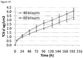
- Some molecularly-thin composite film embodiments of the present invention can be tuned to contain as low as 0.4 ⁇ g/cm 2 of soluble silver and release a total of less than 1 ppm silver ions in buffers. Such films display 99.9999 % bacteria-killing efficiency without any measurable cytotoxicity to mammalian cells. Furthermore, films containing these levels of silver allow adherence and growth of mammalian cells (e.g., NIH-3T3 mouse fibroblasts).
- mammalian cells e.g., NIH-3T3 mouse fibroblasts.
- the releasable antimicrobial silver composition is loaded at an amount of from 1 or 10 to about 50, 100, 200, 300, 400, 500 or 1000 ⁇ g/cm 2 of the polymer multilayer and/or releasable in an amount of about 0.01 and 100 ⁇ g/cm 2 of the polymer multilayer per day. In some embodiments, the releasable antimicrobial silver composition is releasable in an amount of about 0.05 and 100 ⁇ g/cm 2 of the polymer multilayer. In some embodiments, the releasable antimicrobial silver composition is releasable in an amount of about 0.05 and 20 ⁇ g/cm 2 of the polymer multilayer.
- the releasable antimicrobial silver composition is releasable in an amount of about 0.05 and 5 ⁇ g/cm 2 of the polymer multilayer. In some embodiments, the releasable antimicrobial silver composition is releasable in an amount of about 0.05 and 2 ⁇ g/cm 2 of the polymer multilayer. In some embodiments, the releasable antimicrobial silver composition is releasable in an amount of about 0.05 and 1 ⁇ g/cm 2 of the polymer multilayer. In some embodiments, the releasable antimicrobial silver composition is releasable in an amount of about 0.1 to 0.5 ⁇ g/cm 2 of the polymer multilayer.
- ActicoatTM releases a total of over 120 ppm silver on dissolution in water for 24 hr ( Taylor et al., 2005, Biomaterials 26:7221 ), and has been shown to be cytotoxic to keratinocytes and fibroblasts ( Poon et al., 2005, Burns 30:140 ; Trop et al., 2006, J. Trauma 60:648 ).
- the silver-releasing moleculary-thin composite films as manufactured by the process of the present invention have advantage over: 1) other topical delivery agents of silver currently used in clinics; 2) wound-dressings that contain large amounts of silver in them; and 3) other bactericidal composite thin-films containing large amounts of silver, which are cytotoxic to mammalian cells and do not allow mammalian cells to adhere and grow over them.
- Using silver-nanoparticle molecularly-thin films for localized delivery of bactericidal silver in wounds can overcome silver-related toxicity encountered with topical silver-delivery agents for wound healing ( Poon et al., 2004, Burns 30:140 ; Baldi et al., 1988, Toxicol. Lett. 41:261 ; Atiyeh et al., 2007, Burns 33:139 ; Coombs et al., 1992, Burns 18:179 ).
- these polyelectrolyte multilayers are uniform, highly interpenetrated ultrathin nanocomposite films, typically far less than 1 ⁇ m thick ( Decher, 1997, Science 277:1232 ; Hammond, 1999, Curr. Opin. Colloid Interface Sci. 4:430 ).
- Their porous and supramolecular architecture allows incorporating a variety of molecules including DNA, enzymes, viruses, dendrimers, colloids, inorganic particles, and dyes ( Decher, 1997, Science 277:1232 ).
- they can be used for simultaneous localized delivery of other bioactive agents in the wound-matrix (e.g., cell recruiting growth factors, DNA plasmids encoding such growth factors).
- PEMs can conformally coat substrates of any type, size or shape, including natural and synthetic polymers ( Hammond, 1999, Curr. Opin. Colloid Interface Sci. 4:430 ), they can be constructed on any wound-bed or implantable medical device.
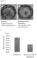
- Figure 1 provides a schematic diagram 100 of a wound bed 110 on which a polyelectrolyte multilayer 130 has been deposited.
- the diagram 100 depicts that the wound bed 110 comprises a heterogeneous surface depicted by shapes 120 which represent different chemical moieties.
- the polyelectrolyte multilayer 130 provides a homogenous surface onto which functional groups can 140 can be attached to form a homogenous functionalized surface 150.
- the functional groups are uniform, however, in some preferred embodiments, different functional groups are utilized.
- a wide variety of active agents can then be attached to the surface via the functional groups 140.
- the wound bed is covalently modified with covalent modification agents.
- the covalent modification agents include, but are not limited to, homobifunctional and heterobifunctional cross-linkers as well as photoactivatable cross linkers.
- Figure 2 provides a schematic diagram 200 of a wound bed 210 comprising a heterogeneous surface depicted by shapes 220 which represent different chemical moieties.
- the wound bed is covalently modified by reacting covalent modification agents 230 with the different chemical moieties to provide a relatively homogenous functionalized surface 250.
- covalent modification agents 230 present functional groups 240.
- the functional groups are uniform, however, in some preferred embodiments, different functional groups are utilized.
- a wide variety of active agents can then be attached to the surface via the functional groups 240. These embodiments are discussed in more detail below.
- a wound active nanoscale polymer matrix microsheet can be manufactured as claimed in claim 1, wherein the wound active nanoscale polymer layer is a polymer multilayer.
- the multilayer structures comprise layers of polymers that form polyelectrolytes, while in other embodiments, the multilayers comprise polymers that do not have a charge (i.e., non-ionic polymers) or a combination of charged and uncharged polymer layers.
- polyelectrolyte films built-up by the alternated adsorption of cationic and anionic polyelectrolyte layers constitute a novel and promising technique to modify wound surfaces in a controlled way ( Decher et al., 1992, Thin Solid Films 210/211:831 ; Decher, 1997, Science 277:1232 ).
- One of the most important properties of such multilayers is that they exhibit an excess of alternatively positive and negative charges ( Caruso et al., 1999, J Am Chem Soc 121:6039 ; Ladam et al., 2000, Langmuir 16:1249 ). Not only can this constitute the motor of their buildup ( Joanny, 1999, Eur.
- the polymer multilayers are nanoscale in dimension. Accordingly, in some embodiments, the polymer multilayers are from about 1 nm to 1000 nm thick, from about 1 nm to 500 nm thick, from about 1 nm to 100 nm thick, from about 1 nm to about 25 nm thick, from about 1 nm to about 10 nm thick, or less than about 500 nm, 100 nm, 25 nm or 10 nm thick.
- the nanoscale dimension of the polymer multilayers allows for the loading of a lower total amount of an active agent while still allowing delivery of an effective amount (i.e., an amount of active agent that accelerates wound healing as compared to controls) of the active agent as compared to matrix structures with greater thickness. It is contemplated that the lower total loading levels result in reduced toxicity in the wound environment, especially when antimicrobial compounds are incorporated into the polymer multilayer.
- the compliance of the polymer multilayers is adjusted to facilitate cell migration in the wound.
- the polymer multilayers exhibit a compliance, measured in kilopascals (kPa) of from about 3 to about 500 kPa, about 7 to about 250 kPa, about 10 to about 250 kPA or from about 10 to about 200 kPa.
- PLL cationic polyelectrolyte poly(L-lysine)
- Polyelectrolyte layers are formed by alternating applications of anionic polyelectrolytes and cationic polyelectrolytes to surfaces to form a polyelectrolyte layer.
- the layers can be used to deliver a wound active agent to a wound.
- at least four layers, and, more preferably, at least six layers are used to form the polyelectrolyte multilayer.
- more than six layers are used, e.g., 7, 8, 9, 10, 15, 20, 25, 30, 35, 40, 45, 50 or more layers.
- a polyelectrolyte multilayer comprises 10.5 layers (e.g., comprising 10 bilayers of the two components and an additional layer of one of the two components).
- the cationic polyelectrolyte used is PLL and the anionic polyelectrolyte used is poly(L-glutamic acid) (PGA).
- PGA poly(L-glutamic acid)
- PES poly(ethylene imine)
- PAH poly(allylamine hydrochloride)
- PSS poly(sodium 4-styrenesulfonate)
- PAC poly(acrylic acid)
- PMA-P poly(maleic acid-co-propylene)
- PAA poly(acrylic acid)
- PVS poly(vinyl sulfate)
- PVS poly(vinyl sulfate)
- the polymer is a dendrimer, grafted polymer, or star architecture polymer.
- the multilayer responds to or is organized in the presence of an electric field, for example an electric field formed by placing electrodes on either side of a wound.
- the polymer multilayer 130 can be comprised of polystyrene sulfonate, an amphiphillic polyelectrolyte with an affinity for hydrophobic regions of the wound bed, and an amphoteric polymer.
- the polymer multilayer is preferably functionalized with one or more crosslinking agents presenting an alkyne so that a uniform surface is presented (e.g., 140 in Figure 1 where x represents an alkyne group). From this point, widely available click chemistries can be used to add desired wound active agents to the modified surface of the wound.
- the wound modifying agent is an azide conjugate or otherwise comprises an azide group and is reacted with the alkyne groups displayed on the wound bed in a Huisgen Cycloaddition.
- Suitable methods for preparing polyelectrolyte multilayers include those described, for example, in Cho and Char, Langmuir 20:4011-4016, 2004 ; Okamura et al., Adv. Mater. 21, 4388-92 (2009 ), Cho et al., Adv. Mat. 13(14):1076-1078 (2001 ); and U.S. pat. Publ. 2010/0062258 .
- Suitable methods include layer by layer deposition, formation on SAMs, and spin coating assisted assembly.
- Cationic polymers useful in the present invention can be any biocompatible water-soluble polycationic polymer, for example, any polymer having protonated heterocycles attached as pendant groups.
- water soluble means that the entire polymer must be soluble in aqueous solutions, such as buffered saline or buffered saline with small amounts of added organic solvents as co-solvents, at a temperature between 20 and 37°C.
- the material will not be sufficiently soluble (defined herein as soluble to the extent of at least one gram per liter) in aqueous solutions per se but can be brought into solution by grafting the polycationic polymer with water-soluble polynonionic materials such as polyethylene glycol.
- Representative cationic polymers include natural and unnatural polyamino acids having net positive charge at neutral pH, positively charged polysaccharides, and positively charged synthetic polymers.
- suitable polycationic materials include polyamines having amine groups on either the polymer backbone or the polymer side chains, such as poly-L-lysine (PLL) and other positively charged polyamino acids of natural or synthetic amino acids or mixtures of amino acids, including, but not limited to, poly(D-lysine), poly(ornithine), poly(arginine), and poly(histidine), and nonpeptide polyamines such as poly(aminostyrene), poly(aminoacrylate), poly (N-methyl aminoacrylate), poly (N-ethylaminoacrylate), poly(N,N-dimethyl aminoacrylate), poly(N,N-diethylaminoacrylate), poly(aminomethacrylate), poly(N-methyl amino-methacrylate), poly(N-ethyl amino methacrylate), poly(N,
- the polymers must include at least five charges, and the molecular weight of the polycationic material must be sufficient to yield the desired degree of binding to a tissue or other surface, having a molecular weight of at least 1000 g/mole.
- Polyanionic materials useful in the present invention can be any biocompatible water-soluble polyanionic polymer, for example, any polymer having carboxylic acid groups attached as pendant groups. Suitable materials include alginate, carrageenan, furcellaran, pectin, xanthan, hyaluronic acid, heparin, heparan sulfate, chondroitin s ulfate, dermatan sulfate, dextran sulfate, poly(meth)acrylic acid, oxidized cellulose, carboxymethyl cellulose and crosmarmelose, synthetic polymers and copolymers containing pendant carboxyl groups, such as those containing maleic acid or fumaric acid in the backbone.
- Suitable materials include alginate, carrageenan, furcellaran, pectin, xanthan, hyaluronic acid, heparin, heparan sulfate, chondroitin s ulfate, dermatan sulfate
- Polyaminoacids of predominantly negative charge are also suitable.
- these materials include polyaspartic acid, polyglutamic acid, and copolymers thereof with other natural and unnatural amino acids.
- Polyphenolic materials such as tannins and lignins can be used if they are sufficiently biocompatible.
- Preferred materials include alginate, pectin, carboxymethyl cellulose, heparin and hyaluronic acid.
- the anionic polymer is poly(acrylic acid) (PAA).
- the multilayer structures are formed from uncharged polymers or from a combination of charged and uncharged polymers.
- uncharged polymers include, but are not limited to, dextran, dextran sulfate, diethylaminoethyl (DEAE)-dextran, hydroxyethyl cellulose, ethyl(hydroxyethyl) cellulose, acrylamide, polyethylene oxide, polypropylene oxide, polyethylene oxide - polypropylene oxide copolymers, PAAN a , Ficoll, polyvinylpyrolidine, and polyacrylic acid.
- DEAE diethylaminoethyl
- the multilayer structures are formed from one or more amphoteric polymers, alone in combination with the other polymers described herein.
- the amphoteric polymers comprise one or more of acrylic acid (AA), DMAEMA (dimethylaminoethyl methacrylate), APA (2-aminopropyl acrylate), MorphEMA (morpholinoethyl methacrylate), DEAEMA (diethylaminoethyl methacrylate), t-ButylAEMA (t-butylaminoethyl methacrylate), PipEMA (piperidinoethyl methacrylate), AEMA (aminoethyl methacrylate), HEMA (2-hydroxyethyl methacrylate), MA (methyl acrylate), MAA (methacrylic acid) APMA (2-aminopropyl methacrylate), AEA (aminoethyl acrylate).
- the amphoteric polymer comprises (a) carboxylic acid, (b) primary amine, and (c) secondary and/or tertiary amine.
- the amphoteric polymers have an isoelectric point of 4 to 8, preferably 5 to 7 and have a number average molecular weight in the range of 10,000 to 150,000.
- a polymer multilayer is produced on a supporting substrate (e.g., PDMS), removed from the supporting substrate, then laid on a wound.
- a polymer multilayer is produced on a supporting substrate (e.g., PDMS), overlaid with a dissolvable sacrificial support layer, e.g., comprising a biocompatible polymer (e.g., polyvinyl alcohol (PVA)).
- PVA polyvinyl alcohol
- the sacrificial support layer facilitates the removal of the polymer multilayer from the supporting substrate, facilitates manual handling of the polymer multilayer, and/or facilitates application of the polymer multilayer on a wound. Then, after placement, the support layer dissolves and thus promotes the adherence of the polymer multilayer to the micro-topography and nano-topography of the wound.
- Wound active agents can be delivered to a wound bed or incorporated into a wound bed using the systems described above that have been manufactured with the process of the invention .
- the wound active agent is bound covalently or non-covalently with the polyelectrolyte layer,.
- the present invention is not limited to a particular mechanism by which the wound active agent binds to the polyelectrolyte layer.
- the polyelectrolyte layer may function as a drug delivery scaffold to deliver one or more wound active agents to the wound.
- Wound active agents that may be desirable to deliver include, but are not limited to, trophic factors, extracellular matrices (ECMs), ECM fragments or synthetic constructs, enzymes, enzyme inhibitors, defensins, polypeptides, anti-infective agents (including antimicrobials, antivirals and antifungals), buffering agents, vitamins and minerals, analgesics, anticoagulants, coagulation factors, anti-inflammatory agents, vasoconstrictors, vasodilators, diuretics, and anti-cancer agents.
- active agents include chlorhexidine, iodine based antimicrobials such as PVP-iodine; selenium based antimicrobials such as 7-azabenzisoselenazol-3(2H)-ones, selenium disulfide, and selenides; and silver based antimicrobials (e.g., silver sulfadiazine, ionic silver, elemental silver, silver nanoparticles)).
- iodine based antimicrobials such as PVP-iodine
- selenium based antimicrobials such as 7-azabenzisoselenazol-3(2H)-ones, selenium disulfide, and selenides
- silver based antimicrobials e.g., silver sulfadiazine, ionic silver, elemental silver, silver nanoparticles
- selenides with the use of standard and variations of typical protein and carbohydrate attachment chemistries, carboxyl and amino containing selenides may be
- wound active agents can be incorporated into the polyelectrolyte layer, wherein the wound active agents are released from the polyelectrolyte layer into the wound.
- wound active agents can be trophic factors, including, but not limited to, agrin, amphiregulin, artemin, cardiotrophin-1, epidermal growth factors including EGF; fibroblast growth factors (e.g., FGF-1, FGF-2, FGF-3, FGF-4, FGF-5, FGF-6, and FGF-7); LIF, CSF-1, CSF-2, CSF-3, erythropoietin, endothelial cell growth factors including ECGF; FGF- related and ECGF-related growth factors (e.g., endothelial cell stimulating angiogenesis factor, tumor angiogenesis factor, retina-derived growth factor (RDGF), vascular endothelium growth factor (VEGF), brain-derived growth factors (BDGF-A and B), astroglial growth factors (AGF 1 and 2), omentum-derived growth factor, fibroblast-stimulating factor (FSF), and embryonal carcinoma-derived growth factor (ECDGF)); neurotrophic growth factors (e.g.
- the wound active agents are integrin binding sequences exemplified by, but not limited to RGD, EILDV, VCAM-1 and their recombined or synthetic analogs, enzymes, enzyme inhibitors, and polypeptides.
- the enzymes include exopeptidases and endopeptidases (also known as proteases and proteinases), including but not limited to the serine proteinases chymotrypsin, trypsin, elastase, and kallikrein, bacterial enzymes, the cysteine proteases papain, actinin, bromelain, cathepsins, cytosolic calpains, parasitic proteases, aspartic proteinases, the pepsin family of proteases pepsin and chymosin, lysosomal cathepsins D, renin, fungal proteases, the viral proteases, AIDS virus retropepsin, and the metalloproteinases (MMPs), collagenases, Maggott enzyme, MMP1, MMP2, MMP8, MMP13, gelatinases, MMP2, MMP9, MMP3, MMP7, MMP10, MMP11, and MMP12.
- MMPs metalloproteinases
- the enzyme inhibitors include captopril, thiorphan, phosphoramidon, teprotide, protease and proteinase inhibitors, metalloproteinase inhibitors and exopeptidase inhibitors.
- defensins including, but not limited to, alpha-defensins HNP 1, 2, 3 and 4, and beta-defensins HBD-1 and HBD-2 are used, or polypeptides such as fibronectin, serotonin, PAF, PDEGF, TNFa, IL1, IL6, IGF, IGF-1, IGF-2, IL-1, PDGF, FGF, KGF, VEGF, bradykinin, prothymosin-alpha, and thymosin-alpha1, or antimicrobials, including, but not limited to, magainin (e.g., magainin I, magainin II, xenopsin, xenopsin precursor fragment, caerulein precursor fragment), magainin I and II analogs (e.g., PGLa, magainin A, magainin G, pexiganin, Z-12, pexigainin acetate, D35, MSI-78A, MG0 (K10), magainin I
- antimicrobial peptides are synthesized from L-amino acids, while in other embodiments, the peptides are synthesized from, or comprise, D-amino acids. Additional antimicrobial polypeptides of use in the present invention are listed in Figure 7 .
- the antimicrobials include loracarbef, cephalexin, cefadroxil, cefixime, ceftibuten, cefprozil, cefpodoxime, cephradine, cefuroxime, cefaclor, neomycin/polymyxin/bacitracin, dicloxacillin, nitrofurantoin, nitrofurantoin macrocrystal, nitrofurantoin/nitrofuran mac, dirithromycin, gemifloxacin, ampicillin, gatifloxacin, penicillin V potassium, ciprofloxacin, enoxacin, amoxicillin, amoxicillin/clavulanate potassium, clarithromycin, levofloxacin, moxifloxacin, azithromycin, sparfloxacin, cefdinir, ofloxacin, trovafloxacin, lomefloxacin, methenamine, erythromycin, norfloxacin, clindamycin/
- antivirals include amantadine, acyclovir, foscarnet, indinavir, ribavirin, enfuvirtide, emtricitabine, lamivudine, abacavir sulfate, fomivirsen, valacyclovir, tenofovir, cidofovir, atazanavir, amprenavir, delavirdine mesylate, famciclovir, adefovir, didanosine, efavirenz, trifluridine, inidinavir, lamivudine, vidarabine, lopinavir/ritonavir, ganciclovir, zanamivir, abacavir/lamivudine/zidovudine, lamivudine/zidovudine, nelfinavir, nelfinavir mesylate, nevirapine, ritonavir
- antifungals includeamphotericin B, nystatin, nystatin/triamcinolone, itraconazole, ketoconazole, miconazole, sulconazole, clotrimazole, clotrimazole/betamethasone, enilconazole, econazole, oxiconazole, tioconazole, terconazole, butoconazole, thiabendazole, flucytosine, butenafine, ciclopirox, haloprogin, naftifine, tolnaftate, natamycin, undecylenic acid, mafenide, dapsone, clioquinol, clioquinol/hydrocortisone, potassium iodide, silver sulfadiazine, gentian violet, carbol-fuchsin, cilofungin, sertaconazole, voriconazole, fluconazole, ter
- the buffering agents include Maleic acid, Phosphoric acid, Glycine, Chloroacetic acid, Formic acid, Benzoic acid, Acetic acid, Pyridine, Piperazine, MES, Bis-tris, Carbonate, ACES, ADA MOPSO, PIPES, Phosphoric acid, BES, MOPS, TES, HEPES, DIPSO, TAPSO, Triethanolamine, HEPSO, Tris, Tricine, Bicine, TAPS, Borate, Ammonia, CHES, Ethanolamine, CAPSO, Glycine, Carbonate, CAPS, Methylamine, Piperidine, and Phosphoric acid.
- the vitamins and minerals include Vitamin A, Carotenoids, Vitamin D, Vitamin E, Vitamin K, Vitamin C/ascorbic acid, B1/thiamin, B2/riboflavin, B3/niacin, B5/pantothenic acid, B6/pyridoxine, B12/cobalamin, Biotin, Calcium, Magnesium, Phosphorus, Sodium, Chloride, Potassium, Boron, Chromium, Copper, Iodine, Iron, Manganese, Selenium, and Zinc.
- the analgesics include acetaminophen, anileridine, acetylsalicylic acid, buprenorphine, butorphanol, fentanyl, fentanyl citrate, codeine, rofecoxib, hydrocodone, hydromorphone, hydromorphone hydrochloride, levorphanol, alfentanil hydrochloride, meperidine, meperidine hydrochloride, methadone, morphine, nalbuphine, opium, levomethadyl, hyaluronate sodium, sufentanil citrate, capsaicin, tramadol, leflunomide, oxycodone, oxymorphone, celecoxib, pentazocine, propoxyphene, benzocaine, lidocaine, dezocine, clonidine, butalbital, phenobarbital, tetracaine, phenazopyridine, sulfameth
- the anticoagulants include coumarins, 1,3-indandione, anisindione, fondaparinux, heparin, lepirudin, antithrombin, warfarin, enoxaparin, dipyridamole, dalteparin, ardeparin, nadroparin, and tinzaparin.
- the coagulation factors includeFactor I (fibrinogen), Factor II (prothrombin), Factor III (thromboplastin, tissue factor), Factor IV (calcium), Factor V (labile factor), Factor VII (stable factor), Factor VIII (antihemophilic globulin, antihemophilic globulin, antihemophilic factor A), Factor IX (plasma thromboplastin component, Christmas factor, antihemophilic factor B), Factor X (Stuart factor, Prower factor, Stuart-Prower factor), Factor XI (plasma thromboplastin antecedent, antihemophilic factor C), Factor XII (Hageman factor, surface factor, contact factor), and Factor XIII (fibrin stabilizing factor, fibrin stabilizing enzyme, fibri-nase).
- Factor I fibrinogen
- Factor II prothrombin
- Factor III thromboplastin, tissue factor
- Factor IV calcium
- Factor V labile factor
- the anti-inflammatory agents include non steroidal anti-inflammatory drugs (NSAIDs) including diclofenac (also known as Voltaren, Abitren, Allvoran, Almiral, Alonpin, Anfenax, Artrites, Betaren, Blesin, Bolabomin, Cataflam, Clofec, Clofen, Cordralan, Curinflam, Diclomax, Diclosian, Dicsnal, Difenac, Ecofenac, Hizemin, Inflamac, Inflanac, Klotaren, Lidonin, Monoflam, Naboal, Oritaren, Remethan, Savismin, Silino, Staren, Tsudohmin, Voltarol, Voren, Voveran, and Vurdon), diflunisal (also known as Dolobid, Adomal, Diflonid, Diflunil, Dolisal, Dolobis, Dolocid, Donobid, Dopanone, Dorbid, Dugodol,
- vasoconstrictors include epinephrine (adrenaline, Susphrine), phenylephrine hydrochloride (Neo-Synephrine), oxymetazoline hydrochloride (Afrin), norepinephrine (Levophed), and caffeine.
- vasodilators include bosentan (Tracleer), epoprostenol (Flolan), treprostinil (Remodulin), sitaxsentan, nifedipine (Adalat, Procardia), nicardipine (Cardene), verapamil (Calan, Covera-HS, Isoptin, Verelan), diltiazem (Dilacor XR, Diltia XT, Tiamate, Tiazac, Cardizem), isradipine (DynaCirc), nimodipine (Nimotop), amlodipine (Norvasc), felodipine (Plendil), nisoldipine (Sular), bepridil (Vascor), hydralazine (Apresoline), minoxidil (Loniten), isosorbide dinitrate (Dilatrate-SR, Iso-Bid, Isonate, Isorbid,
- diuretics include acetazolamide (Diamox), dichlorphenamide (Daranide), methazolamide (Neptazane), bendroflumethiazide (Naturetin), benzthiazide (Exna), chlorothiazide (Diuril), chlorthalidone (Hygroton), hydrochlorothiazide (Esidrix, HydroDiuril, Microzide), hydroflumethiazide (Diucardin), indapamide (Lozol), methyclothiazide (Enduron), metolazone (Zaroxolyn, Mykrox), polythiazide (Renese), quinethazone (Hydromox), trichlormethiazide (Naqua), bumetanide (Bumex), ethacrynic acid (Edecrin), furosemide (Lasix), torsemide (Demadex), amiloride (M
- anti-cancer agents include aldesleukin, alemtuzumab, alitretinoin, allopurinol, altretamine, amifostine, anagrelide, anastrozole, arsenic trioxide, asparaginase, bexarotene, bicalutamide, bleomycin, busulfan, calusterone, capecitabine, carboplatin, carmustine, celecoxib, chlorambucil, cisplatin, cladribine, cyclophosphamide, cytarabine, dacarbazine, dactinomycin, darbepoetin alpha, daunorubicin, daunomycin, dexrazoxane, docetaxel, doxorubicin, epoetin alpha, estramustine, etoposide, etoposide phosphate, exemestane, filgrastim, floxur
- the wound active agent is an siRNA.
- the RNAi constructs of the present invention are gene(s) that express RNAs that base pair to form a dsRNA RNA region.
- the RNAs may be a part of the same molecule or different molecules.
- the RNAi construct comprises a promoter operably linked to a nucleic acid sequence encoding two complementary sequences separated by a loop sequence.
- the complementary regions correspond to a target RNA sequence separated by a loop sequence.
- the RNAi construct is expressed, the complementary regions of the resulting RNA molecule pair with one another to form a double stranded RNA region.
- the present invention is not limited to loop sequences of any particular length.
- the loop sequences range from about 4 to about 20 nucleotides in length. In more preferred embodiments, the loop sequences are from about 6 to about 12 nucleotides in length. In other preferred embodiments, the dsRNA regions are from about 19 to about 23 in length.
- the dsRNA is formed from RNA transcribed from a vector as two separate stands.
- the two strands of DNA used to form the dsRNA may belong to the same or two different duplexes in which they each form with a DNA strand of at least partially complementary sequence.
- the DNA sequence to be transcribed is flanked by two promoters, one controlling the transcription of one of the strands, and the other that of the complementary strand. These two promoters may be identical or different.
- a DNA duplex provided at each end with a promoter sequence can directly generate RNAs of defined length, and which can join in pairs to form a dsRNA. See, e.g., U.S. Pat. No. 5,795,715 .
- RNA duplex formation may be initiated either inside or outside the cell.
- the resulting siRNA can comprise two blunt ends, one blunt end and one end with an overhang, or two ends with overhangs.
- the end or ends with overhangs comprise an overhang of either one or two nucleotides.
- a siRNA of 23 nucleotides in length comprises two 19mers with a two nucleotide overhang at each end.
- a siRNA of 21 nucleotides in length comprises two 19mers with a single nucleotide overhang at each end.
- a siRNA of 22 nucleotides in length comprises two 22mers with no overhangs at either end.
- Inhibition is sequence-specific in that nucleotide sequences corresponding to the duplex region of the RNA are targeted for genetic inhibition. RNA molecules containing a nucleotide sequence identical to a portion of the target gene are preferred for inhibition. RNA sequences with insertions, deletions, and single point mutations relative to the target sequence have also been found to be effective for inhibition.
- sequence identity may optimized by sequence comparison and alignment algorithms known in the art (see Gribskov and Devereux, Sequence Analysis Primer, Stockton Press, 1991 , and references cited therein) and calculating the percent difference between the nucleotide sequences by, for example, the Smith-Waterman algorithm as implemented in the BESTFIT software program using default parameters (e.g., University of Wisconsin Genetic Computing Group).
- the duplex region of the RNA may be defined functionally as a nucleotide sequence that is capable of hybridizing with a portion of the target gene transcript.
- the dsRNA can range from about 21 base pairs (bp) of the gene to the full length of the gene or more.
- the dsRNA used in the methods of the present invention is about 1000 bp in length.
- the dsRNA is about 500 bp in length.
- the dsRNA is about 22 bp in length.
- the sequences that mediate RNAi are from about 21 to about 23 nucleotides.
- the isolated iRNAs of the present invention mediate degradation of the target RNA.
- the double stranded RNA of the present invention need only be sufficiently similar to natural RNA that it has the ability to mediate RNAi for the target RNA.
- the present invention relates to RNA molecules of varying lengths that direct cleavage of specific mRNA to which their sequence corresponds. It is not necessary that there be perfect correspondence of the sequences, but the correspondence must be sufficient to enable the RNA to direct RNAi cleavage of the target mRNA.
- the RNA molecules of the present invention comprise a 3' hydroxyl group.
- microsheets comprising a nanoscale polymer matrix that can be applied to a wound, a biologic tissue, a cornea, a lens, a bone, a tendon, a surgical mesh, a wound dressing, a biomedical device, a device used for healthcare, or other surface.
- the nanoscale polymer matrix is functionalized.
- the nanoscale polymer matrix is not functionalized.
- the nanoscale polymer matrix comprises one or more polymers, preferably biocompatible, or is formed from one or more proteins, or is a combination of polymers and proteins.
- the nanoscale polymer layer is formed from synthetic polymers such as synthetic polyelectrolytes. In other embodiments, the nanoscale polymer layer is formed from naturally occurring polymers such as polysaccharides.
- the nanoscale polymer matrix is functionalized to allow for covalent interaction and/or binding to the tissue surface or the wound bed, or to allow application of - bioactive agents to the nanoscale polymer matrix.
- a - bioactive agent being a wound active agent, for example an antimicrobial agent such as silver, polyhexamethylene biguanide (PHMB), - chlorhexidine, or iodine compound, or an antibiotic, is incorporated into the nanoscale polymer matrix.
- the - bioactive agent is preferably impregnated, incorporated or interspersed throughout the three dimensional structure of the nanoscale polymer matrix.
- the nanoscale polymer matrix is polyelectrolyte multilayer (PEM)
- the - bioactive agent is preferably incorporated between or within the layers of the polymer multilayer.
- a polymer matrix (e.g., a nanoscale polymer matrix and/or a a sacrificial polymer layer) comprising an antibiofilm agent is manufactured.
- the technology is not limited in the antibiofilm agent that is used in embodiments of the device and associated method, kit, and method of treatment embodiments.
- the antibiofilm agent is a small molecule antibiofilm agent, a charged small molecule antibiofilm agent, an antibiofilm polypeptide, an antibiofilm enzyme (e.g., Dispersin B), a metallic particle, or a metal ion antibiofilm agent (e.g., a metal ion, metal ion salt, or metal ion nanoparticle).
- the metal ion antibiofilm agent is a gallium ion, a gallium ion salt, a gallium ion nanoparticle, an alloy of gallium, or an alloy of gallium and silver.
- the nanoscale polymer matrices are nanoscale to microscale in dimension. Accordingly, in some embodiments, the nanoscale polymer matrices are from about 1 nm to 1000 nm thick, from about 1 nm to 500 nm thick, from about 1 nm to 100 nm thick, from about 1 nm to about 25 nm thick, from about 1 nm to about 10 nm thick, or less than about 500 nm, 100 nm, 25 nm or 10 nm thick.
- the nanoscale dimension of the matrices allows for the loading of a lower total amount of an active agent while still allowing delivery of an effective amount (i.e., an amount of active agent that accelerates wound healing as compared to controls) of the active agent as compared to matrix structures with greater thickness. It is contemplated that the lower total loading levels result in reduced toxicity in the wound environment, especially when antimicrobial compounds are incorporated into the polymer multilayer.
- the compliance of the nanoscale polymer matrices is adjusted to facilitate cell migration in the wound.
- the matrices have a compliance, measured in kilopascals (kPa) of from about 100 Pa to about 500 kPa, about 7 to about 250 kPa, about 10 to about 250 kPA or from about 10 to about 200 kPa.
- the matrices, e.g., polymer multilayers have a compliance of from about 3 kPa to 5, 4, 3, 2, 1, 0.5, 0.1 and 0.05 GPa (gigapascals).
- the nanoscale polymer matrix is supported on a sacrificial polymer layer, formed from a degradable or dissolvable support material such as a dissolvable polymer.
- a sacrificial polymer layer is formed on the nanoscale polymer matrix, for example, by spin coating, spraying or casting the sacrificial polymer layer material on the nanoscale polymer matrix.
- the sacrificial polymer layer material is dissolvable in aqueous environments or environments where moisture is present, such as moist surfaces like wound beds, internal body surfaces, epithelial surfaces and the like.
- the sacrificial polymer layer material is dissolvable in aqueous solutions after application of the nanoscale polymer matrix on the surface.
- the present process provides a microsheet article comprising two layers, a first nanoscale polymer matrix layer adjacent to a sacrificial polymer layer. The sacrificial polymer layer dissolves when applied to a moist surface so that the nanoscale polymer matrix is left in contact with the moist surface.
- the nanoscale polymer matrix can be transferred to a wound or tissue using a sacrificial polymer layer that lies on top of nanoscale polymer matrix after transfer to the wound or tissue, or the nanoscale polymer matrix is transferred to a wound or tissue using a sacrificial polymer layer that lies underneath the nanoscale polymer matrix after transfer to the wound or tissue.
- a nanoscale polymer matrix made with PEMs according to a process of the invention and a dissolvable sacrificial cast can be transferred to a wound or tissue surface such that sacrificial cast dissolves completely in wound and PEMs are in direct contact with the wound tissue and a primary/secondary wound can be placed over the wound.
- the nanoscale polymer matrix containing one or more bioactive agents can be transferred to a wound or tissue using a sacrificial polymer layer that lies on top of nanoscale polymer matrix after transfer to the wound or tissue.
- a wound dressing can be placed on top of the sacrificial polymer layer before or after the sacrificial polymer layer is dissolved.
- the nanoscale polymer matrix is a polyelectrolyte multilayer (PEM).
- PEM polyelectrolyte multilayer
- the sacrificial polymer layer of a microsheet contains bioactive agents, antimicrobial agents, antibiofilm agents, microparticles, nanoparticles, magnetic particles.
- microparticles or nanoparticles in the sacrificial polymer layer contain bioactive agents or antimicrobial agents.
- the sacrificial layer comprises an antibiofilm agent.
- the technology is not limited in the antibiofilm agent that is used in embodiments of the device and associated method, kit, and method of treatment embodiments.
- the antibiofilm agent is a small molecule antibiofilm agent, a charged small molecule antibiofilm agent, an antibiofilm polypeptide, an antibiofilm enzyme (e.g., Dispersin B), a metallic particle, or a metal ion antibiofilm agent (e.g., a metal ion, metal ion salt, or metal ion nanoparticle).
- the metal ion antibiofilm agent is a gallium ion, a gallium ion salt, a gallium ion nanoparticle, an alloy of gallium, or an alloy of gallium and silver.
- the sacrificial polymer layer of a microsheet is water soluble.
- the sacrificial polymer layer is made of non-toxic polymer, and in some embodiments the sacrificial polymer layer is poly vinyl alcohol (PVA).
- the sacrificial polymer layer is made of polyacrylic acid (PAA).
- the matrix is a polymer multilayer.
- the multilayer structures comprise layers of polyelectrolytes (i.e., forming a polyelectrolyte multilayer), while in other embodiments, the multilayers comprise polymers that do not have a charge (i.e., non-ionic polymers) or a combination of charged and uncharged polymer layers.
- polyelectrolyte films built-up by the alternated adsorption of cationic and anionic polyelectrolyte layers constitute a novel and promising technique to modify wound surfaces in a controlled way ( Decher et al., 1992, Thin Solid Films 210/211:831 ; Decher, 1997, Science 277:1232 ).
- One of the most important properties of such multilayers is that they exhibit an excess of alternatively positive and negative charges ( Caruso et al., 1999, J Am Chem Soc 121:6039 ; Ladam et al., 2000, Langmuir 16:1249 ). Not only can this constitute the motor of their buildup ( Joanny, 1999, Eur.
- Polyelectrolyte layers are formed by alternating applications of anionic polyelectrolytes and cationic polyelectrolytes to surfaces to form a polyelectrolyte multilayer.
- one or more wound active agents such as those described above, are incorporated into the multilayer.
- at least four layers, and, more preferably, at least six layers are used to form the polyelectrolyte multilayer.
- Cationic polyelectrolytes useful in the present invention can be any biocompatible water-soluble polycationic polymer, for example, any polymer having protonated heterocycles attached as pendant groups.
- water soluble means that the entire polymer must be soluble in aqueous solutions, such as buffered saline or buffered saline with small amounts of added organic solvents as co-solvents, at a temperature between 20 and 37° Centigrade.
- the material will not be sufficiently soluble (defined herein as soluble to the extent of at least one gram per liter) in aqueous solutions per se but can be brought into solution by grafting the polycationic polymer with water-soluble polynonionic materials such as polyethylene glycol.
- Representative cationic polyelectrolytes include natural and unnatural polyamino acids having net positive charge at neutral pH, positively charged polysaccharides, and positively charged synthetic polymers.
- suitable polycationic materials include polyamines having amine groups on either the polymer backbone or the polymer side chains, such as poly-L-lysine (PLL) and other positively charged polyamino acids of natural or synthetic amino acids or mixtures of amino acids, including, but not limited to, poly(D-lysine), poly(ornithine), poly(arginine), and poly(histidine), and nonpeptide polyamines such as poly(aminostyrene), poly(aminoacrylate), poly (N-methyl aminoacrylate), poly (N-ethylaminoacrylate), poly(N,N-dimethyl aminoacrylate), poly(N,N-diethylaminoacrylate), poly(aminomethacrylate), poly(N-methyl amino-methacrylate), poly(N-ethyl amino methacrylate),
- the polymers must include at least five charges, and the molecular weight of the polycationic material must be sufficient to yield the desired degree of binding to a tissue or other surface, having a molecular weight of at least 1000 g/mole.
- Polyanionic materials useful in the present invention can be any biocompatible water-soluble polyanionic polymer, for example, any polymer having carboxylic acid groups attached as pendant groups. Suitable materials include alginate, carrageenan, furcellaran, pectin, xanthan, hyaluronic acid, heparin, heparan sulfate, chondroitin sulfate, polyacrylic acid (PAA), dermatan sulfate, dextran sulfate, poly(meth)acrylic acid, oxidized cellulose, carboxymethyl cellulose and crosmarmelose, synthetic polymers and copolymers containing pendant carboxyl groups, such as those containing maleic acid or fumaric acid in the backbone.
- PAA polyacrylic acid
- PAA polyacrylic acid
- dermatan sulfate dextran sulfate
- poly(meth)acrylic acid oxidized cellulose, carboxymethyl cellulose and crosmarmelose
- Polyaminoacids of predominantly negative charge are also suitable.
- these materials include polyaspartic acid, polyglutamic acid, and copolymers thereof with other natural and unnatural amino acids.
- Polyphenolic materials such as tannins and lignins can be used if they are sufficiently biocompatible.
- Preferred materials include alginate, pectin, carboxymethyl cellulose, heparin and hyaluronic acid.
- the cationic polyelectrolyte used is PLL and the anionic polyelectrolyte used is poly(L-glutamic acid) (PGA).
- PGA poly(L-glutamic acid)
- PES poly(ethylene imine)
- PAH poly(allylamine hydrochloride)
- PSS poly(sodium 4-styrenesulfonate)
- PAC poly(acrylic acid)
- PMA-P poly(maleic acid-co-propylene)
- PVS poly(vinyl sulfate)
- the polymer is a dendrimer, grafted polymer, or star architecture polymer.
- the multilayer structures are formed from uncharged polymers or from a combination of charged and uncharged polymers.
- uncharged polymers include, but are not limited to, dextran, dextran sulfate, diethylaminoethyl (DEAE)-dextran, hydroxyethyl cellulose, ethyl(hydroxyethyl) cellulose, acrylamide, polyethylene oxide, polypropylene oxide, polyethylene oxide - polypropylene oxide copolymers, PAAN a , Ficoll, polyvinylpyrolidine, and polyacrylic acid.
- DEAE diethylaminoethyl
- the multilayer structures are formed from one or more amphoteric polymers, alone in combination with the other polymers described herein.
- the amphoteric polymers comprise one or more of acrylic acid (AA), DMAEMA (dimethylaminoethyl methacrylate), APA (2-aminopropyl acrylate), MorphEMA (morpholinoethyl methacrylate), DEAEMA (diethylaminoethyl methacrylate), t-ButylAEMA (t-butylaminoethyl methacrylate), PipEMA (piperidinoethyl methacrylate), AEMA (aminoethyl methacrylate), HEMA (2-hydroxyethyl methacrylate), MA (methyl acrylate), MAA (methacrylic acid) APMA (2-aminopropyl methacrylate), AEA (aminoethyl acrylate).
- the amphoteric polymer comprises (a) carboxylic acid, (b) primary amine, and (c) secondary and/or tertiary amine.
- the amphoteric polymers have an isoelectric point of 4 to 8, preferably 5 to 7 and have a number average molecular weight in the range of 10,000 to 150,000.
- Polymer layers may be formed by a variety of methods.
- the polymer layers are formed on solid supports as described in detail below.
- the polymer or polymer multilayer is formed by sequential application of polymers using either a pump (including syringes, ink jet printers, and electrojets) or spray, such as an aerosol spray, or by dip coating.
- particle bombardment is utilized.
- the use of a brush including an air brush is contemplated.
- the nanoscale polymer matrix is formed on a solid support, a sacrificial polymer layer is formed on the nanoscale polymer matrix, and the resulting microsheet is peeled from the solid support.
- the matrix comprises one or more proteins.
- the proteins form a hydrogel.
- the matrix comprises one or more extracellular matrix proteins.
- the matrix comprises at least one of collagen, laminin, vitronectin, fibronectin, keratin, and combinations thereof.
- the protein matrix may preferably be formed by a variety of methods.
- the protein matrix is formed on solid supports as described in detail below.
- it is contemplated that the protein matrix is formed by application of proteins or solutions or gels of proteins using either a pump (including syringes, ink jet printers, and electrojets) or spray, such as an aerosol spray.
- a brush including an air brush is contemplated.
- a sponge is utilized.
- a solid support or stamp such as an elastomeric material, for example, PDMS (polydimethylsiloxane), silicone, hydrogel or latex, is used to support the protein matrix and mechanically transfer the protein matrix into or onto the wound bed.
- the matrix is further modified to include cells, including, but not limited to, stem cells, keratinocytes, 3-D skin constructs, corneal epithelial cells, conjunctival cells, corneal limbal stem cell, human embryonic stem cells, pluripotential stem cells, adult induced stem cells, hematopoietic stem cells, hepatocytes, pancreatic cells and the like.
- stem cells including, but not limited to, stem cells, keratinocytes, 3-D skin constructs, corneal epithelial cells, conjunctival cells, corneal limbal stem cell, human embryonic stem cells, pluripotential stem cells, adult induced stem cells, hematopoietic stem cells, hepatocytes, pancreatic cells and the like.
- compositions and methods described herein are used to functionalize harvested and processed (frozen, freeze dried, fixed and stored dry or wet, fresh tissue for direct transplant) animal and human tissue (pig/human aortic valve, harvested human amniotic membrane, human cadaver skin, harvested sclera and cornea, harvested cadaver bone, harvested blood vessels and the like.
- animal and human tissue pig/human aortic valve, harvested human amniotic membrane, human cadaver skin, harvested sclera and cornea, harvested cadaver bone, harvested blood vessels and the like.
- the compositions and methods described herein are used to functionalize skin constructs, including, but not limited to, autograft skin (i.e., skin is harvested from a patient, functionalized as described herein, and returned to the patient), organotypically cultured human skin equivalents, and other keratinocyte products such as DermagraftTM.
- Microsheets comprising a nanoscale polymer matrix and a sacrificial polymer layer
- the present invention provides a process for manufacturing microsheets comprising at least two layers, the first layer being a nanoscale polymer matrix and the second layer being a sacrificial polymer layer.
- Microsheets and methods for making the microsheets are provided in Figures 13 , 16 and 17 .
- a nanoscale polymer matrix is formed on a solid support such as PDMS.
- the nanoscale polymer matrix may comprise any of the polymers described in detail above. Additional support materials are described in more detail below.
- a wound active agent is incorporated into the nanoscale polymer matrix during formation of the matrix. It can also be impregnated into the matrix following formation of the matrix to provide a bioactive nanoscale polymer matrix.
- the wound active agent is preferably incorporated or interspersed within the three dimensional structure of the matrix.
- Figure 16 shows a depiction wherein silver ions and nanoparticles are interspersed within the three dimensional structure of a PAH/PAA nanoscale polyelectrolyte multilayer (PEM) matrix.
- the nanoscale polymer matrix is impregnated with silver nitrate solution to impregnate silver ions; the matrix is then incubated in a solution of a reducing agent to form silver nanoparticles within the matrix.
- Figure 17 shows a depiction wherein chlorhexidine, a positively charged antimicrobial agent, is interspersed between negatively charged polyelectrolyte (PAA) layers in a PAH/PAA nanoscale polyelectrolyte multilayer (PEM) matrix.
- PAA polyelectrolyte
- PEM nanoscale polyelectrolyte multilayer
- a sacrificial polymer layer is then cast on the nanoscale polymer matrix.
- the sacrificial polymer layer preferably comprises a degradable or dissolvable material as described above.
- the sacrificial polymer layer comprises polyvinyl alcohol (PVA).
- the sacrificial polymer layer is made of polyacrylic acid (PAA).
- PAA polyacrylic acid
- the sacrificial polymer layer is preferably cast by dip coating, spin coating or spraying. It is contemplated that dip coating, spin coating or spraying provides a sacrificial polymer layer of uniform thickness.
- the microsheet is from about 0.2 cm 2 to about 600 cm 2 .
- the sacrificial polymer layer has a thickness variation across the entire microsheet of less than 500, 400, 300, 200, 100, 50, 20 or 10% of the average thickness when measured in cross section.
- the microsheet may be peeled from the solid support to provide a free standing microsheet which can be applied to a surface such as a device or wound tissue as depicted in Figures 13 and 14 , respectively.
- the microsheets of the present invention have excellent mechanical strength even though they are microscopically thin and can be handled by hand or by machines.
- the microsheets have a Young modulus of from about 0.1 GPa to about 10 GPa.
- the exposed nanoscale polymer matrix is brought into direct contact with the surface of the device or a wound surface.
- the sacrificial polymer layer dissolves leaving the nanoscale polymer matrix displayed on or adhered to the surface.
- the matrices described above are functionalized.
- the matrices are functionalized with one or more covalent modification agents.
- the crosslinkers comprise either an azide group or an alkyne group so that suitable click chemistries can be utilized.
- the at least one covalent modification agent is a homobifunctional cross-linker. In other embodiments, the at least one covalent modification agent is a heterobifunctional cross-linker.
- the homobifunctional cross-linker is an N-hydroxysuccinimidyl ester (e.g., including, but not limited to, disuccinimidyl ester, dithiobis(succinimidylpropionate), 3,3'-dithiobis(sulfosuccinimidylpropionate), disuccinimidyl suberate, bis(sulfosuccinimidyl)suberate, disuccinimidyl tartarate, disulfosuccinimidyl tartarate, bis[2-(succinimidyloxycarbonyloxy) ethyl]sulfone, bis[2-(sulfosuccinimidooxycarbonyloxy) ethyl]sulfone, ethylene glycolbis(succinimidyl-succinate), ethylene glycolbis(sulfosuccinimidylsuccinate), disuccinimidyl
- the homobifunctional cross-linker is at a concentration between 1 nanomolar and 10 millimolar. In some preferred embodiments, the homobifunctional cross-linker is at a concentration between 10 micromolar and 1 millimolar.
- the at least one covalent modification agent is a heterobifunctional cross-linker (e.g., including, but not limited to, N-succinimidyl 3-(2-pyridyldithio)propionate, succinimidyl 6-(3-[2-pyridyldithio]-propionamido)hexanoate, sulfosuccinimidyl 6-(3'-[2-pyridyldithio]-propionamido)hexanoate, succinimidyloxycarbonyl- ⁇ -methyl- ⁇ -(2-pyridyldithio)toluene, sulfosuccinimidyl-6-[ ⁇ -methyl- ⁇ -
- the heterobifunctional cross-linker is modified with functional groups, rendering it soluble in aqueous solvents for delivery as an aqueous solution.
- the aqueous solution contains additives (e.g., including, but not limited to, surfactants and block copolymers).
- additives e.g., including, but not limited to, surfactants and block copolymers.
- a multiplicity of hetero-bifunctional cross-linkers can be attached to a molecule, polymer or particle to serve as the cross-linking agent.
- the heterobifunctional cross-linker is dissolved in an organic solvent (e.g., including, but not limited to, dimethyl sulfoxide).
- the covalent modifier is a photoactivatable crosslinker.
- Suitable photoactivatable crosslinkers include, but are not limited to, aryl azide N-((2-pyridyldithio)ethyl)-4-azidosalicylamide, 4-azido-2,3,5,6-tetrafluorobenzoic acid, succinimidyl ester, 4-azido-2,3,5,6-tetrafluorobenzoic acid, STP ester, benzophenone maleimide, succinimidyl ester of 4-benzoylbenzoic acid, N-5-azido-2-Nitrobenzoyloxysuccinimide, N-Hydroxysulfo succinimidyl-4- azidobenzo ate, N-hydroxysuccinimidyl-4-azidosalicylic acid, and (4-[p-azidosalicylamido]butylamine).
- the covalent modification agent may be applied to the matrix by any suitable method.
- the covalent modification agent is applied using either a pump (including syringes, ink jet printers, and electrojets) or spray, such as an aerosol spray.
- a pump including syringes, ink jet printers, and electrojets
- spray such as an aerosol spray.
- a brush including an air brush is contemplated.
- a sponge is utilized.
- the functionalized matrix displays one or more reactive moieties, for example, azide moieties, succinimidyl moieties, alkyne moieties or any of the other reactive groups of the compounds described above.
- the matrix is modified by PEGylation with polyethylene glycol. It is contemplated that PEGylation can be used to control interaction of the matrix with the wound bed and to prevent nonspecific adsorption as desired. PEGylation can also be used to control delivery of the wound active agent from the matrix.
- the matrices comprise one or more wound active agents as described in detail above.
- the wound active agent or agents are noncovalently incorporated into the matrix.
- silver containing antimicrobials are incorporated into the functionalized matrix, for example, a polyelectrolyte multilayer as described above.
- the wound active agent or agents are covalently immobilized on the matrix, for example, via a covalent modification agent.
- the one or more wound active agents are applied to form a gradient with respect to the wound modifying agent.
- the gradients present a higher contraction of wound active agent at one or more first desired locations in the wound following application of the wound modifying agent to the wound and a lower concentration of wound active agent at one or second location in the wound following application of the matrix to the wound.
- the concentrations of the wound active agents are layered in a wound bed in a gradient such that higher concentrations of a particular composition is greater proximal to the wound bed than distal to the wound bed in a vertical fashion. The converse, where concentrations of compositions is greater distal to the wound bed than proximal, is also contemplated.
- compositions in a wound bed wherein a horizontal gradient is deposited Concentration of compositions in a wound bed wherein a horizontal gradient is deposited is also contemplated. Topographical gradients are also contemplated, wherein compositions are deposited such that the concentrations of compositions in a wound bed or on a biocompatible particle follow the topography of the substrate, for example, a higher concentration of compositions is deposited in the valleys of undulations of an exemplary substrate compared to the peaks of the undulations.
- the gradient comprises a higher concentration of the wound active agent in the center of the matrix which transitions to a lower concentration of the wound active agent away from the center of the matrix. Accordingly, when the matrix is applied to a wound, the gradient results in a higher concentration of wound active agent in the center of the wound and a lower concentration of wound active agent as one moves to the periphery of the wound. In some embodiments, the gradient comprises a lower concentration of the wound active agent in the center of the matrix which transitions to a higher concentration of the wound active agent away from the center of the matrix. Accordingly, the gradient results in a lower concentration of wound active agent in the center of the wound and a higher concentration of wound active agent as one moves to the periphery of the wound.
- the gradients of high or low concentration can be any shape, such as circular, square, rectangular, oval, oblong, etc. so that the matrix and gradient can conform to a variety of wound shapes.
- the gradient may be centered on a longitudinal axis that extends along the length of the wound and can be centered on the wound.
- the gradient can be circular or oval-shaped for application to open type wounds, burns, sores and ulcers that are roughly circular or oval.
- the gradients comprise a series of features arranged in a pattern.
- the gradients can form a series of stripes or high and low concentrations of one or more wound active agents along a longitudinal axis of the matrix.
- the gradients can form a checkerboard pattern, array, concentric circles, overlapping circles or oval, etc.
- the matrices are formed on a support material.
- Suitable support materials include, but not limited to, solid supports formed from polymeric materials, elastomeric materials such PDMS, nylon, Teflon ® , Gortex ® , silk, polyurethane, and silicon, preferably medical grade silicon, PVDF, polyethylene oxide, and water soluble materials and polymers such as polyvinyl alcohol.
- the polymer(s) used to form the polymer multilayer are different than the polymer(s) forming the support.
- an additional layer of material such as a water soluble material or polymer, is disposed between the polymer multilayer and the support.
- the solid support is a wound dressing or biologic wound dressing that is compatible with functionalization by addition of a matrix material.
- wound dressings that can be modified by addition of a matrix as described below include, but are not limited to, BiobraneTM, gauze, adhesive tape, bandages such as Band-Aids ® , and other commercially available wound dressings including but not limited to COMPEEL ® , DUODERMTM, TAGADERMTM, and OPSITE ® .
- the support is a silicon wafer, surfaces, such as glass or silicon wafers, that are coated with hydrophobic self assembled monolayer, fabrics, such as nylon fabrics, absorbent material, foam, a transparent thin film, a hydrogel, hydro fibers, hydrocolloids, alginate fibers, silica gel, sodium polyacrylate, potassium polyacrylamides and combinations thereof.
- the supports comprise (e.g., is coated with or displays) a biological molecule selected from the group consisting of collagen, hyaluronic acid, glycosaminoglycans, keratin, fibronectin, vitronectin, laminin, and combinations or fragments thereof.
- the polymer multilayers are deposited onto a support by adsorption from solution, spraying, or spin coating.
- the matrices are preferably polymer multilayers, for example, polyelectrolyte multilayers.
- the matrices preferably comprise one more wound active agents as described above and/or or nanoscale or microscale beads as described above.
- the wound active agent is one or more of silver nanoparticles, elemental silver, and silver containing compounds such as silver sulfadiazine and related compounds, preferably included in the concentration ranges described above.
- the microsheets as manufactured by the process of the present invention can be applied to all types of wounds. Furthermore, a wound modifying agent with one or more wound active agents can be applied to skin, mucous membranes, body cavities, and to internal surfaces of bones, tissues, etc. that have been damaged. A wound modifying agent with one or more wound active agents can be used on wounds such as cuts, abrasions, ulcers, surgical incision sites, burns, and to treat other types of tissue damage.
- the microsheets as manufactured by the process of the present invention described above enhance wound healing.
- embodiments of the technology provide for the modification of wound-beds using microfilms comprising PEMs loaded with silver-nanoparticles and a dissolvable layer of PVA.
- the conformal contact of the PEMs to the wound-bed provides for the localized delivery of silver to wounds (see, e.g., Figure 49 ).
- Conventional silver dressings rest on top of a wound and have to release large concentrations of silver ions to diffuse through the wound exudate and kill microbes on the wound bed (see, e.g., Figure 49A ). Most of the silver ions released are inactivated in the wound exudate.
- microfilm silver dressings conform around the micro-contours of wound, providing intimate contact of active silver ions with wound bed, requiring up to 100 ⁇ less silver than conventional dressings to kill microbes on wound-bed (see, e.g., Figure 49B ).
- microfilm dressings reduce loss of silver ions in wound exudate, reducing silver cytotoxicity and tissue staining.
- the PEMs/PVA microfilms find use, e.g., as a proactive application over clean surgical wounds to prevent microbial infections.
- conventional silver dressings are not recommended for use until a 'critical colonization' is established ( M. H. Hermans, Am. J. Nurs. 2006, 106, 69 ) .
- the PEM/PVA microfilm constructs used herein for wound microbial management possess a number of improved and/or novel attributes that are useful in clinical contexts.
- Embodiments of the microfilm constructs are mechanically strong and translucent, allowing clinicians to handle the films and to see through them, thus providing for easy placement on a wound-bed.
- conventional silver-dressings are opaque and are typically removed to examine wounds. Accordingly, embodiments of the PEM/PVA microfilms described herein reduce patient pain, nursing time, and costs associated with dressing changes.
- conventional silver dressings are designed to release large concentrations of silver to replenish silver ions lost to proteins in wound fluid ( M. Trop, M. Novak, S. Rodl, B. Hellbom, W. Kroell, W.
- the approach is generalizable, thus providing embodiments applicable to a heterogeneous variety of wounds and, in some embodiments, to simultaneous release of multiple bioactive agents in wound-beds to support different phases of wound healing.
- the technology contemplates the modification of wound-beds with PEMs comprising multiple bioactive agents.
- some conventional technologies use sacrificial polymer layers for the transfer of polymer nanofilms to wounds ( R. Vendamme, S.-Y. Onoue, A. Nakao, T. Kunitake, Nat. Mater. 2006, 5, 494 ; W. Cheng, M. J. Campolongo, S. J. Tan, D. Luo, Nano Today 2009, 4, 482 ).
- some conventional technologies use PEMs comprised of chitosan and sodium alginate, and the transfer the PEMs to wounds.
- an antibiotic is included to manage microbial burden.
- conventional technologies use a processes of fabrication comprising (i) formation of a PEM on a substrate and coating of the PEM with a film of PVA; (ii) mechanical removal of the PEM/PVA, inversion of the construct, and replacement attachment of the PEM/PVA constructs onto a substrate with the PEM surface exposed, (iii) deposition of antibiotic (tetracycline, TC) from acetone onto the surface of the PEM and encapsulation of the deposited layer of acetone by a layer of hydrophobic polyvinylacetate. Release of the TC from the construct occurred over 6 hours in a manner that was dependent on the thickness of the polyvinylacetate layer (layers of polyvinylacetate thicker than 200 nm were reported to delaminate during experiments).
- antibiotic tetracycline
- embodiments of the techonology provided herein provide a fabrication procedure to prepare PEMs capable of delivering bioactive agents that is novel and provides significant improvement over conventional technologies ( T. Fujie, A. Saito, M. Kinoshita, H. Miyazaki, S. Ohtsubo, D. Saitoh, S. Takeoka, Biomaterials 2010, 31, 6269 ; A. Saito, H. Miyazaki, T. Fujie, S. Ohtsubo, M. Kinoshita, D. Saitoh, S. Takeoka, Acta Biomater. 2012, 8, 2932 ).
- the embodiments of the procedure occur on a single surface, and do not require mechanical removal and replacement of the construct during fabrication.
- embodiments of the technology provide for the controlled release of bioactive agents over 30 days, a period of time that encompasses the time typically involved in wound healing.
- advantages are provided in the use of broad spectrum antimicrobial agents (e.g., silver) for routine management of microbial burden rather than antibiotics such as TC due to the increasing concerns of antibiotic-resistant bacteria to specific antibiotics.
- the silver-loaded PEMs provided herein are more effective in reducing bacterial colonization both in vitro (bacterial reduction of 5.8 and 2.7 log 10 CFU for silver and TC, respectively) and in vivo (bacterial reduction of 2.4 and 1 log 10 CFU for silver and TC, respectively) compared to conventional PEMs.
- biobrane exemplifies a primary dressing with high infection rates
- the data collected indicate that wound treatment with silver nanofilms is broadly useful for management of microbial burden in combination with a range of moist wound dressings such as silicon films, collagen scaffolds, hydrogel sheets, hydrofibers, and hydrocolloids.
- the technology contemplates the use of silver nanofilms in combination with these primary wound dressings to reduce microbial burden in contaminated wounds to provide, e.g., a reduction of microbial colonization in contaminated wounds to expedite wound closure as compared to unmodified wounds.
- Polyelectrolyte multilayers electrostatically assembled alternately from such weak polyacids as poly(allylamine hydrochloride) (PAH) and poly(acrylic acid) (PAA) were tuned to impregnate post-assembly with silver nanoparticles to levels where they kill bacteria effectively yet allow adhesion of mammalian cells on the films without measurable cytotoxicity.
- Silver ions (Ag + ) were incorporated in the films by incubating them with bulk solution of a silver salt (e.g., silver nitrate).
- Silver loading in the PEMs could be varied from -24.2 ⁇ g/cm 2 to 0.4 ⁇ g/cm 2 .
- Cytotoxicity to NIH-3T3 cells seeded on these films was correlated with the concentration of soluble silver in the PEMs, with augmented toxicity at higher silver concentrations, as shown in Figure 8 .
- PEMs prepared with relatively more ionized PAA entrapped lower amounts of silver and demonstrated reduced cytotoxicity. Further, their strongly ionically crosslinked architecture allowed better attachment and spreading of fibroblasts than PEMs prepared using weakly ionized PAA, as shown in Figure 10 .
- PEMs Bacteria killing efficiency of PEMs correlated to the amount of silver entrapped in the films ( Figure 9 ), with PEMs containing as little as -0.4 ⁇ g/cm 2 of silver providing up to 99.9999% kill in a water-borne assay. Furthermore, PEMs with this silver-loading did not exhibit measurable cytotoxicity to NIH-3T3 cells.
- Reference Example 14 Transfer to a soft surface with microscale beads.
- Example 15 Synthesis and use of nanoscale polymer matrices with an accompanying sacrificial polymer layer (microsheet) .
- polymer matrices are assembled as polymer multilayer nanofilms over polydimethylsiloxane (PDMS) sheets; post-assembly, a sacrificial polymer layer of water-soluble polymer polyvinylalcohol (PVA) is casted (by spin coating) over the nanofilms; microsheets are peeled-off the PDMS sheets; sacrificial cast of microsheets dissolves quickly in drops of water when placed over a device surface; polymer nanofilm is immobilized on the device surface.
- Polymer nanofilms synthesized in this manner and loaded with silver nanoparticles or chlorhexidine were tested in-vitro and in an in-vivo wound healing model.
- PEMs Polyelectrolyte multilayers of the oppositely charged polyelectrolytes poly(allylaminehydrochloride) (PAH) (MW 70 kDa; Sigma Aldrich St. Louis, MO) and poly(acrylic acid) (PAA) (MW 60 kDa; Polysciences, Warrington, PA) were assembled on elastomeric poly(dimethylsiloxane) (PDMS) (Dow Chemical, Midland, MI) sheets, or on plasma-cleaned silicon wafers, by 'layer-by-layer' deposition as described in detail elsewhere.
- PAH poly(allylaminehydrochloride)
- PAA poly(acrylic acid)
- PDMS elastomeric poly(dimethylsiloxane)
- PEMs with the desired number of multilayers were assembled on PDMS sheets using a StratoSequence Robot (nanoStrata Inc, Tallahassee, FL), by sequential incubation in solutions (0.01 M by repeat unit) of PAA (between pH 2.5 and pH 7.5) and PAH (pH 7.5) for 10 minutes each.
- PAA between pH 2.5 and pH 7.5
- PAH pH 7.5
- PAH pH 7.5
- PAH pH 7.5
- Fluorescently labeled PEMs were assembly using fluorescein-5-isothiocynate (FITC) (Ext/Em-492/518)-labeled PAH.
- the PEMs were nanometer-thick (10-200 nm). Their porous and supramolecular architecture allows them to be impregnated with a range of bioactive agents post-assembly or during assembly.
- micropartilces or nanoparticles were impregnated during fabrication.
- PAH(PAA/PAH) 10 multilayers deposited on PDMS sheets and terminating in a positively charged PAH layer were incubated with a 1 wt% suspension of microspheres for 2 h, and then washed 3 times with water.
- PAH(PAA/PAH) 10 PS-microspheres
- PAH/PSS PS-microspheres
- PAH/PSS PS-microspheres
- PAH/PSS PAH/PSS 2
- Ag-NP PSS(PAH/PSS) 2
- Ag-NP PAA/PAH
- PAA (MW 60 kDa) was obtained from Polysciences (Warrington, PA).
- PAH (MW 70 kDa), Chlorhexidine diacetate (CX) (MW 625 Da) and all other reagents were obtained from Sigma Aldrich (St. Louis, MO).
- PEMs with desired number of multilayers were assembled on Poly(dimethylsiloxane) (PDMS) sheets, or on plasma-cleaned silicon wafers, using a StratoSequence Robot (nanoStrata Inc, Tallahassee, FL), by sequential incubation in solutions of PAA and PAH (0.01 M by repeat unit) and/or chlorhexidine diacetate (0.1mM) for 10 min each.
- Polyelectrolytes solutions were adjusted to the desired pH using either 1 M HCL or 1 M NaOH; eg: PAH (adjusted to pH 7.5), PAA (adjusted to pH 5.5) and CX (pH of -8.0).
- PAH adjusted to pH 7.5
- PAA adjusted to pH 5.5
- CX pH of -8.0
- the formation of the PEMs onto PDMS was initiated by the adsorption of PAH. Stamps were rinsed with DI water 3 times for 1 min each after immersion in each polyelectrolyte solution. After assembly, the PEMs were dried in vacuum at 60°C for 1 hour.
- PEMs with CX were assembled in different architectures, including (PAH/PAA) 5 (CX/PAA) n architecture with successive depositions of CX and PAA, and a 'tetralayer' structure of (PAH/PAA/CX/PAA) n ) with incorporation of cationic chlorhexidine molecules between alternating PAH and PAA bilayers.
- PAH labeled with fluorescein-5-isothiocyanate (FITC) (Ext/Em- 488/510) (cat# F1906, Invitrogen, Carlsbad, CA) was used to prepare fluorescent films.
- Crimson fluorescent (Ext/Em- 625/645) FluoSpheres ® carboxylate-modified polystyrene (PS) microspheres, 2 ⁇ m in diameter (cat# F8816, Invitrogen), or non-fluorescent carboxyl latex microspheres, 2 ⁇ m in diameter (cat# C37274, Invitrogen) were used to prepare PEMs containing microspheres.
- microspheres were washed three times with DI water in Eppendorf tubes by centrifuging for 5 min at 9000g, and re-suspended in fresh DI water by sonication and vortexing before use.
- PAH/PAA multilayers ending in cationic PAH layer were incubated with a suspension of negatively charged microspheres for 2 h, and then washed 3x with water. Additional layers of polyelectrolytes were assembled subsequently over the microspheres.
- Concentration of chlorhexidine in aqueous solutions was quantified by measuring their UV absorbance at 255 nm wavelength on a UV-Vis spectrophotometer (Beckman Coulter, Fullerton, CA). Fresh standard calibration curves of CX concentrations in solution were prepared for each experiment, with 0.1 ⁇ g/mL CX used as minimum standard concentration.
- PVA polymer- polyvinyl alcohol
- the strain of Staphylococcus aureus subsp. aureus ATCC 25923 was obtained from The American Type Culture Collection (Manassas, VA). Bacteria were grown in Tryptic Soy Broth with Yeast Extract (BD, Franklin Lakes, NJ) overnight at 37°C with shaking (200 rpm) until a cell density of approximately 4 ⁇ 10 9 CFU/mL was reached, as determined from optical density (600 nm) measured on a UV-vis spectrometer (Beckman Coulter, Fullerton, CA). The bacterial suspensions were centrifuged at 2700 rpm for 10 min and the pellet washed and resuspended in PBS.
- Manassas, VA The American Type Culture Collection (Manassas, VA). Bacteria were grown in Tryptic Soy Broth with Yeast Extract (BD, Franklin Lakes, NJ) overnight at 37°C with shaking (200 rpm) until a cell density of approximately 4 ⁇ 10 9 CFU/mL was
- test substrates were placed in the wells of 96-well plates and incubated with 100 ⁇ L HBSS (Hank's Buffered Salt Solution) (pH 7.4) containing 10 7 CFU of S. aureus. Plates were incubated with shaking (200 rpm) at 37°C for 24 h. After incubation, the fluid in each well was collected, the wells rinsed twice with 200 ⁇ L ice cold PBS, and the fluid and washes from each well pooled and brought to 1 mL in PBS. Serial dilutions of the solutions in PBS were spread onto Trypticase Soy Blood Agar plates (#221261, BD, Franklin Lakes, NJ) and incubated at 37°C. Viable bacterial colonies were counted on agar plates after 24 h incubation. All assays were carried out on at least three different days, with at 3-6 replicates of each test sample in each experiment.
- HBSS Hormic Acid Solution
- mice All experimental protocols were approved by the Institutional Animal Care and Use Committee (IACUC) of the University of Wisconsin-Madison. Phenotypically normal male mice (heterozygous for Lepr db , Jackson Laboratories, Inc.) between the ages of 8-12 weeks were used. Mice were housed in groups during a one week acclimation period prior to the study and housed individually thereafter. Throughout the study period, mice were maintained in a temperature-controlled facility with a standard light/dark cycle. All mice were provided with environmental enrichment and food and water ad libitum. Mice were randomly assigned to either control or experimental groups on the day of surgery.
- IACUC Institutional Animal Care and Use Committee
- mice were anaesthetized with inhaled isoflurane, administered using an induction chamber.
- the mice were injected subcutaneously with buprenorphine (0.1 mg/kg) for pain control and the cranial thoracodorsal region was shaved and aseptically prepared for surgery.
- Wounds were splinted to prevent wound contracture and promote wound closure by epithelialiation.
- Silicone O-rings (McMaster-Carr®, inner diameter 11 mm, outer diameter 15 mm) were applied to the skin 4 mm caudal to the base of the ears on each side of the dorsal midline and secured with tissue glue (Tissumend II) and six 5-0 interrupted nylon sutures.
- the splints and sutures were monitored daily. Any loss of contact between the splint and skin was repaired with glue. Broken or missing sutures were replaced under anesthesia as described above, and a dose of buprenorphine was administered subcutaneously (0.1 mg/kg) for pain control. Wounds were removed from analysis if two or more sutures were broken or missing within a 24-hour period. Wounds were digitally traced in the photographs and the area calculated using NIH ImageJ software. Wound closure was calculated at each time point as the percentage of the original wound area.
- a bacterial suspension of 10 6 CFU of S. aureus in 10 ⁇ L PBS was applied topically to the wound and allowed to absorb for 15 minutes before application of a wound dressing (e.g., Biobrane).
- a wound dressing e.g., Biobrane
- all mice were euthanized and the wounds with the attached Biobrane were excised aseptically with a 6 mm biopsy punch. Wounds and Biobrane were homogenized for 15 minutes in 1 mL PBS. Homogenates were serially diluted in PBS and plated on blood agar. Bacterial quantification was performed for each wound and reported as CFU per cm 2 of the wound surface area.
- Figure 15 provides images of microsheets composed of a sacrificial water-soluble cast of PVA and a nanofilm made with PEMs containing silver-nanoparticles.
- Panel A provides an image of a microsheet (1" ⁇ 1") placed on a glass dish.
- Panel B provides an image of a polymer nanofilm matrix (1" ⁇ 1") floating on a water surface after the dissolution of sacrificial cast of the microsheet in solution.
- FIG. 18 demonstrates sustained release of silver-ions in PBS from polymer nanofilms assembled as PEMs of PAH and PAA containing 11 ⁇ 2.1 ug/cm 2 of silver-nanoparticles.
- Figure 19 demonstrates sustained release of chlorhexidine (CX) in PBS from polymer nanofilms fabricated as PEMs containing chlorhexidine acetate molecules.
- FIG. 20 provides images of application of a microsheet as a silver-microfilm wound dressing on excisional wounds in mice.
- A A 6-mm diameter full-thickness splinted wound on the flank of a mice.
- B Coverage of wound by a silver impregnated microfilm wound dressing. The sacrificial cast dissolved in moist wound to immobilize silver-nanofilm polymer matrix on the wound surface.
- Figure 21 provides data showing reduction in the bacterial colonization of excisional wounds in mice on day 3 post-surgery by a microsheet used as silver-microfilm wound dressing containing silver-nanoparticles. All wounds were inoculated with S. aureus on day 0 and covered with IntegraTM bilayer wound dressing, or covered with silver-microfilm wound dressing (microsheet) along with IntegraTM dressing. The bioactive plymer nanofilms expedited wound closure in vivo.
- FIG 22 provides data demonstrating that silver-microfilm wound dressing (microsheet) prevents infection and expedites closure of contaminated wounds in mice. Wounds were inoculated with S. aureus and covered with a biosynthetic dressing (BiobraneTM) in one group (see Figure 22A ) or covered with a silver-microfilm wound dressing (microsheet) before placement of BiobraneTM on the wounds (see Figure 22B).
- Figure 22A and Figure 22B show representative images of wounds in group A and group B on day 9 post surgery.
- Figure 24 shows immobilization of polymer nanofilms by a microsheet on the surface of moist collagen matrix of IntegraTM bilayer wound dressing.
- A Micrograph of the surface of collagen matrix of IntegraTM wound dressing.
- Figure 25 shows immobilization of a polymer nanofilm containing microspheres by a microsheet on the moist collagen matrix of Integra wound dressing.
- A Micrograph of a microsheet with a polymer nanofilm containing 2 ⁇ m size fluorescent microsphers, as assembled on an elastomeric sheet.
- FIG. 26 shows reduction in bacterial colonization in solutions incubated on the surface of IntegraTM Bilayer wound dressing modified by silver-nano films (AgNP) using microsheets with water-soluble sacrificial casts of PVA. Solutions over test dressings were repeatedly inoculated with10 7 CFU/ml of S. aureus every 24 h. Similarly, Figure 26 shows reduction in the bacterial colonization of excisional wounds in mice on day 3 post-surgery by IntegraTM Bilayer wound dressing in which collagen matrix surface was modified with silver-microsheets containing silver-nanoparticles (AgNP). All wounds were inoculated with S. aureus on day 0 and covered with either IntegraTM wound dressing, or IntegraTM dressing surface modified with silver-microsheets.
- AgNP silver-nano films
- PEMs of PAA and PAH were assembled on PDMS sheets, glass substrates, and silicon wafers, by spray coating polyelectrolytes using a multiarm spraying robot.
- An automated SPALASTM (Spray Assisted Layer-by-layer Assembly) coating system was employed.
- Polyelectrolyte solutions, rinse water and air were delivered through four different spray nozzles onto the substrate. Substrates of up to 6" ⁇ 10" were coated with PEMs using this method.
- PEMs fabrication on a PDMS sheet was initiated by first spraying PAH. After that, negatively charged polyelectrolyte solution (PAA) and positively charged polyelectrolyte solution (PAH) was sprayed in alternative cycles with intermittent water rinsing and drying cycles.
- PEMs with (PAH/PAA) 20 bilayers were fabricated in an example. PEMs were subsequently impregnated with silver-ions;that were subsequently reduced to silver-nanoparticles in-situ.
- Silver loading in PEMs was characterized using inductively coupled plasma emission spectroscopy (ICPES) to range from 1 ⁇ g/cm 2 to 21 ⁇ g/cm 2 .
- ICPES inductively coupled plasma emission spectroscopy
- Example 17 Mechanical strength of microsheets.
- Figure 27 provides a stress strain curve showing mechanical strength of a silver-microsheet at 25°C in a Dynamic Strain Sweep test at 0.1 Hz frequency, as measured using a Film and Fiber tool on a Dynamic Mechanical Analyzer (DMA).
- Elastic Young storage modulus (E') was ⁇ 10 9 Pa and elastic loss modulus (E") was ⁇ 10 8 Pa.
- Microsheet was made of a PVA cast over nanofilms (PEMs) of (PAH/PAA) 20 impregnated with silver-nanoparticles. Rectangular microsheet strips of width 3 mm were used for this tensile testing. Microsheet was clamped on the tension clamp such as to provide 10 mm of specimen length.
- Example 18 Polymeric multilayer nanofilms with silver nanoparticles reduce microbial burden in contaminated wounds
- nanofilms were impregnated with silver ions that were subsequently reduced in situ to silver nanoparticles.
- a dissolvable cast of poly(vinylalcohol) PVA was spin-coated over the PEM to create a PEM/PVA composite that is peeled from the underlying support to create an easily handled microfilm.
- PAH 65 kDa
- sodium borohydride sodium borohydride
- PVA 13-23 kDa, 98% hydrolyzed
- Sigma Aldrich Sigma Aldrich
- silver nitrate Alfa Aesar
- PAA 50 kDa
- FITC fluorescein-5-isothiocyanate
- Polished silicon wafers were purchased from Silicon Sense (Nashua, NH). Terminally gamma irradiated human cadaver skin allograft, GammaGraft®, was obtained from Promethean LifeSciences Inc, Pittsburgh, PA. All work with GammaGraft was performed under sterile conditions in an air-flow hood.
- PEMs Poly(dimethylsiloxane) (PDMS) sheets were fabricated by curing Sylgard 184 (10:1 base to catalyst; Dow Chemical, Midland, MI) on OTS-functionalized silicon wafers or glass slides at 60°C for 24 hours. After 24 hours, PDMS was released from the silicon wafers/glass slides and PEMs were assembled on the side of the PDMS that previously was in contact with the OTS-coated silicon wafer/glass slide. The OTS-coated silicon wafer was used to ensure formation of a smooth surface on each PDMS sheet. Aqueous solutions of polyelectrolytes were adjusted to the desired pH using either 1 M HCl or 1 M NaOH for layer-by-layer assembly.
- the PEMs were assembled on PDMS sheet using a StratoSequence IV, a robotic dipping machine, from nanoStrata Inc, Tallahassee, FL.
- the PDMS sheets were first immersed into a PAH solution (0.01 M) for 10 min followed by three 1-minute rinses with deionized water (Millipore, 18.2 M ⁇ ).
- the substrates were immersed in PAA solution (0.01 M) for 10 minutes followed by the rinsing steps described above.
- the adsorption and rinsing steps were repeated until the desired number of multilayers was deposited, as described elsewhere ( A. Agarwal, K. M. Guthrie, C. J. Czuprynski, M. J. Schurr, J. F. McAnulty, C. J. Murphy, N. L. Abbott, Adv. Funct. Mater. 2011, 21, 1863 ).
- Post fabrication the PEMs were dried in vacuum at 60°C for 1 hour and then stored at ambient conditions.
- Ag + ions diffuse into the PEMs and exchange with the carboxylic protons of the PAA.
- the carboxylate-bound Ag + ions within the PEMs were subsequently reduced to form Ag(0) nanoparticles by incubating PEMs in aqueous NaBH 4 (2 mM) solution for 1 minute and again rinsing with water ( M. Logar, B. Jancar, D. Suvorov, R. Kostanj ⁇ ek, Nanotechnology 2007, 18, 325601 ).
- the acidic solutions conditions regenerate the carboxylic acid groups within the PEMs. This allows additional Ag + to be loaded into the PEMs. Repeated incubation in Ag + solutions followed by reducing agent solutions can be used to increase the loading of silver in the PEMs.
- the loading of silver incorporated into the PEMs was determined by extracting the silver from the PEMs (which were punched into circles of 6 mm diameter using a biopsy punch) into 5 mL of 2% nitric acid by incubation for 24 hours.
- the concentration of Ag + extracted from the PEMs was measured by elemental analysis using an inductively coupled plasma (ICP) emission spectrometer (Perkin Elmer Optima 3000 DV) at the wavelength of 328.068 nm ( Z. Shi, K. G. Neoh, S. P. Zhong, L. Y. L. Yung, E. T. Kang, W. Wang, J. Biomed. Mater. Res. A. 2006, 76, 826 ).
- the detection limit of the instrument was specified to be -0.1 ppb. Details regarding the analytical method can be found elsewhere ( A. Agarwal, T. L. Weis, M. J. Schurr, N. G. Faith, C. J. Czuprynski, J. F. McAnulty, C. J. Murphy, N. L. Abbott, Biomaterials 2010, 31, 680 ).
- S. aureus ATCC 6538
- P. aeruginosa ATCC 27853
- ATCC Manassas, VA
- Bacteria were grown in Tryptic Soy Broth Yeast Extract (TSBYE) (BD, Franklin Lakes, NJ) overnight at 37°C with shaking at 200 rpm until a cell density of approximately 4 ⁇ 10 9 CFU/mL was reached.
- TBSYE Tryptic Soy Broth Yeast Extract
- the cell density was calculated from a standard curve for optical density (600 nm) of bacterial suspensions prepared using a UV-Vis spectrometer (Beckman Coulter, Fullerton, CA).
- HBSS Hank's Balance Salt Solutions
- test samples 1) GammaGraft without PEMs, 2) PEMs without silver on GammaGraft, and 3) PEMs with silver on GammaGraft, were placed at the bottom of the wells of 96-well plates and were incubated with 100 ⁇ L PBS buffer (pH 7.4) containing 10 7 CFU of bacteria. Plates were incubated with shaking (150 rpm) at 37°C for 24 to 48 hours.
- each well was collected in Eppendorf tubes pre-filled with 900 ⁇ L of ice cold PBS to make a 1 mL suspension of bacteria. To ensure collection of most of the bacteria present in the well, each well was rinsed twice with 200 ⁇ L diluted bacterial solution collected previously. The viable bacterial cells in the washings were determined by a surface spread-plate method ( B. Herigstad, M. Hamilton, J. Heersink, J. Microbiol. Methods 2001, 44, 121 ). Dilutions of the samples were prepared in PBS and 100 ⁇ l of each diluted sample was spread onto Trypticase Soy Blood Agar plates (#221261, BD, Franklin Lakes, NJ).
- CFU colony forming units
- mice full-thickness wound model All experimental protocols were approved by the Institutional Animal Care and Use Committee (IACUC) of the School of Veterinary Medicine, University of Wisconsin-Madison. Phenotypically normal male mice (heterozygous for Lepr db , Jackson Laboratories, Inc.) or BALB/c mice, 8-12 weeks old, were used in this study. In order to prevent other mice from disturbing the wounds, mice were housed individually in ventilated cages. All mice were acclimatized for a minimum of one week before the surgery. Throughout the study period, mice were maintained in a temperature-controlled facility with a standard light/dark cycle.
- IACUC Institutional Animal Care and Use Committee
- mice All mice were provided with environmental enrichment and food and water ad libitum. Mice were randomly assigned to either control or experimental groups on the day of surgery. For wounding, mice were anaesthetized with inhaled isoflurane, administered using an induction chamber. After buprenorphine (0.001 mg) delivery to the mice (used for pain control), hair on the left and right thorax immediately behind the shoulder was removed using electric clippers. The area was swabbed with 4% chlorhexidine gluconate surgical scrub alternating with 0.9% sterile saline (3 times each).
- silicone O-rings (McMaster-Carr, inner diameter 11 mm, outer diameter 15 mm) were applied to the skin 4-mm caudal to the base of the ears on each side of the dorsal midline and secured with tissue glue (Tissumend II) and six 5-0 interrupted nylon sutures. Two 6 mm diameter full thickness excisional wounds were created on the back of each mouse using a biopsy punch. Wounds were centered in the skin circumscribed by the O-ring. O-ring splint application was used to decrease contracture of the wound margins over the course of the study ( R. D. Galiano, J. Michaels, M. Dobryansky, J. P. Levine, G. C. Gurtner, Wound Repair. Regen.
- mice were euthanized by intra-peritoneal injection of Beuthanasia®-D (Schering-Plough) solution (0.5 mL/mouse) after induction of anesthesia.
- Beuthanasia®-D Schering-Plough
- Wounds were removed from analysis if two or more sutures were broken or missing within a 24-hour period. Wounds were digitally traced in the photographs and the area calculated using NIH ImageJ software. Wound closure was calculated at each time point as the percentage of the original wound area.
- each wound was inoculated with 10 ⁇ L of a bacterial suspension containing ⁇ 10 6 CFU of S. aureus. After the bacterial suspension was incubated on the wound for 15 min, the PEM/PVA microfilms (8 mm diameter) were applied to the surgical site and covered with an 8-mm disc of Biobrane® (UDL Labs). PEM/PVA microfilms (with and without silver) and Biobrane were sterilized by UV light for 30 minutes before application to the wounds. Sterile non-adherent padding (Telfa) was placed on top of the Biobrane, and the entire dressing construct was covered with Tegaderm (3M).
- Tegaderm 3M
- mice were recovered from anesthesia on a warming pad. After three days, the mice were euthanized.
- the wounds (including the Biobrane cover) were harvested with a 6-mm biopsy punch, placed into an Eppendorf tube with 1 mL sterile PBS, and homogenized with a bullet blender for 15 minutes.
- the viable bacterial cells in the homogenate were determined by a surface-spread plate method as described in the in vitro assay.
- mice On day 7 post-operative, 2, 5 and 5 mice from the group without microfilms, from the group with microfilms without silver, and from the group with microfilms with silver, respectively, were euthanized. On day 14, after operation, 2, 3 and 3 mice from each group, respectively, were euthanized.
- the entire wound with adjacent normal skin was excised and fixed in 10% buffered formalin.
- the specimen included the epidermis, dermis and the subcutaneous tissue.
- Excised wound sites fixed in formalin were processed and embedded in paraffin, and wound sections were stained with hematoxylin and eosin (H&E).
- the H&E sections were photographed using a mounted digital camera (Olympus DP72, Melville, NY) and images were analyzed using an image analysis software (CellSence Dimension 1.4, Olympus, Melville, NY) for length of re-epithelialization, epithelial gap, amount of fibrovascular proliferation in the dermis, and inflammatory response.
- PEMs are identified using the notation (Poly1 pH1 /Poly2 pH2 ) n , where Poly1 and Poly2 are the polymers used and pH1 and pH2 are the respective solution conditions at which those polymers were adsorbed; n is the number of bilayers, where one bilayer comprises or consists of one adsorbed layer of Poly1 and one adsorbed layer of Poly2.
- n is the number of bilayers, where one bilayer comprises or consists of one adsorbed layer of Poly1 and one adsorbed layer of Poly2.
- the layers of such "bilayers" are substantially intermixed in PEMs. Growth of (PAH 7.5 /PAA 2.5 and 5 . 5 ) 10.5 multilayers (vacuum dried after fabrication) on piranha-cleaned silicon wafers were confirmed by performing measurements of ellipsometric thicknesses ( Figure 29 ).
- PVA is a non-toxic biodegradable polymer widely used in fabrication of biomaterials including hydrogels ( C. R. Nuttelman, S. M. Henry, K. S. Anseth, Biomaterials 2002, 23, 3617 ; R. H. Schmedlen, K. S. Masters, J. L. West, Biomaterials 2002, 23, 4325 ).
- the final PEM/PVA microfilm was measured to have an elastic Young's modulus (E') of 1 GPa ( Figure 27 ).
- E' elastic Young's modulus
- the microfilms were sufficiently robust that they could be easily handled, e.g., by hand or tweezers, and placed on wound surfaces or skin grafts.
- glass substrates are functionalized with octadecyltrichlorosilane (OTS) before deposition of the PEMs.
- OTS octadecyltrichlorosilane
- PEMs were impregnated with silver by the exchange of silver ions with protons of the carboxylic groups of the PAA in the PEMs.
- the silver ions in the PEMs were subsequently reduced in situ to form silver nanoparticles (see Materials & Methods in Section B for details).
- the PEMs made from (PAH/PAA 2.5 ) 5.5 provided intermediate loadings of silver (1.3 ⁇ 0.01 ⁇ g cm -2 ). These PEMs with 3 different loadings of silver were chosen for further in vitro antibacterial testing. It is noted that the 0.04 ⁇ ⁇ 0.01 ⁇ g cm -2 loading was chosen as a control because the PEMs were subjected to the same processing conditions as the other two loadings yet contain minimal active agent.
- Figure 33 shows that the loadings of silver within the PEMs were measured to be invariant during three months of storage.
- PEM/PVA microfilms were held by tweezers (with PEMs side facing the skin dermis) and contacted with the dermis by first placing an edge of the microfilm into contact with the skin dermis and then releasing the microfilm from the tweezers to allow the remainder of the microfilm to adhere to the skin dermis.
- the PVA cast of each microfilm was observed to dissolve gradually over 10 minutes on the moist dermis and the PEMs were observed to adhere to the dermis.
- the efficiency of transfer of the PEMs from the PDMS support onto the skin dermis was evaluated using fluorescent microscopy.
- PEMs with 40.5 multilayers of FITC-PAH were assembled. Fluorescent imaging was performed immediately after the PEMs were transferred onto the skin dermis. Quantification of the transfer efficiency was performed using Fiji software by comparing the projected fluorescent area of the PEMs on the PDMS sheets ( Figure 34a ) with that of the PEMs after transfer onto the skin dermis ( Figure 34b ), averaged over 13 samples. Repeat experiments confirmed that 99.7 ⁇ 0.5% of the fluorescent area that was projected by the PEMs on the PDMS sheets was transferred onto the skin dermis. The experiments were performed on several different days using different batches of microfilms.
- the rate of dissolution of the PVA cast within a composite microfilm was studied in excess PBS using a Malachite Green spectroscopic dye (0.01 wt% dye) mixed into the PVA solution used for casting.
- the 8 mm diameter composite microfilms were incubated in a multiwell plate in 3 mL PBS, and the cumulative concentration of Malachite Green that was released into the PBS was determined at different time points by measuring the absorbance of the solution at 595 nm wavelength, as shown in Figure 35 . From these measurements, we concluded that -98% of the PVA cast dissolves within 10 minutes of contact with the PBS.
- the persistence of the PEMs that were immobilized on the skin-dermis following dissolution of PVA was evaluated using fluorescent microscopy.
- the fluorescent PEMs were observed to remain adhered to the skin-dermis after repeated pipetting of 1 ml PBS (20 ⁇ L each) onto the PEMs ( Figure 34c ).
- the adherence of the PEMs on the skin dermis was tested against an external vertically applied pressure ( ⁇ 8 kPa) and lateral abrasion generated by the motion of Telfa dressing (a commonly used non-adherent dressing) at ⁇ 1 mm/s ( Figure 36 ). In all treatments, >95% of the fluorescent area of the PEMs remained intact on the skin dermis, indicating good adherence of the PEMs to the skin dermis.
- the strong adherence of the nanofilms on soft tissue is attributed to strong physical interactions, such as through Van der Walls interactions between the PEM and tissue surface ( T. Fujie, N. Matsutani, M. Kinoshita, Y. Okamura, A.
- the release profile of silver ions in PBS from modified skin dermis is presented in Figure 34e .
- the amount of silver released in 24 hours was 0.04 ⁇ ⁇ 0.01, 0.20 ⁇ 0.01, and 0.44 ⁇ 0.04 ⁇ g cm ⁇ 2 from the dermis modified with the three PEM constructs described above, respectively.
- the dermis released none, 0.07 ⁇ 0.01, or 0.18 ⁇ 0.03 ⁇ g cm -2 /day of silver, respectively, for the next 2 days.
- the total amount of silver released, at saturation, over 6 days was 0.04 ⁇ ⁇ 0.01, 0.41 ⁇ 0.01, and 0.80 ⁇ 0.03 ⁇ g cm -2 , respectively.
- Antibacterial activity of skin dermis modified with composite PEM / PVA microfilms were performed using skin dermis (GammaGraft) modified with PEM/PVA microfilms to test for antimicrobial activity against suspensions of either Gram-positive bacteria ( Staphylococcus aureus ) or Gram-negative bacteria ( Pseudomonas aeruginosa ) . Briefly, biopsy punches (6 mm diameter) of dermis placed at the bottom of the wells of a 96-well plate were incubated with 100 ⁇ L HBSS containing ⁇ 10 7 CFU of bacteria on a shaker plate (150 rpm) in a humidified incubator at 37°C. Negative controls in each experiment were no skin dermis, unmodified skin dermis, and skin dermis modified with PEMs without silver. Viable bacterial counts remaining in suspensions were determined by plating, and are reported as colony forming units (CFU).
- CFU colony forming units
- the wounds were 'splinted' to minimize wound closure by contraction and, thus, allow wound closure primarily by epithelialization as in human dermal healing ( R. D. Galiano, J. Michaels, M. Dobryansky, J. P. Levine, G. C. Gurtner, Wound Repair. Regen. 2004, 12, 485 ).
- Initial in vivo studies determined the transfer and persistence of PEMs on the wound-bed.
- Freshly prepared surgical wounds (6 mm diameter) were allowed to exude excess fluid for 5 minutes and then were covered with the PEM/PVA microfilms, with the PEMs facing the wound-bed, as shown in Figure 40a .
- the edge of the composite microfilm was held firmly in tweezers and one edge was brought into contact with the wound-bed. Release of the microfilm from the tweezers resulted in adherence of the remaining film to the wound-bed.
- the PVA cast of each microfilm was observed to start dissolving immediately after contact with the moist wound environment.
- the PEMs were found to adhere conformally to the wound-bed within 10 minutes of dissolution of the PVA cast of the composite microfilms, as shown in Figure 40b .
- Biobrane® is a transparent biosynthetic dressing widely used in hospitals for the treatment of burn wounds ( I. S. Whitaker, S. Prowse, T. S. Potokar, Ann. Plast. Surg. 2008, 60, 333 ; E. M. Lang, C. a. Eiberg, M. Brandis, G. B. Stark, Ann. Plast. Surg. 2005, 55, 485 ). It is composed of a silicone membrane with nylon fibers coated with collagen peptides. Biobrane® seals the wounds and promotes tissue growth. A key advantage of Biobrane is that it integrates into wounds and thus circumvents painful dressing changes ( R. L. Klein, B. F. Rothmann, R.
- Standard of care is to treat wounds with topical solutions of antimicrobial agents before use of Biobrane, or to cover Biobrane with silver dressings that deliver silver to the wound-bed by diffusion across Biobrane. Accordingly, experiments were conducted to assess if wound-beds modified with silver/PEMs prevented and/or reduced bacterial colonization in wounds under Biobrane.
- Three groups of n 8 mice each (e.g., a total of 16 wounds/group) were employed: unmodified wounds, wounds modified with PEMs without silver, and wounds modified with PEMs containing silver nanoparticles.
- a typical silver release profile for these PEMs is presented in Figure 42 As shown, the PEMs provided an initial burst release of 3.1 ⁇ 0.1 ⁇ g cm -2 of silver in the first 24 hours, followed by a release of ⁇ 1.5 ⁇ g cm -2 silver /day for 4 days, and a release rate of -0.4 ⁇ g cm -2 /day thereafter. The cumulative release of silver over the 30 days period was measured to be 17.2 ⁇ 0.6 ⁇ g cm -2 .
- FIG 45a Images of the representative wounds treated with silver/PEMs are shown in Figure 45a, Figure 45b, and Figure 45c for post-operative day 0, 7 and 14. Similar representative images for the control groups are shown in Figure 46 . These images show, qualitatively, that wounds modified with silver/PEMs exhibited similar rates of wound healing as the controls. The epithelial coverage of the wounds was assessed from digital images of wounds, and is presented in Figure 45d as the percentage of original wound size on post-operative days 3, 7, 10 and 14. The results document that the rate of wound closure in all treatment groups was not significantly different on any particular day of observation (p > 0.05, ANOVA Tukey's test).
- FIG 47a and Figure 47b Representative histopathological sections of the wounds modified with silver/PEMs are presented in Figure 47a and Figure 47b for day 7 and 14 post-surgery, respectively. Similar representative histopathological images for control wounds are presented in Figure 48 . Images at day 7 post-surgery clearly show a migrating re-epithelialization cell layer at the wound edge. In addition, wounds modified with silver/PEMs exhibited re-epithelization similar to that in control wounds. Complete closure by re-epithelialization was observed in images on day 14. The quality of granulation tissue and degree of inflammation in all tissue sections was found not to be significantly different between the three test groups, indicating normal progress of wound healing.
- biofilms typically need to be surgically debrided in conventional treatment regimes to allow normal wound healing to progress ( Wolcott et al. Regular debridement is the main tool for maintaining a healthy wound bed in most chronic wounds. Journal of Wound Care. 2009;18:54-6 ).
- the process leading to infection and establishment of biofilms begins at the moment of wounding and needs to be prevented at the onset of wound treatment ( Kennedy et al. Burns, biofilm and a new appraisal of burn wound sepsis. Burns. 2010;36:49-56 ). Accordingly, there is an urgent need for improved dressings and formulations that prevent biofilm formation in acute infections and sensitize bacteria in biofilms in chronic wounds to antimicrobials.
- the transition metal gallium (Ga) inhibits bacterial growth, prevents biofilm formation, and disperses established biofilms ( Kaneko et al. The transition metal gallium disrupts Pseudomonas aeruginosa iron metabolism and has antimicrobial and antibiofilm activity. The Journal of Clinical Investigation. 2007;117:877-88 ; Zhu et al. Pre-treatment with EDTA-gallium prevents the formation of biofilms on surfaces. Experimental and therapeutic medicine. 2013;5:1001-4 ; Valappil et al. Effect of novel antibacterial gallium-carboxymethyl cellulose on Pseudomonas aeruginosa. Dalton Transactions. 2013;42:1778-86 ; Halwani et al.
- Co-encapsulation of gallium with gentamicin in liposomes enhances antimicrobial activity of gentamicin against Pseudomonas aeruginosa. Journal of Antimicrobial Chemotherapy. 2008;62:1291-7 ).
- Ga disrupts iron (Fe) metabolism of bacteria that is essential for the functioning of key enzymes required for bacterial growth ( Kaneko et al. The transition metal gallium disrupts Pseudomonas aeruginosa iron metabolism and has antimicrobial and antibiofilm activity. The Journal of Clinical Investigation. 2007;117:877-88 ).
- Free Fe levels are already extremely low at wound sites due to multiple host defenses that sequester Fe to deter microbial infections.
- Ga taken up by bacteria further decreases Fe uptake of bacteria by repressing Fe-responsive transcriptor regulators ( Kaneko et al. The transition metal gallium disrupts Pseudomonas aeruginosa iron metabolism and has antimicrobial and antibiofilm activity. The Journal of Clinical Investigation. 2007;117:877-88 ).
- Ga3+ has an ionic radius nearly identical to that of Fe 3+ , and many biologic systems are unable to distinguish Ga 3+ from Fe 3+ ( Chitambar et al. Targeting iron-dependent DNA synthesis with gallium and transferrin-gallium. Pathobiology : journal of immunopathology, molecular and cellular biology. 1991;59:3-10 ).
- Ga 3+ cannot be reduced in physiological conditions.
- sequential oxidation and reduction steps critical for many Fe-mediated biological functions are inhibited by Ga 3+ , resulting in bacterial cell death ( Chitambar et al. Targeting iron-dependent DNA synthesis with gallium and transferrin-gallium. Pathobiology : Journal of Immunopathology, Molecular and Cellular Biology. 1991;59:3-10 ).
- studies have shown that Ga is particularly taken up by the nutrient (and, thus, Fe) starved cells in the center of biofilms to which conventional antimicrobials and antibiotics are least effective, making Ga particularly advantageous in biofilm dispersal ( Kaneko et al.
- transition metal gallium disrupts Pseudomonas aeruginosa iron metabolism and has antimicrobial and antibiofilm activity.
- Biofilm growth arising from suspensions of 10 6 CFU/ml P. aeruginosa was quantified after 24, 48, and 72 hours by staining biofilms with crystal violet, following methods described previously ( Brandenburg et al. Tryptophan Inhibits Biofilm Formation by Pseudomonas aeruginosa. Antimicrobial Agents and Chemotherapy. 2013;57:1921-5 ). It was determined that 25 ⁇ M Ga 3+ in solution was sufficient to completely inhibit biofilm formation for 72 hours ( Figure 51 ).
- Ga 3+ is not bactericidal at this concentration, but can be employed to sensitize bacteria in biofilms to other bactericidal agents such as silver.
- TSB disperses established biofilms ( Kaneko et al. The transition metal gallium disrupts Pseudomonas aeruginosa iron metabolism and has antimicrobial and antibiofilm activity. The Journal of Clinical Investigation. 2007;117:877-88 ), and that concentrations of up to 1 mM Ga 3+ are not cytotoxic to mammalian cells ( Chandler et al. Cytotoxicity of Gallium and Indium Ions Compared with Mercuric Ion. Journal of Dental Research. 1994;73:1554-9 ).
- Example 20 Microfilm dressing with gallium in the sacrificial polymeric cast
- microfilm dressings that provide 1) a burst release of an antibiofilm agent (e.g., transition metal gallium) in the wounds to inhibit biofilm formation, followed by 2) a sustained prolonged release of a broad spectrum antimicrobial (e.g., silver) that kill bacteria and prevent microbial colonization.
- an antibiofilm agent e.g., transition metal gallium
- a broad spectrum antimicrobial e.g., silver
- microfilm dressings e.g., 100-200 ⁇ m thick
- the top layer comprises a water-soluble polymeric cast and the bottom layer comprises polymer multilayer nanofilms (e.g., 50-200 nm thick) impregnated with bioactive and/or antimicrobial agents.
- the microfilm dressings comprise silver nanoparticles in the polymeric nanofilms and the antibiofilm transition metal gallium in the soluble polymeric cast (see, e.g., Figure 52 ).
- an antibiofilm agent e.g., gallium
- bactericidal agent e.g., silver
- PEMs of (PAH/PAA) 20 were assembled on PDMS sheets by spray coating, as described herein.
- Post-fabrication PEMs were impregnated with silver nanoparticles by multiple cycles of silver ion exchange and in situ silver ion reduction, as described herein.
- a film of polyvinyl alcohol (PVA, MW 22 kDa) was applied over the PEMs by spin coating (at 1500 rpm for 10 seconds) a 5% w/v PVA (MW 22 kDa) solution containing gallium nitrate.
- Ga 3+ loading in the PVA cast over the PEMs was controlled at between 1 to 10 ⁇ g/cm 2 by adjusting the gallium nitrate concentration in the 5% PVA casting solution in water.
- Ga 3+ loading in the PVA casts was determined by dissolution in 2% nitric acid and subsequent elemental analysis by inductively coupled plasma emission spectroscopy (ICPES) ( Valappil et al. Effect of novel antibacterial gallium-carboxymethyl cellulose on Pseudomonas aeruginosa. Dalton Transactions. 2013;42:1778-86 ).
- ICPES inductively coupled plasma emission spectroscopy
- Ga 3+ loading in PEMs was determined by extraction in 2% nitric acid and elemental analysis by inductively coupled plasma emission spectroscopy (ICPES) ( Valappil et al. Effect of novel antibacterial gallium-carboxymethyl cellulose on Pseudomonas aeruginosa. Dalton Transactions. 2013;42:1778-86 ).
- ICPES inductively coupled plasma emission spectroscopy
- PEMs releasing 1 ⁇ g/cm 2 of Ga 3+ completely inhibited formation of P. aeruginosa biofilms similarly to the effect of 25 ⁇ M Ga(NO 3 ) 3 in solution (Example 19; Figure 51 ).
Landscapes
- Health & Medical Sciences (AREA)
- Chemical & Material Sciences (AREA)
- Life Sciences & Earth Sciences (AREA)
- Engineering & Computer Science (AREA)
- Animal Behavior & Ethology (AREA)
- General Health & Medical Sciences (AREA)
- Public Health (AREA)
- Veterinary Medicine (AREA)
- Materials Engineering (AREA)
- Epidemiology (AREA)
- Hematology (AREA)
- Chemical Kinetics & Catalysis (AREA)
- Biomedical Technology (AREA)
- Vascular Medicine (AREA)
- Heart & Thoracic Surgery (AREA)
- Bioinformatics & Cheminformatics (AREA)
- Nuclear Medicine, Radiotherapy & Molecular Imaging (AREA)
- Organic Chemistry (AREA)
- Pharmacology & Pharmacy (AREA)
- Medicinal Chemistry (AREA)
- General Chemical & Material Sciences (AREA)
- Dermatology (AREA)
- Materials For Medical Uses (AREA)
- Medicinal Preparation (AREA)
- Pharmaceuticals Containing Other Organic And Inorganic Compounds (AREA)
- Peptides Or Proteins (AREA)
Claims (14)
- Verfahren zur Herstellung einer wundaktiven nanoskaligen Polymermatrix-Mikrolage, wobei das Verfahren die folgenden Schritte aufweist:a) Bilden einer nanoskaligen Polymerschicht mit einer Dicke von etwa 0,5 nm bis 1000 nm auf einem Substrat;b) Einbringen eines wundaktiven Mittels in die nanoskalige Polymerschicht, um die wundaktive nanoskalige Polymerschicht bereitzustellen; undc) Bilden der Opferpolymerschicht mit einer Dicke von 0,1 µm bis 100 µm auf der wundaktiven nanoskaligen Polymerschicht, wobei die Opferpolymerschicht auflösbar ist.
- Verfahren nach Anspruch 1, ferner aufweisend den Schritt
d) Entfernen der nanoskaligen Polymermatrix-Mikrolage, die die bioaktive nanoskalige Polymerschicht und die damit assoziierte Opferpolymerschicht aufweist, von dem Substrat. - Verfahren nach Anspruch 1, wobei die wundaktive nanoskalige Polymerschicht eine Polymer-Mehrfachschicht ist.
- Verfahren nach Anspruch 3, wobei die Polymer-Mehrfachschicht durch alternierende Schichten aus mindestens einem positiv geladenen Polyelektrolyten und mindestens einem negativ geladenen Polyelektrolyten gebildet wird, wobei der mindestens eine positiv geladene Polyelektrolyt vorzugsweise ausgewählt ist aus der Gruppe bestehend aus Poly(allylamin-hydrochlorid) (PAH), Polyl-Lysin (PLL), Poly(ethylenimin) (PEI), Poly(histidin), Poly(N,N-dimethylamino-acrylat), Poly(N,N,N-trimethyl-aminoacrylatchlorid), Poly(methylacryl-amidopropyl-trimethyl-ammoniumchlorid) und natürliche oder synthetische Polysaccharide wie Chitosan, wobei der mindestens eine negativ geladene Polyelektrolyt vorzugsweise ausgewählt ist aus der Gruppe bestehend aus Poly(acrylsäure) (PAA), Poly(styrolsulfonat) (PSS), Alginat, Hyaluronsäure, Heparin, Heparansulfat, Chondroitinsulfat, Dextransulfat, Poly(meth)acrylsäure, oxidierte Cellulose, Carboxymethylcellulose, Polyasparaginsäure und Polyglutaminsäure.
- Verfahren nach Anspruch 1, wobei das wundaktive Mittel in die nanoskalige Polymer-Mehrschicht eingearbeitet wird, so dass das wundaktive Mittel in die Schichten der nanoskaligen Polymer-Mehrschicht eingestreut ist.
- Verfahren nach Anspruch 1, wobei das wundaktive Mittel ausgewählt ist aus der Gruppe bestehend aus einem antimikrobiellen Mittel, einem Anti-Biofilm-Mittel, einem Wachstumsfaktor, einem hämostatischen Mittel, einem bioaktiven Peptid, einem bioaktiven Polypeptid, einem Analgetikum, einem Antikoagulans, einem entzündungshemmenden Mittel und einem Arzneimittelmolekül oder einer Arzneimittelverbindung.
- Verfahren nach Anspruch 6, wobei das antimikrobielle Mittel ein antimikrobielles Metallionenmittel ist, wobei das antimikrobielle Metallionenmittel vorzugsweise aus der Gruppe ausgewählt ist, die aus einem Silber-Nanopartikel oder einem Small-Moleculeantimikrobiellen Mittel besteht, wobei das Small-Molecule-antimikrobielle Mittel vorzugsweise aus der Gruppe ausgewählt ist, die aus Silber, Chlorhexidin, Antibiotika, Polyhexamethylenbiguanid (PHMB), Iod, Cadexomer-Iod, Povidon-Iod (PVI), Wasserstoffperoxid und Essig (Essigsäure) besteht.
- Verfahren nach Anspruch 7, wobei das Anti-Biofilm-Mittel ausgewählt ist aus der Gruppe bestehend aus Small-Molecule-Anti-Biofilm-Mitteln, geladenen Small-Molecule-Anti-Biofilm-Mitteln, Anti-Biofilm-Polypeptiden, Anti-Biofilm-Enzymen, Metall-Partikeln und Metallionen-Anti-Biofilm-Mitteln, wobei das Metallionen-Anti-Biofilm-Mittel vorzugsweise ein Gallium-Ion, ein Gallium-Ionen-Salz, ein Gallium-Ionen-Nanopartikel, eine GalliumLegierung oder eine Legierung aus Gallium und Silber ist.
- Verfahren nach Anspruch 1, wobei die Opferpolymerschicht ein wundaktives Mittel aufweist, das aus der Gruppe ausgewählt ist, die aus einem antimikrobiellen Mittel, einem Antibiofilmmittel, einem Wachstumsfaktor, einem hämostatischen Mittel, einem bioaktiven Peptid, einem bioaktiven Polypeptid, einem Analgetikum, einem Antikoagulans, einem entzündungshemmenden Mittel und einem Arzneimittelmolekül oder einer Arzneimittelverbindung besteht.
- Verfahren nach Anspruch 1, wobei das wundaktive Mittel in einer solchen Menge bereitgestellt wird, dass das wundaktive Mittel in der nanoskaligen Polymerschicht mit einer Rate von 0,01 bis 100 µg/cm2 pro Tag freigesetzt wird.
- Verfahren nach Anspruch 1, wobei die Opferpolymerschicht aus einem Polymer gebildet wird, das aus der Gruppe ausgewählt ist, die aus Polyvinylalkohol (PVA), Polyacrylsäure (PAA) besteht.
- Verfahren nach Anspruch 1, wobei die Opferpolymerschicht aus Polyvinylalkohol (PVA) gebildet wird.
- Verfahren nach Anspruch 3, wobei die Polymer-Mehrfachschicht durch alternierende Schichten aus mindestens einem positiv geladenen Polyelektrolyten und mindestens einem negativ geladenen Polyelektrolyten gebildet wird, wobei der mindestens eine positiv geladene Polyelektrolyt Poly(allylaminhydrochlorid) (PAH) ist und wobei der mindestens eine negativ geladene Polyelektrolyt Poly(acrylsäure) (PAA) ist.
- Verfahren nach Anspruch 1, wobei das Substrat ausgewählt ist aus der Gruppe bestehend aus einem Polydimethylsiloxan (PDMS)-Substrat, einem Glassubstrat, einem Kunststoffsubstrat, einem Metallsubstrat und einem Teflonsubstrat.
Priority Applications (1)
| Application Number | Priority Date | Filing Date | Title |
|---|---|---|---|
| EP21203499.5A EP3995158A1 (de) | 2012-11-06 | 2013-11-05 | Verfahren und zusammensetzungen zur wundheilung |
Applications Claiming Priority (2)
| Application Number | Priority Date | Filing Date | Title |
|---|---|---|---|
| US201261723111P | 2012-11-06 | 2012-11-06 | |
| PCT/US2013/068463 WO2014074503A1 (en) | 2012-11-06 | 2013-11-05 | Methods and compositions for wound healing |
Related Child Applications (1)
| Application Number | Title | Priority Date | Filing Date |
|---|---|---|---|
| EP21203499.5A Division EP3995158A1 (de) | 2012-11-06 | 2013-11-05 | Verfahren und zusammensetzungen zur wundheilung |
Publications (3)
| Publication Number | Publication Date |
|---|---|
| EP2916876A1 EP2916876A1 (de) | 2015-09-16 |
| EP2916876A4 EP2916876A4 (de) | 2016-09-07 |
| EP2916876B1 true EP2916876B1 (de) | 2021-10-20 |
Family
ID=50685106
Family Applications (2)
| Application Number | Title | Priority Date | Filing Date |
|---|---|---|---|
| EP21203499.5A Pending EP3995158A1 (de) | 2012-11-06 | 2013-11-05 | Verfahren und zusammensetzungen zur wundheilung |
| EP13853763.4A Active EP2916876B1 (de) | 2012-11-06 | 2013-11-05 | Verfahren und zusammensetzungen zur wundheilung |
Family Applications Before (1)
| Application Number | Title | Priority Date | Filing Date |
|---|---|---|---|
| EP21203499.5A Pending EP3995158A1 (de) | 2012-11-06 | 2013-11-05 | Verfahren und zusammensetzungen zur wundheilung |
Country Status (5)
| Country | Link |
|---|---|
| US (1) | US20150283287A1 (de) |
| EP (2) | EP3995158A1 (de) |
| JP (3) | JP6334550B2 (de) |
| ES (1) | ES2904352T3 (de) |
| WO (1) | WO2014074503A1 (de) |
Cited By (1)
| Publication number | Priority date | Publication date | Assignee | Title |
|---|---|---|---|---|
| WO2025245594A1 (pt) * | 2024-05-27 | 2025-12-04 | Nanowear Ltda | Curativo à base de nanopartículas metálicas |
Families Citing this family (77)
| Publication number | Priority date | Publication date | Assignee | Title |
|---|---|---|---|---|
| CN101801521B (zh) | 2007-05-14 | 2015-06-17 | 纽约州立大学研究基金会 | 生物膜中细菌细胞内的生理学分散响应诱导 |
| EP3574909B1 (de) | 2008-01-30 | 2026-03-04 | Imbed Biosciences, Inc. | Polyelektrolytmultilayer enhaltend silber zur wundheilung |
| US11730760B2 (en) | 2011-04-01 | 2023-08-22 | The Bioregentech Institute, Inc. | Laser assisted wound healing protocol and system |
| US11389663B2 (en) * | 2011-04-01 | 2022-07-19 | Bioregentech, Inc. | Laser assisted wound healing protocol and system |
| US11745026B2 (en) | 2011-04-01 | 2023-09-05 | The Bioregentech Institute, Inc. | Laser assisted wound healing protocol and system |
| DK2964329T3 (da) | 2013-03-08 | 2020-03-16 | Univ California | Polyphosphat-funktionaliserede uorganiske nanopartikler som hæmostatiske sammensætninger og anvendelsesfremgangsmåder |
| CN104069537B (zh) * | 2014-07-17 | 2016-04-06 | 厦门大学 | 海藻酸钠-羧甲基纤维素钠-壳聚糖伤口敷料及其制备方法 |
| CN104225668B (zh) * | 2014-08-26 | 2016-07-06 | 南通大学 | 分阶段作用抗菌水凝胶敷料及其制备方法 |
| WO2016054174A1 (en) * | 2014-10-02 | 2016-04-07 | S. Jackson, Inc. | Method of in vivo monitoring of the condition of an internal surgical repair |
| US9420783B2 (en) * | 2014-10-21 | 2016-08-23 | Eastman Kodak Company | Making imprinted particle structure |
| CN104474570B (zh) * | 2014-11-25 | 2016-08-17 | 广州创赛生物医用材料有限公司 | 一种医用聚维酮碘敷料及其制备方法 |
| CN104548188B (zh) * | 2015-01-27 | 2017-04-19 | 王福亮 | 一种透明质酸‑纳米银基敷料及其制备方法 |
| US10632534B2 (en) * | 2015-02-26 | 2020-04-28 | Purdue Research Foundation | Processes for producing and treating thin-films composed of nanomaterials |
| CN104784744A (zh) * | 2015-05-09 | 2015-07-22 | 南京师范大学 | 一种人发角蛋白复合伤口敷料的制备方法 |
| US11571490B2 (en) * | 2015-06-29 | 2023-02-07 | 3M Innovative Properties Company | Anti-microbial articles and methods of using same |
| US10570194B2 (en) | 2015-08-06 | 2020-02-25 | Grifols Worldwide Operations Limited | Method for treating infectious diseases using a composition comprising plasma-derived immunoglobulin M (IgM) |
| CN105251036A (zh) * | 2015-09-30 | 2016-01-20 | 苏州蔻美新材料有限公司 | 一种医用止血材料及其制备方法 |
| WO2017060489A1 (en) * | 2015-10-09 | 2017-04-13 | INSERM (Institut National de la Santé et de la Recherche Médicale) | Methods for inhibiting or reducing bacterial biofilms |
| IL260038B (en) * | 2016-01-04 | 2022-07-01 | Univ Leland Stanford Junior | Gene therapy for dystrophic blistering of the skin using genetically modified autologous keratinocytes |
| WO2017132770A1 (en) * | 2016-02-04 | 2017-08-10 | O.R. Science Inc. | Wound dressings and methods |
| CN105582576B (zh) * | 2016-02-23 | 2018-08-17 | 武汉大复生物科技有限公司 | 提高内皮祖细胞外泌体释放并促进骨缺损修复的生物材料、制备方法及用途 |
| CN105624833B (zh) * | 2016-03-03 | 2017-11-14 | 青岛大学 | 磁性海藻纤维的制备方法 |
| CN109843343A (zh) * | 2016-07-29 | 2019-06-04 | 因贝德生物科学公司 | 用于伤口愈合的方法和组合物 |
| US20210275440A1 (en) * | 2016-09-29 | 2021-09-09 | The University Of Memphis Research Foundation | Microbead compositions and methods for delivering an agent |
| WO2018075279A1 (en) * | 2016-10-20 | 2018-04-26 | Bard Access Systems, Inc. | Antimicrobial alginate-based microparticles and related materials and methods |
| CN106540238B (zh) * | 2016-11-03 | 2021-02-26 | 东南大学 | 一种自组装抗菌脂肽纳米银颗粒及其制备方法 |
| US11654293B2 (en) | 2016-11-10 | 2023-05-23 | The Bioregentech Institute, Inc. | Laser assisted wound healing protocol and system |
| KR102507679B1 (ko) * | 2016-12-05 | 2023-03-07 | 싸이오레독신 시스템즈 에이비 | 은 및 셀레늄을 포함하는 항생제 조성물 |
| WO2018218096A1 (en) * | 2017-05-26 | 2018-11-29 | Musc Foundation For Research Development | Multi-layered system for wound healing along with methods of its formation and use |
| US20180353174A1 (en) * | 2017-06-13 | 2018-12-13 | Ethicon Llc | Surgical Stapler with Controlled Healing |
| US11058804B2 (en) | 2017-06-13 | 2021-07-13 | Ethicon Llc | Surgical fastener device for the prevention of ECM degradation |
| US10939911B2 (en) | 2017-06-13 | 2021-03-09 | Ethicon Llc | Surgical stapler with end effector coating |
| GB201711179D0 (en) | 2017-07-12 | 2017-08-23 | Smith & Nephew | Wound care materials, devices and uses |
| GB201711181D0 (en) | 2017-07-12 | 2017-08-23 | Smith & Nephew | Polymer foam material, device and use |
| GB201711183D0 (en) * | 2017-07-12 | 2017-08-23 | Smith & Nephew | Antimicrobial or wound care materials, devices and uses |
| WO2019094387A1 (en) | 2017-11-08 | 2019-05-16 | The Regents Of The University Of Colorado, A Body Corporate | Hydrogel drug delivery systems for the treatment of pediatric growth plate injuries |
| WO2019245497A2 (en) * | 2018-02-12 | 2019-12-26 | Demir Caner | A compound for chronic wound treatment |
| US11541105B2 (en) | 2018-06-01 | 2023-01-03 | The Research Foundation For The State University Of New York | Compositions and methods for disrupting biofilm formation and maintenance |
| US11998654B2 (en) | 2018-07-12 | 2024-06-04 | Bard Shannon Limited | Securing implants and medical devices |
| US11793832B2 (en) * | 2018-08-16 | 2023-10-24 | Nicholas L. Abbott | Methods and compositions for wound healing |
| CN108969791B (zh) * | 2018-08-23 | 2021-05-14 | 大连大学 | 一种负载纳米银的复合伤口敷料及其制备方法 |
| US12514711B2 (en) * | 2018-09-27 | 2026-01-06 | Exosseus Llc | Method of producing a custom-fit orthopedic immobilization device |
| US11564848B2 (en) * | 2018-09-27 | 2023-01-31 | Exosseus Llc | Method of producing a custom-fit orthopedic immobilization device |
| CN109395068A (zh) * | 2018-11-26 | 2019-03-01 | 浙江舒是生物科技有限公司 | 一种难愈创口专用消毒液 |
| CN113164278B (zh) | 2018-12-11 | 2024-11-01 | 马优斯珀茨澳大利亚私人有限公司 | 粘合垫 |
| WO2020157633A2 (en) * | 2019-01-28 | 2020-08-06 | Core Scientific Creations Ltd. | Wound dressing compositions and methods |
| RU2706425C1 (ru) * | 2019-02-18 | 2019-11-19 | Андрей Александрович Волков | Раневое покрытие |
| CN109772053B (zh) * | 2019-03-12 | 2021-05-14 | 苏州西部世界机器人有限公司 | 一种触感仿真硅胶皮肤 |
| KR102803665B1 (ko) * | 2019-05-08 | 2025-05-02 | 머스큘로스켈레탈 트랜스플랜트 파운데이션 | 조직 유도된 다공질 매트릭스 및 그의 제조 및 사용 방법 |
| RU2720831C1 (ru) * | 2019-05-31 | 2020-05-13 | Государственное бюджетное учреждение здравоохранения "Научно-исследовательский институт - краевая клиническая больница N 1 имени профессора С.В. Очаповского" Министерства здравоохранения Краснодарского края (ГБУЗ "НИИ-ККБ N 1 им. проф. Очаповского" Минздрава Краснодарского края) | Способ наложения вакуумной повязки |
| US20220339187A1 (en) * | 2019-06-21 | 2022-10-27 | The University Of Melbourne | Antimicrobial peptide-selenium nanoparticles |
| US10688224B1 (en) | 2019-07-03 | 2020-06-23 | King Abdulaziz University | Prosthetic implantable antibacterial surgical mesh |
| US20210046163A1 (en) * | 2019-08-12 | 2021-02-18 | Bioregentech, Inc. | Therapeutic systems, devices, and compositions with wound healing and tissue regenerative properties, uses thereof, and corresponding methods |
| IL270762B (en) * | 2019-11-19 | 2021-10-31 | Sion Biotext Medical Ltd | Wound dressing comprising a combination of hydrogel and honey, method of preparation and uses thereof |
| US20210170072A1 (en) * | 2019-12-05 | 2021-06-10 | Cornell University | Atmosphere-breathing refillable biphasic device for cell replacement therapy |
| CN111228302A (zh) * | 2020-02-27 | 2020-06-05 | 青岛科技大学 | 一种抗菌水凝胶及其制备方法和应用 |
| JP2021166698A (ja) * | 2020-04-09 | 2021-10-21 | 凸版印刷株式会社 | 薄型フィルム、および、転写シート |
| CN111658826B (zh) * | 2020-06-15 | 2022-04-01 | 深圳兰度生物材料有限公司 | 抗菌胶原膜及其制备方法 |
| WO2021257508A1 (en) * | 2020-06-16 | 2021-12-23 | Advanced Dressing, LLC | Controlled oxygen dressing |
| ES3056172T3 (en) * | 2020-06-17 | 2026-02-18 | Otsuka Pharma Factory Inc | Encapsulated pancreatic islet |
| CZ2020409A3 (cs) * | 2020-07-13 | 2022-01-26 | Univerzita Tomáše Bati ve Zlíně | Reaktivní dvousložkový hydrogel pro multifunkční krytí ran a způsob jeho přípravy |
| CN114732936B (zh) * | 2021-01-08 | 2023-05-05 | 烟台宝肤灵生物科技有限公司 | 一种高透气性可降解的载药皮肤创伤敷料 |
| CN112871578B (zh) * | 2021-01-19 | 2022-07-22 | 北京亚宝生物药业有限公司 | 一种贴剂制备装置及制备方法 |
| EP4326309A4 (de) * | 2021-04-20 | 2025-04-09 | Vijay Krishna Raghunathan | Wachstumsfaktorkonjugate |
| CN113181416B (zh) * | 2021-04-26 | 2022-04-05 | 苏州和塑美科技有限公司 | 一种易转移的自粘性纳米纤维抗菌敷料及其制备方法 |
| CN115613219B (zh) * | 2021-07-12 | 2025-08-15 | 苏州博创同康生物工程有限公司 | 一种多功能医用材料及其制备方法和应用 |
| JP2024528700A (ja) * | 2021-07-23 | 2024-07-30 | ヴィーサス セラピューティクス インコーポレイテッド | 細胞喪失を治療又は阻害するためのヒスタチンの組み合わせ及び方法 |
| CN113999410B (zh) * | 2021-09-13 | 2024-01-19 | 西北大学 | 一种用于烧烫伤创面修复的双交联抗菌水凝胶的制备方法 |
| KR102527411B1 (ko) * | 2022-02-08 | 2023-04-28 | 영남이공대학교 산학협력단 | 습윤 밴드용 조성물, 상기 습윤밴드 및 이의 제조방법 |
| EP4482539A4 (de) * | 2022-02-23 | 2026-01-21 | Ccrest Laboratories Inc | Zusammensetzungen auf metall(oid)basis und verwendungen davon gegen bakterielle biofilme |
| KR102442848B1 (ko) * | 2022-05-13 | 2022-09-15 | 주식회사 티앤엘 | 지혈부직포 |
| CN114984301B (zh) * | 2022-05-30 | 2023-03-17 | 浙江大学 | 一种促进创面无瘢痕愈合的抗菌敷料及其制备方法 |
| CN115518195A (zh) * | 2022-09-15 | 2022-12-27 | 广东省科学院生物与医学工程研究所 | 一种长效抗菌复合微球及其制备方法和应用 |
| US20240358884A1 (en) * | 2023-04-28 | 2024-10-31 | Youngstown State University | Ferrocene-containing polyelectrolyte complex and wound dressing |
| CN118340932B (zh) * | 2024-02-28 | 2025-08-05 | 广西医科大学第一附属医院 | 蛋白偶联微球制备方法及其应用 |
| CN121041457A (zh) * | 2025-09-04 | 2025-12-02 | 中国烟草总公司湖南省公司 | 一种烟草提取物创面愈合凝胶的制备方法 |
| CN120827632B (zh) * | 2025-09-16 | 2026-01-06 | 东北大学 | 压电抗菌超分子自凝胶止血粉末及其制备方法和应用 |
Family Cites Families (27)
| Publication number | Priority date | Publication date | Assignee | Title |
|---|---|---|---|---|
| US5268178A (en) | 1989-09-25 | 1993-12-07 | The Board Of Regents, The University Of Texas System | Biodegradable antibiotic implants and methods of their use in treating and preventing infections |
| DE69033606T2 (de) * | 1990-01-12 | 2001-04-26 | New York Society For The Relief Of The Ruptured And Crippled, Maintaining The Hospital For Special Surgery | Verfahren zur verbesserung der wundheilung und gewebeerneuerung |
| US5686116A (en) * | 1990-01-12 | 1997-11-11 | New York Society For The Relief Of The Ruptured And Crippled, Maintaining The Hospital For Special Surgery | Methods of enhancing repair, healing and augmentation of bone implants |
| FR2685346B1 (fr) | 1991-12-18 | 1994-02-11 | Cis Bio International | Procede de preparation d'arn double-brin, et ses applications. |
| US5512131A (en) | 1993-10-04 | 1996-04-30 | President And Fellows Of Harvard College | Formation of microstamped patterns on surfaces and derivative articles |
| US6015880A (en) | 1994-03-16 | 2000-01-18 | California Institute Of Technology | Method and substrate for performing multiple sequential reactions on a matrix |
| US5807522A (en) | 1994-06-17 | 1998-09-15 | The Board Of Trustees Of The Leland Stanford Junior University | Methods for fabricating microarrays of biological samples |
| WO1996029629A2 (en) | 1995-03-01 | 1996-09-26 | President And Fellows Of Harvard College | Microcontact printing on surfaces and derivative articles |
| US5834121A (en) | 1996-01-16 | 1998-11-10 | Solid Phase Sciences Corp. | Composite magnetic beads |
| US5731152A (en) | 1996-05-13 | 1998-03-24 | Motorola, Inc. | Methods and systems for biological reagent placement |
| IL131978A0 (en) | 1997-03-20 | 2001-03-19 | Univ Washington | Solvent for biopolymer synthesis solvent microdots and methods of use |
| US5858770A (en) | 1997-09-30 | 1999-01-12 | Brandeis University | Cell culture plate with oxygen and carbon dioxide-permeable waterproof sealing membrane |
| US6368079B2 (en) | 1998-12-23 | 2002-04-09 | Battelle Pulmonary Therapeutics, Inc. | Piezoelectric micropump |
| US6869858B2 (en) | 1999-01-25 | 2005-03-22 | Taiwan Semiconductor Manufacturing Company, Ltd. | Shallow trench isolation planarized by wet etchback and chemical mechanical polishing |
| EP1183303B1 (de) | 1999-05-10 | 2005-01-12 | Basf Aktiengesellschaft | Biologisch abbaubare polyesterschaumstoffpartikel |
| GB0114399D0 (en) * | 2001-06-13 | 2001-08-08 | Univ Liverpool | Substrates, biomaterials and methods |
| US6935727B2 (en) | 2001-12-18 | 2005-08-30 | Agilent Technologies, Inc. | Pulse jet print head assembly having multiple reservoirs and methods for use in the manufacture of biopolymeric arrays |
| WO2004080502A1 (ja) * | 2003-03-10 | 2004-09-23 | Kawasumi Laboratories, Inc. | 癒着防止材 |
| AU2004296178B2 (en) | 2003-12-04 | 2011-03-24 | The United States Of America As Represented By The Department Of Veterans Affairs | Gallium inhibits biofilm formation |
| WO2005058199A1 (en) * | 2003-12-16 | 2005-06-30 | Avery Dennison Corporation | Electrostatically self-assembled antimicrobial coating for medical applications |
| US20050249791A1 (en) * | 2004-05-07 | 2005-11-10 | 3M Innovative Properties Company | Antimicrobial articles |
| KR100706251B1 (ko) * | 2005-07-14 | 2007-04-12 | 삼성전자주식회사 | 반도체 제조 장치 및 냉매 순환 방법 |
| EP3574909B1 (de) * | 2008-01-30 | 2026-03-04 | Imbed Biosciences, Inc. | Polyelektrolytmultilayer enhaltend silber zur wundheilung |
| WO2010083589A1 (en) * | 2009-01-23 | 2010-07-29 | Kane Biotech Inc. | Biofilm-removing antimicrobial compositions and uses thereof |
| US20120010636A1 (en) * | 2009-02-11 | 2012-01-12 | Nanyang Technological University | Multi-layered surgical prosthesis |
| US8652509B2 (en) * | 2009-11-02 | 2014-02-18 | Moshe Rogosnitzky | Method of wound hemostasis using liquid gallium nitrate |
| WO2011100425A2 (en) * | 2010-02-10 | 2011-08-18 | Wound Engineering Llc | Methods and compositions for wound healing |
-
2013
- 2013-11-05 WO PCT/US2013/068463 patent/WO2014074503A1/en not_active Ceased
- 2013-11-05 JP JP2015541849A patent/JP6334550B2/ja active Active
- 2013-11-05 EP EP21203499.5A patent/EP3995158A1/de active Pending
- 2013-11-05 EP EP13853763.4A patent/EP2916876B1/de active Active
- 2013-11-05 ES ES13853763T patent/ES2904352T3/es active Active
- 2013-11-05 US US14/440,997 patent/US20150283287A1/en active Pending
-
2018
- 2018-04-26 JP JP2018085665A patent/JP6929817B2/ja active Active
-
2020
- 2020-05-21 JP JP2020089038A patent/JP7586654B2/ja active Active
Non-Patent Citations (1)
| Title |
|---|
| None * |
Cited By (1)
| Publication number | Priority date | Publication date | Assignee | Title |
|---|---|---|---|---|
| WO2025245594A1 (pt) * | 2024-05-27 | 2025-12-04 | Nanowear Ltda | Curativo à base de nanopartículas metálicas |
Also Published As
| Publication number | Publication date |
|---|---|
| JP2018127490A (ja) | 2018-08-16 |
| EP3995158A1 (de) | 2022-05-11 |
| EP2916876A1 (de) | 2015-09-16 |
| JP6334550B2 (ja) | 2018-05-30 |
| JP2015536725A (ja) | 2015-12-24 |
| JP2020143125A (ja) | 2020-09-10 |
| WO2014074503A1 (en) | 2014-05-15 |
| US20150283287A1 (en) | 2015-10-08 |
| EP2916876A4 (de) | 2016-09-07 |
| JP7586654B2 (ja) | 2024-11-19 |
| ES2904352T3 (es) | 2022-04-04 |
| JP6929817B2 (ja) | 2021-09-01 |
Similar Documents
| Publication | Publication Date | Title |
|---|---|---|
| EP2916876B1 (de) | Verfahren und zusammensetzungen zur wundheilung | |
| US12409247B2 (en) | Methods and compositions for wound healing | |
| US9174839B2 (en) | Methods and compositions for wound healing | |
| US20210186891A1 (en) | Methods and compositions for wound healing | |
| US12168026B2 (en) | Methods and compositions for wound healing | |
| HK40074104A (en) | Methods and compositions for wound healing | |
| HK40020060A (en) | Methods and compositions for wound healing |
Legal Events
| Date | Code | Title | Description |
|---|---|---|---|
| PUAI | Public reference made under article 153(3) epc to a published international application that has entered the european phase |
Free format text: ORIGINAL CODE: 0009012 |
|
| 17P | Request for examination filed |
Effective date: 20150601 |
|
| AK | Designated contracting states |
Kind code of ref document: A1 Designated state(s): AL AT BE BG CH CY CZ DE DK EE ES FI FR GB GR HR HU IE IS IT LI LT LU LV MC MK MT NL NO PL PT RO RS SE SI SK SM TR |
|
| AX | Request for extension of the european patent |
Extension state: BA ME |
|
| DAX | Request for extension of the european patent (deleted) | ||
| REG | Reference to a national code |
Ref country code: DE Ref legal event code: R079 Ref document number: 602013079750 Country of ref document: DE Free format text: PREVIOUS MAIN CLASS: A61L0015120000 Ipc: A61L0015220000 |
|
| RA4 | Supplementary search report drawn up and despatched (corrected) |
Effective date: 20160804 |
|
| RIC1 | Information provided on ipc code assigned before grant |
Ipc: A61L 15/42 20060101ALI20160729BHEP Ipc: A61F 13/02 20060101ALI20160729BHEP Ipc: A61L 15/44 20060101ALI20160729BHEP Ipc: A61L 15/46 20060101ALI20160729BHEP Ipc: A61L 15/22 20060101AFI20160729BHEP |
|
| STAA | Information on the status of an ep patent application or granted ep patent |
Free format text: STATUS: EXAMINATION IS IN PROGRESS |
|
| 17Q | First examination report despatched |
Effective date: 20180712 |
|
| GRAP | Despatch of communication of intention to grant a patent |
Free format text: ORIGINAL CODE: EPIDOSNIGR1 |
|
| STAA | Information on the status of an ep patent application or granted ep patent |
Free format text: STATUS: GRANT OF PATENT IS INTENDED |
|
| INTG | Intention to grant announced |
Effective date: 20210503 |
|
| GRAS | Grant fee paid |
Free format text: ORIGINAL CODE: EPIDOSNIGR3 |
|
| GRAA | (expected) grant |
Free format text: ORIGINAL CODE: 0009210 |
|
| STAA | Information on the status of an ep patent application or granted ep patent |
Free format text: STATUS: THE PATENT HAS BEEN GRANTED |
|
| AK | Designated contracting states |
Kind code of ref document: B1 Designated state(s): AL AT BE BG CH CY CZ DE DK EE ES FI FR GB GR HR HU IE IS IT LI LT LU LV MC MK MT NL NO PL PT RO RS SE SI SK SM TR |
|
| REG | Reference to a national code |
Ref country code: GB Ref legal event code: FG4D |
|
| REG | Reference to a national code |
Ref country code: CH Ref legal event code: EP |
|
| REG | Reference to a national code |
Ref country code: DE Ref legal event code: R096 Ref document number: 602013079750 Country of ref document: DE |
|
| REG | Reference to a national code |
Ref country code: IE Ref legal event code: FG4D |
|
| REG | Reference to a national code |
Ref country code: AT Ref legal event code: REF Ref document number: 1439384 Country of ref document: AT Kind code of ref document: T Effective date: 20211115 |
|
| REG | Reference to a national code |
Ref country code: SE Ref legal event code: TRGR |
|
| REG | Reference to a national code |
Ref country code: LT Ref legal event code: MG9D |
|
| REG | Reference to a national code |
Ref country code: NL Ref legal event code: MP Effective date: 20211020 |
|
| REG | Reference to a national code |
Ref country code: AT Ref legal event code: MK05 Ref document number: 1439384 Country of ref document: AT Kind code of ref document: T Effective date: 20211020 |
|
| REG | Reference to a national code |
Ref country code: ES Ref legal event code: FG2A Ref document number: 2904352 Country of ref document: ES Kind code of ref document: T3 Effective date: 20220404 |
|
| PG25 | Lapsed in a contracting state [announced via postgrant information from national office to epo] |
Ref country code: RS Free format text: LAPSE BECAUSE OF FAILURE TO SUBMIT A TRANSLATION OF THE DESCRIPTION OR TO PAY THE FEE WITHIN THE PRESCRIBED TIME-LIMIT Effective date: 20211020 Ref country code: LT Free format text: LAPSE BECAUSE OF FAILURE TO SUBMIT A TRANSLATION OF THE DESCRIPTION OR TO PAY THE FEE WITHIN THE PRESCRIBED TIME-LIMIT Effective date: 20211020 Ref country code: FI Free format text: LAPSE BECAUSE OF FAILURE TO SUBMIT A TRANSLATION OF THE DESCRIPTION OR TO PAY THE FEE WITHIN THE PRESCRIBED TIME-LIMIT Effective date: 20211020 Ref country code: BG Free format text: LAPSE BECAUSE OF FAILURE TO SUBMIT A TRANSLATION OF THE DESCRIPTION OR TO PAY THE FEE WITHIN THE PRESCRIBED TIME-LIMIT Effective date: 20220120 Ref country code: AT Free format text: LAPSE BECAUSE OF FAILURE TO SUBMIT A TRANSLATION OF THE DESCRIPTION OR TO PAY THE FEE WITHIN THE PRESCRIBED TIME-LIMIT Effective date: 20211020 |
|
| PG25 | Lapsed in a contracting state [announced via postgrant information from national office to epo] |
Ref country code: IS Free format text: LAPSE BECAUSE OF FAILURE TO SUBMIT A TRANSLATION OF THE DESCRIPTION OR TO PAY THE FEE WITHIN THE PRESCRIBED TIME-LIMIT Effective date: 20220220 Ref country code: PT Free format text: LAPSE BECAUSE OF FAILURE TO SUBMIT A TRANSLATION OF THE DESCRIPTION OR TO PAY THE FEE WITHIN THE PRESCRIBED TIME-LIMIT Effective date: 20220221 Ref country code: PL Free format text: LAPSE BECAUSE OF FAILURE TO SUBMIT A TRANSLATION OF THE DESCRIPTION OR TO PAY THE FEE WITHIN THE PRESCRIBED TIME-LIMIT Effective date: 20211020 Ref country code: NO Free format text: LAPSE BECAUSE OF FAILURE TO SUBMIT A TRANSLATION OF THE DESCRIPTION OR TO PAY THE FEE WITHIN THE PRESCRIBED TIME-LIMIT Effective date: 20220120 Ref country code: NL Free format text: LAPSE BECAUSE OF FAILURE TO SUBMIT A TRANSLATION OF THE DESCRIPTION OR TO PAY THE FEE WITHIN THE PRESCRIBED TIME-LIMIT Effective date: 20211020 Ref country code: LV Free format text: LAPSE BECAUSE OF FAILURE TO SUBMIT A TRANSLATION OF THE DESCRIPTION OR TO PAY THE FEE WITHIN THE PRESCRIBED TIME-LIMIT Effective date: 20211020 Ref country code: HR Free format text: LAPSE BECAUSE OF FAILURE TO SUBMIT A TRANSLATION OF THE DESCRIPTION OR TO PAY THE FEE WITHIN THE PRESCRIBED TIME-LIMIT Effective date: 20211020 Ref country code: GR Free format text: LAPSE BECAUSE OF FAILURE TO SUBMIT A TRANSLATION OF THE DESCRIPTION OR TO PAY THE FEE WITHIN THE PRESCRIBED TIME-LIMIT Effective date: 20220121 |
|
| REG | Reference to a national code |
Ref country code: DE Ref legal event code: R097 Ref document number: 602013079750 Country of ref document: DE |
|
| PG25 | Lapsed in a contracting state [announced via postgrant information from national office to epo] |
Ref country code: SM Free format text: LAPSE BECAUSE OF FAILURE TO SUBMIT A TRANSLATION OF THE DESCRIPTION OR TO PAY THE FEE WITHIN THE PRESCRIBED TIME-LIMIT Effective date: 20211020 Ref country code: SK Free format text: LAPSE BECAUSE OF FAILURE TO SUBMIT A TRANSLATION OF THE DESCRIPTION OR TO PAY THE FEE WITHIN THE PRESCRIBED TIME-LIMIT Effective date: 20211020 Ref country code: RO Free format text: LAPSE BECAUSE OF FAILURE TO SUBMIT A TRANSLATION OF THE DESCRIPTION OR TO PAY THE FEE WITHIN THE PRESCRIBED TIME-LIMIT Effective date: 20211020 Ref country code: MC Free format text: LAPSE BECAUSE OF FAILURE TO SUBMIT A TRANSLATION OF THE DESCRIPTION OR TO PAY THE FEE WITHIN THE PRESCRIBED TIME-LIMIT Effective date: 20211020 Ref country code: LU Free format text: LAPSE BECAUSE OF NON-PAYMENT OF DUE FEES Effective date: 20211105 Ref country code: EE Free format text: LAPSE BECAUSE OF FAILURE TO SUBMIT A TRANSLATION OF THE DESCRIPTION OR TO PAY THE FEE WITHIN THE PRESCRIBED TIME-LIMIT Effective date: 20211020 Ref country code: DK Free format text: LAPSE BECAUSE OF FAILURE TO SUBMIT A TRANSLATION OF THE DESCRIPTION OR TO PAY THE FEE WITHIN THE PRESCRIBED TIME-LIMIT Effective date: 20211020 Ref country code: CZ Free format text: LAPSE BECAUSE OF FAILURE TO SUBMIT A TRANSLATION OF THE DESCRIPTION OR TO PAY THE FEE WITHIN THE PRESCRIBED TIME-LIMIT Effective date: 20211020 Ref country code: BE Free format text: LAPSE BECAUSE OF NON-PAYMENT OF DUE FEES Effective date: 20211130 |
|
| REG | Reference to a national code |
Ref country code: BE Ref legal event code: MM Effective date: 20211130 |
|
| PLBE | No opposition filed within time limit |
Free format text: ORIGINAL CODE: 0009261 |
|
| STAA | Information on the status of an ep patent application or granted ep patent |
Free format text: STATUS: NO OPPOSITION FILED WITHIN TIME LIMIT |
|
| 26N | No opposition filed |
Effective date: 20220721 |
|
| PG25 | Lapsed in a contracting state [announced via postgrant information from national office to epo] |
Ref country code: IE Free format text: LAPSE BECAUSE OF NON-PAYMENT OF DUE FEES Effective date: 20211105 Ref country code: AL Free format text: LAPSE BECAUSE OF FAILURE TO SUBMIT A TRANSLATION OF THE DESCRIPTION OR TO PAY THE FEE WITHIN THE PRESCRIBED TIME-LIMIT Effective date: 20211020 |
|
| PG25 | Lapsed in a contracting state [announced via postgrant information from national office to epo] |
Ref country code: SI Free format text: LAPSE BECAUSE OF FAILURE TO SUBMIT A TRANSLATION OF THE DESCRIPTION OR TO PAY THE FEE WITHIN THE PRESCRIBED TIME-LIMIT Effective date: 20211020 |
|
| PG25 | Lapsed in a contracting state [announced via postgrant information from national office to epo] |
Ref country code: IT Free format text: LAPSE BECAUSE OF FAILURE TO SUBMIT A TRANSLATION OF THE DESCRIPTION OR TO PAY THE FEE WITHIN THE PRESCRIBED TIME-LIMIT Effective date: 20211020 Ref country code: HU Free format text: LAPSE BECAUSE OF FAILURE TO SUBMIT A TRANSLATION OF THE DESCRIPTION OR TO PAY THE FEE WITHIN THE PRESCRIBED TIME-LIMIT; INVALID AB INITIO Effective date: 20131105 |
|
| PG25 | Lapsed in a contracting state [announced via postgrant information from national office to epo] |
Ref country code: CY Free format text: LAPSE BECAUSE OF FAILURE TO SUBMIT A TRANSLATION OF THE DESCRIPTION OR TO PAY THE FEE WITHIN THE PRESCRIBED TIME-LIMIT Effective date: 20211020 |
|
| PG25 | Lapsed in a contracting state [announced via postgrant information from national office to epo] |
Ref country code: MK Free format text: LAPSE BECAUSE OF FAILURE TO SUBMIT A TRANSLATION OF THE DESCRIPTION OR TO PAY THE FEE WITHIN THE PRESCRIBED TIME-LIMIT Effective date: 20211020 |
|
| PG25 | Lapsed in a contracting state [announced via postgrant information from national office to epo] |
Ref country code: MT Free format text: LAPSE BECAUSE OF FAILURE TO SUBMIT A TRANSLATION OF THE DESCRIPTION OR TO PAY THE FEE WITHIN THE PRESCRIBED TIME-LIMIT Effective date: 20211020 |
|
| REG | Reference to a national code |
Ref country code: CH Ref legal event code: U11 Free format text: ST27 STATUS EVENT CODE: U-0-0-U10-U11 (AS PROVIDED BY THE NATIONAL OFFICE) Effective date: 20251201 |
|
| PG25 | Lapsed in a contracting state [announced via postgrant information from national office to epo] |
Ref country code: TR Free format text: LAPSE BECAUSE OF FAILURE TO SUBMIT A TRANSLATION OF THE DESCRIPTION OR TO PAY THE FEE WITHIN THE PRESCRIBED TIME-LIMIT Effective date: 20211020 |
|
| PGFP | Annual fee paid to national office [announced via postgrant information from national office to epo] |
Ref country code: DE Payment date: 20251128 Year of fee payment: 13 |
|
| PGFP | Annual fee paid to national office [announced via postgrant information from national office to epo] |
Ref country code: GB Payment date: 20251127 Year of fee payment: 13 |
|
| PGFP | Annual fee paid to national office [announced via postgrant information from national office to epo] |
Ref country code: FR Payment date: 20251125 Year of fee payment: 13 |
|
| PGFP | Annual fee paid to national office [announced via postgrant information from national office to epo] |
Ref country code: SE Payment date: 20251127 Year of fee payment: 13 Ref country code: CH Payment date: 20251201 Year of fee payment: 13 |
|
| PGFP | Annual fee paid to national office [announced via postgrant information from national office to epo] |
Ref country code: ES Payment date: 20251201 Year of fee payment: 13 |