EP2917731B1 - Protocoles diagnostiques, pronostiques, thérapeutiques et de criblage - Google Patents
Protocoles diagnostiques, pronostiques, thérapeutiques et de criblage Download PDFInfo
- Publication number
- EP2917731B1 EP2917731B1 EP13854034.9A EP13854034A EP2917731B1 EP 2917731 B1 EP2917731 B1 EP 2917731B1 EP 13854034 A EP13854034 A EP 13854034A EP 2917731 B1 EP2917731 B1 EP 2917731B1
- Authority
- EP
- European Patent Office
- Prior art keywords
- pigr
- diga
- igm
- antigen
- complex
- Prior art date
- Legal status (The legal status is an assumption and is not a legal conclusion. Google has not performed a legal analysis and makes no representation as to the accuracy of the status listed.)
- Active
Links
Images


Classifications
-
- G—PHYSICS
- G01—MEASURING; TESTING
- G01N—INVESTIGATING OR ANALYSING MATERIALS BY DETERMINING THEIR CHEMICAL OR PHYSICAL PROPERTIES
- G01N33/00—Investigating or analysing materials by specific methods not covered by groups G01N1/00 - G01N31/00
- G01N33/48—Biological material, e.g. blood, urine; Haemocytometers
- G01N33/50—Chemical analysis of biological material, e.g. blood, urine; Testing involving biospecific ligand binding methods; Immunological testing
- G01N33/68—Chemical analysis of biological material, e.g. blood, urine; Testing involving biospecific ligand binding methods; Immunological testing involving proteins, peptides or amino acids
- G01N33/6854—Immunoglobulins
-
- C—CHEMISTRY; METALLURGY
- C07—ORGANIC CHEMISTRY
- C07K—PEPTIDES
- C07K14/00—Peptides having more than 20 amino acids; Gastrins; Somatostatins; Melanotropins; Derivatives thereof
- C07K14/435—Peptides having more than 20 amino acids; Gastrins; Somatostatins; Melanotropins; Derivatives thereof from animals; from humans
- C07K14/705—Receptors; Cell surface antigens; Cell surface determinants
- C07K14/70503—Immunoglobulin superfamily
-
- G—PHYSICS
- G01—MEASURING; TESTING
- G01N—INVESTIGATING OR ANALYSING MATERIALS BY DETERMINING THEIR CHEMICAL OR PHYSICAL PROPERTIES
- G01N33/00—Investigating or analysing materials by specific methods not covered by groups G01N1/00 - G01N31/00
- G01N33/48—Biological material, e.g. blood, urine; Haemocytometers
- G01N33/50—Chemical analysis of biological material, e.g. blood, urine; Testing involving biospecific ligand binding methods; Immunological testing
- G01N33/53—Immunoassay; Biospecific binding assay; Materials therefor
- G01N33/569—Immunoassay; Biospecific binding assay; Materials therefor for microorganisms, e.g. protozoa, bacteria, viruses
- G01N33/56983—Viruses
-
- C—CHEMISTRY; METALLURGY
- C07—ORGANIC CHEMISTRY
- C07K—PEPTIDES
- C07K2319/00—Fusion polypeptide
-
- G—PHYSICS
- G01—MEASURING; TESTING
- G01N—INVESTIGATING OR ANALYSING MATERIALS BY DETERMINING THEIR CHEMICAL OR PHYSICAL PROPERTIES
- G01N2333/00—Assays involving biological materials from specific organisms or of a specific nature
- G01N2333/005—Assays involving biological materials from specific organisms or of a specific nature from viruses
- G01N2333/08—RNA viruses
- G01N2333/085—Picornaviridae, e.g. coxsackie virus, echovirus, enterovirus
- G01N2333/10—Hepatitis A virus
-
- G—PHYSICS
- G01—MEASURING; TESTING
- G01N—INVESTIGATING OR ANALYSING MATERIALS BY DETERMINING THEIR CHEMICAL OR PHYSICAL PROPERTIES
- G01N2333/00—Assays involving biological materials from specific organisms or of a specific nature
- G01N2333/005—Assays involving biological materials from specific organisms or of a specific nature from viruses
- G01N2333/08—RNA viruses
- G01N2333/15—Retroviridae, e.g. bovine leukaemia virus, feline leukaemia virus, feline leukaemia virus, human T-cell leukaemia-lymphoma virus
- G01N2333/155—Lentiviridae, e.g. visna-maedi virus, equine infectious virus, FIV, SIV
- G01N2333/16—HIV-1, HIV-2
-
- G—PHYSICS
- G01—MEASURING; TESTING
- G01N—INVESTIGATING OR ANALYSING MATERIALS BY DETERMINING THEIR CHEMICAL OR PHYSICAL PROPERTIES
- G01N2333/00—Assays involving biological materials from specific organisms or of a specific nature
- G01N2333/435—Assays involving biological materials from specific organisms or of a specific nature from animals; from humans
- G01N2333/705—Assays involving receptors, cell surface antigens or cell surface determinants
- G01N2333/70503—Immunoglobulin superfamily, e.g. VCAMs, PECAM, LFA-3
- G01N2333/70535—Fc-receptors, e.g. CD16, CD32, CD64 (CD2314/705F)
-
- G—PHYSICS
- G01—MEASURING; TESTING
- G01N—INVESTIGATING OR ANALYSING MATERIALS BY DETERMINING THEIR CHEMICAL OR PHYSICAL PROPERTIES
- G01N2469/00—Immunoassays for the detection of microorganisms
- G01N2469/20—Detection of antibodies in sample from host which are directed against antigens from microorganisms
-
- G—PHYSICS
- G01—MEASURING; TESTING
- G01N—INVESTIGATING OR ANALYSING MATERIALS BY DETERMINING THEIR CHEMICAL OR PHYSICAL PROPERTIES
- G01N2800/00—Detection or diagnosis of diseases
- G01N2800/06—Gastro-intestinal diseases
Definitions
- the present specification relates generally to the fields of diagnostic, prognostic and therapeutic protocols with respect to infectious agents or other conditions associated with immune activation and particularly mucosal immune activation. More particularly, the specification relates to the use of antibodies as biomarkers for immune activation and/or for diagnosis, prognosis and treatment of conditions associated with immune activation.
- the present protocols are proposed for ready translation into both laboratory and point of care formats to reach target populations worldwide.
- Ig immunoglobulin
- detection of antigen-specific IgM-class antibodies is widely used as a diagnostic test for infection with viruses such as hepatitis A virus, hepatitis E virus, West Nile virus, dengue viruses, measles virus, rubella virus; and for infection with bacteria such as syphilis ( Treponema pallidum ), because IgM class antibodies are typically made in the body of an infected host during the acute phase of infection and are detectable for only a few months.
- IgG-class antibodies commonly persist for life and may indicate either current or past infection with a specific agent.
- detection of IgG-class antibodies is diagnostic for infection, whereas for others such as hepatitis C virus (HCV) where a proportion of patients do clear the virus either spontaneously or following treatment, the detection of antigen-specific IgG is not diagnostic of current or ongoing infection.
- IgG-class antibodies are also primarily responsible for antibody-mediated immunity within the plasma compartment of the body.
- IgA-class antibodies have also been used to aid diagnosis of infections including hepatitis E virus, hepatitis A virus, and dengue viruses, as well as in the study of vaccines and immunity to infections.
- IgA is attractive for diagnostic purposes, because it is predominantly made during the acute phase of infection, and high levels of antigen-specific IgA can provide a marker of current infection, with or without the concurrent detection of IgM.
- IgA is the predominant antibody class that is secreted at mucosal epithelial surfaces, its presence is considered as a marker of mucosal immunity.
- the role of different IgA structural forms as biomarkers for infection, such as specifically dIgA, is not understood.
- the role of SIgA in infection and antigens that engender SIgA responses have not been explored nor have diagnostic and prognostic protocols been developed that are designed to rapidly and conveniently assess these responses in patient sera.
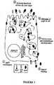
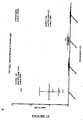
- IgA is synthesized almost exclusively as dimeric or higher polymeric forms, here described collectively as dIgA, which are able to interact with the polymeric Ig receptor (pIgR). This interaction results in secretion of large amounts of secretory IgA (SIgA) into the lumen of epithelial tissues (see Figures 1 and 2 ).
- SIgA secretory IgA
- mIgA monomeric IgA
- dimeric or higher polymeric forms of IgA representing around 10% of the total IgA.
- Detection of IgA, IgM, IgG and other antibody classes or isotypes is usually performed using antibody reagents prepared in another species, for example rabbit antibodies specific for human IgM, or mouse monoclonal antibodies specific for human IgA, or monoclonal antibodies specific for individual antibody subclasses such as IgA1, IgA2 or IgG1, IgG2a, IgG2b, IgG3, IgG4.
- Antibody based capture assay are associated with levels of non-specific binding which is minimised through optimisation protocols.
- WO1991/016061 contemplates prophylactic reagents (such as for passive immunization) comprising polymeric Ig complexed with one or more polymeric-Ig binding domains from the polymeric-Ig receptor, the latter acting as a stabilizer.
- Norderhaug I.N. et al. European journal of Immunology, 1999, vol. 29, pp. 3401-3409
- Roe, M. et al. The Journal of Immunology, 1999, vol. 162, pp.6046-6052
- the specification provides an antibody capture process comprising (i) contacting a biological sample comprising antibodies from a subject with recombinant pIgR or a dIgA-binding variant of pIgR, wherein the pIgR or variant of pIgR binds dIgA and forms a pIgR-dIgA complex.
- the complex may be quantified.
- dIgA may be released from the pIgR and further processed.
- the process is employed for the purification of dIgA antibodies as an alternative to existing processes which employ jacalin agarose. Detection of complexes uses routine methods and agents known in the art such as ELISA or other immunoassay based methods.
- the process further comprises directly or indirectly assessing the level of the pIgR-sIgA complex or the level of a complex between pIgR-dIgA and an antigen of interest.
- the pIgR or a dIgA-binding variant binds dIgA and substantially fails to bind IgM or wherein the pIgR or variant binds dIgA and IgM.
- Processes for detecting antigen specific IgM and dIgA may be used in conjunction with tests for total IgA, IgG and other individual isotypes, subclasses and structural forms and combinations of two or more of these.
- the biological sample is a blood or serum sample.
- Alternative biological samples include samples comprising cells expressing dIgA.
- the process may be used to detect individual B-cells that express dIgA in the screening or isolation of immortalized B-cells.
- the biological sample is obtained from a subject.
- the subject is human.
- Various forms of recombinant pIgR are selected based on the target antibody of interest and species from which the antibody is derived.
- the pIgR is recombinant HpIgA or RpIgR.
- the recombinant pIgR or dIgA-binding variant has the transmembrane domain and/or the cytoplasmic domain deleted.
- the recombinant pIgR comprises a heterologous detection or binding domain.
- the recombinant pIgR or IgA-binding variant is recombinantly produced in a glycan deficient cell, such as a CHO cell.
- the recombinant pIgR is bound to a solid support.
- the biological sample is depleted of IgM or dIgA antibodies prior to use in the process, this allows the pIgR which binds to IgM and dIgA to be employed in assays to specifically detect dIgA.
- depletion of dIgA facilitates the use of HpIgR in assays to specifically detect IgM.
- IgG is depleted prior to sample use in order to reduce competition with dIgA. However, as competing antibodies are washed away in the present antibody/isotype capture formats, lack of such competition is an advantage of the process.
- the antigen of interest is an antigen of an infectious agent or an antigen associated with a condition of a subject that affects a mucosal surface or associated tissues.
- infectious agents include HIV, leprosy, syphilis, hepatitis, dengue virus, measles and rubella.
- the process further comprises contacting the biological sample with an anti-SC binding agent or anti-SC antibody wherein the anti-SC binding agent or anti-SC antibody binds SIgA and forms an SIgA-binding agent/antibody complex.
- the process further comprises contacting a sample comprising the pIgR-dIgA complex with a denaturing solution to remove any SIgA from the complex and measuring the ratio of SIgA and dIgA in the biological sample.
- the specification enables an antibody capture process for determining gut wall integrity in a test subject, the process comprising (i) contacting a biological sample comprising antibodies from the test subject with recombinant pIgR or a dIgA-binding variant of pIgR, wherein the pIgR or variant of pIgR binds dIgA and forms a pIgR-dIgA complex, and (ii) contacting the biological sample with a specific anti-SIgA binding agent or anti-SC binding agent/antibody wherein the anti-SIgA binding agent or anti-SC binding agent/antibody binds SIgA and forms an SIgA-binding agent/antibody complex, and (iii) measuring and comparing the level of the complex formed in (i) with the level of the complex formed in (ii), wherein the ratio of SIgA to dIgA is compared to a corresponding level or ratio from a control subject and provides a measure of gut
- the level or ratio of SIgA2/dIgA2 and/or SIgA1/dIgA1 are determined.
- the antibody capture process comprises (i) contacting a biological sample comprising antibodies from a subject with recombinant pIgR or a dIgA-binding variant of pIgR, wherein the pIgR or variant binds dIgA in the sample and forms a pIgR-dIgA complex, and (ii) directly or indirectly assessing the level of the pIgR-dIgA complex or the level of a complex between pIgR-dIgA and an antigen of interest.
- the antibody capture process may comprise (i) contacting a biological sample comprising antibodies from a subject with recombinant pIgR wherein the pIgR or variant binds IgM and forms a pIgR-IgM complex.
- a process for detecting the presence of antigen-specific dIgA in a subject, the process comprising (i) contacting a biological sample comprising antibodies from a subject with R/HpIgR and antigen and (ii) measuring the level of antigen-specific dIgA.
- a process for detecting the presence of antigen-specific IgM in a subject, the process comprising (i) contacting a biological sample comprising antibodies from a subject with a HpIgR and R/HpIgR and antigen and (ii) measuring the level of antigen-specific IgM and antigen specific dIgA.
- the process is for detecting the presence of antigen-specific IgM and dIgA in a subject, the process comprising (i) contacting a biological sample comprising antibodies from a subject HpIgR and R/HpIgR and antigen and (ii) measuring the level of antigen-specific IgM and antigen specific dIgA.
- the present specification enables a kit for assessing immune status in a biological sample comprising antibodies from a subject, the kit comprising, (a) an immunographic device comprising a porous membrane, a recombinant pIgR molecule or dIgA-binding variant thereof, an antigen of interest and b) instructions for using the immunographic device to detect the presence of antigen specific dIgA antibody in a biological sample obtained from the subject.
- the pIgR is HpIgR and/or R/HpIgR.
- the recombinant pIgR or dIgA-binding variant has the transmembrane domain and/or the cytoplasmic domain deleted.
- the recombinant pIgR comprises a heterologous detection or binding domain.
- the recombinant pIgR or dIgA-binding variant is recombinantly produced in a glycan deficient cell.
- the recombinant pIgR is bound to a solid support.
- the biological sample is depleted of IgM or dIgA antibodies prior to use in the process.
- the kits and reagents contained therein of the present invention are for use ex vivo.
- the antigen of interest is an antigen of an infectious agent or an antigen associated with a condition of a subject that affects a mucosal surface or associated tissues.
- infectious agents are selected from HIV, leprosy, syphilis, hepatitis, dengue virus, measles and rubella.
- the detection of elevated levels of antigen specific dIgA recombinant pIgR relative to control levels facilitates diagnosis and the selection and treatment options.
- a method of treatment is contemplated comprising requesting a test for antigen-specific dIgA levels and administered treatment to the diagnosed subject if the test is positive for an infection or condition.
- the kit further comprises an anti-SIgA binding agent/antibody or anti-SC antibody, wherein the anti-SIgA binding agent/antibody or anti-SC antibody binds SIgA and forms an SIgA-binding agent/antibody complex.
- recombinant pIgR is provided which is suitable for use in capturing or detecting dIgA and/or IgM.
- Illustrative recombinant pIgRs include R/HpIgR or HpIgR or a dIgA and/or IgM binding variant of R/HpIgR or HpIgR.
- Illustrative amino acid and nucleotide sequences are set out in SEQ ID NO:1 to 20, bearing in mind that some of these sequence encode or provide a CD4 cytoplasmic domain which is entirely optional and may be deleted, modified, supplemented or replaced with other binding or detection molecules known in the art.
- SEQ ID NO: Nucleotide and amino acid sequences are referred to by a sequence identifier number (SEQ ID NO:).
- the SEQ ID NOs: correspond numerically to the sequence identifiers ⁇ 400>1 (SEQ ID NO:1), ⁇ 400>2 (SEQ ID NO:2), etc.
- a summary of sequence identifiers is provided in Table 1.
- a sequence listing is provided at the end of the specification.
- the present disclosure provides an antibody capture process comprising determining the level or presence of dimeric or polymeric IgA (dIgA) in a biological sample.
- the process of the present invention may be practised by detecting only the level or presence of dIgA, or it may be practised in combination with protocols to determine the level or presence of one or more further antibody forms (e.g., monomeric, dimeric, polymeric or pentameric complexes), classes (isotypes) or subclasses (e.g., dIgA1, dIgA2, SIgA2, etc.).
- the process is practised using specific antigens that bind to dIgA molecules of interest.
- the process may be practised to identify antigens of interest which engender dIgA responses in a subject that may be detected in a sample from a subject.
- the process enables the development inter alia of diagnostic assays and therapeutic protocols that are useful for assessing secretory IgA responses at mucosal surfaces and associated tissues such as gut-associated lymphoid tissue (GALT).
- GALT gut-associated lymphoid tissue
- the present process is predicated in part on the ability to detect dIgA or dIgA and IgM with high sensitivity and specificity in binding assays using a recombinant polymeric Ig receptor (pIgR).
- the present process employs recombinant pIgR or recombinant variants of pIgR that bind dIgA and IgM, as well as recombinant pIgR or variants of pIgR that preferentially bind dIgA and substantially fail to bind IgM.
- the present specification provides an antibody-capture process comprising detecting or capturing a precursor to secretory dIgA (SIgA), namely dIgA.
- the process comprises step (i) contacting a biological sample from a subject with recombinant polymeric immunoglobulin (Ig) receptor (pIgR) or an dIgA-binding variant thereof, wherein the pIgR or variant binds dIgA and IgM; or wherein the pIgR or variant binds dIgA and substantially fails to bind IgM, and wherein the pIgR substantially does not bind monomeric IgA, and step (ii) determining the level or presence of dIgA that has bound to pIgR.
- step (ii) comprises detecting a complex between dIgA and pIgR or a complex between bound dIgA and an antigen.
- biological sample includes a sample obtained from a subject comprising antibodies.
- the term also includes sample comprising cells expressing dIgA e.g., hybridoma cells, and samples comprising recombinant dIgA expressed from cell lines cultured in vitro.
- Biological samples from subjects include blood and serum samples, other bodily liquids, biopsy etc. Blood and serum samples are preferred.
- Reference to an "antigen” includes a protein or infection agent or part of a protein or part of an infection agent, as known in the art.
- R/HpIgR includes chimeric forms comprising a immunoglobulin domain from a rabbit pIgA sequence or similar dIgA-binding variant sequences derived from rat or mouse or functional (dIgA-binding) variants thereof.
- R includes rabbit, or mouse, or rat-derived sequences.
- Determining the presence or level of dIgA or pIgR or a complex between dIgA and recombinant pIgR or a complex between recombinant dIgA and an antigen may be by any convenient protocol.
- Immunoassays are a particularly useful form of assay that exploits the specificity, strength and diversity of antibody-antigen type or protein-protein reactions to analyse samples and detect specific components therein.
- a wide range of immunoassay techniques are available, such as those described in Wild D. "The Immunoassay Handbook" Nature Publishing Group, 2001 .
- ELISA enzyme-linked immunosorbent assay
- RIA radioimmunoassay
- Immunochromatographic devices comprising dIgA-binding reagents such as recombinant pIgR and further comprising antigens of interest identified as described herein as binding dIgA from infected subjects or subjects exhibiting mucosal immune activation.
- Kits or immunochromatographic devices comprise, for example, reverse-flow or lateral-flow formats.
- a kit for assessing immune status in a biological sample from a subject employs one or more antigens of interest recognised by dIgA from subjects with active infections or conditions associated with mucosal immune activation, and employs a pIgR molecule or dIgA-binding variant thereof as a dIgA-binding reagent.
- the antigen is not a TB antigen.
- the kit comprises:
- the pIgR or dIgA-binding variant thereof is HpIgR or R/HpIgR or a dIgA binding variant thereof.
- the subject assays may employ a wide range of suitable detection markers known in the art.
- the detection marker may be detected using detectable characteristics of the detection marker and a wide range of detection protocols using detectable markers are well known to those of ordinary skill in the art.
- the detection marker is directly or indirectly bound or otherwise associated with an antigen or infectious agent of interest.
- the dIgA binding agent such as pIgA comprises or is designed to interact with a detection marker.
- the detection marker is connected the antigen or dIgA binding agent using binding partners known in the art such as without limitation biotin:avidin or anti-biotin antibody:biotin.
- Polymeric immunoglobulin receptor (pIgR) is encoded by the PIGR gene and is expressed in mucosal epithelial cells where it facilitates uptake of dIgA and secretion of SIgA.
- pIgR has five immunoglobulin-like domains which bind to dIgA including to the J-chain thereof.
- pIgR also binds to pentameric IgM.
- dIgA can be selectively detected using a recombinant form of the polymeric Ig receptor having at least domain 1 derived from the rabbit pIgR, for example, a chimera of rabbit (domain 1) and human (domain 2-5) pIgRs, or with all domains from rabbit pIgR.
- the recombinant pIgR described herein are designed to bind preferentially to dIgA (plus or minus IgM), and can be used either to capture dIgA (IgM) specifically to a solid phase for reaction with an antigen of interest, in which case the pIgR does not need to have an associated detection reagent, or alternatively to detect the presence of dIgA ( ⁇ IgM) bound to an antigen of interest immobilized on a solid phase, in which case the pIgR may be conveniently detected using antibodies or other reagents directed against the pIgR itself, or against epitope tags or other sites introduced into the recombinant pIgR using methods well known in the art.
- a further advantage of pIgR is that it shows very low background reactivity in assays, unlike typical antibody-based detection reagents.
- substitution of human for rabbit (or mouse or rat) D1 provides preferential binding of dIgA, but it would be expected that substitution of any one or more of D2-D5 may also be substituted with the rabbit sequence to give a molecule that preferentially binds to dIgA and these variants are also encompassed. Accordingly, in some embodiments, any one or more of D1, D2, D3, D4 or D5 is substituted with rabbit, mouse or rat homologs.
- the recombinant pIgR lacks a transmembrane domain ( ⁇ TM). In other embodiments, the recombinant pIgR lacks a cytoplasmic domain. In some embodiments, the recombinant pIgR lacks a TM domain and a cytoplasmic domain ( ⁇ CYT). In some embodiments, recombinant pIgR comprises a substitution in the cytoplasmic domain and provides a CD4 cytoplasmic domain.
- Various forms of recombinant pIgR are contemplated and illustrative examples are illustrated in Figures 4 to 7 , further described in the figure legends. The ability to design and test recombinant pIgR having a desired level of specific dIgA is illustrated in Figure 8 and described in the legend to Figure 8 .
- mice hepatocyte pIgR bind only pIgA well and do not translocate IgM into bile. Because mouse liver similarly translocates pIgA but not IgM into bile (48, 49), it is generally assumed that mouse hepatocyte pIgR resembles rat and rabbit pIgR and binds IgM poorly or not at all. If this is true, then the difference between the mouse hepatocyte and the T560 pIgR that makes the latter behave more like human pIgR must be explained.
- a deglycosylated variant of the recombinant pIgR including R/HpIgR is used to improve binding affinity to dIgA.
- this may be achieved by expressing the pIgR in a glycan-deficient cell line known in the art such as, for example, a glycan deficient CHO cell line.
- recombinant pIgR comprises a deletion in the transmembrane domain ( ⁇ TM) to allow for convenient secretion of the recombinant protein and ease of use as a diagnostic/prognostic/screening agent.
- the recombinant pIgR comprises a heterologous detection domain.
- the recombinant pIgR comprises a heterologous binding domain.
- the recombinant pIgR is bound to a solid support.
- Solid supports include plates, wells, beads, agarose particles, nitrocellulose strips, etc.
- recombinant pIgR is produced in glycan deficient cells such as glycan deficient CHO cells to enhance preferential binding to dIgA over IgM.
- the recombinant pIgR is derived from a primate such as human pIgR and comprises at least one immunoglobulin-like domain derived from a non-primate such as rabbit, mouse, rat.
- the recombinant pIgR comprises an amino acid sequence set out in SEQ ID NO:2, or SEQ ID NO: 4, or SEQ ID NO: 6, or SEQ ID NO: 12, or SEQ ID NO: 14, or SEQ ID NO: 16, or an dIgA-binding part thereof or and a dIgA binding variant thereof.
- Illustrative variants comprise at least 70% amino acid sequence identity to one of SEQ ID NO: 2, 4, 6,12, 14 or 16 or deletion variants thereof lacking a cytoplasmic domain.
- Variants include deletion, substitution and insertional variants. Illustrated herein are human derived pIgR varied by one or more immunoglobulin domains (D). Variants include "parts" which includes fragments comprising from about 50%, 60%, 70%, 80%, 85%, 90%, 95% of the reference sequence. Substitution for an equivalent domain from a lower mammal such as a rat, mouse or rabbit domain.
- Variant molecules are also contemplated. Variant molecules are designed to retain the dIgA binding functional activity of the pre-modified recombinant pIgR or to exhibit enhanced activity.
- Polypeptide variants according to the invention can be identified either rationally, or via established methods of mutagenesis (see, for example, Watson, J. D. et al., "Molecular Biology of the Gene", Fourth Edition, Benjamin/Cummings, Menlo Park, California, 1987 ). Random mutagenesis approaches require no a priori information about the sequence that is to be mutated.
- Such methods may be used to selectively alter only those amino acids of the protein that are believed to be important ( Craik C. S., Science, 228:291-297, 1985 ; Cronin et al., Biochem., 27: 4572-4579, 1988 ; Wilks et al., Science, 242:1541-1544, 1988 ).
- Illustrative amino acids affect glycosylation of the recombinant pIgR.
- Polypeptides resulting from rational or established methods of mutagenesis or from combinatorial chemistries, may comprise conservative amino acid substitutions. It is well understood in the art that some amino acids may be changed to others with broadly similar properties without changing the nature of the activity of the polypeptide (conservative substitutions, see Table 3).
- Variant pIgR polypeptides comprises at least 50% sequence identity to herein amino acid sequence at least over the immunoglobulin-like domain region.
- sequence identity refers to the extent that sequences are identical or functionally or structurally similar on an amino acid-by-amino acid basis over a window of comparison.
- a “percentage of sequence identity” is calculated by comparing two optimally aligned sequences over the window of comparison, determining the number of positions at which the identical amino acid residue (e.g.
- sequence identity will be understood to mean the "match percentage” calculated by the DNASIS computer program (Version 2.5 for windows; available from Hitachi Software engineering Co., Ltd., South San Francisco, California, USA) using standard defaults as used in the reference manual accompanying the software. Similar comments apply in relation to sequence similarity which counts as identical, substitutions involving conservative substitutions.
- the percentage similarity between a particular sequence and a reference sequence is at least about 60% or at least about 70% or at least about 80% or at least about 90% or at least about 95% or above such as at least about 96%, 97%, 98%, 99% or greater.
- Percentage similarities or identities between 60% and 100% are also contemplated such as 60, 61, 62, 63, 64, 65, 66, 67, 68, 69, 70, 71, 72, 73, 74, 75, 76, 77, 78, 79, 80, 81, 82, 83, 84, 85, 86, 87, 88, 89, 90, 91, 92, 93, 94, 95, 96, 97, 98, 99 or 100%.
- pIgR encoded by the sequence of nucleotides set out in SEQ ID NO:1, SEQ ID NO:3, SEQ ID NO:5, SEQ ID NO:11, SEQ ID NO:13 or SEQ ID NO:15, or a dIgA-binding and optionally IgM-non binding variant thereof having at least 60% nucleotide sequence identity thereto or at least 60% nucleotide sequence identity to deletion variants thereof lacking a cytoplasmic domain.
- the recombinant pIgR is a human recombinant pIgR variant comprising at least one immunoglobulin-like domain derived from a rabbit.
- (human) pIgR that binds pIgM and dIgA is employed.
- IgG and/or IgM are depleted using known protocols, and, if IgM is depleted then the human pIgR is selective for dIgA among what is left in the sample.
- IgA plus IgM is reported in the literature for hepatitis E and as illustrated herein there are some samples for hepatitis A or hepatitis E in which either IgM or dIgA are much stronger, suggesting that their combined detection is useful.
- recombinant pIgR is also highly advantageous not least because the reagent displays low background (at least 50% less background compared to antibody based reagents) in binding assays unlike most antibody based binding agents.
- recombinant pIgR displays high thermal stability. For example, lyophilised recombinant pIgR retained 50% activity at 60°C and 100% activity at 45°C after three weeks prior to reconstitution, which compares favourably to the rapid loss of activity for dried anti-IgM antibody under the same conditions.
- recombinant human pIgR or dIgA and IgM binding variants may be employed as the binding reagent, but specifically detection of bound dIgA (either dIgA1 or dIgA2) is achieved using anti-IgA1/IgA2 , in this embodiment, the presence of IgM is not a problem.
- the process is for use in a method of assessing conditions or infections of a mucosal surface or associated tissues, or immunity thereto.
- Illustrative applications to specific antigens are described in the Figures and Figure legends for Figures 10 to 22 inclusive and these general protocols are expressly contemplated herein as well as routine variations thereto.
- Illustrative mucosal surfaces include the upper and lower respiratory tracts, the gut and gut-associated lymphoid tissue, the genital tract and the liver.
- infectious agents include, HIV, leprosy, syphilis, hepatitis (e.g., HEV, HAV, HCV) dengue virus, measles, rubella etc.
- Illustrative conditions include diseases of organs such as the respiratory tract, lungs, gut, genital tract and liver.
- Gut conditions include ulcerative colitis, Crohn's disease, IBS, leaky gut syndrome etc.
- subject includes humans and a wide range of mammalian or other animals including wild and domesticated animals, pets, pests and potential vehicles for emerging infectious diseases. In relation to subjects, these may have an infection, they may have had exposure to infection or they have had exposure to an infectious agent.
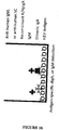
- Figure 10 is a schematic of one preferred experimental approach for detecting the presence of antigen-specific dIgA in a sample such as human serum or plasma.
- Recombinant R/HpIgR-cyto is immobilised on the ELISA plate, and incubated with serum or other samples.
- Dimeric IgA is captured on the solid phase, and after washing to remove other sample components (such as IgA and IgG that are not captured, left), the presence of antigen-specific dIgA is detected by sequential addition of antigen that is, for example, either biotinylated, or reacted with a biotinylated monoclonal antibody against the antigen, and streptavidin-HRP.
- antigen that is, for example, either biotinylated, or reacted with a biotinylated monoclonal antibody against the antigen, and streptavidin-HRP.
- any antigen that is immobilised by reaction with antigen-specific dIgA will give a
- Figure 11 is a schematic of one preferred experimental approach for detecting the presence of hepatitis A virus-specific dIgA in serum (right), compared to detection of HAV-specific IgM using the standard method of anti-IgM capture (left).
- Figure 16 is a schematic of a second preferred method for detection of antigen-specific dIgA (or IgM), in which antigen is coated directly onto the ELISA plate (in this case, hepatitis E virus (HEV) antigen).
- HEV hepatitis E virus
- Serum samples are applied to the plate and antigen-specific antibodies, including IgM and dIgA, bind to the antigens and are then detected with either anti-IgM HRP, or R/HpIgR and anti-human SC HRP. After final washing, signal is generated with TMB substrate or equivalent reagent.
- Figure 23 provides a model outlining the pathogenic consequences of acute human immunodeficiency (HIV) infection, which leads to rapid CD4 depletion in the gut as well as the periphery, with a subsequent reduction in gut barrier function, increased leakage of gut contents and microbial translocation, leading to increased immune activation which drives pathogenesis and further reduction of CD4 T-cell levels ( Brenchley et al, Nat Med 12: 1365 - 1371, 2006 ). There is an unmet need for simple, standardised assays that can detect one or more of these steps in the pathogenesis pathway so that appropriate interventions can be provided to patients, with only CD4 testing having been integrated into the standard of care for HIV-infected patients.
- HIV acute human immunodeficiency
- Detection of CD4 depletion in the gut requires endoscopy; detection of decreased gut barrier function requires complicated sugar challenge studies or other methods; detection of gut leakage and microbial translocation can be achieved using markers such as bacterial LPS or 16sRNA in serum but results are highly variable due in part to the wide variation in gut microbiota between individuals; immune activation requires complex Flow cytometry protocols that are difficult to standardise across instruments/operators.
- Figure 24 provides a schematic illustration of the increase in microbial translocation due to gut leakage induced by pathogenic HIV or SIV infection, compared to normal low levels of translocation in nonpathogenic SIV infection.
- Figure 25 illustrates that one expected consequence of increased microbial translocation is the induction of increased IgA responses due to mucosal antigen exposure.
- M. French et al Journal of Infectious Diseases 200; 2009 demonstrated that indeed the total level of IgA in HIV patients after 6 years of follow up was inversely correlated with the level of CD4 T-cells in patients undergoing highly active antiretroviral therapy, suggesting that even in patients being treated with the most effective current antiviral therapies, microbial translocation contributes to pathogenesis.
- total IgA is highly variable between individuals, and does not provide a prognostic marker that can be used in management of individual patients.
- Figure 26 provides a schematic of the increase in microbial translocation due to gut leakage induced by pathogenic HIV or SIV infection, compared to normal low levels of translocation in nonpathogenic SIV infection, showing the expected effect on dimeric IgA and secretory IgA levels in the plasma compartment.
- gut barrier integrity is maintained and the level of SIgA in the lumen of the gut reflects the amount of its precursor dIgA in the lamina intestinal. Only a minimal amount of SIgA is returned to the plasma compartment, either through active transport by M-cells in the gut, or a small amount of gut leakage.
- the amount of leakage or active transport can be estimated by comparing the serum/plasma concentration of SIgA to that of its precursor dIgA, giving a ratio of SIgA/dIgA.
- the total amount of dIgA is likely to be somewhat elevated and may lead to higher levels of SIgA secretion into the lumen.
- a much higher proportion of SIgA will be returned to the plasma compartment due to passive leakage through the compromised gut barrier, resulting in an elevated SIgA/dIgA ratio.
- the present specification provides a process for assessing gut wall integrity. In some embodiments, and as discussed herein, this assessment provides a prognostic marker for HIV infected patients. In some embodiments, the process comprises determining the relative levels or a ratio of SIgA and dIgA in a sample from a human subject.
- the process comprises the step of (i) contacting a biological sample with recombinant polymeric immunoglobulin (Ig) receptor (pIgR) or an dIgA-binding variant thereof, wherein the pIgR or variant binds dIgA and IgM; or wherein the pIgR or variant binds dIgA and substantially fails to bind IgM, and wherein the pIgR substantially does not bind monomeric IgA or secretory IgA and step (ii) determining the level or presence of dIgA that has bound to pIgR.
- Ig polymeric immunoglobulin
- pIgR polymeric immunoglobulin receptor
- step (ii) comprises detecting a complex between dIgA and pIgR or a complex between bound dIgA and an antigen essentially as described herein.
- the specification contemplates a process of determining the dIgA/SIgA ratio for use in assessing HIV infected subjects.
- SIgA Secretory IgA
- dIgA precursor dIgA
- Figure 27 provides a schematic of one of several typical assays that can be used to measure the relative amount of different IgA forms in order to estimate the SIgA/dIgA ratio.
- the amount of dIgA is measured by capture of dIgA using R/HpIgR, and detection using monoclonal antibodies against either IgA1, or IgA2, or against both IgA subclasses.
- Monomeric IgA does not bind to pIgR; SIgA does bind to R/HpIgR but with lower affinity than dIgA and can be removed by washing with 3.5 M urea if desired.
- SIgA is measured in the same way but using anti-SC antibody capture instead of R/HpIgR.
- the SIgA/dIgA ratio is then calculated as a simple ratio of the assay reactivities for SIgA and dIgA.
- gut leakage can be determined by examining the individual IgA isotypes, because IgA2 (dIgA2, SIgA2) represents around 50% of IgA produced in the gut mucosa, but only around 10% of IgA in other tissues, and thus the ratio of SIgA2 to dIgA2 is likely to provide a very sensitive measurement of gut leakage in an individual patient.
- the process comprises measuring the levels of SIgA2 and dIgA2 and determining the ratio of SIgA2 to dIgA2 wherein the ratio relative to a control is indicative of the presence or absence or degree of gut leakage.
- this ratio provides marker for HIV infected patients thereby potentially facilitating improved management of HIV infection in a subject.
- the present invention enables the use of the specific interaction between the recombinant expressed forms of pIgR and dIgA (plus or minus IgM), allowing pIgR to be used for specific binding or detection of dIgA (plus or minus IgM) to solid surfaces or other assay components as desired.
- the herein described process and/or recombinant pIgR or variants thereof as described herein are sub-licensed for use in antigen screening or antibody selection and purification.
- Treatment protocols are contemplated based upon the results of diagnosis or prognosis testing as described herein.
- the process further comprises: (a) generating data using a process as described herein; (b) transforming the data into computer-readable form; and (c) operating a computer to execute an algorithm, wherein the algorithm determines closeness-of-fit between the computer-readable data and data indicating a diagnosis of a disease or condition.
- the algorithm comprises an artificial intelligence program, such as a fuzzy logic, cluster analysis or neural network. The subject methods may also be used in a personalized or a population medicine approach in the management of pathology platforms.
- the present disclosure provides a computer program and hardware for diagnosis in a subject once off, over time or in response to treatment or other affectors. Values are assigned to complex levels which are stored in a machine readable storage medium.
- a computer program product is one able to convert such values to code and store the code in a computer readable medium and optionally capable of assessing the relationship between the stored data and incoming data and optionally a knowledge database to assess a potential TB status and/or pneumonia.
- the present specification therefore provides a web-based system where data on levels of complex are provided by a client server to a central processor which analyses and compares to a control and optionally considers other information such as patient age, sex, weight and other medical conditions and then provides a diagnostic report.
- the assay may, therefore, be in the form of a kit or computer-based system which comprises the reagents necessary to form and detect the herein described antibody complexes and the computer hardware and/or software including an algorithm to facilitate determination and transmission of reports to a clinician.
- the present invention contemplates a method of allowing a user to determine the status of a subject with respect to TB, the method including:
- the method generally further includes:
- the term "binds specifically," and the like when referring to an antigen-binding molecule refers to a binding reaction which is determinative of the presence of an antigen in the presence of a heterogeneous population of proteins and other biologics.
- the specified antigen-binding molecules bind to a particular antigen and do not bind in a significant amount to other proteins or antigens present in the sample.
- Specific binding to an antigen under such conditions may require an antigen-binding molecule that is selected for its specificity for a particular antigen.
- antigen-binding molecules can be raised to a selected protein antigen, which bind to that antigen but not to other proteins present in a sample.
- immunoassay formats may be used to select antigen-binding molecules specifically immuno-interactive with a particular protein.
- solid-phase ELISA immunoassays are routinely used to select monoclonal antibodies specifically immuno-interactive with a protein. See Harlow and Lane (1988) Antibodies, A Laboratory Manual, Cold Spring Harbor Publications, New York , for a description of immunoassay formats and conditions that can be used to determine specific immunoreactivity.
- a primary antibody polyclonal or monoclonal
- a secondary detection system is used to detect presence (or binding) of the primary antibody.
- Detectable labels can be conjugated to the secondary antibody, such as a fluorescent label, a radiolabel, or an enzyme (e.g., alkaline phosphatase, horseradish peroxidase) which produces a quantifiable, e.g., colored, product.
- the primary antibody itself can be detectably labeled.
- a protein-specific monoclonal antibody can be used both as an immunoadsorbent and as an enzyme-labeled probe to detect and quantify complexes formed in the present process or kit.
- the amount of such protein present in a sample can be calculated by reference to the amount present in a standard or reference preparation using a linear regression computer algorithm (see Lacobilli et al., (1988) Breast Cancer Research and Treatment 11:19-30 ).
- two different monoclonal antibodies to the protein of interest can be employed, one as the immunoadsorbent and the other as an enzyme-labeled probe.
- Assays illustrated in the Examples are done in ELISA format with a single antigen per well, per single antibody form or class or isotype.
- Luminex beads or similar where multiple individual antigens are coated on beads having different intensity of fluorescent label that can be discriminated in an instrument, and the amount of antibody binding to antigen on each bead can be separately measured from the single sample.
- the Luminex beads can be coated with antibody or other reagents to capture the individual antibody forms or isotypes from a sample, and then labelled antigen (or antigens) is added and the different isotype reactivities are assessed. The same can be done in micro-arrays or other arrays.
- Protein capture arrays typically comprise a plurality of protein-capture agents each of which defines a spatially distinct feature of the array.
- the protein-capture agent can be any molecule or complex of molecules which has the ability to bind a protein and immobilize it to the site of the protein-capture agent on the array.
- the protein-capture agent may be a protein whose natural function in a cell is to specifically bind another protein, such as an antibody or a receptor.
- the protein-capture agent may instead be a partially or wholly synthetic or recombinant protein which specifically binds a protein.
- the protein-capture agent may be a protein which has been selected in vitro from a mutagenized, randomized, or completely random and synthetic library by its binding affinity to a specific protein or peptide target.
- the selection method used may optionally have been a display method such as ribosome display or phage display, as known in the art.
- the protein-capture agent obtained via in vitro selection may be a DNA or RNA aptamer which specifically binds a protein target (see, e.g., Potyrailo et al., (1998) Anal. Chem. 70:3419-3425 ; Cohen et al. (1998) Proc. Natl. Acad. Sci.
- aptamers are selected from libraries of oligonucleotides by the SelexTM process and their interaction with protein can be enhanced by covalent attachment, through incorporation of brominated deoxyuridine and UV-activated crosslinking (photoaptamers). Aptamers have the advantages of ease of production by automated oligonucleotide synthesis and the stability and robustness, of DNA; universal fluorescent protein stains can be used to detect binding.
- the in vitro selected protein-capture agent may be a polypeptide (e.g., an antigen) (see, e.g., Roberts and Szostak (1997) Proc. Natl. Acad. Sci. USA 94:12297-12302 ).
- An alternative to an array of capture molecules is one made through 'molecular imprinting' technology, in which peptides (e.g., from the C-terminal regions of proteins) are used as templates to generate structurally complementary, sequence-specific cavities in a polymerisable matrix; the cavities can then specifically capture (denatured) proteins which have the appropriate primary amino acid sequence (e.g., available from ProteinPrintTM and Aspira Biosystems).
- peptides e.g., from the C-terminal regions of proteins
- the cavities can then specifically capture (denatured) proteins which have the appropriate primary amino acid sequence (e.g., available from ProteinPrintTM and Aspira Biosystems).
- Exemplary protein capture arrays include arrays comprising spatially addressed TB antigens or antibody binding agents, which can facilitate extensive parallel analysis of numerous antigens and antibodies. Such arrays have been shown to have the required properties of specificity and acceptable background, and some are available commercially ( e.g ., BD Biosciences, Clontech, BioRad and Sigma). Various methods for the preparation of arrays have been reported (see, e.g., Lopez et al. (2003) J. Chromatogr. B 787:19-27 ; Cahill (2000) Trends in Biotechnology 7:47-51 ; U.S. Pat. App. Pub. 2002/0055186 ; U.S. Pat. App. Pub.
- Immunoglobulin antigen-binding molecules are made either by conventional immunization (e.g ., polyclonal sera and hybridomas), or as recombinant fragments, usually expressed in E. coli, after selection from phage display or ribosome display libraries ( e.g., available from Cambridge Antibody Technology, Biolnvent, Affitech and Biosite).
- 'combibodies' comprising non-covalent associations of VH and VL domains, can be produced in a matrix format created from combinations of diabody-producing bacterial clones ( e.g ., available from Domantis).
- antigen-binding molecules for use as protein-capture agents include monoclonal antibodies, polyclonal antibodies, Fv, Fab, Fab' and F(ab') 2 immunoglobulin fragments, synthetic stabilized Fv fragments, e.g ., single chain Fv fragments (scFv), disulfide stabilized Fv fragments (dsFv), single variable region domains (dAbs) minibodies, combibodies and multivalent antibodies such as diabodies and multi-scFv, single domains from camelids or engineered human equivalents.
- scFv single chain Fv fragments
- dsFv disulfide stabilized Fv fragments
- dAbs single variable region domains minibodies
- combibodies and multivalent antibodies such as diabodies and multi-scFv, single domains from camelids or engineered human equivalents.
- a support surface which is generally planar or contoured.
- Common physical supports include glass slides, silicon, microwells, nitrocellulose or PVDF membranes, and magnetic and other microbeads.
- CD centrifugation devices based on developments in microfluidics (e.g ., available from Gyros) and specialized chip designs, such as engineered microchannels in a plate ( e.g ., The Living ChipTM, available from Biotrove) and tiny 3D posts on a silicon surface (e.g ., available from Zyomyx).
- Particles in suspension can also be used as the basis of arrays, providing they are coded for identification; systems include color coding for microbeads (e.g ., available from Luminex, Bio-Rad and Nanomics Biosystems) and semiconductor nanocrystals ( e.g ., QDotsTM, available from Quantum Dots), and barcoding for beads (UltraPlexTM, available from Smartbeads) and multimetal microrods (NanobarcodesTM particles, available from Surromed). Beads can also be assembled into planar arrays on semiconductor chips ( e.g ., available from LEAPS technology and BioArray Solutions).
- color coding for microbeads e.g ., available from Luminex, Bio-Rad and Nanomics Biosystems
- semiconductor nanocrystals e.g ., QDotsTM, available from Quantum Dots
- barcoding for beads UltraPlexTM, available from Smartbeads
- individual protein-capture agents are typically attached to an individual particle to provide the spatial definition or separation of the array.
- the particles may then be assayed separately, but in parallel, in a compartmentalized way, for example in the wells of a microtiter plate or in separate test tubes.
- a protein sample (see, e.g., U.S. Pat. App. Pub. 2002/0055186 ), is delivered to a protein-capture array under conditions suitable for protein or peptide binding, and the array is washed to remove unbound or non-specifically bound components of the sample from the array.
- the presence or amount of protein or peptide bound to each feature of the array is detected using a suitable detection system.
- the amount of protein bound to a feature of the array may be determined relative to the amount of a second protein bound to a second feature of the array. In certain embodiments, the amount of the second or subsequent protein in the sample is already known or known to be invariant.
- fluorescence labeling can be used for detecting protein bound to the array.
- the same instrumentation as used for reading DNA microarrays is applicable to protein-capture arrays.
- capture arrays e.g. antibody arrays
- fluorophores e.g ., Cy-3 and Cy-5
- Fluorescent readout sensitivity can be amplified 10-100 fold by tyramide signal amplification (TSA) ( e.g ., available from PerkinElmer Lifesciences).
- TSA tyramide signal amplification
- Planar waveguide technology e.g ., available from Zeptosens
- High sensitivity can also be achieved with suspension beads and particles, using phycoerythrin as label (e.g. , available from Luminex) or the properties of semiconductor nanocrystals ( e.g ., available from Quantum Dot).
- Fluorescence resonance energy transfer has been adapted to detect binding of unlabelled ligands, which may be useful on arrays ( e.g ., available from Affibody).
- the techniques used for detection of dIgA or other preselected products will include internal or external standards to permit quantitative or semiquantitative determination of those products, to thereby enable a valid comparison of the level or functional activity of these expression products in a biological sample with the corresponding expression products in a reference sample or samples.
- standards can be determined by the skilled practitioner using standard protocols.
- absolute values for the level or functional activity of individual expression products are determined. Controls may include - individual and population control and samples from diagnostic tests - an earlier time point.
- the diagnostic method is implemented using a system as disclosed, for example, in International Publication No. WO 02/090579 and in copending PCT Application No. PCT/AU03/01517 filed November 14, 2003 , comprising at least one end station coupled to a base station.
- the base station is typically coupled to one or more databases comprising predetermined data from a number of individuals representing the level TB antigen specific antibodies and their isotype structure (dimeric/polymeric) or subclass, when the predetermined data was collected.
- the base station is adapted to receive from the end station, typically via a communications network, subject data representing a measured or normalized level of at least one antibody type in a biological sample obtained from a test subject and to compare the subject data to the predetermined data stored in the database(s). Comparing the subject and predetermined data allows the base station to determine the status of the subject in accordance with the results of the comparison.
- the base station attempts to identify individuals having similar parameter values to the test subject and once the status has been determined on the basis of that identification, the base station provides an indication of the diagnosis to the end station.
- recombinant pIgR is sub-licensed for use in TB antigen screening or TB serological diagnosis.
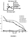
- ELISA shows preferential binding of Chimeric pIgR to dIgA over IgM, but strong binding of human pIgR to IgM
- an ELISA is performed comparing the binding of HpIgR and R/HpIgR to human IgM and dIgA indicating preferential binding of Chimeric pIgR to dIgA over IgM, but strong binding of human pIgR to IgM.
- HpIgR or R/HpIgR were immobilised on 96-well Nunc Immulon plates overnight at 4°C. Dilutions of purified human IgM or dIgA in PBS were bound to the immobilised pIgR forms overnight.
- HRP horseradish peroxidase
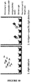
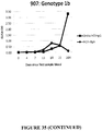
- ELISA shows detection of hepatitis A virus-specific dIgA compared to IgM in an individual patient, with or without depletion of IgM
- An ELISA was conducted demonstrating the detection of HAV-specific dIgA in R/HpIgR capture, using serial dilutions of serum from a patient with acute HAV infection (Accurun HAV panel sample 121).
- the serum sample is either untouched before dilution (untouched, purple) or substantially depleted of IgM using Capture-Select IgM (BAC) (red).
- the results show firstly the strong signal that is obtained demonstrating the detection of HAV-specific dIgA, and secondly that this signal is specific for dIgA not IgM because the IgM depletion method did not substantially reduce the level of HAV-specific reactivity compared to untouched serum, in contrast to the results shown in Figure 12 for IgM detection.
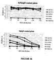
- Comparison of HEV-specific dIgA versus HEV-specific IgM is conducted using sera that are either untouched, or substantially depleted of IgM using Capture-Select IgM, and then serially diluted.
- the results confirm that the IgM assay is specific for IgM, because the reactivity is ablated by IgM depletion, whereas the dIgA assay is predominantly specific for dIgA and not IgM, because the reactivity is only slightly affected by IgM depletion.
- Comparison of HEV-specific dIgA versus HEV-specific IgM is conducted using sera that are either untouched, or substantially depleted of IgM using Capture-Select IgM.
- the results confirm that the IgM assay is specific for IgM, because the reactivity is ablated by IgM depletion, whereas the dIgA assay is predominantly specific for dIgA and not IgM, because the reactivity is only slightly affected by IgM depletion.
- the reduction in dIgA activity following IgM depletion is statistically significant when using a paired T-test to compare samples before and after depletion, but is not significant when using a Mann-Whitney test to compare the overall sample sets before and after depletion.
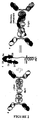
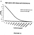
- FIG. 21A An ELISA is conducted demonstrating that the R/HpIgR can be used in both capture (A) and detection (B) of mouse dimeric IgA, with negligible background reactivity to monomeric (human) IgA.
- Figure 21A Dilutions of purified mouse IgA monoclonal antibody 3H1 (anti-HAV) or purified monomeric human IgA were coated on plates and detected with R/HpIgR and anti-SC antibodies.
- B R/HpIgR was coated on plates and dilutions of purified mouse IgA monoclonal antibody 3H1 or purified monomeric human IgA were allowed to bind overnight, then detected with anti-mouse IgA or anti-human IgA.
- the binding of IgA from diverse species to human or rabbit pIgR is known in the art, and this demonstrates that the novel pIgR strategy described herein has utility for diagnosis of infection in other species.
- a schematic of one of several typical assays (see Figure 27 ) that can be used to measure the relative amount of different IgA forms in order to estimate the SIgA/dIgA ratio.
- the amount of dIgA is measured by capture of dIgA using R/HpIgR, and detection using monoclonal antibodies against either IgA1, or IgA2, or against both IgA subclasses.
- Monomeric IgA does not bind to pIgR; SIgA does bind to R/HpIgR but with lower affinity than dIgA and can be removed by washing with 3.5 M urea if desired.
- SIgA is measured in the same way but using anti-SC antibody capture instead of R/HpIgR.
- the SIgA/dIgA ratio is then calculated as a simple ratio of the assay reactivities for SIgA and dIgA.
- SIgA2/dIgA2 S/d
- the assay cutoff for elevated SIgA/dIgA was set as the mean plus 3 standard deviations of the SIgA/dIgA ratio among non-HIV control subjects, and 7/30 HIV-infected subjects showed SIgA/dIgA ratios above this cutoff.
- the range of SIgA/dIgA ratios among normal subjects is smaller than the range for SIgA or dIgA alone, because the role of dIgA as the precursor of SIgA provides a normalising effect for each patient.
- An ELISA is conducted demonstrating the total amount of SIgA in patient and control sera (arbitrary units).
- the amount of SIgA2 in normal patients varies over an 11-fold range, but all normal controls fall within a cutoff of the mean plus 3 standard deviations.
- the amount of SIgA2 in HIV-infected patients varies over a slightly larger range (16-fold), but only 2/30 patients are above the cutoff range (see Figure 29 ).
- those patients who demonstrated elevated SIgA2/dIgA2 ratios in Figure 28 are indicated with red markers (diamonds at ranks 11, 16, 19, 23, 24, 28 and 30).
- SIgA2/dIgA2 ratios are found throughout much of the normal range of the total SIgA2 signal, and cannot be distinguished from the normal controls on the basis of the total SIgA2 alone.
- the R/HpIgR system provides the utility for measuring this ratio.
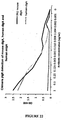
- Figure 30 illustrates a correlation of SIgA2/dIgA2 ratio versus the immune activation marker, CD8+ HLA-DR+ CD38+ T-cells, in a different HIV-infected population to that shown in Figures 28 and 29 . While the overall correlation is low, it is apparent that patients with SIgA2/dIgA2 ratios of >4 in this experiment have elevated levels of immune activation markers (p ⁇ 0.0001).
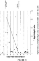
- Figure 31 illustrates a correlation of SIgA1/dIgA1 ratio versus the immune activation marker, CD8+ HLA-DR+ CD38+ T-cells, in the same population as Figure 30 . While the overall correlation is lower again than for IgA2, it is apparent that patients with SIgA1/dIgA1 ratios of >10 in this experiment have elevated levels of immune activation markers (p ⁇ 0.015). The lower correlation for IgA1 and higher cutoff ratio (10 versus 4) for significance highlights the value of specifically measuring IgA2 because of its predominant site of synthesis in the gut, being the tissue in which leakage of SIgA is likely to be clinically relevant marker of gut leakage and immune activation.
- Figure 32 illustrates a correlation of SIgA1/dIgA1 ratio versus SIgA2/dIgA2 ratio, in the same population as Figure 30 and 31 . While SIgA1/dIgA1 ratios are significantly correlated with SIgA2/dIgA2, it is notable that there are some patients with highly elevated SIgAl/dIgA1 ratios and relatively low SIgA2/dIgA2 ratios. This suggests that there may be some value in measuring IgA1, or total IgA, in addition to IgA2 in the calculation of SIgA/dIgA ratios as a measure of gut leakage and immune activation.
Landscapes
- Health & Medical Sciences (AREA)
- Life Sciences & Earth Sciences (AREA)
- Immunology (AREA)
- Chemical & Material Sciences (AREA)
- Engineering & Computer Science (AREA)
- Molecular Biology (AREA)
- Urology & Nephrology (AREA)
- Biomedical Technology (AREA)
- Hematology (AREA)
- Medicinal Chemistry (AREA)
- Cell Biology (AREA)
- General Health & Medical Sciences (AREA)
- Biochemistry (AREA)
- Organic Chemistry (AREA)
- Proteomics, Peptides & Aminoacids (AREA)
- Microbiology (AREA)
- General Physics & Mathematics (AREA)
- Pathology (AREA)
- Biotechnology (AREA)
- Analytical Chemistry (AREA)
- Physics & Mathematics (AREA)
- Food Science & Technology (AREA)
- Genetics & Genomics (AREA)
- Gastroenterology & Hepatology (AREA)
- Zoology (AREA)
- Toxicology (AREA)
- Biophysics (AREA)
- Virology (AREA)
- Tropical Medicine & Parasitology (AREA)
- Peptides Or Proteins (AREA)
Claims (16)
- Procédé de capture d'anticorps afin de déterminer l'intégrité de la paroi intestinale chez un sujet, le procédé comprenant (i) la mise en contact d'un échantillon biologique comprenant des anticorps, issu du sujet, avec un pIgR recombiné ou un variant de pIgR fixateur de dIgA, où le pIgR ou le variant de pIgR fixe la dIgA et forme un complexe pIgR-dIgA, et (ii) la mise en contact de l'échantillon biologique avec un agent fixateur anti-SIgA ou un anticorps/agent fixateur anti-SC spécifique, où l'agent fixateur anti-SIgA ou l'anticorps/agent fixateur anti-SC fixe la SIgA et forme un complexe anticorps/agent fixateur de SIgA, et (iii) la mesure et la comparaison du taux du complexe formé dans (i) avec le taux du complexe formé dans (ii), où le rapport de la SIgA à la dIgA est comparé à un taux ou rapport correspondant issu d'un sujet témoin et fournit une mesure de l'intégrité/fuite intestinale.
- Procédé de capture d'anticorps comprenant (i) la mise en contact d'un échantillon biologique comprenant des anticorps, issu d'un sujet, avec un pIgR recombiné ou un variant de pIgR fixateur de dIgA, où le pIgR ou le variant fixe la dIgA dans l'échantillon et forme un complexe pIgR-dIgA, et (ii) l'évaluation directe ou indirecte du taux de complexe pIgR-dIgA ou du taux d'un complexe entre pIgR-dIgA et un antigène d'intérêt.
- Procédé de capture d'anticorps selon la revendication 2, dans lequel le pIgR ou un variant de pIgR fixateur de dIgA fixe la dIgA et n'arrive sensiblement pas à fixer l'IgM ou où le pIgR ou un variant de pIgR fixe la dIgA et l'IgM et forme un complexe pIgR-dIgM et/ou forme un complexe pIgR-dIgA.
- Procédé de détection de la présence d'une dIgA antigène-spécifique chez un sujet, le procédé comprenant (i) la mise en contact d'un échantillon biologique comprenant des anticorps, issu d'un sujet, avec un R/HpIgR recombiné et un antigène et (ii) la mesure du taux de dIgA antigène-spécifique.
- Procédé de détection de la présence d'une IgM et d'une dIgA antigène-spécifiques chez un sujet, le procédé comprenant (i) la mise en contact d'un échantillon biologique comprenant des anticorps, issu d'un sujet, avec un HpIgR et un R/HpIgR et un antigène et (ii) la mesure du taux d'IgM antigène-spécifique et de dIgA antigène-spécifique.
- Kit destiné à évaluer l'état immunitaire dans un échantillon biologique comprenant des anticorps, issu d'un sujet, le kit comprenant (a) un dispositif immunographique comprenant une membrane poreuse, une molécule de pIgR recombinée ou un variant fixateur de dIgA de celle-ci, un antigène d'intérêt et (b) des instructions pour l'utilisation du dispositif immunographique afin de détecter la présence d'un anticorps dIgA antigène-spécifique dans l'échantillon.
- Procédé de capture d'anticorps selon l'une quelconque des revendications 1 à 3, ou kit selon la revendication 6, où le sujet est un être humain.
- Procédé de capture d'anticorps selon l'une quelconque des revendications 1 à 3 ou 7, ou kit selon la revendication 6, où le pIgR est un HpIgR ou RpIgR recombiné.
- Procédé de capture d'anticorps selon l'une quelconque des revendications 1 à 3, 7 ou 8, ou kit selon la revendication 6, où (a) le pIgR recombiné ou le variant de pIgR fixateur de dIgA possède une délétion du domaine transmembranaire et/ou du domaine cytoplasmique, et/ou (b) où le pIgR recombiné comprend un domaine de fixation ou de détection hétérologue, et/ou (c) où le pIgR recombiné ou le variant de pIgR fixateur de dIgA est produit de manière recombinée dans une cellule déficiente en glycanes, et/ou (d) où le pIgR recombiné est fixé à un support solide, et/ou (e) où l'échantillon biologique est appauvri en anticorps IgM ou dIgA préalablement à l'utilisation dans le procédé, ou (f) où le pIgR est le HpIgA, ou (g) où le pIgR est le R/HpIgR.
- Procédé selon l'une quelconque des revendications 2 et 3, ou 7 à 9, ou kit selon la revendication 6, où l'antigène d'intérêt est un antigène d'un agent infectieux ou un antigène associé à une condition d'un sujet qui affecte une surface mucosale ou des tissus associés, ou où l'agent infectieux ou la condition sont choisis parmi le VIH, la lèpre, la syphilis, l'hépatite, le virus de la dengue, la rougeole, la tuberculose et la rubéole.
- Procédé selon l'une quelconque des revendications 2 et 3, ou 7 à 10, comprenant en outre la mise en contact de l'échantillon biologique avec un agent fixateur anti-SC ou un anticorps anti-SC, où l'agent fixateur anti-SC ou l'anticorps anti-SC fixe la SIgA et forme un complexe anticorps/agent fixateur de SIgA.
- Procédé selon l'une quelconque des revendications 2 et 3, ou 7 à 10, comprenant en outre la mise en contact d'un échantillon comprenant le complexe pIgR-dIgA avec une solution dénaturante afin d'éliminer toute SIgA du complexe et mesurer le rapport de SIgA et de dIgA dans l'échantillon biologique.
- Kit selon l'une quelconque des revendications 6 à 10, où l'échantillon biologique est un échantillon de sang.
- Kit selon la revendication 6, où la molécule de pIgR recombiné ou un variant fixateur de dIgA de celle-ci est (i) un pIgR recombiné ou un variant de pIgR fixateur de dIgA, où le pIgR ou le variant fixe la dIgA et n'arrive sensiblement pas à fixer l'IgM dans l'échantillon et forme un complexe pIgR-dIgA et (ii) un pIgR recombiné ou un variant de pIgR fixateur de dIgA, où le pIgR ou le variant de pIgR fixe la dIgA et l'IgM et forme un complexe pIgR-IgM et un complexe pIgR-dIgA, et où les instructions sont destinées à l'utilisation du dispositif immunographique afin de détecter les taux de dIgA antigène-spécifique et d'IgM antigène-spécifique.
- Procédé de capture d'anticorps selon la revendication 2, comprenant (i) la mise en contact d'un échantillon biologique comprenant des anticorps, issu du sujet, avec un pIgR recombiné ou un variant de pIgR fixateur de dIgA, où le pIgR ou le variant fixe la dIgA et n'arrive sensiblement pas à fixer l'IgM dans l'échantillon et forme un complexe pIgR-dIgA, et (ii) la mise en contact de l'échantillon biologique avec un pIgR recombiné ou un variant de pIgR fixateur de dIgA, où le pIgR ou le variant de pIgR fixe la dIgA et l'IgM et forme un complexe pIgR-IgM et un complexe pIgR-dIgA, et (iii) l'évaluation directe ou indirecte du taux de complexe pIgR-dIgA ou du taux d'un complexe entre pIgR-dIgA et un antigène d'intérêt formé dans (i), et du taux de complexe pIgR-IgM ou du taux d'un complexe entre pIgR-IgM et un antigène d'intérêt formé dans (ii).
- Kit selon la revendication 14 ou procédé de capture d'anticorps selon la revendication 15, où les taux de dIgA antigène-spécifique et d'IgM antigène-spécifique sont utilisés pour détecter une infection récente (aiguë) par un agent infectieux comprenant l'antigène.
Applications Claiming Priority (2)
| Application Number | Priority Date | Filing Date | Title |
|---|---|---|---|
| AU2012904887A AU2012904887A0 (en) | 2012-11-08 | Diagnostic, prognostic, therapeutic and screening protocols | |
| PCT/AU2013/001291 WO2014071456A1 (fr) | 2012-11-08 | 2013-11-08 | Protocoles diagnostiques, pronostiques, thérapeutiques et de criblage |
Publications (3)
| Publication Number | Publication Date |
|---|---|
| EP2917731A1 EP2917731A1 (fr) | 2015-09-16 |
| EP2917731A4 EP2917731A4 (fr) | 2016-06-01 |
| EP2917731B1 true EP2917731B1 (fr) | 2019-12-25 |
Family
ID=50683832
Family Applications (1)
| Application Number | Title | Priority Date | Filing Date |
|---|---|---|---|
| EP13854034.9A Active EP2917731B1 (fr) | 2012-11-08 | 2013-11-08 | Protocoles diagnostiques, pronostiques, thérapeutiques et de criblage |
Country Status (5)
| Country | Link |
|---|---|
| US (3) | US20150330993A1 (fr) |
| EP (1) | EP2917731B1 (fr) |
| CN (1) | CN104903727B (fr) |
| AU (1) | AU2013344322B2 (fr) |
| WO (1) | WO2014071456A1 (fr) |
Families Citing this family (8)
| Publication number | Priority date | Publication date | Assignee | Title |
|---|---|---|---|---|
| CR20220042A (es) * | 2019-08-02 | 2022-03-22 | Janssen Biotech Inc | Materiales y métodos para el direccionamiento al receptor de anticuerpos poliméricos |
| US20230055382A1 (en) * | 2020-01-24 | 2023-02-23 | Macfarlane Burnet Institute For Medical Research And Public Health Limited | Detecting gut barrier dysfunction and/or cirrhosis |
| CN115003798B (zh) * | 2020-06-17 | 2025-08-01 | 兰迪·莱曼·阿伦 | 用于检测分析物的方法和试剂盒 |
| EP4188960A4 (fr) | 2020-08-03 | 2024-09-11 | Janssen Biotech, Inc. | Matériaux et procédés pour le biotransport multidirectionnel dans des agents virothérapeutiques |
| CN116635719A (zh) * | 2020-10-06 | 2023-08-22 | 麦克法兰布奈特医疗研究与公共健康研究所有限公司 | 基于纵向研究的方法和组合物 |
| WO2022073066A1 (fr) * | 2020-10-06 | 2022-04-14 | Macfarlane Burnet Institute For Medical Research And Public Health Limited | Méthodes et compositions fondées sur des études longitudinales |
| WO2022081716A1 (fr) * | 2020-10-13 | 2022-04-21 | Accubit LLC - Biotechnology | Méthodes de traitement de la néphropathie à iga à l'aide de molécules contenant des thiols |
| US20240425570A1 (en) * | 2021-09-17 | 2024-12-26 | The Board Of Trustees Of The University Of Illinois | Chimeric secretory component polypeptides and uses thereof |
Family Cites Families (16)
| Publication number | Priority date | Publication date | Assignee | Title |
|---|---|---|---|---|
| US6596476B1 (en) | 1989-12-22 | 2003-07-22 | Abbott Laboratories | Hepatitis C assay |
| CN1057785A (zh) * | 1990-04-16 | 1992-01-15 | 哈佛大学校长及研究员协会 | 合成多聚免疫球蛋白受体,受体-抗体复合物,生产及应用 |
| EP0799310A1 (fr) | 1994-12-16 | 1997-10-08 | Novartis AG | Protection d'un constituant secretoire recombine |
| CA2319828A1 (fr) | 1998-01-29 | 1999-08-05 | Miller, Samuel | Series haute densite pour l'analyse des proteomes et procedes et compositions pour ces derniers |
| US6406921B1 (en) | 1998-07-14 | 2002-06-18 | Zyomyx, Incorporated | Protein arrays for high-throughput screening |
| US20010041349A1 (en) | 2000-04-17 | 2001-11-15 | Andrew Patron | Protein expression system arrays and use in biological screening |
| GB0022978D0 (en) | 2000-09-19 | 2000-11-01 | Oxford Glycosciences Uk Ltd | Detection of peptides |
| EP1324778A2 (fr) | 2000-10-02 | 2003-07-09 | Arizeke Pharmaceuticals, Inc. | Compositions et procedes pour le transport d'agents biologiquement actifs a travers les barrieres cellulaires |
| AU2002220236A1 (en) | 2000-11-09 | 2002-05-21 | Bionova Pharmaceutials, Inc. | A method for identifying the proteome of cells using an antibody library microarray |
| EP1360490B1 (fr) | 2001-01-23 | 2011-12-21 | President and Fellows of Harvard College | Reseaux de proteines a acide nucleique programmable |
| AUPR480901A0 (en) | 2001-05-04 | 2001-05-31 | Genomics Research Partners Pty Ltd | Diagnostic method for assessing a condition of a performance animal |
| WO2003062444A2 (fr) | 2001-11-13 | 2003-07-31 | Emory University | Systemes matriciels et procedes |
| EP1502102B1 (fr) | 2002-03-11 | 2009-01-14 | caprotec bioanalytics GmbH | Composes et procedes pour analyser le proteome |
| US20060165675A1 (en) * | 2002-08-02 | 2006-07-27 | Richman-Eisenstat Janice Beth | Modulation of mesenchymal cells via iga-receptors |
| AU2002952696A0 (en) | 2002-11-14 | 2002-11-28 | Genomics Research Partners Pty Ltd | Status determination |
| CN101287503A (zh) * | 2005-08-03 | 2008-10-15 | Rq生物科技有限公司 | 用于诊断IgA和IgM介导的肾脏疾病的方法和组合物 |
-
2013
- 2013-11-08 US US14/441,606 patent/US20150330993A1/en not_active Abandoned
- 2013-11-08 AU AU2013344322A patent/AU2013344322B2/en active Active
- 2013-11-08 EP EP13854034.9A patent/EP2917731B1/fr active Active
- 2013-11-08 WO PCT/AU2013/001291 patent/WO2014071456A1/fr not_active Ceased
- 2013-11-08 CN CN201380069755.0A patent/CN104903727B/zh active Active
-
2019
- 2019-02-08 US US16/271,588 patent/US12168682B2/en active Active
-
2024
- 2024-11-06 US US18/938,734 patent/US20250122262A1/en active Pending
Non-Patent Citations (1)
| Title |
|---|
| M. S. JOSHI ET AL: "Evaluation of Urine as a Clinical Specimen for Diagnosis of Hepatitis A", CLINICAL AND VACCINE IMMUNOLOGY, vol. 9, no. 4, 1 July 2002 (2002-07-01), US, pages 840 - 845, XP055424081, ISSN: 1556-6811, DOI: 10.1128/CDLI.9.4.840-845.2002 * |
Also Published As
| Publication number | Publication date |
|---|---|
| AU2013344322A1 (en) | 2015-05-14 |
| US20150330993A1 (en) | 2015-11-19 |
| CN104903727A (zh) | 2015-09-09 |
| CN104903727B (zh) | 2019-10-22 |
| US20190359679A1 (en) | 2019-11-28 |
| EP2917731A4 (fr) | 2016-06-01 |
| AU2013344322B2 (en) | 2019-08-01 |
| US20250122262A1 (en) | 2025-04-17 |
| EP2917731A1 (fr) | 2015-09-16 |
| WO2014071456A1 (fr) | 2014-05-15 |
| US12168682B2 (en) | 2024-12-17 |
Similar Documents
| Publication | Publication Date | Title |
|---|---|---|
| US20250122262A1 (en) | Diagnostic, prognostic, therapeutic and screening protocols | |
| CN102112489B (zh) | 抗-hepcidin-25选择性抗体及其用途 | |
| JP7105970B1 (ja) | SARS-CoV-2の免疫測定方法及び免疫測定キット | |
| WO2022007304A1 (fr) | Anticorps d'iga reconnaissant spécifiquement une protéine rbd et kit de test | |
| WO2014071455A1 (fr) | Protocoles diagnostiques, pronostiques, thérapeutiques et de criblage par rapport à l'espèce mycobacterium infectieuse | |
| JP2004035534A (ja) | Hcv抗コア・モノクローナル抗体 | |
| CN112979795A (zh) | 抗体组合产品及其在新冠肺炎检测中应用 | |
| CN117280214A (zh) | SARS-CoV-2的免疫测定方法及免疫测定试剂盒、以及单克隆抗体或其抗体片段 | |
| CN113045646B (zh) | 抗新型冠状病毒SARS-CoV-2的抗体 | |
| Ch’ng et al. | Phage display-derived antibodies: Application of recombinant antibodies for diagnostics | |
| CN117683121A (zh) | 抗水痘-带状疱疹病毒抗体及其应用 | |
| US20230176054A1 (en) | Coronavirus assay | |
| KR20230084469A (ko) | Sars-cov-2를 검출하기 위한 검정법 | |
| KR101329344B1 (ko) | 돼지인플루엔자 바이러스 헤마글루티닌 검출용 항체 및 이의 용도 | |
| US12584917B2 (en) | Recombinant antibodies, kits comprising the same, and uses thereof | |
| CN113980099B (zh) | 新冠病毒的n抗原特定表位及其应用 | |
| CN112646006B (zh) | 一种用于covid-19轻重症分型诊断的标志物抗原表位多肽及其应用 | |
| JP7778702B2 (ja) | 腸管バリア機能障害および/または肝硬変の検出 | |
| Class et al. | Patent application title: Diagnostic, prognostic, therapeutic and screening protocols Inventors: David Andrew Anderson (Melbourne, AU) Mary Louise Garcia (Melbourne, AU) Nadine Carmel Barnes (Melbourne, AU) Khayriyyah Mohd Hanafiah (Melbourne, AU) Alan Lee Landay (Melbourne, AU) | |
| CN120647764B (zh) | 一种PLA2R IgG4抗体及其应用 | |
| JP7366411B2 (ja) | ヒトαディフェンシンHD5を検出する方法及びキット、並びにこれらにおいて用いられる抗体 | |
| US20240288440A1 (en) | Methods and compositions based on longitudinal studies | |
| Burciaga-Flores et al. | A New Approach in DENV Diagnostic based on Linear Peptides Obtained by Phage Display | |
| CN106415270B (zh) | 测定法 | |
| WO2024081915A1 (fr) | Agents de capture pour la détection de la maladie de kawasaki |
Legal Events
| Date | Code | Title | Description |
|---|---|---|---|
| PUAI | Public reference made under article 153(3) epc to a published international application that has entered the european phase |
Free format text: ORIGINAL CODE: 0009012 |
|
| 17P | Request for examination filed |
Effective date: 20150511 |
|
| AK | Designated contracting states |
Kind code of ref document: A1 Designated state(s): AL AT BE BG CH CY CZ DE DK EE ES FI FR GB GR HR HU IE IS IT LI LT LU LV MC MK MT NL NO PL PT RO RS SE SI SK SM TR |
|
| AX | Request for extension of the european patent |
Extension state: BA ME |
|
| DAX | Request for extension of the european patent (deleted) | ||
| RA4 | Supplementary search report drawn up and despatched (corrected) |
Effective date: 20160504 |
|
| RIC1 | Information provided on ipc code assigned before grant |
Ipc: C12N 15/12 20060101ALI20160428BHEP Ipc: G01N 33/53 20060101AFI20160428BHEP |
|
| STAA | Information on the status of an ep patent application or granted ep patent |
Free format text: STATUS: EXAMINATION IS IN PROGRESS |
|
| 17Q | First examination report despatched |
Effective date: 20170403 |
|
| REG | Reference to a national code |
Ref country code: DE Ref legal event code: R079 Ref document number: 602013064460 Country of ref document: DE Free format text: PREVIOUS MAIN CLASS: G01N0033530000 Ipc: G01N0033680000 |
|
| RIC1 | Information provided on ipc code assigned before grant |
Ipc: G01N 33/68 20060101AFI20190304BHEP Ipc: C07K 14/705 20060101ALI20190304BHEP |
|
| GRAP | Despatch of communication of intention to grant a patent |
Free format text: ORIGINAL CODE: EPIDOSNIGR1 |
|
| STAA | Information on the status of an ep patent application or granted ep patent |
Free format text: STATUS: GRANT OF PATENT IS INTENDED |
|
| INTG | Intention to grant announced |
Effective date: 20190418 |
|
| GRAS | Grant fee paid |
Free format text: ORIGINAL CODE: EPIDOSNIGR3 |
|
| GRAA | (expected) grant |
Free format text: ORIGINAL CODE: 0009210 |
|
| STAA | Information on the status of an ep patent application or granted ep patent |
Free format text: STATUS: THE PATENT HAS BEEN GRANTED |
|
| AK | Designated contracting states |
Kind code of ref document: B1 Designated state(s): AL AT BE BG CH CY CZ DE DK EE ES FI FR GB GR HR HU IE IS IT LI LT LU LV MC MK MT NL NO PL PT RO RS SE SI SK SM TR |
|
| REG | Reference to a national code |
Ref country code: GB Ref legal event code: FG4D |
|
| REG | Reference to a national code |
Ref country code: CH Ref legal event code: EP |
|
| REG | Reference to a national code |
Ref country code: AT Ref legal event code: REF Ref document number: 1217691 Country of ref document: AT Kind code of ref document: T Effective date: 20200115 |
|
| REG | Reference to a national code |
Ref country code: DE Ref legal event code: R096 Ref document number: 602013064460 Country of ref document: DE |
|
| REG | Reference to a national code |
Ref country code: IE Ref legal event code: FG4D |
|
| REG | Reference to a national code |
Ref country code: NL Ref legal event code: MP Effective date: 20191225 |
|
| PG25 | Lapsed in a contracting state [announced via postgrant information from national office to epo] |
Ref country code: GR Free format text: LAPSE BECAUSE OF FAILURE TO SUBMIT A TRANSLATION OF THE DESCRIPTION OR TO PAY THE FEE WITHIN THE PRESCRIBED TIME-LIMIT Effective date: 20200326 Ref country code: BG Free format text: LAPSE BECAUSE OF FAILURE TO SUBMIT A TRANSLATION OF THE DESCRIPTION OR TO PAY THE FEE WITHIN THE PRESCRIBED TIME-LIMIT Effective date: 20200325 Ref country code: FI Free format text: LAPSE BECAUSE OF FAILURE TO SUBMIT A TRANSLATION OF THE DESCRIPTION OR TO PAY THE FEE WITHIN THE PRESCRIBED TIME-LIMIT Effective date: 20191225 Ref country code: LT Free format text: LAPSE BECAUSE OF FAILURE TO SUBMIT A TRANSLATION OF THE DESCRIPTION OR TO PAY THE FEE WITHIN THE PRESCRIBED TIME-LIMIT Effective date: 20191225 Ref country code: NO Free format text: LAPSE BECAUSE OF FAILURE TO SUBMIT A TRANSLATION OF THE DESCRIPTION OR TO PAY THE FEE WITHIN THE PRESCRIBED TIME-LIMIT Effective date: 20200325 Ref country code: SE Free format text: LAPSE BECAUSE OF FAILURE TO SUBMIT A TRANSLATION OF THE DESCRIPTION OR TO PAY THE FEE WITHIN THE PRESCRIBED TIME-LIMIT Effective date: 20191225 Ref country code: LV Free format text: LAPSE BECAUSE OF FAILURE TO SUBMIT A TRANSLATION OF THE DESCRIPTION OR TO PAY THE FEE WITHIN THE PRESCRIBED TIME-LIMIT Effective date: 20191225 |
|
| REG | Reference to a national code |
Ref country code: LT Ref legal event code: MG4D |
|
| PG25 | Lapsed in a contracting state [announced via postgrant information from national office to epo] |
Ref country code: RS Free format text: LAPSE BECAUSE OF FAILURE TO SUBMIT A TRANSLATION OF THE DESCRIPTION OR TO PAY THE FEE WITHIN THE PRESCRIBED TIME-LIMIT Effective date: 20191225 Ref country code: HR Free format text: LAPSE BECAUSE OF FAILURE TO SUBMIT A TRANSLATION OF THE DESCRIPTION OR TO PAY THE FEE WITHIN THE PRESCRIBED TIME-LIMIT Effective date: 20191225 |
|
| PG25 | Lapsed in a contracting state [announced via postgrant information from national office to epo] |
Ref country code: AL Free format text: LAPSE BECAUSE OF FAILURE TO SUBMIT A TRANSLATION OF THE DESCRIPTION OR TO PAY THE FEE WITHIN THE PRESCRIBED TIME-LIMIT Effective date: 20191225 |
|
| PG25 | Lapsed in a contracting state [announced via postgrant information from national office to epo] |
Ref country code: EE Free format text: LAPSE BECAUSE OF FAILURE TO SUBMIT A TRANSLATION OF THE DESCRIPTION OR TO PAY THE FEE WITHIN THE PRESCRIBED TIME-LIMIT Effective date: 20191225 Ref country code: NL Free format text: LAPSE BECAUSE OF FAILURE TO SUBMIT A TRANSLATION OF THE DESCRIPTION OR TO PAY THE FEE WITHIN THE PRESCRIBED TIME-LIMIT Effective date: 20191225 Ref country code: PT Free format text: LAPSE BECAUSE OF FAILURE TO SUBMIT A TRANSLATION OF THE DESCRIPTION OR TO PAY THE FEE WITHIN THE PRESCRIBED TIME-LIMIT Effective date: 20200520 Ref country code: CZ Free format text: LAPSE BECAUSE OF FAILURE TO SUBMIT A TRANSLATION OF THE DESCRIPTION OR TO PAY THE FEE WITHIN THE PRESCRIBED TIME-LIMIT Effective date: 20191225 Ref country code: RO Free format text: LAPSE BECAUSE OF FAILURE TO SUBMIT A TRANSLATION OF THE DESCRIPTION OR TO PAY THE FEE WITHIN THE PRESCRIBED TIME-LIMIT Effective date: 20191225 |
|
| PG25 | Lapsed in a contracting state [announced via postgrant information from national office to epo] |
Ref country code: IS Free format text: LAPSE BECAUSE OF FAILURE TO SUBMIT A TRANSLATION OF THE DESCRIPTION OR TO PAY THE FEE WITHIN THE PRESCRIBED TIME-LIMIT Effective date: 20200425 Ref country code: SK Free format text: LAPSE BECAUSE OF FAILURE TO SUBMIT A TRANSLATION OF THE DESCRIPTION OR TO PAY THE FEE WITHIN THE PRESCRIBED TIME-LIMIT Effective date: 20191225 Ref country code: SM Free format text: LAPSE BECAUSE OF FAILURE TO SUBMIT A TRANSLATION OF THE DESCRIPTION OR TO PAY THE FEE WITHIN THE PRESCRIBED TIME-LIMIT Effective date: 20191225 |
|
| REG | Reference to a national code |
Ref country code: DE Ref legal event code: R097 Ref document number: 602013064460 Country of ref document: DE |
|
| PG25 | Lapsed in a contracting state [announced via postgrant information from national office to epo] |
Ref country code: ES Free format text: LAPSE BECAUSE OF FAILURE TO SUBMIT A TRANSLATION OF THE DESCRIPTION OR TO PAY THE FEE WITHIN THE PRESCRIBED TIME-LIMIT Effective date: 20191225 Ref country code: DK Free format text: LAPSE BECAUSE OF FAILURE TO SUBMIT A TRANSLATION OF THE DESCRIPTION OR TO PAY THE FEE WITHIN THE PRESCRIBED TIME-LIMIT Effective date: 20191225 |
|
| PLBE | No opposition filed within time limit |
Free format text: ORIGINAL CODE: 0009261 |
|
| STAA | Information on the status of an ep patent application or granted ep patent |
Free format text: STATUS: NO OPPOSITION FILED WITHIN TIME LIMIT |
|
| REG | Reference to a national code |
Ref country code: AT Ref legal event code: MK05 Ref document number: 1217691 Country of ref document: AT Kind code of ref document: T Effective date: 20191225 |
|
| PG25 | Lapsed in a contracting state [announced via postgrant information from national office to epo] |
Ref country code: SI Free format text: LAPSE BECAUSE OF FAILURE TO SUBMIT A TRANSLATION OF THE DESCRIPTION OR TO PAY THE FEE WITHIN THE PRESCRIBED TIME-LIMIT Effective date: 20191225 |
|
| 26N | No opposition filed |
Effective date: 20200928 |
|
| PG25 | Lapsed in a contracting state [announced via postgrant information from national office to epo] |
Ref country code: AT Free format text: LAPSE BECAUSE OF FAILURE TO SUBMIT A TRANSLATION OF THE DESCRIPTION OR TO PAY THE FEE WITHIN THE PRESCRIBED TIME-LIMIT Effective date: 20191225 |
|
| PG25 | Lapsed in a contracting state [announced via postgrant information from national office to epo] |
Ref country code: PL Free format text: LAPSE BECAUSE OF FAILURE TO SUBMIT A TRANSLATION OF THE DESCRIPTION OR TO PAY THE FEE WITHIN THE PRESCRIBED TIME-LIMIT Effective date: 20191225 |
|
| PG25 | Lapsed in a contracting state [announced via postgrant information from national office to epo] |
Ref country code: MC Free format text: LAPSE BECAUSE OF FAILURE TO SUBMIT A TRANSLATION OF THE DESCRIPTION OR TO PAY THE FEE WITHIN THE PRESCRIBED TIME-LIMIT Effective date: 20191225 |
|
| REG | Reference to a national code |
Ref country code: CH Ref legal event code: PL |
|
| PG25 | Lapsed in a contracting state [announced via postgrant information from national office to epo] |
Ref country code: LU Free format text: LAPSE BECAUSE OF NON-PAYMENT OF DUE FEES Effective date: 20201108 |
|
| REG | Reference to a national code |
Ref country code: BE Ref legal event code: MM Effective date: 20201130 |
|
| PG25 | Lapsed in a contracting state [announced via postgrant information from national office to epo] |
Ref country code: LI Free format text: LAPSE BECAUSE OF NON-PAYMENT OF DUE FEES Effective date: 20201130 Ref country code: CH Free format text: LAPSE BECAUSE OF NON-PAYMENT OF DUE FEES Effective date: 20201130 |
|
| PG25 | Lapsed in a contracting state [announced via postgrant information from national office to epo] |
Ref country code: IE Free format text: LAPSE BECAUSE OF NON-PAYMENT OF DUE FEES Effective date: 20201108 |
|
| PG25 | Lapsed in a contracting state [announced via postgrant information from national office to epo] |
Ref country code: TR Free format text: LAPSE BECAUSE OF FAILURE TO SUBMIT A TRANSLATION OF THE DESCRIPTION OR TO PAY THE FEE WITHIN THE PRESCRIBED TIME-LIMIT Effective date: 20191225 Ref country code: MT Free format text: LAPSE BECAUSE OF FAILURE TO SUBMIT A TRANSLATION OF THE DESCRIPTION OR TO PAY THE FEE WITHIN THE PRESCRIBED TIME-LIMIT Effective date: 20191225 Ref country code: CY Free format text: LAPSE BECAUSE OF FAILURE TO SUBMIT A TRANSLATION OF THE DESCRIPTION OR TO PAY THE FEE WITHIN THE PRESCRIBED TIME-LIMIT Effective date: 20191225 |
|
| PG25 | Lapsed in a contracting state [announced via postgrant information from national office to epo] |
Ref country code: MK Free format text: LAPSE BECAUSE OF FAILURE TO SUBMIT A TRANSLATION OF THE DESCRIPTION OR TO PAY THE FEE WITHIN THE PRESCRIBED TIME-LIMIT Effective date: 20191225 |
|
| PG25 | Lapsed in a contracting state [announced via postgrant information from national office to epo] |
Ref country code: BE Free format text: LAPSE BECAUSE OF NON-PAYMENT OF DUE FEES Effective date: 20201130 |
|
| P01 | Opt-out of the competence of the unified patent court (upc) registered |
Effective date: 20230518 |
|
| PGFP | Annual fee paid to national office [announced via postgrant information from national office to epo] |
Ref country code: GB Payment date: 20250918 Year of fee payment: 13 |
|
| PGFP | Annual fee paid to national office [announced via postgrant information from national office to epo] |
Ref country code: FR Payment date: 20250908 Year of fee payment: 13 |
|
| PGFP | Annual fee paid to national office [announced via postgrant information from national office to epo] |
Ref country code: DE Payment date: 20250916 Year of fee payment: 13 |
|
| PGFP | Annual fee paid to national office [announced via postgrant information from national office to epo] |
Ref country code: IT Payment date: 20251022 Year of fee payment: 13 |