EP2921990A2 - Procédé et appareil de segmentation non supervisée d'une image couleur microscopique d'un spécimen non coloré et coloration numérique de structures histologiques segmentées - Google Patents
Procédé et appareil de segmentation non supervisée d'une image couleur microscopique d'un spécimen non coloré et coloration numérique de structures histologiques segmentées Download PDFInfo
- Publication number
- EP2921990A2 EP2921990A2 EP15000801.9A EP15000801A EP2921990A2 EP 2921990 A2 EP2921990 A2 EP 2921990A2 EP 15000801 A EP15000801 A EP 15000801A EP 2921990 A2 EP2921990 A2 EP 2921990A2
- Authority
- EP
- European Patent Office
- Prior art keywords
- image
- specimen
- histological structures
- histological
- unstained
- Prior art date
- Legal status (The legal status is an assumption and is not a legal conclusion. Google has not performed a legal analysis and makes no representation as to the accuracy of the status listed.)
- Granted
Links
Images








Classifications
-
- G—PHYSICS
- G16—INFORMATION AND COMMUNICATION TECHNOLOGY [ICT] SPECIALLY ADAPTED FOR SPECIFIC APPLICATION FIELDS
- G16B—BIOINFORMATICS, i.e. INFORMATION AND COMMUNICATION TECHNOLOGY [ICT] SPECIALLY ADAPTED FOR GENETIC OR PROTEIN-RELATED DATA PROCESSING IN COMPUTATIONAL MOLECULAR BIOLOGY
- G16B45/00—ICT specially adapted for bioinformatics-related data visualisation, e.g. displaying of maps or networks
-
- G—PHYSICS
- G06—COMPUTING OR CALCULATING; COUNTING
- G06V—IMAGE OR VIDEO RECOGNITION OR UNDERSTANDING
- G06V20/00—Scenes; Scene-specific elements
- G06V20/60—Type of objects
- G06V20/69—Microscopic objects, e.g. biological cells or cellular parts
- G06V20/695—Preprocessing, e.g. image segmentation
-
- G—PHYSICS
- G01—MEASURING; TESTING
- G01N—INVESTIGATING OR ANALYSING MATERIALS BY DETERMINING THEIR CHEMICAL OR PHYSICAL PROPERTIES
- G01N33/00—Investigating or analysing materials by specific methods not covered by groups G01N1/00 - G01N31/00
- G01N33/48—Biological material, e.g. blood, urine; Haemocytometers
- G01N33/483—Physical analysis of biological material
- G01N33/4833—Physical analysis of biological material of solid biological material, e.g. tissue samples, cell cultures
-
- G—PHYSICS
- G06—COMPUTING OR CALCULATING; COUNTING
- G06F—ELECTRIC DIGITAL DATA PROCESSING
- G06F18/00—Pattern recognition
- G06F18/20—Analysing
- G06F18/21—Design or setup of recognition systems or techniques; Extraction of features in feature space; Blind source separation
- G06F18/213—Feature extraction, e.g. by transforming the feature space; Summarisation; Mappings, e.g. subspace methods
- G06F18/2133—Feature extraction, e.g. by transforming the feature space; Summarisation; Mappings, e.g. subspace methods based on naturality criteria, e.g. with non-negative factorisation or negative correlation
-
- G—PHYSICS
- G06—COMPUTING OR CALCULATING; COUNTING
- G06V—IMAGE OR VIDEO RECOGNITION OR UNDERSTANDING
- G06V10/00—Arrangements for image or video recognition or understanding
- G06V10/70—Arrangements for image or video recognition or understanding using pattern recognition or machine learning
- G06V10/77—Processing image or video features in feature spaces; using data integration or data reduction, e.g. principal component analysis [PCA] or independent component analysis [ICA] or self-organising maps [SOM]; Blind source separation
- G06V10/7715—Feature extraction, e.g. by transforming the feature space, e.g. multi-dimensional scaling [MDS]; Mappings, e.g. subspace methods
Definitions
- the invention relates to a computing device-implemented method and apparatus for unsupervised segmentation of microscopic color image of unstained specimen and digital staining of segmented histological structures. Segmented histological structures and digitally stained image are displayed in order to diagnose a disease.
- Some benefits of application of the invention when compared against existing staining techniques, are: ( i ) shortening of slide preparation process; ( ii ) reduction of variation in diagnosis between histologist; ( iii ) total elimination of adding chemical effects on a specimen; ( iv ) elimination of morphological changes of a specimen e.g.
- shrinkage ( v ) simplification of histological and intra-surgical tissue analysis; ( vi ) being significantly cheaper; ( vii ) harmless to the user because toxic chemical stains are not used; ( viii ) discrimination of several types of histological structures present in the specimen; ( ix ) using the same specimen for more than one analysis.
- Various stains and tags can be attached to biological tissues to enhance contrast of tissue components and thereby improve visibility.
- the presence, concentration, localization and distribution of biological molecules (such as nucleic acids, proteins or lipids for example) or different portions and structures of the tissue can be determined by selecting a specific combination of chemical fixatives and stains.
- Visualization of the histological structures in a biological tissue sample is a basic procedure undertaken by pathologist in order to establish diagnosis of the disease that might have afflicted a patient, for example, kidney disease, liver disease, and the like. Staining, however, involves few hours of preprocessing of the specimen, during that some chemical effects can be added to the nature of the cells or tissues, causing their shrinkage and/or other type of morphological changes.
- fluorescent probes cannot be employed to stain the cell nuclei because the viability of the cultures must not be comprised.
- fluorescent dyes due to their toxicity, cannot be used to mark their nuclei.
- Subcellular localization of the genetically encoded proteins imposes constraints on cell recognition method. That is necessary to draw conclusion about a protein's function: staining of the cell is not allowed in order to preserve the quality of the specimen and not influence the result of an investigation. Once a biological tissue is stained with a particular stain or tag to visualize one tissue component, the same tissue generally cannot be stained again with another dye or tag to visualize another tissue component.
- digital staining does not destroy the biological sample and therefore the same sample could be analyzed by multiple digital staining protocols.
- the digital staining process does not involve toxic chemical stains, and is, therefore, intrinsically harmless to the user.
- Image segmentation refers to the partitioning of an image into sets of pixels (segments) corresponding to distinct objects. It is understood within the scope of the present invention that the expression distinct objects refers to distinct histological structures present in the image of unstained specimen. Segmentation results are often displayed by a region coloring, i.e. assigning colors to the pixels such that different colors correspond to different objects. This results in compact representation of an image in terms of its useful parts. It is important to distinguish between single- and multi-channel images. In the former case, segmentation is performed by detection of changes of intensity or texture by thresholding some kind of spatial derivative of an image: D. Marr and E. Hildredth, "Theory of Edge Detection," Proc.
- the present invention is related to an algorithm for unsupervised segmentation of microscopic color (RGB) image of unstained specimen in histopathology and digital staining of segmented histological structures.
- unsupervised segmentation is performed by novel algorithm for underdetermined blind separation of binary ⁇ 0, 1 ⁇ sources, whereas in the present invention the term sources means histological structures.
- Underdetermined blind separation implies that number of histological structures present in the image is greater than number of channels and that is 3 for color (RGB) image.
- the color image is represented by a linear mixture model comprised of a product of mixing matrix, the columns of which stand for spectral profiles of the histological structures, and binary source matrix. It is further assumed that color image has good spatial resolution and that at each pixel only one histological structure is present.
- source matrix indicates whether at some spatial (pixel) location specific histological structure is present or not.
- this model represents microscopic color image of unstained specimen
- spectral profiles columns of the mixing matrix
- the image has a poor spectral resolution and histological structures are hard to distinguish. That is why image segmentation algorithms, including existing blind source separation methods, yield inaccurate results in segmentation of color microscopic image of unstained specimen.
- the present invention performs pixel-wise nonlinear mapping of the color image of unstained specimen by using empirical kernel map onto high-dimensional space.
- Mapped image is represented by the linear mixture model comprised of the same binary sources but with new mixing matrix comprised of high-dimensional mixing vectors that are less collinear than in the case of original image.
- spectral discrimination between the histological structures present in the image is improved by nonlinear mapping.
- Image segmentation is executed by applying sparseness constrained nonnegative matrix factorization (NMF) algorithm to a mapped image.
- NMF sparseness constrained nonnegative matrix factorization
- Papers cited below present methods for blind separation of finite-alphabet and binary sources from linear or nonlinear mixtures. They are indirectly related to the subject of the present invention: underdetermined blind separation of binary ⁇ 0, 1 ⁇ sources from nonlinear mixtures. That is because binary sources are special case of finite-alphabet sources but also because in case of binary sources nonlinear mixture model is reduced to the linear one.
- nonlinear mapping increases number of mixtures which makes possible to separate sources from ill-posed linear mixture model and that occurs when histological structures, present in the color microscopic image, have similar spectral profiles, that is when spectral resolution between them is poor.
- Competing methods for blind separation of binary and/or finite-alphabet sources are presented in: K. Diamantaras, T. Papadimitrou, G. Vranou, "Blind separation of multiple binary sources from one nonlinear mixture," Proc. IEEE Int. Conf. Acoust. Speech and Sig. Proc. (ICASSP-2011), pp.2108-2111, 2011 ; K. Diamantaras, "Blind separation of multiple binary sources using a single linear mixture," in Proc.
- ICA Independent Component Analysis and Signal Separation
- nonlinear mapping in the present invention is empirical kernel map (EKM) while cited methods use explicit feature map (EFM). While EFM maps LMM in the finite dimensional space, EKM maps LMM in the low-dimensional subspace approximation of the infinite dimensional space.
- nonnegative matrix factorization (NMF) algorithm employed in mapped space by present invention is regularized the by the l 0 quasi-norm of the source matrix (the l 0 quasi-norm counts number of non-zero coefficients of the source matrix). That is due to the fact that source amplitudes belong to ⁇ 0, 1 ⁇ and regularization that emulates indicator function, such as l 0 quasi-norm, is a natural choice.
- US Patent 8,532,376 "Signal processing system and computer readable medium for recording signal processing program,” relates to endoscope based system and image analysis method that looks for a presence of an object with predefined spectral response. As opposed to that, the method of present invention performs unsupervised segmentation of microscopic color image of unstained specimen into constituent objects without using any prior information.
- the US Patent Application 20130071002 "System and method for support of medical diagnosis” relates to automated evaluation of a conventionally prepared sample by a standard dye such as hemotoxylin-eosin by preparing digitally stained sample as a response to staining with a disease specific dye in order to estimate whether staining with disease specific dye is necessary.
- the present invention relates to a method for digital staining of the microscopic color image of an unstained specimen, that is no staining with either standard or disease specific dye is required.
- the US Patent Application 20120269417 “Computer-aided staining of multispectral images,” relates to evaluation and analysis of histological structures and, in particular, to revealing the morphology of these structures by digitally emulating the effects produced by staining the histological structures with dyes.
- this patent application is proposing a method for enhancement of spectral signals of the multispectral image of unstained specimen in order to discriminate objects with similar spectral attributes. That is also a goal of the present invention.
- method proposed in commented patent application is achieving this goal by using multispectral image of unstained specimen and training image related to target chemical stain.
- the method of the present invention is fully unsupervised, that is it only uses as its input microscopic color (RGB) image of unstained specimen and does not include any human involvement.
- RGB input microscopic color
- Patent application WO2011078980 "A method for performing a blood count and determining the morphology of a blood cell,” presents a method that counts blood cells in a sample of whole blood. Thereby, sample can be stained or unstained.
- application of the subject of the present invention relates to an unsupervised image segmentation method that can be applied to color image of arbitrary unstained sample and in order to discriminate between different histological structures (objects) that are present in the image.
- Patent application CN101667299 “Method for staining digital image,” presents a method for staining a digital image. For this purpose disclosed method selects color image with the content similar to the black and white image as a color source. As opposed to that, the method of the present invention is fully unsupervised. That is the method of the present invention is using only microscopic color image of unstained specimen to perform segmentation and digitally stain segmented histological structures.
- Patent application US2010111382 discloses a method for digital staining of image of unstained sample of corneocytes in order to differentiate skin conditions.
- the image of unstained corneocytes is converted into pseudocolor image by means of conversion table.
- the method of the present invention employs novel blind source separation algorithm for unsupervised segmentation of color image of arbitrary unstained specimen and optionally digitally coloring segmented histological structures and displaying them as pseudocolor image.
- Patent application US2013317369 discloses a method for virtual staining of unstained biological tissue.
- image of unstained tissue can be generated by a plurality of detectors (sensors).
- Image of unstained tissue is transformed digitally such that output image mimics response of tissue to staining by specific stain, dye or group of them.
- the virtual staining transforms, that are stored in computer system memory, have to be learned. That is achieved by analyzing image of unstained tissue as well as by analyzing image of tissues stained by predefined dyes (stains).
- the method of the present invention is fully unsupervised, that is it uses only microscopic color image of unstained specimen to segment it into different histological structures and, afterwards, optionally digitally color segmented structures according to predefined color map and display them as a synthetic color (RGB) image.
- RGB synthetic color
- Patent application US20110228072 “Generation of a multicolor image of an unstained biological specimen,” discloses a method for digital (virtual) staining of an image of unstained biological specimen. That is achieved by generating at least two different chemical substances of the unstained specimen and that is used to generate multicolor image of unstained specimen.
- the invention comprises an optical system for exposing to ultraviolet light at different frequencies unstained biological sample and measuring for the intensity of transmitted ultraviolet light for various regions of the specimen. Measured intensity images are used by computer program to generate multicolor image of unstained specimen.
- the method of the present invention records only one color (RGB) image of unstained biological specimen.
- An algorithm stored in computer program is used to segment the image into different histological structures and, optionally, generate synthetic multicolor image.
- Patent application US20110134233 "Imaging system and method for enhancing microscopic images of unstained cells,” discloses a method for enhancing microscopic image of unstained cells. That is achieved by an imaging system configured to acquire image of unstained cells at different focal planes. Processing of these images yields enhanced image of unstained cell. As opposed to described invention, the method of the present invention records only one color (RGB) microscopic image of unstained biological specimen. An algorithm stored in computer program is used to segment the image into different histological structures and, optionally, generate synthetic multicolor image.
- RGB color
- Patent application US2011005817 discloses an invention based upon spectral imaging technique which depending on the wavelength produces images of morphological and chemical constituents of a chromosome by means of its interference properties.
- the method of the present invention records color (RGB) microscopic image of unstained biological specimen that can be composed of various histological structures. That offers an advantage over invention disclosed in US20110058177 because the RGB imaging system is more standard and simpler than multispectral imaging system.
- Patent application US20080032325 discloses an invention for counting cells in living tissue. That is achieved by obtaining microscopic image of unstained tissue and subtraction of ellipses-based cell models from acquired image. Thus, disclosed image processing method is highly specific. As opposed to described invention, the method of the present invention records color (RGB) microscopic image of unstained biological specimen that can be composed of arbitrary histological structures.
- RGB color
- Patent application US20070109874 “Time-lapse cell cycle analysis of unstained nuclei,” discloses method for automatic tracking of cell progress over time.
- level set algorithm is used to segment sequence of images, whereas specimen can be stained or unstained.
- the level set is a supervised image segmentation method that requires user intervention/information during segmentation process.
- the method of the present invention performs unsupervised segmentation of a single color (RGB) microscopic image of unstained biological specimen that can be composed of arbitrary histological structures.
- RGB single color
- Patent application JPH0225251 "Microscope for Microsurgery” discloses an apparatus (microscope system) capable of processing an unstained specimen by arranging special construction of a microscope. As opposed to described invention, the method of the present invention discriminates histological structures in color microscopic image of unstained specimen by means of algorithm that performs unsupervised segmentation of the image.
- Patent application CA1036385 "Biological cell analyzing system,” discloses a method for automatic categorization of unstained biological cells as normal and non-normal. Thereby, the cells are made to flow through a transparent tube and are scanned with a mixture of ultraviolet and visible light. Categorization is achieved by subtracting visible light signal from ultraviolet light signal. As opposed to described invention, the method of the present invention discriminates arbitrary histological structures in color microscopic image of unstained specimen by means of algorithm that performs unsupervised segmentation of the image.
- Patent application US20120147002 discloses a method for virtual staining of cellular structures in order to prevent or minimize crosstalk among emitted colors in an imaging experiment that involves multiplex staining.
- virtual cellular staining involves displaying one or more structures of an exemplary cell on a display of an electronic device and allowing a user to change the individual colors of the one or more structures of the cell. It is thus assumed in the embodiment of disclosed invention that cellular structures present in the image are distinguishable to the user.
- the method of the present invention discriminates arbitrary histological structures in color microscopic image of unstained specimen by means of algorithm that performs unsupervised segmentation of the image. Since the specimen is unstained, histological structures present in the image are hardly visible to the user.
- sources represent presence or absence of the histological structure (histological structure can refer to cell, nuclei, tissue types and the like) at particular spatial location (pixel element) in the image and mixtures represent image intensities recorded at particular color (wavelength) such as red, green and blue (RGB).
- the invention is composed of empirical kernel map (EKM)-based nonlinear mapping of recorded microscopic color (RGB) image of unstained specimen onto high-dimensional space and l 0 quasi-norm constrained nonnegative matrix factorization (NMF) of mapped image, characterised in that said underdetermined blind separation of non-overlapping binary ⁇ 0, 1 ⁇ sources comprises the following steps:
- a system for unsupervised image segmentation by means of underdetermined blind separation of nonnegative binary sources comprising: light microscope 101 and color (RGB) camera 102 for recording image X of unstained specimen, an input storing device/medium 103 for storing the image X , a computing device 104 wherein code is implemented or carried out for executing a method according to anyone of the claims 1 to 12 based on image X stored in/on the input storing device/medium 103, an output storing and displaying device or medium 105 for storing and displaying the result of the method carried out by the processor.
- RGB color
- subspace dimension in empirical kernel map induced space in [III] is D ⁇ 150.
- number of histological structures present M in [II], [III] and [V] is selected as M ⁇ ⁇ 4, 5, 6 ⁇ .
- sNMF algorithm in [V] is nonnegative matrix factorization algorithm constrained with l 0 - quasi-norm of S : R. Peharz, F. Pernkopf, "Sparse nonnegative matrix factorization with l0 constraints," Neurocomputing, vol. 80, pp. 38-46, 2012 .
- a method of the present invention is applied to discrimination of histological structures present in the color microscopic image of unstained specimen.
- stain and “staining” are broad terms and can include without limitation staining with a dye or a stain, immunohistochemical staining, aptamer staining, tagging, chemical staining, antibody staining, or any other alteration to a tissue sample.
- sample tissue sample
- biological sample biological sample
- specimen samples comprise without limitation tissue samples, tissue specimen, bulk tissue, surgical site, biopsy, bacteria, cell or cell components.
- a sample can be analyzed in vivo or in vitro.
- the present invention provides a computer-readable medium having computer executable instructions stored thereon, which, when executed by computer will cause the computer to carry out a method of the present invention.
- computer-readable media include, for example, a floppy disk, a flexible disk, a hard disk, a magnetic tape, or any other magnetic medium, a CD-ROM, any other optical medium, a punch card, a papertape, any other physical medium with patterns of holes, a RAM, a PROM, an EPROM, a FLASH-EPROM, any other memory chip or cartridge, or any other tangible and non-transitory present or future medium from which a computer can read.
- the output storing and displaying device is a printer, plotter or display monitor and the output storing medium is a memory based device that is readable by computer.
- the novelty of proposed method for unsupervised segmentation of color microscopic image of unstained specimen and digital staining of segmented histological structures in relation to state-of-the-art is in empirical kernel map based nonlinear mapping of the color image of unstained specimen onto high-dimensional space.
- This mapping embeds spectral profiles of the histological structures from 3-dimensional space to space with much higher dimensionality (for example 150-dimensional space). That enables discrimination between histological structures with very similar spectral profiles and that occurs when color microscopic image of unstained specimen is recorded.
- factorization of mapped image constrained by nonnegativity and indicator function of the sources such as l 0 quasi-norm of the sources, will discriminate/segment histological structures even though, due to spectral similarity, they are hardly visible in the image of unstained specimen.
- FIG. 1 A schematic block-diagram of a device for unsupervised segmentation of color microscopic image of unstained specimen and digital staining of segmented histological structures, that is defined by equation [II] and employing methodology of empirical kernel map-based nonlinear mapping and nonnegativity and l 0 -norm constrained matrix factorization, according to an embodiment of the present invention is shown in FIG. 1 .
- the device consists of: light microscope 101; color (RGB) camera 102 used to acquire color microscopic image of unstained specimen; input storing device 103 used to store acquired image; CPU or computer 104 where algorithm based on empirical kernel map and nonnegativity and l 0 -norm constrained factorization is implemented for unsupervised segmentation of color microscopic image of unstained specimen and digital staining of segmented histological structures; and output storing and/or display device 105 used to store and display segmented histological structures and synthetic color (RGB) image obtained by digital staining of segmented histological structures.
- RGB color
- Segmented histological structures are stored and/or displayed at the output device 105. Furthermore, segmented histological structures are digitally stained according to [VI] to obtain synthetic color (RGB) image Y that is stored and/or displayed at the output device 105.
- procedure for unsupervised segmentation of color microscopic image of unstained specimen and digital staining of segmented histological structures consists of the following steps:
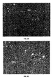
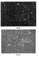
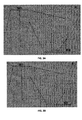
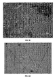
- FIGS. 2 to 6 demonstrate unsupervised segmentation of experimental microscopic color images of unstained specimen and digital staining of segmented histological structures on experimental image according to an embodiment of the present invention.
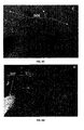
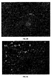
- FIG. 2A shows gray scale version of color microscopic image of unstained specimen of the human liver with diagnoses of obesity with marked blood vessels 201 and vacuoles 202. Histological structures segmented by means of nonnegative matrix factorization algorithm constrained with l 0 quasi-norm (NMF_L0) and related to blood vessels 203 and vacuoles 204 are respectively shown in FIG. 2B and FIG. 2C . These structures are digitally stained according to [VI] whereas gray scale version of synthetic color image Y is shown in FIG. 2D.
- FIG. 2A shows gray scale version of color microscopic image of unstained specimen of the human liver with diagnoses of obesity with marked blood vessels 201 and vacuoles 202. Histological structures segmented by means of nonnegative matrix factorization algorithm constrained with l 0 quasi-nor
- FIGS. 2E shows gray scale version of the color image of the same specimen stained subsequently with hemotoxylin-eosin with marked histological structures related to blood vessels 207 and vacuoles 208. It is seen the correspondence between segmented histological structures shown in FIGS. 2B and 2C and histological structures colored by hemotoxylin-eosin in FIG. 2E . It is, however, important to emphasize that histological structures related to blood vessels and vacuoles were segmented (discriminated) from the image of unstained specimen.
- FIGS. 3B to 3E show results related to unsupervised segmentation and digital staining of histological structures present in the image of unstained specimen, FIG. 3A , of the human liver with widespread liver metastases from pancreatic cancer.
- segmented histological structure that corresponds with the metastasis is shown in FIG. 3B
- segmented histological structure that corresponds with the liver parenchyma is shown in FIG. 3C
- FIGS. 4B to 4E show results related to unsupervised segmentation and digital staining of histological structures present in the image of unstained specimen, FIG.
- FIGS. 4A and 4C segmented histological structure that corresponds with the metastasis is shown in FIG. 4B
- FIG. 4C segmented histological structure that corresponds with the liver parenchyma
- FIGS. 4B and 4C histological structures colored by standard hemotoxylin-eosin procedure in FIG. 4E .
- histological structures related to metastasis and liver parenchyma were segmented (discriminated) from the image of unstained specimen of the liver.
- FIGS. 5B to 5G show results related to unsupervised segmentation and digital staining of histological structures present in the image of unstained specimen, FIG. 5A , of liver of patient with diagnosis of hepatocellular carcinoma.
- FIGS. 5B to 5E segmented histological structures related to tumor, liver parenchyma, blood vessels and vacuoles are respectively shown in FIGS. 5B to 5E .
- histological structures related to blood vessels and vacuoles are very hard to distinguish even on the image stained by hemotoxylin-eosin, i.e. due to very similar spectral profile they are colored equally, see FIG. 5G .
- FIGS. 6B to 6F show results related to unsupervised segmentation and digital staining of histological structures present in unstained specimen of the liver tissue of NOD mice, FIG. 6A . Segmented structures related to vena centralis and sinusoids, vacuoles filled with lipids and cell membranes are respectively shown in FIGS. 6B to 6D . Again, it is seen the correspondence between segmented histological structures shown in FIGS.
- FIG. 6F histological structures colored by hemotoxylin-eosin in FIG. 6F .
- histological structures related to metastasis and liver parenchyma were segmented (discriminated) from the image of unstained specimen of the mouse liver.
- Empirical kernel map-based nonlinear mapping of the color microscopic image of unstained specimen combined with non-overlapping and binary ⁇ 0, 1 ⁇ constraint on the sources (histological structures) is what enables unsupervised segmentation of the image of unstained specimen and digital staining of segmented histological structures. That is distinction with respect to state-of-the-art linear and nonlinear algorithms for blind separation of binary sources that do not employ empirical kernel map based nonlinear mapping of the image. That, however, is of crucial importance for discrimination of spectrally very similar histological structures present in the image of unstained specimen.
- the invention relates to a computing device-implemented method and apparatus for unsupervised segmentation of color microscopic image of unstained specimen and digital staining of segmented histological structures.
- the present invention can be applied to the discrimination and digital staining of the histological structures present in the microscopic color image of the unstained specimen. Elimination of staining brings the following benefits in clinical pathology: ( i ) shortening of slide preparation process; ( ii ) reduction of intra-histologist variation in diagnosis; ( iii ) elimination of adding chemical effects on specimen; ( iv ) elimination of additional morphological changes of a specimen; ( v ) simplification of histological and intra-surgical tissue analysis; ( vi ) significantly cheaper than existing staining techniques; ( vii ) harmless to the user because toxic chemical stains are not used; ( viii ) discrimination of several types of histological structures present in the specimen; ( ix ) allowing the use of the same specimen for more than one analysis.
Landscapes
- Engineering & Computer Science (AREA)
- Health & Medical Sciences (AREA)
- Life Sciences & Earth Sciences (AREA)
- Physics & Mathematics (AREA)
- Theoretical Computer Science (AREA)
- Biomedical Technology (AREA)
- General Health & Medical Sciences (AREA)
- General Physics & Mathematics (AREA)
- Computer Vision & Pattern Recognition (AREA)
- Data Mining & Analysis (AREA)
- Chemical & Material Sciences (AREA)
- Biophysics (AREA)
- Molecular Biology (AREA)
- Medical Informatics (AREA)
- Multimedia (AREA)
- Artificial Intelligence (AREA)
- Bioinformatics & Cheminformatics (AREA)
- Bioinformatics & Computational Biology (AREA)
- Evolutionary Biology (AREA)
- Evolutionary Computation (AREA)
- Food Science & Technology (AREA)
- Analytical Chemistry (AREA)
- Pathology (AREA)
- Hematology (AREA)
- Optics & Photonics (AREA)
- Urology & Nephrology (AREA)
- Immunology (AREA)
- Biochemistry (AREA)
- Medicinal Chemistry (AREA)
- Software Systems (AREA)
- Computing Systems (AREA)
- Databases & Information Systems (AREA)
- General Engineering & Computer Science (AREA)
- Biotechnology (AREA)
- Spectroscopy & Molecular Physics (AREA)
- Investigating Or Analysing Biological Materials (AREA)
Applications Claiming Priority (1)
| Application Number | Priority Date | Filing Date | Title |
|---|---|---|---|
| US14/221,017 US20150269314A1 (en) | 2014-03-20 | 2014-03-20 | Method and apparatus for unsupervised segmentation of microscopic color image of unstained specimen and digital staining of segmented histological structures |
Publications (3)
| Publication Number | Publication Date |
|---|---|
| EP2921990A2 true EP2921990A2 (fr) | 2015-09-23 |
| EP2921990A3 EP2921990A3 (fr) | 2016-01-06 |
| EP2921990B1 EP2921990B1 (fr) | 2016-07-06 |
Family
ID=52780749
Family Applications (1)
| Application Number | Title | Priority Date | Filing Date |
|---|---|---|---|
| EP15000801.9A Not-in-force EP2921990B1 (fr) | 2014-03-20 | 2015-03-18 | Procédé et appareil de segmentation non supervisée d'une image couleur microscopique d'un spécimen non coloré et coloration numérique de structures histologiques segmentées |
Country Status (3)
| Country | Link |
|---|---|
| US (1) | US20150269314A1 (fr) |
| EP (1) | EP2921990B1 (fr) |
| CA (1) | CA2866258A1 (fr) |
Cited By (5)
| Publication number | Priority date | Publication date | Assignee | Title |
|---|---|---|---|---|
| CN107578381A (zh) * | 2017-08-09 | 2018-01-12 | 天津恒宇医疗科技有限公司 | 一种内窥光干涉断层成像颜色映射方法 |
| WO2019191697A1 (fr) * | 2018-03-30 | 2019-10-03 | The Regents Of The University Of California | Procédé et système de coloration numérique d'images de fluorescence sans étiquette à l'aide d'un apprentissage profond |
| US10935773B2 (en) | 2017-08-09 | 2021-03-02 | Allen Institute | Systems, devices, and methods for image processing to generate an image having predictive tagging |
| US12106552B2 (en) | 2018-07-19 | 2024-10-01 | The Regents Of The University Of California | Method and system for digital staining of label-free phase images using deep learning |
| US12399100B2 (en) | 2020-08-12 | 2025-08-26 | Allen Institute | Systems, devices, and methods for quality control of cell production using predictive tagging |
Families Citing this family (10)
| Publication number | Priority date | Publication date | Assignee | Title |
|---|---|---|---|---|
| WO2017205853A1 (fr) * | 2016-05-26 | 2017-11-30 | John Abrams | Appareil pour créer des symboles de type chémotypes |
| CN110326024B (zh) * | 2017-02-21 | 2023-11-03 | 株式会社高迎科技 | 用于处理由医学成像装置捕获的组织学图像的方法和装置 |
| GB2567155B (en) * | 2017-10-02 | 2022-03-02 | Room4 Group Ltd | Histopathological image analysis |
| WO2019172901A1 (fr) * | 2018-03-07 | 2019-09-12 | Google Llc | Coloration virtuelle pour images de lame de tissu |
| EP3818693A4 (fr) | 2018-07-02 | 2021-10-13 | Stowers Institute for Medical Research | Reconnaissance d'image faciale à l'aide de pseudo-images |
| CN110111391B (zh) * | 2019-05-20 | 2021-01-12 | 厦门大学 | 基于改进的下逼近非负矩阵对极几何估计方法 |
| CN111079615B (zh) * | 2019-12-10 | 2023-03-31 | 哈尔滨工程大学 | 一种基于莱维飞行细菌觅食进化的盲源分离方法 |
| CN111666892B (zh) * | 2020-06-08 | 2023-04-25 | 西南交通大学 | 一种基于经验小波希尔伯特变换的电力机车空转识别方法 |
| CN114943723B (zh) * | 2022-06-08 | 2024-05-28 | 北京大学口腔医学院 | 一种对不规则细胞进行分割计数的方法及相关设备 |
| CN115471634B (zh) * | 2022-10-28 | 2023-03-24 | 吉奥时空信息技术股份有限公司 | 一种城市绿植孪生的建模方法及装置 |
Citations (17)
| Publication number | Priority date | Publication date | Assignee | Title |
|---|---|---|---|---|
| CA1036385A (fr) | 1972-08-30 | 1978-08-15 | Milton Stoller | Systeme d'analyse de cellule biologique |
| US20070016081A1 (en) | 2005-07-12 | 2007-01-18 | Globalmedia Group, Llc | Chroma-photon staining |
| US20070109874A1 (en) | 2005-11-12 | 2007-05-17 | General Electric Company | Time-lapse cell cycle analysis of unstained nuclei |
| US20080032325A1 (en) | 2006-08-07 | 2008-02-07 | Northeastern University | Phase subtraction cell counting method |
| CN101667299A (zh) | 2009-09-27 | 2010-03-10 | 汲业 | 一种数字图像染色方法 |
| US20100111382A1 (en) | 2007-02-14 | 2010-05-06 | Pola Chemical Industries, Inc. | Method of supporting the differentiation of corneocytes |
| US20110005817A1 (en) | 2008-03-31 | 2011-01-13 | Mitsui Mining & Smelting Co., Ltd. | Capacitor-forming material and printed wiring board provided with capacitor |
| US20110026803A1 (en) | 2009-07-31 | 2011-02-03 | General Electric Company | Methods and systems for digitally enhancing an image of a stained material |
| US20110058177A1 (en) | 2008-02-27 | 2011-03-10 | Rudolf Kessler | Marker-free chromosome screening |
| US20110134233A1 (en) | 2008-08-05 | 2011-06-09 | Ge Healthcare Uk Limited | Imaging system and method for enhancing microscopic images of unstained cells |
| WO2011078980A1 (fr) | 2009-12-22 | 2011-06-30 | Abbott Laboratories | Méthode de numération globulaire et de détermination de la morphologie d'un frottis sanguin |
| US20110228072A1 (en) | 2008-11-27 | 2011-09-22 | Koninklijke Philips Electronics N.V. | Generation of a multicolour image of an unstained biological specimen |
| US20120147002A1 (en) | 2010-12-07 | 2012-06-14 | Life Technologies Corporation | Virtual cellular staining |
| US20120269417A1 (en) | 2011-04-25 | 2012-10-25 | Bautista Pinky A | Computer-aided staining of multispectral images |
| US20130071002A1 (en) | 2011-09-15 | 2013-03-21 | Takeshi Otsuka | System and method for support of medical diagnosis |
| US8532376B2 (en) | 2008-07-18 | 2013-09-10 | Olympus Corporation | Signal processing system and computer readable medium for recording signal processing program |
| US20130317369A1 (en) | 2012-03-19 | 2013-11-28 | Genetic Innovations, Inc. | Devices, systems, and methods for virtual staining |
Family Cites Families (1)
| Publication number | Priority date | Publication date | Assignee | Title |
|---|---|---|---|---|
| WO2004111930A2 (fr) * | 2003-05-30 | 2004-12-23 | Rabinovich Andrew M | Demelange de couleurs et detection de zones d'interet dans des echantillons de tissu |
-
2014
- 2014-03-20 US US14/221,017 patent/US20150269314A1/en not_active Abandoned
- 2014-10-06 CA CA2866258A patent/CA2866258A1/fr not_active Abandoned
-
2015
- 2015-03-18 EP EP15000801.9A patent/EP2921990B1/fr not_active Not-in-force
Patent Citations (17)
| Publication number | Priority date | Publication date | Assignee | Title |
|---|---|---|---|---|
| CA1036385A (fr) | 1972-08-30 | 1978-08-15 | Milton Stoller | Systeme d'analyse de cellule biologique |
| US20070016081A1 (en) | 2005-07-12 | 2007-01-18 | Globalmedia Group, Llc | Chroma-photon staining |
| US20070109874A1 (en) | 2005-11-12 | 2007-05-17 | General Electric Company | Time-lapse cell cycle analysis of unstained nuclei |
| US20080032325A1 (en) | 2006-08-07 | 2008-02-07 | Northeastern University | Phase subtraction cell counting method |
| US20100111382A1 (en) | 2007-02-14 | 2010-05-06 | Pola Chemical Industries, Inc. | Method of supporting the differentiation of corneocytes |
| US20110058177A1 (en) | 2008-02-27 | 2011-03-10 | Rudolf Kessler | Marker-free chromosome screening |
| US20110005817A1 (en) | 2008-03-31 | 2011-01-13 | Mitsui Mining & Smelting Co., Ltd. | Capacitor-forming material and printed wiring board provided with capacitor |
| US8532376B2 (en) | 2008-07-18 | 2013-09-10 | Olympus Corporation | Signal processing system and computer readable medium for recording signal processing program |
| US20110134233A1 (en) | 2008-08-05 | 2011-06-09 | Ge Healthcare Uk Limited | Imaging system and method for enhancing microscopic images of unstained cells |
| US20110228072A1 (en) | 2008-11-27 | 2011-09-22 | Koninklijke Philips Electronics N.V. | Generation of a multicolour image of an unstained biological specimen |
| US20110026803A1 (en) | 2009-07-31 | 2011-02-03 | General Electric Company | Methods and systems for digitally enhancing an image of a stained material |
| CN101667299A (zh) | 2009-09-27 | 2010-03-10 | 汲业 | 一种数字图像染色方法 |
| WO2011078980A1 (fr) | 2009-12-22 | 2011-06-30 | Abbott Laboratories | Méthode de numération globulaire et de détermination de la morphologie d'un frottis sanguin |
| US20120147002A1 (en) | 2010-12-07 | 2012-06-14 | Life Technologies Corporation | Virtual cellular staining |
| US20120269417A1 (en) | 2011-04-25 | 2012-10-25 | Bautista Pinky A | Computer-aided staining of multispectral images |
| US20130071002A1 (en) | 2011-09-15 | 2013-03-21 | Takeshi Otsuka | System and method for support of medical diagnosis |
| US20130317369A1 (en) | 2012-03-19 | 2013-11-28 | Genetic Innovations, Inc. | Devices, systems, and methods for virtual staining |
Non-Patent Citations (17)
| Title |
|---|
| D. MARR; E. HILDREDTH: "Theory of Edge Detection", PROC. ROYAL SAC LONDON SER B BIOL SCI, vol. 207, 1980, pages 187 - 217, XP000865964 |
| I. KOPRIVA; A. CICHOCKI: "Proc. of 9th Int. Conf. on Latent Variable Analysis and Signal Separation", vol. 6365, 27 September 2010, article "Nonlinear Band Expansion and 3D Nonnegative Tensor Factorization for Blind Decomposition of Magnetic Resonance Image of the Brain", pages: 490 - 497 |
| I. KOPRIVA; A. PERIN: "Unsupervised decomposition of low-intensity low-dimensional multi-spectral fluorescent images for tumour demarcation", MEDICAL IMAGE ANALYSIS, vol. 13, 2009, pages 507 - 518, XP026089348, DOI: doi:10.1016/j.media.2009.02.002 |
| I. KOPRIVA; M. HADZIJA; M. POPOVIÉ-HADZIJA; M. KOROLIJA; A. CICHOCKI: "Rational Variety Mapping for Contrast-Enhanced Nonlinear Unsupervised Segmentation of Multispectral Images of Unstained Specimen", THE AMERICAN JOURNAL OF PATHOLOGY, vol. 179, no. 2, 2011, pages 547 - 553 |
| I. KOPRIVA; X. CHEN; Y. JAO: "SPIE Medical Imaging-Image Processing, Orlando, FL, USA", vol. 7962, 2011, article "Nonlinear Band Expansion and Nonnegative Matrix Underapproximation for Unsupervised Segmentation of a Liver from a Multi-phase CT image", pages: 79623A - 1,79623A |
| K. 1. DIAMANTARAS; T. PAPADIMITRIOU: "Blind separation of three binary sources from one nonlinear mixture", PROC. 2010 INT. WORKSHOP ON MACHINE LEARNING FOR SIGNAL PROCESSING (MLSP-2010), 29 July 2010 (2010-07-29), pages 301 - 306 |
| K. DIAMANTARAS: "Blind separation of multiple binary sources using a single linear mixture", PROC. IEEE INT. CONF. ACOUSTIC, SPEECH AND SIGNAL PROCESSING (ICASSP-2000), vol. V, June 2000 (2000-06-01), pages 2889 - 2892 |
| K. DIAMANTARAS; T. PAPADIMITROU; Q. VRANOU: "Blind separation of multiple binary sources from one nonlinear mixture", PROC. IEEE INT. CONF ACOUST. SPEECH AND SIG. PROC. (ICASSP-2011), 2011, pages 2108 - 2111, XP032001075, DOI: doi:10.1109/ICASSP.2011.5946742 |
| K. I. DIAMANTARAS: "Signal Processing", vol. 86, April 2006, ELSEVIER, article "A Clustering Approach for Blind Separation of Multiple Finite Alphabet Sequences from a Single Linear Mixture", pages: 877 - 891 |
| K. I. DIAMANTARAS; E. CHASSIOTI: "Blind separation of n binary sources from one observation: A deterministic approach", PROC. INT. CONF INDEPENDENT COMPONENT ANALYSIS AND SIGNAL SEPARATION (ICA), HELSINKI, FINLAND, June 2000 (2000-06-01) |
| K. I. DIAMANTARAS; T. PAPADIMITRIOU: "Blind deconvolution of multi-input single-output systems with binary sources", IEEE; TRANS. SIGNAL PROCESS., vol. 54, no. 10, October 2006 (2006-10-01), pages 3720 - 3731 |
| K. I. DIAMANTARAS; T. PAPADIMITRIOU: "Separating two binary sources from a single nonlinear mixture", PROC. INT. CONF ACOUSTICS, SPEECH AND SIGNAL PROCESSING (7CASSP 2010), 14 March 2010 (2010-03-14), pages 1946 - 1949, XP031697258 |
| M. CASTOLLA: "Inversion of Polynomial Systems and Separation of Nonlinear Mixtures of Finite-Alphabet Sources", IEEE TRANS. ON SIG. PROC., vol. 56, no. 8, August 2008 (2008-08-01), pages 3905 - 3917, XP011231478, DOI: doi:10.1109/TSP.2008.921788 |
| OUYANG, Y. C.; CHEN, H. M.; CHAI, J. W.; CHEN, C. C. C.; POON, S. K.; YANG, C. W.; LEE, S. K.; CHANG, C. I.: "Band Expansion-Based Over-Complete Independent Component Analysis for Multispectral Processing of Magnetic Resonance Image", IEEE TRANS. BIOMED ENG., vol. 55, 2008, pages 1666 - 1677, XP011342701, DOI: doi:10.1109/TBME.2008.919107 |
| Q. DU; I. KOPRIVA; H.SZU: "Independent Component Analysis for Classifying Multispectral Images with Dimensionality Limitation", INTERNATIONAL JOURNAL OFLNFORMATION ACQUISITION, vol. 1, no. 3, September 2004 (2004-09-01), pages 201 - 216 |
| R. PEHARZ; F. PEMKOPF: "Sparse nonnegative matrix factorization with ?° constraints", NEUROCOMPUTING, vol. 80, 2012, pages 38 - 46, XP028356707, DOI: doi:10.1016/j.neucom.2011.09.024 |
| Y. LI; A. CICHOCKI; L. ZHANG: "Blind separation and extraction of binary sources", IEICE TRANS FUNDAMENTALS, vol. E86-A, no. 3, March 2003 (2003-03-01), pages 580 - 589 |
Cited By (10)
| Publication number | Priority date | Publication date | Assignee | Title |
|---|---|---|---|---|
| CN107578381A (zh) * | 2017-08-09 | 2018-01-12 | 天津恒宇医疗科技有限公司 | 一种内窥光干涉断层成像颜色映射方法 |
| US10935773B2 (en) | 2017-08-09 | 2021-03-02 | Allen Institute | Systems, devices, and methods for image processing to generate an image having predictive tagging |
| US11614610B2 (en) | 2017-08-09 | 2023-03-28 | Allen Institute | Systems, devices, and methods for image processing to generate an image having predictive tagging |
| US12008764B2 (en) | 2017-08-09 | 2024-06-11 | Allen Institute | Systems, devices, and methods for image processing to generate an image having predictive tagging |
| WO2019191697A1 (fr) * | 2018-03-30 | 2019-10-03 | The Regents Of The University Of California | Procédé et système de coloration numérique d'images de fluorescence sans étiquette à l'aide d'un apprentissage profond |
| JP2021519924A (ja) * | 2018-03-30 | 2021-08-12 | ザ リージェンツ オブ ザ ユニバーシティ オブ カリフォルニア | ディープラーニングを使用して無標識蛍光画像をデジタル染色する方法及びシステム |
| US11893739B2 (en) | 2018-03-30 | 2024-02-06 | The Regents Of The University Of California | Method and system for digital staining of label-free fluorescence images using deep learning |
| US12327362B2 (en) | 2018-03-30 | 2025-06-10 | The Regents Of The University Of California | Method and system for digital staining of label-free fluorescence images using deep learning |
| US12106552B2 (en) | 2018-07-19 | 2024-10-01 | The Regents Of The University Of California | Method and system for digital staining of label-free phase images using deep learning |
| US12399100B2 (en) | 2020-08-12 | 2025-08-26 | Allen Institute | Systems, devices, and methods for quality control of cell production using predictive tagging |
Also Published As
| Publication number | Publication date |
|---|---|
| EP2921990B1 (fr) | 2016-07-06 |
| US20150269314A1 (en) | 2015-09-24 |
| EP2921990A3 (fr) | 2016-01-06 |
| CA2866258A1 (fr) | 2015-09-20 |
Similar Documents
| Publication | Publication Date | Title |
|---|---|---|
| EP2921990B1 (fr) | Procédé et appareil de segmentation non supervisée d'une image couleur microscopique d'un spécimen non coloré et coloration numérique de structures histologiques segmentées | |
| EP3486836B1 (fr) | Procédé d'analyse d'image, appareil, programme et algorithme d'apprentissage profond appris | |
| EP2252974B1 (fr) | Procédé et système de segmentation automatisée de populations de cellules denses | |
| Dougherty | Medical image processing: techniques and applications | |
| McCann et al. | Automated histology analysis: Opportunities for signal processing | |
| JP4947589B2 (ja) | 類似画像検索装置 | |
| EP3127084B1 (fr) | Procédé et dispositif d'examen et analyse d'une image | |
| US8320655B2 (en) | Process and system for analyzing the expression of biomarkers in cells | |
| US20080240527A1 (en) | Methods, Compositions and Systems for Analyzing Imaging Data | |
| Binder et al. | Multi-organ gland segmentation using deep learning | |
| JP2013509629A (ja) | ハイパースペクトル画像を解析するための方法および装置 | |
| US12493964B2 (en) | Methods and systems for providing training data sets for training a machine-learned segmentation algorithm for use in digital pathology | |
| Ruusuvuori et al. | Spatial analysis of histology in 3D: quantification and visualization of organ and tumor level tissue environment | |
| US20260017790A1 (en) | Information processing apparatus and information processing system | |
| Jaworek-Korjakowska | A deep learning approach to vascular structure segmentation in dermoscopy colour images | |
| Phillips et al. | Segmentation of prognostic tissue structures in cutaneous melanoma using whole slide images | |
| Jütte et al. | Integrating generative AI with ABCDE rule analysis for enhanced skin cancer diagnosis, dermatologist training and patient education | |
| Shmueli et al. | Deep learning based multiple sclerosis lesion detection utilizing synthetic data generation and soft attention mechanism | |
| Hu et al. | Multi-scale multi-site renal microvascular structures segmentation for whole slide imaging in renal pathology | |
| Bendiabdallah et al. | Deep learning for segmentation and counting of white blood cells in clinical diagnosis | |
| Olliverre et al. | Pairwise mixture model for unmixing partial volume effect in multi-voxel MR spectroscopy of brain tumour patients | |
| Antony | From Imaging to Insight: Advanced Computer Vision in Life Sciences: From Imaging to Insight: Advanced Computer Vision in Life Sciences | |
| Gagniuc | Computational Analysis of Microscopy Images | |
| Sabata | Digital pathology imaging-The next frontier in medical imaging | |
| Waithe et al. | 3-D density kernel estimation for counting in microscopy image volumes using 3-D image filters and random decision trees |
Legal Events
| Date | Code | Title | Description |
|---|---|---|---|
| PUAI | Public reference made under article 153(3) epc to a published international application that has entered the european phase |
Free format text: ORIGINAL CODE: 0009012 |
|
| 17P | Request for examination filed |
Effective date: 20150319 |
|
| AK | Designated contracting states |
Kind code of ref document: A2 Designated state(s): AL AT BE BG CH CY CZ DE DK EE ES FI FR GB GR HR HU IE IS IT LI LT LU LV MC MK MT NL NO PL PT RO RS SE SI SK SM TR |
|
| AX | Request for extension of the european patent |
Extension state: BA ME |
|
| PUAL | Search report despatched |
Free format text: ORIGINAL CODE: 0009013 |
|
| AK | Designated contracting states |
Kind code of ref document: A3 Designated state(s): AL AT BE BG CH CY CZ DE DK EE ES FI FR GB GR HR HU IE IS IT LI LT LU LV MC MK MT NL NO PL PT RO RS SE SI SK SM TR |
|
| AX | Request for extension of the european patent |
Extension state: BA ME |
|
| RIC1 | Information provided on ipc code assigned before grant |
Ipc: G06K 9/00 20060101AFI20151202BHEP Ipc: G06K 9/62 20060101ALI20151202BHEP |
|
| GRAP | Despatch of communication of intention to grant a patent |
Free format text: ORIGINAL CODE: EPIDOSNIGR1 |
|
| RIN1 | Information on inventor provided before grant (corrected) |
Inventor name: POPOVIC-HADZIJA, MARIJANA Inventor name: HADYIJA, MIRKO Inventor name: KOPRIVA, IVICA Inventor name: ARALICA, GORANA |
|
| INTG | Intention to grant announced |
Effective date: 20160224 |
|
| RIN1 | Information on inventor provided before grant (corrected) |
Inventor name: ARALICA, GORANA Inventor name: POPOVIC-HADZIJA, MARIJANA Inventor name: KOPRIVA, IVICA Inventor name: HADZIJA, MIRKO |
|
| GRAS | Grant fee paid |
Free format text: ORIGINAL CODE: EPIDOSNIGR3 |
|
| GRAA | (expected) grant |
Free format text: ORIGINAL CODE: 0009210 |
|
| AK | Designated contracting states |
Kind code of ref document: B1 Designated state(s): AL AT BE BG CH CY CZ DE DK EE ES FI FR GB GR HR HU IE IS IT LI LT LU LV MC MK MT NL NO PL PT RO RS SE SI SK SM TR |
|
| REG | Reference to a national code |
Ref country code: GB Ref legal event code: FG4D |
|
| REG | Reference to a national code |
Ref country code: AT Ref legal event code: REF Ref document number: 811179 Country of ref document: AT Kind code of ref document: T Effective date: 20160715 Ref country code: CH Ref legal event code: EP |
|
| REG | Reference to a national code |
Ref country code: IE Ref legal event code: FG4D |
|
| REG | Reference to a national code |
Ref country code: DE Ref legal event code: R096 Ref document number: 602015000093 Country of ref document: DE |
|
| REG | Reference to a national code |
Ref country code: NL Ref legal event code: MP Effective date: 20160706 |
|
| REG | Reference to a national code |
Ref country code: LT Ref legal event code: MG4D |
|
| REG | Reference to a national code |
Ref country code: AT Ref legal event code: MK05 Ref document number: 811179 Country of ref document: AT Kind code of ref document: T Effective date: 20160706 |
|
| PG25 | Lapsed in a contracting state [announced via postgrant information from national office to epo] |
Ref country code: HR Free format text: LAPSE BECAUSE OF FAILURE TO SUBMIT A TRANSLATION OF THE DESCRIPTION OR TO PAY THE FEE WITHIN THE PRESCRIBED TIME-LIMIT Effective date: 20160706 Ref country code: NL Free format text: LAPSE BECAUSE OF FAILURE TO SUBMIT A TRANSLATION OF THE DESCRIPTION OR TO PAY THE FEE WITHIN THE PRESCRIBED TIME-LIMIT Effective date: 20160706 Ref country code: NO Free format text: LAPSE BECAUSE OF FAILURE TO SUBMIT A TRANSLATION OF THE DESCRIPTION OR TO PAY THE FEE WITHIN THE PRESCRIBED TIME-LIMIT Effective date: 20161006 Ref country code: RS Free format text: LAPSE BECAUSE OF FAILURE TO SUBMIT A TRANSLATION OF THE DESCRIPTION OR TO PAY THE FEE WITHIN THE PRESCRIBED TIME-LIMIT Effective date: 20160706 Ref country code: FI Free format text: LAPSE BECAUSE OF FAILURE TO SUBMIT A TRANSLATION OF THE DESCRIPTION OR TO PAY THE FEE WITHIN THE PRESCRIBED TIME-LIMIT Effective date: 20160706 Ref country code: IT Free format text: LAPSE BECAUSE OF FAILURE TO SUBMIT A TRANSLATION OF THE DESCRIPTION OR TO PAY THE FEE WITHIN THE PRESCRIBED TIME-LIMIT Effective date: 20160706 Ref country code: IS Free format text: LAPSE BECAUSE OF FAILURE TO SUBMIT A TRANSLATION OF THE DESCRIPTION OR TO PAY THE FEE WITHIN THE PRESCRIBED TIME-LIMIT Effective date: 20161106 Ref country code: LT Free format text: LAPSE BECAUSE OF FAILURE TO SUBMIT A TRANSLATION OF THE DESCRIPTION OR TO PAY THE FEE WITHIN THE PRESCRIBED TIME-LIMIT Effective date: 20160706 |
|
| PG25 | Lapsed in a contracting state [announced via postgrant information from national office to epo] |
Ref country code: LV Free format text: LAPSE BECAUSE OF FAILURE TO SUBMIT A TRANSLATION OF THE DESCRIPTION OR TO PAY THE FEE WITHIN THE PRESCRIBED TIME-LIMIT Effective date: 20160706 Ref country code: BE Free format text: LAPSE BECAUSE OF FAILURE TO SUBMIT A TRANSLATION OF THE DESCRIPTION OR TO PAY THE FEE WITHIN THE PRESCRIBED TIME-LIMIT Effective date: 20160706 Ref country code: PT Free format text: LAPSE BECAUSE OF FAILURE TO SUBMIT A TRANSLATION OF THE DESCRIPTION OR TO PAY THE FEE WITHIN THE PRESCRIBED TIME-LIMIT Effective date: 20161107 Ref country code: PL Free format text: LAPSE BECAUSE OF FAILURE TO SUBMIT A TRANSLATION OF THE DESCRIPTION OR TO PAY THE FEE WITHIN THE PRESCRIBED TIME-LIMIT Effective date: 20160706 Ref country code: SE Free format text: LAPSE BECAUSE OF FAILURE TO SUBMIT A TRANSLATION OF THE DESCRIPTION OR TO PAY THE FEE WITHIN THE PRESCRIBED TIME-LIMIT Effective date: 20160706 Ref country code: AT Free format text: LAPSE BECAUSE OF FAILURE TO SUBMIT A TRANSLATION OF THE DESCRIPTION OR TO PAY THE FEE WITHIN THE PRESCRIBED TIME-LIMIT Effective date: 20160706 Ref country code: GR Free format text: LAPSE BECAUSE OF FAILURE TO SUBMIT A TRANSLATION OF THE DESCRIPTION OR TO PAY THE FEE WITHIN THE PRESCRIBED TIME-LIMIT Effective date: 20161007 Ref country code: ES Free format text: LAPSE BECAUSE OF FAILURE TO SUBMIT A TRANSLATION OF THE DESCRIPTION OR TO PAY THE FEE WITHIN THE PRESCRIBED TIME-LIMIT Effective date: 20160706 |
|
| REG | Reference to a national code |
Ref country code: DE Ref legal event code: R097 Ref document number: 602015000093 Country of ref document: DE |
|
| PG25 | Lapsed in a contracting state [announced via postgrant information from national office to epo] |
Ref country code: EE Free format text: LAPSE BECAUSE OF FAILURE TO SUBMIT A TRANSLATION OF THE DESCRIPTION OR TO PAY THE FEE WITHIN THE PRESCRIBED TIME-LIMIT Effective date: 20160706 Ref country code: RO Free format text: LAPSE BECAUSE OF FAILURE TO SUBMIT A TRANSLATION OF THE DESCRIPTION OR TO PAY THE FEE WITHIN THE PRESCRIBED TIME-LIMIT Effective date: 20160706 |
|
| PLBE | No opposition filed within time limit |
Free format text: ORIGINAL CODE: 0009261 |
|
| STAA | Information on the status of an ep patent application or granted ep patent |
Free format text: STATUS: NO OPPOSITION FILED WITHIN TIME LIMIT |
|
| PG25 | Lapsed in a contracting state [announced via postgrant information from national office to epo] |
Ref country code: DK Free format text: LAPSE BECAUSE OF FAILURE TO SUBMIT A TRANSLATION OF THE DESCRIPTION OR TO PAY THE FEE WITHIN THE PRESCRIBED TIME-LIMIT Effective date: 20160706 Ref country code: SM Free format text: LAPSE BECAUSE OF FAILURE TO SUBMIT A TRANSLATION OF THE DESCRIPTION OR TO PAY THE FEE WITHIN THE PRESCRIBED TIME-LIMIT Effective date: 20160706 Ref country code: BG Free format text: LAPSE BECAUSE OF FAILURE TO SUBMIT A TRANSLATION OF THE DESCRIPTION OR TO PAY THE FEE WITHIN THE PRESCRIBED TIME-LIMIT Effective date: 20161006 Ref country code: SK Free format text: LAPSE BECAUSE OF FAILURE TO SUBMIT A TRANSLATION OF THE DESCRIPTION OR TO PAY THE FEE WITHIN THE PRESCRIBED TIME-LIMIT Effective date: 20160706 Ref country code: CZ Free format text: LAPSE BECAUSE OF FAILURE TO SUBMIT A TRANSLATION OF THE DESCRIPTION OR TO PAY THE FEE WITHIN THE PRESCRIBED TIME-LIMIT Effective date: 20160706 |
|
| 26N | No opposition filed |
Effective date: 20170407 |
|
| PG25 | Lapsed in a contracting state [announced via postgrant information from national office to epo] |
Ref country code: SI Free format text: LAPSE BECAUSE OF FAILURE TO SUBMIT A TRANSLATION OF THE DESCRIPTION OR TO PAY THE FEE WITHIN THE PRESCRIBED TIME-LIMIT Effective date: 20160706 |
|
| REG | Reference to a national code |
Ref country code: DE Ref legal event code: R119 Ref document number: 602015000093 Country of ref document: DE |
|
| PG25 | Lapsed in a contracting state [announced via postgrant information from national office to epo] |
Ref country code: MC Free format text: LAPSE BECAUSE OF FAILURE TO SUBMIT A TRANSLATION OF THE DESCRIPTION OR TO PAY THE FEE WITHIN THE PRESCRIBED TIME-LIMIT Effective date: 20160706 |
|
| REG | Reference to a national code |
Ref country code: IE Ref legal event code: MM4A |
|
| REG | Reference to a national code |
Ref country code: FR Ref legal event code: ST Effective date: 20171130 |
|
| PG25 | Lapsed in a contracting state [announced via postgrant information from national office to epo] |
Ref country code: DE Free format text: LAPSE BECAUSE OF NON-PAYMENT OF DUE FEES Effective date: 20171003 Ref country code: LU Free format text: LAPSE BECAUSE OF NON-PAYMENT OF DUE FEES Effective date: 20170318 Ref country code: FR Free format text: LAPSE BECAUSE OF NON-PAYMENT OF DUE FEES Effective date: 20170331 |
|
| PG25 | Lapsed in a contracting state [announced via postgrant information from national office to epo] |
Ref country code: IE Free format text: LAPSE BECAUSE OF NON-PAYMENT OF DUE FEES Effective date: 20170318 |
|
| PG25 | Lapsed in a contracting state [announced via postgrant information from national office to epo] |
Ref country code: MT Free format text: LAPSE BECAUSE OF NON-PAYMENT OF DUE FEES Effective date: 20170318 |
|
| PG25 | Lapsed in a contracting state [announced via postgrant information from national office to epo] |
Ref country code: AL Free format text: LAPSE BECAUSE OF FAILURE TO SUBMIT A TRANSLATION OF THE DESCRIPTION OR TO PAY THE FEE WITHIN THE PRESCRIBED TIME-LIMIT Effective date: 20160706 |
|
| REG | Reference to a national code |
Ref country code: CH Ref legal event code: PL |
|
| PG25 | Lapsed in a contracting state [announced via postgrant information from national office to epo] |
Ref country code: CH Free format text: LAPSE BECAUSE OF NON-PAYMENT OF DUE FEES Effective date: 20180331 Ref country code: LI Free format text: LAPSE BECAUSE OF NON-PAYMENT OF DUE FEES Effective date: 20180331 |
|
| PG25 | Lapsed in a contracting state [announced via postgrant information from national office to epo] |
Ref country code: HU Free format text: LAPSE BECAUSE OF FAILURE TO SUBMIT A TRANSLATION OF THE DESCRIPTION OR TO PAY THE FEE WITHIN THE PRESCRIBED TIME-LIMIT; INVALID AB INITIO Effective date: 20150318 |
|
| PG25 | Lapsed in a contracting state [announced via postgrant information from national office to epo] |
Ref country code: CY Free format text: LAPSE BECAUSE OF FAILURE TO SUBMIT A TRANSLATION OF THE DESCRIPTION OR TO PAY THE FEE WITHIN THE PRESCRIBED TIME-LIMIT Effective date: 20160706 |
|
| GBPC | Gb: european patent ceased through non-payment of renewal fee |
Effective date: 20190318 |
|
| PG25 | Lapsed in a contracting state [announced via postgrant information from national office to epo] |
Ref country code: MK Free format text: LAPSE BECAUSE OF FAILURE TO SUBMIT A TRANSLATION OF THE DESCRIPTION OR TO PAY THE FEE WITHIN THE PRESCRIBED TIME-LIMIT Effective date: 20160706 |
|
| PG25 | Lapsed in a contracting state [announced via postgrant information from national office to epo] |
Ref country code: GB Free format text: LAPSE BECAUSE OF NON-PAYMENT OF DUE FEES Effective date: 20190318 |
|
| PG25 | Lapsed in a contracting state [announced via postgrant information from national office to epo] |
Ref country code: TR Free format text: LAPSE BECAUSE OF FAILURE TO SUBMIT A TRANSLATION OF THE DESCRIPTION OR TO PAY THE FEE WITHIN THE PRESCRIBED TIME-LIMIT Effective date: 20160706 |