EP2943565B1 - Compositions et procédés pour l'administration de cellules immunitaires pour traiter des cellules tumorales non résécables ou non réséquées et une récidive de tumeur - Google Patents
Compositions et procédés pour l'administration de cellules immunitaires pour traiter des cellules tumorales non résécables ou non réséquées et une récidive de tumeur Download PDFInfo
- Publication number
- EP2943565B1 EP2943565B1 EP14737656.0A EP14737656A EP2943565B1 EP 2943565 B1 EP2943565 B1 EP 2943565B1 EP 14737656 A EP14737656 A EP 14737656A EP 2943565 B1 EP2943565 B1 EP 2943565B1
- Authority
- EP
- European Patent Office
- Prior art keywords
- composition
- tumor
- cell
- cancer cell
- cells
- Prior art date
- Legal status (The legal status is an assumption and is not a legal conclusion. Google has not performed a legal analysis and makes no representation as to the accuracy of the status listed.)
- Active
Links
Images













Classifications
-
- A—HUMAN NECESSITIES
- A01—AGRICULTURE; FORESTRY; ANIMAL HUSBANDRY; HUNTING; TRAPPING; FISHING
- A01K—ANIMAL HUSBANDRY; AVICULTURE; APICULTURE; PISCICULTURE; FISHING; REARING OR BREEDING ANIMALS, NOT OTHERWISE PROVIDED FOR; NEW BREEDS OF ANIMALS
- A01K67/00—Rearing or breeding animals, not otherwise provided for; New or modified breeds of animals
- A01K67/027—New or modified breeds of vertebrates
- A01K67/0271—Chimeric vertebrates, e.g. comprising exogenous cells
-
- C—CHEMISTRY; METALLURGY
- C12—BIOCHEMISTRY; BEER; SPIRITS; WINE; VINEGAR; MICROBIOLOGY; ENZYMOLOGY; MUTATION OR GENETIC ENGINEERING
- C12N—MICROORGANISMS OR ENZYMES; COMPOSITIONS THEREOF; PROPAGATING, PRESERVING, OR MAINTAINING MICROORGANISMS; MUTATION OR GENETIC ENGINEERING; CULTURE MEDIA
- C12N5/00—Undifferentiated human, animal or plant cells, e.g. cell lines; Tissues; Cultivation or maintenance thereof; Culture media therefor
- C12N5/06—Animal cells or tissues; Human cells or tissues
- C12N5/0602—Vertebrate cells
- C12N5/0693—Tumour cells; Cancer cells
- C12N5/0694—Cells of blood, e.g. leukemia cells, myeloma cells
-
- A—HUMAN NECESSITIES
- A61—MEDICAL OR VETERINARY SCIENCE; HYGIENE
- A61K—PREPARATIONS FOR MEDICAL, DENTAL OR TOILETRY PURPOSES
- A61K39/00—Medicinal preparations containing antigens or antibodies
- A61K39/395—Antibodies; Immunoglobulins; Immune serum, e.g. antilymphocytic serum
- A61K39/44—Antibodies bound to carriers
-
- A—HUMAN NECESSITIES
- A61—MEDICAL OR VETERINARY SCIENCE; HYGIENE
- A61K—PREPARATIONS FOR MEDICAL, DENTAL OR TOILETRY PURPOSES
- A61K40/00—Cellular immunotherapy
- A61K40/10—Cellular immunotherapy characterised by the cell type used
- A61K40/11—T-cells, e.g. tumour infiltrating lymphocytes [TIL] or regulatory T [Treg] cells; Lymphokine-activated killer [LAK] cells
-
- A—HUMAN NECESSITIES
- A61—MEDICAL OR VETERINARY SCIENCE; HYGIENE
- A61K—PREPARATIONS FOR MEDICAL, DENTAL OR TOILETRY PURPOSES
- A61K40/00—Cellular immunotherapy
- A61K40/40—Cellular immunotherapy characterised by antigens that are targeted or presented by cells of the immune system
- A61K40/41—Vertebrate antigens
- A61K40/42—Cancer antigens
-
- A—HUMAN NECESSITIES
- A61—MEDICAL OR VETERINARY SCIENCE; HYGIENE
- A61K—PREPARATIONS FOR MEDICAL, DENTAL OR TOILETRY PURPOSES
- A61K45/00—Medicinal preparations containing active ingredients not provided for in groups A61K31/00 - A61K41/00
- A61K45/06—Mixtures of active ingredients without chemical characterisation, e.g. antiphlogistics and cardiaca
-
- A—HUMAN NECESSITIES
- A61—MEDICAL OR VETERINARY SCIENCE; HYGIENE
- A61K—PREPARATIONS FOR MEDICAL, DENTAL OR TOILETRY PURPOSES
- A61K9/00—Medicinal preparations characterised by special physical form
- A61K9/0012—Galenical forms characterised by the site of application
- A61K9/0019—Injectable compositions; Intramuscular, intravenous, arterial, subcutaneous administration; Compositions to be administered through the skin in an invasive manner
- A61K9/0024—Solid, semi-solid or solidifying implants, which are implanted or injected in body tissue
-
- A—HUMAN NECESSITIES
- A61—MEDICAL OR VETERINARY SCIENCE; HYGIENE
- A61K—PREPARATIONS FOR MEDICAL, DENTAL OR TOILETRY PURPOSES
- A61K9/00—Medicinal preparations characterised by special physical form
- A61K9/14—Particulate form, e.g. powders, Processes for size reducing of pure drugs or the resulting products, Pure drug nanoparticles
- A61K9/141—Intimate drug-carrier mixtures characterised by the carrier, e.g. ordered mixtures, adsorbates, solid solutions, eutectica, co-dried, co-solubilised, co-kneaded, co-milled, co-ground products, co-precipitates, co-evaporates, co-extrudates, co-melts; Drug nanoparticles with adsorbed surface modifiers
- A61K9/146—Intimate drug-carrier mixtures characterised by the carrier, e.g. ordered mixtures, adsorbates, solid solutions, eutectica, co-dried, co-solubilised, co-kneaded, co-milled, co-ground products, co-precipitates, co-evaporates, co-extrudates, co-melts; Drug nanoparticles with adsorbed surface modifiers with organic macromolecular compounds
-
- C—CHEMISTRY; METALLURGY
- C07—ORGANIC CHEMISTRY
- C07K—PEPTIDES
- C07K16/00—Immunoglobulins [IG], e.g. monoclonal or polyclonal antibodies
- C07K16/18—Immunoglobulins [IG], e.g. monoclonal or polyclonal antibodies against material from animals or humans
- C07K16/28—Immunoglobulins [IG], e.g. monoclonal or polyclonal antibodies against material from animals or humans against receptors, cell surface antigens or cell surface determinants
- C07K16/2803—Immunoglobulins [IG], e.g. monoclonal or polyclonal antibodies against material from animals or humans against receptors, cell surface antigens or cell surface determinants against the immunoglobulin superfamily
- C07K16/2809—Immunoglobulins [IG], e.g. monoclonal or polyclonal antibodies against material from animals or humans against receptors, cell surface antigens or cell surface determinants against the immunoglobulin superfamily against the T-cell receptor (TcR)-CD3 complex
-
- C—CHEMISTRY; METALLURGY
- C07—ORGANIC CHEMISTRY
- C07K—PEPTIDES
- C07K16/00—Immunoglobulins [IG], e.g. monoclonal or polyclonal antibodies
- C07K16/18—Immunoglobulins [IG], e.g. monoclonal or polyclonal antibodies against material from animals or humans
- C07K16/28—Immunoglobulins [IG], e.g. monoclonal or polyclonal antibodies against material from animals or humans against receptors, cell surface antigens or cell surface determinants
- C07K16/2803—Immunoglobulins [IG], e.g. monoclonal or polyclonal antibodies against material from animals or humans against receptors, cell surface antigens or cell surface determinants against the immunoglobulin superfamily
- C07K16/2818—Immunoglobulins [IG], e.g. monoclonal or polyclonal antibodies against material from animals or humans against receptors, cell surface antigens or cell surface determinants against the immunoglobulin superfamily against CD28 or CD152
-
- C—CHEMISTRY; METALLURGY
- C07—ORGANIC CHEMISTRY
- C07K—PEPTIDES
- C07K16/00—Immunoglobulins [IG], e.g. monoclonal or polyclonal antibodies
- C07K16/18—Immunoglobulins [IG], e.g. monoclonal or polyclonal antibodies against material from animals or humans
- C07K16/28—Immunoglobulins [IG], e.g. monoclonal or polyclonal antibodies against material from animals or humans against receptors, cell surface antigens or cell surface determinants
- C07K16/2878—Immunoglobulins [IG], e.g. monoclonal or polyclonal antibodies against material from animals or humans against receptors, cell surface antigens or cell surface determinants against the NGF-receptor/TNF-receptor superfamily, e.g. CD27, CD30, CD40, CD95
-
- A—HUMAN NECESSITIES
- A01—AGRICULTURE; FORESTRY; ANIMAL HUSBANDRY; HUNTING; TRAPPING; FISHING
- A01K—ANIMAL HUSBANDRY; AVICULTURE; APICULTURE; PISCICULTURE; FISHING; REARING OR BREEDING ANIMALS, NOT OTHERWISE PROVIDED FOR; NEW BREEDS OF ANIMALS
- A01K2207/00—Modified animals
- A01K2207/12—Animals modified by administration of exogenous cells
-
- A—HUMAN NECESSITIES
- A01—AGRICULTURE; FORESTRY; ANIMAL HUSBANDRY; HUNTING; TRAPPING; FISHING
- A01K—ANIMAL HUSBANDRY; AVICULTURE; APICULTURE; PISCICULTURE; FISHING; REARING OR BREEDING ANIMALS, NOT OTHERWISE PROVIDED FOR; NEW BREEDS OF ANIMALS
- A01K2227/00—Animals characterised by species
- A01K2227/10—Mammal
- A01K2227/105—Murine
-
- A—HUMAN NECESSITIES
- A01—AGRICULTURE; FORESTRY; ANIMAL HUSBANDRY; HUNTING; TRAPPING; FISHING
- A01K—ANIMAL HUSBANDRY; AVICULTURE; APICULTURE; PISCICULTURE; FISHING; REARING OR BREEDING ANIMALS, NOT OTHERWISE PROVIDED FOR; NEW BREEDS OF ANIMALS
- A01K2267/00—Animals characterised by purpose
- A01K2267/03—Animal model, e.g. for test or diseases
- A01K2267/0331—Animal model for proliferative diseases
-
- A—HUMAN NECESSITIES
- A61—MEDICAL OR VETERINARY SCIENCE; HYGIENE
- A61K—PREPARATIONS FOR MEDICAL, DENTAL OR TOILETRY PURPOSES
- A61K2239/00—Indexing codes associated with cellular immunotherapy of group A61K40/00
- A61K2239/31—Indexing codes associated with cellular immunotherapy of group A61K40/00 characterized by the route of administration
-
- A—HUMAN NECESSITIES
- A61—MEDICAL OR VETERINARY SCIENCE; HYGIENE
- A61K—PREPARATIONS FOR MEDICAL, DENTAL OR TOILETRY PURPOSES
- A61K2239/00—Indexing codes associated with cellular immunotherapy of group A61K40/00
- A61K2239/38—Indexing codes associated with cellular immunotherapy of group A61K40/00 characterised by the dose, timing or administration schedule
-
- A—HUMAN NECESSITIES
- A61—MEDICAL OR VETERINARY SCIENCE; HYGIENE
- A61K—PREPARATIONS FOR MEDICAL, DENTAL OR TOILETRY PURPOSES
- A61K2239/00—Indexing codes associated with cellular immunotherapy of group A61K40/00
- A61K2239/46—Indexing codes associated with cellular immunotherapy of group A61K40/00 characterised by the cancer treated
- A61K2239/49—Breast
-
- C—CHEMISTRY; METALLURGY
- C12—BIOCHEMISTRY; BEER; SPIRITS; WINE; VINEGAR; MICROBIOLOGY; ENZYMOLOGY; MUTATION OR GENETIC ENGINEERING
- C12N—MICROORGANISMS OR ENZYMES; COMPOSITIONS THEREOF; PROPAGATING, PRESERVING, OR MAINTAINING MICROORGANISMS; MUTATION OR GENETIC ENGINEERING; CULTURE MEDIA
- C12N2510/00—Genetically modified cells
-
- C—CHEMISTRY; METALLURGY
- C12—BIOCHEMISTRY; BEER; SPIRITS; WINE; VINEGAR; MICROBIOLOGY; ENZYMOLOGY; MUTATION OR GENETIC ENGINEERING
- C12N—MICROORGANISMS OR ENZYMES; COMPOSITIONS THEREOF; PROPAGATING, PRESERVING, OR MAINTAINING MICROORGANISMS; MUTATION OR GENETIC ENGINEERING; CULTURE MEDIA
- C12N2533/00—Supports or coatings for cell culture, characterised by material
- C12N2533/50—Proteins
- C12N2533/54—Collagen; Gelatin
-
- C—CHEMISTRY; METALLURGY
- C12—BIOCHEMISTRY; BEER; SPIRITS; WINE; VINEGAR; MICROBIOLOGY; ENZYMOLOGY; MUTATION OR GENETIC ENGINEERING
- C12N—MICROORGANISMS OR ENZYMES; COMPOSITIONS THEREOF; PROPAGATING, PRESERVING, OR MAINTAINING MICROORGANISMS; MUTATION OR GENETIC ENGINEERING; CULTURE MEDIA
- C12N2533/00—Supports or coatings for cell culture, characterised by material
- C12N2533/70—Polysaccharides
- C12N2533/74—Alginate
Definitions
- compositions and methods for the delivery of immune cells to treat un-resectable or non-resected tumor cells and tumor relapse comprise (i) a structure; (ii) lymphocytes, (iii) lymphocyte-adhesion moieties; and (iv) lymphocyte-activating moieties, and optionally an immune stimulant.
- ACT adoptive cell therapy
- compositions comprising (i) a structure; (ii) lymphocytes, (iii) lymphocyte-adhesion moieties; and (iv) lymphocyte-activating moieties, and optionally an immune stimulant.
- the compositions can be surgically implanted at a site of an un-resectable tumor or at a tumor resection site following the resection. Such compositions can act as active depots, releasing lymphocytes into the tumor area or resection bed to purge residual tumor cells.
- dispersed immune stimulants can activate the subject's immune system to destroy distant deposits of tumor cells.
- compositions can provide a clinical device to provide surgeons with a more effective treatment option for tumors that currently cannot be resected or can only be managed by palliative surgery.
- Treatment with the compositions and methods disclosed herein can save patients from complicated second or third surgeries, costly extended hospital stays, rounds of radiation or chemotherapy, and expensive palliative care.
- composition comprising (i) a structure comprising an injectable polymer or scaffold comprising pores; (ii) lymphocytes disposed within the structure, (iii) at least one lymphocyte-adhesion moiety associated with the structure; and (iv) at least one lymphocyte-activating moiety associated with the structure.
- the lymphocytes are T-cells and/or natural killer cells. In another embodiment, the lymphocytes are CD8+ T-cells. In yet another embodiment, the composition comprises at least 7x10 6 lymphocytes.
- the lymphocyte-adhesion moiety comprises a collagen-mimetic peptide, a peptide that binds ⁇ 1 ⁇ 1 , integrin, ⁇ 2 ⁇ 1 , integrin, ⁇ 4 ⁇ 1 , integrin, ⁇ 5 ⁇ 1 , integrin, or lymphocyte function associated antigen (LFA-1), a GFOGER peptide, an ICAM-1 peptide, or a FNIII 7-10 peptide.
- the lymphocyte-adhesion moiety comprises a peptide of SEQ ID NO:1, SEQ ID NO:2, SEQ ID NO:3, or SEQ ID NO:4.
- the lymphocyte-activating moieties are bound to or incorporated in one or more particles, wherein the particles are microparticles or nanoparticles.
- the particles are microparticles with a diameter of 10-20 ⁇ m and the ratio of microparticles to lymphocytes within the composition is 0.5:1; 1:1; 5;1 or 10;1.
- the particles are nanoparticles with a diameter of 100-150 nm and the ratio of nanoparticles to lymphocytes within the composition is 500:1; 1000:1 or 5000;1.
- the composition comprises 7x10 6 to 1x10 10 particles,
- lymphocyte-activating moiety comprises antibodies specific for CD3, CD28, and/or CD137.
- the composition further comprises an immune stimulant.
- the particles further comprise an immune stimulant.
- the immune stimulant is a cytokine, an antibody, a small molecule, an siRNA, a plasmid DNA, and/or a vaccine adjuvant.
- the cytokine is IL-2, IL-4, IL-10, IL-11, IL-12, IL-15, IL-18, TNF ⁇ , IFN- ⁇ , IFN- ⁇ , IFN- ⁇ , or GM-CSF.
- the immune stimulant is the interleukin-15 superagonist RLI.
- the immune stimulant is a vaccine adjuvant such as CpG oligodeoxynucleotide or Poly(I:C).
- the structure is injectable.
- the lymphocyte-adhesion moieties and/or lymphocyte-activating moieties are associated with the structure in a bioactive coating on the scaffold.
- the lymphocyte-activating moieties are associated with particles embedded in the pores of the scaffold.
- the lymphocyte-activating moieties are associated with particles attached to the surface of the scaffold or are embedded in the scaffold.
- the scaffold is an alginate scaffold.
- the scaffold is a polymeric calcium cross-linked alginate scaffold.
- the lymphocytes, lymphocyte-adhesion moieties, and lymphocyte-activating moieties are within the structure of the composition.
- Also disclosed herein is a method of treating a tumor in a subject comprising implanting a composition of any one of claims 1-33 into a subject within a proximity to a tumor cell sufficient to lead to the destruction of the tumor cell in the subject, thereby treating the tumor.
- a method of reducing surgical treatment failure caused by metastatic relapse after resection of a primary tumor comprising administering a composition of any one of 1-33 to a tumor resection bed of a subject thereby reducing surgical treatment failure caused by metastatic relapse after primary tumor resection.
- the implanting may be within a tumor resection bed.
- the implanting may lead to the destruction of a tumor cell of an incompletely resected tumor or a tumor cell of a metastasized tumor.
- the destroyed tumor cell is a cell of an incompletely resected tumor.
- the destroyed tumor cell is a cell of a metastasized tumor.
- the tumor cell is a seminoma cell, a melanoma cell, a teratoma cell, a neuroblastoma cell, a glioma cell, a rectal cancer cell, an endometrial cancer cell, a kidney cancer cell, an adrenal cancer cell, a thyroid cancer cell, a skin cancer cell, a brain cancer cell, a cervical cancer cell, an intestinal cancer cell, a liver cancer cell, a colon cancer cell, a stomach cancer cell, a head and neck cancer cell, a gastrointestinal cancer cell, a lymph node cancer cell, an esophageal cancer cell, a colorectal cancer cell, a pancreatic cancer cell, an ear, nose and throat (ENT) cancer cell, a breast cancer cell, a prostate cancer cell, a uterine cancer cell, an ovarian cancer cell, or a
- An ideal anti-tumor immunotherapy should not only eradicate residual tumor cells quickly to prevent tumor relapse, but should also activate the patient's own immune system to induce systemic anti-tumor memory for control of metastatic tumors and long-term tumor resistance.
- cancer vaccines are capable of eliciting such anti-tumor activity, however in reality, residual tumors progress very rapidly and literally outpace the immune system.
- tumor-reactive T-cells infused in ACT can lyse large tumor deposits immediately, yet the majority of transferred cells never reach their intended target or are rendered dysfunctional by the tumor microenvironment.
- Another limitation currently restricting the widespread use of ACT is the need to grow sufficient numbers of tumor-reactive cells in the laboratory (10 10 -10 11 cells/patient). This is an extremely costly, laborious process performed in a few locations worldwide.
- compositions and methods that can treat un-resectable tumors or non-resected tumor cells and therefore treat cancer, metastasis, and/or tumor relapse.
- the compositions include a structure, lymphocytes, lymphocyte-adhesion moieties and lymphocyte-activating moieties. Lymphocyte-adhesion moieties assist with lymphocyte exit from the structure following implantation at a treatment site. Lymphocyte-activating moieties support activation, multiplication and/or maintenance of the lymphocytes within the structure.
- the structure can be an injectable structure or a scaffold with pores.
- the pores can provide a structure to embed tumor-targeting lymphocytes.
- the scaffold is formed from a material having lymphocyte-adhesion moieties.
- lymphocyte-adhesion moieties can be provided as part of a bioactive coating that fully or partially coats the scaffold.
- lymphocyte-activating moieties can be a part of the scaffold itself, can be provided as part of a bioactive coating and/or can be provided on particles.
- the described particles can be micro- or nanoparticles and can be embedded in the pores of the scaffold, attached to the surface of the scaffold and/or be embedded within the scaffold itself.
- the composition may further comprise one or more immune stimulants.
- compositions and methods provide surgeons with a powerful tool to effectively deliver and functionally support tumor-reactive lymphocytes to target un-resectable tumors and/or to purge healthy tissue and lymph nodes of residual tumor cells following a resection.
- the compositions sustain the viability of, stimulate the proliferation of, and/or amplify the anti-tumor activity of embedded lymphocytes.
- the compositions and methods also allow the controlled release of lymphocytes directly into the tumor or tumor bed following resection to treat cancer and/or to protect against disease metastasis and/or recurrence.
- compositions obviate the need to grow billions of cells in the laboratory as all essential stimulatory lymphocyte-activating moieties for activation of embedded lymphocytes are incorporated into the structure of the composition itself. Thus, only a small number (10 6 -10 7 ) of minimally cultured lymphocytes are needed to prepare the described compositions. Embedded lymphocytes then rapidly expand in situ within the structure and migrate out of the composition with the assistance of lymphocyte-adhesion moieties. Already at the treatment site, the lymphocytes immediately begin eliminating tumor cells including residual tumor cells in a resection bed. As used herein, the terms “resection bed” and “tumor resection bed” refer to the area immediately surrounding the previously resected tumor. The smaller number of required lymphocytes to effectuate the treatments allows many cancer centers, where even only rudimentary cell-processing facilities are available, to take advantage of the treatment benefits offered by the currently disclosed compositions and methods.
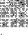
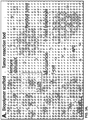
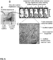
- the compositions of the current disclosure can deliver tumor-reactive lymphocytes ( FIG. 3A ) along with immune stimulants at high local concentrations and over an extended period of time.
- Lymphocytes seeded within the composition exit the composition following implantation and disperse at high densities throughout a tumor resection bed and into draining lymph nodes to destroy remaining residual tumor cells following a resection ( FIG. 3B ).
- This step releases large amounts of tumor antigens from dying tumor cells into the tissue, which are subsequently taken up by antigen presenting cells (APCs).
- APCs antigen presenting cells
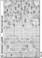
- This "second wave" of anti-tumor immunity is broader and involves multiple cell types ( FIG. 3C ) acting in synergy to eliminate remaining tumor cells.
- This described approach provides immediate efficacy by implantation of anti-tumor lymphocytes.
- the composition is designed to turn the tumor site or tumor resection cavity into a "self" vaccine site using dying tumor cells directly as the source of antigen to launch an effective anti-tumor immune response in the host.
- compositions also provide in vivo screening tools to (1) test which immune cell types, cell phenotypes or combinations of immune cells are most potent at destroying tumors, and (2) identify agents that boost their anti-tumor activity.
- the conventional approach currently used to answer these critical questions usually involves intravenous injection of the immune cells to be tested into tumor-bearing animals. However once infused, cells poorly traffic to the tumor and quickly change their phenotype, which prevents the determination of their intrinsic anti-tumor activity.
- small molecules, cytokines or stimulatory antibodies are often rapidly cleared from the circulation following intravenous administration and poorly penetrate into solid tumors. This makes it extremely difficult to study their direct effects on immune cells in tumors.
- any immune cell type can be embedded into the compositions along with a given therapeutic compound and, upon implantation, is directly exposed to tumor under clinically relevant in vivo conditions.
- This approach provides previously inaccessible knowledge about how immune cells collaborate and how drugs affect their function, which could ultimately speed up the transition of cell-based immunotherapies to the clinic.
- compositions can be constructed from a variety of material including, without limitation, biocompatible polymers.
- biocompatible polymers include, but not limited to, agar, agarose, alginate, alginate/calcium phosphate cement (CPC), beta-galactosidase ( ⁇ -GAL), (1,2,3,4,6-pentaacetyl ⁇ -D-galactose), cellulose, chitin, chitosan, collagen, elastin, gelatin, hyaluronic acid collagen, hydroxyapatite, poly(3-hydroxybutyrate-co-3-hydroxy-hexanoate) (PHBHHx), poly(lactide), poly(caprolactone) (PCL), poly(lactide-co-glycolide) (PLG), polyethylene oxide (PEO), poly(lactic-co-glycolic acid) (PLGA), polypropylene oxide (PPO), poly(vinyl alcohol) (PVA), silk, soy protein, and so
- the polymers can be responsive to a changed environmental condition following implantation. Polymers with these characteristics are known to those of ordinary skill in the art.
- an injectable in situ gel-forming system is used.
- the polymer formulation can gel in vivo in response to temperature change (thermal gelation), in response to pH change or in response to light.
- thermal gelation temperature change
- UV ultraviolet
- the polymer formulation can gel in vivo in response to ionic cross-linking.
- the polymer formulation can gel in vivo in response to solvent exchange.
- the gel used is thermoreversible, pH reversible, or light reversible.
- the gel used is high-viscosity and shear-thinning.
- the gel can be a gel formed from, without limitation, any polymer described herein.
- alginate is used as a structure material, either separately or in combination with one or more other materials.
- Alginate is easily processed, water soluble, and non-immunogenic.
- Alginate is a biodegradable anionic polysaccharide with free hydroxyl groups that offer easy gelling.
- the polymer may be a polyelectrolyte complex mixture (PEC) formed from a 1:1 solution of alginate and chitosan.
- a structure may formed from an alginate/calcium carbonate/glucono-delta-lactone mixture, such as 0.5-5% alginate, 0.5-15 g/L calcium carbonate, and 1-50 g/L gluconon-delta-lactone in a ratio of 2:1:1 (alginate:CaCO 3 :GDL).
- Polymer structures may also include varying amounts of gelatin in combination with varying amounts of alginate. Depending on the materials and material ratios in mixture, the structures may optionally be cross-linked.
- polymer solutions having varying amounts of polymer dissolved in an acidic solution can be used to form the structures disclosed herein.
- the concentration of the acid can be adjusted depending on the amount of polymer dissolved.
- the acidic solution is 1% (v/v) acetic acid.
- the amount of polymer in solution is between about 0.5-5% (w/v) and any whole or partial increments therebetween.
- the amount of polymer in solution (w/v) can be 0.5%, 1%, 1.5%, 2%, 2.5%, 3%, 3.5%, 4%, 4.5% or 5%.
- the amount of polymer in solution is 2.4% (w/v).
- the polymer is dissolved in at least one of water, acid, acetic acid, camphene, or camphene-naphthalene.
- the concentration of the acid can be adjusted depending on the amount of gelatin in combination with polymer (in one embodiment, alginate) that is dissolved.
- the acidic solution is 1% (v/v) acetic acid.
- the amount of gelatin in solution is between about 1-10% (w/v) and any whole or partial increments therebetween.
- the amount of alginate in solution (w/v) can be 1%, 2%, 3%, 4%, 5%, 6%, 7%, 8%, 9% or 10%.
- the amount of alginate in solution is 5.5% (w/v).
- the polymer solution includes a combination of 2.4% (w/v) alginate solution and a 5.5% (w/v) gelatin solution.
- the gelatin in combination with varying amounts of alginate is dissolved in at least one of water, acid, acetic acid, camphene, or camphene-naphthalene.
- alginate-based scaffolds can be formed as follows: a weight by volume (w/v) alginate solution in deionized (DI) water can be prepared and filtered with a 0.45 micrometer bottle filter to remove any particles and then frozen to -80°C. The frozen sample can be lyophilized in a 4.5 liter benchtop freeze dry system (Labconco, Kansas City, MO). The filtered lyophilized alginate can be reconstituted into solutions of various concentrations (0.1%-5%) with water or buffer.
- DI deionized
- Crosslinking can be performed with, without limitation, calcium chloride and/or calcium carbonate.
- Calcium carbonate is a slow crosslinker, with samples taking up to several hours to fully crosslink.
- GDL gluconodeltalactone
- Calcium chloride is a fast crosslinker and the samples will fully gel in a few minutes.
- the addition of CaCl 2 to the alginate solution can occur prior to freezing.
- Other methods include use of a 5.5% (w/v) solution of calcium carbonate+GDL added to the alginate solution prior to initial freezing.
- alginate solutions can be degassed in a speed mixer and poured slowly into casts to prevent bubbles from forming.
- air bubble formation can be avoided by placing a micropipette on the open end of mold grooves and repeatedly flushing the entire canal system until the residual air is flushed out.
- Freeze casting can be used to form the scaffolds disclosed herein.
- Various polymer solutions can be freeze cast into various sized casts as would be understood by those skilled in the art.
- the rate of cooling should be controlled as it affects the size and alignment of pores, as well as the formation of ridges.
- the cooling rate can range between 0.1-100°C per minute (m) and any whole or partial increments therebetween.
- the cooling rate can range between 1-10°C/m, and any whole or partial increments therebetween.
- the cooling rate (°C/m) can be 0.1, 0.5, 1, 2, 3, 4, 5, 5.5, 6, 6.5, 7, 7.5, 8, 8.5, 9, 9.5, or 10.
- compositions disclosed herein include lymphocyte-adhesion moieties to promote lymphocyte mobility out of the implanted structures.
- Lymphocyte-adhesion moieties include, without limitation, cell-adhesion moieties such as cell-adhesion polypeptides that mimic the extracellular matrix (such as collagen).
- cell adhesion polypeptides refer to compounds having at least two amino acids per molecule which are capable of binding via cell surface molecules, such as integrin.
- the cell adhesion polypeptides may be any of the proteins of the extracellular matrix which are known to play a role in cell adhesion, including fibronectin, vitronectin, laminin, elastin, fibrinogen, collagen types I, II, and V, as described in Boateng et al., Am. J. Physiol. - Cell Physio. 288:30-38 (2005 ). Additionally, the cell adhesion polypeptides may be any peptide derived from any of these proteins, including fragments or sequences containing the binding domains. Cell adhesion polypeptides include those having integrin-binding motifs, such as the ICAM-1 motif , and related peptides that are functional equivalents.
- Cell adhesion polypeptides may also be any of the peptides described in U.S. Patent Publication No. 20060067909 .
- the structures include compounds having lymphocyte-adhesion moieties, such as a ligand for ⁇ 1 ⁇ 1 , integrin, a ligand for ⁇ 2 ⁇ 1 , integrin, a ligand for ⁇ 4 ⁇ 1 integrin, a ligand for ⁇ 5 ⁇ 1 , integrin, a ligand for lymphocyte function-associated antigen (LFA-1), or combinations thereof.
- the ligand interacts specifically with one integrin.
- the ligand is not a complete fibronectin molecule or is not a complete collagen molecule.
- the lymphocyte-adhesion moiety can be a peptide, antibody, or a small organic molecule.
- a small organic molecule refers to a carbon-based molecule having a molecular weight of 500 daltons or less.
- the antibody or an integrin binding fragment thereof can be single chained, humanized, or chimeric.
- the lymphocyte-adhesion moiety can be a collagen-mimetic peptide, for example a stable triple-helical, collagen-mimetic peptide that contains the GFOGER adhesion motif from type I collagen (SEQ ID NO:1) that is recognized by the ⁇ 2 ⁇ 1 , integrin. This peptide adopts a stable triple-helical conformation similar to the native structure of type I collagen.
- an exemplary collagen-mimetic peptide has the following amino acid sequence GGYGGGPC(GPP) 5 GFOGER(GPP) 5 GPC (also referred to herein as "GFOGER peptide").
- GFOGER peptide comprises, consists, or consists essentially of SEQ ID NO:2.
- ICAM-1 is an Ig-like cell adhesion molecule that binds integrins promoting cell-cell adhesion and is a ligand for lymphocyte function-associated (LFA) antigens.
- LFA lymphocyte function-associated
- ICAM-1 is found primarily on monocytes and endothelial cells, and is widely inducible, or upregulated, on many cells including T-cells, B-cells, thymocytes, dendritic cells, endothelial cells, fibroblasts, keratinocytes, chondrocytes, and epithelial cells. This protein has a co-stimulatory effect upon cytotoxic T-cell interaction, and is utilized in a number of intercellular binding interactions.
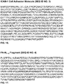
- ICAM-1 comprises, consists, or consists essentially of SEQ ID NO:3.
- FNIII 7-10 as a lymphocyte-adhesion moiety.
- FNIII 7-10 is a fibronectin fragment spanning the 7-10th type III repeats of fibronectin.
- the sequence of fibronectin is known in the art.
- FNIII 7-10 comprises, consists or consists essentially of SEQ ID NO:4.
- Variants include peptides having one or more conservative amino acid substitutions.
- a "conservative substitution” involves a substitution of one amino acid for another found in one of the following conservative substitutions groups: Group 1: Alanine (Ala), Glycine (Gly), Serine (Ser), Threonine (Thr); Group 2: Aspartic acid (Asp), Glutamic acid (Glu); Group 3: Asparagine (Asn), Glutamine (Gln); Group 4: Arginine (Arg), Lysine (Lys), Histidine (His); Group 5: Isoleucine (Ile), Leucine (Leu), Methionine (Met), Valine (Val); and Group 6: Phenylalanine (Phe), Tyrosine (Tyr), Tryptophan (Trp).
- amino acids can be grouped into conservative substitution groups by similar function or chemical structure or composition (e.g., acidic, basic, aliphatic, aromatic, sulfur-containing).
- an aliphatic grouping may include, for purposes of substitution, Gly, Ala, Val, Leu, and Ile.
- Other groups containing amino acids that are considered conservative substitutions for one another include: sulfur-containing: Met and Cysteine (Cys); acidic: Asp, Glu, Asn, and Gln; small aliphatic, nonpolar or slightly polar residues: Ala, Ser, Thr, Pro, and Gly; polar, negatively charged residues and their amides: Asp, Asn, Glu, and Gln; polar, positively charged residues: His, Arg, and Lys; large aliphatic, nonpolar residues: Met, Leu, Ile, Val, and Cys; and large aromatic residues: Phe, Tyr, and Trp. Additional information is found in Creighton (1984) Proteins, W.H. Freeman and Company .
- Variants also include sequences with at least 70% sequence identity, 80% sequence identity, 85% sequence, 90% sequence identity, 95% sequence identity, 96% sequence identity, 97% sequence identity, 98% sequence identity, or 99% sequence identity to SEQ ID NO:1; SEQ ID NO:2; SEQ ID NO:3; and SEQ ID NO:4.
- % identity refers to a relationship between two or more protein sequences, as determined by comparing the sequences.
- identity also means the degree of sequence relatedness between proteins as determined by the match between strings of such sequences.
- Identity (often referred to as “similarity") can be readily calculated by known methods, including (but not limited to) those described in: Computational Molecular Biology (Lesk, A. M., ed.) Oxford University Press, NY (1988 ); Biocomputing: Informatics and Genome Projects (Smith, D. W., ed.) Academic Press, NY (1994 ); Computer Analysis of Sequence Data, Part I (Griffin, A. M., and Griffin, H.
- Relevant programs also include the GCG suite of programs (Wisconsin Package Version 9.0, Genetics Computer Group (GCG), Madison, WI); BLASTP, BLASTN, BLASTX ( Altschul, et al., J. Mol. Biol. 215:403-410 (1990 )); DNASTAR®; and the FASTA program incorporating the Smith-Waterman algorithm ( Pearson, Comput. Methods Genome Res., [Proc. Int. Symp.] (1994), Meeting Date 1992, 111-20.
- the lymphocyte-adhesion moieties may be incorporated into the backbone of a polymer chain.
- a polymer can be created containing YIGSR in the backbone of a polymer as described in Jun et al., J. Biomaterials Sci., Polymer Ed. 15(1), 73-94 (2004 ).
- One of skill in the art could incorporate other cell adhesion polypeptides into the backbone of alginate or other polymers.
- the lymphocyte-adhesion moieties may be grafted onto a polymer.
- the lymphocyte-adhesion moieties are polypeptides that may be grafted onto polymers using various methods known in the art.
- polymers having side branches containing reactive functional groups such as epoxide, halide, amine, alcohol, sulfonate, azido, anhydride, or carboxylic acid moieties can be covalently linked to the amine terminus of the polypeptides via the reactive side branches using conventional coupling techniques such as carbodiimide reactions.
- RGD (Arg-Gly-Asp)-containing peptides have been grafted onto the backbone of polymers as described in Lin, et al., J. Biomedical Materials Res, 28(3), 329-42 (1994 ).
- RGD-containing peptides have been grafted onto the side branches of polyethylene glycol based polymers, as described in Hansson, et al., Biomaterials, 26, 861-872 (2005 ).
- the scaffold can comprise fibrin scaffolds, collagen scaffolds or fibrin and/or collagen scaffold blends (blended with, for example, alginate). Certain embodiments do not require bioactive coatings to support lymphocyte mobility. Similarly, injectable forms of the structures do not require bioactive coatings.
- Scaffolds can also be coated with a bioactive coating comprising a lymphocyte-activating moiety.
- the scaffold is at least partially coated with a bioactive coating comprising a lymphocyte-adhesion moiety.
- the bioactive coating can be applied onto the surface of the scaffold in various ways, including the use of coating methods that are known in the art.
- the bioactive coating may be sprayed onto the scaffold by a conventional electrostatic spraying process, resulting in charged droplets being deposited onto the surface of the composition
- the bioactive compound for example, a polypeptide
- the deposited polypeptide may form a monolayer on the surface of the scaffolding.
- the bioactive coating may be bonded to the surface of a scaffold by any type of chemical or physical bonding means, including covalent, polar, ionic, coordinate, metallic, electrostatic, or intermolecular dipolar (including Van der Waals) bonds.
- the surface of the scaffold is coated with GFOGER peptide.
- the purified GFOGER peptide could be stored as a trifluoroacetic acid (TFA) salt and reconstituted to 10 mg/mL in 0.1% TFA and 0.01% sodium azide and stored at 4°C prior to use.
- TFA trifluoroacetic acid
- the scaffolds After the scaffolds are rinsed with ethanol to remove contaminants, cleaned in fresh ethanol, rinsed in ddH 2 O, they can be soaked in phosphate buffered saline (PBS).
- PBS phosphate buffered saline
- the GFOGER peptide can then be absorbed onto the scaffolds passively by incubating the scaffolds in a solution of GFOGER in PBS. Prior to implantation, scaffolds could be rinsed in PBS to remove unbound GFOGER peptides.
- an alginate/calcium carbonate/glucono-delta-lactone mixture can be prepared by stirring, with concentrations ranging from 0.5-5 wt% alginate, 0.5-15 g/L calcium carbonate, and 1-50 g/L glucono-delta-lactone in a volume ratio of 2:1:1 (alginate:CaCO 3 :GDL) as a "pre-gelling" process.
- the resulting mixture can be freeze cast (directionally frozen) at a constant cooling rate (0.1°/min-10°/min) until solid and lyophilized until dry.
- compositions can be crosslinked in 0.1-2.5 wt.% calcium chloride for 5-30 minutes and washed in HEPES buffered saline prior to any further use of the scaffold.
- surfaces of the compositions can be coated in polylysine or polyornithine (0.1-1.0 mg/ml for 3-10 minutes) followed by coating in a GFOGER peptide (10 ⁇ g/ml-250 ⁇ g/ml for 30 minutes-24 hours).
- an alginate-chitosan polyelectrolyte complex (PEC) mixture can be prepared by sonicating or homogenizing on ice in a range of 1:1 to 1:9 solutions (both ways) of alginate (prepared in water) and chitosan (prepared in 1% acetic acid) and total polymer content ranging from 0.5%-5%.
- the pH of the resulting mixture can be adjusted with NaOH up to 10.0.
- the alginate-chitosan PEC mixture can be freeze cast at a constant cooling rate (0.1°/min-10°/min) until solid and lyophilized until dry.
- Dried compositions can be crosslinked in 0.1-2.5% calcium chloride for 5-30 minutes and washed in PBS prior to any further use of the composition.
- scaffolds can be coated in polylysine or polyornithine (0.5 mg/ml for 6 minutes) followed by coating in a GFOGER peptide (10 ⁇ g/ml-250 ⁇ g/ml for 30 minutes-24 hours).
- GFOGER peptides are immobilized onto alginate using aqueous carbodiimide chemistry.
- Bioactive coatings can additionally include other components to alter the surface of the scaffold, for example polylysine, polyornitine, or other glycoproteins.
- Lymphocyte-activating moieties include any compound that activates a lymphocyte and can be incorporated in or attached to the structures disclosed herein.
- activation of a lymphocyte refers to the state of a lymphocyte that has been sufficiently stimulated to induce detectable cellular proliferation, cytokine production, or effector function such as tumor targeting and/or killing. If the lymphocyte is a T-cell, activation also results in expression of cell surface markers particular to the T-cell type.
- lymphoctye-activating moieties include CD3, CD27, CD28, CD80, CD86, 4-1BB, CD137, OX40, CD30, CD40, LFA-1, CD2, CD7, LIGHT, NKG2C, B7-H3, and CD83 ligands or antibodies, CD1d, recombinant CD1d molecules preloaded with ⁇ -galactosyl ceramide and/or recombinant major histocompatibility complex (MHC) molecules loaded with defined tumor antigens or peptides to selectively expand particular lymphocyte types embedded within a scaffold.
- MHC major histocompatibility complex
- Lymphocyte-activating moieties can be found within injectable forms of the structures or embedded within the pores of the scaffolds, attached to the surface of the scaffolds, and/or embedded within the scaffolds themselves. As discussed further below, lymphocyte-activating moieties can also be associated with particles.
- immune stimulants can be included within the compositions.
- the immune stimulant is a cytokine, an antibody, a small molecule, an siRNA, a plasmid DNA, and/or a vaccine adjuvant.
- cytokines include, without limitation, IL-2, IL-7, IL-12, IL-15, IL-18, IL-21, TNF ⁇ , IFN- ⁇ , IFN- ⁇ , IFN- ⁇ , or GM-CSF.
- the immune stimulant may be a cytokine composition comprising combinations of cytokines, such as IL-2, IL-12 or IL-15 in combination with IFN- ⁇ , IFN- ⁇ or IFN- ⁇ , or GM-CSF, or any effective combination thereof, or any other effective combination of cytokines.
- the above-identified cytokines stimulate T H 1 responses, but cytokines that stimulate T H 2 responses may also be used, such as IL-4, IL-10, IL-11, or any effective combination thereof.
- combinations of cytokines that stimulate T H 1 responses along with cytokines that stimulate T H 2 responses may be used.
- Exemplary antibodies include, without limitation, anti-PD1, anti-PDL1, anti-CTLA-4, anti-TIM3, agonistic anti-CD40, agonistic anti-4-1 BB, and/or bispecific antibodies (e.g., BITE-antibodies: anti-CD3/anti-tumor antigen).
- Exemplary small molecule drugs include, without limitation, TGF-beta inhibitors, SHP-inhibitors, STAT-3 inhibitors, and/or STAT-5 inhibitors. Any siRNA capable of down-regulating immune-suppressive signals or oncogenic pathways (such as kras) can be used whereas any plasmid DNA (such as minicircle DNA) encoding immune-stimulatory proteins can be used.
- Exemplary vaccine adjuvants include, without limitation, any kind of Toll-like receptor ligand or combinations thereof (e.g. CpG, Poly(I:C), ⁇ -galactoceramide, MPLA, cyclic dinucleotides, VTX-2337 (novel TLR8 agonist developed by VentiRx), and/or inhibitors of heat-shock protein 90 (Hsp90), such as 17-DMAG (17-dimethylaminoethylamino-17-demethoxygeldanamycin).
- Toll-like receptor ligand or combinations thereof e.g. CpG, Poly(I:C), ⁇ -galactoceramide, MPLA, cyclic dinucleotides, VTX-2337 (novel TLR8 agonist developed by VentiRx), and/or inhibitors of heat-shock protein 90 (Hsp90), such as 17-DMAG (17-dimethylaminoethylamino-17-demethoxygeldanamycin
- Immune stimulants derived from the molecules noted in the preceding paragraphs can also be used.
- RLI is an IL-15-IL-15 receptor- ⁇ fusion protein that exhibits 50-fold greater potency than IL-15 alone.
- IL-15 impacts the anti-tumor immune response at multiple points. It can differentiate monocytes into stimulatory antigen presenting cells; promote the effector functions and proliferation of tumor-reactive T-cells; and recruit and activate NK cells.
- Immune stimulants can be found within injectable forms of the structures or embedded within the pores of the scaffolds, attached to the surface of the scaffolds and/or embedded within the scaffolds themselves. As discussed further below, immune stimulants can also be associated with particles.
- Release of the immune stimulants from particles can be modified by incorporation of surfactants, detergents, complexing agents, internal phase viscosity enhancers, surface active molecules, co-solvents, chelators, stabilizers, derivatives of cellulose, polysorbates, PVA or sucrose. Salts and buffers can also be used to alter release characteristics.
- particles can be included as means to deliver/present lymphocyte-activating moieties to lymphocytes because they can mimic physiological antigen presenting cells.
- Another advantage of particles is that they are highly modular and can be customized without affecting the chemical properties of the structure itself.
- lymphocyte-activating moieties and/or immune stimulants are provided in association with particles.
- Particles can be included within injectable structures and/or within scaffolds.
- the particles can be formed from any biocompatible polymer including, without limitation, agar, agarose, alginate, alginate/ CPC, ⁇ -GAL, (1,2,3,4,6-pentaacetyl ⁇ -D-galactose), cellulose, chitin, chitosan, collagen, elastin, gelatin, hyaluronic acid collagen, hydroxyapatite, PHBHHx, poly(lactide), PCL, PLG, PEO, PLGA, PPO, PVA, silk, soy protein, and soy protein isolate, alone or in combination with any other polymer composition, in any concentration and in any ratio. Blending different polymer types in different ratios using various grades can result in characteristics that borrow from each of the contributing polymers.
- Various terminal group chemistries can also be adopted.
- the particles can be microparticles or nanoparticles.
- Microparticles can have a diameter of 10 - 20 ⁇ m while nanoparticles have a diameter of 100-150 nm.
- Particles may be formed according to any method known to those of ordinary skill in the art. Common methods include, without limitation, spray-drying or emulsion.
- an organic phase of PLGA polymer and dioleoylphosphocholine (DOPC), dioleoylphosphoglycerol (DOPG) and malemimide-phycoerythin (PE) lipids are emulsified in water, leading to self-assembled lipid coatings surrounding each particle.
- Particles may also be formed using a Buchi-190 Mini Spray wherein 1-2% polymer can be spray dried and collected in a dry container.
- the particles can be stirred in 0.4% sodium hydroxide in ethanol solution for 15 minutes to 1 hour before washing in PBS.
- the particles can be coated in 0.1-1% alginate solution for 5-20 minutes before washing in water, freezing, and lyophilization until dry.
- the particles can be stirred in a 0.001%-1% genipin solution prepared in PBS, or 0.001-25% glutaraldehyde solution for 0-48 hours.
- Crosslinking is stopped by stirring the particles in a 10% glycine (prepared in PBS) solution for 30 minutes.
- the particles can also be coated in 0.1-1% alginate solution for 5-20 minutes, or alginate solution followed by 0.1-1% polyethylene glycol solution for 5-20 minutes before washing in water, freezing, and lyophilizing until dry.
- alginate particles can be cross-linked as follows. Alginate particles (collected dry or collected in calcium chloride) can be suspended in ethanol. Epichlorohydrin (1-25% v/v) can be added to the particle mixture in ethanol. The mixture can be sonicated or homogenized on ice while adding 1M-6M sodium hydroxide. The mixture can then be stirred at room temperature for 6-24 hours and the reaction can be stopped by adjusting the pH to 7 with 1M hydrochloric acid. The crosslinked particle can be washed in ethanol in decreasing concentrations (e.g., 75%, 50%, 25%), followed by washing in water three times.
- concentrations e.g., 75%, 50%, 25%
- Alternate covalent crosslinking can be performed by suspending the particles in methanol containing 1-25% glutaraldehyde and 0.05-5% hydrochloric acid, stirring for 0-48 hours. Remaining calcium chloride can be removed by stirring microcapsules in 55 mM sodium citrate for 10 minutes, followed by washing in water. Particles can also be coated here in A) 0.1-1% chitosan solution (prepared in 1% acetic acid) for 5-20 minutes, B) the solution of (A) followed by 0.1-1% alginate solution for 5-20 minutes; and/or C) the solution of (A) followed by 0.1-1% polyethylene glycol solution for 5-20 minutes. The additional crosslinking/coating steps are followed by washing in water, freezing, and lyophilizing until dry.
- Lymphocyte-activating moieties can be incorporated on the surface of the particles by soaking (either from a dry state or pre-hydrated in PBS) in the desired lymphocyte-activating moiety solution (with or without stabilizers such as trehalose).
- concentrations and timing can range from 1 ⁇ g/ml to 1 g/ml for 15 minutes to 24 hours before rinsing with PBS.
- the lymphocyte-activating moieties are antibodies that are mildly reduced with dithiothreitol (DTT) and covalently coupled to maleimide on the surface of the particles.
- DTT dithiothreitol
- the lymphocyte-activating moiety-surface coated particles can be stirred into the polymer mixture prior to freeze casting.
- the prepared particles are added dropwise to a 2% aqueous GFOGER-peptide modified alginate solution before cross-linking alginate with calcium chloride and molding three-dimensional scaffolds by freeze drying. This process yields compositions with 7X10 6 antibody-coated PLGA microparticles that are homogenously dispersed within the scaffold's pore network.
- lipid stock solutions can be prepared in chloroform.
- DOPC, DSPE-PEG(2000) maleimide, cholesterol and 18:1 PEG(2000) PE can be combined in a scintillation vial to attain a DOPC:DSPE-PEG(2000) maleimide:cholesterol:PEG(2000) with a PE mass ratio of 55:5:30:10 and 2.5 mg total lipid.
- Chloroform can be evaporated and residual solvent removed.
- a suspension of spherical silica gel can be prepared in PBS.
- the suspension can be combined with an immune stimulant.
- the suspension can be gently agitated and diluted.
- the entire suspension can be added to a batch of lipid film.
- the mixture can be vortexed.
- the particles can be centrifuged and the supernatant can be removed.
- the pellet can be washed and redispersed.
- the hinge-region disulfide bonds of anti-CD3, CD28, and CD137 can be selectively reduced as described by Kwong et al, Biomaterials 32, 5134 (2011 ) which is incorporated by reference herein for its teachings regarding the same.
- the mildly-reduced antibodies can be added to the maleimide-functionalized particles.
- the mixture can be vortexed briefly and the resulting antibody-labeled particles can be centrifuged and supernatant removed. The pellet can then be washed.
- compositions disclosed herein include embedded lymphocytes. Any type of lymphocyte capable of targeting and killing tumor cells, targeting tumor cells for killing by other cell types, or otherwise mediating tumor cell killing can be used.
- the lymphocytes are autologous to the individual for whom the composition is administered.
- Lymphocytes include T-cells, B cells and natural killer (NK) cells.
- NK natural killer cells.
- the current disclosure focuses on the use of embedded T-cells, but other types of lymphocytes may be used as well, alone or in combination.
- T-cells include helper cells (CD4+ T-cells) and cytotoxic T-cells (CTLs, CD8+ T-cells) which comprise cytolytic T-cells.
- helper cells CD4+ T-cells
- CTLs cytotoxic T-cells
- T helper cells assist other white blood cells in immunologic processes, including maturation of B cells into plasma cells and activation of cytotoxic T-cells and macrophages, among other functions. These cells are also known as CD4+ T-cells because they express the CD4 protein on their surface. Helper T-cells become activated when they are presented with peptide antigens by MHC class II molecules that are expressed on the surface of antigen presenting cells (APCs). Once activated, they divide rapidly and secrete small proteins called cytokines that regulate or assist in the active immune response.
- APCs antigen presenting cells
- Cytotoxic T-cells destroy virally infected cells and tumor cells, and are also implicated in transplant rejection. These cells are also known as CD8+ T-cells because they express the CD8 glycoprotein at their surface. These cells recognize their targets by binding to antigen associated with MHC class I, which is present on the surface of nearly every cell of the body.
- T-cell receptor A majority of T-cells have a T-cell receptor (TCR) existing as a complex of several proteins.
- the actual TCR is composed of two separate peptide chains, which are produced from the independent T-cell receptor alpha and beta (TCR ⁇ and TCR ⁇ ) genes and are called ⁇ - and ⁇ -TCR chains.
- TCR ⁇ and TCR ⁇ TCR alpha and beta
- ⁇ - and ⁇ -TCR chains Gamma-delta ( ⁇ ) T-cells represent a small subset of T-cells that possess a distinct TCR on their surface. However, in ⁇ T-cells, the TCR is made up of one ⁇ -chain and one ⁇ -chain. This group of T-cells is much less common (2% of total T-cells) than the ⁇ T-cells.
- Central memory T-cells refers to an antigen experienced CTL that expresses CD62L or CCR-7 and CD45RO on the surface thereof, and does not express or has decreased expression of CD45RA as compared to naive cells.
- central memory cells are positive for expression of CD62L, CCR7, CD2S, CD127, CD45RO, and CD95, and have decreased expression of CD54RA as compared to naive cells.
- Effective memory T-cell refers to an antigen experienced T-cell that does not express or has decreased expression of CD62L on the surface thereof as compared to central memory cells, and does not express or has decreased expression of CD45RA as compared to a naive cell.
- effector memory cells are negative for expression of CD62L and CCR7, compared to naive cells or central memory cells, and have variable expression of CD28 and CD45RA.
- naive T-cells refers to a non-antigen experienced T lymphocyte that expresses CD62L and CD45RA, and does not express CD45RO as compared to central or effector memory cells.
- naive CD8+ T lymphocytes are characterized by the expression of phenotypic markers of naive T-cells including CD62L, CCR7, CD28, CD127, and CD45RA.
- TE T-cells refers to a antigen experienced cytotoxic T lymphocyte cells that do not express or have decreased expression of CD62L, CCR7, CD28, and are positive for granzyme B and perforin as compared to central memory or naive T-cells.
- the primary lymphocyte cell type will be CTL.
- CTLs can be included at 50% or more of the embedded lymphocyte population, 55% or more of the embedded lymphocyte population, 60% or more of the embedded lymphocyte population, 65% or more of the embedded lymphocyte population, 70% or more of the embedded lymphocyte population, 75% or more of the embedded lymphocyte population, 80% or more of the embedded lymphocyte population, 85% or more of the embedded lymphocyte population, 90% or more of the embedded lymphocyte population, 95% or more of the embedded lymphocyte population, or 100% the embedded lymphocyte population.
- the composition includes a mixture of CD8+ cells, NK cells, invariant NKT cells (iNKT cells), Th17 CD4+ cells and/or B cells.
- the compositions include a mixture of CD8+ cells and NK cells.
- the mixture of CD8+ cells and NK cells is a 50:50 mix.
- the compositions include a mixture of CD8+ cells and iNKT cells.
- the mixture of CD8+ cells and iNKT cells is a 50:50 mix. All other possible combinations of the disclosed cell types can also be used within the compositions disclosed herein.
- the lymphocytes can be isolated and expanded from resected tumor.
- subjects can be vaccinated with a tumor antigen (e.g., against Her2) and vaccine-induced T-cell populations can be expanded and embedded into the composition.
- a tumor antigen e.g., against Her2
- vaccine-induced T-cell populations can be expanded and embedded into the composition.
- Lymphocytes within the compositions can be non-genetically modified or genetically-modified or can be provided in a combination of non-genetically-modified and genetically-modified forms. Genetic modifications can be made to enhance growth, survival, immune function and/or tumor cell targeting. Examples of genetic modifications include those allowing expression of a chimeric antigen receptor (CAR), a ⁇ T-cell receptor (or modification thereof), and/or pro-inflammatory cytokines. CAR modification and/or ⁇ T-cell receptor modifications allow the modified lymphocytes to specifically target cell types.
- CAR chimeric antigen receptor
- ⁇ T-cell receptor or modification thereof
- pro-inflammatory cytokines pro-inflammatory cytokines
- modified lymphocytes can have improved tumor recognition, trigger increased native T-cell proliferation and/or cytokine production.
- Different potential CAR nucleic acid constructs that encode different ligand binding domains, different spacer region lengths, different intracellular binding domains and/or different transmembrane domains, can be tested in vivo (in an animal model) and/or in vitro to identify CARs with improved function over non-genetically modified lymphocytes and/or other CARs and in particular embodiments, using the compositions disclosed herein as an in vivo screening tool.
- Exemplary CARs express ligand binding domains targeting, without limitation, mesothelin, Her2, WT-1 and/or EGRF.
- An exemplary T-cell receptor modification targets melanoma-associated antigen (MAGE) A3 TCR.
- Negative selectable genes include, inter alia the following: the Herpes simplex virus type I thymidine kinase (HSV-I TK) gene, which confers ganciclovir sensitivity; the cellular hypoxanthine phosphribosyltransferase (HPRT) gene, the cellular adenine phosphoribosyltransferase (APRT) gene, and bacterial cytosine deaminase.
- HSV-I TK Herpes simplex virus type I thymidine kinase
- HPRT hypoxanthine phosphribosyltransferase
- APRT cellular adenine phosphoribosyltransferase
- Desired genes can be introduced into the lymphocytes prior to embedding in a composition disclosed herein. Such introduction can be carried out by any method known in the art, including but not limited to transfection, electroporation, microinjection, lipofection, calcium phosphate mediated transfection, infection with a viral or bacteriophage vector containing the gene sequences, cell fusion, chromosome-mediated gene transfer, microcell-mediated gene transfer, sheroplast fusion, etc. Numerous techniques are known in the art for the introduction of foreign genes into cells (see e.g., Loeffler and Behr, Meth. Enzymol, 217, 599-618 (1993 ); Cohen et al., Meth.
- the technique provides for the stable transfer of the gene to the cell, so that the gene is expressible by the cell and preferably heritable and expressible by its cell progeny. In other embodiments, the technique provides for transient expression of the gene within a cell.
- lymphocytes will be embedded within the compositions at or near the time of composition implantation in a subject, for example within 48 hours of implantation, within 36 hours of implantation, within 24 hours of implantation, within 12 hours of implantation, within 6 hours of implantation, within 3 hours of implantation, within 1 hour of implantation or within 30 minutes of implantation.
- lymphocyte loading into pre-molded scaffolds will occur within 30 minutes of implantation whereas the loading will more often occur closer (i.e., within 5 minutes; within 2 minutes, within 1 minute or within 30 seconds) to the actual implantation time when injectable forms of the compositions are used.
- the lymphocytes can be fresh lymphocytes or can be previously cryo-preserved lymphocytes. If previously-cryopreserved lymphocytes are used, they should be thawed quickly (e.g., in a water bath maintained at 37°-41°C) and chilled immediately upon thawing. It may be desirable to further treat the lymphocytes in order to prevent cellular clumping upon thawing. To prevent clumping, various procedures can be used, including but not limited to, the addition before and/or after freezing of DNase, low molecular weight dextran and citrate, hydroxyethyl starch, etc. Where necessary due to potential cytotoxicities, cryoprotective agents should be removed. After removal of cryoprotective agents, when necessary, cell count and/or viability testing can be performed.
- lymphocytes are resuspended in cell culture medium (e.g., RPMI). This cell suspension is then added dropwise on top of a lyophilized scaffold.
- cell culture medium e.g., RPMI
- static seeding a lymphocyte suspension is seeded onto a structure and afterwards incubated for a certain time in the absence of agitation before being exposed to dynamic culture conditions, for example into a spinner flask that is slowly agitated.
- dynamic seeding can be used.
- the structure and the lymphocyte suspension can be placed together in, e.g., a container and the container is then incubated with gentle agitation for a certain time allowing the lymphocytes to embed themselves within the structure.
- rotational systems including centrifuges
- vacuum systems can be used.
- sheet-based lymphocyte seeding, electrostatic lymphocyte seeding, magnetic lymphocyte seeding, filtration lymphocyte seeding, and/or oscillating perfusion lymphocyte seeding can be used.
- Various combinations, such as, and without limitation, rotational vacuum seeding can also be used.
- the use of various biological hydrogels is also appropriate. For discussions of the various seeding options, see Li et al., Biotechnol.
- compositions described herein can be placed in the vicinity of un-resecatable tumor and/or non-resected tumor cells to have an anti-tumor effect in a subject.
- subject or “individual” typically refer to a mammal, such as a human, but can also be another mammal such as, but not limited to, dogs, cats, rabbits, cows, horses, etc.
- a "tumor” is a swelling or lesion formed by an abnormal growth of cells (called neoplastic cells or tumor cells).
- a “tumor cell” is an abnormal cell that divides by a rapid, uncontrolled cellular proliferation and continues to divide after the stimuli that initiated the new division cease. Tumors show partial or complete lack of structural organization and functional coordination with the normal tissue, and usually form a distinct mass of tissue, which may be either benign, pre-malignant or malignant.
- an anti-tumor effect refers to a biological effect, which can be manifested by a decrease in tumor volume, a decrease in the number of tumor cells, a decrease in the number of metastases, an increase in life expectancy, or a decrease of various physiological symptoms associated with the cancerous condition.
- An anti-tumor effect can also be manifested by a decrease in recurrence or an increase in the time before recurrence.
- the compositions disclosed herein can be used to treat a variety of cancers, can prevent or significantly delay metastasis, and/or can prevent or significantly delay relapse.
- Cancer refers to a class of diseases in which a group of cells display uncontrolled growth (division beyond the normal limits), invasion (intrusion on and destruction of adjacent tissues), and sometimes metastasis.
- Metalastasis refers to the spread of cancer cells from their original site of proliferation to another part of the body. The formation of metastasis is a very complex process and depends on detachment of malignant cells from the primary tumor, invasion of the extracellular matrix, penetration of the endothelial basement membranes to enter the body cavity and vessels, and then, after being transported by the blood, infiltration of target organs. Finally, the growth of a new tumor, i.e. a secondary tumor or metastatic tumor, at the target site depends on angiogenesis. Tumor metastasis often occurs even after the removal of the primary tumor because tumor cells or components may remain and develop metastatic potential.
- Cancers that can be treated with the anti-tumor effects of the compositions and methods disclosed herein include, without limitation, seminomas, melanomas, teratomas, neuroblastomas, gliomas, rectal cancer, endometrial cancer, kidney cancer, adrenal cancer, thyroid cancer, skin cancer, cancer of the brain, cervical cancer, intestinal cancer, liver cancer, colon cancer, stomach cancer, head and neck cancer, gastrointestinal cancer, lymph node cancer, esophagus cancer, colorectal cancer, pancreas cancer, ear, nose and throat (ENT) cancer, breast cancer, prostate cancer, cancer of the uterus, ovarian cancer, lung cancer, and metastases thereof.
- seminomas melanomas, teratomas, neuroblastomas, gliomas, rectal cancer, endometrial cancer, kidney cancer, adrenal cancer, thyroid cancer, skin cancer, cancer of the brain, cervical cancer, intestinal cancer, liver cancer, colon cancer, stomach cancer, head and neck cancer, gastrointestinal cancer, lymph node cancer, es
- compositions and methods disclosed herein without limiting the scope of the compositions and methods disclosed herein, the following cancer types are noted:
- Brain tumor (Glioblastoma): An estimated 10,000 new cases/year in the U.S. are seen. Currently no curative therapy is available. Gliobastoma shows very infiltrative growth and cannot be resected completely. 90% of tumors relapse within a 2 cm margin from the originally resected tumor. Biomaterial wafers loaded with chemotherapy are United States Food and Drug Administration (FDA)-approved (GLIADEL®, MGI Pharma.) for glioblastoma. However, due to insufficient tissue penetration, biomaterial implant delivered chemotherapy is mostly ineffective. In contrast, tumor-reactive lymphocytes deployed from the compositions disclosed herein can actively migrate to affected tissue, seeking out and destroying residual tumor cells.
- FDA United States Food and Drug Administration
- Pancreatic adenocarcinoma An estimated 43,920 new cases of pancreatic cancer were expected to occur in the U.S. in 2012. Only 20% will have resectable disease at the time of diagnosis (80% of patients do not undergo surgery as their tumor is too advanced at the time of diagnosis). Even surgery is considered a palliative venture with a 5-year survival rate of only 20%. Local recurrence is usually attributed to the difficulty of achieving microscopically negative surgical margins. Beyond the current composition's primary application to eradicate residual disease following surgical tumor resection, the compositions could also provide pancreatic tumor patients with inoperable disease ( ⁇ 80% of patients) with a highly effective treatment option. Compositions are implanted directly onto un-resectable established pancreatic adenocarcinomas.
- Ovarian cancer An estimated 22,000 new cases in 2012 in the U.S. were seen. Despite multimodality therapy with surgery and chemotherapy, most ovarian cancer patients have a poor prognosis (15,500 estimated deaths/year in U.S.). Ovarian cancer primarily disseminates within the peritoneal cavity. Adoptive T-cell therapy in ovarian cancer patients is currently being investigated at several centers. However, to date clinical results have been disappointing due to a poor survival of infused T-cells and a failure to combat immunosuppressive factors released by tumor cells to render T-cells dysfunctional. Multiple compositions embedded with tumor-reactive lymphocytes could be implanted laparoscopically into the peritoneal cavity of ovarian cancer patients, where they release tumor-reactive lymphocytes and immune stimulants over an extended time period.
- compositions are implanted in close proximity to un-resectable tumor cells and/or in tumor resection beds following resection.
- the compositions can be available in a number of different sizes and shapes and can be shape-conformable to fit the particular needs of individual subjects.
- the compositions are injected using ultrasound guidance in close proximity to (or in physical contact with) un-resected or non-resected tumor cells. Depending on the stage, size or severity of a tumor, compositions may be provided with different therapeutic strengths.
- Therapeutic strength can be manipulated by altering the size of the composition, volume of the composition, the number of lymphocytes embedded within a composition, the number of lymphocyte-activating moieties within a composition, the presence or amount of immune stimulants within the composition, etc. Each of these parameters can be assessed and determined by a treating physician.
- proximity refers to a distance within 10 cm, within 9 cm, within 8 cm, within 7 cm, within 6 cm, within 5 cm, within 4 cm, within 3 cm, within 2 cm, within 1 cm, within 0.9 cm, within 0.8 cm, within 0.7 cm, within 0.6 cm, within 0.5 cm, within 0.4 cm, within 0.3 cm, within 0.2 cm, or within 0.1 cm of an un-resectable tumor, un-resectable tumor cells, and/or a tumor resection bed.
- compositions can be implanted only once, at the time of resection or at a first treatment time in an individual with an un-resectable tumor. Additionally, the compositions can be implanted a plurality of times to provide ongoing therapy over months or years. Such treatment regimens can be determined by a treating physician.
- surgical treatment failure refers to relapse of cancer in a subject who had previously undergone tumor resection. Surgical treatment failure may include metastatic relapse.
- Example 1 Generation and ex vivo expansion of 4T1 breast tumor-reactive mouse T-cells.
- 4T1-STIM Irradiated 4T1 B7.1/4-1 BBL (hereafter 4T1-STIM) cells act as a whole cell cancer vaccine and prime tumor-specific T-cells in BALB/c mice following tail-base injection (4 x 10 6 tumor cells).
- the adjuvants CpG oligodeoxynucleotide and poly(I:C) were mixed with 4T1-STIM cells before injection.
- 3x10 5 4T1 tumor-reactive T-cells from inguinal and axillary lymph nodes of a single mouse can routinely be isolated.
- Example 2 Tumor-reactive T-cells injected intravenously or locally into the tumor resection bed fail to prevent tumor relapse due to inefficient tumor homing and/or poor persistence.
- 4T1 breast tumor model very closely mimics the tumor growth and metastatic spread of human breast cancer to lymph nodes, liver, lung, and bone.
- Tumor cells retrovirally tagged with Gaussia luciferase for bioluminescence imaging, are easily transplanted into the right mammary gland of BALB/c mice and develop tumors that are -10 mm in size after two weeks.
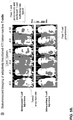
- mice were preconditioned for the adoptive transfer of T-cells by removing homeostatic cytokine sinks by lymphodepletion (250 mg/kg cyclophosphamide, injected intraperitoneally). The following day, tumors were resected, leaving behind 0.1-1% residual disease as quantified by bioluminescent imaging ( FIG. 5A , "Residual tumor”). Mice were either infused with T-cells the same day or received no treatment. Unexpectedly, all of the 10 animals treated intravenously with 7x10 6 tumor-specific T-cells relapsed with tumor ( FIG.
- T-cell treatments failed to control disease recurrence was investigated.
- T-cells were retrovirally-transduced with clickbeetle red luciferase (CBR-luc).
- CBR-luc clickbeetle red luciferase
- Intravenously infused T-cells accumulated at high levels in the spleen and the liver, but poorly trafficked to relapsing tumor ( FIG. 5B , upper panel).
- T-cells injected directly into the tumor bed cavity were readily detectable using bioluminescence on day 0.
- serial imaging showed a gradual CBR-luc signal decline following T-cell injection, consistent with poor T-cell expansion and persistence ( FIG. 5B , lower panel).
- Example 3 Porous polysaccharide scaffolds coated with collagen-mimetic peptide support rapid "lymph nodelike” motility and sustain the viability of embedded T-cells.
- compositions were created to produce a new microenvironment at the tumor resection site conducive to the sustained proliferation of transferred lymphocytes.
- the compositions can be used to deliver tumor-reactive lymphocytes to residual tumor following resection while sustaining their effector function and survival.
- a composition needs to provide sufficient mechanical support for embedded cells, a cell-adhesive coating to enable loaded cells to migrate through the material and exit into tissue, and appropriate stimulatory signals to trigger cell proliferation.
- GFOGER collagen-mimetic peptide
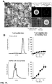
- Peptide binding efficiencies ranged from 83% (0.005 mg peptide/mg alginate) for the lowest peptide concentration tested, to 53% (0.102 mg peptide/mg alginate) for the highest peptide concentration tested ( FIG. 6B ). The highest peptide concentration was selected for use in subsequent experiments.
- FIG. 6C T-cell migration inside scaffolds using time-lapse video microscopy was recorded.
- the GFOGER peptide coating supported a rapid pore-to-pore T-cell migration quantitatively similar to the high motility of these cells in native secondary lymphoid organs (7.9 ⁇ m/min, FIG. 6 ).
- T-cells poorly migrated through uncoated scaffolds (3.6 ⁇ m/min), with most cells merely circling within their initial pore space ( FIG. 6C , lower left panel).
- contact with GFOGER peptide sustained the viability of loaded T-cells ( FIG. 6E ), which is consistent with reports describing the activation of pro-survival signaling pathways in T-cells binding collagen.
- the results suggest that GFOGER peptide-functionalized alginate scaffolds support rapid T-cell migration and sustain T-cell survival.
- Example 4 Scaffolds release functional tumor-reactive T-cells into surrounding tissue.
- T-cells can migrate outward from GFOGER-coated scaffolds into surrounding tissue was examined.
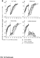
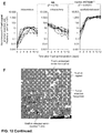
- 3D collagen gels containing the inflammatory cytokine IP-10 were prepared ( FIG. 7A ). Scaffolds loaded with 7x10 6 4T1 tumor-reactive T-cells were then embedded inside the collagen gel. T-cell egress from the composition was quantified every 12 hours for four days by counting viable cells in the scaffold and the collagen gel. T-cells gradually populated the surrounding tissue mimetic at high numbers, reaching a peak of 18.9x10 6 T-cells after 72 hours ( FIG. 7B , C ).
- composition-dispersed T-cells efficiently killed 4T1 tumor targets and released high amounts of the effector cytokines IL-2, IFN- ⁇ , and TNF- ⁇ , following co-culture on 4T1-STIM cells ( FIG. 7E ).
- GFOGER-coated scaffolds can efficiently disperse fully functional tumor-reactive T-cells into tissue.
- Example 5 Incorporating lymphocyte-stimulating ligands into compositions will help delivered lymphocytes overcome poor tumor immunogenicity and an adverse tumor microenvironment.
- Polymer microparticles coated with a combination of anti-CD3, CD28 and CD137 antibodies are incorporated into the compositions and support proliferation of embedded lymphocytes and increase the number and functionality of the cells that migrate from the composition into surrounding tissue.
- Example 5a Fabrication of alginate scaffolds carrying stimulatory micro/nanoparticles.
- T-cells To become activated, T-cells must not only recognize antigen but also receive costimulatory signals from antigen presenting cells (APCs).
- APCs antigen presenting cells
- PLGA poly(lactic-co-glycolic acid) microparticles similar in size to APCs (10-20 ⁇ m in diameter, FIG. 8A.1 , right panel) were synthesized. Particles were coated with avidin lipid so that biotinylated anti-CD3 antibodies and co-stimulatory anti-CD28/CD137 antibodies could be anchored to their surface.
- Stimulatory microparticle preparation preparation of lipid film.
- Lipid stock solutions were prepared in chloroform.
- 140 ⁇ L DOPC (10 mg/mL), 30 ⁇ L DSPE-PEG(2000) maleimide (5 mg/mL), 150 ⁇ L cholesterol (5 mg/mL), and 50 ⁇ L 18:1 PEG(2000) PE (5 mg/mL) were combined in a scintillation vial to attain a DOPC:DSPE-PEG(2000) maleimide:cholesterol:PEG(2000) PE mass ratio of 55:5:30:10 and 2.5 mg total lipid.
- Chloroform was evaporated under a stream of nitrogen and residual solvent was removed under vacuum overnight.
- cytokine into mesoporous silica microparticles.
- a suspension of spherical silica gel (15 ⁇ m particle diameter, 100 A pore diameter) was prepared in PBS (100 mg/mL). 120 ⁇ L of the suspension was combined in a 1.5-mL polypropylene tube with 400 ⁇ L of IL15 SA (22 ⁇ g/mL). The suspension was gently agitated on a vortexer for 1 hour at room temperature (RT) then diluted with 480 ⁇ L PBS.
- Antibody conjugation to silica-supported liposomes The hinge-region disulfide bonds of anti-CD3, CD28, and CD137 were selectively reduced with dithiothreitol (DTT) as previously described [ B. Kwong et al, Biomaterials (2011) 32:5134 ]. After removal of DTT with a desalting column, the mildly-reduced antibodies (anti-CD3: 200 ⁇ g; anti-CD28 and CD137: 400 ⁇ g) were added to the maleimide-functionalized particles. The mixture was vortexed briefly then rotated at RT for 2 h. The resulting antibody-labeled particles were centrifuged at 3500 x g for 2 min then the supernatant was removed. The pellet was washed with PBS (3 x 1 mL) then suspended in 125 ⁇ L PBS.
- PBS 3 x 1 mL
- Anti-CD3/CD28/CD137 antibodies mildly reduced with DTT, are then covalently coupled to maleimide on the surface of nanoparticles as previously described. Bershteyn et al., Soft Matter, 4, 1787-1791 (2008 ). The amount of bioactive antibodies coupled to micro/nanoparticles is quantified by a functional ELISA assay. The lipid envelopes of particles are first solubilized in 0.5% Tween 20 surfactant.
- Example 5b Comparison of lymphocyte expansion in microparticle vs. nanoparticle-functionalized compositions.
- the findings presented in FIG. 8 demonstrate that alginate scaffolds with incorporated stimulatory microparticles or nanoparticles can be successfully fabricated.
- in vitro T-cell assays described in detail in FIG. 7 are conducted. 7 million 4T1 tumor-reactive CD8 + T-cells are seeded either onto composite alginate/microparticle scaffolds ( FIG. 8A.1 ), alginate/nanoparticle scaffolds ( FIG. 8A.2 ) or plain scaffolds.
- an equal number (7x10 6 ) of microparticles and T-cells are embedded in each scaffold.
- Example 5c Determination of optimal concentration of stimulatory signals to support lymphocyte expansion.
- the optimal concentration of stimulatory signals to support maximum lymphocyte expansion without compromising lymphocyte viability or key effector functions is determined.
- the main advantage of composite alginate/particle scaffolds is that the amount of stimulatory signal inside the scaffold can be finely tuned without further modifying the alginate backbone itself just by adding more or fewer stimulatory particles to the alginate solution before scaffold fabrication.
- the optimal particle:lymphocyte ratio inside the composition is determined.
- Alginate/particle scaffolds with a final microparticle:T-cell ratio of 0.5:1, 1:1, 5:1 and 10:1 (or 500:1, 1,000:1, 5,000:1 and 10,000:1, if nanoparticles are chosen) are fabricated and compared to T-cell proliferation and functionality using the same assays described in Example 5b.
- the phenotype of composition-released lymphocytes is characterized. Phenotypic traits are predictive of the ability of lymphocytes to survive long-term, to serially kill tumor cells, to migrate into tumor-draining lymph nodes and, in embodiments utilizing T-cells, to differentiate into memory T-cells. Therefore, in parallel studies, how increasing stimulation inside the composition affects the phenotype of T-cells exiting the implant is investigated. T-cells that have migrated out of alginate scaffolds (fabricated with various numbers of stimulatory particles as in previous experiments) into collagen gel after 72 hours are recovered.
- T-cells are analyzed by flow cytometry for the expression of the pro-survival factor Bcl-xL, the proliferation marker Ki-67, the marker for terminal differentiation and replicative senescence KLRG1, and the memory markers CD44, CD62L and CD122.
- Early apoptotic T-cells are identified by staining cells with Annexin-V and PI.
- the major goal of providing lymphocytes with (co-)stimulatory signals inside the composition is to compensate for the absence of these ligands on tumor cells - a mechanism used by tumors to render attacking lymphocytes dysfunctional.
- combined CD137 and CD28 signaling can synergistically enhance the anti-tumor effector function of T-cells while decreasing their susceptibility to apoptosis.
- alginate scaffolds with incorporated anti-CD3/CD28/CD137 antibodies will mount a robust proliferative T-cell response.
- functionally exhausting T-cells or causing activation induced cell death by stimulating them excessively is avoided.
- T-cell receptor activation is lowered. This is readily achievable by reducing the number of anti-CD3 antibodies and increasing the number of costimulatory anti-CD28/CD137 antibodies on the surface of particles.
- mice exit implanted scaffolds at high densities over time and infiltrate the tumor resection bed and tumor-draining lymph nodes ( FIG. 9 ).
- the objectives of Example 6 is to (1) compare the therapeutic effectiveness of composition-supported lymphocyte delivery with conventional lymphocyte injections, and (2) elucidate underlying mechanism(s) by analyzing in vivo migration, expansion, persistence and phenotypic differences of transferred lymphocytes.
- Example 6a Comparison of the therapeutic effectiveness of composition-supported lymphocyte delivery with conventional lymphocyte injections. Differences in the frequency of tumor relapse in BALB/c mice following incomplete 4T1 tumor resection (as shown in FIGS. 2 , 5 , 9 ) are measured. Five different treatment groups are studied (18 mice/group). In one group, 7 x 10 6 4T1 tumor-reactive T-cells from optimized compositions are delivered directly into the tumor resection cavity (as shown in FIG. 9 ). Two groups of mice receive the same T-cell dose, but cells are either injected intravenously or locally into the resection bed. To assess therapeutic effects of the biomaterial itself, "empty" scaffolds (no T-cells) are implanted into one additional group of animals. All control mice are left untreated after surgery.
- 4T1 tumors (retrovirally tagged with the Gaussia luciferase gene) are serially imaged every two days over a period of 42 days using bioluminescence imaging, as described in relation to FIG. 5A ) using a state-of-the-art IVIS Spectrum (Caliper/Xenogen) whole-mouse imaging system.
- a whole animal bioluminescent signal of >40 x 10 6 photons per second as the surrogate endpoint for death was defined to avoid unnecessary pain and distress in treated animals.
- compositions are loaded with (co-)stimulatory antibodies ( FIG. 8 ), whereas T-cells injected as a cell suspension are not supported by these ligands. Accordingly, composition-released T-cells would be expected to eradicate tumors more effectively merely as a result of receiving additional stimulation. Intravenously injected T-cells therefore are activated locally with an equivalent amount of stimulatory antibody incorporated into the compositions. To this end, T-cells are stimulated in 6-well plates with immobilized anti-CD3/CD28/CD137 antibody 24 hours prior to injection.
- Example 6b Analysis of in vivo migration, expansion, persistence and phenotypic differences of transferred lymphocytes. Appropriate localization and migration of lymphocytes is a prerequisite for anti-tumor responses.
- the described studies showed that dual bioluminescence imaging of Gaussia luciferase (Gau-luc) in 4T1 tumor cells and of clickbeetle red luciferase (CBR-luc) in tumor-reactive T-cells allowed simultaneously monitoring of tumor regrowth and T-cell biodistribution ( FIG. 5 ).
- This assay is used to serially track the tissue distribution, expansion and persistence of composition-administered lymphocytes. Animals are imaged every two days for a period of 42 days or until they need to be euthanized.
- CBR-luc T-cell
- Gau-luc tumor cell
- Example 6c Effect of delivery mode on phenotype or functionality of composition-administered lymphocytes. Differences in lymphocyte phenotype and function between treatment groups are determined by flow cytometry. To distinguish composition-administered lymphocytes from host lymphocytes, 4T1 tumor-specific T-cells are generated in BALB/c mice that are congenic for the CD45.1 marker, as illustrated in FIG. 4 , and used to treat CD45.2 congenic recipient BALB/c mice. On day 4, 8 and 16 after transfer, three mice per treatment group are euthanized and single cell suspensions prepared from the tumor-resection bed, tumor draining lymph nodes (inguinal, axillary), and the spleen.
- lymphocytes are restimulated for 12 hours on 4T1-STIM monolayers in the presence of brefeldin A, and intracellular IFN- ⁇ or perforin are measured by flow cytometry, as shown in FIG. 4C .
- IFN- ⁇ is a key effector cytokine, while perforin is a key mediator of target cell killing.
- Example7 Co-delivering immune stimulants from implanted compositions can enhance act and trigger systemic host anti-tumor immunity.
- Example 7 the ability of compositions, beyond their primary function as lymphocyte delivery vehicles, to mount an effective host anti-tumor immune response capable of eliminating untreated distant metastases are tested.
- RLI is an IL-15-IL-15 receptor a fusion protein ( FIG. 10A ) that exhibits 50-fold greater potency than IL-15 alone.
- IL-15 impacts the anti-tumor immune response at multiple points. It can differentiate monocytes into stimulatory antigen presenting cells; promote the effector functions and proliferation of tumor-reactive T-cells; and recruit and activate NK cells.
- IL-15 or its superagonist RLI requires high and sustained systemic doses to achieve the desired effect, leading to dose-limiting toxicities.
- Compositions disclosed herein focus drug action only on immune cells for which they were intended, thereby avoiding systemic overexposure to these agonists.
- Example 7 investigate whether dispersing RLI from compositions into the tumor resection bed and the draining lymph nodes can (1) confer composition-delivered lymphocytes with markedly amplified anti-tumor effector functions and viability, and (2) orchestrate the destruction of untreated tumors throughout the body by activating dendritic cells, T-cells and NK-cells in the host.
- the results will show that combining delivery of RLI and tumor-reactive T-cells from compositions amplifies the expansion and persistence of transferred T-cells, using low RLI doses that have no effect when administered by traditional systemic routes.
- composition-released RLI transforms the peritumoral tissue and the tumor-draining lymph nodes from sites favoring immune suppression into "self" vaccine sites launching systemic anti-tumor immunity.
- Example 7a This example investigates whether dispersing RLI from implanted compositions can amplify the effector function and persistence of scaffold-delivered lymphocytes such as T-cells.
- 293-F cells are transduced with a plasmid encoding His-tagged RLI protein and RLI is purified from culture supernatant using a standard Cobalt agarose resin ( FIG. 10B ).
- Isolated RLI is fully functional, as demonstrated by its ability to enhance the proliferation of tumor-specific T-cells co-cultured on tumor monolayers ( FIG. 10C ).
- Poly(lactic-co-glycolic acid) (PLGA) micro-or nanoparticles fabricated as described in Example 5a (including surface-anchored stimulatory antibodies), efficiently encapsulate RLI and slowly release it over a one-week period ( FIG. 10D ).
- RLI-loaded micro- or nanoparticles are loaded into alginate compositions.
- Biodistributions of RLI following delivery via compositions disclosed herein relative to local or systemic bolus injections are measured.
- 10 ⁇ g RLI is administered either from compositions into 4T1 tumor resection cavities, or it is injected in its soluble form intravenously or into resection cavities.
- Blood serum and tissue samples of the tumor resection bed and tumor-draining (inguinal, axillary) lymph nodes re collected every two days for a period of 10 days.
- Tissue is homogenized with an ultrasonic dismembrator and centrifuged to collect supernatant. The amount of RLI per gram of tissue using a commercially available IL-1 5/IL-1 5R ⁇ ELISA is measured.
- Example 7b Enhancement of lymphocyte function and viability following composition-released rli versus infusion-administered RLI.
- One group of mice is treated with T-cell-loaded compositions that contain 10-50 ⁇ g RLI.
- a second group of mice is treated with T-cell-loaded compositions and 10-50 ⁇ g intravenous RLI. 4T1 tumor relapse and T-cell expansion are quantitated in response to RLI given via different routes using bioluminescence imaging as in Example 2.
- Example 7c Composition-released RLI orchestration of untreated and/or distant tumor.
- Tumors render antigen-presenting cells in draining lymph nodes dysfunctional to prevent tumor-specific T-cells in the host from differentiating into cytolytic effectors.
- IL-15 has been reported to restore the antigen presenting capacity of dendritic cells (DCs) and reverse tolerance in tumor-specific T-cells.
- DCs dendritic cells
- one predicted effect of local RLI delivery from compositions disclosed herein is activation of DCs in TDLNs coupled with the stimulation of anti-tumor T-cells in the host.
- the frequency and phenotype of DCs in the TDLNs (inguinal, axillary) in mice treated with RLI-loaded scaffold implants versus systemically injected RLI (10 ⁇ g) or no exogenous RLI is analyzed. Lymph nodes and spleens are recovered from animals at day 2, 4 and 6 after treatment, digested with collagenase, and cells are stained with antibodies against CD11c, CD11b, CD40, CD80, and MHC I and II to detect costimulatory receptors and MHC molecules by flow cytometry.
- In situ cytokine induction in DCs is analyzed by intracellular IL-12p40 staining.
- a fraction of cells on 4T1-STIM monolayers is restimulated and analyzed for IFN- ⁇ production, surface CD107a and perforin expression in CD8 + T-cells by flow cytometry.
- Another cell fraction is stained with antibodies against CD49b to assess whether RLI increases the number of NK cells in the TDLNs or the spleen.
- Example 7d Ability of compositions to eliminate distant tumor metastases.
- the ultimate goal of local immunotherapy is the generation of a systemic immune response capable of eliminating disseminated tumors and distant metastases following treatment of an accessible tumor site.
- 4T1 tumors in the lungs of mice that just underwent incomplete 4T1 breast tumor resection are established.
- 4T1 tumor cells are known to form lung metastases when injected through the lateral tail vein.
- One million 4T1 tumor cells tagged with luciferase are infused to allow for bioluminescence imaging of 4T1 tumors in the lungs simultaneously with relapsing 4T1 tumors at the primary tumor resection site.
- compositions loaded with 10-50 ⁇ g RLI only are treated with: (1) compositions loaded with 10-50 ⁇ g RLI only, (2) compositions loaded with 7x10 6 4TI tumor-reactive CD8 + T-cells only, (3) compositions loaded with both RLI plus T-cells, or (4) as control, "empty" (cell-free and RLI-free) compositions.
- TDLNs and in the lungs using the bioluminescence tumor imaging assays described in Example 2 are quantitated.
- tissue from the tumor resection cavity, the TDLNs, the lung, and the spleen are harvested at days 4, 8, and 12 after treatment, and analyzed for frequencies and phenotypes of tumor-reactive T-cells, dendritic cells (DCs), and NK cells in the host by flow cytometry as described above in relation to Examples 5c and 6b.
- DCs dendritic cells
- NK cells in the host by flow cytometry as described above in relation to Examples 5c and 6b.
- CD45.2 + recipient mice are treated with CD45.1 + T-cells.
- mice/condition For biodistribution studies and flow cytometry assays, 12 mice/condition are studied (four mice/group, three experiments). For tumor imaging experiments 18 mice/group are studied. Statistical analyses is performed as described in Example 5.
- Particles were created by coating porous silica microparticles with lipid bilayers that mimic cell membranes.
- Light microscopy image of alginate scaffold with incorporated microspheres is shown in FIG. 18E .
- the high pore volume and surface area of the silica core allow high-capacity encapsulation and sustained release of soluble biomolecules.
- the T-cell stimulant interleukin 15 superagonist was encapsulated.
- the lipid membrane used to envelop particles serves as a modular scaffold for the attachment of a variety of lymphocyte-stimulating ligands.
- Agonistic anti-CD3, anti-CD28 and anti-CD137 monoclonal antibodies were covalently coupled to the surface of microspheres containing IL-15/IL-15R ⁇ .
- FIG. 18C An in vitro assay, a schematic of which is shown in FIG. 18C , is used to quantify the migration of tumor-reactive T-cells from an alginate scaffold into a tissue mimetic (3D collagen gel). Light microscope images of tumor-reactive T-cells that have migrated from the scaffold into the 3D collagen gel are also shown in FIG. 18C .
- FIG. 18A A photomicrograph of a T-cell loaded alginate scaffold and time-lapse images of T-cells migrating through unmodified or GFOGER-peptide coated alginate scaffolds are shown in FIG. 18A .
- the trajectories of individual T-cells tracked for 30 minutes are shown.
- FIG. 18B shows a graph of mean displacements of T-cells during the 30 minute imaging interval.
- T-cells Quantification of T-cells in the alginate scaffold and in the collagen matrix. At indicated time points, T-cells were recovered from scaffolds and collagen gel by alginase or collagenase enzyme digestion, respectively. The number of viable T-cells was determined by Trypan Blue exclusion and graphed. ( FIG. 18D ).
- Tumor cells were transplanted into the mammary gland, and ten days later, tumors were resected such that ⁇ 1% residual diseased tissue remained.
- Four different treatment groups were compared. In one group, 7 x10 6 4T1 breast tumor-specific T-cells contained in scaffold were delivered directly into the tumor resection cavity. Two groups of mice received lymphocytes injected intravenously or locally into the resection bed, and control mice were left untreated. Sequential in vivo bioluminescence imaging of luciferase-expressing 4T1 breast tumors is show in in FIG. 12A . Representative acquisitions from a total of 10 mice/group imaged every two days are shown. Bioluminescent tumor signal quantified per animal every to days over a period of 30 days is shown in FIG.
- FIG. 12B Figure 19C shows survival of animals following T-cell therapy illustrated by Kaplan-Meier curves. Sequential bioluminescence imaging of adoptively transferred 4T1 tumor-reactive T-cell retrovirally transduced with luciferase is shown in FIG. 12D . Bioluminescent T-cell signal was quantified per animal every to days over a period of 12 days ( FIG. 12E).
- FIG. 12F shows a confocal image of tumor-reactive T-cell (labeled with CellTracker Green) as they exit the scaffold (Alexa-647-labeled) to populate the tumor resection bed four days after implantation.
- each embodiment of the claimed invention can comprise, consist essentially of or consist of its particular stated element, step, ingredient or component.
- the transition term “comprise” or “comprises” means includes, but is not limited to, and allows for the inclusion of unspecified elements, steps, ingredients, or components, even in major amounts.
- the transitional phrase “consisting of” excludes any element, step, ingredient or component not specified.
- the transition phrase “consisting essentially of” limits the scope of the embodiment to the specified elements, steps, ingredients or components and to those that do not materially affect the embodiment.
- a material effect would cause a statistically-significant reduction in the anti-tumor effects of a claimed composition or method in at least two measures of anti-tumor activity.
- the term "about” has the meaning reasonably ascribed to it by a person skilled in the art when used in conjunction with a stated numerical value or range, i.e. denoting somewhat more or somewhat less than the stated value or range, to within a range of ⁇ 20% of the stated value; ⁇ 19% of the stated value; ⁇ 18% of the stated value; ⁇ 17% of the stated value; ⁇ 16% of the stated value; ⁇ 15% of the stated value; ⁇ 14% of the stated value; ⁇ 13% of the stated value; ⁇ 12% of the stated value; ⁇ 11% of the stated value; ⁇ 10% of the stated value; ⁇ 9% of the stated value; ⁇ 8% of the stated value; ⁇ 7% of the stated value; ⁇ 6% of the stated value; ⁇ 5% of the stated value; ⁇ 4% of the stated value; ⁇ 3% of the stated value; ⁇ 2% of the stated value; or ⁇ 1% of the stated value.
Landscapes
- Health & Medical Sciences (AREA)
- Life Sciences & Earth Sciences (AREA)
- Chemical & Material Sciences (AREA)
- General Health & Medical Sciences (AREA)
- Immunology (AREA)
- Engineering & Computer Science (AREA)
- Organic Chemistry (AREA)
- Medicinal Chemistry (AREA)
- Animal Behavior & Ethology (AREA)
- Veterinary Medicine (AREA)
- Public Health (AREA)
- Epidemiology (AREA)
- Bioinformatics & Cheminformatics (AREA)
- Genetics & Genomics (AREA)
- Biomedical Technology (AREA)
- Zoology (AREA)
- Biochemistry (AREA)
- Pharmacology & Pharmacy (AREA)
- Cell Biology (AREA)
- Biotechnology (AREA)
- Wood Science & Technology (AREA)
- Environmental Sciences (AREA)
- Oncology (AREA)
- Molecular Biology (AREA)
- Proteomics, Peptides & Aminoacids (AREA)
- Biophysics (AREA)
- Microbiology (AREA)
- Hematology (AREA)
- Biodiversity & Conservation Biology (AREA)
- Animal Husbandry (AREA)
- General Engineering & Computer Science (AREA)
- Mycology (AREA)
- Neurosurgery (AREA)
- Dermatology (AREA)
- Medicines That Contain Protein Lipid Enzymes And Other Medicines (AREA)
- Developmental Biology & Embryology (AREA)
- Virology (AREA)
- Medicines Containing Antibodies Or Antigens For Use As Internal Diagnostic Agents (AREA)
- Peptides Or Proteins (AREA)
- Medicines Containing Material From Animals Or Micro-Organisms (AREA)
Claims (41)
- Une composition pour utilisation dans un procédé de traitement d'une tumeur chez un sujet, le procédé comprenant l'implantation de la composition dans le sujet à proximité suffisante d'une cellule tumorale pour entraîner la destruction de la cellule tumorale chez le sujet, la composition comprenant (i) une structure comprenant un polymère injectable ou un échafaudage comprenant des pores ; (ii) des lymphocytes disposés à l'intérieur de la structure, (iii) au moins une fraction d'adhésion aux lymphocytes associée à la structure ; et (iv) au moins une fraction activant les lymphocytes associée à la structure.
- La composition pour utilisation de la revendication 1, dans laquelle les lymphocytes sont des cellules T et/ou des cellules tueuses naturelles.
- La composition pour utilisation de la revendication 2, dans laquelle les lymphocytes sont des cellules T CD8+.
- La composition pour utilisation de n'importe laquelle des revendications 1-3, comprenant au moins 7x10⁶ lymphocytes.
- La composition pour utilisation de n'importe laquelle des revendications 1-4, dans laquelle la fraction d'adhésion aux lymphocytes comprend un peptide mimétique du collagène.
- La composition pour utilisation de n'importe laquelle des revendications 1-4, dans laquelle la fraction d'adhésion aux lymphocytes comprend un peptide qui se lie à une intégrine α1β1, une intégrine α2β1, une intégrine α4β1, une intégrine α5β1, ou un antigène associé à la fonction lymphocytaire (LFA-1).
- La composition pour utilisation de n'importe laquelle des revendications 1-4, dans laquelle la fraction d'adhésion aux lymphocytes comprend un peptide GFOGER.
- La composition pour utilisation de la revendication 7, dans laquelle la fraction d'adhésion aux lymphocytes comprend un peptide GFOGER de SEQ ID NO:1 ou SEQ ID NO:2.
- La composition pour utilisation de n'importe laquelle des revendications 1-4, dans laquelle la fraction d'adhésion aux lymphocytes comprend un peptide ICAM-1.
- La composition pour utilisation de la revendication 9, dans laquelle la fraction d'adhésion aux lymphocytes comprend un peptide ICAM-1 de SEQ ID NO:3.
- La composition pour utilisation de n'importe laquelle des revendications 1-4, dans laquelle la fraction d'adhésion aux lymphocytes comprend un peptide FNIII7-10.
- La composition pour utilisation de la revendication 11, dans laquelle la fraction d'adhésion aux lymphocytes comprend un peptide FNIM7.10 de SEQ ID NO. 4.
- La composition pour utilisation de n'importe laquelle des revendications 1-12, dans laquelle les fractions activant les lymphocytes sont liées à ou incorporées à une ou plusieurs particules.
- La composition pour utilisation de la revendication 13, dans laquelle les particules sont des microparticules ou des nanoparticules.
- La composition pour utilisation de la revendication 14, dans laquelle les particules sont des microparticules ayant un diamètre de 10-20 µm.
- La composition pour utilisation de la revendication 15, dans laquelle les particules sont des microparticules et la proportion de microparticules aux lymphocytes à l'intérieur de la composition est de 0,5:1 ; 1:1 ; 5:1 ou 10:1.
- La composition pour utilisation de la revendication 13, dans laquelle les particules sont des nanoparticules ayant un diamètre de 100-150 nm.
- La composition pour utilisation de la revendication 17, dans laquelle les particules sont des nanoparticules et la proportion de nanoparticules aux lymphocytes à l'intérieur de la composition est de 500:1 ; 1000:1 ou 5000:1.
- La composition pour utilisation de n'importe laquelle des revendications 1-18, dans laquelle la fraction activant les lymphocytes comprend des anticorps spécifiques pour CD3, CD28, et/ou CD137.
- La composition pour utilisation de n'importe laquelle des revendications 13-19, dans laquelle la composition comprend 7x10⁶ à 1x10¹⁰ particules.
- La composition pour utilisation de n'importe laquelle des revendications 1-20, comprenant en sus un stimulant immunitaire.
- La composition pour utilisation de n'importe laquelle des revendications 1-20, dans laquelle les particules comprennent en sus un stimulant immunitaire.
- La composition pour utilisation de la revendication 22, dans laquelle le stimulant immunitaire est une cytokine, un anticorps, une petite molécule, un siARN, un ADN plasmide, et/ou un adjuvant vaccinal.
- La composition pour utilisation de la revendication 23, dans laquelle la cytokine est IL-2, IL-4, IL-10, IL-11, IL-12, IL-15, IL-18, TNFα, IFN-α, IFN-β, IFN-γ, ou GM-CSF.
- La composition pour utilisation de la revendication 24, dans laquelle le stimulant immunitaire est le superagoniste interleukine-15 RLI.
- La composition pour utilisation de la revendication 25, dans laquelle l'adjuvant vaccinal est l'oligodéoxynucléotide CpG ou Poly(I:C).
- La composition pour utilisation de n'importe laquelle des revendications 1-26, dans laquelle la structure est injectable.
- La composition pour utilisation de la revendication 1, dans laquelle les fractions d'adhésion aux lymphocytes et/ou les fractions activant les lymphocytes sont associées à la structure dans un revêtement bioactif sur l'échafaudage.
- La composition pour utilisation de la revendication 1, dans laquelle les fractions activant les lymphocytes sont associées à des particules incorporées dans les pores de l'échafaudage.
- La composition pour utilisation de la revendication 29, dans laquelle les fractions activant les lymphocytes sont associées à des particules attachées à la surface de l'échafaudage ou sont incorporées dans l'échafaudage.
- La composition pour utilisation de la revendication 1, dans laquelle l'échafaudage est un échafaudage d'alginate.
- La composition pour utilisation de la revendication 31, dans laquelle l'échafaudage est un échafaudage d'alginate réticulé de calcium polymère.
- La composition pour utilisation de n'importe laquelle des revendications 1-32, dans laquelle les lymphocytes, les fractions d'adhésion aux lymphocytes et les fractions activant les lymphocytes sont à l'intérieur de la structure de la composition.
- La composition pour utilisation de la revendication 1, dans laquelle l'implantation est à l'intérieur d'un lit de résection d'une tumeur.
- La composition pour utilisation de la revendication 34, dans laquelle la cellule tumorale détruite est une cellule d'une tumeur non complètement réséquée.
- La composition pour utilisation de la revendication 34, dans laquelle la cellule tumorale détruite est une cellule d'une tumeur métastasée.
- La composition pour utilisation de la revendication 36, dans laquelle l'implantation entraîne la destruction d'une cellule tumorale d'une tumeur non complètement réséquée ou d'une cellule tumorale d'une tumeur métastasée.
- La composition pour utilisation de la revendication 37, dans laquelle la cellule tumorale est une cellule de séminome, une cellule de mélanome, une cellule de tératome, une cellule de neuroblastome, une cellule de gliome, une cellule de cancer du rectum, une cellule de cancer de l'endomètre, une cellule de cancer du rein, une cellule de cancer de la glande surrénale, une cellule de cancer de la thyroïde, une cellule de cancer de la peau, une cellule de cancer du cerveau, une cellule de cancer du col utérin, une cellule de cancer de l'intestin, une cellule de cancer du foie, une cellule de cancer du côlon, une cellule de cancer de l'estomac, une cellule de cancer des voies aérodigestives supérieures, une cellule de cancer gastro-intestinal, une cellule de cancer des ganglions lymphatiques, une cellule de cancer de l'oesophage, une cellule de cancer colorectal, une cellule de cancer du pancréas, une cellule de cancer otorhinolaryngologique (ORL), une cellule de cancer du sein, une cellule de cancer de la prostate, une cellule de cancer de l'utérus, une cellule de cancer de l'ovaire ou une cellule de cancer du poumon.
- La composition pour utilisation de la revendication 38, dans laquelle la cellule tumorale est une cellule de glioblastome, une cellule d'adénocarcinome pancréatique ou une cellule du cancer de l'ovaire.
- La composition pour utilisation de n'importe laquelle des revendications 1-33, dans laquelle le procédé est destiné à réduire l'échec d'un traitement chirurgical causé par une rechute métastasique après résection d'une tumeur primaire, comprenant l'administration de la composition à un lit de résection de la tumeur d'un sujet, ce qui réduit l'échec du traitement chirurgical causé par la rechute métastasique après la résection de la tumeur primaire.
- La composition pour utilisation de la revendication 40, dans laquelle dans laquelle la tumeur primaire comprend une cellule de séminome, une cellule de mélanome, une cellule de tératome, une cellule de neuroblastome, une cellule de gliome, une cellule de cancer du rectum, une cellule de cancer de l'endomètre, une cellule de cancer du rein, une cellule de cancer de la glande surrénale, une cellule de cancer de la thyroïde, une cellule de cancer de la peau, une cellule de cancer du cerveau, une cellule de cancer du col utérin, une cellule de cancer de l'intestin, une cellule de cancer du foie, une cellule de cancer du côlon, une cellule de cancer de l'estomac, une cellule de cancer des voies aérodigestives supérieures, une cellule de cancer gastro-intestinal, une cellule de cancer des ganglions lymphatiques, une cellule de cancer de l'oesophage, une cellule de cancer colorectal, une cellule de cancer du pancréas, une cellule de cancer otorhinolaryngologique (ORL), une cellule de cancer du sein, une cellule de cancer de la prostate, une cellule de cancer de l'utérus, une cellule de cancer de l'ovaire ou une cellule de cancer du poumon.
Applications Claiming Priority (3)
| Application Number | Priority Date | Filing Date | Title |
|---|---|---|---|
| US201361752423P | 2013-01-14 | 2013-01-14 | |
| US201361900922P | 2013-11-06 | 2013-11-06 | |
| PCT/US2014/011526 WO2014110591A1 (fr) | 2013-01-14 | 2014-01-14 | Compositions et procédés pour l'administration de cellules immunitaires pour traiter des cellules tumorales non résécables ou non réséquées et une récidive de tumeur |
Publications (3)
| Publication Number | Publication Date |
|---|---|
| EP2943565A1 EP2943565A1 (fr) | 2015-11-18 |
| EP2943565A4 EP2943565A4 (fr) | 2016-08-03 |
| EP2943565B1 true EP2943565B1 (fr) | 2018-03-28 |
Family
ID=51167457
Family Applications (1)
| Application Number | Title | Priority Date | Filing Date |
|---|---|---|---|
| EP14737656.0A Active EP2943565B1 (fr) | 2013-01-14 | 2014-01-14 | Compositions et procédés pour l'administration de cellules immunitaires pour traiter des cellules tumorales non résécables ou non réséquées et une récidive de tumeur |
Country Status (3)
| Country | Link |
|---|---|
| US (5) | US20160008399A1 (fr) |
| EP (1) | EP2943565B1 (fr) |
| WO (1) | WO2014110591A1 (fr) |
Families Citing this family (49)
| Publication number | Priority date | Publication date | Assignee | Title |
|---|---|---|---|---|
| WO2014110591A1 (fr) | 2013-01-14 | 2014-07-17 | Fred Hutchinson Cancer Research Center | Compositions et procédés pour l'administration de cellules immunitaires pour traiter des cellules tumorales non résécables ou non réséquées et une récidive de tumeur |
| WO2015069745A2 (fr) * | 2013-11-05 | 2015-05-14 | The Board Of Regents Of The University Of Texas System | Induction d'une forte réponse lymphocytaire t cd8 sous l'effet de tumeurs exprimant le fragment fc de l'igg1 |
| EP3593812A3 (fr) | 2014-03-15 | 2020-05-27 | Novartis AG | Traitement du cancer à l'aide d'un récepteur d'antigène chimérique |
| IL322264A (en) | 2014-04-07 | 2025-09-01 | Novartis Ag | Cancer treatment using chimeric antigen receptor (CAR) against CD19 |
| WO2016014530A1 (fr) | 2014-07-21 | 2016-01-28 | Novartis Ag | Combinaisons de faibles doses renforçant l'immunité d'inhibiteurs de mtor et car |
| US9777061B2 (en) | 2014-07-21 | 2017-10-03 | Novartis Ag | Treatment of cancer using a CD33 chimeric antigen receptor |
| US11542488B2 (en) | 2014-07-21 | 2023-01-03 | Novartis Ag | Sortase synthesized chimeric antigen receptors |
| US20170209492A1 (en) | 2014-07-31 | 2017-07-27 | Novartis Ag | Subset-optimized chimeric antigen receptor-containing t-cells |
| WO2016025880A1 (fr) | 2014-08-14 | 2016-02-18 | Novartis Ag | Traitement du cancer à l'aide du récepteur d'antigène chimérique gfr alpha-4 |
| SG11201700770PA (en) | 2014-08-19 | 2017-03-30 | Novartis Ag | Anti-cd123 chimeric antigen receptor (car) for use in cancer treatment |
| JP6692834B2 (ja) | 2015-02-09 | 2020-05-13 | スリングショット バイオサイエンシーズ, インコーポレイテッド | 調整可能な光学特性を有するヒドロゲル粒子およびその使用の方法 |
| CN107531805A (zh) | 2015-03-05 | 2018-01-02 | 弗雷德哈钦森癌症研究中心 | 免疫调节融合蛋白及其用途 |
| CN104829730A (zh) * | 2015-04-14 | 2015-08-12 | 苏静 | 一种能联合免疫细胞增强肿瘤杀伤能力的双特异性抗体及其制备方法和应用 |
| AU2016249005B2 (en) | 2015-04-17 | 2022-06-16 | Novartis Ag | Methods for improving the efficacy and expansion of chimeric antigen receptor-expressing cells |
| EP3286211A1 (fr) | 2015-04-23 | 2018-02-28 | Novartis AG | Traitement du cancer à l'aide de protéine récepteur antigénique chimérique et un inhibiteur de protéine kinase |
| GB2557123B (en) * | 2015-07-31 | 2021-11-03 | Univ Minnesota | Modified cells and methods of therapy |
| US10188749B2 (en) | 2016-04-14 | 2019-01-29 | Fred Hutchinson Cancer Research Center | Compositions and methods to program therapeutic cells using targeted nucleic acid nanocarriers |
| CN109689692B (zh) | 2016-06-22 | 2021-11-12 | 本康生物制药(深圳)有限公司 | 用于肿瘤治疗的双特异性抗体和抗体偶联物及其应用 |
| US12171809B2 (en) | 2016-07-18 | 2024-12-24 | University Of Delaware | Collagen-mimetic peptide mediated delivery of nucleic acid carriers for efficient delivery from collagen |
| CN109641947B (zh) | 2016-07-20 | 2023-04-14 | 犹他大学研究基金会 | Cd229 car t细胞及其使用方法 |
| WO2018067992A1 (fr) | 2016-10-07 | 2018-04-12 | Novartis Ag | Récepteurs antigéniques chimériques pour le traitement du cancer |
| WO2018075940A1 (fr) * | 2016-10-21 | 2018-04-26 | Georgia Tech Research Corporation | Méthodes et systèmes d'expansion de lymphocytes t |
| WO2018119274A1 (fr) * | 2016-12-21 | 2018-06-28 | Fred Hutchinson Cancer Research Center | Échafaudages pour traiter des cellules de tumeurs solides et des variants d'échappement |
| WO2018129270A1 (fr) * | 2017-01-05 | 2018-07-12 | Fred Hutchinson Cancer Research Center | Systèmes et procédés pour améliorer l'efficacité d'un vaccin |
| MX2019010812A (es) | 2017-03-17 | 2019-12-11 | Hutchinson Fred Cancer Res | Proteinas de fusion inmunomoduladoras y sus usos. |
| EP3601359A4 (fr) * | 2017-03-20 | 2021-02-17 | QT Holdings Corp | Procédés et compositions pour la modulation de cellules immunitaires |
| US20200055948A1 (en) | 2017-04-28 | 2020-02-20 | Novartis Ag | Cells expressing a bcma-targeting chimeric antigen receptor, and combination therapy with a gamma secretase inhibitor |
| US11884934B2 (en) | 2017-07-21 | 2024-01-30 | Washington University | Methods and compositions for T cell activation |
| SG11202004512XA (en) | 2017-11-15 | 2020-06-29 | Novartis Ag | Bcma-targeting chimeric antigen receptor, cd19-targeting chimeric antigen receptor, and combination therapies |
| WO2019108900A1 (fr) | 2017-11-30 | 2019-06-06 | Novartis Ag | Récepteur d'antigène chimérique ciblant le bcma et ses utilisations |
| CN112218651A (zh) | 2018-01-08 | 2021-01-12 | 诺华公司 | 用于与嵌合抗原受体疗法组合的免疫增强rna |
| EP3740198A4 (fr) | 2018-01-18 | 2023-08-09 | Fred Hutchinson Cancer Center | Modification des états inflammatoires de cellules immunitaires in vivo par modulation des états d'activation cellulaire |
| AU2019215031C1 (en) | 2018-01-31 | 2026-02-26 | Novartis Ag | Combination therapy using a chimeric antigen receptor |
| WO2019160956A1 (fr) | 2018-02-13 | 2019-08-22 | Novartis Ag | Thérapie par récepteur antigénique chimérique en combinaison avec il-15 r et il15 |
| AU2019275406B2 (en) | 2018-05-24 | 2024-11-14 | Celanese Eva Performance Polymers Llc | Implantable device for sustained release of a macromolecular drug compound |
| WO2019226519A1 (fr) | 2018-05-24 | 2019-11-28 | Celanese EVA Performance Polymers Corporation | Dispositif implantable pour la libération prolongée d'un composé médicamenteux macromoléculaire |
| WO2019222856A1 (fr) | 2018-05-24 | 2019-11-28 | Nureva Inc. | Procédé, appareil et supports lisibles par ordinateur pour gérer des sources sonores semi-constantes (persistantes) dans des zones de capture/foyer de microphones |
| AU2019284911B2 (en) | 2018-06-13 | 2026-04-09 | Novartis Ag | BCMA chimeric antigen receptors and uses thereof |
| EP3820453B1 (fr) * | 2018-07-10 | 2024-09-04 | The Regents of The University of California | Particules et films enrobés de biomolécules et leurs utilisations |
| KR20220004077A (ko) * | 2019-05-02 | 2022-01-11 | 스티밋 코퍼레이션 | 암 치료 |
| WO2021055658A1 (fr) * | 2019-09-18 | 2021-03-25 | The Regents Of The University Of California | Échafaudages implantables et leurs utilisations en immunothérapie et pour d'autres utilisations |
| WO2021055697A2 (fr) * | 2019-09-18 | 2021-03-25 | The Regents Of The University Of California | Microparticules immunoactives et utilisations associées |
| KR20220104217A (ko) | 2019-11-26 | 2022-07-26 | 노파르티스 아게 | Cd19 및 cd22 키메라 항원 수용체 및 이의 용도 |
| CN116096862A (zh) | 2020-06-11 | 2023-05-09 | 诺华股份有限公司 | Zbtb32抑制剂及其用途 |
| IL305182A (en) * | 2021-02-25 | 2023-10-01 | Bristol Myers Squibb Co | Patient response-based biomarker topology quantification and assessment for multiple tissue types |
| WO2022231930A1 (fr) | 2021-04-26 | 2022-11-03 | Celanese Eva Performance Polymers Llc | Dispositif implantable pour la libération prolongée d'un composé médicamenteux macromoléculaire |
| TW202307210A (zh) | 2021-06-01 | 2023-02-16 | 瑞士商諾華公司 | Cd19和cd22嵌合抗原受體及其用途 |
| CN115281150A (zh) * | 2022-07-05 | 2022-11-04 | 四川大学华西医院 | 脾脏切除注射法建立肺腺癌肝转移小鼠模型的方法 |
| CN115725501B (zh) * | 2022-09-27 | 2023-11-14 | 北京大学口腔医学院 | 专用于增强t淋巴细胞功能的培养基材、方法及用途 |
Family Cites Families (24)
| Publication number | Priority date | Publication date | Assignee | Title |
|---|---|---|---|---|
| US5827642A (en) | 1994-08-31 | 1998-10-27 | Fred Hutchinson Cancer Research Center | Rapid expansion method ("REM") for in vitro propagation of T lymphocytes |
| US20020062154A1 (en) | 2000-09-22 | 2002-05-23 | Ayers Reed A. | Non-uniform porosity tissue implant |
| US8791073B2 (en) | 2004-05-14 | 2014-07-29 | William Marsh Rice University | Peptide-modified polyurethane compositions and associated methods |
| DK2347775T3 (da) * | 2005-12-13 | 2020-07-13 | Harvard College | Skabeloner til celletransplantation |
| US8114431B2 (en) | 2006-09-19 | 2012-02-14 | Georgia Tech Research Corporation | Biomolecular coating for implants |
| WO2011063336A2 (fr) | 2009-11-20 | 2011-05-26 | President And Fellows Of Harvard College | Site secondaire de stimulation d'antigène pour vaccination thérapeutique |
| US9370558B2 (en) * | 2008-02-13 | 2016-06-21 | President And Fellows Of Harvard College | Controlled delivery of TLR agonists in structural polymeric devices |
| US8658178B2 (en) * | 2008-03-19 | 2014-02-25 | Yale University | Carbon nanotube compositions and methods of use thereof |
| EP3936122A1 (fr) * | 2008-11-24 | 2022-01-12 | Massachusetts Institute Of Technology | Procédés et compositions pour l'administration localisée d'un agent |
| EP2558024B1 (fr) * | 2010-04-12 | 2017-03-08 | The University Of Miami | Échafaudages biotechnologiques macroporeux pour la transplantation de cellules |
| US9925296B2 (en) | 2011-03-23 | 2018-03-27 | The Regents Of The University Of California | Mesh enclosed tissue constructs |
| US8900862B2 (en) | 2011-03-23 | 2014-12-02 | The Regents Of The University Of California | Mesh enclosed tissue constructs |
| EP2701686A4 (fr) | 2011-04-28 | 2014-11-05 | Stc Unm | Bicouches lipidiques supportées sur des nanoparticules poreuses (protocellules) pour l'administration ciblée, et procédés d'utilisation associés |
| JP5372297B1 (ja) | 2012-12-20 | 2013-12-18 | 三菱電機株式会社 | 車載装置及びプログラム |
| WO2014110591A1 (fr) | 2013-01-14 | 2014-07-17 | Fred Hutchinson Cancer Research Center | Compositions et procédés pour l'administration de cellules immunitaires pour traiter des cellules tumorales non résécables ou non réséquées et une récidive de tumeur |
| US9108442B2 (en) | 2013-08-20 | 2015-08-18 | Ricoh Company, Ltd. | Image forming apparatus |
| WO2015061496A1 (fr) | 2013-10-23 | 2015-04-30 | Neurosigma, Inc. | Dispositifs tridimensionnels à base de nitinol en couche mince |
| EP3071209A4 (fr) | 2013-11-19 | 2017-08-16 | The University of Chicago | Utilisation d'un agoniste de sting en tant que traitement anti-cancéreux |
| JP6762030B2 (ja) | 2014-11-20 | 2020-09-30 | 国立研究開発法人医薬基盤・健康・栄養研究所 | 異なる核酸アジュバントの組み合わせによる、新規Th1誘導性アジュバントおよびその用途 |
| US20170340658A1 (en) | 2014-12-16 | 2017-11-30 | Invivogen | Combined use of a chemotherapeutic agent and a cyclic dinucleotide for cancer treatment |
| JO3746B1 (ar) | 2015-03-10 | 2021-01-31 | Aduro Biotech Inc | تركيبات وطرق لتنشيط الإشارات المعتمدة على "منبه أو تحفيز جين انترفيرون" |
| GB201504502D0 (en) | 2015-03-17 | 2015-04-29 | Immatics Biotechnologies Gmbh | Novel peptides and combination of peptides for use in immunotherapy against pancreatic cancer and other cancers |
| US20180117171A1 (en) | 2015-04-01 | 2018-05-03 | President And Fellows Of Harvard College | Immunoconjugates for programming or reprogramming of cells |
| WO2018119274A1 (fr) | 2016-12-21 | 2018-06-28 | Fred Hutchinson Cancer Research Center | Échafaudages pour traiter des cellules de tumeurs solides et des variants d'échappement |
-
2014
- 2014-01-14 WO PCT/US2014/011526 patent/WO2014110591A1/fr not_active Ceased
- 2014-01-14 US US14/760,695 patent/US20160008399A1/en not_active Abandoned
- 2014-01-14 EP EP14737656.0A patent/EP2943565B1/fr active Active
-
2018
- 2018-10-09 US US16/155,801 patent/US10702551B2/en active Active
- 2018-10-10 US US16/156,996 patent/US10806756B2/en active Active
-
2020
- 2020-09-30 US US17/039,671 patent/US11890303B2/en active Active
-
2024
- 2024-01-03 US US18/403,553 patent/US12584114B2/en active Active
Non-Patent Citations (1)
| Title |
|---|
| None * |
Also Published As
| Publication number | Publication date |
|---|---|
| US20210015863A1 (en) | 2021-01-21 |
| US10806756B2 (en) | 2020-10-20 |
| US11890303B2 (en) | 2024-02-06 |
| US20190046572A1 (en) | 2019-02-14 |
| US12584114B2 (en) | 2026-03-24 |
| WO2014110591A1 (fr) | 2014-07-17 |
| US20190054121A1 (en) | 2019-02-21 |
| US10702551B2 (en) | 2020-07-07 |
| EP2943565A1 (fr) | 2015-11-18 |
| EP2943565A4 (fr) | 2016-08-03 |
| US20160008399A1 (en) | 2016-01-14 |
| US20240122984A1 (en) | 2024-04-18 |
Similar Documents
| Publication | Publication Date | Title |
|---|---|---|
| US12584114B2 (en) | Compositions and methods for delivery of immune cells to treat un-resectable or non-resected tumor cells and tumor relapse | |
| JP7595099B2 (ja) | 固形腫瘍細胞及びエスケープバリアントを治療するための足場 | |
| Zhang et al. | In situ tumor vaccine for lymph nodes delivery and cancer therapy based on small size nanoadjuvant | |
| Zhao et al. | Neoantigen immunotherapeutic-gel combined with TIM-3 blockade effectively restrains orthotopic hepatocellular carcinoma progression | |
| Kuang et al. | Cryo-shocked cancer cell microgels for tumor postoperative combination immunotherapy and tissue regeneration | |
| US20250313801A1 (en) | Specific t cell for preventing or treating cancer, and preparation method thereof | |
| Lei et al. | Efficient tumor immunotherapy through a single injection of injectable antigen/adjuvant-loaded macroporous silk fibroin microspheres | |
| CN1119080C (zh) | 含有产生扩散性生物产物的细胞的可植入的琼脂糖-胶原珠及其应用 | |
| Lanis et al. | Hydrogels in the Immune Context: In Vivo Applications for Modulating Immune Responses in Cancer Therapy | |
| CN119371557B (zh) | 一种靶向ca9的嵌合抗原受体及其应用 | |
| Zhang et al. | A smart DNA hydrogel for isolation of bacterial outer membrane vesicles and localized cancer immunotherapy | |
| Luo et al. | Engineering self-assembling peptide hydrogel to enhance the capacity of dendritic cells to activate in vivo T-cell immunity | |
| Mozafari et al. | Recent biomaterial-assisted approaches for immunotherapeutic inhibition of cancer recurrence | |
| US20260132383A1 (en) | Compositions and methods for delivery of immune cells to treat un-resectable or non-resected tumor cells and tumor relapse | |
| WO2025152901A1 (fr) | Chimère virus oncolytique-lymphocyte t pour thérapie anticancéreuse et son utilisation | |
| US8691568B2 (en) | Method for preparing cell populations with anti-tumor immune response activity | |
| HK40106300A (en) | Scaffolds to treat solid tumor cells and escape variants | |
| CN118416238A (zh) | 一种治疗性细胞的递送支架及其制备方法 | |
| Komeri et al. | Prospects of Cell Immobilization in Cancer Research and Immunotherapy | |
| CN121015860A (zh) | 一种疫苗及其制备方法和应用 | |
| Koshy | Injectable Biomaterial Systems for Immuno-oncology | |
| HK1019037B (en) | Implantable agarose-collagen beads containing cells which produce a diffusible biological product, and uses thereof | |
| HK1088928A (en) | Conditioned medium containing a tumor cell proliferation suppressive factor and process of making the same |
Legal Events
| Date | Code | Title | Description |
|---|---|---|---|
| PUAI | Public reference made under article 153(3) epc to a published international application that has entered the european phase |
Free format text: ORIGINAL CODE: 0009012 |
|
| 17P | Request for examination filed |
Effective date: 20150814 |
|
| AK | Designated contracting states |
Kind code of ref document: A1 Designated state(s): AL AT BE BG CH CY CZ DE DK EE ES FI FR GB GR HR HU IE IS IT LI LT LU LV MC MK MT NL NO PL PT RO RS SE SI SK SM TR |
|
| AX | Request for extension of the european patent |
Extension state: BA ME |
|
| DAX | Request for extension of the european patent (deleted) | ||
| A4 | Supplementary search report drawn up and despatched |
Effective date: 20160706 |
|
| RIC1 | Information provided on ipc code assigned before grant |
Ipc: C12N 5/0783 20100101AFI20160630BHEP |
|
| STAA | Information on the status of an ep patent application or granted ep patent |
Free format text: STATUS: EXAMINATION IS IN PROGRESS |
|
| 17Q | First examination report despatched |
Effective date: 20170209 |
|
| REG | Reference to a national code |
Ref country code: DE Ref legal event code: R079 Ref document number: 602014022985 Country of ref document: DE Free format text: PREVIOUS MAIN CLASS: C12N0005078300 Ipc: A01K0067027000 |
|
| RIC1 | Information provided on ipc code assigned before grant |
Ipc: A61K 9/00 20060101ALI20170829BHEP Ipc: A61K 39/44 20060101ALI20170829BHEP Ipc: C07K 16/28 20060101ALI20170829BHEP Ipc: C12N 5/09 20100101ALI20170829BHEP Ipc: A01K 67/027 20060101AFI20170829BHEP Ipc: A61K 45/06 20060101ALI20170829BHEP Ipc: A61K 35/17 20150101ALI20170829BHEP Ipc: A61K 9/14 20060101ALI20170829BHEP |
|
| GRAP | Despatch of communication of intention to grant a patent |
Free format text: ORIGINAL CODE: EPIDOSNIGR1 |
|
| STAA | Information on the status of an ep patent application or granted ep patent |
Free format text: STATUS: GRANT OF PATENT IS INTENDED |
|
| INTG | Intention to grant announced |
Effective date: 20171006 |
|
| GRAS | Grant fee paid |
Free format text: ORIGINAL CODE: EPIDOSNIGR3 |
|
| GRAA | (expected) grant |
Free format text: ORIGINAL CODE: 0009210 |
|
| STAA | Information on the status of an ep patent application or granted ep patent |
Free format text: STATUS: THE PATENT HAS BEEN GRANTED |
|
| AK | Designated contracting states |
Kind code of ref document: B1 Designated state(s): AL AT BE BG CH CY CZ DE DK EE ES FI FR GB GR HR HU IE IS IT LI LT LU LV MC MK MT NL NO PL PT RO RS SE SI SK SM TR |
|
| REG | Reference to a national code |
Ref country code: GB Ref legal event code: FG4D |
|
| REG | Reference to a national code |
Ref country code: CH Ref legal event code: EP |
|
| REG | Reference to a national code |
Ref country code: CH Ref legal event code: NV Representative=s name: ISLER AND PEDRAZZINI AG, CH |
|
| REG | Reference to a national code |
Ref country code: AT Ref legal event code: REF Ref document number: 982531 Country of ref document: AT Kind code of ref document: T Effective date: 20180415 |
|
| REG | Reference to a national code |
Ref country code: IE Ref legal event code: FG4D |
|
| REG | Reference to a national code |
Ref country code: DE Ref legal event code: R096 Ref document number: 602014022985 Country of ref document: DE |
|
| PG25 | Lapsed in a contracting state [announced via postgrant information from national office to epo] |
Ref country code: FI Free format text: LAPSE BECAUSE OF FAILURE TO SUBMIT A TRANSLATION OF THE DESCRIPTION OR TO PAY THE FEE WITHIN THE PRESCRIBED TIME-LIMIT Effective date: 20180328 Ref country code: HR Free format text: LAPSE BECAUSE OF FAILURE TO SUBMIT A TRANSLATION OF THE DESCRIPTION OR TO PAY THE FEE WITHIN THE PRESCRIBED TIME-LIMIT Effective date: 20180328 Ref country code: LT Free format text: LAPSE BECAUSE OF FAILURE TO SUBMIT A TRANSLATION OF THE DESCRIPTION OR TO PAY THE FEE WITHIN THE PRESCRIBED TIME-LIMIT Effective date: 20180328 Ref country code: NO Free format text: LAPSE BECAUSE OF FAILURE TO SUBMIT A TRANSLATION OF THE DESCRIPTION OR TO PAY THE FEE WITHIN THE PRESCRIBED TIME-LIMIT Effective date: 20180628 |
|
| REG | Reference to a national code |
Ref country code: NL Ref legal event code: MP Effective date: 20180328 |
|
| REG | Reference to a national code |
Ref country code: LT Ref legal event code: MG4D |
|
| PG25 | Lapsed in a contracting state [announced via postgrant information from national office to epo] |
Ref country code: RS Free format text: LAPSE BECAUSE OF FAILURE TO SUBMIT A TRANSLATION OF THE DESCRIPTION OR TO PAY THE FEE WITHIN THE PRESCRIBED TIME-LIMIT Effective date: 20180328 Ref country code: BG Free format text: LAPSE BECAUSE OF FAILURE TO SUBMIT A TRANSLATION OF THE DESCRIPTION OR TO PAY THE FEE WITHIN THE PRESCRIBED TIME-LIMIT Effective date: 20180628 Ref country code: LV Free format text: LAPSE BECAUSE OF FAILURE TO SUBMIT A TRANSLATION OF THE DESCRIPTION OR TO PAY THE FEE WITHIN THE PRESCRIBED TIME-LIMIT Effective date: 20180328 Ref country code: SE Free format text: LAPSE BECAUSE OF FAILURE TO SUBMIT A TRANSLATION OF THE DESCRIPTION OR TO PAY THE FEE WITHIN THE PRESCRIBED TIME-LIMIT Effective date: 20180328 Ref country code: GR Free format text: LAPSE BECAUSE OF FAILURE TO SUBMIT A TRANSLATION OF THE DESCRIPTION OR TO PAY THE FEE WITHIN THE PRESCRIBED TIME-LIMIT Effective date: 20180629 |
|
| PG25 | Lapsed in a contracting state [announced via postgrant information from national office to epo] |
Ref country code: EE Free format text: LAPSE BECAUSE OF FAILURE TO SUBMIT A TRANSLATION OF THE DESCRIPTION OR TO PAY THE FEE WITHIN THE PRESCRIBED TIME-LIMIT Effective date: 20180328 Ref country code: PL Free format text: LAPSE BECAUSE OF FAILURE TO SUBMIT A TRANSLATION OF THE DESCRIPTION OR TO PAY THE FEE WITHIN THE PRESCRIBED TIME-LIMIT Effective date: 20180328 Ref country code: NL Free format text: LAPSE BECAUSE OF FAILURE TO SUBMIT A TRANSLATION OF THE DESCRIPTION OR TO PAY THE FEE WITHIN THE PRESCRIBED TIME-LIMIT Effective date: 20180328 Ref country code: RO Free format text: LAPSE BECAUSE OF FAILURE TO SUBMIT A TRANSLATION OF THE DESCRIPTION OR TO PAY THE FEE WITHIN THE PRESCRIBED TIME-LIMIT Effective date: 20180328 Ref country code: ES Free format text: LAPSE BECAUSE OF FAILURE TO SUBMIT A TRANSLATION OF THE DESCRIPTION OR TO PAY THE FEE WITHIN THE PRESCRIBED TIME-LIMIT Effective date: 20180328 Ref country code: AL Free format text: LAPSE BECAUSE OF FAILURE TO SUBMIT A TRANSLATION OF THE DESCRIPTION OR TO PAY THE FEE WITHIN THE PRESCRIBED TIME-LIMIT Effective date: 20180328 |
|
| PG25 | Lapsed in a contracting state [announced via postgrant information from national office to epo] |
Ref country code: SK Free format text: LAPSE BECAUSE OF FAILURE TO SUBMIT A TRANSLATION OF THE DESCRIPTION OR TO PAY THE FEE WITHIN THE PRESCRIBED TIME-LIMIT Effective date: 20180328 Ref country code: SM Free format text: LAPSE BECAUSE OF FAILURE TO SUBMIT A TRANSLATION OF THE DESCRIPTION OR TO PAY THE FEE WITHIN THE PRESCRIBED TIME-LIMIT Effective date: 20180328 Ref country code: CZ Free format text: LAPSE BECAUSE OF FAILURE TO SUBMIT A TRANSLATION OF THE DESCRIPTION OR TO PAY THE FEE WITHIN THE PRESCRIBED TIME-LIMIT Effective date: 20180328 |
|
| REG | Reference to a national code |
Ref country code: AT Ref legal event code: MK05 Ref document number: 982531 Country of ref document: AT Kind code of ref document: T Effective date: 20180328 |
|
| PG25 | Lapsed in a contracting state [announced via postgrant information from national office to epo] |
Ref country code: PT Free format text: LAPSE BECAUSE OF FAILURE TO SUBMIT A TRANSLATION OF THE DESCRIPTION OR TO PAY THE FEE WITHIN THE PRESCRIBED TIME-LIMIT Effective date: 20180730 |
|
| REG | Reference to a national code |
Ref country code: DE Ref legal event code: R097 Ref document number: 602014022985 Country of ref document: DE |
|
| PG25 | Lapsed in a contracting state [announced via postgrant information from national office to epo] |
Ref country code: AT Free format text: LAPSE BECAUSE OF FAILURE TO SUBMIT A TRANSLATION OF THE DESCRIPTION OR TO PAY THE FEE WITHIN THE PRESCRIBED TIME-LIMIT Effective date: 20180328 Ref country code: DK Free format text: LAPSE BECAUSE OF FAILURE TO SUBMIT A TRANSLATION OF THE DESCRIPTION OR TO PAY THE FEE WITHIN THE PRESCRIBED TIME-LIMIT Effective date: 20180328 |
|
| PLBE | No opposition filed within time limit |
Free format text: ORIGINAL CODE: 0009261 |
|
| STAA | Information on the status of an ep patent application or granted ep patent |
Free format text: STATUS: NO OPPOSITION FILED WITHIN TIME LIMIT |
|
| PG25 | Lapsed in a contracting state [announced via postgrant information from national office to epo] |
Ref country code: IT Free format text: LAPSE BECAUSE OF FAILURE TO SUBMIT A TRANSLATION OF THE DESCRIPTION OR TO PAY THE FEE WITHIN THE PRESCRIBED TIME-LIMIT Effective date: 20180328 |
|
| 26N | No opposition filed |
Effective date: 20190103 |
|
| REG | Reference to a national code |
Ref country code: DE Ref legal event code: R082 Ref document number: 602014022985 Country of ref document: DE Representative=s name: WEIDNER STERN JESCHKE PATENTANWAELTE PARTNERSC, DE |
|
| PG25 | Lapsed in a contracting state [announced via postgrant information from national office to epo] |
Ref country code: SI Free format text: LAPSE BECAUSE OF FAILURE TO SUBMIT A TRANSLATION OF THE DESCRIPTION OR TO PAY THE FEE WITHIN THE PRESCRIBED TIME-LIMIT Effective date: 20180328 |
|
| PG25 | Lapsed in a contracting state [announced via postgrant information from national office to epo] |
Ref country code: MC Free format text: LAPSE BECAUSE OF FAILURE TO SUBMIT A TRANSLATION OF THE DESCRIPTION OR TO PAY THE FEE WITHIN THE PRESCRIBED TIME-LIMIT Effective date: 20180328 |
|
| PG25 | Lapsed in a contracting state [announced via postgrant information from national office to epo] |
Ref country code: LU Free format text: LAPSE BECAUSE OF NON-PAYMENT OF DUE FEES Effective date: 20190114 |
|
| REG | Reference to a national code |
Ref country code: BE Ref legal event code: MM Effective date: 20190131 |
|
| REG | Reference to a national code |
Ref country code: IE Ref legal event code: MM4A |
|
| PG25 | Lapsed in a contracting state [announced via postgrant information from national office to epo] |
Ref country code: BE Free format text: LAPSE BECAUSE OF NON-PAYMENT OF DUE FEES Effective date: 20190131 |
|
| PG25 | Lapsed in a contracting state [announced via postgrant information from national office to epo] |
Ref country code: IE Free format text: LAPSE BECAUSE OF NON-PAYMENT OF DUE FEES Effective date: 20190114 |
|
| PG25 | Lapsed in a contracting state [announced via postgrant information from national office to epo] |
Ref country code: TR Free format text: LAPSE BECAUSE OF FAILURE TO SUBMIT A TRANSLATION OF THE DESCRIPTION OR TO PAY THE FEE WITHIN THE PRESCRIBED TIME-LIMIT Effective date: 20180328 |
|
| PG25 | Lapsed in a contracting state [announced via postgrant information from national office to epo] |
Ref country code: MT Free format text: LAPSE BECAUSE OF NON-PAYMENT OF DUE FEES Effective date: 20190114 |
|
| PG25 | Lapsed in a contracting state [announced via postgrant information from national office to epo] |
Ref country code: CY Free format text: LAPSE BECAUSE OF FAILURE TO SUBMIT A TRANSLATION OF THE DESCRIPTION OR TO PAY THE FEE WITHIN THE PRESCRIBED TIME-LIMIT Effective date: 20180328 |
|
| PG25 | Lapsed in a contracting state [announced via postgrant information from national office to epo] |
Ref country code: IS Free format text: LAPSE BECAUSE OF FAILURE TO SUBMIT A TRANSLATION OF THE DESCRIPTION OR TO PAY THE FEE WITHIN THE PRESCRIBED TIME-LIMIT Effective date: 20180728 |
|
| PG25 | Lapsed in a contracting state [announced via postgrant information from national office to epo] |
Ref country code: HU Free format text: LAPSE BECAUSE OF FAILURE TO SUBMIT A TRANSLATION OF THE DESCRIPTION OR TO PAY THE FEE WITHIN THE PRESCRIBED TIME-LIMIT; INVALID AB INITIO Effective date: 20140114 |
|
| PG25 | Lapsed in a contracting state [announced via postgrant information from national office to epo] |
Ref country code: MK Free format text: LAPSE BECAUSE OF FAILURE TO SUBMIT A TRANSLATION OF THE DESCRIPTION OR TO PAY THE FEE WITHIN THE PRESCRIBED TIME-LIMIT Effective date: 20180328 |
|
| P01 | Opt-out of the competence of the unified patent court (upc) registered |
Effective date: 20230529 |
|
| REG | Reference to a national code |
Ref country code: CH Ref legal event code: U11 Free format text: ST27 STATUS EVENT CODE: U-0-0-U10-U11 (AS PROVIDED BY THE NATIONAL OFFICE) Effective date: 20260201 |
|
| PGFP | Annual fee paid to national office [announced via postgrant information from national office to epo] |
Ref country code: GB Payment date: 20260127 Year of fee payment: 13 |
|
| PGFP | Annual fee paid to national office [announced via postgrant information from national office to epo] |
Ref country code: DE Payment date: 20260128 Year of fee payment: 13 |
|
| PGFP | Annual fee paid to national office [announced via postgrant information from national office to epo] |
Ref country code: FR Payment date: 20260126 Year of fee payment: 13 |
|
| PGFP | Annual fee paid to national office [announced via postgrant information from national office to epo] |
Ref country code: CH Payment date: 20260201 Year of fee payment: 13 |