EP2948062B1 - Procédé pour identifier une partie spécifique d'une colonne vertébrale dans une image - Google Patents
Procédé pour identifier une partie spécifique d'une colonne vertébrale dans une image Download PDFInfo
- Publication number
- EP2948062B1 EP2948062B1 EP14701169.6A EP14701169A EP2948062B1 EP 2948062 B1 EP2948062 B1 EP 2948062B1 EP 14701169 A EP14701169 A EP 14701169A EP 2948062 B1 EP2948062 B1 EP 2948062B1
- Authority
- EP
- European Patent Office
- Prior art keywords
- image
- spine
- vertebra
- parts
- contour
- Prior art date
- Legal status (The legal status is an assumption and is not a legal conclusion. Google has not performed a legal analysis and makes no representation as to the accuracy of the status listed.)
- Active
Links
Images

Classifications
-
- A—HUMAN NECESSITIES
- A61—MEDICAL OR VETERINARY SCIENCE; HYGIENE
- A61B—DIAGNOSIS; SURGERY; IDENTIFICATION
- A61B6/00—Apparatus or devices for radiation diagnosis; Apparatus or devices for radiation diagnosis combined with radiation therapy equipment
- A61B6/50—Apparatus or devices for radiation diagnosis; Apparatus or devices for radiation diagnosis combined with radiation therapy equipment specially adapted for specific body parts; specially adapted for specific clinical applications
- A61B6/505—Apparatus or devices for radiation diagnosis; Apparatus or devices for radiation diagnosis combined with radiation therapy equipment specially adapted for specific body parts; specially adapted for specific clinical applications for diagnosis of bone
-
- A—HUMAN NECESSITIES
- A61—MEDICAL OR VETERINARY SCIENCE; HYGIENE
- A61B—DIAGNOSIS; SURGERY; IDENTIFICATION
- A61B5/00—Measuring for diagnostic purposes; Identification of persons
- A61B5/103—Measuring devices for testing the shape, pattern, colour, size or movement of the body or parts thereof, for diagnostic purposes
- A61B5/107—Measuring physical dimensions, e.g. size of the entire body or parts thereof
-
- A—HUMAN NECESSITIES
- A61—MEDICAL OR VETERINARY SCIENCE; HYGIENE
- A61B—DIAGNOSIS; SURGERY; IDENTIFICATION
- A61B5/00—Measuring for diagnostic purposes; Identification of persons
- A61B5/103—Measuring devices for testing the shape, pattern, colour, size or movement of the body or parts thereof, for diagnostic purposes
- A61B5/107—Measuring physical dimensions, e.g. size of the entire body or parts thereof
- A61B5/1075—Measuring physical dimensions, e.g. size of the entire body or parts thereof for measuring dimensions by non-invasive methods, e.g. for determining thickness of tissue layer
-
- A—HUMAN NECESSITIES
- A61—MEDICAL OR VETERINARY SCIENCE; HYGIENE
- A61B—DIAGNOSIS; SURGERY; IDENTIFICATION
- A61B6/00—Apparatus or devices for radiation diagnosis; Apparatus or devices for radiation diagnosis combined with radiation therapy equipment
- A61B6/02—Arrangements for diagnosis sequentially in different planes; Stereoscopic radiation diagnosis
- A61B6/03—Computed tomography [CT]
- A61B6/032—Transmission computed tomography [CT]
-
- A—HUMAN NECESSITIES
- A61—MEDICAL OR VETERINARY SCIENCE; HYGIENE
- A61B—DIAGNOSIS; SURGERY; IDENTIFICATION
- A61B6/00—Apparatus or devices for radiation diagnosis; Apparatus or devices for radiation diagnosis combined with radiation therapy equipment
- A61B6/52—Devices using data or image processing specially adapted for radiation diagnosis
- A61B6/5211—Devices using data or image processing specially adapted for radiation diagnosis involving processing of medical diagnostic data
- A61B6/5217—Devices using data or image processing specially adapted for radiation diagnosis involving processing of medical diagnostic data extracting a diagnostic or physiological parameter from medical diagnostic data
-
- G—PHYSICS
- G06—COMPUTING OR CALCULATING; COUNTING
- G06T—IMAGE DATA PROCESSING OR GENERATION, IN GENERAL
- G06T5/00—Image enhancement or restoration
- G06T5/70—Denoising; Smoothing
-
- G—PHYSICS
- G06—COMPUTING OR CALCULATING; COUNTING
- G06T—IMAGE DATA PROCESSING OR GENERATION, IN GENERAL
- G06T7/00—Image analysis
- G06T7/0002—Inspection of images, e.g. flaw detection
- G06T7/0012—Biomedical image inspection
-
- G—PHYSICS
- G16—INFORMATION AND COMMUNICATION TECHNOLOGY [ICT] SPECIALLY ADAPTED FOR SPECIFIC APPLICATION FIELDS
- G16H—HEALTHCARE INFORMATICS, i.e. INFORMATION AND COMMUNICATION TECHNOLOGY [ICT] SPECIALLY ADAPTED FOR THE HANDLING OR PROCESSING OF MEDICAL OR HEALTHCARE DATA
- G16H50/00—ICT specially adapted for medical diagnosis, medical simulation or medical data mining; ICT specially adapted for detecting, monitoring or modelling epidemics or pandemics
- G16H50/30—ICT specially adapted for medical diagnosis, medical simulation or medical data mining; ICT specially adapted for detecting, monitoring or modelling epidemics or pandemics for calculating health indices; for individual health risk assessment
-
- A—HUMAN NECESSITIES
- A61—MEDICAL OR VETERINARY SCIENCE; HYGIENE
- A61B—DIAGNOSIS; SURGERY; IDENTIFICATION
- A61B6/00—Apparatus or devices for radiation diagnosis; Apparatus or devices for radiation diagnosis combined with radiation therapy equipment
- A61B6/46—Arrangements for interfacing with the operator or the patient
- A61B6/467—Arrangements for interfacing with the operator or the patient characterised by special input means
- A61B6/468—Arrangements for interfacing with the operator or the patient characterised by special input means allowing annotation or message recording
-
- G—PHYSICS
- G06—COMPUTING OR CALCULATING; COUNTING
- G06F—ELECTRIC DIGITAL DATA PROCESSING
- G06F2218/00—Aspects of pattern recognition specially adapted for signal processing
- G06F2218/12—Classification; Matching
-
- G—PHYSICS
- G06—COMPUTING OR CALCULATING; COUNTING
- G06T—IMAGE DATA PROCESSING OR GENERATION, IN GENERAL
- G06T2207/00—Indexing scheme for image analysis or image enhancement
- G06T2207/10—Image acquisition modality
- G06T2207/10072—Tomographic images
- G06T2207/10081—Computed x-ray tomography [CT]
-
- G—PHYSICS
- G06—COMPUTING OR CALCULATING; COUNTING
- G06T—IMAGE DATA PROCESSING OR GENERATION, IN GENERAL
- G06T2207/00—Indexing scheme for image analysis or image enhancement
- G06T2207/30—Subject of image; Context of image processing
- G06T2207/30004—Biomedical image processing
- G06T2207/30008—Bone
- G06T2207/30012—Spine; Backbone
-
- G—PHYSICS
- G06—COMPUTING OR CALCULATING; COUNTING
- G06V—IMAGE OR VIDEO RECOGNITION OR UNDERSTANDING
- G06V2201/00—Indexing scheme relating to image or video recognition or understanding
- G06V2201/03—Recognition of patterns in medical or anatomical images
- G06V2201/033—Recognition of patterns in medical or anatomical images of skeletal patterns
Definitions
- the present invention relates to a method and corresponding apparatus and system for identifying a specific part, in particular a specific vertebra, of a spine in an image, in particular a computed tomography (CT) image, of a human or animal body according to the independent claims.
- CT computed tomography
- the acquisition of CT images with and without contrast agent of abdomen, thorax and/or neck is a routine procedure for the diagnosis of a multitude of diseases or injuries.
- the spinal column represents a natural reference structure of the upper part of the body for describing the locations of organs and pathologies.
- the vertebrae and/or intervertebral disks in the image have to be labeled.
- a manual labeling can be time consuming, especially if only arbitrary parts of the spine are visible in the data. Therefore, automatic approaches which deliver labeling results after image acquisition without any user interaction are of interest.
- the T12 vertebra and its associated intervertebral discs are natural candidates for starting points of a spine labeling algorithm. It turned out, however, that a reliable discrimination of the T12 vertebra from other vertebrae in a lumbar-thoracic CT scan is not trivial and can even become hard especially in partial scans, i.e., when image data showing only some of lumbar and/or some of thoracic vertebrae.
- the ribs attached to the T12 vertebra vary in length and form and may exhibit anatomical similarity with the transversal processes of the L1 vertebra. This may confuse even domain experts and will generally bias any unaware attempts on automated initialization of the labeling algorithm from T12.
- the invention is based on the approach to automatically identify a specific vertebra, preferably the T12 vertebra, in an image of a spine or a part thereof.
- one or more discriminative parameters for each of at least two vertebrae contained in the image of the spine are determined, wherein the discriminative parameters relate to the shape of each of the vertebrae.
- the discriminative parameters characterize the length and/or angle of transverse processes and/or ribs attached to each of the vertebrae.
- the discriminative parameters determined for each of the vertebrae are classified into two or more different classes. For example, the discriminative parameters are separated, i.e.
- a respective vertebra is identified as the specific vertebra, e.g. the T12 vertebra.
- a specific part, in particular a specific vertebra, of a spine in an image of a human or animal body can be identified reliably and easily.
- the image of the spine contains image information about at least one, preferably at least two, lumbar vertebra and at least one, preferably at least two, thoracic vertebra.
- the T12 vertebra can be discriminated in a particular reliable way.
- the discriminative parameters may relate to transverse processes at, preferably each of, said parts, in particular vertebra, of the spine in the image.
- the transverse processes of a vertebra two in number, serve for the attachment of muscles and ligaments and project one at either side from the point of a vertebra where the lamina joins the pedicle, between the superior and inferior articular processes.
- Discriminative parameters relating to transverse processes of the vertebrae turned out to be highly conclusive characteristic values for discriminating specific vertebrae. Thus, the reliability of the identification of specific vertebra is further enhanced.
- the discriminative parameters relate to ribs at, preferably each of, said parts, in particular vertebra, of the spine in the image.
- discriminative parameters relating to a rib or the ribs attached to a vertebra are highly conclusive characteristic values for discriminating specific vertebrae. By this means, the reliability of the identification of specific vertebra is also enhanced.
- the discriminative parameters relate to a length of the transverse processes or ribs, respectively, at said parts of the spine in the image.
- the length of a transverse or rib corresponds to a distance between a centroid of a vertebra and a tip of a transverse process or rib, respectively, associated with the vertebra.
- the coordinates of the tip of the associated transverse process or rib, respectively, correspond to the coordinates of a local maximum in the image of the spine.
- the discriminative parameters relate to an angle of the transverse processes or ribs, respectively, at said parts of the spine in the image.
- the angle of the transverse processes or ribs, respectively corresponds to an angle between two straight lines connecting a centroid of a vertebra with the tips of two transverse processes or ribs, respectively, associated with the vertebra.
- the identification of a specific vertebra in the image becomes particularly reliable.
- the discriminative parameters of a part of the spine, in particular of a vertebra comprise both the length and the angle of the transverse processes or ribs, respectively, associated with the vertebra. It was surprisingly found that an evaluation of these two parameters for each of the vertebrae allows for a particularly reliable discrimination of specific vertebrae in the image, in particular the T12 vertebra.
- the discriminative parameters of said parts of the spine in the image are classified by means of a support vector machine (SVM).
- SVM support vector machine
- a SVM according to the invention is a learning model with an associated learning algorithm which analyzes input data for performing a classification by assigning a set of input data, i.e. a set of discriminative parameters of a vertebra, into one category or class out of a given number of categories or classes, respectively.
- Said parts, in particular vertebrae, of the spine in the image of the body are determined on the basis of a contour image which is derived from the image of the body by means of the following steps: detecting the spinal canal of the spine in the, preferably three-dimensional, image of the body; pruning away a frontal part of a rib cage from the image of the body; calculating a, preferably two-dimensional, maximum image projection (MIP) of the image of the body; deriving a binary image from the maximum image projection (MIP) of the image of the body by comparing pixel values of the maximum image projection (MIP) with at least one bone threshold value; deriving the contour image from the binary image.
- MIP maximum image projection
- Said parts, in particular vertebrae, of the spine in the image of the body are determined on the basis of the contour image by means of the following steps: smoothing the contour image; calculating local minima and local maxima in the contour image; determining vertebra centers, in particular centroids, on the basis of said local minima and/or local maxima; establishing connections between said vertebra centers with corresponding local maxima; determining said discriminative parameters for said parts of the spine in the image on the basis of said connections, in particular determining the lengths and/or angles of said connections.
- a particularly preferred and advantageous application of the invention relates to a method for labeling one or more parts of a spine in an image, in particular a computed tomography (CT) image, of a human or animal body, comprising the following steps: i) matching a model of a spine segment with segments of the spine in the image by starting matching said model of a spine segment with an initial segment of the spine in the image, wherein said initial segment of the spine in the image comprising an initial part of the spine in the image, and continuing matching said model of a spine segment with one or more further segments of the spine in the image, wherein said further segments of the spine in the image comprising further parts of the spine in the image, and ii) labeling one or more parts of the spine in the image dependent on the result of the matching according to step i), wherein the initial part of the spine in the image corresponds to the specific part of the spine in the image which is identified by the method according to the invention.
- CT computed tomography
- the term "part of a spine” preferably relates to a vertebra or intervertebral disk of a spine.
- the terms “spine segment” and “segment of a spine” preferably relate to a portion of a spine comprising one or more parts of the spine, in particular one or more vertebrae and/or intervertebral disks.
- an "initial segment of the spine” or a “further segment of the spine” comprises one or more parts of the spine located at an initial or a further position, respectively, on or along the spine.
- the term “matching” relates to a comparison of said model of a spine segment with segments of the spine in the image and/or an examination whether the model of a spine segment corresponds and/or correlates with segments of the spine in the image.
- FIG. 1 shows an example of an apparatus 10 and a system according to the invention.
- a medical image data set 11 comprising a plurality of images, in particular slice images, of a human or animal body is acquired by a medical imaging apparatus 12, in particular a computer tomography (CT) apparatus.
- CT computer tomography
- the apparatus 10 comprises a control unit 13, e.g. a workstation or a personal computer (PC), to which the image data set 11 is fed.
- the image data set 11 can be transferred from the medical imaging apparatus 12 to the control unit 13 via a data network 18 to which the control unit 13 is, at least temporarily, connected.
- the data network 18 can be a local area network (LAN) or wireless LAN (WLAN) in a hospital environment or the internet.
- control unit 13 is configured to generate a slice view 15 and/or a volume reconstruction (not shown) of the image data set 11 on a display 14, e.g. a TFT screen of the workstation or PC, respectively.
- control unit 13 is designed to identify a specific part of a spine, in particular a specific vertebra, which can preferably serve as a starting point for an algorithm for labeling one or more further parts, in particular further vertebrae and/or intervertebral discs, of the spine in the image data set 11.
- a vertebra T12 corresponding to the 12 th thoracic vertebra was identified and accordingly labeled in the axial slice view 15 of the image data set of a spine.
- the method relates to a, preferably machine learning-based, algorithm for reliable discrimination of the 12 th thoracic vertebra T12 in lumbar-thoracic parts of CT scans by means of two discriminative features for separating the lumbar vertebrae from the thoracic ones.
- the two discriminative features relate to the length and the angle of the attached transversal process or rib, both measured in a curved coronal projection of bones close to the spinal canal.
- the image data set 11 is a three-dimensional (3D) data set and the discriminative parameters are determined on the basis of a two-dimensional (2D) data set which is derived from the original 3D data set 11. Based on spinal canal extraction, the frontal part of the rib cage is cropped out and the remaining bones attached to the spine are projected in a frontal curved maximum intensity manner.
- Post processing of this projection yields a single closed contour in 2D, further referred to as "rib contour” or “contour image”. Its interior is split into vertebral segments containing 2D projections of transverse processes or ribs. For each vertebral segment, a pair of discriminative features is computed and classified with a pre-computed support vector machine (SVM). The derivation of the 2D rib contour from the 3D volume data set 11 is detailed in the following.
- SVM support vector machine
- Detection of the spinal canal is a first logical step for any algorithm dealing with spine.
- the spinal canal is detected or extracted by the method disclosed by F. Schulze, D. Major, and K. Bühler, Fast and memory efficient feature detection using multi-resolution probabilistic boosting trees, in Journal of WSCG, 19(1):33-40, 2011 .
- the frontal part of the rib cage is pruned, i.e. image data relating to the frontal part of the rib cage are eliminated from the image data set 11.
- the remaining 3D data set is converted and/or displayed by a frontal, maximum intensity projection (MIP), wherein in the visualization plane the voxels, i.e. 3D image data, with maximum intensity that fall in the way of parallel rays traced from the viewpoint to the plane of projection are selected.
- MIP maximum intensity projection
- the MIP is adopted in a curved planar reformation (CPR) manner according to the method disclosed by A. Kanitsar, D. Fleischmann, R. Wegenkittl, P. Felkel, and M. E. Gröller, CPR - Curved Planar Reformation, in IEEE Visualization 2002, pages 37-44, 2002 .
- CPR curved planar reformation
- the MIP projection is thresholded to obtain bone pixels.
- the resulting binary mask is processed by elementary image processing operations resulting in several closed connected contours. The longest one, the rib contour, contains all the lumbar vertebrae and some lower thoracic vertebrae and is a subject for further analysis.
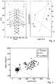
- fig. 2 shows results of different processing steps, i.e. a maximum intensity projection of the input CT scan with a ground labeling overlay (left part), a curved planar projection of bones attached to the spine, with frontal ribs cropped out (middle part) and contours extracted from the mask, wherein the longest one is referred to as the rib contour 20 (right part).
- the rib contour coordinates are smoothed by means of a Gaussian kernel. This also yields the rib contour's differentiable parameterization (x(t); y(t)), wherein x(t) and y(t) denote spatial coordinates of the contour with respect to continuous, real-valued parameter t.
- Equation (1) denotes a signed curvature of the rib contour.
- Centroids of the segments annotated with reference letter “a” are connected to the two associated rib/process tips ⁇ .
- centroids ⁇ of ten centroids in total see symbols annotated with "L”, "X” and “T"
- six local maxima ⁇ of twenty-four maxima in total are denoted with corresponding reference letters.
- each segment is connected with the two tips ⁇ of the associated ribs/processes via a straight line ⁇ .
- Two measures which are also called “discriminative parameters" in the context of the invention, are calculated for the connecting lines ⁇ .
- the length and the angle enclosed by the left and right part the connecting lines ⁇ are calculated in order to get a measure for distinguishing between Alike (+1), dash-like (0), i.e. straight, and eventual V-like (-1) connecting lines ⁇ .
- Algorithm 2 Vertebra segments and features function GETSEGMENTS(RibContour) > returns list of polylines Smooth rib contour Compute curvatures, minima and maxima ⁇ fig.
- two linear support vector machines are trained: the first SVM separating the lumbar segments ⁇ L1 ... L5 ⁇ from the thoracic ones ⁇ T7 ... T12 ⁇ and the second SVM separating the ⁇ T7 ... T11 ⁇ from ⁇ T12 ... L5 ⁇ .
- An SVM is a so-called classifier, which corresponds to a mathematical method for separating, i.e. classifying, an amount of objects into two or more classes.
- SVMs are supervised learning models with associated learning algorithms that analyze data and recognize patterns used for classification.
- a basic SVM takes a set of input data and predicts, for each given input, which of two possible classes forms the output, making it a non-probabilistic binary linear classifier.
- Given a set of training examples, each marked as belonging to one of two categories, an SVM training algorithm builds a model that assigns new examples into one category or the other.
- An SVM model is a representation of the examples as points in space, mapped so that the examples of the separate categories are divided by a clear gap that is as wide as possible.
- New examples are then mapped into that same space and predicted to belong to a category based on which side of the gap they fall on.
- Methods of training SVMs are disclosed by V. N. Vapnik, The nature of statistical learning theory, Springer-Verlag, New York, Inc., New York, NY, USA, 1995 , which is herewith incorporated into this patent application by reference.
- Fig. 3 shows the calculated length and angle of feature polylines, i.e. connecting lines ⁇ , plotted in a two-dimensional feature space.
- the solid lines correspond to the two pre-learned support vector machines (SVM) that split the feature space into four sub-regions, i.e. lumbar region ("L"), T12 region ("X”), thoracic region ("T”) and blank.
- SVM support vector machines
- Figure 4 shows an example of a feature space in which discriminative parameters together with two linear SVMs learned from 29 CT images are depicted.
- there was no linear SVM i.e. line
- a linear SVM was found to discriminate between the lumbar vertebrae segments ⁇ L1...L5 ⁇ from thoracic ones ⁇ T7...T12 ⁇ .
Landscapes
- Health & Medical Sciences (AREA)
- Engineering & Computer Science (AREA)
- Life Sciences & Earth Sciences (AREA)
- Medical Informatics (AREA)
- General Health & Medical Sciences (AREA)
- Physics & Mathematics (AREA)
- Public Health (AREA)
- Biomedical Technology (AREA)
- Pathology (AREA)
- Surgery (AREA)
- Molecular Biology (AREA)
- Heart & Thoracic Surgery (AREA)
- Animal Behavior & Ethology (AREA)
- Biophysics (AREA)
- Veterinary Medicine (AREA)
- Radiology & Medical Imaging (AREA)
- Nuclear Medicine, Radiotherapy & Molecular Imaging (AREA)
- High Energy & Nuclear Physics (AREA)
- Optics & Photonics (AREA)
- Dentistry (AREA)
- Oral & Maxillofacial Surgery (AREA)
- Theoretical Computer Science (AREA)
- Computer Vision & Pattern Recognition (AREA)
- General Physics & Mathematics (AREA)
- Pulmonology (AREA)
- Quality & Reliability (AREA)
- Orthopedic Medicine & Surgery (AREA)
- Physiology (AREA)
- Primary Health Care (AREA)
- Data Mining & Analysis (AREA)
- Databases & Information Systems (AREA)
- Epidemiology (AREA)
- Apparatus For Radiation Diagnosis (AREA)
Claims (2)
- Procédé pour identifier une partie spécifique d'une colonne vertébrale, ladite partie spécifique de la colonne vertébrale se rapportant à une vertèbre ou un disque intervertébral, dans une image obtenue par tomographie assistée par ordinateur (image CT) d'un corps humain ou animal, comprenant les étapes consistant à:a) déterminer une ou plusieurs parties de la colonne vertébrale dans l'image,b) déterminer un ou plusieurs paramètres discriminatifs pour chacune desdites parties de la colonne vertébrale dans l'image, lesdits paramètres discriminatifs se rapportant à au moins une caractéristique anatomique de chacune desdites parties de la colonne vertébrale,c) classifier les paramètres discriminatifs desdites parties de la colonne vertébrale dans l'image etd) identifier de manière automatique, au moyen d'une unité de traitement d'image (13), ladite partie spécifique de la colonne vertébrale sur la base de la classification des paramètres discriminatifs desdites parties de la colonne vertébrale dans l'image, lesdits paramètres discriminatifs se rapportant à des côtes dans lesdites parties de la colonne vertébrale dans l'image, caractérisé en ce que lesdites parties de la colonne vertébrale dans l'image sont déterminées, sur la base d'une image de contour (20, 21) dérivée de ladite image, par les étapes consistant à:- détecter le canal rachidien de la colonne vertébrale dans l'image,- éliminer une zone frontale d'une cage thoracique de l'image,- calculer une projection d'image maximale frontale (MIP) de l'image,- dériver une image binaire de la projection d'image maximale frontale (MIP) de l'image en comparant des valeurs de pixel de la projection d'image maximale frontale (MIP) à au moins une valeur seuil d'os et- dériver l'image de contour (20, 21) à partir de l'image binaire, etoù, en outre, lesdites parties de la colonne vertébrale dans l'image sont déterminées sur la base de l'image de contour (20, 21) par les étapes consistant à:- lisser l'image de contour (20, 21),- calculer des valeurs locales minimales (µ) et des valeurs locales maximales (τ) dans l'image de contour (20, 21),- déterminer des centres vertébraux (α) sur la base desdites valeurs locales minimales (µ) et/ou desdites valeurs locales maximales (τ),- établir des connexions (δ) entres lesdits centres vertébraux (α) et les valeurs locales maximales (τ) correspondantes et- déterminer lesdits paramètres discriminatifs pour chacune desdites parties de la colonne vertébrale dans l'image sur la base desdites connexions (δ) comprenant les longueurs et les angles desdites connexions (δ).
- Procédé selon la revendication 1, caractérisé en ce que la partie spécifique de la colonne vertébrale dans l'image correspond à la vertèbre T12.
Priority Applications (1)
| Application Number | Priority Date | Filing Date | Title |
|---|---|---|---|
| EP14701169.6A EP2948062B1 (fr) | 2013-01-22 | 2014-01-20 | Procédé pour identifier une partie spécifique d'une colonne vertébrale dans une image |
Applications Claiming Priority (4)
| Application Number | Priority Date | Filing Date | Title |
|---|---|---|---|
| EP13152169.2A EP2756804A1 (fr) | 2013-01-22 | 2013-01-22 | Procédé, appareil et système pour identifier une partie spécifique d'une colonne vertébrale dans une image |
| US201361755995P | 2013-01-24 | 2013-01-24 | |
| PCT/EP2014/051014 WO2014114588A1 (fr) | 2013-01-22 | 2014-01-20 | Procédé, appareil et système pour identifier une partie spécifique d'une colonne vertébrale dans une image |
| EP14701169.6A EP2948062B1 (fr) | 2013-01-22 | 2014-01-20 | Procédé pour identifier une partie spécifique d'une colonne vertébrale dans une image |
Publications (2)
| Publication Number | Publication Date |
|---|---|
| EP2948062A1 EP2948062A1 (fr) | 2015-12-02 |
| EP2948062B1 true EP2948062B1 (fr) | 2021-04-28 |
Family
ID=47632840
Family Applications (2)
| Application Number | Title | Priority Date | Filing Date |
|---|---|---|---|
| EP13152169.2A Withdrawn EP2756804A1 (fr) | 2013-01-22 | 2013-01-22 | Procédé, appareil et système pour identifier une partie spécifique d'une colonne vertébrale dans une image |
| EP14701169.6A Active EP2948062B1 (fr) | 2013-01-22 | 2014-01-20 | Procédé pour identifier une partie spécifique d'une colonne vertébrale dans une image |
Family Applications Before (1)
| Application Number | Title | Priority Date | Filing Date |
|---|---|---|---|
| EP13152169.2A Withdrawn EP2756804A1 (fr) | 2013-01-22 | 2013-01-22 | Procédé, appareil et système pour identifier une partie spécifique d'une colonne vertébrale dans une image |
Country Status (6)
| Country | Link |
|---|---|
| US (1) | US9763635B2 (fr) |
| EP (2) | EP2756804A1 (fr) |
| CN (1) | CN105338902B (fr) |
| BR (1) | BR112015017413B1 (fr) |
| ES (1) | ES2874455T3 (fr) |
| WO (1) | WO2014114588A1 (fr) |
Families Citing this family (28)
| Publication number | Priority date | Publication date | Assignee | Title |
|---|---|---|---|---|
| EP3244799B1 (fr) * | 2015-01-16 | 2018-05-23 | Koninklijke Philips N.V. | Identification d'éléments vertébraux |
| US9704066B2 (en) | 2015-03-20 | 2017-07-11 | Microsoft Technology Licensing, Llc | Multi-stage image classification |
| CN105496563B (zh) * | 2015-12-04 | 2018-05-04 | 上海联影医疗科技有限公司 | 标定脊柱医学图像定位线的方法 |
| US10460488B2 (en) * | 2016-09-06 | 2019-10-29 | International Business Machines Corporation | Spine labeling automation |
| EP3662444B1 (fr) | 2017-07-31 | 2022-06-29 | Shanghai United Imaging Healthcare Co., Ltd. | Systèmes et procédés de segmentation et d'identification automatiques de vertèbres dans des images médicales |
| CN107680134B (zh) * | 2017-09-29 | 2020-06-12 | 东软医疗系统股份有限公司 | 医学图像中脊椎标定方法、装置及设备 |
| CN108309334B (zh) * | 2017-12-08 | 2023-05-30 | 李书纲 | 一种脊柱x线影像的数据处理方法 |
| CN108670302B (zh) * | 2018-06-06 | 2020-11-06 | 西北工业大学 | 一种基于2.5维超声宽景成像的脊柱三维结构再现方法 |
| CN109697713B (zh) * | 2018-11-14 | 2022-09-02 | 吉林大学 | 基于深度学习和空间关系推理的椎间盘定位标注方法 |
| CN109360213B (zh) * | 2018-12-14 | 2020-10-30 | 浙江工业大学 | 一种基于脊柱超声冠状面图像的自动化椎体识别方法 |
| CN113138191B (zh) * | 2020-01-20 | 2022-08-05 | 上海各瑟信息科技有限公司 | 检测对象部位名称分析系统及方法 |
| US11450435B2 (en) | 2020-04-07 | 2022-09-20 | Mazor Robotics Ltd. | Spinal stenosis detection and generation of spinal decompression plan |
| US11426119B2 (en) | 2020-04-10 | 2022-08-30 | Warsaw Orthopedic, Inc. | Assessment of spinal column integrity |
| CN115485720A (zh) | 2020-05-04 | 2022-12-16 | 美敦力导航股份有限公司 | 用于检测解剖特征的系统、方法和装置 |
| US20220039770A1 (en) * | 2020-08-10 | 2022-02-10 | The Board Of Trustees Of The Leland Stanford Junior University | Systems and Methods for Identification of Pulmonary Conditions |
| US12394043B2 (en) * | 2021-04-19 | 2025-08-19 | GE Precision Healthcare LLC | Workflow management for labeling the subject anatomy |
| CN113470004A (zh) * | 2021-07-22 | 2021-10-01 | 上海嘉奥信息科技发展有限公司 | 一种基于ct的单个椎体的分割方法、系统及介质 |
| CN113689388B (zh) * | 2021-08-03 | 2024-02-06 | 慧影医疗科技(北京)股份有限公司 | 用于主动脉曲面重建的三维中心线起始点定位方法及装置 |
| CN113888548B (zh) * | 2021-09-28 | 2025-07-18 | 上海商汤善萃医疗科技有限公司 | 图像处理方法及装置、电子设备和存储介质 |
| JP7775075B2 (ja) * | 2021-12-27 | 2025-11-25 | 富士フイルム株式会社 | 画像処理装置、方法およびプログラム |
| CN114533094B (zh) * | 2022-01-07 | 2025-09-05 | 宁波大学 | 一种c型臂x光机图像的识别方法 |
| JP2023154994A (ja) * | 2022-04-08 | 2023-10-20 | キヤノン株式会社 | 画像処理装置、画像処理装置の作動方法、およびプログラム |
| CN115330753B (zh) * | 2022-10-10 | 2022-12-20 | 博志生物科技(深圳)有限公司 | 椎骨识别方法、装置、设备及存储介质 |
| US12433678B2 (en) | 2022-10-10 | 2025-10-07 | Warsaw Orthopedic, Inc. | Devices, methods, and systems for assessing suitability of spinal implants |
| CN116664078B (zh) * | 2023-07-24 | 2023-10-10 | 杭州所思互连科技有限公司 | 基于语意特征向量的rpa对象识别方法 |
| CN119887634A (zh) * | 2024-12-13 | 2025-04-25 | 脊影慧(苏州)智能科技有限公司 | 椎体位置识别模型的训练方法及椎体位置识别方法 |
| CN119810120B (zh) * | 2024-12-19 | 2026-01-13 | 山东师范大学 | 一种基于ct图像的多脊柱结构分割方法及系统 |
| CN120859534B (zh) * | 2025-07-24 | 2026-01-30 | 中世康恺科技有限公司 | 一种x射线成像系统的几何参数标定方法 |
Citations (1)
| Publication number | Priority date | Publication date | Assignee | Title |
|---|---|---|---|---|
| US20090245608A1 (en) * | 2008-03-31 | 2009-10-01 | Wan Caihua | Bone number determination apparatus and recording medium having stored therein program |
Family Cites Families (6)
| Publication number | Priority date | Publication date | Assignee | Title |
|---|---|---|---|---|
| US5570404A (en) * | 1994-09-30 | 1996-10-29 | Siemens Corporate Research | Method and apparatus for editing abdominal CT angiographic images for blood vessel visualization |
| WO2008030247A2 (fr) * | 2006-03-24 | 2008-03-13 | Kunio Doi | Procédé de détection de fractures vertébrales sur des radiographies latérales du thorax |
| WO2011021181A1 (fr) * | 2009-08-16 | 2011-02-24 | Ori Hay | Evaluation de l'anatomie vertébrale |
| JP4940340B2 (ja) * | 2009-11-27 | 2012-05-30 | 富士フイルム株式会社 | 椎骨セグメンテーション装置、方法及びプログラム |
| US8571285B2 (en) * | 2010-10-29 | 2013-10-29 | Siemens Aktiengesellschaft | Automated rib ordering and pairing |
| US8913817B2 (en) * | 2011-10-28 | 2014-12-16 | Carestream Health, Inc. | Rib suppression in radiographic images |
-
2013
- 2013-01-22 EP EP13152169.2A patent/EP2756804A1/fr not_active Withdrawn
-
2014
- 2014-01-20 WO PCT/EP2014/051014 patent/WO2014114588A1/fr not_active Ceased
- 2014-01-20 US US14/761,317 patent/US9763635B2/en active Active
- 2014-01-20 CN CN201480005669.8A patent/CN105338902B/zh not_active Expired - Fee Related
- 2014-01-20 BR BR112015017413-2A patent/BR112015017413B1/pt active IP Right Grant
- 2014-01-20 EP EP14701169.6A patent/EP2948062B1/fr active Active
- 2014-01-20 ES ES14701169T patent/ES2874455T3/es active Active
Patent Citations (1)
| Publication number | Priority date | Publication date | Assignee | Title |
|---|---|---|---|---|
| US20090245608A1 (en) * | 2008-03-31 | 2009-10-01 | Wan Caihua | Bone number determination apparatus and recording medium having stored therein program |
Also Published As
| Publication number | Publication date |
|---|---|
| ES2874455T3 (es) | 2021-11-05 |
| CN105338902B (zh) | 2019-02-15 |
| WO2014114588A1 (fr) | 2014-07-31 |
| BR112015017413B1 (pt) | 2022-03-08 |
| EP2756804A1 (fr) | 2014-07-23 |
| EP2948062A1 (fr) | 2015-12-02 |
| CN105338902A (zh) | 2016-02-17 |
| US9763635B2 (en) | 2017-09-19 |
| US20150356729A1 (en) | 2015-12-10 |
| BR112015017413A2 (pt) | 2017-07-11 |
Similar Documents
| Publication | Publication Date | Title |
|---|---|---|
| US9763635B2 (en) | Method, apparatus and system for identifying a specific part of a spine in an image | |
| EP2690596B1 (fr) | Procédé, appareil et système de marquage dorsal automatisé | |
| US8170306B2 (en) | Automatic partitioning and recognition of human body regions from an arbitrary scan coverage image | |
| Oktay et al. | Simultaneous localization of lumbar vertebrae and intervertebral discs with SVM-based MRF | |
| US20220414869A1 (en) | Detecting and segmenting regions of interest in biomedical images using neural networks | |
| CN111210897A (zh) | 处理医学图像 | |
| EP2639763B1 (fr) | Procédé, appareil et système de localisation d'une colonne vertébrale | |
| Oktay et al. | Localization of the lumbar discs using machine learning and exact probabilistic inference | |
| Basha et al. | Computer aided fracture detection system | |
| Ren et al. | Detection of trabecular landmarks for osteoporosis prescreening in dental panoramic radiographs | |
| KR102132564B1 (ko) | 진단 장치 및 진단 방법 | |
| Lenga et al. | Deep learning based rib centerline extraction and labeling | |
| Saeed et al. | Multimodal lumbar spine segmentation pipeline with optimized deep learning and network pruning | |
| Arifin et al. | Detection of overlapping teeth on dental panoramic radiograph | |
| Wang et al. | Combining statistical and geometric features for colonic polyp detection in CTC based on multiple kernel learning | |
| Lu et al. | Accurate 3D bone segmentation in challenging CT images: Bottom-up parsing and contextualized optimization | |
| Apostolopoulos et al. | Computer aided classification of mammographic tissue using shapelets and support vector machines | |
| Gornale et al. | Cognitive informatics, computer modeling and cognitive science assessment of knee osteoarthritis in radiographic images: a machine learning approach | |
| Ramakrishnan et al. | Automatic three-dimensional rib centerline extraction from CT scans for enhanced visualization and anatomical context | |
| Antani et al. | Vertebra shape classification using MLP for content-based image retrieval | |
| EP3869453A1 (fr) | Traitement d'une image médicale pour fournir un indicateur visuel | |
| Sungkhun et al. | Vertebral body segmentation using aggregate superpixels | |
| Wu et al. | Automated identification of thoracolumbar vertebrae using orthogonal matching pursuit | |
| Ray et al. | Identification of abnormal brain images from CT image dataset | |
| Kundu et al. | Automated lumbar-lordosis angle computation from digital X-ray image based on unsupervised learning |
Legal Events
| Date | Code | Title | Description |
|---|---|---|---|
| PUAI | Public reference made under article 153(3) epc to a published international application that has entered the european phase |
Free format text: ORIGINAL CODE: 0009012 |
|
| 17P | Request for examination filed |
Effective date: 20150824 |
|
| AK | Designated contracting states |
Kind code of ref document: A1 Designated state(s): AL AT BE BG CH CY CZ DE DK EE ES FI FR GB GR HR HU IE IS IT LI LT LU LV MC MK MT NL NO PL PT RO RS SE SI SK SM TR |
|
| AX | Request for extension of the european patent |
Extension state: BA ME |
|
| DAX | Request for extension of the european patent (deleted) | ||
| STAA | Information on the status of an ep patent application or granted ep patent |
Free format text: STATUS: EXAMINATION IS IN PROGRESS |
|
| 17Q | First examination report despatched |
Effective date: 20200313 |
|
| GRAP | Despatch of communication of intention to grant a patent |
Free format text: ORIGINAL CODE: EPIDOSNIGR1 |
|
| STAA | Information on the status of an ep patent application or granted ep patent |
Free format text: STATUS: GRANT OF PATENT IS INTENDED |
|
| INTG | Intention to grant announced |
Effective date: 20201130 |
|
| RIN1 | Information on inventor provided before grant (corrected) |
Inventor name: BUEHLER, KATJA Inventor name: MAJOR, DAVID Inventor name: HLADUVKA, JIRI |
|
| GRAS | Grant fee paid |
Free format text: ORIGINAL CODE: EPIDOSNIGR3 |
|
| RAP1 | Party data changed (applicant data changed or rights of an application transferred) |
Owner name: VRVIS ZENTRUM FUER VIRTUAL REALITY UND VISUALISIERUNG FORSCHUNGS-GMBH Owner name: IPM FORSCHUNGINSTITUT FUER MOLEKULARE PATHOLOGIE GMBH Owner name: AGFA HEALTHCARE NV |
|
| GRAA | (expected) grant |
Free format text: ORIGINAL CODE: 0009210 |
|
| STAA | Information on the status of an ep patent application or granted ep patent |
Free format text: STATUS: THE PATENT HAS BEEN GRANTED |
|
| AK | Designated contracting states |
Kind code of ref document: B1 Designated state(s): AL AT BE BG CH CY CZ DE DK EE ES FI FR GB GR HR HU IE IS IT LI LT LU LV MC MK MT NL NO PL PT RO RS SE SI SK SM TR |
|
| REG | Reference to a national code |
Ref country code: GB Ref legal event code: FG4D |
|
| REG | Reference to a national code |
Ref country code: CH Ref legal event code: EP |
|
| REG | Reference to a national code |
Ref country code: AT Ref legal event code: REF Ref document number: 1386192 Country of ref document: AT Kind code of ref document: T Effective date: 20210515 |
|
| REG | Reference to a national code |
Ref country code: DE Ref legal event code: R096 Ref document number: 602014076957 Country of ref document: DE |
|
| REG | Reference to a national code |
Ref country code: NL Ref legal event code: FP Ref country code: IE Ref legal event code: FG4D |
|
| REG | Reference to a national code |
Ref country code: SE Ref legal event code: TRGR |
|
| REG | Reference to a national code |
Ref country code: LT Ref legal event code: MG9D |
|
| REG | Reference to a national code |
Ref country code: AT Ref legal event code: MK05 Ref document number: 1386192 Country of ref document: AT Kind code of ref document: T Effective date: 20210428 |
|
| PG25 | Lapsed in a contracting state [announced via postgrant information from national office to epo] |
Ref country code: HR Free format text: LAPSE BECAUSE OF FAILURE TO SUBMIT A TRANSLATION OF THE DESCRIPTION OR TO PAY THE FEE WITHIN THE PRESCRIBED TIME-LIMIT Effective date: 20210428 Ref country code: BG Free format text: LAPSE BECAUSE OF FAILURE TO SUBMIT A TRANSLATION OF THE DESCRIPTION OR TO PAY THE FEE WITHIN THE PRESCRIBED TIME-LIMIT Effective date: 20210728 Ref country code: AT Free format text: LAPSE BECAUSE OF FAILURE TO SUBMIT A TRANSLATION OF THE DESCRIPTION OR TO PAY THE FEE WITHIN THE PRESCRIBED TIME-LIMIT Effective date: 20210428 Ref country code: LT Free format text: LAPSE BECAUSE OF FAILURE TO SUBMIT A TRANSLATION OF THE DESCRIPTION OR TO PAY THE FEE WITHIN THE PRESCRIBED TIME-LIMIT Effective date: 20210428 Ref country code: FI Free format text: LAPSE BECAUSE OF FAILURE TO SUBMIT A TRANSLATION OF THE DESCRIPTION OR TO PAY THE FEE WITHIN THE PRESCRIBED TIME-LIMIT Effective date: 20210428 |
|
| REG | Reference to a national code |
Ref country code: ES Ref legal event code: FG2A Ref document number: 2874455 Country of ref document: ES Kind code of ref document: T3 Effective date: 20211105 |
|
| PG25 | Lapsed in a contracting state [announced via postgrant information from national office to epo] |
Ref country code: LV Free format text: LAPSE BECAUSE OF FAILURE TO SUBMIT A TRANSLATION OF THE DESCRIPTION OR TO PAY THE FEE WITHIN THE PRESCRIBED TIME-LIMIT Effective date: 20210428 Ref country code: GR Free format text: LAPSE BECAUSE OF FAILURE TO SUBMIT A TRANSLATION OF THE DESCRIPTION OR TO PAY THE FEE WITHIN THE PRESCRIBED TIME-LIMIT Effective date: 20210729 Ref country code: IS Free format text: LAPSE BECAUSE OF FAILURE TO SUBMIT A TRANSLATION OF THE DESCRIPTION OR TO PAY THE FEE WITHIN THE PRESCRIBED TIME-LIMIT Effective date: 20210828 Ref country code: RS Free format text: LAPSE BECAUSE OF FAILURE TO SUBMIT A TRANSLATION OF THE DESCRIPTION OR TO PAY THE FEE WITHIN THE PRESCRIBED TIME-LIMIT Effective date: 20210428 Ref country code: PL Free format text: LAPSE BECAUSE OF FAILURE TO SUBMIT A TRANSLATION OF THE DESCRIPTION OR TO PAY THE FEE WITHIN THE PRESCRIBED TIME-LIMIT Effective date: 20210428 Ref country code: NO Free format text: LAPSE BECAUSE OF FAILURE TO SUBMIT A TRANSLATION OF THE DESCRIPTION OR TO PAY THE FEE WITHIN THE PRESCRIBED TIME-LIMIT Effective date: 20210728 Ref country code: PT Free format text: LAPSE BECAUSE OF FAILURE TO SUBMIT A TRANSLATION OF THE DESCRIPTION OR TO PAY THE FEE WITHIN THE PRESCRIBED TIME-LIMIT Effective date: 20210830 |
|
| PG25 | Lapsed in a contracting state [announced via postgrant information from national office to epo] |
Ref country code: SK Free format text: LAPSE BECAUSE OF FAILURE TO SUBMIT A TRANSLATION OF THE DESCRIPTION OR TO PAY THE FEE WITHIN THE PRESCRIBED TIME-LIMIT Effective date: 20210428 Ref country code: SM Free format text: LAPSE BECAUSE OF FAILURE TO SUBMIT A TRANSLATION OF THE DESCRIPTION OR TO PAY THE FEE WITHIN THE PRESCRIBED TIME-LIMIT Effective date: 20210428 Ref country code: EE Free format text: LAPSE BECAUSE OF FAILURE TO SUBMIT A TRANSLATION OF THE DESCRIPTION OR TO PAY THE FEE WITHIN THE PRESCRIBED TIME-LIMIT Effective date: 20210428 Ref country code: CZ Free format text: LAPSE BECAUSE OF FAILURE TO SUBMIT A TRANSLATION OF THE DESCRIPTION OR TO PAY THE FEE WITHIN THE PRESCRIBED TIME-LIMIT Effective date: 20210428 Ref country code: DK Free format text: LAPSE BECAUSE OF FAILURE TO SUBMIT A TRANSLATION OF THE DESCRIPTION OR TO PAY THE FEE WITHIN THE PRESCRIBED TIME-LIMIT Effective date: 20210428 Ref country code: RO Free format text: LAPSE BECAUSE OF FAILURE TO SUBMIT A TRANSLATION OF THE DESCRIPTION OR TO PAY THE FEE WITHIN THE PRESCRIBED TIME-LIMIT Effective date: 20210428 |
|
| REG | Reference to a national code |
Ref country code: DE Ref legal event code: R097 Ref document number: 602014076957 Country of ref document: DE |
|
| PLBE | No opposition filed within time limit |
Free format text: ORIGINAL CODE: 0009261 |
|
| STAA | Information on the status of an ep patent application or granted ep patent |
Free format text: STATUS: NO OPPOSITION FILED WITHIN TIME LIMIT |
|
| 26N | No opposition filed |
Effective date: 20220131 |
|
| PG25 | Lapsed in a contracting state [announced via postgrant information from national office to epo] |
Ref country code: IS Free format text: LAPSE BECAUSE OF FAILURE TO SUBMIT A TRANSLATION OF THE DESCRIPTION OR TO PAY THE FEE WITHIN THE PRESCRIBED TIME-LIMIT Effective date: 20210828 Ref country code: AL Free format text: LAPSE BECAUSE OF FAILURE TO SUBMIT A TRANSLATION OF THE DESCRIPTION OR TO PAY THE FEE WITHIN THE PRESCRIBED TIME-LIMIT Effective date: 20210428 |
|
| PG25 | Lapsed in a contracting state [announced via postgrant information from national office to epo] |
Ref country code: MC Free format text: LAPSE BECAUSE OF FAILURE TO SUBMIT A TRANSLATION OF THE DESCRIPTION OR TO PAY THE FEE WITHIN THE PRESCRIBED TIME-LIMIT Effective date: 20210428 |
|
| REG | Reference to a national code |
Ref country code: CH Ref legal event code: PL |
|
| PG25 | Lapsed in a contracting state [announced via postgrant information from national office to epo] |
Ref country code: LU Free format text: LAPSE BECAUSE OF NON-PAYMENT OF DUE FEES Effective date: 20220120 |
|
| PG25 | Lapsed in a contracting state [announced via postgrant information from national office to epo] |
Ref country code: LI Free format text: LAPSE BECAUSE OF NON-PAYMENT OF DUE FEES Effective date: 20220131 Ref country code: CH Free format text: LAPSE BECAUSE OF NON-PAYMENT OF DUE FEES Effective date: 20220131 |
|
| PG25 | Lapsed in a contracting state [announced via postgrant information from national office to epo] |
Ref country code: IE Free format text: LAPSE BECAUSE OF NON-PAYMENT OF DUE FEES Effective date: 20220120 |
|
| PG25 | Lapsed in a contracting state [announced via postgrant information from national office to epo] |
Ref country code: HU Free format text: LAPSE BECAUSE OF FAILURE TO SUBMIT A TRANSLATION OF THE DESCRIPTION OR TO PAY THE FEE WITHIN THE PRESCRIBED TIME-LIMIT; INVALID AB INITIO Effective date: 20140120 |
|
| PG25 | Lapsed in a contracting state [announced via postgrant information from national office to epo] |
Ref country code: MK Free format text: LAPSE BECAUSE OF FAILURE TO SUBMIT A TRANSLATION OF THE DESCRIPTION OR TO PAY THE FEE WITHIN THE PRESCRIBED TIME-LIMIT Effective date: 20210428 Ref country code: CY Free format text: LAPSE BECAUSE OF FAILURE TO SUBMIT A TRANSLATION OF THE DESCRIPTION OR TO PAY THE FEE WITHIN THE PRESCRIBED TIME-LIMIT Effective date: 20210428 |
|
| PG25 | Lapsed in a contracting state [announced via postgrant information from national office to epo] |
Ref country code: MT Free format text: LAPSE BECAUSE OF FAILURE TO SUBMIT A TRANSLATION OF THE DESCRIPTION OR TO PAY THE FEE WITHIN THE PRESCRIBED TIME-LIMIT Effective date: 20210428 |
|
| PGFP | Annual fee paid to national office [announced via postgrant information from national office to epo] |
Ref country code: NL Payment date: 20250113 Year of fee payment: 12 |
|
| PGFP | Annual fee paid to national office [announced via postgrant information from national office to epo] |
Ref country code: DE Payment date: 20250103 Year of fee payment: 12 |
|
| PGFP | Annual fee paid to national office [announced via postgrant information from national office to epo] |
Ref country code: ES Payment date: 20250207 Year of fee payment: 12 |
|
| PGFP | Annual fee paid to national office [announced via postgrant information from national office to epo] |
Ref country code: SE Payment date: 20250109 Year of fee payment: 12 |
|
| PGFP | Annual fee paid to national office [announced via postgrant information from national office to epo] |
Ref country code: BE Payment date: 20250103 Year of fee payment: 12 |
|
| PGFP | Annual fee paid to national office [announced via postgrant information from national office to epo] |
Ref country code: FR Payment date: 20250106 Year of fee payment: 12 |
|
| PGFP | Annual fee paid to national office [announced via postgrant information from national office to epo] |
Ref country code: IT Payment date: 20250131 Year of fee payment: 12 Ref country code: GB Payment date: 20250106 Year of fee payment: 12 |
|
| PG25 | Lapsed in a contracting state [announced via postgrant information from national office to epo] |
Ref country code: TR Free format text: LAPSE BECAUSE OF FAILURE TO SUBMIT A TRANSLATION OF THE DESCRIPTION OR TO PAY THE FEE WITHIN THE PRESCRIBED TIME-LIMIT Effective date: 20210428 |