EP2948775B1 - Compositions et procédés de détection de néoplasie - Google Patents
Compositions et procédés de détection de néoplasie Download PDFInfo
- Publication number
- EP2948775B1 EP2948775B1 EP14704237.8A EP14704237A EP2948775B1 EP 2948775 B1 EP2948775 B1 EP 2948775B1 EP 14704237 A EP14704237 A EP 14704237A EP 2948775 B1 EP2948775 B1 EP 2948775B1
- Authority
- EP
- European Patent Office
- Prior art keywords
- biomarkers
- dimethylarginine
- subject
- cancer
- biomarkers related
- Prior art date
- Legal status (The legal status is an assumption and is not a legal conclusion. Google has not performed a legal analysis and makes no representation as to the accuracy of the status listed.)
- Not-in-force
Links
Images











Classifications
-
- C—CHEMISTRY; METALLURGY
- C12—BIOCHEMISTRY; BEER; SPIRITS; WINE; VINEGAR; MICROBIOLOGY; ENZYMOLOGY; MUTATION OR GENETIC ENGINEERING
- C12Q—MEASURING OR TESTING PROCESSES INVOLVING ENZYMES, NUCLEIC ACIDS OR MICROORGANISMS; COMPOSITIONS OR TEST PAPERS THEREFOR; PROCESSES OF PREPARING SUCH COMPOSITIONS; CONDITION-RESPONSIVE CONTROL IN MICROBIOLOGICAL OR ENZYMOLOGICAL PROCESSES
- C12Q1/00—Measuring or testing processes involving enzymes, nucleic acids or microorganisms; Compositions therefor; Processes of preparing such compositions
- C12Q1/68—Measuring or testing processes involving enzymes, nucleic acids or microorganisms; Compositions therefor; Processes of preparing such compositions involving nucleic acids
- C12Q1/6876—Nucleic acid products used in the analysis of nucleic acids, e.g. primers or probes
- C12Q1/6883—Nucleic acid products used in the analysis of nucleic acids, e.g. primers or probes for diseases caused by alterations of genetic material
- C12Q1/6886—Nucleic acid products used in the analysis of nucleic acids, e.g. primers or probes for diseases caused by alterations of genetic material for cancer
-
- G—PHYSICS
- G01—MEASURING; TESTING
- G01N—INVESTIGATING OR ANALYSING MATERIALS BY DETERMINING THEIR CHEMICAL OR PHYSICAL PROPERTIES
- G01N33/00—Investigating or analysing materials by specific methods not covered by groups G01N1/00 - G01N31/00
- G01N33/48—Biological material, e.g. blood, urine; Haemocytometers
- G01N33/50—Chemical analysis of biological material, e.g. blood, urine; Testing involving biospecific ligand binding methods; Immunological testing
- G01N33/53—Immunoassay; Biospecific binding assay; Materials therefor
- G01N33/575—Immunoassay; Biospecific binding assay; Materials therefor for cancer
- G01N33/57535—Immunoassay; Biospecific binding assay; Materials therefor for cancer of the large intestine, e.g. colon, rectum or anus
-
- G—PHYSICS
- G01—MEASURING; TESTING
- G01N—INVESTIGATING OR ANALYSING MATERIALS BY DETERMINING THEIR CHEMICAL OR PHYSICAL PROPERTIES
- G01N33/00—Investigating or analysing materials by specific methods not covered by groups G01N1/00 - G01N31/00
- G01N33/48—Biological material, e.g. blood, urine; Haemocytometers
- G01N33/50—Chemical analysis of biological material, e.g. blood, urine; Testing involving biospecific ligand binding methods; Immunological testing
- G01N33/68—Chemical analysis of biological material, e.g. blood, urine; Testing involving biospecific ligand binding methods; Immunological testing involving proteins, peptides or amino acids
- G01N33/6803—General methods of protein analysis not limited to specific proteins or families of proteins
- G01N33/6806—Determination of free amino acids
-
- C—CHEMISTRY; METALLURGY
- C12—BIOCHEMISTRY; BEER; SPIRITS; WINE; VINEGAR; MICROBIOLOGY; ENZYMOLOGY; MUTATION OR GENETIC ENGINEERING
- C12Q—MEASURING OR TESTING PROCESSES INVOLVING ENZYMES, NUCLEIC ACIDS OR MICROORGANISMS; COMPOSITIONS OR TEST PAPERS THEREFOR; PROCESSES OF PREPARING SUCH COMPOSITIONS; CONDITION-RESPONSIVE CONTROL IN MICROBIOLOGICAL OR ENZYMOLOGICAL PROCESSES
- C12Q2600/00—Oligonucleotides characterized by their use
- C12Q2600/154—Methylation markers
-
- C—CHEMISTRY; METALLURGY
- C12—BIOCHEMISTRY; BEER; SPIRITS; WINE; VINEGAR; MICROBIOLOGY; ENZYMOLOGY; MUTATION OR GENETIC ENGINEERING
- C12Q—MEASURING OR TESTING PROCESSES INVOLVING ENZYMES, NUCLEIC ACIDS OR MICROORGANISMS; COMPOSITIONS OR TEST PAPERS THEREFOR; PROCESSES OF PREPARING SUCH COMPOSITIONS; CONDITION-RESPONSIVE CONTROL IN MICROBIOLOGICAL OR ENZYMOLOGICAL PROCESSES
- C12Q2600/00—Oligonucleotides characterized by their use
- C12Q2600/158—Expression markers
-
- G—PHYSICS
- G01—MEASURING; TESTING
- G01N—INVESTIGATING OR ANALYSING MATERIALS BY DETERMINING THEIR CHEMICAL OR PHYSICAL PROPERTIES
- G01N2800/00—Detection or diagnosis of diseases
- G01N2800/50—Determining the risk of developing a disease
-
- G—PHYSICS
- G01—MEASURING; TESTING
- G01N—INVESTIGATING OR ANALYSING MATERIALS BY DETERMINING THEIR CHEMICAL OR PHYSICAL PROPERTIES
- G01N2800/00—Detection or diagnosis of diseases
- G01N2800/52—Predicting or monitoring the response to treatment, e.g. for selection of therapy based on assay results in personalised medicine; Prognosis
-
- G—PHYSICS
- G01—MEASURING; TESTING
- G01N—INVESTIGATING OR ANALYSING MATERIALS BY DETERMINING THEIR CHEMICAL OR PHYSICAL PROPERTIES
- G01N2800/00—Detection or diagnosis of diseases
- G01N2800/56—Staging of a disease; Further complications associated with the disease
Definitions
- the disclosure features a kit for aiding the diagnosis of neoplasia or colorectal cancer, the kit including at least one reagent capable of detecting, binding, or capturing at least one biomarker selected from the group of biomarkers related to methylation including asymmetric-dimethylarginine, symmetric-dimethylarginine, carnitine, methionine, dimethylglycine, and betaine and at least one reagent capable of detecting, binding, or capturing at least one additional marker selected from a group from among the following:
- the disclosure features a kit for aiding the diagnosis of neoplasia or colorectal cancer, the kit containing at least one reagent capable of detecting, binding, or capturing asymmetric-dimethylarginine and at least one reagent capable of detecting, binding, or capturing symmetric-dimethylarginine.
- the kit further contains at least one reagent capable of detecting the M264 biomarker.
- the kit further contains directions for using the reagent to analyze the level of asymmetric-dimethylarginine, symmetric-dimethylarginine, carnitine, methionine, dimethylglycine, betaine, xanthosine, inosine, deoxyuridine, thymidine, deoxycytidine, cytosine, hypoxanthine, xanthine, uracil, guanosine, adenosine, N1-acetylspermidine, N8-acetylspermidine, spermine, arginine ornithine, citrulline, proline, glutamine, glutamic acid, threonine, and N ⁇ -acetyllysine, M264 ( ⁇ -D-Ribofuranosylcreatine), or a combination thereof.
- M264 ⁇ -D-Ribofuranosylcreatine
- the kit further contains directions for contacting a test sample with the reagent and detecting asymmetric-dimethylarginine, symmetric-dimethylarginine, carnitine, methionine, dimethylglycine, betaine, xanthosine, inosine, deoxyuridine, thymidine, deoxycytidine, cytosine, hypoxanthine, xanthine, uracil, guanosine, adenosine, N1-acetylspermidine, N8-acetylspermidine, spermine, arginine ornithine, citrulline, proline, glutamine, glutamic acid, threonine, and N ⁇ -acetyllysine, M264 ( ⁇ -D-Ribofuranosylcreatine), or a combination thereof retained by the reagent.
- asymmetric-dimethylarginine symmetric-dimethylarginine
- carnitine methionine
- the disclosure features a panel for aiding the diagnosis of neoplasia or colorectal cancer, the panel including at least one reagent capable of detecting, binding, or capturing at least one biomarker selected from the group of biomarkers related to methylation: biomarkers related to nucleic acid metabolism; biomarkers related to urea cycle and polyamine metabolism; and biomarkers related to amino acid metabolism.
- the disclosure provides a panel for aiding the diagnosis of neoplasia or colorectal cancer, the panel containing at least one reagent capable of detecting, binding, or capturing a first biomarker selected from a first group and at least one reagent capable of detecting, binding, or capturing at least one additional marker selected from a different group, where the groups are selected from among the following:
- the disclosure features a panel for aiding the diagnosis of neoplasia or colorectal cancer, the panel including at least one reagent capable of detecting, binding, or capturing at least one biomarker selected from the group of biomarkers related to methylation including asymmetric-dimethylarginine, symmetric-dimethylarginine, carnitine, methionine, dimethylglycine, and betaine and at least one reagent capable of detecting, binding, or capturing at least one additional marker selected from a group from among the following:
- the groups are selected from the following:
- a decrease in the level of biomarkers in one or more of the groups identifies a subject as having an increased chance of survival; identifies a therapy as effective; or identifies a neoplasia as less aggressive.
- the biological sample is a biological fluid (e.g., urine, blood, blood serum, plasma, bile, fecal aspirate, intestinal aspirate, cerebrospinal fluid and saliva) or tissue sample.
- the method further involves measuring the level of carnitine in the biological sample.
- a decrease in the level of carnitine in a biological sample that is a biological fluid identifies a subject as having or having a propensity to develop neoplasia; identifies a subject as having a reduced chance of survival; or identifies a neoplasia as aggressive.
- an increase in the level of carnitine in a biological sample that is a biological fluid identifies a subject as having an increased chance of survival; identifies a therapy as effective; or identifies a neoplasia as less aggressive.
- an increase in the level of carnitine in a biological sample that is a tissue sample identifies a subject as having or having a propensity to develop neoplasia; identifies a subject as having a reduced chance of survival; or identifies a neoplasia as aggressive.
- a decrease in the level of carnitine in a biological sample that is a tissue sample identifies a subject as having an increased chance of survival; identifies a therapy as effective; or identifies a neoplasia as less aggressive.
- the reference is a control or a corresponding biological sample derived from a healthy subject.
- the level of the biomarker is normalized to the level of creatinine in the sample, absolute concentration, 24-hr excretion, or with respect to specific gravity.
- the subject is human.
- the level is detected by chromatography, mass spectrometry, spectroscopy, immunoassay, ultra performance liquid chromatography (UPLC).
- the mass spectrometry is electrospray ionization quadruple time-of-flight mass spectrometry (ESI-QTF-MS).
- spectroscopy is NMR spectroscopy.
- the immunoassay is ELISA.
- the method further involves measuring the level of biomarkers in one or more groups from among biomarkers related to methylation; biomarkers related to polyamine metabolism; and biomarkers related to amino acid metabolism.
- an increase in the level of biomarkers in one or more of the groups identifies a subject as having or having a propensity to develop neoplasia; identifies a subject as having a reduced chance of survival; or identifies a neoplasia as aggressive.
- an increase in the level of biomarkers in one or more of the groups identifies a subject as having an increased chance of survival; identifies a therapy as effective; or identifies a neoplasia as less aggressive.
- biomarker generally refers to a molecule that is differentially present in a sample (e.g.. biopsy, biological or physiological fluid) taken from a subject of one phenotypic status (e.g., having a disease) as compared with another phenotypic status (e.g., not having the disease).
- a biomarker is differentially present between different phenotypic statuses if the mean or median level of the biomarker in a first phenotypic status relative to a second phenotypic status is calculated to represent statistically significant differences. Common tests for statistical significance include, among others, t-test, ANOVA, Kruskal-Wallis, Wilcoxon, Mann-Whitney and odds ratio.
- biomarker related to methylation refers to metabolic intermediates and products related to increased methylation (e.g., protein methylation, DNA methylation) in a subject or a tissue or a cell of the subject (e.g., a neoplastic tissue or cell).
- the biomarker related to methylation is a small molecule compound.
- the biomarker related to methylation is present in a sample derived from the subject (e.g., a biological or physiological fluid).
- an alteration in the levels of one or more biomarkers related to methylation, relative to a reference indicates a tissue or cell is neoplastic.
- biomarker related to nucleic acid metabolism refers to metabolic intermediates and products related to increased nucleic acid metabolism in a subject or a tissue or a cell of the subject (e.g., a neoplastic tissue or cell).
- the biomarker related to nucleic acid metabolism is a small molecule compound.
- the biomarker related to nucleic acid metabolism is present in a sample derived from the subject (e.g., a biological or physiological fluid).
- an alteration in the levels of one or more biomarkers related to nucleic acid metabolism, relative to a reference indicates a tissue or cell is neoplastic.
- biomarkers related to nucleic acid metabolism include without limitation: xanthosine ( CAS: 146-80-5 ); inosine ( CAS: 58-63-9 ); deoxyuridine ( CAS: 951-78-0 ); thymidine ( CAS: 50-89-5 ); Uric acid ( CAS: 69-93-2 ); S-allantoin ( CAS: 97-59-6 ); Xanthine ( CAS: 69-89-6 ); Hypoxanthine ( CAS: 68-94-0 ); Guanine ( CAS: 73-40-5 ); Guanosine ( CAS: 118-00-3 ); Deoxyguanosine ( CAS: 961-07-9 ); Adenine ( CAS: 73-24-5 ); Adenosine ( CAS: CAS: 146
- an alteration in the levels of one or more biomarkers related to polyamine metabolism, relative to a reference, indicates a subject has or is at risk of having neoplasia.
- biomarkers related to urea cycle and polyamine metabolism include without limitation: N1-acetylspermidine ( CAS: 14278-49-0 ); N8-acetylspermidine ( CAS: 34450-15-2 ); spermine ( CAS: 71-44-3 ); Putrescine ( CAS: 110-60-1 ); spermidine ( CAS: 124-20-9 ); ornithine ( CAS: 70-26-8 ); Citrulline ( CAS: 372-75-8 ); L-arginine ( CAS: 74-79-3 ); N-acetylputrelcine ( CAS: 124-20-9 ); N1-acetylspermine ( CAS: 25593-72-0 ); and L-argininosuccinic acid ( CAS: 2387-71
- asymmetric-dimethylarginine refers to a compound having the CAS number 102783-24-4 , including a pharmaceutically acceptable salt, solvate, hydrate, geometrical isomer, tautomer, optical isomer, isotopic derivative, polymorph, prodrug, or N -oxide thereof.
- An increase in asymmetric-dimethylarginine in a biological sample is related to an increase in methylation (e.g. protein methylation, DNA methylation) in a subject or a tissue or a cell of the subject (e.g., a neoplastic tissue or cell).
- symmetric-dimethylarginine refers to a compound having the CAS number 30344-00-4 , including a pharmaceutically acceptable salt, solvate, hydrate, geometrical isomer, tautomer, optical isomer, isotopic derivative, polymorph, prodrug, or N -oxide thereof.
- An increase in symmetric-dimethylarginine in a biological sample is related to an increase in methylation (e.g. protein methylation, DNA methylation) in a subject or a tissue or a cell of the subject (e.g., a neoplastic tissue or cell).
- xanthosine refers to a compound having the CAS number 146-80-5 , including a pharmaceutically acceptable salt, solvate, hydrate, geometrical isomer, tautomer, optical isomer, isotopic derivative, polymorph, prodrug, or N -oxide thereof.
- An increase in xanthosine in a biological sample is related to an increase in nucleic acid metabolism in a subject or a tissue or a cell of the subject (e.g.. a neoplastic tissue or cell).
- deoxycytidine refers to a compound having the CAS number 951-77-9 , including a pharmaceutically acceptable salt, solvate, hydrate, geometrical isomer, tautomer, optical isomer, isotopic derivative, polymorph, prodrug, or N-oxide thereof.
- An increase in deoxycytidine in a biological sample is related to an increase in nucleic acid metabolism in a subject or a tissue or a cell of the subject (e.g., a neoplastic tissue or cell).
- cytosine refers to a compound having the CAS number 71-30-7 , including a pharmaceutically acceptable salt, solvate, hydrate, geometrical isomer, tautomer, optical isomer, isotopic derivative, polymorph, prodrug, or N-oxide thereof.
- An increase in cytosine in a biological sample is related to an increase in nucleic acid metabolism in a subject or a tissue or a cell of the subject (e.g., a neoplastic tissue or cell).
- xanthine refers to a compound having the CAS number CAS: 69-89-6 , including a pharmaceutically acceptable salt, solvate, hydrate, geometrical isomer, tautomer, optical isomer, isotopic derivative, polymorph, prodrug, or N-oxide thereof.
- An increase in dxanthine in a biological sample is related to an increase in nucleic acid metabolism in a subject or a tissue or a cell of the subject (e.g.. a neoplastic tissue or cell).
- arginine refers to a compound having the CAS number CAS: 74-79-3 . including a pharmaceutically acceptable salt, solvate, hydrate, geometrical isomer, tautomer, optical isomer, isotopic derivative, polymorph, prodrug, or N-oxide thereof.
- An increase in arginine in a biological sample is related to an increase in polyamine metabolism in a subject or a tissue or a cell of the subject (e.g.. a neoplastic tissue or cell).
- N ⁇ -acetyllysine refers to a compound having the CAS number 1946-82-3 , including pharmaceutically acceptable salts, solvates, hydrates, geometrical isomers, tautomers, optical isomers, isotopic derivatives, polymorphs, prodrugs, or N -oxides thereof.
- An increase in N ⁇ -acetyllysine in a biological sample is related to an increase in amino acid metabolism in a subject or a tissue or a cell of the subject (e.g.. a neoplastic tissue or cell).
- the IUPAC name of the compound is 2- ⁇ 2-[(2R,3R,4S,5R)-3,4-dihydroxy-5-(hydroxymethyl)-oxolan-2-yl]-1-methylcarbamimidamido ⁇ acetic acid and the common name P-D-Ribofuranosylcreatine.
- the biosynthesis of the compound is expected to involve arginine metabolism, methylation and pentose phosphate pathway, which is also involved in nucleic acid biosynthesis.
- the term “antibody” means not only intact antibody molecules, but also fragments of antibody molecules that retain immunogen-binding ability. Such fragments are also well known in the art and are regularly employed both in vitro and in vivo. Accordingly, as used herein, the term “antibody” means not only intact immunoglobulin molecules but also the well-known active fragments F(ab')2, and Fab. F(ab')2, and Fab fragments that lack the Fc fragment of intact antibody, clear more rapidly from the circulation, and may have less non-specific tissue binding of an intact antibody ( Wahl et al., J. Nucl. Med. 24:316-325 (1983 ).
- the antibodies of the invention comprise whole native antibodies, bispecific antibodies; chimeric antibodies; Fab, Fab', single chain V region fragments (scFv), fusion polypeptides, and unconventional antibodies.
- biological sample any tissue, cell, fluid, or other material derived from an organism.
- Clinical aggressiveness is meant the severity of the neoplasia. Aggressive neoplasias are more likely to metastasize than less aggressive neoplasias. While conservative methods of treatment are appropriate for less aggressive neoplasias, more aggressive neoplasias require more aggressive therapeutic regimens.
- the terms “determining”. “assessing”, “assaying”, “measuring” and “detecting” refer to both quantitative and qualitative determinations, and as such, the term “determining” is used interchangeably herein with “assaying,” “measuring,” and the like. Where a quantitative determination is intended, the phrase “determining an amount” of an analyte and the like is used. Where a qualitative and/or quantitative determination is intended, the phrase “determining a level" of an analyte or “detecting” an analyte is used.
- Purity and homogeneity are typically determined using analytical chemistry techniques, for example, polyacrylamide gel electrophoresis or high performance liquid chromatography.
- the term "purified" can denote that a nucleic acid or protein gives rise to essentially one band in an electrophoretic gel.
- modifications for example, phosphorylation or glycosylation, different modifications may give rise to different isolated proteins, which can be separately purified.
- Waldenstrom's macroglobulinemia, heavy chain disease, and solid tumors such as sarcomas and carcinomas (e.g., fibrosarcoma, myxosarcoma, liposarcoma, chondrosarcoma, osteogenic sarcoma, chordoma.
- sarcomas and carcinomas e.g., fibrosarcoma, myxosarcoma, liposarcoma, chondrosarcoma, osteogenic sarcoma, chordoma.
- angiosarcoma e.g., endotheliosarcoma, lymphangiosarcoma, lymphangioendotheliosarcoma, synovioma.
- Lymphoproliferative disorders are also considered to be proliferative diseases.
- Periodic patient monitoring includes, for example, a schedule of tests that are administered daily, bi-weekly, bi-monthly, monthly, biannually, or annually.
- Marker profile is meant a characterization of the level of two or more compounds, analytes, or metabolites.
- Marker profiles of the following groups of biomarkers are useful in characterizing neoplasia in the methods of the invention: asymmetric-dimethylarginine and symmetric-dimethylarginine, and optionally carnitine (methylation); proline, glutamine, Na-acetyllysine (amino acid metabolism); xanthosine, inosine, deoxyuridine, thymidine (nucleic acid metabolism); and (polyamine metabolism); or a combination thereof
- subject refers to an animal which is the object of treatment, observation, or experiment.
- a subject includes, but is not limited to, a mammal, including, but not limited to, a human or a non-human mammal, such as a non-human primate, murine, bovine, equine, canine, ovine, or feline.
- an affinity agent e.g., an antibody
- a compound or agent of interest e.g., a biomarker
- treat refers to reducing or ameliorating a disease or condition, e.g., neoplasia, and/or symptoms associated therewith. It will be appreciated that, although not precluded, treating a disease or condition does not require that the disease, condition, or symptoms associated therewith be completely eliminated.
- the term "about” is understood as within a range of normal tolerance in the art, for example within 2 standard deviations of the mean. About can be understood as within 10%, 9%, 8%, 7%, 6%, 5%, 4%, 3%. 2%. 1%, 0.5%. 0.1%, 0.05%, or 0.01% of the stated value. Unless otherwise clear from context, all numerical values provided herein are modified by the term about.
- metabolomic and transcriptomic analysis revealed changes in amino acid, nucleic acid, polyamine metabolism and methylation (e.g. protein methylation, DNA methylation) in tumor tissue.
- This disclosure is based, at least in pan, on the discovery that the following groups or panels of biomarkers are useful and effective for identifying a subject as having or having a propensity to develop neoplasia (e.g., colorectal cancer, small intestine cancer, duodenal cancer, gastric cancer, pancreatic cancer, liver cancer, esophageal cancer, ovarian cancer, prostate cancer, uterine cancer, breast cancer, thyroid cancer and melanoma): biomarkers related to methylation; biomarkers related to nucleic acid metabolism: biomarkers related to urea cycle and polyamine metabolism; and biomarkers related to amino acid metabolism.
- neoplasia e.g., colorectal cancer, small intestine cancer, duodenal cancer, gastric cancer, pancreatic
- the biomarkers related to methylation include asymmetric-dimethylarginine, symmetric-dimethylarginine, carnitine, methionine, dimethylglycine, and betaine;
- the biomarkers related to nucleic acid metabolism include xanthosine, inosine, deoxyuridine, thymidine, deoxycytidine, cytosine, hypoxanthine, xanthine, uracil, guanosine, and adenosine;
- the biomarkers related to urea cycle and polyamine metabolism include N1-acetylspermidine, N8-acetylspermidine, spermine, arginine ornithine, and citrulline: and the biomarkers related to amino acid metabolism include proline, glutamine, glutamic acid, threonine, and Na-acetyllysine.
- urinary metabolites to prospectively identify animals that would eventually develop tumor, indicate a combined panel of urinary biomarkers representing synchronized deregulation of amino acid, nucleic acid, polyamine metabolism and methylation may provide a powerful high-throughput noninvasive method for screening as well as early diagnosis of colorectal cancer.
- the present invention features diagnostic assays for the detection of neoplasia, including colorectal cancer.
- the level of a biomarker(s) is measured in a subject sample and used to characterize neoplasia or the propensity to develop neoplasia.
- the levels of a group of methylation biomarkers are measured including at least asymmetric-dimethylarginine and symmetric-dimethylarginine, and optionally carnitine.
- Elevated levels of the biomarker(s) are considered a positive indicator of neoplasia.
- an increase in the levels of one or more of biomarkers related to methylation; biomarkers related to nucleic acid metabolism; biomarkers related to urea cycle and polyamine metabolism; biomarkers related to amino acid metabolism and M264 is indicative of neoplasia or the propensity to develop neoplasia.
- the increase or decrease in biomarker levels may be by at least about 1.5, 2, 2.5, 3, 3.5, 4, 4.5, 5, 5.5, 6, 6.5, 7, 7.5, 8, 8.5, 9, 9.5, 10, 10.5, 11, 11.5, 12, 12.5, 13, 13.5, 14, 14.5, 15, 16, 17, 18, 19, 20, 25, 30, 35, 40, 45, 50, 55, 60, 65, 70, 75, 80, 85, 90, 95-fold or more.
- multiple biomarkers are measured (e.g ., asymmetric-dimethylarginine, symmetric-dimethylarginine, carnitine, methionine, dimethylglycine, and betaine (methylation); proline, glutamine, glutamic acid, threonine; and N ⁇ -acetyllysine (amino acid metabolism): xanthosine, inosine, deoxyuridine, thymidine, deoxycytidine, cytosine, hypoxanthine, xanthine, uracil, guanosine, and adenosine (nucleic acid metabolism); N1-acetylspermidine, N8-acetylspermidine, spermine, arginine ornithine, and citrulline (urea cycle and polyamine metabolism). Additionally, one or more additional neoplasia biomarkers that are known in the art. (e.g., an APC variant associated with colon cancer).
- Detection of an alteration relative to a reference sample can be used as a diagnostic indicator of neoplasia or colorectal cancer.
- the invention provides methods for identifying a subject as having or having a propensity to develop neoplasia or colorectal cancer.
- the methods involve detecting the levels of one or more groups or panels of biomarkers as follows: asymmetric-dimethylarginine, symmetric-dimethylarginine, carnitine, methionine, dimethylglycine, and betaine (methylation): proline, glutamine, glutamic acid, threonine, and Na-acetyllysine (amino acid metabolism); xanthosine, inosine, deoxyuridine, thymidine, deoxycytidine, cytosine, hypoxanthine, xanthine, uracil, guanosine, and adenosine (nucleic acid metabolism); N1-acetylspermidine, N8-acetylspermidine, spermine, arginine ornithine, and citrulline (urea cycle and polyamine metabolism);, or a combination thereof as specified herein, in a sample obtained from the subject.
- biomarkers as follows: asymmetric-di
- the methods involve comparing the level of the biomarker to a reference. In embodiments, the methods involve identifying the subject as having or having a propensity to develop neoplasia or colorectal cancer when the level of the biomarker is increased relative to the reference.
- the invention provides methods for identifying neoplasia or colorectal cancer in a subject.
- the methods involve detecting the levels of one or more groups or panels of biomarkers as follows: asymmetric-dimethylarginine, symmetric-dimethylarginine, carnitine, methionine, dimethylglycine, and betaine (methylation); proline, glutamine, glutamic acid, threonine, and N ⁇ -acetyllysine (amino acid metabolism); xanthosine, inosine, deoxyuridine, thymidine, deoxycytidine, cytosine, hypoxanthine, xanthine, uracil, guanosine, and adenosine (nucleic acid metabolism); N1-acetylspermidine, N8-acetylspermidine, spermine, arginine ornithine, and citrulline (urea cycle and polyamine metabolism); or
- the methods involve detecting the levels of one or more groups or panels of biomarkers as follows: a symmetric-dimethylarginine, symmetric-dimethylarginine, carnitine, methionine, dimethylglycine, and betaine (methylation); proline, glutamine, glutamic acid, threonine, and N ⁇ -acetyllysine (amino acid metabolism); xanthosine, inosine, deoxyuridine, thymidine, deoxycytidine, cytosine, hypoxanthine, xanthine, uracil, guanosine, and adenosine (nucleic acid metabolism); N1-acetylspermidine, N8-acetyispermidine, spermine, arginine ornithine, and citrulline (urea cycle and polyamine metabolism); or a combination thereof as specified here
- the methods involve detecting the level s of one or more groups or panels of biomarkers as follows: asymmetric-dimethylarginine, symmetric-dimethylarginine, carnitine, methionine, dimethylglycine, and betaine (methylation); proline, glutamine, glutamic acid, threonine, and N ⁇ -acetyllysine (amino acid metabolism); xanthosine, inosine, deoxyuridine, thymidine, deoxycytidine, cytosine, hypoxanthine, xanthine, uracil, guanosine, and adenosine (nucleic acid metabolism); N1-acetylspermidine, N8-acetylspermidine, spermine, arginine ornithine, and citrulline (urea cycle and poly
- the methods involve comparing the level of the biomarker to a reference.
- the methods involve identifying the subject as having a higher level of lipid accumulation during the early stage of neoplasia when there is an increase in the level of the biomarker relative to the reference.
- the methods involve detecting the levels of one or more groups or panels of biomarkers as follows: asymmetric-dimethylarginine, symmetric-dimethylarginine, carnitine, methionine, dimethylglycine, and betaine (methylation); proline, glutamine, glutamic acid, threonine, and N ⁇ -acetyllysine (amino acid metabolism); xanthosine, inosine, deoxyuridine, thymidine, deoxycytidine, cytosine, hypoxanthine, xanthine, uracil, guanosine, and adenosine (nucleic acid metabolism); N1-acetylspermidine, N8-acetylspermidine, spermine, arginine ornithine, and citrulline (urea cycle and polyamine metabolism); or a combination thereof as specified herein in
- the methods involve detecting an alteration in the levels of one or more groups or panels of biomarkers as follows: asymmetric-dimethylarginine, symmetric-dimethylarginine, carnitine, methionine, dimethylglycine, and betaine (methylation); proline, glutamine, glutamic acid, threonine, and Na-acetyllysine (amino acid metabolism): xanthosine, inosine, deoxyuridine, thymidine, deoxycytidine, cytosine, hypoxanthine, xanthine, uracil, guanosine, and adenosine (nucleic acid metabolism); N1-acetylspermidine, N8-acetylspermidine, spermine, arginine ornithine, and citrulline (urea cycle and polyamine metabolism); or
- the methods involve comparing the level of the biomarker to a reference (e.g., a patient sample taken at an earlier time point or prior to treatment).
- a reference e.g., a patient sample taken at an earlier time point or prior to treatment.
- the methods involve identifying the agent as having therapeutic efficacy in the subject when there is a decrease in the level.
- the methods involve identifying the agent as lacking therapeutic efficacy in the subject when there maintenance or increase in the level.
- the level of the biomarker(s) is measured on at least two different occasions and an alteration in the levels as compared to normal reference levels over time is used as an indicator of neoplasia, including colorectal cancer.
- the level of the biomarker(s) in a sample from a subject e.g., bodily fluids such as blood, blood scrum, plasma, bile, fecal aspirate, intestinal aspirate, cerebrospinal fluid, saliva, and urine
- a subject having neoplasia or the propensity to develop such a condition may be altered by as little as 10%, 20%, 30%, or 40%, or by as much as 50%, 60%, 70%, 80%, or 90% or more relative to the level of such biomarker(s) in a normal control.
- biomarker(s) can be used alone, in combination with other biomarkers in any set, or with entirely different markers in aiding neoplasia diagnosis.
- the markers are differentially present in samples of a neoplasia patient and a normal subject in whom neoplasia is undetectable. Therefore, detection of one or more of these biomarkers in a person would provide useful information regarding the probability that the person may have neoplasia or regarding the stage of neoplasia progression.
- the correlation may take into account the amount of the biomarker(s) in the sample compared to a control amount of biomarker(s) (e.g.. in normal subjects or in non-neoplasia subjects such as where neoplasia is undetectable).
- a control can be, e.g ., the average or median amount of the biomarker(s) present in comparable samples of normal subjects in normal subjects or in non-neoplasia subjects such as where neoplasia is undetectable.
- the control amount is measured under the same or substantially similar experimental conditions as in measuring the test amount.
- control can be employed as a reference standard, where the normal (non-neoplasia) phenotype is known, and each result can be compared to that standard (e.g , a standardized curve for use), rather than re-running a control.
- control is derived from the patient and provides a reference level of the patient prior to, during, or after treatment for neoplasia.
- a biomarker profile may be obtained from a subject sample and compared to a reference biomarker profile obtained from a reference population, so that it is possible to classify the subject as belonging to or not belonging to the reference population.
- the correlation may take into account the presence or absence of the biomarkers in a test sample and the frequency of detection of the same biomarkers in a control.
- the correlation may take into account both of such factors to facilitate determination of neoplasia status.
- the methods further comprise managing subject treatment based on the status.
- the disclosure also provides for such methods where the biomarker(s) are measured again after subject management.
- the methods are used to monitor the status of neoplasia, e.g ., response to neoplasia treatment, including improvement, maintenance, or progression of the disease.
- a biomarker individually, can be useful in aiding in the determination of neoplasia status.
- the selected biomarker is detected in a subject sample using well known methods, including, but not limited to, the methods described herein. Then, the result is compared with a control that distinguishes neoplasia status from non-neoplasia status.
- the techniques can be adjusted to increase sensitivity or specificity of the diagnostic assay depending on the preference of the diagnostician.
- biomarker While an individual biomarker is a useful diagnostic marker, in some instances, a combination of biomarkers provides greater predictive value than single markers alone.
- the detection of a plurality of biomarkers (or absence thereof, as the case may be) in a sample can increase the percentage of true positive and true negative diagnoses and decrease the percentage of false positive or false negative diagnoses.
- methods of the present invention comprise the measurement of more than one biomarker.
- Colorectal cancer is the second leading cause of cancer mortality in United States and fourth worldwide. Although colorectal cancer has good therapeutic response at early stages, advanced stages are frequently associated with metastasis and poor prognosis. Regular screening and early diagnosis of the disease is pivotal to therapeutic success.
- TNM American Joint Committee on Cancer
- N the extent of spread to nearby lymph nodes. Lymph nodes are small bean-shaped collections of immune system cells that are important in fighting infections.
- M indicates whether the cancer has spread (metastasized) to other organs of the body. (Colorectal cancer can spread almost anywhere in the body, but the most common sites of spread are the liver and lungs.). Numbers appearing after T, N, and M (0-4) indicate increasing severity.
- T categories of colorectal cancer describe the extent of spread through the layers that form the wall of the colon and rectum.
- Tx No description of the tumor's extent is possible because of incomplete information.
- Tis The cancer is in the earliest stage (in situ); involves only the mucosa; and/or has not grown beyond the muscularis mucosa (inner muscle layer).
- T1 The cancer has grown through the muscularis mucosa and extends into the submucosa.
- T2 The cancer has grown through the submucosa and extends into the muscularis basement (thick outer muscle layer).
- T3 The cancer has grown through the muscularis intestinal and into the outermost layers of the colon or rectum but not through them. It has not reached any nearby organs or tissues.
- T4a The cancer has grown through the serosa (also known as the visceral peritoneum), the outermost lining of the intestines.
- T4b The cancer has grown through the wall of the colon or rectum and is attached to or invades into nearby tissues or organs.
- N categories indicate whether or not the cancer has spread to nearby lymph nodes and, if so, how many lymph nodes are involved.
- Nx No description of lymph node involvement is possible because of incomplete information.
- N0 No cancer in nearby lymph nodes.
- N1 Cancer cells are found in or near 1 to 3 nearby lymph nodes; small deposits of cancer cells are found in areas of fat near lymph nodes, but not in the lymph nodes themselves.
- N2 Cancer cells are found in 4 or more nearby lymph nodes
- stage grouping Information from T. N, and M categories is combined in a process called stage grouping.
- the stage is expressed in Roman numerals from stage I (the least advanced) to stage IV (the most advanced).
- N0, M0 The cancer has grown through the muscularis mucosa into the submucosa (T1) or it may also have grown into the muscularis basement (T2); the cancer has not spread to nearby lymph nodes or distant sites.
- N0, M0 The cancer has grown into the outermost layers of the colon or rectum but has not gone through them (T3). The cancer has not reached nearby organs. The cancer has not yet spread to the nearby lymph nodes or distant sites.
- Stage IIB - T4a, N0, M0 The cancer has grown through the wall of the colon or rectum but has not grown into other nearby tissues or organs (T4a). The cancer has not yet spread to the nearby lymph nodes or distant sites.
- Stage IIC - T4b, N0, M0 The cancer has grown through the wall of the colon or rectum and is attached to or has grown into other nearby tissues or organs (T4b). The cancer has not yet spread to the nearby lymph nodes or distant sites.
- Stage IIIA One of the following applies.
- T1-T2, N1. M0 The cancer has grown through the mucosa into the submucosa (T1) and it may also have grown into the muscularis basement (T2). The cancer has spread to 1 to 3 nearby lymph nodes (N1a/N1b) or into areas of fat near the lymph nodes but not the nodes themselves (N1c). It has not spread to distant sites.
- T1, N2a, M0 The cancer has grown through the mucosa into the submucosa (T1). It has spread to 4 to 6 nearby lymph nodes (N2a). The cancer has not spread to distant sites.
- Stage IIIB One of the following applies.
- T3-T4a, N1, M0 The cancer has grown into the outermost layers of the colon or rectum (T3) or through the visceral peritoneum (T4a) but has not reached nearby organs. The cancer has spread to 1 to 3 nearby lymph nodes (N1a/N1b) or into areas of fat near the lymph nodes but not the nodes themselves (N1c). It has not spread to distant sites.
- T2-T3, N2a, M0 The cancer has grown into the muscularis intestinal (T2) or into the outermost layers of the colon or rectum (T3). The cancer has spread to 4 to 6 nearby lymph nodes (N2a). It has not spread to distant sites. T1-T2, N2b.
- T4a The cancer has grown through the mucosa into the submucosa (T1) or it may also have grown into the muscularis basement (T2). The cancer has spread to 7 or more nearby lymph nodes (N2b). The cancer has not spread to distant sites. Stage IIIC - One of the following applies.
- T4a. N2a, M0 The cancer has grown through the wall of the colon or rectum (including the visceral peritoneum) but has not reached nearby organs (T4a). The cancer has spread to 4 to 6 nearby lymph nodes (N2a). The cancer has not spread to distant sites.
- T3-T4a. N2b The cancer has grown through the mucosa into the submucosa (T1) or it may also have grown into the muscularis basement (T2). The cancer has spread to 7 or more nearby lymph nodes (N2b). The cancer has not spread to distant sites.
- T4a. N2a, M0 The cancer has grown through the wall
- Stage IVA - Any T, Any N, M1a The cancer may or may not have grown through the wall of the colon or rectum, and it may or may not have spread to nearby lymph nodes. The cancer has spread to 1 distant organ (such as the liver or lung) or set of lymph nodes (M1a).
- Stage IVB - Any T, Any N, M1b The cancer may or may not have grown through the wall of the colon or rectum, and it may or may not have spread to nearby lymph nodes. The cancer has spread to more than 1 distant organ (such as the liver or lung) or set of lymph nodes, or it has spread to distant parts of the peritoneum (the lining of the abdominal cavity) (M1b).
- Diagnosis of colorectal cancer is via tumor biopsy typically done during sigmoidoscopy or colonoscopy. The extent of the disease is then usually determined by a CT scan of the chest, abdomen and pelvis. There are other potential imaging test such as PET and MRI which may be used in certain cases.
- the three main screening tests are fecal occult blood testing, flexible sigmoidoscopy and colonoscopy. Of the three, only sigmoidoscopy can not screen the right side of the colon where 42% of malignancies are found.
- Virtual colonoscopy via a CT scan can detect cancers and large adenomas but is expensive, associated with radiation exposure, and can not remove any detected abnormal growths like standard colonoscopy can.
- Metabolomics has the potential to be a useful tool for identification of changes in biochemical signature associated with pathogenesis.
- tissue metabolomics requires biopsy samples and is invasive.
- Urine metabolomics has been shown to be a powerful approach to identify noninvasive biomarkers for diagnosis.
- a number of metabolomic studies have actually shown that metabolite composition of the tissue as well as biofluids from colorectal cancer patients are different from that of healthy controls thus raising the hope for an alternative noninvasive method for screening, diagnosis and therapeutic monitoring.
- sensitivity of the metabolome to factors such as genetic composition, food and environment, warrants exploration of the mechanistic link between biofluid biomarkers and molecular signatures of tumor tissue to identify robust biomarkers of pathogenesis and high-fidelity target pathways.
- Adenomatous polyposis coli APC
- the most commonly mutated gene in all colorectal cancer is the APC gene, which produces the APC protein.
- the APC protein which is a tumor suppressor gene, inhibits the accumulation of ⁇ -catenin protein. Without APC, ⁇ -catenin accumulates to high levels and translocates (moves) into the nucleus, binds to DNA, and activates the transcription of genes that are normally important for stem cell renewal and differentiation but when inappropriately expressed at high levels can cause cancer. While APC is mutated in most colon cancers, some cancers have mutations in other Wnt signaling pathway proteins.
- APC is a 2843-amino acid polypeptide.
- the nucleotide sequence of human APC corresponds to nucleotides 86-8617 of the human APC mRNA transcript (variant 3) provided at GenBank Accession No. NM_000038.5, which is reproduced below:
- Familial adenomatous polyposis is caused by mutations in the APC gene. More than 800 mutations in the APC gene have been identified in families with classic and attenuated types of familial adenomatous polyposis. Most of these mutations cause the production of an APC protein that is abnormally short and nonfunctional. This short protein cannot suppress the cellular overgrowth that leads to the formation of polyps, which can become cancerous.
- the most common mutation in familial adenomatous polyposis is a deletion of five bases in the APC gene. This mutation changes the sequence of amino acids in the resulting APC protein beginning at position 1309.
- Another mutation is carried by approximately 6 percent of people of Ashkenazi (eastern and central European) Jewish heritage. This mutation results in the substitution of the amino acid lysine for isoleucine at position 1307 in the APC protein (also written as I1307K or Ile1307Lys). This change was initially thought to be harmless, but has recently been shown to be associated with a 10 to 20 percent increased risk of colon cancer.
- Any suitable method can be used to detect the biomarker(s).
- Successful practice of the invention can be achieved with one or a combination of methods that can detect and, in embodiments, quantify the biomarker(s).
- Detection of the biomarkers of the invention e.g. symmetric-dimethylarginine, symmetric-dimethylarginine, carnitine, methionine, dimethylglycine, betaine, proline, glutamine, glutamic acid, threonine, and N ⁇ -acetyllysine.
- xanthosine, inosine, deoxyuridine, thymidine, deoxycytidine, cytosine, hypoxanthine, xanthine, uracil, guanosine, adenosine, N1-acetylspermidine, N8-acetylspermidine, spermine, arginine ornithine, citrulline and M264 can be detected using one or more methods well known in the art, including, without limitation, mass spectrometry, chromatography, spectroscopy (e.g., NMR), elemental analysis, conventional chemical methods, immunoassays, and the like.
- Detection of the biomarkers can be conducted in the same or different samples, the same or separate assays, and may be conducted in the same or different reaction mixtures.
- the samples are usually obtained from the subject during the same procedure (e.g., urine collection, blood draw, tissue extraction, and the like) or with only a relative short time intervening so as to avoid an incorrect result due to passage of time.
- the samples assayed are can be derived from the same or different samples obtained from the subject to be tested.
- Suitable mass spectrometers for use include, without limit, electrospray ionization mass spectrometry (ESI-MS), ESIMS/MS, BSI-MS/(MS)n (n is an integer greater than zero), matrix-assisted laser desorption ionization time-of-flight mass spectrometry (MALDI-TOF-MS), electron impact ionization mess spectrometry (EI-MS), chemical ionization mass spectrometry (CI-MS), surface-enhanced laser desorption/ ionization time-of-flight mass spectrometry (SELDI-TOF-MS), desorption/ionization on silicon (DIOS), secondary ion mass spectrometry (SIMS), quadrupole timeof-flight (Q-TOF), atmospheric pressure chemical ionization mass spectrometry (APCI-MS), APCI-MS/MS, APCI(MS)11, atmospheric pressure photoionization mass spectrometry (APPI-MS),
- the methods may be performed in an automated ( Villanueva, et al., Nature Protocols (2006) 1(2):880-891 ) or semi-automated format. This can be accomplished, for example with MS operably linked to a liquid chromatography device (LC-MS/MS or LC-MS) or gas chromatography device (GC-MS or GC-MS/MS).
- MS operably linked to a liquid chromatography device (LC-MS/MS or LC-MS) or gas chromatography device (GC-MS or GC-MS/MS).
- Methods for performing MS are known in the field and have been disclosed, for example, in US Patent Application Publication Nos: 20050023454 and 20050035286 ; US Patent No. 5,800,979 ; and WO 2012/024612 A1 , which discloses determination of ADMA and other biomarkers in cancer prognosis.
- Samples are collected on a collection layer. They may then be analyzed by a spectroscopic method based on matrix-ussisted laser desorption/ionization (MALDI), electrospray ionization (ESI), and the like.
- MALDI matrix-ussisted laser desorption/ionization
- ESI electrospray ionization
- MALDI-TOF MS can be used to analyze the analytes obtained on the collection membrane. These include the use of delayed ion extraction, energy reflectors and ion-trap modules. In addition, post source decay and MS--MS analysis are useful to provide further structural analysis. With ESI, the sample is in the liquid phase and the analysis can be by ion-trap, TOF, single quadrupole or multi-quadrupole mass spectrometers. The use of such devices (other than a single quadrupole) allows MS--MS or MS" analysis to be performed. Tandem mass spectrometry allows multiple reactions to be monitored at the same time.
- Capillary infusion may be employed to introduce the marker to a desired MS implementation, for instance, because it can efficiently introduce small quantities of a sample into a mass spectrometer without destroying the vacuum.
- Capillary columns are routinely used to interface the ionization source of a MS with other separation techniques including gas chromatography (GC) and liquid chromatography (LC).
- GC and LC can serve to separate a solution into its different components prior to mass analysis.
- Such techniques are readily combined with MS, for instance.
- One variation of the technique is that high performance liquid chromatography (HPLC) can now be directly coupled to mass spectrometer for integrated sample separation/and mass spectrometer analysis.
- HPLC high performance liquid chromatography
- Quadrupole mass analyzers may also be employed as needed to practice the invention.
- Fourier-transform ion cyclotron resonance (FTMS) can also be used for some invention embodiments. It offers high resolution and the ability of tandem MS experiments.
- FTMS is based on the principle of a charged particle orbiting in the presence of a magnetic field. Coupled to ESI and MALDI, FTMS offers high accuracy with errors as low as 0.001%.
- the diagnostic methods of the invention may further comprise identifying significant peaks from combined spectra.
- the methods may also further comprise searching for outlier spectra.
- the methods of the invention further comprise determining distant dependent K-nearest neighbors.
- an ion mobility spectrometer can be used to detect and characterize the biomarker(s).
- the principle of ion mobility spectrometry is based on different mobility of ions. Specifically, ions of a sample produced by ionization move at different rates, due to their difference in, e.g ., mass, charge, or shape, through a tube under the influence of an electric field. The ions (typically in the form of a current) are registered at the detector which can then be used to identify a biomarker or other substances in a sample.
- One advantage of ion mobility spectrometry is that it can operate at atmospheric pressure.
- detection of the biomarker(s) involves chemical methods well known in the art.
- the chemical method is chemical extraction.
- the chemical method is chemical derivitization.
- detection of the biomarker(s) involves use of chromatography methods that are well known in the art.
- chromatography methods include, without limit, column chromatography, ion exchange chromatography, hydrophobic (reverse phase) liquid chromatography, normal phase chromatography, hydrophilic interaction liquid chromatography, or other chromatography, such as thinlayer, gas, or liquid chromatography (e.g., high-performance or ultraperformance liquid chromatography), or any combination thereof.
- detection of the biomarker(s) involves use of spectroscopy methods that arc well known in the art.
- spectroscopy methods include, without limitation, NMR, IR, and the like.
- detection of the biomarker(s) involves elemental analysis methods that are well known in the art.
- elemental analysis methods include, without limitation, combustion analysis, gravimetry, atomic spectroscopy, and the like.
- detection of the biomarker(s) involves use of immunoassays.
- the immunoassays involve the use of antibodies. Suitable immunoassays include, without limition, ELISA, flow chamber adhesion, colorimetric assays (e.g., antibody based colorimetric assays), biochip (e.g., antibody based biochip), and the like.
- Analytes can be detected by a variety of detection methods. Detection methods may include use of a biochip array.
- Biochip arrays useful in the invention include protein and polynucleotide arrays.
- One or more markers are captured on the biochip array and subjected to analysis to detect the level of the markers in a sample.
- analytes can be detected by a variety of detection methods selected from, for example, a gas phase ion spectrometry method, an optical method, an electrochemical method, atomic force microscopy and a radio frequency method.
- mass spectrometry and in particular, SELDI, is used.
- Optical methods include, for example, detection of fluorescence, luminescence, chemiluminescence, absorbance, reflectance, transmittance, birefringence or refractive index (e.g., surface plasmon resonance, ellipsometry, a resonant mirror method, a grating coupler waveguide method or interferometry).
- Optical methods include microscopy (both confocal and non-confocal), imaging methods and non-imaging methods.
- Immunoassays in various formats e.g., ELISA
- Electrochemical methods include voltametry and amperometry methods.
- Radio frequency methods include multipolar resonance spectroscopy.
- symmetric-dimethylargininc The level of symmetric-dimethylargininc. symmetric-dimethylarginine, carnitine, methionine, dimethylglycine, betaine, proline, glutamine, glutamic acid, threonine, and Na-acetyllysine, xanthosine, inosine, deoxyuridine, thymidine, deoxycytidine, cytosine, hypoxanthine, xanthine, uracil, guanosine, adenosine, N1-acetylspermidine, N8-acetylspermidine, spermine, arginine ornithine, citrulline and M264, or a combination thereof, is measured in different types of samples.
- the level of the biomarker(s) is measured in a biologic sample.
- Suitable biologic samples include, without limit, a tissue sample (e.g., from a biopsy) and biological or physiological fluids (e.g.. urine, blood, blood serum, plasma, fecal aspirate, intestinal aspirate, bile, saliva, cerebrospinal fluid or any other biological fluid useful in the methods of the invention).
- the sample is a urine sample derived from the patient.
- the collection of a biological fluid is noninvasive (e.g., urine).
- the disease state or treatment of a subject having neoplasia can be monitored using the methods and biomarkers of the invention.
- methods and biomarkers of the invention are used by a clinician to identify subjects as having or not having neoplasia.
- a general practitioner may use the methods delineated herein to screen patients for the presence of neoplasia.
- the level of biomarker(s) present in a patient sample e.g ., bodily fluid such as urine, blood, blood serum, plasma, fecal aspirate, bile, intestinal aspirate, or saliva, is monitored.
- Such monitoring may be useful, for example, in assessing disease progression or in assessing the efficacy of a particular drug in a subject.
- An increase in the level of a biomarker of the invention e.g ., symmetric-dimethylarginine, symmetric-dimethylarginine, carnitine, methionine, dimethylglycine, betaine, proline, glutamine, glutamic acid, threonine, and N ⁇ -acetyllysine, xanthosine, inosine, deoxyuridine, thymidine, deoxycytidine, cytosine, hypoxanthine, xanthine, uracil, guanosine, adenosine, N1-acetylspermidine, N8-acetylspermidine, spermine, arginine ornithine, citrulline and M264, or a combination thereof as specified herein) may indicate cancer onset or relapse.
- a decrease in the level of a biomarker of the invention may indicate cancer remission or the effectiveness of a therapy.
- a biomarker of the invention e.g ., symmetric-dimethylarginine, symmetric-dimethylarginine, carnitine, methionine, dimethylglycine, betaine, proline, glutamine, glutamic acid, threonine, and N ⁇ -acetyllysine, xanthosine, inosine, deoxyuridine, thymidine, deoxycytidine, cytosine, hypoxanthine, xanthine, uracil, guanosine, adenosine, N1-ocetylspermidine, N8-acetylspermidine, spermine, arginine ornithine, citrulline and M264, or a combination thereof as specified herein
- a biomarker of the invention e.g ., symmetric-dimethylarginine
- Therapeutics that decrease the level of a biomarker of the invention are taken as particularly useful in the invention.
- the biomarker(s) are monitored prior to administering therapy. These results provide a baseline that describes the level of the biomarker(s) prior to treatment. In embodiments, the biomarker(s) are monitored periodically. In embodiments, the biomarker(s) are monitored periodically throughout treatment. A therapy is identified as efficacious when a diagnostic assay of the invention detects a decrease in marker levels relative to the baseline level of marker prior to treatment.
- a method of treatment is selected.
- colorectal cancer for example, a number of standard treatment regimens are available.
- the marker profile of the neoplasia is used in selecting a treatment method.
- Increased levels of symmetric-dimethylarginine, symmetric-dimethylarginine, carnitine, methionine, dimethylglycine, or betaine (methylation) correlate with a poor prognosis and an increased risk of death.
- the levels of the methylation biomarkers may be used alone or in combination with the levels of biomarkers from one or more of the following groups: biomarkers related to nucleic acid metabolism including xanthosine, inosine, deoxyuridine, thymidine, deoxycytidine, cytosine, hypoxanthine, xanthine, uracil, guanosine, and adenosine; biomarkers related to urea cycle and polyamine metabolism including N1-acetylspermidine, N8-acetylspermidine, spermine, arginine ornithine, and citrulline; and biomarkers related to amino acid metabolism including proline, glutamine, glutamic acid, threonine, and N ⁇ -acetyllysine, and M264 or a combination thereof.
- biomarkers related to nucleic acid metabolism including xanthosine, inosine, deoxyuridine, thymidine, deoxycytidine,
- neoplasias are identified as aggressive neoplasias. Marker profiles that correlate with good clinical outcomes (e.g., decreased or basal levels of asymmetric-dimethylarginine and symmetric-dimethylarginine) are identified as less aggressive neoplasias.
- Conservative treatment methods include, for example, cancer surveillance, which involves periodic patient monitoring using diagnostic assays of the invention. Cancer surveillance is selected when diagnostic assays indicate that the adverse effects of treatment (e.g., impotence, urinary, and bowel disorders) are likely to outweigh therapeutic benefits.
- cancer surveillance is selected when diagnostic assays indicate that the adverse effects of treatment (e.g., impotence, urinary, and bowel disorders) are likely to outweigh therapeutic benefits.
- Aggressive therapeutic regimens typically include one or more of the following therapies: radical surgery, radiation therapy (e.g., external beam and brachytherapy), and chemotherapy.
- Treatment may be provided wherever cancer therapy is performed: at home, the doctor's office, a clinic, a hospital's outpatient department, or a hospital. Treatment generally begins at a hospital so that the doctor can observe the therapy's effects closely and make any adjustments that are needed. The duration of the therapy depends on the kind of cancer being treated, the age and condition of the patient, the stage and type of the patient's disease, and how the patient's body responds to the treatment. Drug administration may be performed at different intervals (e.g., daily, weekly, or monthly). Therapy may be given in on-and-off cycles that include rest periods so that the patient's body has a chance to build healthy new cells and regain its strength.
- the therapy can be used to slow the spreading of the cancer, to slow the cancer's growth, to kill or arrest cancer cells that may have spread to other parts of the body from the original tumor, to relieve symptoms caused by the cancer, or to prevent cancer in the first place.
- colonal cancer is meant a collection of colon, rectum, large intestine, or appendix cells multiplying in an abnormal manner. Cancer growth is uncontrolled and progressive, and occurs under conditions that would not elicit, or would cause cessation of, multiplication of normal cells.
- the diagnostic methods of the invention are also useful for monitoring the course of a colon cancer in a patient or for assessing the efficacy of a therapeutic regimen.
- the diagnostic methods of the invention are used periodically to monitor the levels of biomarkers in one or more of the following groups: biomarkers related to methylation including asymmetric-dimethylarginine, symmetric-dimethylarginine, carnitine, methionine, dimethylglycine, and betaine; biomarkers related to nucleic acid metabolism including xanthosine, inosine, deoxyuridine, thymidine, deoxycytidine, cytosine, hypoxanthine, xanthine, uracil, guanosine, and adenosine; the biomarker related to urea cycle and polyamine metabolism including N1-acetylspermidine, N8-acetylspermidine, spermine, arginine ornithine, and citrulline; and biomarkers related to amino
- the neoplasia is characterized using a diagnostic assay of the invention prior to administering therapy.
- This assay provides a baseline that describes the level of one or more markers of the neoplasia prior to treatment. Additional diagnostic assays are administered during the course of therapy to monitor the efficacy of a selected therapeutic regimen. A therapy is identified as efficacious when a diagnostic assay of the invention detects a decrease in marker levels relative to the baseline level of marker prior to treatment.
- nucleic acid microarrays Methods for making nucleic acid microarrays are known to the skilled artisan and are described, for example, in U.S. Pat. No. 5,837,832 , Lockhart, et al. (Nat. Biotech. 14:1675-1680, 1996 ), and Schema, et al. (Proc. Natl. Acad. Sci. 93:10614-10619, 1996 . Methods for making polypeptide microarrays are described, for example, by Ge (Nucleic Acids Res. 28:e3.i-e3.vii, 2000 ), MacBeath et al., (Science 289:1760-1763, 2000 ), Zhu et al. (Nature Genet.
- nucleic acid microarray oligonucleotides may be synthesized or bound to the surface of a substrate using a chemical coupling procedure and an ink jet application apparatus, as described in PCT application WO95/251116 (Baldeschweiler et al. ).
- a gridded array may be used to arrange and link cDNA fragments or oligonucleotides to the surface of a substrate using a vacuum system, thermal, UV, mechanical or chemical bonding procedure.
- Metabolic biomaikers such as those described herein, may also be analyzed using protein microarrays. Such arrays are useful in high-throughput low-cost screens to identify peptide or candidate compounds that bind a polypeptide of the invention, or fragment thereof.
- protein microarrays feature a protein, or fragment thereof, bound to a solid support.
- Suitable solid supports include membranes (e.g., membranes composed of nitrocellulose, paper, or other material), polymer-based films (e.g., polystyrene), beads, or glass slides.
- proteins are spotted on a substrate using any convenient method known to the skilled artisan (e.g., by hand or by inkjet printer). Preferably, such methods retain the biological activity or function of the protein bound to the substrate (Ge et al., supra; Zhu et al., supra).
- the microarray is hybridized with a detectable probe.
- probes can be small molecules, polypeptides, or nucleic acids.
- small molecule probes are derived from a biological sample taken from a patient, such as a bodily fluid (e.g., urine, blood, blood serum, plasma, bile, fecal aspirate, intestinal aspirate, cerebrospinal fluid, and saliva); a homogenized tissue sample (e.g. a tissue sample obtained by biopsy); or cultured cells (e.g., colorectal cancer cells). Binding or capture conditions (e.g., temperature, pH. protein concentration, and ionic strength) are optimized to promote specific interactions.
- a bodily fluid e.g., urine, blood, blood serum, plasma, bile, fecal aspirate, intestinal aspirate, cerebrospinal fluid, and saliva
- a homogenized tissue sample e.g. a tissue sample obtained by biopsy
- cultured cells e.g., color
- probes are detected, for example, by fluorescence, enzyme activity (e.g., an enzyme-linked calorimetric assay), direct immunoassay, radiometric assay, or any other suitable detectable method known to the skilled artisan.
- a detection system may be used to measure the absence, presence, and amount of binding or capture for all of the biomarkers simultaneously (e.g.. Heller et al., Proc. Natl. Acad. Sci. 94:2150-2155, 1997 ).
- a scanner is used to determine the levels and patterns of fluorescence.
- kits for diagnosing or monitoring neoplasia e.g., colorectal cancer
- the kits include one or more reagents capable of detecting and/or capturing one or more of the following groups of biomarkers: biomarkers related to methylation including asymmetric-dimethylarginine, symmetric-dimethylarginine, carnitine, methionine, dimethylglycine, and betaine; biomarkers related to nucleic acid metabolism including xanthosine, inosine, deoxyuridine, thymidine, deoxycytidine, cytosine, hypoxanthine, xanthine, uracil, guanosine, and adenosine; biomarkers related to urea cycle and polyamine metabolism including N1-acetylspermidine, N8-acetylspermidine, spermine, arginine ornithine, and citrulline
- kits include an affinity agent or adsorbent that retains one or more of the biomarkers, or a combination thereof.
- the kit further contains directions for contacting a test sample with the affinity agent or adsorbent and detecting one or more of the biomarkers, or a combination thereof retained by the affinity agent or adsorbent.
- the reagents and/or affinity agents are provided on a solid support (e.g., chip, microtiter plate, bead, resin, and the like).
- the kits include washing solution(s) or instructions for making a washing solution, in which the combination of the reagent/adsorbent and the washing solution allows capture of the biomarkers on the reagent/adsorbent.
- kits include the biomarkers, which can be used as standard(s) for calibration as may be desired.
- the kit contains a container(s) that houses the components of the kit (e.g., reagent, adsorbant, solid support, and the like).
- Such containers can be boxes, ampoules, bottles, vials, tubes, bags, pouches, blister-packs, or other suitable container forms known in the art.
- Such containers can be made of plastic, glass, laminated paper, metal foil, and the like.
- kits further contain directions for using the kit in any of the methods described herein (e.g., diagnosing neoplasia, monitoring neoplasia, characterizing neoplasia, selecting a treatment for neoplasia, and the like).
- the instructions include at least one of the following: description of the reagents, supports, and/or affinity agents; warnings; indications; counter-indications; animal study data; clinical study data; and/or references.
- the instructions may be printed directly on the container (when present), or as a label applied to the container, or as a separate sheet, pamphlet, card, or folder supplied in or with the container.
- Example 1 Distinct metabolic traits correlated with colorectal tumor development in APC Min/+ mice.
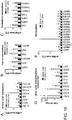
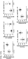
- the creatinine-normalized excretion of glutamine, proline, citrulline, Na-acetyllysine, spermine, N1-acetylspermidine, N8-acetylspermidine, spermine, xanthosine, inosine, xanthine, deoxyuridine, cytidine, and thymidine were progressively elevated whereas those of ornithine and carnitine were reduced in tumor-bearing animals.
- a detailed description of quantitative ranges and statistical significances (P value and odds ratio) of these urinary biomarkers is provided at Table 1.
- the urinary excretion of glutamic acid, aspartic acid, lysine, arginine, hypoxanthine, creatine, N ⁇ -monomethylarginine, and uridine were found to be unaffected during tumorigenesis.
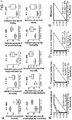
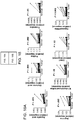
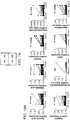
- the urinary excretion of hypermethylated arginine metabolite symmetric-dimethylarginine was elevated by 1.7-fold and 1.6-fold at five and six months ( Figure 4A ), while that of asymmetric-dimethylarginine ( Figure 4B ) was also elevated by 1.7-fold and 2-fold at four and six months, respectively.
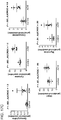
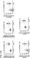
- the urinary excretion of xanthosine ( Figure 6A ) was elevated 2.0- and 2.8-fold at five and six months, respectively, whereas inosine ( Figure 6B ) and xanthine ( Figure 6C ) were elevated by 3.5- and 4.2-fold, respectively, at six months.
- the urinary excretion of cytidine ( Figure 6D ) was elevated 28.7- and 6.8-fold at five and six months, respectively whereas that of deoxyuridine ( Figure 6E ) and thymidine ( Figure 6F ) were elevated by 5.0- and 7.4-fold, respectively, at five months.
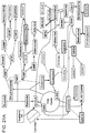
- the ODC expression was upregulated in tumor with concomitant downregulation of OTC expression that would result in funneling of ornithine into biosynthesis of putrescine. This indicates that the arginine pool is channelized in bifurcated fashion into both urea cycle (via NOS) and polyamine biosynthesis (via ODC).
- the next step in polyamine biosynthesis involves successive addition of propylamine groups from S-adenosylmethioninamine (SAMA) by SRM to produce spermidine and then, by SRM or SMS to produce spermine.
- SAMA S-adenosylmethioninamine
- SAMA S-adenosylmethioninamine
- Arginine residues are methylated either symmetrically (by type-I PRMTs such as PRMT1, PRMT3, CARM1, PRMT6) or asymmetrically (by type-II PRMTs such as PRMT5, PRMT7) to produce symmetric or asymmetric dimethylarginine.
- the expression of all of these genes was upregulated along with Prmt8. which was recently shown to interact with hnRNPs and RNA-helicases, in the tumor tissue. Consequently, the abundance of dimethylarginines in tumor tissue as well as their excretion in the urine was significantly elevated. Wnt-signaling has been shown to cause arginine methylation of G3BP1 that acts as a molecular switch for ⁇ -catenin activity. Thus, increased arginine methylation due to activation of Wnt-signaling may compound the activation of the proliferative signal and viciously accelerate tumorigenesis.
- Nucleic acid metabolites such as uracil, hypoxanthine and xanthine were elevated. Similar to Apc Min /+ mice, human tumors showed elevation of hypermethylated metabolites such as carnitine, symmetric-dimethylarginine, asymmetric-dimethylarginine, dimethylglycine and betaine along with S-adenosylmethionine and S-adenosylhomocysteine.
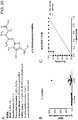
- FIG. 20A shows the structure of the metabolite which has a molecular formula C 9 H 17 N 3 O 6 . M264 has been found to be significantly (p ⁇ .0001) elevated only in the urine samples of CRC patients.
- Serum samples were collected by retro-orbital bleeding before euthanasia. Animals were killed through CO 2 asphyxiation at the end of the study, intestine flushed with normal saline, longitudinally opened, tumor counted under light microscope, tissue samples harvested and all samples were stored at -80°C.
- the quantitaion of the urinary metabolites were performed on a Xevo triple-quadruple ESIQToF platform coupled with an amide column through multiple reaction monitoring and all data were presented as normalized with respect to creatinine concentrations. Changes in relative abundance of the metabolites in tissue were calculated from internal standard-normalized area under extracted chromatograms. See Supplementary methods for delail.
- Urine samples were deproteinated using 50% or 70% acetonitrile containing 5 ⁇ M chloropropamide or ⁇ -aminopimelic acid as internal standards for reverse-phase (RP) or hydrophilic interaction liquid chromatography (HILIC), respectively. Supernatants were transferred into 96-well sample plates. All pipetting and dilution were performed using a MICROLAB STAR LET automated liquid handler (Hamilton Robotics; Reno, NV).
- the gradient elution was performed over 10 min using: 1-60% B in 4 min (0.4 ml/min), 60-80% B at 8 min (0.4 ml/min), holding at 80% B up to 8.5 min (0.3 ml/min), bringing back to 1% B at 8.8 min and holding at 1% until end (0.3 ml/min), Column temperature was maintained at 40°C. Column was re-equilibrated with 99% A at the end of each run prior to injection of next sample. For reverse-phase analysis, 5 ⁇ L aliquot of samples were injected into a 2.1x50 mm Acquity UPLC BEH C 18 column (1.7 ⁇ M) connected to a SYNAPT HDMS ESIQTOF mass spectrometer (Waters Corporation).
- Chromatographic separation was achieved by using a mixture of (A) water containing 1% formic acid and (B) acetonitrile containing 1% formic acid as mobile phase.
- the gradient elution was performed over 6min at a flow rate of 0.3 mL using: 1-99% B in 4 min, holding at 99% B up to 5.0 min, bringing back to 1% at 5.5 min and holding at 1% until end.
- Column temperature was maintained at 40°C. Column was re-equilibrated with 98% A at the end of each run prior to injection of next sample. Mass spectrometric analysis was performed in both positive and negative ionization modes.
- the multivariate data matrix was analyzed by SIMCA-P+12 software (Umetrics, Kinnelon, NJ). Data quality inspection as well as distribution and unsupervised segregation of wild-type and mutants on global metabolomic space were checked by principal components analysis (PCA) of the Pareto-scaled data.
- PCA principal components analysis
- the supervised orthogonal projection to latent structures (OPLS) model was used identify ions contributing to discrimination (indicated by their distance from origin along Y-axes) of metabolic traits. Consistency of longitudinal trend of ions in contributing to difference of wild-type and mutant metabolome was examined to select potential biomarker candidates. Candidate metabolites for these ions were identified on the basis of accurate mass measurement using metabolomic databases such as METLIN (http://metlin.scripps.edu) or HMDB (http://www.hmdb.ca/) and MS/MS fragmentation pattern. Finally, identities of biomarker were confirmed by comparison of retention time and fragmentation pattern with authentic standards.
- METLIN http://metlin.scripps.edu
- HMDB http://www.hmdb.ca/
- Metabolites in the deproteinated urine samples were quantified in multiple reactions monitoring mode on a XEVO triple quadruple mass spectrometer (Water Corporation).
- ⁇ -Aminopimelic acid (5 ⁇ M) was used as internal standard.
- the following metabolites were quantified by monitoring characteristic fragmentation reactions (in bracket); ⁇ -aminopimelic acid (176 ⁇ 112, ESI+), creatinine (114 ⁇ 86, ESI+), aspartic acid (132 ⁇ 88, ESI-), glutamine (147 ⁇ 84, ESI+), glutamic acid (148 ⁇ 84, ESI+), creatine (132 ⁇ 90, ESI+), lysine (147 ⁇ 130, ESI+), citrulline (176 ⁇ 159, ESI+), ornithine (133 ⁇ 70, ESI+), arginine (175 ⁇ 60, ESI+), N ⁇ -monomethylarginine (189 ⁇ 84, ESI+), sym -dimethyl
- Spermine was measured by single ion monitoring (203, ESI+) and relative abundance was measured by area under the peak. Chromatographic separation was achieved on a 2.1 x 50 mm Acquity UPLC BEH amide column using the mobile phase as mentioned above. The gradient elution was performed over 10 min at a flow rate of 0.5 ml/min using: 1-40% B in 6 min, 40-60% B at 8 min, 60-80% at 8.2 min, bringing back to 1% B at 9 min and holding at 1% until end. All data were processed using TargetLynx software (Waters Corporation). Internal standard-normalized area under the peak (response) from serially diluted authentic standard solution was used to build calibration curve for each metabolite. The concentration of metabolite was determined from the calibration curve and divided by creatinine concentration to determine creatinine-normalized excretion of the metabolite in 24-hour urine.
- Metabolite composition was quantitated using MRM and normalized as mentioned above.
- MRMs the following MRMs were also monitored for respective metabolites; adenosine (268 ⁇ 136, ESI+), UMP (325 ⁇ 97, ESI-), dUMP (309 ⁇ 81, ESI+), CMP (324 ⁇ 112, ESI+), ATP (508 ⁇ 136, BSI+), N-acetylglutamic acid (190 ⁇ 84, ESI+), acetyl CoA (810 ⁇ 303, BSI+), uracil (111 ⁇ 42, ESI-), dimethylglycine (104 ⁇ 58, ESI+), S-adenosylhomocysteine (385 ⁇ 134, ESI+), and S-adenosylmethionine (399 ⁇ 250, ESI+).
- the fold change in abundance of metabolites in Cre+ colon epithelium were calculated by dividing the normalized response for
- Targeted measurement of metabolite abundances in the reconstituted aqueous extract were performed by MRM reaction monitoring coupled with hydrophilic interaction liquid chromatography as mentioned above for analysis of urine samples.
- the area under the peak for each metabolite was normalized step-by-step for initial tissue weight, extraction efficiency ( ⁇ -aminopimelic acid) and instrument response (difluromethylornithine) and presented as normalized response.
Landscapes
- Health & Medical Sciences (AREA)
- Life Sciences & Earth Sciences (AREA)
- Engineering & Computer Science (AREA)
- Chemical & Material Sciences (AREA)
- Immunology (AREA)
- Molecular Biology (AREA)
- Hematology (AREA)
- Urology & Nephrology (AREA)
- Biomedical Technology (AREA)
- Pathology (AREA)
- Physics & Mathematics (AREA)
- Analytical Chemistry (AREA)
- Proteomics, Peptides & Aminoacids (AREA)
- Biochemistry (AREA)
- Biotechnology (AREA)
- Microbiology (AREA)
- General Health & Medical Sciences (AREA)
- Organic Chemistry (AREA)
- Food Science & Technology (AREA)
- General Physics & Mathematics (AREA)
- Cell Biology (AREA)
- Medicinal Chemistry (AREA)
- Zoology (AREA)
- Biophysics (AREA)
- Genetics & Genomics (AREA)
- Wood Science & Technology (AREA)
- Bioinformatics & Cheminformatics (AREA)
- Hospice & Palliative Care (AREA)
- Oncology (AREA)
- Bioinformatics & Computational Biology (AREA)
- General Engineering & Computer Science (AREA)
- Measuring Or Testing Involving Enzymes Or Micro-Organisms (AREA)
- Investigating Or Analysing Biological Materials (AREA)
- Medicines Containing Antibodies Or Antigens For Use As Internal Diagnostic Agents (AREA)
Claims (15)
- Procédé d'identification in vitro d'un sujet ayant ou ayant une propension à développer une néoplasie, le procédé comprenant :l'identification d'un taux accru de M264 (β-D-ribofuranosylcréatine) dans un échantillon biologique provenant du sujet par rapport au taux présent dans une référence, en combinaison avec un taux accru d'un ou plusieurs biomarqueurs supplémentaires parmi les suivants :biomarqueurs associés à une méthylation, comprenant diméthylarginine symétrique, carnitine, méthionine, diméthylglycine et bétaïne ;biomarqueurs associés au métabolisme de l'acide nucléique comprenant xanthosine, inosine, désoxy-uridine, thymidine, désoxycytidine, cytosine, hypoxanthine, xanthine, uracile, guanosine, et adénosine ; etbiomarqueurs associés au cycle de l'urée et au métabolisme des polyamines comprenant N1-acétylspermidine, N8-acétylspermidine, spermine, arginine ornithine et citrulline ;biomarqueurs associés au métabolisme des acides aminés comprenant proline, glutamine, acide glutamique, thréonine, et Nα-acétyllysine,dans lequel une augmentation desdits taux de M264 et de l'un ou plusieurs biomarqueur(s) identifie un sujet comme ayant ou ayant une propension à développer une néoplasie, dans lequel le procédé comprend en outre éventuellement la sélection d'un procédé de traitement pour traiter une néoplasie, dans lequel ledit traitement correspond de préférence à une néoplasie plus agressive.
- Procédé selon la revendication 1, dans lequel les groupes d'un ou plusieurs biomarqueur(s) supplémentaire(s) sont sélectionnés parmi les suivants :biomarqueurs associés à une méthylation, comprenant diméthylarginine asymétrique et diméthylarginine symétrique ;biomarqueurs associés au métabolisme de l'acide nucléique comprenant xanthosine, inosine, désoxy-uridine et thymidine ;biomarqueurs associés au métabolisme des polyamines comprenant N1-acétylspermidine, N8-acétylspermidine, et spermine ; etbiomarqueurs associés au métabolisme des acides aminés comprenant proline, glutamine, et Nα-acétyllysine.
- Procédé selon l'une quelconque des revendications 1 ou 2, dans lequel l'échantillon biologique est un fluide biologique ou un échantillon de tissu ; dans lequel le fluide biologique est sélectionné de préférence dans le groupe consistant en urine, sang, sérum sanguin, plasma, bile, aspirat fécal, aspirat intestinal, liquide céphalorachidien et salive.
- Procédé selon l'une quelconque des revendications 1 à 3, comprenant en outre une mesure du taux de carnitine dans l'échantillon biologique.
- Procédé selon l'une quelconque des revendications 1 à 4, dans lequel une diminution du taux de carnitine dans un échantillon biologique qui est un fluide biologique identifie un sujet comme ayant ou ayant une propension à développer une néoplasie, ou dans lequel une diminution du taux de carnitine dans un échantillon biologique qui est un échantillon de tissu identifie un sujet comme ayant une chance de survie accrue.
- Procédé selon l'une quelconque des revendications 1 à 4, dans lequel une augmentation du taux de carnitine dans un échantillon biologique qui est un fluide biologique identifie un sujet comme ayant une chance de survie accrue, ou dans lequel une augmentation du taux de carnitine dans un échantillon biologique qui est un échantillon de tissu identifie un sujet comme ayant ou ayant une propension à développer une néoplasie.
- Procédé selon l'une quelconque des revendications 1 à 6, dans lequel la néoplasie est un cancer colorectal, un cancer de l'intestin grêle, un cancer du duodénum, un cancer de l'estomac, un cancer du pancréas, un cancer du foie, un cancer de l'oesophage, un cancer de l'ovaire, un cancer de la prostate, un cancer de l'utérus, un cancer du sein, un cancer de la thyroïde ou un mélanome.
- Procédé selon l'une quelconque des revendications 1 à 7, dans lequel la néoplasie comprend une mutation de l'APC ou d'une protéine de la voie de signalisation de Wnt.
- Procédé selon l'une quelconque des revendications 1 à 8, comprenant en outre la détection d'une mutation de l'APC ou d'une protéine de la voie de signalisation de Wnt.
- Procédé selon l'une quelconque des revendications 1 à 9, dans lequel le taux de M264 (β-D-ribofuranosylcréatine) ou de l'un ou plusieurs biomarqueurs supplémentaires est accru de 1,5, 2, 2,5, 3, 3,5, 4, 5, 6, 7, 8, 9, 10, 11, 12, 13, 14, 15 fois ou plus par rapport à la référence.
- Procédé selon l'une quelconque des revendications 1 à 10, dans lequel la référence est un témoin ou un échantillon biologique correspondant provenant d'un sujet sain ; et/ou le taux de biomarqueur est détecté par chromatographie, spectrométrie de masse, spectroscopie ou immuno-essai.
- Procédé selon l'une quelconque des revendications 1 à 11, dans lequel le taux du biomarqueur est normalisé par rapport au taux de créatinine dans l'échantillon, à une concentration absolue, à une élimination sur 24 h, ou par rapport à la densité relative.
- Ensemble pour aider au diagnostic d'une néoplasie ou d'un cancer colorectal, l'ensemble comprenant au moins un réactif capable de se lier spécifiquement à M264 (β-D-ribofuranosylcréatine) et au moins un réactif capable de se lier spécifiquement à au moins un marqueur supplémentaire sélectionné parmi les suivants :biomarqueurs associés à une méthylation, comprenant diméthylarginine asymétrique, diméthylarginine symétrique, carnitine, méthionine, diméthylglycine et bétaïne ;biomarqueurs associés au métabolisme de l'acide nucléique comprenant xanthosine, inosine, désoxy-uridine, thymidine, désoxycytidine, cytosine, hypoxanthine, xanthine, uracile, guanosine, et adénosine ; etbiomarqueurs associés au cycle de l'urée et au métabolisme des polyamines comprenant N1-acétylspermidine, N8-acétylspermidine, spermine, arginine ornithine et citrulline ; etbiomarqueurs associés au métabolisme des acides aminés comprenant proline, glutamine, acide glutamique, thréonine, et Nα-acétyllysine ; dans lequel les groupes sont sélectionnés de préférence parmi les suivants :biomarqueurs associés à une méthylation, comprenant diméthylarginine asymétrique et diméthylarginine symétrique ;biomarqueurs associés au métabolisme de l'acide nucléique comprenant xanthosine, inosine, désoxy-uridine et thymidine ;biomarqueurs associés au métabolisme des polyamines comprenant N1-acétylspermidine, N8-acétylspermidine, et spermine ; etbiomarqueurs associés au métabolisme des acides aminés comprenant proline, glutamine, et Nα-acétyllysine ; etdans lequel le réactif est un anticorps.
- Ensemble selon la revendication 13, comprenant au moins un réactif capable de se lier spécifiquement avec M264 (β-D-ribofuranosylcréatine).
- Utilisation d'un ensemble selon les procédés selon l'une quelconque des revendications 1 à 12, pour aider au diagnostic d'une néoplasie ou d'un cancer colorectal, l'ensemble comprenant au moins un réactif capable de détecter, se lier ou capturer M264 (β-D-ribofuranosylcréatine) et au moins un réactif capable de détecter, se lier ou capturer au moins un marqueur supplémentaire sélectionné parmi les suivants :biomarqueurs associés à une méthylation, comprenant diméthylarginine asymétrique, diméthylarginine symétrique, carnitine, méthionine, diméthylglycine et bétaïne ;biomarqueurs associés au métabolisme de l'acide nucléique comprenant xanthosine, inosine, désoxy-uridine, thymidine, désoxycytidine, cytosine, hypoxanthine, xanthine, uracile, guanosine, et adénosine ;biomarqueurs associés au cycle de l'urée et au métabolisme des polyamines comprenant N1-acétylspermidine, N8-acétylspermidine, spermine, arginine ornithine et citrulline ; etbiomarqueurs associés au métabolisme des acides aminés comprenant proline, glutamine, acide glutamique, thréonine, et Nα-acétyllysine ;dans lequel les groupes sont sélectionnés de préférence parmi les suivants :biomarqueurs associés à une méthylation, comprenant diméthylarginine asymétrique et diméthylarginine symétrique ;biomarqueurs associés au métabolisme de l'acide nucléique comprenant xanthosine, inosine, désoxy-uridine et thymidine ;biomarqueurs associés au métabolisme des polyamines comprenant N1-acétylspermidine, N8-acétylspermidine, et spermine ; etbiomarqueurs associés au métabolisme des acides aminés comprenant proline, glutamine, et Nα-acétyllysine ; etdans lequel le réactif est un ou plusieurs parmi un composé organique, un peptide, un peptido-mimétique, un polypeptide, un ligand d'acide nucléique, un aptamère, ou un anticorps.
Applications Claiming Priority (2)
| Application Number | Priority Date | Filing Date | Title |
|---|---|---|---|
| US201361755891P | 2013-01-23 | 2013-01-23 | |
| PCT/US2014/012758 WO2014116833A2 (fr) | 2013-01-23 | 2014-01-23 | Compositions et procédés de détection de néoplasie |
Publications (2)
| Publication Number | Publication Date |
|---|---|
| EP2948775A2 EP2948775A2 (fr) | 2015-12-02 |
| EP2948775B1 true EP2948775B1 (fr) | 2018-10-03 |
Family
ID=50097864
Family Applications (1)
| Application Number | Title | Priority Date | Filing Date |
|---|---|---|---|
| EP14704237.8A Not-in-force EP2948775B1 (fr) | 2013-01-23 | 2014-01-23 | Compositions et procédés de détection de néoplasie |
Country Status (3)
| Country | Link |
|---|---|
| US (1) | US10487363B2 (fr) |
| EP (1) | EP2948775B1 (fr) |
| WO (1) | WO2014116833A2 (fr) |
Cited By (1)
| Publication number | Priority date | Publication date | Assignee | Title |
|---|---|---|---|---|
| CN109030665A (zh) * | 2018-10-08 | 2018-12-18 | 黑龙江大学 | 亲水性液相色谱串联质谱法测定红甜菜中甜菜碱含量的检测方法 |
Families Citing this family (22)
| Publication number | Priority date | Publication date | Assignee | Title |
|---|---|---|---|---|
| KR101824937B1 (ko) | 2012-11-27 | 2018-02-02 | 아지노모토 가부시키가이샤 | 췌장암의 평가 방법, 췌장암 평가 장치, 췌장암 평가 방법, 췌장암 평가 프로그램, 췌장암 평가 시스템 및 정보 통신 단말 장치 |
| US10393745B2 (en) | 2013-07-11 | 2019-08-27 | The USA, as represented by the Secretary, Department of Heath and Human Services | Method for the diagnosis and prognosis of cancer |
| EP3578984A3 (fr) * | 2013-10-28 | 2020-03-04 | Salivatech Co., Ltd. | Biomarqueurs salivaires pour cancer de la bouche |
| CN104251895B (zh) * | 2014-09-24 | 2016-08-31 | 珠海健帆生物科技股份有限公司 | 一种快速检测吸附剂吸附性能的方法 |
| CN105116000B (zh) * | 2015-09-01 | 2017-07-18 | 上海交通大学 | 用于检测胃癌相关代谢小分子的核磁共振模型及制备方法 |
| US10001501B2 (en) * | 2015-12-31 | 2018-06-19 | Universal Diagnostics, S. L. | Systems and methods for automated, customizable sample preparation for detection of metabolites and lipids |
| HUE055153T2 (hu) | 2016-05-24 | 2021-11-29 | In Ovo B V | Módszer és rendszer a roncsolásmentes in ovo meghatározásához a szárnyas nemnek |
| US10006925B2 (en) | 2016-05-30 | 2018-06-26 | Universal Diagnostics, S. L. | Methods and systems for metabolite and/or lipid-based detection of colorectal cancer and/or adenomatous polyps |
| JPWO2018066621A1 (ja) * | 2016-10-04 | 2019-07-18 | 味の素株式会社 | 大腸癌の評価方法、評価装置、評価プログラム、評価システム、及び端末装置 |
| JP7230336B2 (ja) | 2018-04-02 | 2023-03-01 | 味の素株式会社 | 取得方法、算出方法、評価装置、算出装置、評価プログラム、算出プログラム、記録媒体、及び評価システム |
| JP7230335B2 (ja) * | 2018-04-02 | 2023-03-01 | 味の素株式会社 | 取得方法、算出方法、評価装置、算出装置、評価プログラム、算出プログラム、記録媒体、及び評価システム |
| US20190391092A1 (en) * | 2018-06-21 | 2019-12-26 | Oregon Institute of Science and Medicine | Metabolic profiling with magnetic resonance mass spectrometry (mrms) |
| US11840720B2 (en) | 2019-12-23 | 2023-12-12 | Metabolomic Technologies Inc. | Urinary metabolomic biomarkers for detecting colorectal cancer and polyps |
| CN112834644A (zh) * | 2020-12-31 | 2021-05-25 | 郑州大学第一附属医院 | 膀胱癌相关的组合标志物和检测试剂盒 |
| CN115372490A (zh) * | 2021-05-21 | 2022-11-22 | 深圳市绘云生物科技有限公司 | 用于评估腺瘤及结直肠癌风险的生物标志物及其应用 |
| CN113567584A (zh) * | 2021-07-26 | 2021-10-29 | 郑州大学第一附属医院 | 一种基于血清代谢组学的贲门癌筛查的标志物及试剂盒 |
| US20240402180A1 (en) * | 2021-09-21 | 2024-12-05 | The Regents Of The University Of Michigan | Metabolite biomarkers for differential diagnosis of pacreatic cysts |
| CN114705782B (zh) * | 2022-04-07 | 2023-12-01 | 中国人民解放军总医院第一医学中心 | 用于诊断或监测结直肠癌的血浆代谢标志物组合及应用 |
| CN114965800B (zh) * | 2022-05-12 | 2024-02-20 | 山西医科大学 | 肾透明细胞癌生物标志物及其在早期筛查中的应用 |
| CN116334223B (zh) * | 2023-03-02 | 2023-12-22 | 武汉大学 | 可变剪接功能性位点rs61746794的检测试剂在制备结直肠癌辅助诊断试剂盒中的应用 |
| CN117388495B (zh) * | 2023-12-13 | 2024-02-09 | 哈尔滨脉图精准技术有限公司 | 用于诊断肺癌分期的代谢标志物的应用及试剂盒 |
| CN119804728B (zh) * | 2025-02-06 | 2026-03-10 | 哈尔滨脉图精准技术有限公司 | 用于检测多种妇科肿瘤的血浆代谢标志物及其应用 |
Citations (1)
| Publication number | Priority date | Publication date | Assignee | Title |
|---|---|---|---|---|
| WO2012024612A1 (fr) * | 2010-08-20 | 2012-02-23 | Pangea Biosciences, Inc. | Diagnostic du cancer et agent thérapeutique anticancéreux |
Family Cites Families (8)
| Publication number | Priority date | Publication date | Assignee | Title |
|---|---|---|---|---|
| US5837832A (en) | 1993-06-25 | 1998-11-17 | Affymetrix, Inc. | Arrays of nucleic acid probes on biological chips |
| US6015880A (en) | 1994-03-16 | 2000-01-18 | California Institute Of Technology | Method and substrate for performing multiple sequential reactions on a matrix |
| US5800979A (en) | 1994-12-01 | 1998-09-01 | Kolhouse; J. Fred | Gas chromatography/mass spectrometric determination of folic acid coenzymes |
| US6436665B1 (en) | 1999-08-27 | 2002-08-20 | Phylos, Inc | Methods for encoding and sorting in vitro translated proteins |
| US7202473B2 (en) | 2003-04-10 | 2007-04-10 | Micromass Uk Limited | Mass spectrometer |
| US7041972B2 (en) | 2003-05-09 | 2006-05-09 | Waters Investments Limited | Mass spectrometer |
| US9217032B2 (en) * | 2010-01-08 | 2015-12-22 | Les Laboratoires Servier | Methods for treating colorectal cancer |
| CN101945027B (zh) * | 2010-08-30 | 2014-03-19 | 中兴通讯股份有限公司 | 一种家庭有线物联网关获取uim卡号的方法及系统 |
-
2014
- 2014-01-23 EP EP14704237.8A patent/EP2948775B1/fr not_active Not-in-force
- 2014-01-23 US US14/762,426 patent/US10487363B2/en not_active Expired - Fee Related
- 2014-01-23 WO PCT/US2014/012758 patent/WO2014116833A2/fr not_active Ceased
Patent Citations (1)
| Publication number | Priority date | Publication date | Assignee | Title |
|---|---|---|---|---|
| WO2012024612A1 (fr) * | 2010-08-20 | 2012-02-23 | Pangea Biosciences, Inc. | Diagnostic du cancer et agent thérapeutique anticancéreux |
Non-Patent Citations (6)
| Title |
|---|
| DENKERT CARSTEN ET AL: "Metabolite profiling of human colon carcinoma - deregulation of TCA cycle and amino acid turnover", MOLECULAR CANCER, BIOMED CENTRAL, LONDON, GB, vol. 7, no. 1, 18 September 2008 (2008-09-18), pages 72, XP021043743, ISSN: 1476-4598, DOI: 10.1186/1476-4598-7-72 * |
| MASANORI YOSHIMATSU ET AL: "Dysregulation of PRMT1 and PRMT6, Type I arginine methyltransferases, is involved in various types of human cancers", INTERNATIONAL JOURNAL OF CANCER, vol. 128, no. 3, 27 November 2010 (2010-11-27), pages 562 - 573, XP055114363, ISSN: 0020-7136, DOI: 10.1002/ijc.25366 * |
| SOUMEN K. MANNA ET AL: "Biomarkers of Coordinate Metabolic Reprogramming in Colorectal Tumors in Mice and Humans", GASTROENTEROLOGY, 1 January 2014 (2014-01-01), XP055114314, ISSN: 0016-5085, DOI: 10.1053/j.gastro.2014.01.017 * |
| STEPHANOS PAVLIDES ET AL: "The autophagic tumor stroma model of cancer: Role of oxidative stress and ketone production in fueling tumor cell metabolism", CELL CYCLE, vol. 9, no. 17, 1 September 2010 (2010-09-01), pages 3485 - 3505, XP055114408, ISSN: 1538-4101, DOI: 10.4161/cc.9.17.12721 * |
| YUNPING QIU ET AL: "Serum Metabolite Profiling of Human Colorectal Cancer Using GC-TOFMS and UPLC-QTOFMS", JOURNAL OF PROTEOME RESEARCH, vol. 8, no. 10, 2 October 2009 (2009-10-02), pages 4844 - 4850, XP055080029, ISSN: 1535-3893, DOI: 10.1021/pr9004162 * |
| YUNPING QIU ET AL: "Urinary Metabonomic Study on Colorectal Cancer", JOURNAL OF PROTEOME RESEARCH, vol. 9, no. 3, 5 March 2010 (2010-03-05), pages 1627 - 1634, XP055079026, ISSN: 1535-3893, DOI: 10.1021/pr901081y * |
Cited By (1)
| Publication number | Priority date | Publication date | Assignee | Title |
|---|---|---|---|---|
| CN109030665A (zh) * | 2018-10-08 | 2018-12-18 | 黑龙江大学 | 亲水性液相色谱串联质谱法测定红甜菜中甜菜碱含量的检测方法 |
Also Published As
| Publication number | Publication date |
|---|---|
| US10487363B2 (en) | 2019-11-26 |
| EP2948775A2 (fr) | 2015-12-02 |
| WO2014116833A3 (fr) | 2014-11-13 |
| US20150344969A1 (en) | 2015-12-03 |
| WO2014116833A2 (fr) | 2014-07-31 |
Similar Documents
| Publication | Publication Date | Title |
|---|---|---|
| EP2948775B1 (fr) | Compositions et procédés de détection de néoplasie | |
| US11493516B2 (en) | Biomarkers for assessing breast cancer | |
| US11181525B2 (en) | Metabolomic signatures for predicting, diagnosing, and prognosing various diseases including cancer | |
| Moreno et al. | Metabolomic profiling of human lung tumor tissues–nucleotide metabolism as a candidate for therapeutic interventions and biomarkers | |
| Manna et al. | Biomarkers of coordinate metabolic reprogramming in colorectal tumors in mice and humans | |
| US20250298025A1 (en) | Methods for the detection and treatment of pancreatic ductal adenocarcinoma | |
| Pan et al. | Proteomics analysis of bodily fluids in pancreatic cancer | |
| EP2597464B1 (fr) | Profilage métabolomique du cancer de la prostate | |
| US20240084394A1 (en) | Urinary metabolomic biomarkers for detecting colorectal cancer and polyps | |
| CN103229052A (zh) | 用于前列腺癌的生物标记及其使用方法 | |
| US20150133331A1 (en) | Metabolite biomarkers for the detection of liver cancer | |
| Martínez‐Aguilar et al. | Quantitative mass spectrometry for colorectal cancer proteomics | |
| Ma et al. | Mass spectrometry based translational proteomics for biomarker discovery and application in colorectal cancer | |
| CN108107134A (zh) | 预测培美曲塞联合铂类治疗非小细胞肺癌的疗效的新标志物及其应用 | |
| CA3065979C (fr) | Biomarqueurs metabolomiques urinaires pour detecter le cancer et les polypes colorectaux | |
| US20260009085A1 (en) | Methods and compositions for the diagnosis and treatment of cancer |
Legal Events
| Date | Code | Title | Description |
|---|---|---|---|
| PUAI | Public reference made under article 153(3) epc to a published international application that has entered the european phase |
Free format text: ORIGINAL CODE: 0009012 |
|
| 17P | Request for examination filed |
Effective date: 20150729 |
|
| AK | Designated contracting states |
Kind code of ref document: A2 Designated state(s): AL AT BE BG CH CY CZ DE DK EE ES FI FR GB GR HR HU IE IS IT LI LT LU LV MC MK MT NL NO PL PT RO RS SE SI SK SM TR |
|
| AX | Request for extension of the european patent |
Extension state: BA ME |
|
| RIN1 | Information on inventor provided before grant (corrected) |
Inventor name: MANNA, SOUMEN, KANTI Inventor name: GONZALEZ, FRANK, J. Inventor name: KRAUSZ, KRISTOPHER, W. |
|
| DAX | Request for extension of the european patent (deleted) | ||
| PUAG | Search results despatched under rule 164(2) epc together with communication from examining division |
Free format text: ORIGINAL CODE: 0009017 |
|
| STAA | Information on the status of an ep patent application or granted ep patent |
Free format text: STATUS: EXAMINATION IS IN PROGRESS |
|
| 17Q | First examination report despatched |
Effective date: 20170623 |
|
| B565 | Issuance of search results under rule 164(2) epc |
Effective date: 20170623 |
|
| RIC1 | Information provided on ipc code assigned before grant |
Ipc: G01N 33/574 20060101AFI20170620BHEP |
|
| GRAP | Despatch of communication of intention to grant a patent |
Free format text: ORIGINAL CODE: EPIDOSNIGR1 |
|
| STAA | Information on the status of an ep patent application or granted ep patent |
Free format text: STATUS: GRANT OF PATENT IS INTENDED |
|
| RAP1 | Party data changed (applicant data changed or rights of an application transferred) |
Owner name: THE UNITED STATES OF AMERICA, AS REPRESENTED BY TH |
|
| INTG | Intention to grant announced |
Effective date: 20180416 |
|
| GRAS | Grant fee paid |
Free format text: ORIGINAL CODE: EPIDOSNIGR3 |
|
| GRAA | (expected) grant |
Free format text: ORIGINAL CODE: 0009210 |
|
| STAA | Information on the status of an ep patent application or granted ep patent |
Free format text: STATUS: THE PATENT HAS BEEN GRANTED |
|
| AK | Designated contracting states |
Kind code of ref document: B1 Designated state(s): AL AT BE BG CH CY CZ DE DK EE ES FI FR GB GR HR HU IE IS IT LI LT LU LV MC MK MT NL NO PL PT RO RS SE SI SK SM TR |
|
| REG | Reference to a national code |
Ref country code: GB Ref legal event code: FG4D |
|
| REG | Reference to a national code |
Ref country code: CH Ref legal event code: EP Ref country code: AT Ref legal event code: REF Ref document number: 1049180 Country of ref document: AT Kind code of ref document: T Effective date: 20181015 |
|
| REG | Reference to a national code |
Ref country code: IE Ref legal event code: FG4D Ref country code: DE Ref legal event code: R096 Ref document number: 602014033311 Country of ref document: DE |
|
| REG | Reference to a national code |
Ref country code: NL Ref legal event code: MP Effective date: 20181003 |
|
| REG | Reference to a national code |
Ref country code: LT Ref legal event code: MG4D |
|
| REG | Reference to a national code |
Ref country code: AT Ref legal event code: MK05 Ref document number: 1049180 Country of ref document: AT Kind code of ref document: T Effective date: 20181003 |
|
| PG25 | Lapsed in a contracting state [announced via postgrant information from national office to epo] |
Ref country code: NL Free format text: LAPSE BECAUSE OF FAILURE TO SUBMIT A TRANSLATION OF THE DESCRIPTION OR TO PAY THE FEE WITHIN THE PRESCRIBED TIME-LIMIT Effective date: 20181003 |
|
| PG25 | Lapsed in a contracting state [announced via postgrant information from national office to epo] |
Ref country code: ES Free format text: LAPSE BECAUSE OF FAILURE TO SUBMIT A TRANSLATION OF THE DESCRIPTION OR TO PAY THE FEE WITHIN THE PRESCRIBED TIME-LIMIT Effective date: 20181003 Ref country code: CZ Free format text: LAPSE BECAUSE OF FAILURE TO SUBMIT A TRANSLATION OF THE DESCRIPTION OR TO PAY THE FEE WITHIN THE PRESCRIBED TIME-LIMIT Effective date: 20181003 Ref country code: LV Free format text: LAPSE BECAUSE OF FAILURE TO SUBMIT A TRANSLATION OF THE DESCRIPTION OR TO PAY THE FEE WITHIN THE PRESCRIBED TIME-LIMIT Effective date: 20181003 Ref country code: HR Free format text: LAPSE BECAUSE OF FAILURE TO SUBMIT A TRANSLATION OF THE DESCRIPTION OR TO PAY THE FEE WITHIN THE PRESCRIBED TIME-LIMIT Effective date: 20181003 Ref country code: PL Free format text: LAPSE BECAUSE OF FAILURE TO SUBMIT A TRANSLATION OF THE DESCRIPTION OR TO PAY THE FEE WITHIN THE PRESCRIBED TIME-LIMIT Effective date: 20181003 Ref country code: BG Free format text: LAPSE BECAUSE OF FAILURE TO SUBMIT A TRANSLATION OF THE DESCRIPTION OR TO PAY THE FEE WITHIN THE PRESCRIBED TIME-LIMIT Effective date: 20190103 Ref country code: LT Free format text: LAPSE BECAUSE OF FAILURE TO SUBMIT A TRANSLATION OF THE DESCRIPTION OR TO PAY THE FEE WITHIN THE PRESCRIBED TIME-LIMIT Effective date: 20181003 Ref country code: FI Free format text: LAPSE BECAUSE OF FAILURE TO SUBMIT A TRANSLATION OF THE DESCRIPTION OR TO PAY THE FEE WITHIN THE PRESCRIBED TIME-LIMIT Effective date: 20181003 Ref country code: NO Free format text: LAPSE BECAUSE OF FAILURE TO SUBMIT A TRANSLATION OF THE DESCRIPTION OR TO PAY THE FEE WITHIN THE PRESCRIBED TIME-LIMIT Effective date: 20190103 Ref country code: IS Free format text: LAPSE BECAUSE OF FAILURE TO SUBMIT A TRANSLATION OF THE DESCRIPTION OR TO PAY THE FEE WITHIN THE PRESCRIBED TIME-LIMIT Effective date: 20190203 Ref country code: AT Free format text: LAPSE BECAUSE OF FAILURE TO SUBMIT A TRANSLATION OF THE DESCRIPTION OR TO PAY THE FEE WITHIN THE PRESCRIBED TIME-LIMIT Effective date: 20181003 |
|
| PG25 | Lapsed in a contracting state [announced via postgrant information from national office to epo] |
Ref country code: AL Free format text: LAPSE BECAUSE OF FAILURE TO SUBMIT A TRANSLATION OF THE DESCRIPTION OR TO PAY THE FEE WITHIN THE PRESCRIBED TIME-LIMIT Effective date: 20181003 Ref country code: SE Free format text: LAPSE BECAUSE OF FAILURE TO SUBMIT A TRANSLATION OF THE DESCRIPTION OR TO PAY THE FEE WITHIN THE PRESCRIBED TIME-LIMIT Effective date: 20181003 Ref country code: GR Free format text: LAPSE BECAUSE OF FAILURE TO SUBMIT A TRANSLATION OF THE DESCRIPTION OR TO PAY THE FEE WITHIN THE PRESCRIBED TIME-LIMIT Effective date: 20190104 Ref country code: RS Free format text: LAPSE BECAUSE OF FAILURE TO SUBMIT A TRANSLATION OF THE DESCRIPTION OR TO PAY THE FEE WITHIN THE PRESCRIBED TIME-LIMIT Effective date: 20181003 Ref country code: PT Free format text: LAPSE BECAUSE OF FAILURE TO SUBMIT A TRANSLATION OF THE DESCRIPTION OR TO PAY THE FEE WITHIN THE PRESCRIBED TIME-LIMIT Effective date: 20190203 |
|
| REG | Reference to a national code |
Ref country code: DE Ref legal event code: R097 Ref document number: 602014033311 Country of ref document: DE |
|
| PG25 | Lapsed in a contracting state [announced via postgrant information from national office to epo] |
Ref country code: DK Free format text: LAPSE BECAUSE OF FAILURE TO SUBMIT A TRANSLATION OF THE DESCRIPTION OR TO PAY THE FEE WITHIN THE PRESCRIBED TIME-LIMIT Effective date: 20181003 Ref country code: IT Free format text: LAPSE BECAUSE OF FAILURE TO SUBMIT A TRANSLATION OF THE DESCRIPTION OR TO PAY THE FEE WITHIN THE PRESCRIBED TIME-LIMIT Effective date: 20181003 |
|
| PLBE | No opposition filed within time limit |
Free format text: ORIGINAL CODE: 0009261 |
|
| STAA | Information on the status of an ep patent application or granted ep patent |
Free format text: STATUS: NO OPPOSITION FILED WITHIN TIME LIMIT |
|
| PG25 | Lapsed in a contracting state [announced via postgrant information from national office to epo] |
Ref country code: SK Free format text: LAPSE BECAUSE OF FAILURE TO SUBMIT A TRANSLATION OF THE DESCRIPTION OR TO PAY THE FEE WITHIN THE PRESCRIBED TIME-LIMIT Effective date: 20181003 Ref country code: RO Free format text: LAPSE BECAUSE OF FAILURE TO SUBMIT A TRANSLATION OF THE DESCRIPTION OR TO PAY THE FEE WITHIN THE PRESCRIBED TIME-LIMIT Effective date: 20181003 Ref country code: MC Free format text: LAPSE BECAUSE OF FAILURE TO SUBMIT A TRANSLATION OF THE DESCRIPTION OR TO PAY THE FEE WITHIN THE PRESCRIBED TIME-LIMIT Effective date: 20181003 Ref country code: EE Free format text: LAPSE BECAUSE OF FAILURE TO SUBMIT A TRANSLATION OF THE DESCRIPTION OR TO PAY THE FEE WITHIN THE PRESCRIBED TIME-LIMIT Effective date: 20181003 Ref country code: SM Free format text: LAPSE BECAUSE OF FAILURE TO SUBMIT A TRANSLATION OF THE DESCRIPTION OR TO PAY THE FEE WITHIN THE PRESCRIBED TIME-LIMIT Effective date: 20181003 |
|
| REG | Reference to a national code |
Ref country code: CH Ref legal event code: PL |
|
| 26N | No opposition filed |
Effective date: 20190704 |
|
| PG25 | Lapsed in a contracting state [announced via postgrant information from national office to epo] |
Ref country code: LU Free format text: LAPSE BECAUSE OF NON-PAYMENT OF DUE FEES Effective date: 20190123 |
|
| REG | Reference to a national code |
Ref country code: BE Ref legal event code: MM Effective date: 20190131 |
|
| REG | Reference to a national code |
Ref country code: IE Ref legal event code: MM4A |
|
| PG25 | Lapsed in a contracting state [announced via postgrant information from national office to epo] |
Ref country code: SI Free format text: LAPSE BECAUSE OF FAILURE TO SUBMIT A TRANSLATION OF THE DESCRIPTION OR TO PAY THE FEE WITHIN THE PRESCRIBED TIME-LIMIT Effective date: 20181003 |
|
| PG25 | Lapsed in a contracting state [announced via postgrant information from national office to epo] |
Ref country code: BE Free format text: LAPSE BECAUSE OF NON-PAYMENT OF DUE FEES Effective date: 20190131 |
|
| PG25 | Lapsed in a contracting state [announced via postgrant information from national office to epo] |
Ref country code: LI Free format text: LAPSE BECAUSE OF NON-PAYMENT OF DUE FEES Effective date: 20190131 Ref country code: CH Free format text: LAPSE BECAUSE OF NON-PAYMENT OF DUE FEES Effective date: 20190131 |
|
| PG25 | Lapsed in a contracting state [announced via postgrant information from national office to epo] |
Ref country code: IE Free format text: LAPSE BECAUSE OF NON-PAYMENT OF DUE FEES Effective date: 20190123 |
|
| PG25 | Lapsed in a contracting state [announced via postgrant information from national office to epo] |
Ref country code: TR Free format text: LAPSE BECAUSE OF FAILURE TO SUBMIT A TRANSLATION OF THE DESCRIPTION OR TO PAY THE FEE WITHIN THE PRESCRIBED TIME-LIMIT Effective date: 20181003 |
|
| PG25 | Lapsed in a contracting state [announced via postgrant information from national office to epo] |
Ref country code: MT Free format text: LAPSE BECAUSE OF NON-PAYMENT OF DUE FEES Effective date: 20190123 |
|
| PGFP | Annual fee paid to national office [announced via postgrant information from national office to epo] |
Ref country code: FR Payment date: 20210125 Year of fee payment: 8 |
|
| PG25 | Lapsed in a contracting state [announced via postgrant information from national office to epo] |
Ref country code: CY Free format text: LAPSE BECAUSE OF FAILURE TO SUBMIT A TRANSLATION OF THE DESCRIPTION OR TO PAY THE FEE WITHIN THE PRESCRIBED TIME-LIMIT Effective date: 20181003 |
|
| PGFP | Annual fee paid to national office [announced via postgrant information from national office to epo] |
Ref country code: DE Payment date: 20210127 Year of fee payment: 8 Ref country code: GB Payment date: 20210128 Year of fee payment: 8 |
|
| PG25 | Lapsed in a contracting state [announced via postgrant information from national office to epo] |
Ref country code: HU Free format text: LAPSE BECAUSE OF FAILURE TO SUBMIT A TRANSLATION OF THE DESCRIPTION OR TO PAY THE FEE WITHIN THE PRESCRIBED TIME-LIMIT; INVALID AB INITIO Effective date: 20140123 |
|
| PG25 | Lapsed in a contracting state [announced via postgrant information from national office to epo] |
Ref country code: MK Free format text: LAPSE BECAUSE OF FAILURE TO SUBMIT A TRANSLATION OF THE DESCRIPTION OR TO PAY THE FEE WITHIN THE PRESCRIBED TIME-LIMIT Effective date: 20181003 |
|
| REG | Reference to a national code |
Ref country code: DE Ref legal event code: R119 Ref document number: 602014033311 Country of ref document: DE |
|
| GBPC | Gb: european patent ceased through non-payment of renewal fee |
Effective date: 20220123 |
|
| PG25 | Lapsed in a contracting state [announced via postgrant information from national office to epo] |
Ref country code: GB Free format text: LAPSE BECAUSE OF NON-PAYMENT OF DUE FEES Effective date: 20220123 Ref country code: DE Free format text: LAPSE BECAUSE OF NON-PAYMENT OF DUE FEES Effective date: 20220802 |
|
| PG25 | Lapsed in a contracting state [announced via postgrant information from national office to epo] |
Ref country code: FR Free format text: LAPSE BECAUSE OF NON-PAYMENT OF DUE FEES Effective date: 20220131 |