EP2951585B1 - Erfassung und freisetzung von teilchen aus flüssigen proben - Google Patents
Erfassung und freisetzung von teilchen aus flüssigen proben Download PDFInfo
- Publication number
- EP2951585B1 EP2951585B1 EP14745825.1A EP14745825A EP2951585B1 EP 2951585 B1 EP2951585 B1 EP 2951585B1 EP 14745825 A EP14745825 A EP 14745825A EP 2951585 B1 EP2951585 B1 EP 2951585B1
- Authority
- EP
- European Patent Office
- Prior art keywords
- gelatin
- layer
- members
- binding pair
- bound
- Prior art date
- Legal status (The legal status is an assumption and is not a legal conclusion. Google has not performed a legal analysis and makes no representation as to the accuracy of the status listed.)
- Active
Links
Images


Classifications
-
- G—PHYSICS
- G01—MEASURING; TESTING
- G01N—INVESTIGATING OR ANALYSING MATERIALS BY DETERMINING THEIR CHEMICAL OR PHYSICAL PROPERTIES
- G01N33/00—Investigating or analysing materials by specific methods not covered by groups G01N1/00 - G01N31/00
- G01N33/48—Biological material, e.g. blood, urine; Haemocytometers
- G01N33/50—Chemical analysis of biological material, e.g. blood, urine; Testing involving biospecific ligand binding methods; Immunological testing
- G01N33/53—Immunoassay; Biospecific binding assay; Materials therefor
- G01N33/543—Immunoassay; Biospecific binding assay; Materials therefor with an insoluble carrier for immobilising immunochemicals
- G01N33/54393—Improving reaction conditions or stability, e.g. by coating or irradiation of surface, by reduction of non-specific binding, by promotion of specific binding
-
- G—PHYSICS
- G01—MEASURING; TESTING
- G01N—INVESTIGATING OR ANALYSING MATERIALS BY DETERMINING THEIR CHEMICAL OR PHYSICAL PROPERTIES
- G01N33/00—Investigating or analysing materials by specific methods not covered by groups G01N1/00 - G01N31/00
- G01N33/48—Biological material, e.g. blood, urine; Haemocytometers
- G01N33/50—Chemical analysis of biological material, e.g. blood, urine; Testing involving biospecific ligand binding methods; Immunological testing
- G01N33/53—Immunoassay; Biospecific binding assay; Materials therefor
- G01N33/543—Immunoassay; Biospecific binding assay; Materials therefor with an insoluble carrier for immobilising immunochemicals
- G01N33/54366—Apparatus specially adapted for solid-phase testing
-
- G—PHYSICS
- G01—MEASURING; TESTING
- G01N—INVESTIGATING OR ANALYSING MATERIALS BY DETERMINING THEIR CHEMICAL OR PHYSICAL PROPERTIES
- G01N33/00—Investigating or analysing materials by specific methods not covered by groups G01N1/00 - G01N31/00
- G01N33/48—Biological material, e.g. blood, urine; Haemocytometers
- G01N33/50—Chemical analysis of biological material, e.g. blood, urine; Testing involving biospecific ligand binding methods; Immunological testing
- G01N33/53—Immunoassay; Biospecific binding assay; Materials therefor
- G01N33/543—Immunoassay; Biospecific binding assay; Materials therefor with an insoluble carrier for immobilising immunochemicals
- G01N33/544—Immunoassay; Biospecific binding assay; Materials therefor with an insoluble carrier for immobilising immunochemicals the carrier being organic
-
- G—PHYSICS
- G01—MEASURING; TESTING
- G01N—INVESTIGATING OR ANALYSING MATERIALS BY DETERMINING THEIR CHEMICAL OR PHYSICAL PROPERTIES
- G01N33/00—Investigating or analysing materials by specific methods not covered by groups G01N1/00 - G01N31/00
- G01N33/48—Biological material, e.g. blood, urine; Haemocytometers
- G01N33/50—Chemical analysis of biological material, e.g. blood, urine; Testing involving biospecific ligand binding methods; Immunological testing
- G01N33/53—Immunoassay; Biospecific binding assay; Materials therefor
- G01N33/569—Immunoassay; Biospecific binding assay; Materials therefor for microorganisms, e.g. protozoa, bacteria, viruses
- G01N33/56966—Animal cells
-
- G—PHYSICS
- G01—MEASURING; TESTING
- G01N—INVESTIGATING OR ANALYSING MATERIALS BY DETERMINING THEIR CHEMICAL OR PHYSICAL PROPERTIES
- G01N33/00—Investigating or analysing materials by specific methods not covered by groups G01N1/00 - G01N31/00
- G01N33/48—Biological material, e.g. blood, urine; Haemocytometers
- G01N33/50—Chemical analysis of biological material, e.g. blood, urine; Testing involving biospecific ligand binding methods; Immunological testing
- G01N33/53—Immunoassay; Biospecific binding assay; Materials therefor
- G01N33/575—Immunoassay; Biospecific binding assay; Materials therefor for cancer
- G01N33/5758—Immunoassay; Biospecific binding assay; Materials therefor for cancer involving compounds serving as markers for tumours, cancers or neoplasias, e.g. cellular determinants, receptors, heat shock/stress proteins, A-protein, oligosaccharides or metabolites
- G01N33/5759—Immunoassay; Biospecific binding assay; Materials therefor for cancer involving compounds serving as markers for tumours, cancers or neoplasias, e.g. cellular determinants, receptors, heat shock/stress proteins, A-protein, oligosaccharides or metabolites involving compounds localised on the membrane of tumour or cancer cells
-
- G—PHYSICS
- G01—MEASURING; TESTING
- G01N—INVESTIGATING OR ANALYSING MATERIALS BY DETERMINING THEIR CHEMICAL OR PHYSICAL PROPERTIES
- G01N2333/00—Assays involving biological materials from specific organisms or of a specific nature
- G01N2333/435—Assays involving biological materials from specific organisms or of a specific nature from animals; from humans
- G01N2333/705—Assays involving receptors, cell surface antigens or cell surface determinants
- G01N2333/71—Assays involving receptors, cell surface antigens or cell surface determinants for growth factors; for growth regulators
-
- G—PHYSICS
- G01—MEASURING; TESTING
- G01N—INVESTIGATING OR ANALYSING MATERIALS BY DETERMINING THEIR CHEMICAL OR PHYSICAL PROPERTIES
- G01N2400/00—Assays, e.g. immunoassays or enzyme assays, involving carbohydrates
Definitions
- This disclosure relates to systems and methods for selectively binding and releasing particles, e.g., living cells, from liquid samples, e.g., blood.
- particles e.g., living cells
- Isolation of specific target particle populations e.g., living cells or microvesicles
- complex mixtures such as whole blood
- a variety of approaches may be used to separate particles from a heterogeneous sample.
- some techniques can use functionalized materials to capture cells based on cell surface markers that are particular to the target cell population using specific capture moieties present on or in the functionalized materials.
- capture moieties can include antibodies or other specific binding molecules, such as aptamers or selectins.
- a microfluidic affinity-based chip that is configured to isolate rare circulating tumor cells (CTCs) from whole blood of cancer patients is described, e.g., in Nagrath et al., "Isolation of rare circulating tumour cells in cancer patients by microchip technology," Nature 450 (2007), pp. 1235-1239 , or Stott et. al., ""Isolation of circulating tumor cells using microvortex-generating herringbone-chip," PNAS 107 (2010), pp. 18392 - 18397 .
- CTCs may disseminate from the tumor and are observed to be present in numbers that tend to correlate with patients' clinical courses.
- These CTCs may also be involved in metastasis.
- microfluidic chip technology may be used in diagnostic and prognostic devices for oncological applications.
- limited phenotyping and genotyping of these rare cells can be achieved because the CTCs tend to remain attached to the substrate (e.g., a silicon-based chip).
- US 2012/0003711 describes devices and methods for capturing rare cells that has a substrate containing a nanostructured surface region. Attached to the nanostructured surface region is a plurality of binding agents, which are capable of selectively capturing target cells in a cell sample.
- the nanostructured surface region contains a plurality of nanostructures.
- the nanostructures have a longitudinal dimension and a lateral dimension, and in some embodiments, the longitudinal dimension is at least ten times greater than the lateral dimension.
- biochips that include a matrix layer coupled to a substrate, wherein the matrix layer includes a plurality of ligands in a plurality of predetermined positions and wherein ligands bind to an anti-ligand disposed in a sample fluid.
- the present disclosure provides systems and methods for the selective capture and release of target particles, e.g., living cells, microvesicles, or exosomes, from liquid samples, e.g., blood or serum.
- target particles e.g., living cells, microvesicles, or exosomes
- the present disclosure is based, at least in part, on the development of new particle capture systems that include a substrate; a first layer of gelatin bound to the substrate by physical adsorption, wherein the gelatin is functionalized with a plurality of first members of a binding pair; a second layer of gelatin wherein the gelatin is functionalized with a plurality of the first members of the binding pair and the second layer is bound to the first layer via a plurality of second members of the binding pair that are associated with the first members of the binding pair on both the first and the second layers.
- the systems can further include one or more subsequent layers of gelatin, each bound to a previous layer by second members of the binding pair.
- All systems further include a plurality of nanostructures, wherein the nanostructures are bound to the second members of the binding pair and to one or more particle-binding moieties that selectively bind to the target particles, wherein the nanostructures are bound to a top layer of gelatin by the second members of the binding pair.
- the target particles can be selectively captured from a liquid sample by contacting the liquid sample to the particle capture systems to enable the one or more particle-binding moieties to bind to target particles in the sample.
- the captured particles can then be bulk released from the systems by melting the gelatin layers at an increased temperature, e.g., a temperature over 30 °C, e.g., 37°C.
- the captured particles can also be selectively released from the systems by increasing a localized shear stress on the gelatin layers, e.g., by applying a frequency-controlled force with a microtip.
- the present disclosure features systems for selectively capturing and releasing one or more target particles from a liquid sample.
- particle capture systems include a substrate, e.g., a flat slide, a channel in a microfluidic device, or a microbead, which is bound with a first layer of gelatin that is functionalized with a first member of a binding pair, e.g., biotin.
- the first layer of gelatin is bound to the substrate by physical adsorption.
- These particle capture systems further include a second layer of gelatin that is also functionalized with a plurality of the first members of the binding pair and is bound to the first layer via a plurality of second members of the binding pair, e.g., avidin, streptavidin, or neutravidin to bind to biotin.
- the second members of the binding pair are associated with the first members of the binding pair on both the first and the second layers.
- one or more subsequent layers of functionalized gelatin can be included, wherein each new layer is bound to a previous layer by the second members of the binding pair.
- These particle capture systems also include a plurality of nanostructures, e.g., nanoparticles, nanospheres, nanotubes, or nanorods, which are bound to the surface of the top gelatin layer by the second members of the binding pair.
- nanostructures e.g., nanoparticles, nanospheres, nanotubes, or nanorods
- Each of the nanostructures is bound to the second members of the binding pair and to one or more particle-binding moieties that selectively bind to the target particles.
- the present disclosure features methods of selectively capturing and releasing one or more target particles from a liquid sample using the particle capture systems described herein. These methods include contacting the liquid sample to the particle capture systems to enable the one or more particle-binding moieties on the nanostructures to bind to target particles in the sample. In some embodiments, these methods further include releasing the bound target particles from the system by melting the gelatin layers at an increased temperature, e.g., at a temperature over 30°C, e.g., 37°C.
- these methods further include selectively releasing the bound target particles from the system by applying a localized shear stress to one or more of the gelatin layers, e.g., by applying a frequency-controlled force with a microtip to at least a top gelatin layer.
- the target particles are living cells or microvesicles, e.g., leucocytes, CD4 + T-cells, fetal cells in maternal blood, or circulating tumor cells (CTC).
- the particle-binding moieties can include antibodies that specifically bind living cells or microvesicles.
- the antibodies can be, for example, any one or more of anti-EpCAM, anti-HER2, and anti-EGFR antibodies.
- the antibodies can include, e.g., any one or more of anti-CD45, anti-CD16, and anti-CD14 antibodies.
- the antibodies can include, for example, any one or more of anti-fetal hemoglobin and anti-embryonic hemoglobin antibodies.
- the antibodies include any one or more of anti-CD9, anti-CD63, anti-CD81, and anti-CD31.
- the present disclosure features methods of making a target particle capturing system.
- the methods include (a) obtaining a substrate, e.g., a flat slide, a channel in a microfluidic device, or a microbead; (b) forming a first layer of gelatin on the substrate by physical adsorption, wherein the gelatin is functionalized with a plurality of first members of a binding pair, e.g., biotin; (c) contacting the first layer of gelatin with second members of the binding pair, e.g., avidin, streptavidin, or neutravidin; (d) forming a second layer of gelatin that is functionalized with a plurality of the first members of the binding pair and the second layer is bound to the first layer via a plurality of second members of the binding pair that are associated with the first members of the binding pair on both the first and the second layers; (d) optionally forming one or more subsequent layers of gelatin, each bound to a previous layer by second members of the binding pair
- the present disclosure features devices, not being part of the invention, for selectively releasing one or more particles from a gel.
- These devices can include a microtip and a vibrator that is connected to the microtip and moves the microtip at a controlled frequency. When contacting with the gel, the microtip can produce a localized shear stress in the gel at the controlled frequency, and thereby induces the release of one or a few selected particles from the gel.
- the vibrator includes one or more magnets and an electromagnet.
- the vibrator includes a piezoelectric element.
- a material or a “functionalized” material refers to a chemical modification of the material to modify the reactivity of the material.
- functionalizing a surface or a functionalized surface refers to the chemical modification of the surface to modify the reactivity of the surface.
- the material can be chemically modified by oxidizing, reducing, aminating, or carboxylating one or more chemical functional groups.
- Functionalizing the surface can include, for example, contacting the surface (e.g., glass) with a chemical compound that introduces amine moieties to the surface. Functionalizing can be performed in one or more chemical reaction steps.
- a material can be functionalized by reactive contact with one or more functionalizing agents, which can be one or more chemical compounds that react with at least a portion of the hydrogel.
- biotin-NHS can be bound to primary amines on a gelatin.
- a substrate is any material that has a surface to which functionalized gelatin layers can be applied as described herein.
- a substrate can be a material with a relatively flat or curved surface such as a plastic or glass microscope slide.
- the substrate can be a device with a complex, three-dimensional surface, such as the one or more channels in a microfluidic device, or the substrate can be a bead or particle or a collection or a plurality of beads or other particles of various sizes, e.g., microbeads or microparticles, and materials, e.g., glass, ceramic, metal, or plastic, to the surface of which the functionalized gelatin can be applied.
- the systems provide a low cost, unique unable capacity for the isolation as well as the release of rare particles, such as target circulating tumor cells ("CTCs"), microvesicles, exosomes, fungal cells, and fetal cells in maternal blood, at the single cell level.
- CTCs target circulating tumor cells
- These extremely rare cells can be captured with high yield and accuracy, and then released either in bulk or one or a few cells at a time.
- the ability to release a few cells at a time is a major advantage for downstream molecular assays, because it provides highly pure samples.
- the release efficiency and viability of the captured and released cells is very high.
- the different release methods enable multiple type or assays within the same sample (e.g., staining for immunofluorescence, cell culture, molecular analysis).
- the non-fouling, physiologically friendly methods to release the cells (by raising the temperature, e.g., to body temperature) also help to maintain cell integrity for downstream cell culture and/or RNA/DNA analysis.
- the extremely thin (nanometer-scale) coatings applied by the new methods enable one to coat complex 3D geometries of microfluidic devices such as herringbone structures, e,g., as described and illustrated in Toner et al., US Published Patent Application No. US2011/0294187 , and other microfluidic devices, e.g., as described in Toner et al., US Patent No. 8,304,230 , and Toner et al., US Patent No. 8,186,913 .
- the present disclosure provides systems and methods for selective capture and release of target particles of different sizes, e.g., living cells or microvesicles, for example rare cells, from liquid samples, e.g., blood or serum.
- the new particle capture systems include a substrate, e.g., a 3D substrate, such as one or more channels within a microfluidic device, a microbead or other spherical or particulate substrate, or a flat substrate, such as a microscope slide, which is coated with a first layer of gelatin that is functionalized with a first member of a binding pair, e.g., biotin. The first layer of gelatin is bound to the substrate by physical adsorption.
- a substrate e.g., a 3D substrate, such as one or more channels within a microfluidic device, a microbead or other spherical or particulate substrate, or a flat substrate, such as a microscope slide, which is coated with a first layer of gelatin that
- a second layer of gelatin also functionalized with a plurality of the first members of the binding pair is bound to the first layer via a plurality of second members of the binding pair, e.g., avidin or streptavidin to bind to biotin, that are associated with the first members of the binding pair on both the first and the second layers.
- a plurality of second members of the binding pair e.g., avidin or streptavidin to bind to biotin, that are associated with the first members of the binding pair on both the first and the second layers.
- one or more subsequent layers of functionalized gelatin can be included, wherein each new layer is bound to a previous layer by second members of the binding pair.
- the presently disclosed methods of making the particle capture systems use protein physisorption physics (e.g., hydrogen bonding, electrostatic interactions, hydrophobic interactions) to deposit a first gelatin layer on a substrate, thus nanometer-size thicknesses can be achieved.
- the new methods enable the coating of complex 3D geometries in microfluidic devices such as herringbone structures.
- gelatin exhibits an amphoteric behavior (positive and negative charges are present in the polymer at certain values of pH), and therefore self-assembly of gelatin in multiple layers is possible. This is different from forming polymeric layers using standard layer-by-layer methodologies that require the use of two oppositely charged polymers.
- the gelatin is functionalized as described herein, the gelatin is provided with increased thermal stability.
- nanostructures e.g., nanoparticles, nanospheres, nanotubes, or nanorods
- Each of the nanostructures is bound to the second members of the binding pair and to one or more particle-binding moieties that selectively bind to the target particles.
- the nanostructures are bound to the top layer of gelatin by the second members of the binding pair.
- the nanostructures add surface roughness to the top gelatin layer and create two additional benefits: (i) higher local concentration of the particle-binding moieties per unit area, and (ii) multiple orientations of the particle-binding moieties around the surface of the nanostructures which can provide high particle capture efficiency.
- Target particles can be selectively captured from a liquid sample by contacting the liquid sample to the bioresponsive-nanostructured particle capture systems disclosed herein to enable the one or more particle-binding moieties to bind to target particles in the sample.
- the target particles can then be released from the substrate, e.g., a slide or a channel within a microfluidic device, via either of two release mechanisms.
- the substrate e.g., a slide or a channel within a microfluidic device, via either of two release mechanisms.
- single cells can be selectively released from the substrate.
- a vibrating device e.g., the microtip devices described herein.
- the versatility of the particle capture systems allows a practitioner to perform various assays with low or high complexity. For low complexity assays such as cell enumeration, culturing and staining, bulk release is sufficient. For high complexity assays such as single cell genomics, the selective release may be preferred.
- the particle capture systems disclosed herein can be formed by using a modified layer-by-layer (LBL) process.
- LBL process can start with obtaining a substrate, e.g., glass or PDMS.
- the substrate can be a microscope slide or one or more channels, e.g., within a microfluidic device.
- the substrate can then be exposed to a gelatin solution comprising gelatin functionalized with a plurality of the first members of a binding pair, e.g., biotinylated gelatin, at a concentration and for a period of time sufficient for the gelatin to bind to the substrate, e.g., 5, 10, 15, or 20 minutes, thereby forming a first layer of gelatin on the substrate by physical adsorption.
- a gelatin solution comprising gelatin functionalized with a plurality of the first members of a binding pair, e.g., biotinylated gelatin, at a concentration and for a period of time sufficient for the gelatin to bind to the
- the time of exposure is one factor in selecting the thickness of the coating, with the maximum thickness being attained at about 15 minutes. Longer times will not add much to the thickness and shorter times will result in a thinner coating.
- the exposure can be accomplished by flowing the gelatin solution over or onto the substrate and is done at room temperature. The idea is for the gelatin to physically adsorb to the substrate without gelling.
- the excess gelatin solution can be washed out with a buffer, e.g., phosphate-buffer saline (PBS).
- PBS phosphate-buffer saline
- This physisorbed gelatin layer interacts strongly with the substrate, e.g., PDMS or glass surface, and is not sensitive to temperature changes up to 37°C.
- the thickness of the physisorbed layer also depends on the initial concentration of the functionalized gelatin solution, which can be about 0.1% to about 2.5%, e.g., about 0.5% to about 2%, about 0.5% to about 1.5%, about 0.6% to about 1.4%, about 0.7% to about 1.3%, about 0.8% to about 1.2%, about 0.9% to about 1.1%. All concentrations are weight/volume.
- Figure 1C shows the thickness of one or multiple gelatin layers in an exemplary particle capture system.
- the dotted-line represents the initial physisorbed, e.g., physically adsorbed, gelatin layer.
- the first layer of gelatin can be contacted with a solution comprising the second members of the binding pair, e.g., avidin, neutravidin, or streptavidin, for a period of time sufficient to enable the members of the binding pair to bind each other, e.g., 15 minutes.
- a second layer of gelatin can be formed by exposing the deposited layers to the functionalized gelatin solution again for a period of time sufficient to enable the first members of the binding pair on the gelatin in solution to bind the second members of the binding pair on the deposited layers, e.g., 15 minutes.
- the second layer is bound to the first layer via a plurality of the second members of the binding pair that are associated with the first members of the binding pair on both the first and the second layers.
- the deposited second layer of gelatin can then be contacted with a solution comprising the second members of the binding pair again to allow the members of the binding pair to bind to each other.
- These steps can be repeated to form one or more subsequent layers of gelatin, each bound to a previous layer by the second members of the binding pair.
- the second and subsequent gelatin layers are temperature-responsive layers.
- the gelatin layers are formed at a temperature of about 10°C to about 23°C.
- the solidified gelatin layers can be contacted with a solution comprising nanostructures that are bound to the second members of the binding pair and to one or more particle-binding moieties that can selectively bind the target particles for a period of time sufficient for the nanostructures to bind to the functional groups on the gelatin layers, e.g., 30 minutes.
- the nanostructures are about 50 to 250 nm, e.g., 75 to 150 nm, e.g., 100 nm, in size and can be, for example, nanoparticles, nanotubes, nanorods, or nanospheres.
- Figure 1A illustrates one example of a particle capture and release system 50, which includes a substrate 51 (e.g., a glass slide, microbead surface, or channel of a microfluidic device); a first layer of gelatin 52 bound to the substrate 50 by physical adsorption, wherein the gelatin is functionalized with a plurality of first members of a binding pair 54; a second layer of gelatin 55 wherein the gelatin is functionalized with a plurality of the first members of the binding pair 54 and the second layer is bound to the first layer 52 via a plurality of second members of the binding pair 56 that are associated with the first members of the binding pair on both the first and the second layers; optionally one or more subsequent layers of gelatin, each bound to a previous layer by second members of the binding pair; and a plurality of nanostructures 58 that are bound to second members of the binding pair 56 and
- Figure 1B is a representation of a microfluidic device with a so-called "herringbone” pattern of channels.
- the gelatin layers are formed on the surfaces of the channels, e.g., the bottom and/or side walls, within the microfluidic device.
- the binding pairs used in the particle capture systems can be those commonly known in the art, e.g., biotin/streptavidin, biotin/neutravidin, nucleic acid/complementary nucleic acid, substrate/binding protein, antigen/antibody, hormone/receptor, enzyme/substrate, opposite charged polyelectrolytes polymers, and the like.
- the antibodies can include, for example, anti-EpCAM, anti-HER2, and anti-EGFR antibodies.
- the substrate comprises a silica-containing material (e.g., glass, PDMS, sol-gel product or reactant).
- the substrate can be a polymeric thermoplastic material, e.g., commodity or engineered polyolefin polymers or copolymers including but not limited to polyacrylics (Lucite®, polymethylmethacrylate); polycarbonate (Lexan®, Calibre®, etc.); polyvinyl chloride, polyethylene, polypropylene, polyethylene terephtalate, cycloolefins (cycloolefin copolymer (COC, or TOPAS), or cycloolefin polymer (COP or Zeonor®); polystyrene; epoxies, etc.
- polyacrylics Luciite®, polymethylmethacrylate
- polycarbonate Liexan®, Calibre®, etc.
- polyvinyl chloride polyethylene, polypropylene, polyethylene terephtalate, cycl
- the substrate could be a thermosetting plastic, such as epoxies (mixture of epoxide resin with polyamine resin), including fiber-reinforced plastic materials.
- the substrate could be any of these polymeric materials functionalized with silica.
- the Young's modulus can be within a range of about 0.2 to 4.0 GPa, e.g., 250 to 2000 KPa, or 360 to 870 KPa, so that the substrate can easily be used for selective release with an applied shear stress as described herein.
- the substrate can be metallic (gold, silver, platinum, copper, or aluminum), metal oxides (copper oxide, aluminum oxide, silver oxide, indium tin oxide, etc.); inorganic materials including semiconductor materials and magnetic materials.
- the substrate can be a combination of silica, polymeric, metallic, or inorganic materials listed above.
- the particle capture systems described herein can be used to selectively capture target particles from a liquid sample.
- a liquid sample can be applied to the particle capture systems for a period of time sufficient to enable the particle-binding moieties on the nanostructures to bind target particles in the liquid sample.
- the liquid sample can be flowed onto or into the substrate, or the substrate can be dipped into the liquid sample.
- the captured particles can be released from the particle capture systems by two ways: bulk release or selective release.
- Bulk release occurs when the bound target particles are released from the capture systems by melting the external gelatin layers at an increased temperature.
- the particle capture systems must include the second and a few subsequent gelatin layers, for example, three, four, five, or more gelatin layers in total.
- the temperature is below 30°C, e.g., at room temperature, the gelatin layers stay deposited and the release of captured particles is negligible.
- An increase in the temperature to over 30°C, e.g., to 37°C leads to melting and detachment of the external gelatin layers from the substrate.
- Selective release occurs when a localized shear stress is generated in the gelatin layer, e.g., by applying a frequency-controlled force with a vibrating device, e.g., a microtip device described herein, to the gelatin layers.
- the microtip device can produce a controlled vibration at the surface of the gelatin such that the gelatin is locally removed by shear stress.
- the size of the gelatin removed depends on the frequency of vibration and a release radius is defined.
- a microtip device 60 for selectively releasing one or more captured particles in a gel, comprises (1) a microtip 72; and (2) a vibrator mechanism that is connected to the microtip and moves the microtip at a controlled frequency, wherein the microtip when contacting the gel produces a localized shear stress in the gel at the controlled frequency, and releases one or more captured particles from the gel.
- the vibrator mechanism can consist of copper coil and cone 66 mounted on a support 61, and magnets 64. The copper coil and cone 66 are connected with an electricity source through connector 62.
- the microtip device 60 can further include a film 70, which wraps around the copper coil and cone 66.
- One end of the microtip 72 itself is mounted, e.g., perpendicularly, on the copper coil and cone 66 to ensure movement only in the z-direction (to bring it into contact with the gel).
- the microtip can have a diameter of 50-150 ⁇ m, e.g., 80 ⁇ m.
- the device 60 can further include one or more holders 68 that provide support to the film 70, e.g., by connecting the film 70 with another components of the device 60, e.g., the support 61 or the magnets 64. In operation, the vibrator mechanism generates vibration at a controlled frequency when electric current passes through the copper coil 66.
- the vibration moves the microtip 72 at the controlled frequency.
- the microtip 72 contacts the gelatin layers of the particle capture systems, it creates a localized shear stress in the gelatin layer and releases one or more captured particles from the system.
- the size of release radius is based on the magnitude of the localized shear stress, which can be controlled by the frequency of vibration.
- shear stress as a mechanism of particle release
- one example is the device presented in Sheng et.al., Lab on a Chip, 14 (29), 2014, p 89 - 98 .
- flow rates presented in this device are excessively high ( ⁇ 18 mL/h) and the amount of shear stress may damage potential target cells and it does not offer release selectivity at all since the shear stress is uniformly applied to the entire device.
- the target particles are living cells or microvesicles, e.g., leucocytes, CD4 + T-cells, fetal cells in maternal blood, or circulating tumor cells (CTC).
- the particle-binding moieties can include antibodies that specifically bind living cells or microvesicles.
- the antibodies can be, for example, any one or more of anti-EpCAM, anti-HER2, and anti-EGFR antibodies.
- the antibodies can include, e.g., any one or more of anti-CD45, anti-CD16, and anti-CD14 antibodies.
- the antibodies can include, for example, any one or more of anti-fetal hemoglobin and anti-embryonic hemoglobin antibodies.
- the antibodies include any one or more of anti-CD9, anti-CD63, anti-CD81, and anti-CD31.
- the particle capture systems described herein can be included in microfluidic devices to capture and then release living cells from a biological sample, e.g., blood.
- a biological sample e.g., blood.
- elastomer microchannels are described, e.g., in Cheng et al., "A microfluidic device for practical label-free CD4+ T cell counting of HIV-infected subjects.” Lab on a Chip 7 (2007), pp. 170-178 .
- Preparation of microchannels with functionalized gels are described, e.g., in Nagrath et al., "Isolation of rare circulating tumour cells in cancer patients by microchip technology," Nature 450 (2007), pp. 1235-1239 .
- Example 1 Synthesis and Caracterization of One Example of a Particle Capture System: Gel-Chip
- herringbone microfluidic devices were fabricated using standard photolithography techniques on a 4" Silicon wafer to create a negative mold of herringbone structures ( Stott, S.L., et al., PNAS 107: 18392-18398, 2010 ; Duffy, D. C., et. al., Anal. Chem. 70: 4974 - 4984, 1998 ).
- a 10:1 mass ratio of a base and a curing agent SYLGARD® 184 A/B (Dow Corning) was poured on the wafer mold and baked overnight in an oven at 80°C.
- the cured polydimethyl-siloxane (PDMS) replica was removed from the mold and holes were punched through the PDMS for fluid connections.
- the PDMS devices were irreversible bonded to glass slides using oxygen plasma at 50 mW, 5 ccm for 30 seconds (PX-250, March Plasma Systems).
- the gelatin coating was constructed using a modified layer-by-layer technique (LBL).
- LBL layer-by-layer technique
- a 1 % (w/v) gelatin-biotin solution was directly flushed on the plasma activated surface of a microfluidic chip and incubated for 15 minutes.
- the excess of gelatin-biotin was washed with phosphate-buffer saline (PBS).
- PBS phosphate-buffer saline
- a solution of 100 ⁇ g/ml Neutravidin was flushed into the device and incubated for 15 minutes.
- the incremental deposition of Neutravidin was verified with fluorescence microscopy.
- the modified LBL process produced very uniform layers on complex 3-D microfluidic structures such as grooves or herringbones.
- the thickness of the gelatin coating was characterized using a Dektar 150 Surface Profiler (Veeco). A minimum of three different gels for each condition were quantification. Thickness measurements of the gelatin showed that there was approximately 33.75 nm increase per deposited layer ( Figure 1C ) and four to six layers were deposited. The thickness of the physisorbed layer ( ⁇ ) depended on the initial concentration of gelatin-biotin.
- the thickness of the physisorbed layer was 5.4 ⁇ 2.34 nm; while for a 1% (V/W) solution, the thickness was 18.2 ⁇ 7.51 nm.
- L D 135.0 ⁇ 11.2 nm > ⁇ .
- the Temperature on the microfluidic device was increased from room temperature (RT) to 37°C and maintained for 10 min without flow conditions. During this time, cells started to detach from the surface of the microfluidic chip and were collected at the outlet of the device applying a flow rate of 1.5 to 2.5 ml/h.
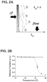
- Figure 3A illustrates the mechanism of selective release.
- an external controlled force was applied on the surface of the chip such that it created a localized shear stress on the surface of the gelatin film.
- the shear force at the surface of the film produced the detachment of the cell.
- Cells released within the release radius were collected at the outlet of the device applying a flow rate of 1.5 to 2.5 ml/h.
- An experimental device for releasing CTCs was built (see Figure 4A ).
- the microtip produced a controlled vibration at the surface of the chip such that the gelatin layer was locally removed by shear stress.
- the sizes of the gelatin layer removed depended on the frequency of vibration and a release radius was defined ( Figure 3B ).
- Nanoparticles were incorporated into the system for the immunoaffinity capture of target cells, e.g., CTCs.
- This capture-layer consisted of a uniform deposition of streptavidin coated nanoparticles (at 7.31 nmole of antibody per 1 mg of nanoparticle) on top of the gelatin film.
- the nanoparticles interacted with the surface by the strong (K d ⁇ 10 -15 M) biotin-streptavidin binding.
- Fluorescently labeled Biotin-RPE was used to determine that the nanoparticles still had available binding sites for capture antibodies and they were not completely embedded on the gelatin film.
- Example 2 A Device for Selectively Releasing Captured Particles in a Gel
- a prototype device for selectively releasing captured particles was built by converting a stereo speaker system into a microtip transducer ( Figure 4A ).
- a microtip of diameter 80 ⁇ m (Warner Instruments) was mounted directly on the center of a copper coil using superglue.
- the microtip transducer was controlled with the presence of two magnetic fields ( Figure 4B ): one permanent magnetic field produced by the magnet; and an alternating magnetic field produced by the cupper coil.
- the alternating magnetic field was created using a function generator (BK Precision) with a fixed voltage peak to peak (VPP) of 20 V.
- the range of the input frequencies was 5 to 60 Hz.
- a sinusoidal input signal was used for all the release experiments.
- the displacement of the microtip transducer was controlled with the input frequency.
- the microtip transducer was mounted in a stage with only movement in the z -direction (to bring it into contact with the Gel-Chip).
- the Gel-Chip was mounted on a motorized microscope stage such that target cells were positioned within the release radius of the microtip.
- PC3 Prostate (PC3), Breast (SKBR3, MDA-MD-231), and lung (H1650) cancer cell lines were obtained from the American Type Culture Collection (ATCC) and expanded according standard cell culture protocols. At 85% cell confluence, cells were stained with cell tracker green (Invitrogen), and then trypsinized. Stained cells were spiked into blood at 1000 cells/ml of healthy donor blood and run through the microfluidic device. For CTC capture and non-specific cell binding quantification, an automatic imaging protocol was used. For quantification of release cells, the microfluidic chip was imaged before and after release and the difference of the counted cells was defined as the percentage of released cells.
- the particle capture system was optimized for maximum isolation of CTCs from whole blood.
- Target PC3 prostate cancer cells were used (1000 cells spiked in 1 ml of blood).
- capture efficiency was 10 % ⁇ 2.34 %.
- an increase in nanoparticles concentration also reflected as an increase in capture efficiency.
- a nanoparticle concentration of 0.11 mg/ml was optimal; producing a capture efficiency of over 95 % ⁇ 1.45 %.
- Further addition of nanoparticle concentration did not increase capture efficiency of the Gel-chip. Specificity of antibody capture on the Gel-Chip was compared with our previous functionalization chemistry protocols ( Stott, S., et.
- the purity of captured cancer cells among contaminating leukocytes was 47.23 % ⁇ 0.34 % for the Gel-Chip, compared to 14.0 % ⁇ 0.1 % for the HB Chip. Additionally, the Gel-Chip offered the advantage of low NSB compared to the HB Chip ( Figure 5D ).
- the overall value for NSB per ml of blood in the Gel-Chip was 1307 ⁇ 376.83 cells, while in the HB-Chip was 3507 ⁇ 676.16 cells for two different flow rates tested (1.2 and 1.5 ml/h, p ⁇ 0.05).
- the Gel-Chip allowed the recovery of capture cells from its surface with two different mechanisms.
- the process had an efficiency of 91.49 % ⁇ 2.32 %, 93.83 % ⁇ 2.89 %, and 97 % ⁇ 4.45 for PC3, H1650, and SKBR3 cells respectively (p > 0.05). Therefore, release efficiency was considered independent of cancer cell phenotype or levels of EpCAM expression (data not shown).
- PC3 cancer cells were individually released and grouped in three categories ( Figure 5F , data not shown).
- the viability of the recovered cells for the different cell groups were over 91.67 ⁇ 1.42 % with no significant difference between groups (p > 0.05), which was consisted with viability values obtained for bulk release ( Figure 5F ).
- Recovered cells were able to grow overtime and form colonies.
- selective release achieved remarkable values of purity, with values up to 89.74 % ⁇ 3.25 % ( Figure 5F ) when cells where released sequentially and many individual events of 100% purity.
- an antibody cocktail of biotinylated anti-EpCAM, anti-HER2, and anti-EGFR was used.
- CTCs were capture in 14 of 16 patient samples (87.5 %). Some samples were analyzed on the Gel-Chip.
- anti-EPCAM, anti-EGFR, anti-MET, anti-SOX2, and anti-WSCK were used as tumor markers, and anti-CD45 as leukocyte marker.
- Nuclear DNA content was stained with 4, 6-diamidino-2-phenylindole or DAPI.
- captured cells on the HB chip were fixed with 4 % paraformaldehyde and washed with PBS immediately following blood processing. The fixed cells were permeabilized with 1 % NP40 and blocked with 2 % normal goat serum / 3 % BSA before the addition of primary antibodies for immunostaining.
- the primary antibodies used for CTC targeting were rabbit wide spectrum anti-cytokeratin (1:100, ABCAM), rabbit anti-MET (1:1000, BD Biosciences), anti-SOX2 (1:50, BD Biosciences) and anti-EGFR (1:200, BD Biosciences).
- Anti-CD45 mIgG1 (1:100, BD Biosciences) was added to target white blood cells.
- secondary immunofluorescent labeled antibodies were added to amplify the signal along with DAPI to label the nuclei.
- the secondary antibodies used were goat anti-rabbit Alexa Flour 488 (1:200, Jackson) and goat anti-mouse IgG1 Alexa Flour 594 (1:200, Jackson). Following staining, the devices were washed with PBS and stored at 4°C.
- CTCs were identified with mouse anti-EpCAM (3:100, Cell Signaling), anti-Cadherin11 (1:10, R&D Systems), and anti-Axl (1:10, R&D Systems) conjugated to Alexa Flour 488.
- White blood cells were targeted with mouse anti-CD45 (1:20, BD Biosciences), anti-CD16 (1:20, BD Biosciences), and anti-CD-14 (1:20, BD Biosciences) conjugated to PE-CF 594.
- CTCs for electron microscopy were fixed with 2 % glutaraldehyde in a 0.2 M sodium cacodylate for 2 hours. Fixed samples were dehydrated in steps in ethanol dilutions. Ethanol was removed using a critical point dryer (Auto Samdri 815 A, Tousimis), and sputtered with gold/palladium for 60 sec at 20 mA (208HR Cressington). Imaging was performed using a field emission scanning electron microscope (SUPRA 55 VP, Zeiss).
- the Gel-Chip allowed the capture of clusters of CTCs from different cancer patients. Cluster of CTCs varied in size, shape, and number of cells. The minimum number of CTCs to be considered as a cluster was four tumor cells. Comparisons of the surface morphology of different CTCs revealed they were very heterogeneous with some CTCs having micrometer-size vesicles attached to them ( Figures 7A-C ).
- the number of captured CTCs for breast cancer patients varied from 0 to 45.43 CTCs/mL, whereas the number was 0 to 6.56 CTCs/mL for lung cancer patients.
- Clusters of CTCs were identified in 37.5 % and 25 % of breast and lung cancer patients, respectively.
- Captured CTCs were released from the Gel-Chip using bulk or selective degradation of the film. Released CTCs were immobilized in PLL coated glass slides and stained for immunofluorescence. The shape, morphology of CTCs as well as their interactions with leukocytes or other CTCs has been maintained during the release process (data not shown). For patients who showed CTC aggregates or clusters, the number of clusters was quantified (single, double, triple or cluster of CTCs).
- H&E Hematoxylin and eosin stain of the primary tumors is performed for each of the four patients (data not shown).
- FNAs fine needle aspirates
- CTCs Single CTCs isolated from the blood of the same breast and lung cancer patients were used for comparison.
- CTCs were captured on the Gel-Chip and then stained with Alexa488-conjugated antibodies against EpCAM, and anti-Cadherin11.
- PE-CF 594-conjugated antibodies against CD14, CD16 and CD45 were used to exclude contaminating leukocytes.
- Selective release was used to recover Alexa488-positive single CTCs without additional steps.
- Genomic DNA was then extracted from the released CTCs with AllPrep® DNA/RNA micro kit (Qiagen) according to the manufacturer's instructions, and subjected to targeted PCR to amplify the region of interest.
- AllPrep® DNA/RNA micro kit Qiagen
- two PCR rounds of 35 cycles each were performed with previously described primers ( Barbi, S. et al. J Exp Clin Cancer Res. 29: 32, 2010 ; Mitsudomi, T. & Yatabe, Y., FEBS Journal 277: 301-308, 2010 ) or primers were designed to be specific for the known mutations to be detected.
Landscapes
- Health & Medical Sciences (AREA)
- Life Sciences & Earth Sciences (AREA)
- Immunology (AREA)
- Engineering & Computer Science (AREA)
- Chemical & Material Sciences (AREA)
- Urology & Nephrology (AREA)
- Biomedical Technology (AREA)
- Hematology (AREA)
- Molecular Biology (AREA)
- Cell Biology (AREA)
- Food Science & Technology (AREA)
- General Health & Medical Sciences (AREA)
- Biotechnology (AREA)
- Medicinal Chemistry (AREA)
- Physics & Mathematics (AREA)
- Analytical Chemistry (AREA)
- Biochemistry (AREA)
- Microbiology (AREA)
- General Physics & Mathematics (AREA)
- Pathology (AREA)
- Chemical Kinetics & Catalysis (AREA)
- Zoology (AREA)
- Tropical Medicine & Parasitology (AREA)
- Virology (AREA)
- Apparatus Associated With Microorganisms And Enzymes (AREA)
- Oncology (AREA)
Claims (16)
- System (50) zum selektiven Einfangen und Freigeben eines oder mehrerer Zielpartikel aus einer flüssigen Probe, wobei das System umfasst:ein Substrat (51);eine erste Gelatineschicht (52), die durch physikalische Adsorption an das Substrat gebunden ist, wobei die Gelatine mit einer Vielzahl von ersten Elementen eines Bindungspaars (54) funktionalisiert ist;eine zweite Gelatineschicht (55), wobei die Gelatine mit einer Vielzahl der ersten Elemente des Bindungspaars funktionalisiert ist und die zweite Schicht über eine Vielzahl von zweiten Elementen des Bindungspaars (56), die mit den ersten Elementen des Bindungspaars sowohl an der ersten als auch an der zweiten Schicht verbunden sind, an die erste Schicht gebunden ist;gegebenenfalls eine oder mehrere nachfolgende Gelatineschichten, die jeweils durch die zweiten Elemente des Bindungspaars an eine vorhergehende Schicht gebunden sind; undeine Vielzahl von Nanostrukturen (58), wobei die Nanostrukturen an die zweiten Elemente des Bindungspaars und an eine oder mehrere partikelbindende Einheiten, die selektiv an die Zielpartikel binden, gebunden sind, wobei die Nanostrukturen durch die zweiten Elemente des Bindungspaars an die oberste Gelatineschicht gebunden sind.
- Verfahren zum selektiven Einfangen und Freigeben eines oder mehrerer Zielpartikel aus einer flüssigen Probe, wobei das Verfahren umfasst:Beschaffen eines Systems gemäß Anspruch 1;Inkontaktbringen der Probe mit dem System, um zu ermöglichen, dass die eine oder mehreren partikelbindenden Einheiten an Zielpartikel in der Probe binden.
- Verfahren gemäß Anspruch 2, ferner umfassend Freigeben der gebundenen Zielpartikel von dem System gemäß Anspruch 1 durch Schmelzen der Gelatineschichten bei einer erhöhten Temperatur.
- Verfahren gemäß Anspruch 3, wobei die erhöhte Temperatur 37 °C beträgt.
- Verfahren gemäß Anspruch 2, ferner umfassend Freigeben der gebundenen Zielpartikel von dem System durch Anwenden einer lokalisierten Scherspannung an eine oder mehrere der Gelatineschichten.
- Verfahren gemäß Anspruch 5, wobei Anwenden einer lokalisierten Scherspannung an eine oder mehrere der Gelatineschichten Anwenden einer frequenzregulierten Kraft an wenigstens die oberste Gelatineschicht umfasst.
- System oder Verfahren gemäß einem der Ansprüche 1 bis 6, wobei die Zielpartikel lebende Zellen oder Mikrovesikel sind.
- System oder Verfahren gemäß Anspruch 7, wobei die lebenden Zellen zirkulierende Tumorzellen sind.
- System oder Verfahren gemäß einem der Ansprüche 1 bis 8, wobei die eine oder mehreren partikelbindenden Einheiten Antikörper umfassen.
- Verfahren gemäß Anspruch 9, wobei die Antikörper anti-EpCAM-, anti-HER2- und anti-EGFR-Antikörper umfassen.
- System oder Verfahren gemäß einem der Ansprüche 1 bis 10, wobei das Substrat einen flachen Objektträger, einen Kanal in einer Mikrofluidikvorrichtung oder ein Mikrokügelchen umfasst.
- Verfahren zum Herstellen eines Zielpartikel-Einfangsystems, wobei das Verfahren umfasst:(a) Beschaffen eines Substrats (51);(b) Bilden einer ersten Gelatineschicht (52) auf dem Substrat durch physikalische Adsorption, wobei die Gelatine mit einer Vielzahl von ersten Elementen eines Bindungspaars (54) funktionalisiert ist;(c) Inkontaktbringen der ersten Gelatineschicht mit zweiten Elementen des Bindungspaars (56);(d) Bilden einer zweiten Gelatineschicht (55), wobei die Gelatine mit einer Vielzahl der ersten Elemente des Bindungspaars funktionalisiert ist und die zweite Schicht über eine Vielzahl von zweiten Elementen des Bindungspaars, die mit den ersten Elementen des Bindungspaars sowohl an der ersten als auch an der zweiten Schicht verbunden sind, an die erste Schicht gebunden wird;(d) gegebenenfalls Bilden einer oder mehrerer nachfolgender Gelatineschichten, die jeweils durch zweite Elemente des Bindungspaars an eine vorhergehende Schicht gebunden werden; und(e) Inkontaktbringen der letzten gebildeten Gelatineschicht mit einer Lösung, die Nanostrukturen (58) umfasst, wobei die Nanostrukturen an die zweiten Elemente des Bindungspaars und an eine oder mehrere partikelbindende Einheiten, die selektiv an die Zielpartikel binden, gebunden werden.
- Verfahren gemäß Anspruch 12, wobei die Zielpartikel lebende Zellen oder Mikrovesikel sind.
- Verfahren gemäß Anspruch 13, wobei die lebenden Zellen zirkulierende Tumorzellen sind.
- Verfahren gemäß einem der Ansprüche 12 bis 14, wobei die eine oder mehreren partikelbindenden Einheiten Antikörper umfassen.
- Verfahren gemäß Anspruch 15, wobei die Antikörper anti-EpCAM-, anti-HER2- und anti-EGFR-Antikörper umfassen.
Applications Claiming Priority (2)
| Application Number | Priority Date | Filing Date | Title |
|---|---|---|---|
| US201361759684P | 2013-02-01 | 2013-02-01 | |
| PCT/US2014/014463 WO2014121204A1 (en) | 2013-02-01 | 2014-02-03 | Capture and release of particles from liquid samples |
Publications (3)
| Publication Number | Publication Date |
|---|---|
| EP2951585A1 EP2951585A1 (de) | 2015-12-09 |
| EP2951585A4 EP2951585A4 (de) | 2016-09-07 |
| EP2951585B1 true EP2951585B1 (de) | 2018-01-03 |
Family
ID=51263029
Family Applications (1)
| Application Number | Title | Priority Date | Filing Date |
|---|---|---|---|
| EP14745825.1A Active EP2951585B1 (de) | 2013-02-01 | 2014-02-03 | Erfassung und freisetzung von teilchen aus flüssigen proben |
Country Status (3)
| Country | Link |
|---|---|
| US (2) | US10551376B2 (de) |
| EP (1) | EP2951585B1 (de) |
| WO (1) | WO2014121204A1 (de) |
Families Citing this family (11)
| Publication number | Priority date | Publication date | Assignee | Title |
|---|---|---|---|---|
| US10551376B2 (en) | 2013-02-01 | 2020-02-04 | The General Hospital Corporation | Capture and release of particles from liquid samples |
| EP3177904A4 (de) | 2014-08-07 | 2018-01-03 | The General Hospital Corporation | Gegen thrombozyten gerichtete mikrofluidische isolierung von zellen |
| CN105486865B (zh) * | 2014-09-15 | 2017-04-19 | 浙江大学 | 一种用于细胞分选和富集的微流控芯片及其应用 |
| CN105734013B (zh) * | 2016-02-02 | 2019-07-05 | 苏州大学 | 目标细胞的多重捕获配体修饰的多层纳米粒柔性支架及其应用 |
| US20200030819A1 (en) * | 2017-03-01 | 2020-01-30 | Cidra Corporate Services Llc | Polymer coating for selective separation of hydrophobic particles in aqueous slurry |
| US11548002B2 (en) | 2017-05-19 | 2023-01-10 | The General Hospital Corporation | Engineered nano-interfaces for microfluidic isolation of extracellular vesicles |
| KR101996218B1 (ko) * | 2017-09-05 | 2019-10-01 | 주식회사 싸이토젠 | Axl 기반 암환자 스크리닝 방법 |
| CN113272648A (zh) * | 2018-10-25 | 2021-08-17 | 萨夫兰技术公司 | 粒子捕获系统和方法 |
| CN112718027B (zh) * | 2020-12-10 | 2022-10-18 | 致慧医疗科技(上海)有限公司 | 微流控芯片和温敏材料复合系统及其制备方法和应用 |
| CN113340859A (zh) * | 2021-05-10 | 2021-09-03 | 武汉大学 | 一种抗体-明胶纳米颗粒修饰的芯片及其制备方法与在选择性分离单个循环肿瘤细胞中的应用 |
| WO2025188758A1 (en) * | 2024-03-04 | 2025-09-12 | Ohio State Innovation Foundation | Light-induced extracellular vesicle and particle adsorption for label-free capture and quantification of molecular cargo |
Family Cites Families (12)
| Publication number | Priority date | Publication date | Assignee | Title |
|---|---|---|---|---|
| US5073341A (en) | 1985-08-21 | 1991-12-17 | Biotope, Inc. | Devices for conducting specific binding assays |
| US5637469A (en) | 1992-05-01 | 1997-06-10 | Trustees Of The University Of Pennsylvania | Methods and apparatus for the detection of an analyte utilizing mesoscale flow systems |
| US20050244843A1 (en) | 2001-11-16 | 2005-11-03 | Wen-Tien Chen | Blood test prototypes and methods for the detection of circulating tumor and endothelial cells |
| US20050221283A1 (en) * | 2001-12-11 | 2005-10-06 | Mahant Vijay K | Biochip |
| US7141369B2 (en) | 2002-04-25 | 2006-11-28 | Semibio Technology, Inc. | Measuring cellular metabolism of immobilized cells |
| US8911957B2 (en) | 2006-03-15 | 2014-12-16 | The General Hospital Corporation | Devices and methods for detecting cells and other analytes |
| US9140697B2 (en) | 2009-03-18 | 2015-09-22 | The Regents Of The University Of California | Device for capturing circulating cells |
| WO2010129283A2 (en) * | 2009-04-27 | 2010-11-11 | The General Hospital Corporation | Microfluidic analyte capture using a thermoflowable material |
| WO2010132795A2 (en) | 2009-05-15 | 2010-11-18 | The General Hospital Corporation | Systems, devices, and methods for specific capture and release of biological sample components |
| US9879310B2 (en) | 2011-01-31 | 2018-01-30 | The Regents Of The University Of California | Nano/microscale vehicles for capture and isolation of target biomolecules and living organisms |
| JP6120778B2 (ja) * | 2011-02-03 | 2017-04-26 | ノースイースタン ユニヴァーシティ | 生物学的材料の極めて特異的な捕獲および遊離のための方法および組成物 |
| US10551376B2 (en) | 2013-02-01 | 2020-02-04 | The General Hospital Corporation | Capture and release of particles from liquid samples |
-
2014
- 2014-02-03 US US14/764,855 patent/US10551376B2/en active Active
- 2014-02-03 EP EP14745825.1A patent/EP2951585B1/de active Active
- 2014-02-03 WO PCT/US2014/014463 patent/WO2014121204A1/en not_active Ceased
-
2020
- 2020-01-30 US US16/777,241 patent/US11971406B2/en active Active
Non-Patent Citations (1)
| Title |
|---|
| None * |
Also Published As
| Publication number | Publication date |
|---|---|
| WO2014121204A1 (en) | 2014-08-07 |
| US20200378964A1 (en) | 2020-12-03 |
| US20150369804A1 (en) | 2015-12-24 |
| EP2951585A4 (de) | 2016-09-07 |
| EP2951585A1 (de) | 2015-12-09 |
| US11971406B2 (en) | 2024-04-30 |
| US10551376B2 (en) | 2020-02-04 |
Similar Documents
| Publication | Publication Date | Title |
|---|---|---|
| US11971406B2 (en) | Capture and release of particles from liquid samples | |
| Dong et al. | Nanostructured substrates for detection and characterization of circulating rare cells: from materials research to clinical applications | |
| Chen et al. | Review on strategies and technologies for exosome isolation and purification | |
| Xu et al. | Magnetic-based microfluidic device for on-chip isolation and detection of tumor-derived exosomes | |
| Lim et al. | Direct isolation and characterization of circulating exosomes from biological samples using magnetic nanowires | |
| Zou et al. | Advances in isolation and detection of circulating tumor cells based on microfluidics | |
| JP5759443B2 (ja) | 循環細胞の捕獲用デバイス | |
| CN106796164B (zh) | 细胞的血小板靶向微流体分离 | |
| US10018632B2 (en) | Microfluidic devices for the capture of biological sample components | |
| US9556485B2 (en) | Methods and compositions for detecting non-hematopoietic cells from a blood sample | |
| Wu et al. | Microfluidic technologies in cell isolation and analysis for biomedical applications | |
| US20070059781A1 (en) | System for size based separation and analysis | |
| US20070059719A1 (en) | Business methods for prenatal Diagnosis | |
| CN103889556A (zh) | 在引入亲和性和大小两者的微流体芯片上的循环肿瘤细胞捕获 | |
| Liu et al. | Nanomaterial-based immunocapture platforms for the recognition, isolation, and detection of circulating tumor cells | |
| Guo et al. | Programmable DNA-responsive microchip for the capture and release of circulating tumor cells by nucleic acid hybridization | |
| Medlock et al. | Cancer bioimprinting and cell shape recognition for diagnosis and targeted treatment | |
| Jiang et al. | Rapid enrichment and detection of extracellular vesicles enabled by CuS-enclosed microgels | |
| KR102029156B1 (ko) | 순환종양세포 검출용 미세유체칩 | |
| Kim et al. | Novel streptavidin-functionalized silicon nanowire arrays for CD4+ T lymphocyte separation | |
| WO2015048315A1 (en) | Biodegradable layer-by-layer (lbl) films for cell capture and release | |
| JP2017512314A (ja) | 末梢循環腫瘍細胞を捕獲するための生体模倣マイクロ流体装置 | |
| Lou et al. | Dual-functional lipid coating for the nanopillar-based capture of circulating tumor cells with high purity and efficiency | |
| Piffoux et al. | Potential of on‐chip analysis and engineering techniques for extracellular vesicle bioproduction for therapeutics | |
| Zhang et al. | Rapid prototyping of nanoroughened polydimethylsiloxane surfaces for the enhancement of immunomagnetic isolation and recovery of rare tumor cells |
Legal Events
| Date | Code | Title | Description |
|---|---|---|---|
| PUAI | Public reference made under article 153(3) epc to a published international application that has entered the european phase |
Free format text: ORIGINAL CODE: 0009012 |
|
| 17P | Request for examination filed |
Effective date: 20150831 |
|
| AK | Designated contracting states |
Kind code of ref document: A1 Designated state(s): AL AT BE BG CH CY CZ DE DK EE ES FI FR GB GR HR HU IE IS IT LI LT LU LV MC MK MT NL NO PL PT RO RS SE SI SK SM TR |
|
| AX | Request for extension of the european patent |
Extension state: BA ME |
|
| DAX | Request for extension of the european patent (deleted) | ||
| A4 | Supplementary search report drawn up and despatched |
Effective date: 20160804 |
|
| RIC1 | Information provided on ipc code assigned before grant |
Ipc: G01N 33/544 20060101ALI20160729BHEP Ipc: G01N 33/543 20060101AFI20160729BHEP Ipc: G01N 33/569 20060101ALI20160729BHEP |
|
| 17Q | First examination report despatched |
Effective date: 20160817 |
|
| GRAP | Despatch of communication of intention to grant a patent |
Free format text: ORIGINAL CODE: EPIDOSNIGR1 |
|
| INTG | Intention to grant announced |
Effective date: 20170724 |
|
| GRAS | Grant fee paid |
Free format text: ORIGINAL CODE: EPIDOSNIGR3 |
|
| GRAA | (expected) grant |
Free format text: ORIGINAL CODE: 0009210 |
|
| AK | Designated contracting states |
Kind code of ref document: B1 Designated state(s): AL AT BE BG CH CY CZ DE DK EE ES FI FR GB GR HR HU IE IS IT LI LT LU LV MC MK MT NL NO PL PT RO RS SE SI SK SM TR |
|
| REG | Reference to a national code |
Ref country code: GB Ref legal event code: FG4D |
|
| REG | Reference to a national code |
Ref country code: CH Ref legal event code: EP Ref country code: AT Ref legal event code: REF Ref document number: 960767 Country of ref document: AT Kind code of ref document: T Effective date: 20180115 |
|
| REG | Reference to a national code |
Ref country code: IE Ref legal event code: FG4D |
|
| REG | Reference to a national code |
Ref country code: DE Ref legal event code: R096 Ref document number: 602014019423 Country of ref document: DE |
|
| REG | Reference to a national code |
Ref country code: FR Ref legal event code: PLFP Year of fee payment: 5 |
|
| REG | Reference to a national code |
Ref country code: NL Ref legal event code: MP Effective date: 20180103 |
|
| REG | Reference to a national code |
Ref country code: LT Ref legal event code: MG4D |
|
| REG | Reference to a national code |
Ref country code: AT Ref legal event code: MK05 Ref document number: 960767 Country of ref document: AT Kind code of ref document: T Effective date: 20180103 |
|
| PG25 | Lapsed in a contracting state [announced via postgrant information from national office to epo] |
Ref country code: NL Free format text: LAPSE BECAUSE OF FAILURE TO SUBMIT A TRANSLATION OF THE DESCRIPTION OR TO PAY THE FEE WITHIN THE PRESCRIBED TIME-LIMIT Effective date: 20180103 |
|
| PG25 | Lapsed in a contracting state [announced via postgrant information from national office to epo] |
Ref country code: ES Free format text: LAPSE BECAUSE OF FAILURE TO SUBMIT A TRANSLATION OF THE DESCRIPTION OR TO PAY THE FEE WITHIN THE PRESCRIBED TIME-LIMIT Effective date: 20180103 Ref country code: FI Free format text: LAPSE BECAUSE OF FAILURE TO SUBMIT A TRANSLATION OF THE DESCRIPTION OR TO PAY THE FEE WITHIN THE PRESCRIBED TIME-LIMIT Effective date: 20180103 Ref country code: NO Free format text: LAPSE BECAUSE OF FAILURE TO SUBMIT A TRANSLATION OF THE DESCRIPTION OR TO PAY THE FEE WITHIN THE PRESCRIBED TIME-LIMIT Effective date: 20180403 Ref country code: CY Free format text: LAPSE BECAUSE OF FAILURE TO SUBMIT A TRANSLATION OF THE DESCRIPTION OR TO PAY THE FEE WITHIN THE PRESCRIBED TIME-LIMIT Effective date: 20180103 Ref country code: LT Free format text: LAPSE BECAUSE OF FAILURE TO SUBMIT A TRANSLATION OF THE DESCRIPTION OR TO PAY THE FEE WITHIN THE PRESCRIBED TIME-LIMIT Effective date: 20180103 Ref country code: HR Free format text: LAPSE BECAUSE OF FAILURE TO SUBMIT A TRANSLATION OF THE DESCRIPTION OR TO PAY THE FEE WITHIN THE PRESCRIBED TIME-LIMIT Effective date: 20180103 |
|
| PG25 | Lapsed in a contracting state [announced via postgrant information from national office to epo] |
Ref country code: BG Free format text: LAPSE BECAUSE OF FAILURE TO SUBMIT A TRANSLATION OF THE DESCRIPTION OR TO PAY THE FEE WITHIN THE PRESCRIBED TIME-LIMIT Effective date: 20180403 Ref country code: GR Free format text: LAPSE BECAUSE OF FAILURE TO SUBMIT A TRANSLATION OF THE DESCRIPTION OR TO PAY THE FEE WITHIN THE PRESCRIBED TIME-LIMIT Effective date: 20180404 Ref country code: IS Free format text: LAPSE BECAUSE OF FAILURE TO SUBMIT A TRANSLATION OF THE DESCRIPTION OR TO PAY THE FEE WITHIN THE PRESCRIBED TIME-LIMIT Effective date: 20180503 Ref country code: SE Free format text: LAPSE BECAUSE OF FAILURE TO SUBMIT A TRANSLATION OF THE DESCRIPTION OR TO PAY THE FEE WITHIN THE PRESCRIBED TIME-LIMIT Effective date: 20180103 Ref country code: LV Free format text: LAPSE BECAUSE OF FAILURE TO SUBMIT A TRANSLATION OF THE DESCRIPTION OR TO PAY THE FEE WITHIN THE PRESCRIBED TIME-LIMIT Effective date: 20180103 Ref country code: RS Free format text: LAPSE BECAUSE OF FAILURE TO SUBMIT A TRANSLATION OF THE DESCRIPTION OR TO PAY THE FEE WITHIN THE PRESCRIBED TIME-LIMIT Effective date: 20180103 Ref country code: AT Free format text: LAPSE BECAUSE OF FAILURE TO SUBMIT A TRANSLATION OF THE DESCRIPTION OR TO PAY THE FEE WITHIN THE PRESCRIBED TIME-LIMIT Effective date: 20180103 Ref country code: PL Free format text: LAPSE BECAUSE OF FAILURE TO SUBMIT A TRANSLATION OF THE DESCRIPTION OR TO PAY THE FEE WITHIN THE PRESCRIBED TIME-LIMIT Effective date: 20180103 |
|
| REG | Reference to a national code |
Ref country code: CH Ref legal event code: PL |
|
| REG | Reference to a national code |
Ref country code: DE Ref legal event code: R097 Ref document number: 602014019423 Country of ref document: DE |
|
| PG25 | Lapsed in a contracting state [announced via postgrant information from national office to epo] |
Ref country code: RO Free format text: LAPSE BECAUSE OF FAILURE TO SUBMIT A TRANSLATION OF THE DESCRIPTION OR TO PAY THE FEE WITHIN THE PRESCRIBED TIME-LIMIT Effective date: 20180103 Ref country code: EE Free format text: LAPSE BECAUSE OF FAILURE TO SUBMIT A TRANSLATION OF THE DESCRIPTION OR TO PAY THE FEE WITHIN THE PRESCRIBED TIME-LIMIT Effective date: 20180103 Ref country code: IT Free format text: LAPSE BECAUSE OF FAILURE TO SUBMIT A TRANSLATION OF THE DESCRIPTION OR TO PAY THE FEE WITHIN THE PRESCRIBED TIME-LIMIT Effective date: 20180103 Ref country code: MC Free format text: LAPSE BECAUSE OF FAILURE TO SUBMIT A TRANSLATION OF THE DESCRIPTION OR TO PAY THE FEE WITHIN THE PRESCRIBED TIME-LIMIT Effective date: 20180103 Ref country code: AL Free format text: LAPSE BECAUSE OF FAILURE TO SUBMIT A TRANSLATION OF THE DESCRIPTION OR TO PAY THE FEE WITHIN THE PRESCRIBED TIME-LIMIT Effective date: 20180103 |
|
| PLBE | No opposition filed within time limit |
Free format text: ORIGINAL CODE: 0009261 |
|
| STAA | Information on the status of an ep patent application or granted ep patent |
Free format text: STATUS: NO OPPOSITION FILED WITHIN TIME LIMIT |
|
| REG | Reference to a national code |
Ref country code: BE Ref legal event code: MM Effective date: 20180228 |
|
| PG25 | Lapsed in a contracting state [announced via postgrant information from national office to epo] |
Ref country code: SK Free format text: LAPSE BECAUSE OF FAILURE TO SUBMIT A TRANSLATION OF THE DESCRIPTION OR TO PAY THE FEE WITHIN THE PRESCRIBED TIME-LIMIT Effective date: 20180103 Ref country code: CZ Free format text: LAPSE BECAUSE OF FAILURE TO SUBMIT A TRANSLATION OF THE DESCRIPTION OR TO PAY THE FEE WITHIN THE PRESCRIBED TIME-LIMIT Effective date: 20180103 Ref country code: LI Free format text: LAPSE BECAUSE OF NON-PAYMENT OF DUE FEES Effective date: 20180228 Ref country code: CH Free format text: LAPSE BECAUSE OF NON-PAYMENT OF DUE FEES Effective date: 20180228 Ref country code: LU Free format text: LAPSE BECAUSE OF NON-PAYMENT OF DUE FEES Effective date: 20180203 Ref country code: DK Free format text: LAPSE BECAUSE OF FAILURE TO SUBMIT A TRANSLATION OF THE DESCRIPTION OR TO PAY THE FEE WITHIN THE PRESCRIBED TIME-LIMIT Effective date: 20180103 Ref country code: SM Free format text: LAPSE BECAUSE OF FAILURE TO SUBMIT A TRANSLATION OF THE DESCRIPTION OR TO PAY THE FEE WITHIN THE PRESCRIBED TIME-LIMIT Effective date: 20180103 |
|
| 26N | No opposition filed |
Effective date: 20181005 |
|
| PG25 | Lapsed in a contracting state [announced via postgrant information from national office to epo] |
Ref country code: BE Free format text: LAPSE BECAUSE OF NON-PAYMENT OF DUE FEES Effective date: 20180228 Ref country code: SI Free format text: LAPSE BECAUSE OF FAILURE TO SUBMIT A TRANSLATION OF THE DESCRIPTION OR TO PAY THE FEE WITHIN THE PRESCRIBED TIME-LIMIT Effective date: 20180103 |
|
| PG25 | Lapsed in a contracting state [announced via postgrant information from national office to epo] |
Ref country code: MT Free format text: LAPSE BECAUSE OF NON-PAYMENT OF DUE FEES Effective date: 20180203 |
|
| PG25 | Lapsed in a contracting state [announced via postgrant information from national office to epo] |
Ref country code: TR Free format text: LAPSE BECAUSE OF FAILURE TO SUBMIT A TRANSLATION OF THE DESCRIPTION OR TO PAY THE FEE WITHIN THE PRESCRIBED TIME-LIMIT Effective date: 20180103 |
|
| PG25 | Lapsed in a contracting state [announced via postgrant information from national office to epo] |
Ref country code: PT Free format text: LAPSE BECAUSE OF FAILURE TO SUBMIT A TRANSLATION OF THE DESCRIPTION OR TO PAY THE FEE WITHIN THE PRESCRIBED TIME-LIMIT Effective date: 20180103 |
|
| PG25 | Lapsed in a contracting state [announced via postgrant information from national office to epo] |
Ref country code: MK Free format text: LAPSE BECAUSE OF NON-PAYMENT OF DUE FEES Effective date: 20180103 Ref country code: HU Free format text: LAPSE BECAUSE OF FAILURE TO SUBMIT A TRANSLATION OF THE DESCRIPTION OR TO PAY THE FEE WITHIN THE PRESCRIBED TIME-LIMIT; INVALID AB INITIO Effective date: 20140203 |
|
| P01 | Opt-out of the competence of the unified patent court (upc) registered |
Effective date: 20230528 |
|
| P02 | Opt-out of the competence of the unified patent court (upc) changed |
Effective date: 20230530 |
|
| PGFP | Annual fee paid to national office [announced via postgrant information from national office to epo] |
Ref country code: GB Payment date: 20260227 Year of fee payment: 13 |
|
| PGFP | Annual fee paid to national office [announced via postgrant information from national office to epo] |
Ref country code: DE Payment date: 20260227 Year of fee payment: 13 Ref country code: IE Payment date: 20260227 Year of fee payment: 13 |
|
| PGFP | Annual fee paid to national office [announced via postgrant information from national office to epo] |
Ref country code: FR Payment date: 20260225 Year of fee payment: 13 |