EP2957912A1 - Verfahren zur Markierung eines Zielmoleküls als Teil einer Korona von Molekülen auf der Oberfläche eines Objekts im Nanogrößenbereich - Google Patents
Verfahren zur Markierung eines Zielmoleküls als Teil einer Korona von Molekülen auf der Oberfläche eines Objekts im Nanogrößenbereich Download PDFInfo
- Publication number
- EP2957912A1 EP2957912A1 EP14172854.3A EP14172854A EP2957912A1 EP 2957912 A1 EP2957912 A1 EP 2957912A1 EP 14172854 A EP14172854 A EP 14172854A EP 2957912 A1 EP2957912 A1 EP 2957912A1
- Authority
- EP
- European Patent Office
- Prior art keywords
- target molecule
- nanosized object
- nanosized
- molecules
- corona
- Prior art date
- Legal status (The legal status is an assumption and is not a legal conclusion. Google has not performed a legal analysis and makes no representation as to the accuracy of the status listed.)
- Withdrawn
Links
- 238000000034 method Methods 0.000 title claims abstract description 101
- 238000002372 labelling Methods 0.000 title claims abstract description 44
- 239000002105 nanoparticle Substances 0.000 claims abstract description 116
- 239000000523 sample Substances 0.000 claims abstract description 62
- 238000009826 distribution Methods 0.000 claims abstract description 17
- 102000004169 proteins and genes Human genes 0.000 claims description 43
- 108090000623 proteins and genes Proteins 0.000 claims description 43
- 230000000704 physical effect Effects 0.000 claims description 35
- 230000008859 change Effects 0.000 claims description 32
- 230000027455 binding Effects 0.000 claims description 15
- 229920006395 saturated elastomer Polymers 0.000 claims description 10
- 238000004062 sedimentation Methods 0.000 claims description 8
- 230000005389 magnetism Effects 0.000 claims description 7
- 102000008394 Immunoglobulin Fragments Human genes 0.000 claims description 4
- 108010021625 Immunoglobulin Fragments Proteins 0.000 claims description 4
- 239000003446 ligand Substances 0.000 claims description 4
- 238000011534 incubation Methods 0.000 claims description 3
- 238000012544 monitoring process Methods 0.000 claims description 3
- 239000004793 Polystyrene Substances 0.000 description 36
- 239000002245 particle Substances 0.000 description 35
- 102000004338 Transferrin Human genes 0.000 description 22
- 108090000901 Transferrin Proteins 0.000 description 22
- 229920002223 polystyrene Polymers 0.000 description 22
- 239000012581 transferrin Substances 0.000 description 22
- 102000007238 Transferrin Receptors Human genes 0.000 description 17
- 108010033576 Transferrin Receptors Proteins 0.000 description 17
- PCHJSUWPFVWCPO-UHFFFAOYSA-N gold Chemical compound [Au] PCHJSUWPFVWCPO-UHFFFAOYSA-N 0.000 description 13
- 229910052737 gold Inorganic materials 0.000 description 12
- 239000010931 gold Substances 0.000 description 12
- 238000004458 analytical method Methods 0.000 description 8
- 238000001493 electron microscopy Methods 0.000 description 8
- 230000005291 magnetic effect Effects 0.000 description 8
- 102000005962 receptors Human genes 0.000 description 8
- 108020003175 receptors Proteins 0.000 description 8
- 238000000799 fluorescence microscopy Methods 0.000 description 7
- 238000003384 imaging method Methods 0.000 description 7
- 230000003993 interaction Effects 0.000 description 7
- 238000005259 measurement Methods 0.000 description 7
- 239000002096 quantum dot Substances 0.000 description 7
- 239000000126 substance Substances 0.000 description 7
- 238000001327 Förster resonance energy transfer Methods 0.000 description 6
- 108010020147 Protein Corona Proteins 0.000 description 6
- 238000004630 atomic force microscopy Methods 0.000 description 6
- 238000002292 fluorescence lifetime imaging microscopy Methods 0.000 description 6
- 239000000463 material Substances 0.000 description 6
- 230000009467 reduction Effects 0.000 description 6
- 238000004626 scanning electron microscopy Methods 0.000 description 6
- 238000001350 scanning transmission electron microscopy Methods 0.000 description 6
- 238000004574 scanning tunneling microscopy Methods 0.000 description 6
- 239000002086 nanomaterial Substances 0.000 description 5
- 108090000765 processed proteins & peptides Proteins 0.000 description 5
- 238000004448 titration Methods 0.000 description 5
- 238000000684 flow cytometry Methods 0.000 description 4
- 238000004949 mass spectrometry Methods 0.000 description 4
- 238000000386 microscopy Methods 0.000 description 4
- 239000000203 mixture Substances 0.000 description 4
- 230000003287 optical effect Effects 0.000 description 4
- 230000005540 biological transmission Effects 0.000 description 3
- 239000003795 chemical substances by application Substances 0.000 description 3
- 239000000975 dye Substances 0.000 description 3
- 238000001433 helium-ion microscopy Methods 0.000 description 3
- 230000009871 nonspecific binding Effects 0.000 description 3
- 102000004196 processed proteins & peptides Human genes 0.000 description 3
- 238000000954 titration curve Methods 0.000 description 3
- XEEYBQQBJWHFJM-UHFFFAOYSA-N Iron Chemical compound [Fe] XEEYBQQBJWHFJM-UHFFFAOYSA-N 0.000 description 2
- PXHVJJICTQNCMI-UHFFFAOYSA-N Nickel Chemical compound [Ni] PXHVJJICTQNCMI-UHFFFAOYSA-N 0.000 description 2
- VYPSYNLAJGMNEJ-UHFFFAOYSA-N Silicium dioxide Chemical compound O=[Si]=O VYPSYNLAJGMNEJ-UHFFFAOYSA-N 0.000 description 2
- CZMRCDWAGMRECN-UGDNZRGBSA-N Sucrose Chemical compound O[C@H]1[C@H](O)[C@@H](CO)O[C@@]1(CO)O[C@@H]1[C@H](O)[C@@H](O)[C@H](O)[C@@H](CO)O1 CZMRCDWAGMRECN-UGDNZRGBSA-N 0.000 description 2
- 229930006000 Sucrose Natural products 0.000 description 2
- 238000003917 TEM image Methods 0.000 description 2
- 238000000441 X-ray spectroscopy Methods 0.000 description 2
- 238000013459 approach Methods 0.000 description 2
- 230000001413 cellular effect Effects 0.000 description 2
- 238000012512 characterization method Methods 0.000 description 2
- 239000002872 contrast media Substances 0.000 description 2
- 230000009260 cross reactivity Effects 0.000 description 2
- 238000001514 detection method Methods 0.000 description 2
- 238000000635 electron micrograph Methods 0.000 description 2
- 238000004020 luminiscence type Methods 0.000 description 2
- 238000013507 mapping Methods 0.000 description 2
- 238000012986 modification Methods 0.000 description 2
- 230000004048 modification Effects 0.000 description 2
- 239000002159 nanocrystal Substances 0.000 description 2
- 108020004707 nucleic acids Proteins 0.000 description 2
- 102000039446 nucleic acids Human genes 0.000 description 2
- 150000007523 nucleic acids Chemical class 0.000 description 2
- 210000003463 organelle Anatomy 0.000 description 2
- 238000000926 separation method Methods 0.000 description 2
- 230000009870 specific binding Effects 0.000 description 2
- 238000004611 spectroscopical analysis Methods 0.000 description 2
- 239000005720 sucrose Substances 0.000 description 2
- 235000000346 sugar Nutrition 0.000 description 2
- 238000012800 visualization Methods 0.000 description 2
- 238000005406 washing Methods 0.000 description 2
- 108091023037 Aptamer Proteins 0.000 description 1
- 102000004506 Blood Proteins Human genes 0.000 description 1
- 108010017384 Blood Proteins Proteins 0.000 description 1
- KCXVZYZYPLLWCC-UHFFFAOYSA-N EDTA Chemical compound OC(=O)CN(CC(O)=O)CCN(CC(O)=O)CC(O)=O KCXVZYZYPLLWCC-UHFFFAOYSA-N 0.000 description 1
- 108090001090 Lectins Proteins 0.000 description 1
- 102000004856 Lectins Human genes 0.000 description 1
- 108090001030 Lipoproteins Proteins 0.000 description 1
- 102000004895 Lipoproteins Human genes 0.000 description 1
- BQCADISMDOOEFD-UHFFFAOYSA-N Silver Chemical compound [Ag] BQCADISMDOOEFD-UHFFFAOYSA-N 0.000 description 1
- 108010090804 Streptavidin Proteins 0.000 description 1
- 101710120037 Toxin CcdB Proteins 0.000 description 1
- 241000700605 Viruses Species 0.000 description 1
- 230000002776 aggregation Effects 0.000 description 1
- 238000004220 aggregation Methods 0.000 description 1
- 230000004075 alteration Effects 0.000 description 1
- 125000000539 amino acid group Chemical group 0.000 description 1
- 238000003556 assay Methods 0.000 description 1
- 239000011173 biocomposite Substances 0.000 description 1
- 230000004071 biological effect Effects 0.000 description 1
- 239000013060 biological fluid Substances 0.000 description 1
- 239000012620 biological material Substances 0.000 description 1
- 230000008512 biological response Effects 0.000 description 1
- 230000015572 biosynthetic process Effects 0.000 description 1
- 230000000903 blocking effect Effects 0.000 description 1
- 238000011088 calibration curve Methods 0.000 description 1
- 125000003178 carboxy group Chemical group [H]OC(*)=O 0.000 description 1
- 210000004027 cell Anatomy 0.000 description 1
- 230000033077 cellular process Effects 0.000 description 1
- 238000005119 centrifugation Methods 0.000 description 1
- 125000003636 chemical group Chemical group 0.000 description 1
- 229910017052 cobalt Inorganic materials 0.000 description 1
- 239000010941 cobalt Substances 0.000 description 1
- GUTLYIVDDKVIGB-UHFFFAOYSA-N cobalt atom Chemical compound [Co] GUTLYIVDDKVIGB-UHFFFAOYSA-N 0.000 description 1
- 239000003086 colorant Substances 0.000 description 1
- 239000002131 composite material Substances 0.000 description 1
- 238000010276 construction Methods 0.000 description 1
- 238000013461 design Methods 0.000 description 1
- 229940079593 drug Drugs 0.000 description 1
- 239000003814 drug Substances 0.000 description 1
- 210000001163 endosome Anatomy 0.000 description 1
- 238000005516 engineering process Methods 0.000 description 1
- 210000001808 exosome Anatomy 0.000 description 1
- 239000003302 ferromagnetic material Substances 0.000 description 1
- 238000001506 fluorescence spectroscopy Methods 0.000 description 1
- 239000007850 fluorescent dye Substances 0.000 description 1
- 210000000987 immune system Anatomy 0.000 description 1
- 238000003119 immunoblot Methods 0.000 description 1
- 238000003126 immunogold labeling Methods 0.000 description 1
- 230000008676 import Effects 0.000 description 1
- 238000011835 investigation Methods 0.000 description 1
- 229910052742 iron Inorganic materials 0.000 description 1
- WTFXARWRTYJXII-UHFFFAOYSA-N iron(2+);iron(3+);oxygen(2-) Chemical compound [O-2].[O-2].[O-2].[O-2].[Fe+2].[Fe+3].[Fe+3] WTFXARWRTYJXII-UHFFFAOYSA-N 0.000 description 1
- 239000002523 lectin Substances 0.000 description 1
- 239000002502 liposome Substances 0.000 description 1
- 238000000504 luminescence detection Methods 0.000 description 1
- 238000005404 magnetometry Methods 0.000 description 1
- 238000000838 magnetophoresis Methods 0.000 description 1
- 229910052751 metal Inorganic materials 0.000 description 1
- 239000002184 metal Substances 0.000 description 1
- 150000002739 metals Chemical class 0.000 description 1
- 210000003470 mitochondria Anatomy 0.000 description 1
- 229940031182 nanoparticles iron oxide Drugs 0.000 description 1
- 229910052759 nickel Inorganic materials 0.000 description 1
- MUJIDPITZJWBSW-UHFFFAOYSA-N palladium(2+) Chemical class [Pd+2] MUJIDPITZJWBSW-UHFFFAOYSA-N 0.000 description 1
- 238000000059 patterning Methods 0.000 description 1
- HRGDZIGMBDGFTC-UHFFFAOYSA-N platinum(2+) Chemical compound [Pt+2] HRGDZIGMBDGFTC-UHFFFAOYSA-N 0.000 description 1
- 229920000642 polymer Polymers 0.000 description 1
- 229920001184 polypeptide Polymers 0.000 description 1
- 150000004032 porphyrins Chemical class 0.000 description 1
- 230000008569 process Effects 0.000 description 1
- 238000012545 processing Methods 0.000 description 1
- 238000003908 quality control method Methods 0.000 description 1
- 230000001105 regulatory effect Effects 0.000 description 1
- 238000011160 research Methods 0.000 description 1
- 238000009738 saturating Methods 0.000 description 1
- 239000000377 silicon dioxide Substances 0.000 description 1
- 229910052709 silver Inorganic materials 0.000 description 1
- 239000004332 silver Substances 0.000 description 1
- 238000002415 sodium dodecyl sulfate polyacrylamide gel electrophoresis Methods 0.000 description 1
- 238000001179 sorption measurement Methods 0.000 description 1
- 150000008163 sugars Chemical class 0.000 description 1
- 230000003746 surface roughness Effects 0.000 description 1
- 238000003786 synthesis reaction Methods 0.000 description 1
- 230000008685 targeting Effects 0.000 description 1
- 238000004627 transmission electron microscopy Methods 0.000 description 1
- 238000010200 validation analysis Methods 0.000 description 1
Images




Classifications
-
- G—PHYSICS
- G01—MEASURING; TESTING
- G01N—INVESTIGATING OR ANALYSING MATERIALS BY DETERMINING THEIR CHEMICAL OR PHYSICAL PROPERTIES
- G01N33/00—Investigating or analysing materials by specific methods not covered by groups G01N1/00 - G01N31/00
- G01N33/48—Biological material, e.g. blood, urine; Haemocytometers
- G01N33/50—Chemical analysis of biological material, e.g. blood, urine; Testing involving biospecific ligand binding methods; Immunological testing
- G01N33/53—Immunoassay; Biospecific binding assay; Materials therefor
- G01N33/543—Immunoassay; Biospecific binding assay; Materials therefor with an insoluble carrier for immobilising immunochemicals
- G01N33/54313—Immunoassay; Biospecific binding assay; Materials therefor with an insoluble carrier for immobilising immunochemicals the carrier being characterised by its particulate form
- G01N33/54346—Nanoparticles
Definitions
- the invention relates to a method of labelling a target molecule forming part of a corona of molecules on a surface of a nanosized object.
- the invention relates to a method of determining a spatial characteristic, or an abundance, of at least one target molecule forming part of a corona of molecules on a surface of a nanosized object.
- nanoparticles in a biological milieu form a 'corona' of biomolecules which may be sufficiently long lived, and appropriately organised, to confer key aspects of biological identity to the particle.
- hard corona the most tightly nanoparticlebound biomolecules
- mass spectrometry yielding a macroscopically averaged composition of the biomolecules on the particles.
- Other methods of analysing the protein corona include X-ray spectroscopy, mass spectrometry and array based studies.
- X-ray spectroscopy is a time consuming and technically difficult method, which cannot be carried out in high-throughput.
- Mass spectrometry yields a macroscopically averaged composition of the biomoleulces attached, however, gives no indication about the arrangement or even composition on a single particle basis.
- Array-based studies are difficult to achieve for nanoparticles; due to the fact that the population is mixed and it is necessary to select for particles which interact, and as such it is not representative of the whole sample.
- the invention provides techniques and methods for labelling and subsequently visualising a corona of molecules, generally visualising the biomolecules on the surface of nanoparticles such as are found in a protein corona formed on the surface of a nano-sized object such as a nanoparticle, or bio-functionalised nanomaterials, or a nano-sized biomolecule or hybrid bio-material.
- the methods of the invention involve labelling specific molecules in the corona (or specific parts of molecules such as epitopes) with probes comprising small nanoparticles with a recognition motif for the target molecule attached. Once labelled, the corona of the nanoparticle can be imaged to determine the spatial characteristics of the corona, for example the location or spatial distribution of target molecule.
- a multi parametric fingerprint of the nanomaterial can be constructed with varying degrees of detail ranging from single protein distributions to multi correlative patterns which describe the biological identity of the nanosurface. In this instance the latter is of high importance to describe the biological surface of nanomaterials for regulatory or quality control purposes.
- biomolecular corona or biological identity of nanoparticles Due to the highly sensitive nature of the biomolecular corona or biological identity of nanoparticles, which in most cases is context specific, it provides us with a unique opportunity to use the distribution of biomolecules across the surface as a characterisation tool assessing in higher detail the reproducibility of nanoparticles, of high import for the regulation of such materials. Moreover the flexibility of this platform allows one to target specific key amino acid residues, or epitopes, which are known to cause specific biological responses, e.g. epitopes which engage cellular receptors facilitating uptake, alert the immune system to potential hazards or trigger other biological effects.
- the abundance of the target molecule on the nano-sized object can optionally be quantified by determining a change in physical property e.g. density, once the nanoparticle are labelled, typically by means of density centrifugal sedimentation.
- the relative abundance of two target molecules forming part of the corona can be determined by determining using contrast in the same physical property or using multiple labels of different types, conferring contrast in multiple parameters e.g. density, fluorescence magnetism etc. Suitable design of the small nanoparticle probe allows specific epitopes of a target protein to be identified and mapped out.
- the invention thus provides a method for obtaining information on the location, distribution and orientation of molecules in a nanoparticle corona.
- the technique is versatile and fast, and provides a quick method for identification of proteins in a protein corona, with the option of further detailed analysis of these proteins.
- the interaction of biomolecules whilst on a nanoparticle surface can be investigated.
- the invention provides a method of labelling a target molecule forming part of a corona of molecules on a nanosized object, the method comprising the steps of:
- the small nanoparticle is configured to act as a contrast agent through one or more parameters such as size, shape, density, fluorescence, charge or magnetism, enabling the visualisation of the small nanoparticle on the surface of the nano-sized object.
- imaging techniques may be employed including electron microscopy, for example TEM (transmission, electron microscopy), SEM (scanning electron microscopy), Helium Ion microscopy.
- AFM Atomic force microscopy
- STM scanning tunnelling microscopy
- fluorescence imaging and advanced fluorescence techniques such as Förster resonance energy transfer (FRET) and fluorescence lifetime imaging microscopy (FLIM), as well as super resolution fluorescence imaging techniques e.g. Stochastic optical reconstruction microscopy (STORM) and Stimulated emission depletion (STED) can be used.
- the corona is a biomolecular corona, and in which the recognition motif is generally a biological recognition motif.
- the target molecule is a protein or an epitope of a protein.
- the biological recognition motif is an antibody, antibody fragment or peptide.
- the nanosized object is a nanoparticle.
- the method involves labelling a specific part of the target molecule, for example a specific epitope of the target molecule.
- the recognition motif is specific for the epitope.
- Suitable recognition motifs for labelling specific epitopes of target molecules include monoclonal antibodies and peptides, and other biomolecules with defined interactions e.g. lectins and receptors.
- the method comprises a step of monitoring a change in a physical property of the nanosized object during the incubation step to determine when the nanosized object is saturated with probe, wherein the separating step is generally carried out after the nanosized object has been determined to be saturated with probe.
- the physical property is selected from density, light emission, magnetism, or size.
- the physical property is density, in which case the small nanoparticle is a heavy probe e.g. gold.
- the term “heavy probe” should be understood to mean a small nanoparticle capable of increasing the net density of the large nanoparticle once bound, typically these particles are dense metals.
- a "light probe” may be employed.
- the term “light probe” is defined as a small nanoparticle of lower density than the large nanoparticle under analysis, capable of reducing the net density once bound.
- the change is density is determined using centrifugal sedimentation, ideally density centrifugal sedimentation (DCS).
- the physical property is light emission, in which the small nanoparticle is a fluorescent or luminescent probe.
- probes include probes in which the small nanoparticle emits light, for example nanocrystals, or small nanoparticles labelled with a light emitting moiety, for example a molecular beacon or a fluorescent or phosphorescent dye.
- dyes include platinum (II) or palladium (II) complexes of porphyrin dyes, organic molecules and luminescent biomolecules.
- the change is light emission is determined using fluorescence/ luminescence spectroscopy or an equivalent technique, the details of which will be known to a person skilled in the art.
- the physical property is magnetism, in which case the small nanoparticle has a magnetic character, for example a magnetic core or a magnetic shell.
- magnetic probes include superparamagnetic Iron oxide nanoparticles, or other ferromagnetic materials such as are derived from Iron, Cobalt or Nickel.
- the change in magnetism is determined by magnetometry or magnetophoresis.
- the physical property is the scattering of the particle in which small nanoparticles alter the way in which large nanoparticles scatter light.
- This change in scattered light can indicate a change in size, or morphology of the nanoparticle e.g. roughness.
- Each of the probes discussed previously could be used to change the scattering profile of large nanoparticles.
- the change in scattering is measured using Flow Cytometry.
- the small nanoparticles have a diameter of less than 20 nm, 15nm, 14nm, 13nm, 12nm, 11nm, 10nm, 9mm, 8nm, 7nm, 6nm, or 5nm.
- the small nanoparticles have a dimension of at least 0.1nm, 0.5nm, or 1.0nm.
- the method of measuring the diameter (or dimension) of small nanoparticles (or nanosized objects) is described in National Institute of Standards and Technology (NIST) - NCL Joint Assay Protocol, PCC-7
- the invention also provides a method of labelling at least first and second target molecules forming part of a corona of molecules on a nanosized object, the method comprising the steps of:
- the identification and separation of the labelled large nanoparticle is preferably carried out by, so called "multi dimensional” e.g. 2D, 3D techniques, where it is separated in one dimension using contrast in the first physical property and in another dimension with contrast in a second physical property.
- multi dimensional e.g. 2D, 3D techniques
- the number of dimensions is directly proportional to the number of probes conferring different physical properties.
- the invention also provides a method of determining a spatial characteristic of at least one target molecule forming part of a corona of molecules on a nanosized object, the method comprising the steps of:
- the invention also provides a method of determining a spatial characteristic of at least first and second target molecules forming part of a corona of molecules on a nanosized object, the method comprising the steps of:
- the invention also provides a method of determining a spatial characteristic of at least first and second target molecules forming part of a corona of molecules on a nanosized object, the method comprising the steps of:
- one probe comprises a first small nanoparticle having a first density and the second probe comprises a second small nanoparticle having a different density to the first small nanoparticle.
- one probe comprises a first small nanoparticle having first magnetic characteristic and the second probe comprises a second small nanoparticle having a different magnetic characteristic to the first small nanoparticle.
- one probe comprises a first small nanoparticle having a first density and the second probe comprises a second small nanoparticle having a fluorescence, specific combinations of probes are not limited to the examples listed here.
- the spatial characteristic is selected from the spatial location of the target molecule on the surface of, or attached to the nanosized object, spatial distribution of the target molecules on the surface of the nanosized object, either in relation to other molecules of the same class or, using multiple probes, in relation to molecules of different classes, and orientation of the target molecule on the surface of the nanosized object.
- the image is generated by microscopy, preferably electron microscopy, for example TEM (transmission, electron microscopy), SEM (scanning electron microscopy), and Helium Ion microscopy.
- microscopy preferably electron microscopy, for example TEM (transmission, electron microscopy), SEM (scanning electron microscopy), and Helium Ion microscopy.
- Other methods of generating an image include AFM (Atomic force microscopy), STM (scanning tunnelling microscopy), fluorescence imaging including advanced fluorescence techniques such as Förster resonance energy transfer (FRET) and fluorescence lifetime imaging microscopy (FLIM), as well as super resolution fluorescence imaging techniques e.g. Stochastic optical reconstruction microscopy (STORM) and Stimulated emission depletion (STED).
- FRET Förster resonance energy transfer
- FLIM fluorescence lifetime imaging microscopy
- TRANSM Stochastic optical reconstruction microscopy
- STED Stimulated emission depletion
- the small nanoparticle is a heavy probe, for example gold.
- the probe has a fluorescence or luminescence characteristic, for example a nanocrystal or a small nanoparticle labelled with a fluorescent probe.
- the probe is at least partially magnetic.
- the first target molecule is a first a protein
- the second target molecule is a second protein
- the first target molecule is a first epitope of a protein
- the second target molecule is a second epitope of the same protein
- the first target molecule is a first epitope of a protein
- the second target molecule is a different epitope of a different protein
- the invention also provides a method of determining the abundance of at least one target molecule forming part of a corona of molecules on a surface of a nanosized object, the method comprising the steps of:
- the invention also provides a method of determining relative abundance of at least first and second target molecules forming part of a corona of molecules on a surface of a nanosized object, the method comprising the steps of:
- the physical property is selected from density, light emission, charge, magnetism, or size. Examples of methods to determine the change in these physical properties, and suitable probes to employ, are described in more detail above.
- the invention also relates to a method of mapping out the distribution of epitopes on a nanoparticle surface comprising the steps of determining the determining the spatial location of each of the epitopes on the nanoparticle surface according to a method of the invention.
- nanosized object should be understood to mean objects having a size range of 10-5000nm, typically 10-1000nm, 200-1000nm, 300-1000nm, 400-1000nm or 500nm to 1000nm, including nanoparticles and sub-micron sized biological molecules and clusters including virus, cellular organelles (for example endosomes, exosomes, mitochondria), and biological complexes (for example lipoprotein molecules).
- nanoparticles include polymer nanoparticles such as polystyrene, inorganic nanoparticles, for example silica, gold or silver, composite and biocomposite nanoparticles including hybrid materials, combining a biological and synthetic element, and biological objects on the nanoscale eg. liposomes and organelles.
- small nanoparticle should be understood to mean nanoparticles that have a dimension not greater than 20% of the dimension of the nanosized object to be labelled, and having a size range of 0.1 to 20nm.
- suitable small nanoparticles are colloidal gold, quantum dots, superparamagnetic nanoparticles, nanoclusters and nanostructures.
- the small nanoparticle will have a dimension that is not greater than 10% or 5% of the dimension of the nano-sized object.
- the term "corona of molecules” should be understood to mean the molecules that adhere to the surface of a nano-sized object.
- the molecules are biomolecules.
- the molecules are protein in which case the corona is referred to as a protein corona.
- the corona of molecules may include other biomolecules (for example monomeric and polymeric sugar molecules, and nucleic acids), chemical molecules, or a mixture of biomolecule and chemical molecules.
- the corona of molecules will comprise a plurality of different molecules, for example a multiplicity of proteins. For example, when a nanoparticle is located in a biological fluid comprising a milieu of biomolecules, a corona of the biomolecules comprising predominantly protein molecules will form on the surface.
- target molecule should be understood to mean a specific biomolecule or chemical molecule that forms part of the corona of molecules on the curved surface of the nano-sized object.
- target molecules include specific proteins, peptides, polypeptides, sugars, nucleic acids, and chemical molecules, or a conjugate of any of these.
- the target molecule a a protein.
- the methods of the invention comprise labelling a specific part of a target molecule, for example a specific epitope of the target molecule.
- the term "recognition motif specific for the target molecule” refers to a ligand forming part of the probe that is capable of specific binding with the target molecule.
- the recognition motif may be a biomolecule or a chemical molecule.
- the recognition motif is a biological recognition motif, for example an antibody or antibody fragment, protein, peptide, or aptamer.
- epitope should be understood to mean an exposed part of a protein that is recognised by an antibody or antibody fragment.
- a protein will have a number of different epitopes.
- the method of the invention comprises separating the nano-sized objects from unbound probe. Methods for separation will be apparent to those skilled in the art, and generally include washing the nano-sized objects to remove the excess probe.
- imaging or “generating an image” should be understood to mean any technique in which the surface of the nanosized object can be visualised such that bound probe can be differentiated from the surface of the nanosized object (i.e. the probe acts as an imaging contrast agent.
- imaging techniques include, but are not limited to , electron microscopy for example TEM (transmission, electron microscopy), SEM (scanning electron microscopy), and Helium Ion microscopy.
- AFM Atomic force microscopy
- STM scanning tunnelling microscopy
- fluorescence imaging including advanced fluorescence techniques such as Förster resonance energy transfer (FRET) and fluorescence lifetime imaging microscopy (FLIM), as well as super resolution fluorescence imaging techniques e.g. Stochastic optical reconstruction microscopy (STORM) and Stimulated emission depletion (STED).
- FRET Förster resonance energy transfer
- FLIM fluorescence lifetime imaging microscopy
- TRANSM Stochastic optical reconstruction microscopy
- STED Stimulated emission depletion
- the small nanoparticles are labelled with the recognition motif using techniques that are known to the person skilled in the art, for example by covalent attachment through the use of linkers or short molecules to couple the small nanoparticle and the recognition motif, or through modification of the small nanoparticle, or recognition motif to allow specific attachment, direct attachment to the surface of the nanoparticle, adsorbing the recognition motif.
- modification include modifying the chemical groups of the small nanoparticle to facilitate specific chemical attachment, modifying the surface of the small nanoparticle with a biological molecule e.g. streptavidin, protein G, Protein A to facilitate attachment of the recognition motif.
- Small gold nanoparticles were produced according to the Slot-Geuze method. [ Slot, J. W. & Geuze, H. J. A new method of preparing gold probes for multiple-labeling cytochemistry. European journal of cell biology 38, 87-93 (1985 ).] The particles were characterised using UV-visible spectroscopy, Differential Centrifugal Sedimentation and Transmission Electron Microscopy.
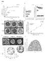
- the Transferrin coated polystyrene was saturated with IG-mTf and washed free of excess gold by three cycles of centrifugation at 20000 x g for 10 min. These particles were subsequently titrated with IG-pTf. Analysis of the washed samples indicated that negligible gold was removed from the surface during the washing procedure ( Fig.2a ).
- Two controls were used to check the level of nonspecific binding of gold nanoparticles. The first was IG with no antibodies in which the surface was blocked with BSA (IG-BSA). The second control used an antibody raised against R-phycoetherin (IG-cAb), carefully chosen to have minimal cross-reactivity with human plasma proteins. Both controls showed minimal binding at the same concentrations where IG-mTf and IG-pTf were already saturated confirming that the shifts are due to specific binding of the antibodies to their target epitopes ( Fig.2a ).
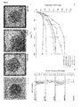
- Carboxylated PS nanoparticles of nominal diameter 200 nm were incubated for 1 h at room temperature in 80% v/v plasma at a nanoparticle concentration of 1 mg/mL. These nanoparticles complete with corona (PS@HC) were washed six times by centrifuging 250 ⁇ L at 20000 x g for 10 min and re-suspending in PBS (137mM, pH 7.4, with 1mM EDTA). The biomolecular corona was analysed by SDS-PAGE. Immunogold labelling was carried out by incubating PS @HCwith different immunogold concentrations 1-300 nM overnight at 4°C prior to analysis using DCS. These incubation conditions were chosen to reduce nonspecific binding of IG labels. The results shown are the average of three measurements carried out on independently prepared samples ( Fig.5 ).
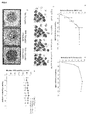
- Transferrin coated polystyrene (50 ⁇ g/mL) was incubated with increasing amounts of soluble TfR ca. 1-300 nM for 1 h at room temperature. The apparent diameter of these particles was measured by DCS and compared with particles in the absence of any receptor. The results shown are representative of duplicate measurements ( Fig.4a ), a single data point at high receptor concentrations ca. 350 nM was measured to ensure saturation had been reached. Transferrin coated polystyrene nanoparticles were incubated with different concentrations of TfR and subsequently labelled using IG-mTf at a concentration corresponding to saturation. The samples were analysed by DCS and the percentage reduction in IG binding was calculated using Equation (2).
- d block is the apparent diameter of the sample after blocking of the surface with TfR and labelling with IG-mTf
- d sat is the diameter for the particles saturated with IG-mTf
- STEM imaging was used to generate a 3D reconstruction of a Transferrin coated polystyrene saturated with IG-mTf ( Fig.3e ).
- the difference in focus between particles on the top and the bottom can be achieved through careful analysis of each particle on the surface by comparing bright field, and the two STEM modes. It should be noted that identification around the circumference of the sphere is the most problematic.
- Protein structures were obtained from the protein databank www.rcsb.org (PDB ID: 1SUV) (Scheme1). 16 These structures were visualised and labelled using UCSF Chimera ( Pettersen, E. F. et al. UCSF chimera - A visualization system for exploratory research and analysis. J Comput Chem 25, 1605-1612 (2004 )).
- Figs 6 and 7 demonstrate the feasibility of using quantum dot particles conjugated to antibodies to label the epitopes of protiens on the surface of a nanoparticle.
- different techniques can be employed to examine the extent of labelling e.g. luminescent spectroscopy, or the distribution of luminescent intensitites across the sample flow cytometry.
- Figure 6 shows a titration curve in which polystyrene nanoparticles of nominal diameter 200nm are incubated with quantum dots conjugated with an antibody which recognises transferrin. The samples are washed to remove any unbound quantum dots and thus the fluorescent intensity is proportional to the number of quantum dots attached to the polystyrene nanoparticles.
- FIG. 7(a) shows the change in the forward (xaxis) and side scatter (y axis) of PS@Tf nanoparticles post labeling with QD-Ab's. This change is the scattering arises due to the increase in size of the object and change in optical properties.
- Figure 7(b) Highlights that it is possible to separate different populations using flow cytometry and these populations can be sorted to obtain distinct samples if neccisary.
- Figure 7(c) shows that in additon to forward and side scatter the luminescent intensity can be used to show follow a change in the sample post labelling with QD-Ab's.
- Figure 7(d) shows the luminescent distributions for each of the samples demonstrating a quick method to discribe the distribution of labeled proteins across a larege number of nanoparticles.
- the methods of the invention may be employed to map out the proteins fon a nanosized object such as a nanoparticle, to determine the location and spatial distribution of the protein.
- the methods may also be employed to map out specific epitopes of biomolecules on a nanoparticle, providing information on the orientation of the proteins that contain the specific epitope.
- the methods of the invention may also be employed to determine the abundance of specific protein, or epitope, on a nanoparticle, and to determine relative abundance of a plurality of different proteins, different epitopes, including different epitopes from the same protein.
Landscapes
- Health & Medical Sciences (AREA)
- Engineering & Computer Science (AREA)
- Immunology (AREA)
- Life Sciences & Earth Sciences (AREA)
- Chemical & Material Sciences (AREA)
- Hematology (AREA)
- Urology & Nephrology (AREA)
- Biomedical Technology (AREA)
- Molecular Biology (AREA)
- Microbiology (AREA)
- Analytical Chemistry (AREA)
- Biotechnology (AREA)
- Nanotechnology (AREA)
- Food Science & Technology (AREA)
- Medicinal Chemistry (AREA)
- Physics & Mathematics (AREA)
- Cell Biology (AREA)
- Biochemistry (AREA)
- General Health & Medical Sciences (AREA)
- General Physics & Mathematics (AREA)
- Pathology (AREA)
- Investigating Or Analysing Biological Materials (AREA)
- Investigating, Analyzing Materials By Fluorescence Or Luminescence (AREA)
Priority Applications (3)
| Application Number | Priority Date | Filing Date | Title |
|---|---|---|---|
| EP14172854.3A EP2957912A1 (de) | 2014-06-17 | 2014-06-17 | Verfahren zur Markierung eines Zielmoleküls als Teil einer Korona von Molekülen auf der Oberfläche eines Objekts im Nanogrößenbereich |
| US15/319,627 US20170146527A1 (en) | 2014-06-17 | 2015-06-17 | A method of labelling a target molecule forming part of a corona of molecules on a surfaces of a nanosized object |
| PCT/EP2015/001220 WO2015192964A1 (en) | 2014-06-17 | 2015-06-17 | A method of labelling a target molecule forming part of a corona of molecules on a surfaces of a nanosized object |
Applications Claiming Priority (1)
| Application Number | Priority Date | Filing Date | Title |
|---|---|---|---|
| EP14172854.3A EP2957912A1 (de) | 2014-06-17 | 2014-06-17 | Verfahren zur Markierung eines Zielmoleküls als Teil einer Korona von Molekülen auf der Oberfläche eines Objekts im Nanogrößenbereich |
Publications (1)
| Publication Number | Publication Date |
|---|---|
| EP2957912A1 true EP2957912A1 (de) | 2015-12-23 |
Family
ID=50942223
Family Applications (1)
| Application Number | Title | Priority Date | Filing Date |
|---|---|---|---|
| EP14172854.3A Withdrawn EP2957912A1 (de) | 2014-06-17 | 2014-06-17 | Verfahren zur Markierung eines Zielmoleküls als Teil einer Korona von Molekülen auf der Oberfläche eines Objekts im Nanogrößenbereich |
Country Status (3)
| Country | Link |
|---|---|
| US (1) | US20170146527A1 (de) |
| EP (1) | EP2957912A1 (de) |
| WO (1) | WO2015192964A1 (de) |
Cited By (1)
| Publication number | Priority date | Publication date | Assignee | Title |
|---|---|---|---|---|
| US11598779B2 (en) | 2016-09-06 | 2023-03-07 | The University Of Manchester | Detection of cancer biomarkers using nanoparticles |
Families Citing this family (8)
| Publication number | Priority date | Publication date | Assignee | Title |
|---|---|---|---|---|
| US20210072255A1 (en) | 2016-12-16 | 2021-03-11 | The Brigham And Women's Hospital, Inc. | System and method for protein corona sensor array for early detection of diseases |
| GB2595583A (en) | 2018-11-07 | 2021-12-01 | Seer Inc | Compositions, methods and systems for protein corona analysis and uses thereof |
| US20210072143A1 (en) * | 2019-07-01 | 2021-03-11 | Davinci Bio, Inc. | High capacity molecule detection |
| WO2021087407A1 (en) * | 2019-11-02 | 2021-05-06 | Seer, Inc. | Systems for protein corona analysis |
| CN114787628A (zh) * | 2020-01-20 | 2022-07-22 | 纳肽得(青岛)生物医药有限公司 | 通过蛋白冠调制构建基于纳米颗粒的药物载体的方法 |
| US20230212647A1 (en) * | 2020-05-29 | 2023-07-06 | Seer, Inc. | Systems and methods for rapid identification of proteins |
| IL299998A (en) * | 2020-07-20 | 2023-03-01 | Seer Inc | Particles and test methods |
| WO2024118850A1 (en) * | 2022-11-30 | 2024-06-06 | Cedars-Sinai Medical Center | Methods for detecting metabolic markers |
Citations (2)
| Publication number | Priority date | Publication date | Assignee | Title |
|---|---|---|---|---|
| WO2010097785A1 (en) * | 2009-02-26 | 2010-09-02 | University College Dublin, National University Of Ireland, Dublin | A method for the selective concentration of a specific low abundance biomolecule |
| WO2011088128A2 (en) * | 2010-01-14 | 2011-07-21 | University Of Central Florida Research Foundation, Inc. | Methods for biomolecule and biomolecule complex (bmc) detection and analysis and the use of such for research and medical diagnosis |
Family Cites Families (5)
| Publication number | Priority date | Publication date | Assignee | Title |
|---|---|---|---|---|
| US7169556B2 (en) * | 1996-07-29 | 2007-01-30 | Nanosphere, Inc. | Nanoparticles having oligonucleotides attached thereto and uses therefor |
| US7214427B2 (en) * | 2002-03-21 | 2007-05-08 | Aviva Biosciences Corporation | Composite beads comprising magnetizable substance and electro-conductive substance |
| EP1760467A1 (de) * | 2005-09-02 | 2007-03-07 | Schering AG | Optisch fluoreszierende Nanopartikel |
| EP2017006A1 (de) * | 2007-07-20 | 2009-01-21 | Koninklijke Philips Electronics N.V. | Mikrofluidische Verfahren und Systeme zur Verwendung bei der Analytdetektion |
| US20120295285A1 (en) * | 2008-08-27 | 2012-11-22 | New York Blood Center, Inc. | Ordered Assembly of Membrane Proteins During Differentiation of Erythroblasts |
-
2014
- 2014-06-17 EP EP14172854.3A patent/EP2957912A1/de not_active Withdrawn
-
2015
- 2015-06-17 WO PCT/EP2015/001220 patent/WO2015192964A1/en not_active Ceased
- 2015-06-17 US US15/319,627 patent/US20170146527A1/en not_active Abandoned
Patent Citations (2)
| Publication number | Priority date | Publication date | Assignee | Title |
|---|---|---|---|---|
| WO2010097785A1 (en) * | 2009-02-26 | 2010-09-02 | University College Dublin, National University Of Ireland, Dublin | A method for the selective concentration of a specific low abundance biomolecule |
| WO2011088128A2 (en) * | 2010-01-14 | 2011-07-21 | University Of Central Florida Research Foundation, Inc. | Methods for biomolecule and biomolecule complex (bmc) detection and analysis and the use of such for research and medical diagnosis |
Non-Patent Citations (4)
| Title |
|---|
| MARA CANOVI ET AL: "Applications of Surface Plasmon Resonance (SPR) for the Characterization of Nanoparticles Developed for Biomedical Purposes", SENSORS, vol. 12, no. 12, 27 December 2012 (2012-12-27), pages 16420 - 16432, XP055153960, ISSN: 1424-8220, DOI: 10.3390/s121216420 * |
| PETTERSEN, E. F. ET AL.: "UCSF chimera - A visualization system for exploratory research and analysis", J COMPUT CHEM, vol. 25, 2004, pages 1605 - 1612 |
| SALVATI, A. ET AL.: "Transferrin-functionalized nanoparticles lose their targeting capabilities when a biomolecule corona adsorbs on the surface", NATURE NANOTECHNOLOGY, vol. 8, 2013, pages 137 - 143, XP055133158, DOI: doi:10.1038/nnano.2012.237 |
| SLOT, J. W.; GEUZE, H. J.: "A new method of preparing gold probes for multiple-labeling cytochemistry", EUROPEAN JOURNAL OF CELL BIOLOGY, vol. 38, 1985, pages 87 - 93, XP008050370 |
Cited By (1)
| Publication number | Priority date | Publication date | Assignee | Title |
|---|---|---|---|---|
| US11598779B2 (en) | 2016-09-06 | 2023-03-07 | The University Of Manchester | Detection of cancer biomarkers using nanoparticles |
Also Published As
| Publication number | Publication date |
|---|---|
| WO2015192964A1 (en) | 2015-12-23 |
| US20170146527A1 (en) | 2017-05-25 |
Similar Documents
| Publication | Publication Date | Title |
|---|---|---|
| US20170146527A1 (en) | A method of labelling a target molecule forming part of a corona of molecules on a surfaces of a nanosized object | |
| Sapsford et al. | Analyzing nanomaterial bioconjugates: a review of current and emerging purification and characterization techniques | |
| Nienhaus et al. | Studying the protein corona on nanoparticles by FCS | |
| KR101195957B1 (ko) | 표면증강 라만 산란 복합 프로브 및 이를 이용하여 표적 물질을 검출하는 방법 | |
| JP6104868B2 (ja) | 磁気センサに基づく結合反応速度の定量的な分析 | |
| US10656117B2 (en) | Nanopore-based determination of protein charge, sharp, volume, rotational diffusion coefficient, and dipole moment | |
| US9528995B2 (en) | Systems and methods for high-throughput detection of an analyte in a sample | |
| US10191041B2 (en) | Detection of analytes using metal nanoparticle probes and dynamic light scattering | |
| JP5970153B2 (ja) | 凝集パラメータの測定 | |
| Shipunova et al. | MPQ-cytometry: a magnetism-based method for quantification of nanoparticle–cell interactions | |
| US20060148104A1 (en) | Detection of ion channel or receptor activity | |
| Omidfar et al. | A high sensitive electrochemical nanoimmunosensor based on Fe3O4/TMC/Au nanocomposite and PT-modified electrode for the detection of cancer biomarker epidermal growth factor receptor | |
| Smith et al. | Optimization of antibody-conjugated magnetic nanoparticles for target preconcentration and immunoassays | |
| EP2401619A1 (de) | Verfahren zur selektiven konzentration eines spezifischen low-abundance-biomoleküls | |
| JP2021081359A (ja) | 分子間相互作用の解析方法および解析装置 | |
| JP6593840B2 (ja) | 量子ドット蛍光増強免疫測定法 | |
| TWI745593B (zh) | 包覆式功能化鑽石晶體 | |
| US10083341B2 (en) | Cellular activity quantification using labeled probes | |
| Woolley et al. | From particle to platelet: Optimization of a stable, high brightness fluorescent nanoparticle based cell detection platform | |
| WO2018052912A1 (en) | Pathogen quantification using labeled probes | |
| Yang et al. | An early cancer diagnosis platform based on micro-magnetic sensor array demonstrates ultra-high sensitivity | |
| Taranova et al. | Comparative characteristics of nanodisperse labels for immunochromatographic test systems | |
| US20150044665A1 (en) | Target-specific probe comprising t7 bacteriophage and detecting for biomarker using the same | |
| Sousa et al. | Characterization of Bio-nanosystems | |
| WO2007044712A2 (en) | Portable materials and methods for ultrasensitive detection of pathogens and bioparticles |
Legal Events
| Date | Code | Title | Description |
|---|---|---|---|
| PUAI | Public reference made under article 153(3) epc to a published international application that has entered the european phase |
Free format text: ORIGINAL CODE: 0009012 |
|
| AK | Designated contracting states |
Kind code of ref document: A1 Designated state(s): AL AT BE BG CH CY CZ DE DK EE ES FI FR GB GR HR HU IE IS IT LI LT LU LV MC MK MT NL NO PL PT RO RS SE SI SK SM TR |
|
| AX | Request for extension of the european patent |
Extension state: BA ME |
|
| STAA | Information on the status of an ep patent application or granted ep patent |
Free format text: STATUS: THE APPLICATION IS DEEMED TO BE WITHDRAWN |
|
| 18D | Application deemed to be withdrawn |
Effective date: 20160624 |