EP2970895B1 - Podozytenkulturen und verwendungen davon - Google Patents
Podozytenkulturen und verwendungen davon Download PDFInfo
- Publication number
- EP2970895B1 EP2970895B1 EP14769052.3A EP14769052A EP2970895B1 EP 2970895 B1 EP2970895 B1 EP 2970895B1 EP 14769052 A EP14769052 A EP 14769052A EP 2970895 B1 EP2970895 B1 EP 2970895B1
- Authority
- EP
- European Patent Office
- Prior art keywords
- podocytes
- podocyte
- cell
- cells
- days
- Prior art date
- Legal status (The legal status is an assumption and is not a legal conclusion. Google has not performed a legal analysis and makes no representation as to the accuracy of the status listed.)
- Active
Links
Images




Classifications
-
- C—CHEMISTRY; METALLURGY
- C12—BIOCHEMISTRY; BEER; SPIRITS; WINE; VINEGAR; MICROBIOLOGY; ENZYMOLOGY; MUTATION OR GENETIC ENGINEERING
- C12N—MICROORGANISMS OR ENZYMES; COMPOSITIONS THEREOF; PROPAGATING, PRESERVING, OR MAINTAINING MICROORGANISMS; MUTATION OR GENETIC ENGINEERING; CULTURE MEDIA
- C12N5/00—Undifferentiated human, animal or plant cells, e.g. cell lines; Tissues; Cultivation or maintenance thereof; Culture media therefor
- C12N5/06—Animal cells or tissues; Human cells or tissues
- C12N5/0602—Vertebrate cells
- C12N5/0684—Cells of the urinary tract or kidneys
- C12N5/0686—Kidney cells
-
- C—CHEMISTRY; METALLURGY
- C12—BIOCHEMISTRY; BEER; SPIRITS; WINE; VINEGAR; MICROBIOLOGY; ENZYMOLOGY; MUTATION OR GENETIC ENGINEERING
- C12N—MICROORGANISMS OR ENZYMES; COMPOSITIONS THEREOF; PROPAGATING, PRESERVING, OR MAINTAINING MICROORGANISMS; MUTATION OR GENETIC ENGINEERING; CULTURE MEDIA
- C12N5/00—Undifferentiated human, animal or plant cells, e.g. cell lines; Tissues; Cultivation or maintenance thereof; Culture media therefor
- C12N5/06—Animal cells or tissues; Human cells or tissues
- C12N5/0602—Vertebrate cells
- C12N5/0684—Cells of the urinary tract or kidneys
- C12N5/0687—Renal stem cells; Renal progenitors
-
- G—PHYSICS
- G01—MEASURING; TESTING
- G01N—INVESTIGATING OR ANALYSING MATERIALS BY DETERMINING THEIR CHEMICAL OR PHYSICAL PROPERTIES
- G01N33/00—Investigating or analysing materials by specific methods not covered by groups G01N1/00 - G01N31/00
- G01N33/48—Biological material, e.g. blood, urine; Haemocytometers
- G01N33/50—Chemical analysis of biological material, e.g. blood, urine; Testing involving biospecific ligand binding methods; Immunological testing
- G01N33/5005—Chemical analysis of biological material, e.g. blood, urine; Testing involving biospecific ligand binding methods; Immunological testing involving human or animal cells
- G01N33/5008—Chemical analysis of biological material, e.g. blood, urine; Testing involving biospecific ligand binding methods; Immunological testing involving human or animal cells for testing or evaluating the effect of chemical or biological compounds, e.g. drugs, cosmetics
- G01N33/502—Chemical analysis of biological material, e.g. blood, urine; Testing involving biospecific ligand binding methods; Immunological testing involving human or animal cells for testing or evaluating the effect of chemical or biological compounds, e.g. drugs, cosmetics for testing non-proliferative effects
-
- G—PHYSICS
- G01—MEASURING; TESTING
- G01N—INVESTIGATING OR ANALYSING MATERIALS BY DETERMINING THEIR CHEMICAL OR PHYSICAL PROPERTIES
- G01N33/00—Investigating or analysing materials by specific methods not covered by groups G01N1/00 - G01N31/00
- G01N33/48—Biological material, e.g. blood, urine; Haemocytometers
- G01N33/50—Chemical analysis of biological material, e.g. blood, urine; Testing involving biospecific ligand binding methods; Immunological testing
- G01N33/5005—Chemical analysis of biological material, e.g. blood, urine; Testing involving biospecific ligand binding methods; Immunological testing involving human or animal cells
- G01N33/5008—Chemical analysis of biological material, e.g. blood, urine; Testing involving biospecific ligand binding methods; Immunological testing involving human or animal cells for testing or evaluating the effect of chemical or biological compounds, e.g. drugs, cosmetics
- G01N33/502—Chemical analysis of biological material, e.g. blood, urine; Testing involving biospecific ligand binding methods; Immunological testing involving human or animal cells for testing or evaluating the effect of chemical or biological compounds, e.g. drugs, cosmetics for testing non-proliferative effects
- G01N33/5023—Chemical analysis of biological material, e.g. blood, urine; Testing involving biospecific ligand binding methods; Immunological testing involving human or animal cells for testing or evaluating the effect of chemical or biological compounds, e.g. drugs, cosmetics for testing non-proliferative effects on expression patterns
-
- G—PHYSICS
- G01—MEASURING; TESTING
- G01N—INVESTIGATING OR ANALYSING MATERIALS BY DETERMINING THEIR CHEMICAL OR PHYSICAL PROPERTIES
- G01N33/00—Investigating or analysing materials by specific methods not covered by groups G01N1/00 - G01N31/00
- G01N33/48—Biological material, e.g. blood, urine; Haemocytometers
- G01N33/50—Chemical analysis of biological material, e.g. blood, urine; Testing involving biospecific ligand binding methods; Immunological testing
- G01N33/5005—Chemical analysis of biological material, e.g. blood, urine; Testing involving biospecific ligand binding methods; Immunological testing involving human or animal cells
- G01N33/5008—Chemical analysis of biological material, e.g. blood, urine; Testing involving biospecific ligand binding methods; Immunological testing involving human or animal cells for testing or evaluating the effect of chemical or biological compounds, e.g. drugs, cosmetics
- G01N33/502—Chemical analysis of biological material, e.g. blood, urine; Testing involving biospecific ligand binding methods; Immunological testing involving human or animal cells for testing or evaluating the effect of chemical or biological compounds, e.g. drugs, cosmetics for testing non-proliferative effects
- G01N33/5026—Chemical analysis of biological material, e.g. blood, urine; Testing involving biospecific ligand binding methods; Immunological testing involving human or animal cells for testing or evaluating the effect of chemical or biological compounds, e.g. drugs, cosmetics for testing non-proliferative effects on cell morphology
-
- G—PHYSICS
- G01—MEASURING; TESTING
- G01N—INVESTIGATING OR ANALYSING MATERIALS BY DETERMINING THEIR CHEMICAL OR PHYSICAL PROPERTIES
- G01N33/00—Investigating or analysing materials by specific methods not covered by groups G01N1/00 - G01N31/00
- G01N33/48—Biological material, e.g. blood, urine; Haemocytometers
- G01N33/50—Chemical analysis of biological material, e.g. blood, urine; Testing involving biospecific ligand binding methods; Immunological testing
- G01N33/5005—Chemical analysis of biological material, e.g. blood, urine; Testing involving biospecific ligand binding methods; Immunological testing involving human or animal cells
- G01N33/5008—Chemical analysis of biological material, e.g. blood, urine; Testing involving biospecific ligand binding methods; Immunological testing involving human or animal cells for testing or evaluating the effect of chemical or biological compounds, e.g. drugs, cosmetics
- G01N33/5044—Chemical analysis of biological material, e.g. blood, urine; Testing involving biospecific ligand binding methods; Immunological testing involving human or animal cells for testing or evaluating the effect of chemical or biological compounds, e.g. drugs, cosmetics involving specific cell types
-
- G—PHYSICS
- G01—MEASURING; TESTING
- G01N—INVESTIGATING OR ANALYSING MATERIALS BY DETERMINING THEIR CHEMICAL OR PHYSICAL PROPERTIES
- G01N33/00—Investigating or analysing materials by specific methods not covered by groups G01N1/00 - G01N31/00
- G01N33/48—Biological material, e.g. blood, urine; Haemocytometers
- G01N33/50—Chemical analysis of biological material, e.g. blood, urine; Testing involving biospecific ligand binding methods; Immunological testing
- G01N33/53—Immunoassay; Biospecific binding assay; Materials therefor
- G01N33/569—Immunoassay; Biospecific binding assay; Materials therefor for microorganisms, e.g. protozoa, bacteria, viruses
- G01N33/56966—Animal cells
-
- C—CHEMISTRY; METALLURGY
- C12—BIOCHEMISTRY; BEER; SPIRITS; WINE; VINEGAR; MICROBIOLOGY; ENZYMOLOGY; MUTATION OR GENETIC ENGINEERING
- C12N—MICROORGANISMS OR ENZYMES; COMPOSITIONS THEREOF; PROPAGATING, PRESERVING, OR MAINTAINING MICROORGANISMS; MUTATION OR GENETIC ENGINEERING; CULTURE MEDIA
- C12N2510/00—Genetically modified cells
- C12N2510/04—Immortalised cells
-
- G—PHYSICS
- G01—MEASURING; TESTING
- G01N—INVESTIGATING OR ANALYSING MATERIALS BY DETERMINING THEIR CHEMICAL OR PHYSICAL PROPERTIES
- G01N2333/00—Assays involving biological materials from specific organisms or of a specific nature
- G01N2333/435—Assays involving biological materials from specific organisms or of a specific nature from animals; from humans
- G01N2333/46—Assays involving biological materials from specific organisms or of a specific nature from animals; from humans from vertebrates
- G01N2333/47—Assays involving proteins of known structure or function as defined in the subgroups
- G01N2333/4701—Details
- G01N2333/4712—Muscle proteins, e.g. myosin, actin, protein
-
- G—PHYSICS
- G01—MEASURING; TESTING
- G01N—INVESTIGATING OR ANALYSING MATERIALS BY DETERMINING THEIR CHEMICAL OR PHYSICAL PROPERTIES
- G01N2800/00—Detection or diagnosis of diseases
- G01N2800/34—Genitourinary disorders
- G01N2800/347—Renal failures; Glomerular diseases; Tubulointerstitial diseases, e.g. nephritic syndrome, glomerulonephritis; Renovascular diseases, e.g. renal artery occlusion, nephropathy
Definitions
- Kidney-failure End stage renal disease, ESRD is a debilitating disease with no treatments or therapeutics. Patients suffering from ESRD go on dialysis and require a kidney transplant in order to regain kidney function. A majority of kidney diseases and ESRD originate within the glomerulus and are associated with proteinuria. ESRD imposes a significant burden on the patients and the health care system worldwide in is in urgent need for effective therapies and better treatment options.
- kidney disease The study of kidney disease is complex because the onset is often undetected, diseases may be acute or chronic in nature, the genetic makeup of the host leads to variable clinical syndromes, and multiple organs are often involved simultaneously.
- Cell cultures and animal models are necessary to study disease susceptibility, mechanisms, prognosis, and potential therapies.
- the use of experimental animal models has proved invaluable. Nevertheless, animal models are often limited because they do not always fully replicate the human diseases. For example, the current mouse models of diabetic nephropathy do not typically demonstrate the features of human diseases, such as Kimmelstiel-Wilson nodules.
- podocytes form the final filtration barrier for blood in the glomerulus and play a central role in glomerular diseases that ultimately result in ESRD ( Mundel P. and Reiser J. Kidney Int 2010, 77: 571-80 ).
- Podocytes are terminally differentiated pericyte-like cells that reside on the outer surface of the glomerular basement membrane and give rise to long major processes that branch into structures known as foot processes (FPs).
- FPs foot processes
- FPs of adjacent podocyte cells interdigitate and form narrow filtration slits, a structure known as the slit diaphragm (SD), which forms a molecular sieve that the body uses to retain proteins in the blood, while filtering small molecules and other agents in to the urinary space.
- SD slit diaphragm
- podocyte injury is a common theme in many proteinuric kidney diseases ( Mundel P. and Reiser J. Kidney Int 2010, 77: 571-80 ).
- podocytes represent a key target for the development of novel therapeutics to treat a variety of kidney diseases.
- podocytes play a key role in the prevention of proteinuria in the healthy situation, they are important targets of injury in a variety of renal diseases and are important determinants of outcome. Improved understanding of podocyte biology has come from two main arenas: first, molecular genetics of single gene disorders which lead to rare forms of congenital nephrotic syndrome; and second, focused study of this specialized cell type in vivo and in vitro. Research has also shed light on specific proteins, RNA and cell signaling mechanisms in the podocytes that represent good targets for drug discovery efforts, diagnostics and therapeutics.
- podocyte cultures do not fully replicate the in vivo environment, they have several major advantages. These include the ability to directly study mechanistic events, to control the environment such that specific hypotheses can be tested, and that multiple experiments can be performed to validate the initial observations.
- podocytes are a challenging cell type for use in such assays.
- the invention provides a method of culturing podocytes according to claim 1.
- the cultures and terminally-differentiated podocytes obtained from the cultures can be used for a variety of purposes, such as drug screening (such as medium or high throughput drug screening), studying molecular pathways involved in glomerular diseases, and diagnosing and screening patients for kidney disease or predisposition for kidney disease.
- drug screening such as medium or high throughput drug screening
- studying molecular pathways involved in glomerular diseases and diagnosing and screening patients for kidney disease or predisposition for kidney disease.
- the method comprises maintaining a first culture of podocyte cells (e.g, primary podocytes, proliferating podocytes, podocyte precursor cells or podocyte cell lines, such as conditionally-immortalized podocyte cell lines) under a permissive condition or proliferation condition to produce a population of podocytes, maintaining that population of podocytes (or a fraction thereof) under a non-permissive condition or differentiation condition for a period of time sufficient to obtain a population of partially differentiated podocytes, and then maintaining the partially differentiated podocytes (or a fraction thereof) in a second culture under a non-permissive condition or differentiation condition for a period of time sufficient to obtain a population of terminally-differentiated podocytes.
- podocyte cells e.g, primary podocytes, proliferating podocytes, podocyte precursor cells or podocyte cell lines, such as conditionally-immortalized podocyte cell lines
- the cultures and terminally-differentiated podocytes obtained from the cultures can be used for a variety of purposes, such as drug screening (such as medium or high throughput drug screening), studying molecular pathways involved in glomerular diseases, and diagnosing and screening patients for kidney disease or predisposition for kidney disease.
- drug screening such as medium or high throughput drug screening
- studying molecular pathways involved in glomerular diseases and diagnosing and screening patients for kidney disease or predisposition for kidney disease.
- podocyte cultures A significant limitation in using podocyte cultures is the difficulty in handling podocytes and using them in medium- or high-throughput assays.
- Podocytes show heterogeneous morphology in in vitro cultures, which makes it hard to get robust, reproducible data with low-variability between different wells, plates, and on different days.
- the numbers of podocyte cells per well are also highly variable from well-to-well and plate-to-plate, because of the long culture time needed for the proliferation and differentiate these cells in vitro. Additionally, differentiated podocytes have shown limited ability to propagate.
- the inventors have developed new methods of culturing podocytes in vitro, which produce podocyte cultures that are suitable for, among other purposes, medium- or high-throughput assays.
- conditionally-immortalized podocytes were proliferated under a permissive condition to achieve a desired number of cells (in this example, 100% confluency).
- the cells were maintained under a non-permissive condition to induce partial differentiation.
- these partially differentiated cells were re-seeded in a multi-well plate, in which the partially differentiated cells were maintained under a non-permissive condition to produce terminally-differentiated podocytes.
- podocytes produced by this method are particularly suitable for use in medium- and high-throughput assays (such as drug screenings).
- the inventors also developed assays and methods to analyze cellular and physiological characteristics of terminally differentiated podocytes, such as membrane permeability, morphology, viability, or expression of a podocyte marker.
- image analysis algorithms were developed to determine podocyte health using specific measurements of the actin cytoskeleton, focal adhesions, and morphology of the podocyte in vitro.
- the methods were used to quantify changes in podocytes caused by a damaging agent, or a damaging agent together with an agent to maintain or improve podocyte health.
- the inventors quantified the changes in podocytes using multiple parameters to segregate different population subtypes in a single well.
- a “conditionally-immortalized” podocyte refers to an immortalized podocyte in which the mitotic proliferation of the podocyte can be activated or inactivated under appropriate conditions. Under a “permissive condition,” the cell proceeds with mitotic proliferation, and under a “non-permissive condition,” the cell proceeds to differentiate.
- a conditionally immortalized podocyte line can be constructed in a number of ways. For example, a foreign gene encoding a temperature-sensitive immortalizing molecule, such as the polyoma-large T antigen, can be integrated in the genome of a cell. The resulting cell line can be grown at temperatures at which the immortalizing molecule is active, or at which the molecule becomes inactive, such as between about 30°C and 40°C.
- Conditionally immortalized human podocyte cell lines have been developed by transfection using both the temperature-sensitive mutant U19 ts A58 of the SV40 large T antigen (SV40) and the essential catalytic subunit of the hTERT telomerase gene.
- the hTERT vector expresses telomerase activity to maintain telomere length, preventing the occurrence of replicative senescence. Transfection of cells with SV40T allows cells to proliferate at the "permissive" temperature of 33°C. Transfer to the "non-permissive" temperature of 37°C results in the inactivation of large T antigen with minor changes in gene expression.
- Podocytes then enter growth arrest and express markers of differentiated in vivo podocytes, including the podocyte proteins, nephrin, podocin, CD2AP, and synaptopodin, and known molecules of the slit diaphragm ZO-1, alpha-, beta-, and gamma-catenin and P-cadherin. See, e.g., Ni et al., Podocyte culture: Tricks of the trade, Nephrology 17 (2012) 525-531 .
- the term "permissive condition” refers to a cell culture condition that allows a podocyte to divide and propagate, without inducing apparent terminal differentiation.
- a conditionally immortalized murine podocyte cell line Mundel, Reiser et al., Experimental Cell Research, 236, p248 (1997 )
- Mundel can be propagated at 33 degree centigrade and media containing 10 U/ml recombinant mouse ⁇ -interferon (permissive condition).
- non-permissive condition refers to a cell culture condition that induces terminal differentiation of a podocyte, which may be demonstrated, for example, by the expression of makers that are typically expressed in fully differentiated podocytes in the kidney.
- a conditionally immortalized murine podocyte cell line Mundel, Reiser et al., Experimental Cell Research, 236, p248 (1997 )
- Mundel can be differentiated by shifting the cells from 33 degree centigrade to a 37 degree centigrade cell culture environment and excluding recombinant mouse ⁇ -interferon from the media (non-permissive condition).
- a “terminally differentiated” podocyte refers to a cell that typically doesn't enter mitotic cell cycle and is further characterized by its arborized appearance, large cell size, an organized actin cytoskeleton, and podocyte specific markers.
- the following proteins have been identified as being markers for a terminally differentiated podocyte: ⁇ 3 ⁇ 1 integrin, ⁇ -Actinin-4, CD2AP, Nephrin, Podocalyxin, Podocin, Synaptopodin, VEGF, WT-1.
- a “partially differentiated” podocyte refers to a cell that has differentiated from a conditionally-immortalized podocyte, a proliferating podocyte or a podocyte precursor cell, but is not terminally differentiated.
- a partially differentiated podocyte is phenotypically different from a conditionally-immortalized podocyte, a proliferating podocyte, a podocyte precursor cell and a terminally differentiated podocyte, and can be further characterized by only partial expression of differentiated podocyte markers and phenotype.
- immortalized podocytes show a typical cobblestone morphology.
- a "partialy differentiated" podocyte shows partial expression of markers typically present in a "terminally differentiated” podocyte. This includes proteins, such as Synaptopodin and podocin. The level of expression of these proteins in "partially-differentiated" podocytes is less than that of the levels typically present in a "terminally differentiated” podocyte.
- a primary cell culture refers to a cell culture in which the cells are isolated directly from an organism or a tissue sample and proliferated under the appropriate conditions, with no passages.
- a "proliferation condition" for a podocyte culture refers to a condition that allows a podocyte to divide and propagate, without inducing apparent terminal differentiation. Typically, a primary podocyte remains proliferative when the cell density of the culture is below about 50% confluent (when grown as an adherent culture).
- a “differentiation condition" for a podocyte culture refers to a condition that induces terminal differentiation of a podocyte.
- a primary podocyte starts differentiation when the cell density of the culture is above about 50% confluent (when grown as an adherent culture).
- a terminally differentiated podocyte is "healthy" when the podocyte is viable and has other characteristics of normal podocytes.
- healthy podocytes expresses the marker proteins Podocin and Synaptopodin at levels typical of normal terminally differentiated podocytes, which are known in the art.
- the podocytes can be from any desired animal species, such as a mammal (e.g., murine (mouse, rat), human) or non-mammal, such as fish (e.g., zebra fish) or insect (e.g. Drosophila).
- a mammal e.g., murine (mouse, rat), human
- non-mammal such as fish (e.g., zebra fish) or insect (e.g. Drosophila).
- Also described herein is a method of culturing podocytes in vitro, comprising: (a) maintaining podocyte cells (e.g, primary podocytes, proliferating podocytes, podocyte precursor cells or podocyte cell lines, such as conditionally-immortalized podocyte cell lines) in a first cell culture under a permissive condition or proliferation condition for a period of time sufficient for the conditionally-immortalized podocyte to double at least once, thereby producing a population of podocytes; (b) maintaining the population of podocytes obtained in (a) under a non-permissive condition or differentiation condition for a period of time sufficient to obtain a population of partially differentiated podocytes; and (c) maintaining the partially differentiated podocytes obtained in (b) in a second cell culture under a non-permissive condition or differentiation condition for a period of time sufficient to obtain a population of terminally-differentiated podocytes.
- podocyte cells e.g, primary podocytes, proliferating podocytes, podocyte precursor
- the first cell culture may contain a primary podocyte, a podocyte precursor, a proliferative podocyte or a podocyte cell line.
- a primary podocyte remains proliferative when the cell density of the culture is below about 50% confluent (when grown as an adherent culture), and starts differentiation when the cell density of the culture is above about 50% confluent (when grown as an adherent culture).
- the first cell culture contains a conditionally-immortalized podocyte cell line.
- podocyte cell lines including wild-type and knockout murine cell lines, as well as wild-type and genetically altered human podocyte lines, are available and can be used in the methods described herein.
- Table 1 summarizes some of the podocyte cell lines that may be used.
- Conditionally immortalized podocyte cell lines are proliferative when cultured under permissive conditions. Non-permissive conditions cause growth arrest in the majority of podocytes within a couple of days and induces many characteristics of differentiated podocytes.
- podocytes Similar to undifferentiated primary podocytes in culture, actively proliferating conditionally immortalized podocytes growing under permissive conditions display an epithelial morphology. They are small in size, exhibit a polygonal or "cobblestone" appearance, and have a relatively small cytoplasmic volume. When podocytes are placed in non-permissive conditions, again similar to primary differentiated podocytes, they substantially increase in size, stop replicating, and take on a more complex arborized morphology including the formation of cellular structures comparable with filtration slits in vivo. Podocytes are typically plated in non-permissive conditions at a concentration of 5,000-10,000 cells/cm 2 .
- conditionally immortalized podocyte cell lines may also be made from primary cultures. Methods of isolating podocytes to produce a primary culture, and immortalizing podocytes are known in the art. See, e.g., Ni et al., Podocyte culture: Tricks of the trade, Nephrology 17 (2012) 525-531 .
- Terminally differentiated podocytes typically do not enter mitotic cell cycle and are further characterized by arborized appearance, large cell size, an organized actin cytoskeleton, and podocyte specific markers.
- Table 2 summarizes certain characteristics of terminally-differentiated podocytes. These characteristics may be used individually or in combination to determine the differentiation state of podocytes.
- ATRA all-trans retinoic acid
- PAN puromycin aminonucleoside
- PCNA proliferating cell nuclear antigen
- TGF transforming growth factor
- VEGF vascular endothelial growth factor.
- the first podocyte culture is maintained under a permissive condition (e.g., for conditionally-amortized cells) or a proliferation condition (e.g., for primary podocytes, podocyte precursors, or other types proliferative podocytes) for between about 3 days and about 30 days, between about 5 days and about 20 days, or between about 7 days and about 14 days.
- a permissive condition e.g., for conditionally-amortized cells
- a proliferation condition e.g., for primary podocytes, podocyte precursors, or other types proliferative podocytes
- the first podocyte culture can be any desired type of culture, such as a suspension culture or an adherent culture.
- the first podocyte culture is an adherent culture.
- the population of podocytes are maintained at least about 50% confluent, at least about 60% confluent, at least about 70% confluent, at least about 80% confluent, or at least about 90% confluent, in an adherent culture before partial differentiation is initiated.
- the population of podocytes are maintained under a non-permissive condition (e.g., for conditionally-amortized cells) or a differentiation condition (e.g., for primary podocytes, podocyte precursors, or other types proliferative podocytes) for between about 1 days and about 12 days, between about 3 days and about 10 days, between about 4 days and about 8 days, or between about 5 days and about 7 days.
- the culture can be any desired type of culture, such as a suspension culture or an adherent culture.
- the partially differentiated podocytes are maintained under a non-permissive condition (e.g., for conditionally-amortized cells) or a differentiation condition (e.g., for primary podocytes, podocyte precursors, or other types proliferative podocytes) for about 3 days and about 30 days, between about 5 days and about 20 days, or between about 6 days and about 15 days.
- the culture can be any desired type of culture, such as a suspension culture or an adherent culture.
- the podocyte cultures described herein are suitable for medium- to high-throughput screening, among other purposes. Accordingly, the partially-differentiated podocytes can be reseeded into second cell cultures that are in any desired format suitable for screening or other purposes. For screening, the partially-differentiated podocytes can be reseeded into an array of second cell cultures, for example in a multiwall plate.
- the multi-well plate can be suitable for high throughput screening, including, e.g., 96-well plates, 384-well plates, or 1536-well plates.
- the podocyte cultures described herein can be used to screen candidate compounds for treating a kidney disease.
- the method comprises: (a) providing a culture of terminally differentiated podocytes; (b) contacting a candidate compound with said terminally differentiated podocyte(s); and (c) determining a cellular or physiological characteristic of the terminally differentiated podocyte(s).
- a change in the cellular or physiological characteristic relative to a suitable control is indicative that said compound is a candidate for treating a kidney disease.
- Suitable cellular or physiological characteristics include, e.g., membrane permeability, morphology, viability, or expression of a podocyte marker.
- a change in the cellular characteristic relative to a suitable control is indicative that said compound is a candidate for treating a kidney disease.
- Suitable cellular characteristics include, e.g., membrane permeability, morphology, viability, or expression of a podocyte marker.
- substantially all of the units (e.g. cultures) in the array are either controls or are contacted with the same or different candidate compound.
- the cellular or physiological characteristic may be the healthiness of the terminally-differentiated podocyte, which is determined using the parameters described herein in section B.
- the array may comprise a multi-well plate, each well comprising one or more terminally differentiated podocytes.
- the corresponding cellular or physiological characteristic from a known healthy podocyte may be used as a control.
- the control may be cellular or physiological characteristic from a parallel assay in which the podocyte is cultured in the absence of the candidate compound.
- Still other suitable controls may be data or cellular characteristics that are present in a database or in publications.
- the suitable control is obtained form a culture that is not exposed to the candidate compound but is optionally exposed to any solvent, vehicle or diluent that is used to test the candidate compound.
- Also described herein is a method of identifying healthy podocytes, such as terminally-differentiated podocytes including those produced by the culture methods described herein.
- the method is used to identify healthy podocytes (e.g., terminally-differentiated podocytes) in a culture, such as the cultures described herein.
- the method comprises determining one or more of the parameters described in sections B1 - B6 either individually or any desired combinations.
- kidney diseases or a predisposition to kidney disease in a patient.
- podocytes from a subject may be cultured using methods described herein, and the cellular characteristic (such as healthiness) of the podocytes can be determined using methods described herein.
- the presence of unhealthy podocytes from the subject e.g., in a terminally differentiated podocyte population produced using the methods described herein is indicative that the subject has or is predisposed to a kidney disease.
- serum or other suitable biological sample obtained from a subject to be diagnosed or screened.
- the serum of suitable biological sample is added to a culture of podocytes (e.g., a terminally differentiated podocyte population produced using the methods described herein) and after a sufficient period of time, the healthiness of the podocytes is determined using methods described herein.
- the presence of unhealthy podocytes in the culture is indicative that the subject has or is susceptible to a kidney disease.
- the podocytes are cultured with the serum or other suitable biological sample for at least about 6 hours, preferably at least about 12 hours or at least about 24 hours.
- Cultured podocytes provide a validated model system that reliably recapitulates critical aspects of rodent and human glomerular disease.
- culturing podocytes in vitro has long been hampered by a number of issues that have hampered their use in a medium- or high-throughput assay environment.
- Podocytes show heterogeneous morphology in culture in vitro that makes it hard to get robust, reproducible data with low-variability between different wells, plates and on different days.
- Podocyte cell number is also highly variable from well-to-well and plate-to-plate, because of the long culture time needed to grow and appropriately differentiate these cells in vitro.
- differentiated podocytes have shown limited ability to propagate in currently used cell culture system.
- HTS high-throughput screening
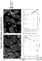
- Changes in the podocyte actin cytoskeleton were measured by the intensity and number of F-actin stress fibers.
- the F-actin stress fibers were sectioned from the image and contrasted to the intensity of the rest of the cell to give a ratio of intensities.
- the intensity ratio of the F-actin stress fibers to the cell along with the number of sectioned F-actin stress fibers per cell were able to quantitate changes in the podocyte induced by PAN.
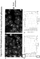
- Changes in the podocytes focal adhesions were also monitored using the previously mentioned parameters and were also able to quantitate changes in a dose response fashion to PAN.
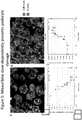
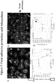
- the aforementioned parameters for quantitation of the podocyte actin cytoskeleton and focal adhesion were also found to measure in a dose response fashion the protection from PAN treatment by Mizouribine.
- these parameters were found to better differentiate a healthy podocyte phenotype from a damaged podocyte phenotype when combined.
- One example of multiparametric analysis would be a two dimensional comparison between the sectioned F-actin stress fibers intensity to the cell versus the cell's total area of sectioned F-actin stress fibers to sort cells into populations of healthy versus damaged cells. This multiparametric method was found to increase the sensitivity of the assay over using one parameter. Overall this assay is a robust quantitative method to determine in-vitro podocyte health and is a marked improvement over the current art of the field which is qualitative observation of podocyte health.
- Murine podocytes were cultured as have been described previously ( Saleem et al., JASN 13, 630-638 (2002 ); Mundel, Reiser et al., Experimental Cell Research, 236, p248 (1997 )).
- murine podocytes derived from the mouse line H-2K b -tsA58 were cultured under permissive conditions at 33°C on rat-tail collagen coated dishes in RPMI medium supplemented with 10% heat inactivated FBS, penicillin and streptomycin (100 U/ml), and 10 U/ml recombinant mouse ⁇ -interferon. Cells were then trypsinized and re-plated for differentiation at 37°C in medium without ⁇ -interferon.
- the cells were differentiated for 0 to 7 days and then reseeded to rat-tail collagen coated multi-well plates at a density of 1500/well or 500/well for 96 and 384 well plates, respectively. Cells were then further differentiated for another 5-14 days in the multi-well plates for subsequent experimentation with various reagents prior to fixation and analyses.
- Methods for isolating and culturing primary podocytes are well known in the art. For example, see: Kabgani et al., Primary Cultures of Glomerular Parietal Epithelial Cells or Podocytes with Proven Origin, Plos One, PLoS ONE (2012) 7(4): e34907. doi:10.1371/journal.pone.0034907 .
- Differentiated podocytes were cultured in 96- well or 384-well multiwall optical plates (PerkinElmer, Boston, MA) and treated with various amounts of puromycin, PAN, LPS, Adriamycin and other agents.
- the cells were co-treated with chemical and other agents (such as mizouribine) to prevent damage from damaging agents (such as PAN).
- the rescue agents were added 0-48 h after the addition of injury causing agents.
- the cells were fixed with a solution containing a 4% paraformaldehyde and 2% sucrose (added directly to cell culture media, without disturbing the cells or removing the culture media) 1 hour followed by permeabilization with 0.3% Triton X-100 in PBS for 10 min at room temperature.
- F-actin was visualized with Alexa fluor 594 phalloidin (Invitrogen), HCS CellMask Blue (Invitrogen) was used for a nuclear/cytoplasmic stain, and anti-Paxillin antibody clone Y113 was used for detecting focal adhesions.
- the anti-paxillin antibody was further detected using alexa fluor 488 conjugated secondary antibody. Confocal microscopy was performed using the Opera XL (Perkin Elmer) with appropriate filters. Images were used in subsequent image analysis for quantitation of a variety of cellular parameters. Assays were performed in replicate wells for assay robustness.
- splitting coefficients of 0.45 and 0.46, respectively, were applied. Healthy versus damaged cell populations were sorted by the parameters "Relative Spot to Background Intensity” and “Total Spot Area” parameters using the "Select Populations” analysis module. Healthy versus damaged cell populations were then used for determining the percentage of healthy cells per well. Approximately 100 to 1500 cells per well were analyzed.
Landscapes
- Health & Medical Sciences (AREA)
- Life Sciences & Earth Sciences (AREA)
- Engineering & Computer Science (AREA)
- Biomedical Technology (AREA)
- Chemical & Material Sciences (AREA)
- Urology & Nephrology (AREA)
- Immunology (AREA)
- Cell Biology (AREA)
- Biotechnology (AREA)
- Molecular Biology (AREA)
- Hematology (AREA)
- Bioinformatics & Cheminformatics (AREA)
- Microbiology (AREA)
- Biochemistry (AREA)
- General Health & Medical Sciences (AREA)
- Zoology (AREA)
- Organic Chemistry (AREA)
- Wood Science & Technology (AREA)
- Genetics & Genomics (AREA)
- Tropical Medicine & Parasitology (AREA)
- Medicinal Chemistry (AREA)
- General Physics & Mathematics (AREA)
- Pathology (AREA)
- Analytical Chemistry (AREA)
- Physics & Mathematics (AREA)
- Food Science & Technology (AREA)
- Toxicology (AREA)
- General Engineering & Computer Science (AREA)
- Virology (AREA)
- Physiology (AREA)
- Measuring Or Testing Involving Enzymes Or Micro-Organisms (AREA)
- Investigating Or Analysing Biological Materials (AREA)
- Micro-Organisms Or Cultivation Processes Thereof (AREA)
- Apparatus Associated With Microorganisms And Enzymes (AREA)
Claims (7)
- Verfahren zum Kultivieren von Podozyten in vitro, umfassend:(a) Unteralten einer bedingt-immortalisierten Podozytenzelle in einer ersten Zellkultur unter einer permissiven Bedingung oder einer Proliferationsbedingung für einen Zeitraum, ausreichend für die Podozytenzelle um sich wenigstens einmal zu verdoppeln, dadurch Herstellen einer Population von Podozyten;(b) Unterhalten der Population von Podozyten, erhalten in a), unter einer nicht-permissiven Bedingung oder einer Differenzierungsbedingung für einen Zeitraum, ausreichend, um eine Population von teilweise differenzierten Podozyten, aber nicht endgültig differenzierten Podozyten zu erhalten; und(c) Wiederansetzen der teilweise differenzierten Podozyten, erhalten in b), und Unterhalten der teilweise differenzierten Podozyten, erhalten in b), in einer zweiten Zellkultur unter einer nicht-permissiven Bedingung oder Differenzierungsbedingung für einen Zeitraum, ausreichend, um eine homogene Population von endgültig differenzierten Podozyten zu erhalten.
- Verfahren nach Anspruch 1, wobei die zweite Zellkultur in einer Multi-Well-Platte enthalten ist,
vorzugsweise wobei die Multi-Well-Platte für ein Hochdurchsatz-Screening geeignet ist,
vorzugsweise wobei die Podozytenzelle in einer adhärenten Kultur unterhalten wird. - Verfahren nach einem der Ansprüche 1 - 2, wobei der bedingt immortalisierte Podozyt eine Mauszelle oder eine menschliche Zelle ist.
- Verfahren nach Anspruch 3, wobei in Schritt (a) die Population von Podozyten in einer adhärenten Kultur unterhalten wird, die zu wenigstens ungefähr 60 % konfluent ist.
- Verfahren nach Anspruch 3 oder 4, wobei in Schritt (a) der bedingt-immortalisierte Podozyt unter den permissiven Bedingungen für zwischen ungefähr 5 Tagen und ungefähr 20 Tagen unterhalten wird,
vorzugsweise wobei in Schritt (a) der bedingt-immortalisierte Podozyt unter der permissiven Bedingung für zwischen ungefähr 7 Tagen und ungefähr 14 Tagen unterhalten wird. - Verfahren nach einem der Ansprüche 3 - 5, wobei in Schritt (b) die Population von Podozyten unter der nicht-permissiven Bedingung für zwischen ungefähr 4 Tagen und ungefähr 8 Tagen unterhalten wird.
- Verfahren nach einem der Ansprüche 3 - 6, wobei in Schritt (c) die teilweise differenzierten Podozyten unter der nicht-permissiven Bedingung für zwischen ungefähr 5 Tagen und ungefähr 20 Tagen unterhalten werden, vorzugsweise wobei in Schritt (c) die teilweise differenzierten Podozyten unter den nicht-permissiven Bedingungen für zwischen ungefähr 6 Tagen und ungefähr 15 Tagen unterhalten werden.
Applications Claiming Priority (2)
| Application Number | Priority Date | Filing Date | Title |
|---|---|---|---|
| US201361798385P | 2013-03-15 | 2013-03-15 | |
| PCT/US2014/022497 WO2014150178A1 (en) | 2013-03-15 | 2014-03-10 | Podocyte cultures and uses thereof |
Publications (3)
| Publication Number | Publication Date |
|---|---|
| EP2970895A1 EP2970895A1 (de) | 2016-01-20 |
| EP2970895A4 EP2970895A4 (de) | 2016-08-10 |
| EP2970895B1 true EP2970895B1 (de) | 2020-12-23 |
Family
ID=51580710
Family Applications (1)
| Application Number | Title | Priority Date | Filing Date |
|---|---|---|---|
| EP14769052.3A Active EP2970895B1 (de) | 2013-03-15 | 2014-03-10 | Podozytenkulturen und verwendungen davon |
Country Status (4)
| Country | Link |
|---|---|
| US (1) | US10526579B2 (de) |
| EP (1) | EP2970895B1 (de) |
| JP (2) | JP6524055B2 (de) |
| WO (1) | WO2014150178A1 (de) |
Families Citing this family (4)
| Publication number | Priority date | Publication date | Assignee | Title |
|---|---|---|---|---|
| EP2970895B1 (de) | 2013-03-15 | 2020-12-23 | Rush University Medical Center | Podozytenkulturen und verwendungen davon |
| JP2018514515A (ja) * | 2015-03-27 | 2018-06-07 | ラッシュ・ユニバーシティ・メディカル・センター | 腎疾患の骨髄系前駆細胞 |
| CA3112813A1 (en) | 2018-09-21 | 2020-03-26 | Berkeley Lights, Inc. | Functionalized well plate, methods of preparation and use thereof |
| US20240000930A1 (en) | 2020-12-09 | 2024-01-04 | Siwa Corporation | Methods and compositions for treating kidney diseases |
Family Cites Families (8)
| Publication number | Priority date | Publication date | Assignee | Title |
|---|---|---|---|---|
| CA2540529C (en) | 2003-10-03 | 2012-03-13 | Vib Vzw | Means and methods for the recruitment and identification of stem cells |
| WO2011019980A1 (en) | 2009-08-14 | 2011-02-17 | University Of Miami | Novel role of alpha-galactosidase activity as a biomarker in kidney disease |
| WO2011057172A1 (en) * | 2009-11-06 | 2011-05-12 | University Of Miami | Podocyte specific assays and uses thereof |
| US20110135574A1 (en) | 2009-11-16 | 2011-06-09 | Anna Greka | Methods of treating kidney disease |
| IT1397084B1 (it) * | 2009-12-17 | 2012-12-28 | Fond Irccs Ca Granda Ospedale Maggiore Policlinico | Metodo di co-coltura tridimensionale di podociti e cellule endoteliali e relativo sistema di co-coltura in vitro |
| US10052345B2 (en) * | 2010-10-19 | 2018-08-21 | University Of Miami | Assays, methods and kits for predicting renal disease and personalized treatment strategies |
| WO2013066879A2 (en) | 2011-10-31 | 2013-05-10 | The University Of Miami | SOLUBLE UROKINASE RECEPTOR (suPAR) IN DIABETIC KIDNEY DISEASE |
| EP2970895B1 (de) | 2013-03-15 | 2020-12-23 | Rush University Medical Center | Podozytenkulturen und verwendungen davon |
-
2014
- 2014-03-10 EP EP14769052.3A patent/EP2970895B1/de active Active
- 2014-03-10 WO PCT/US2014/022497 patent/WO2014150178A1/en not_active Ceased
- 2014-03-10 US US14/767,104 patent/US10526579B2/en active Active
- 2014-03-10 JP JP2016500983A patent/JP6524055B2/ja active Active
-
2019
- 2019-04-26 JP JP2019085695A patent/JP6824326B2/ja active Active
Non-Patent Citations (1)
| Title |
|---|
| REISERGUPTA ET AL., KIDNEY INT., vol. 77, 2010, pages 662 * |
Also Published As
| Publication number | Publication date |
|---|---|
| EP2970895A4 (de) | 2016-08-10 |
| WO2014150178A1 (en) | 2014-09-25 |
| US20150376575A1 (en) | 2015-12-31 |
| JP6524055B2 (ja) | 2019-06-05 |
| JP6824326B2 (ja) | 2021-02-03 |
| US10526579B2 (en) | 2020-01-07 |
| JP2016516406A (ja) | 2016-06-09 |
| JP2019162118A (ja) | 2019-09-26 |
| EP2970895A1 (de) | 2016-01-20 |
Similar Documents
| Publication | Publication Date | Title |
|---|---|---|
| Voges et al. | Vascular cells improve functionality of human cardiac organoids | |
| Lester et al. | Tau aggregates are RNA-protein assemblies that mislocalize multiple nuclear speckle components | |
| Short et al. | The contribution of branching morphogenesis to kidney development and disease | |
| Renner et al. | Self‐organized developmental patterning and differentiation in cerebral organoids | |
| Gilbert et al. | NEXMIF/KIDLIA knock-out mouse demonstrates autism-like behaviors, memory deficits, and impairments in synapse formation and function | |
| JP6824326B2 (ja) | 有足突起培養物およびその使用 | |
| Schroeder et al. | Model system identification of novel congenital heart disease gene candidates: focus on RPL13 | |
| De Silva et al. | Nuclear envelope disruption triggers hallmarks of aging in lung alveolar macrophages | |
| Dubaic et al. | Role of ciliopathy protein TMEM107 in eye development: insights from a mouse model and retinal organoid | |
| Louis et al. | A reductionist approach to determine the effect of cell-cell contact on human epidermal stem cell differentiation | |
| Osaki et al. | Early differential impact of MeCP2 mutations on functional networks in Rett syndrome patient-derived human cerebral organoids | |
| Lei et al. | Lymphatic muscle cells are unique cells that undergo aging induced changes | |
| Bouillet et al. | Revisiting the involvement of tau in complex neural network remodeling: Analysis of the extracellular neuronal activity in organotypic brain slice co-cultures | |
| Schulz et al. | Piezo mediates Rho activation and actin stress fibre formation in Drosophila nephrocytes | |
| Kumar et al. | Systematic discovery of transcription factors that improve hPSC-derived cardiomyocyte maturation via temporal analysis of bioengineered cardiac tissues | |
| Vermaercke et al. | SYNGAP1 deficiency disrupts neoteny in human cortical neurons in vivo | |
| Ilamathi et al. | Mitochondrial fission is required for proper nucleoid distribution within mitochondrial networks | |
| Barkan et al. | Embryo-scale single-cell chemical transcriptomics reveals dependencies between cell types and signaling pathways | |
| Delgado et al. | The Arp2/3 complex maintains genome integrity and survival of epidermal Langerhans cells | |
| Dell’Oca et al. | The CDKL5 kinase undergoes liquid-liquid phase separation driven by a serine-rich C-terminal region and impaired by neurodevelopmental disease-related truncations | |
| Drube et al. | Dynamic balance of H3K9me2 heterochromatin by CoREST-2 and RE-1 in growing neurons | |
| Weidling et al. | hiPSC-neurons recapitulate the subtype-specific cell intrinsic nature of susceptibility to neurodegenerative disease-relevant aggregation | |
| Belle et al. | Genetic analysis and multimodal imaging confirm m. 12148 T> C mitochondrial variant pathogenicity leading to multisystem dysfunction | |
| Cerrato et al. | Live cell imaging of LC3 dynamics | |
| Gutiérrez-García et al. | Temporal Dynamics of Azot Expression Following Traumatic Brain Injury |
Legal Events
| Date | Code | Title | Description |
|---|---|---|---|
| PUAI | Public reference made under article 153(3) epc to a published international application that has entered the european phase |
Free format text: ORIGINAL CODE: 0009012 |
|
| 17P | Request for examination filed |
Effective date: 20151015 |
|
| AK | Designated contracting states |
Kind code of ref document: A1 Designated state(s): AL AT BE BG CH CY CZ DE DK EE ES FI FR GB GR HR HU IE IS IT LI LT LU LV MC MK MT NL NO PL PT RO RS SE SI SK SM TR |
|
| AX | Request for extension of the european patent |
Extension state: BA ME |
|
| DAX | Request for extension of the european patent (deleted) | ||
| A4 | Supplementary search report drawn up and despatched |
Effective date: 20160708 |
|
| RIC1 | Information provided on ipc code assigned before grant |
Ipc: C12Q 1/68 20060101ALI20160704BHEP Ipc: C12N 5/071 20100101AFI20160704BHEP Ipc: G01N 33/50 20060101ALI20160704BHEP Ipc: G01N 33/569 20060101ALI20160704BHEP |
|
| STAA | Information on the status of an ep patent application or granted ep patent |
Free format text: STATUS: EXAMINATION IS IN PROGRESS |
|
| 17Q | First examination report despatched |
Effective date: 20170710 |
|
| GRAP | Despatch of communication of intention to grant a patent |
Free format text: ORIGINAL CODE: EPIDOSNIGR1 |
|
| STAA | Information on the status of an ep patent application or granted ep patent |
Free format text: STATUS: GRANT OF PATENT IS INTENDED |
|
| INTG | Intention to grant announced |
Effective date: 20200716 |
|
| GRAS | Grant fee paid |
Free format text: ORIGINAL CODE: EPIDOSNIGR3 |
|
| GRAA | (expected) grant |
Free format text: ORIGINAL CODE: 0009210 |
|
| STAA | Information on the status of an ep patent application or granted ep patent |
Free format text: STATUS: THE PATENT HAS BEEN GRANTED |
|
| AK | Designated contracting states |
Kind code of ref document: B1 Designated state(s): AL AT BE BG CH CY CZ DE DK EE ES FI FR GB GR HR HU IE IS IT LI LT LU LV MC MK MT NL NO PL PT RO RS SE SI SK SM TR |
|
| REG | Reference to a national code |
Ref country code: GB Ref legal event code: FG4D |
|
| REG | Reference to a national code |
Ref country code: DE Ref legal event code: R096 Ref document number: 602014073600 Country of ref document: DE |
|
| REG | Reference to a national code |
Ref country code: AT Ref legal event code: REF Ref document number: 1347751 Country of ref document: AT Kind code of ref document: T Effective date: 20210115 |
|
| REG | Reference to a national code |
Ref country code: IE Ref legal event code: FG4D |
|
| PG25 | Lapsed in a contracting state [announced via postgrant information from national office to epo] |
Ref country code: GR Free format text: LAPSE BECAUSE OF FAILURE TO SUBMIT A TRANSLATION OF THE DESCRIPTION OR TO PAY THE FEE WITHIN THE PRESCRIBED TIME-LIMIT Effective date: 20210324 Ref country code: RS Free format text: LAPSE BECAUSE OF FAILURE TO SUBMIT A TRANSLATION OF THE DESCRIPTION OR TO PAY THE FEE WITHIN THE PRESCRIBED TIME-LIMIT Effective date: 20201223 Ref country code: FI Free format text: LAPSE BECAUSE OF FAILURE TO SUBMIT A TRANSLATION OF THE DESCRIPTION OR TO PAY THE FEE WITHIN THE PRESCRIBED TIME-LIMIT Effective date: 20201223 Ref country code: NO Free format text: LAPSE BECAUSE OF FAILURE TO SUBMIT A TRANSLATION OF THE DESCRIPTION OR TO PAY THE FEE WITHIN THE PRESCRIBED TIME-LIMIT Effective date: 20210323 |
|
| REG | Reference to a national code |
Ref country code: AT Ref legal event code: MK05 Ref document number: 1347751 Country of ref document: AT Kind code of ref document: T Effective date: 20201223 |
|
| REG | Reference to a national code |
Ref country code: NL Ref legal event code: MP Effective date: 20201223 |
|
| PG25 | Lapsed in a contracting state [announced via postgrant information from national office to epo] |
Ref country code: LV Free format text: LAPSE BECAUSE OF FAILURE TO SUBMIT A TRANSLATION OF THE DESCRIPTION OR TO PAY THE FEE WITHIN THE PRESCRIBED TIME-LIMIT Effective date: 20201223 Ref country code: SE Free format text: LAPSE BECAUSE OF FAILURE TO SUBMIT A TRANSLATION OF THE DESCRIPTION OR TO PAY THE FEE WITHIN THE PRESCRIBED TIME-LIMIT Effective date: 20201223 Ref country code: BG Free format text: LAPSE BECAUSE OF FAILURE TO SUBMIT A TRANSLATION OF THE DESCRIPTION OR TO PAY THE FEE WITHIN THE PRESCRIBED TIME-LIMIT Effective date: 20210323 |
|
| PG25 | Lapsed in a contracting state [announced via postgrant information from national office to epo] |
Ref country code: NL Free format text: LAPSE BECAUSE OF FAILURE TO SUBMIT A TRANSLATION OF THE DESCRIPTION OR TO PAY THE FEE WITHIN THE PRESCRIBED TIME-LIMIT Effective date: 20201223 Ref country code: HR Free format text: LAPSE BECAUSE OF FAILURE TO SUBMIT A TRANSLATION OF THE DESCRIPTION OR TO PAY THE FEE WITHIN THE PRESCRIBED TIME-LIMIT Effective date: 20201223 |
|
| REG | Reference to a national code |
Ref country code: LT Ref legal event code: MG9D |
|
| PG25 | Lapsed in a contracting state [announced via postgrant information from national office to epo] |
Ref country code: PT Free format text: LAPSE BECAUSE OF FAILURE TO SUBMIT A TRANSLATION OF THE DESCRIPTION OR TO PAY THE FEE WITHIN THE PRESCRIBED TIME-LIMIT Effective date: 20210423 Ref country code: RO Free format text: LAPSE BECAUSE OF FAILURE TO SUBMIT A TRANSLATION OF THE DESCRIPTION OR TO PAY THE FEE WITHIN THE PRESCRIBED TIME-LIMIT Effective date: 20201223 Ref country code: SK Free format text: LAPSE BECAUSE OF FAILURE TO SUBMIT A TRANSLATION OF THE DESCRIPTION OR TO PAY THE FEE WITHIN THE PRESCRIBED TIME-LIMIT Effective date: 20201223 Ref country code: LT Free format text: LAPSE BECAUSE OF FAILURE TO SUBMIT A TRANSLATION OF THE DESCRIPTION OR TO PAY THE FEE WITHIN THE PRESCRIBED TIME-LIMIT Effective date: 20201223 Ref country code: SM Free format text: LAPSE BECAUSE OF FAILURE TO SUBMIT A TRANSLATION OF THE DESCRIPTION OR TO PAY THE FEE WITHIN THE PRESCRIBED TIME-LIMIT Effective date: 20201223 Ref country code: CZ Free format text: LAPSE BECAUSE OF FAILURE TO SUBMIT A TRANSLATION OF THE DESCRIPTION OR TO PAY THE FEE WITHIN THE PRESCRIBED TIME-LIMIT Effective date: 20201223 Ref country code: EE Free format text: LAPSE BECAUSE OF FAILURE TO SUBMIT A TRANSLATION OF THE DESCRIPTION OR TO PAY THE FEE WITHIN THE PRESCRIBED TIME-LIMIT Effective date: 20201223 |
|
| PG25 | Lapsed in a contracting state [announced via postgrant information from national office to epo] |
Ref country code: PL Free format text: LAPSE BECAUSE OF FAILURE TO SUBMIT A TRANSLATION OF THE DESCRIPTION OR TO PAY THE FEE WITHIN THE PRESCRIBED TIME-LIMIT Effective date: 20201223 Ref country code: AT Free format text: LAPSE BECAUSE OF FAILURE TO SUBMIT A TRANSLATION OF THE DESCRIPTION OR TO PAY THE FEE WITHIN THE PRESCRIBED TIME-LIMIT Effective date: 20201223 |
|
| REG | Reference to a national code |
Ref country code: DE Ref legal event code: R097 Ref document number: 602014073600 Country of ref document: DE |
|
| PG25 | Lapsed in a contracting state [announced via postgrant information from national office to epo] |
Ref country code: IS Free format text: LAPSE BECAUSE OF FAILURE TO SUBMIT A TRANSLATION OF THE DESCRIPTION OR TO PAY THE FEE WITHIN THE PRESCRIBED TIME-LIMIT Effective date: 20210423 |
|
| PG25 | Lapsed in a contracting state [announced via postgrant information from national office to epo] |
Ref country code: AL Free format text: LAPSE BECAUSE OF FAILURE TO SUBMIT A TRANSLATION OF THE DESCRIPTION OR TO PAY THE FEE WITHIN THE PRESCRIBED TIME-LIMIT Effective date: 20201223 Ref country code: MC Free format text: LAPSE BECAUSE OF FAILURE TO SUBMIT A TRANSLATION OF THE DESCRIPTION OR TO PAY THE FEE WITHIN THE PRESCRIBED TIME-LIMIT Effective date: 20201223 Ref country code: IT Free format text: LAPSE BECAUSE OF FAILURE TO SUBMIT A TRANSLATION OF THE DESCRIPTION OR TO PAY THE FEE WITHIN THE PRESCRIBED TIME-LIMIT Effective date: 20201223 |
|
| PLBE | No opposition filed within time limit |
Free format text: ORIGINAL CODE: 0009261 |
|
| STAA | Information on the status of an ep patent application or granted ep patent |
Free format text: STATUS: NO OPPOSITION FILED WITHIN TIME LIMIT |
|
| PG25 | Lapsed in a contracting state [announced via postgrant information from national office to epo] |
Ref country code: DK Free format text: LAPSE BECAUSE OF FAILURE TO SUBMIT A TRANSLATION OF THE DESCRIPTION OR TO PAY THE FEE WITHIN THE PRESCRIBED TIME-LIMIT Effective date: 20201223 Ref country code: ES Free format text: LAPSE BECAUSE OF FAILURE TO SUBMIT A TRANSLATION OF THE DESCRIPTION OR TO PAY THE FEE WITHIN THE PRESCRIBED TIME-LIMIT Effective date: 20201223 |
|
| 26N | No opposition filed |
Effective date: 20210924 |
|
| REG | Reference to a national code |
Ref country code: BE Ref legal event code: MM Effective date: 20210331 |
|
| PG25 | Lapsed in a contracting state [announced via postgrant information from national office to epo] |
Ref country code: IE Free format text: LAPSE BECAUSE OF NON-PAYMENT OF DUE FEES Effective date: 20210310 Ref country code: LU Free format text: LAPSE BECAUSE OF NON-PAYMENT OF DUE FEES Effective date: 20210310 |
|
| PG25 | Lapsed in a contracting state [announced via postgrant information from national office to epo] |
Ref country code: SI Free format text: LAPSE BECAUSE OF FAILURE TO SUBMIT A TRANSLATION OF THE DESCRIPTION OR TO PAY THE FEE WITHIN THE PRESCRIBED TIME-LIMIT Effective date: 20201223 |
|
| PG25 | Lapsed in a contracting state [announced via postgrant information from national office to epo] |
Ref country code: IS Free format text: LAPSE BECAUSE OF FAILURE TO SUBMIT A TRANSLATION OF THE DESCRIPTION OR TO PAY THE FEE WITHIN THE PRESCRIBED TIME-LIMIT Effective date: 20210423 |
|
| PG25 | Lapsed in a contracting state [announced via postgrant information from national office to epo] |
Ref country code: BE Free format text: LAPSE BECAUSE OF NON-PAYMENT OF DUE FEES Effective date: 20210331 |
|
| PG25 | Lapsed in a contracting state [announced via postgrant information from national office to epo] |
Ref country code: HU Free format text: LAPSE BECAUSE OF FAILURE TO SUBMIT A TRANSLATION OF THE DESCRIPTION OR TO PAY THE FEE WITHIN THE PRESCRIBED TIME-LIMIT; INVALID AB INITIO Effective date: 20140310 |
|
| P01 | Opt-out of the competence of the unified patent court (upc) registered |
Effective date: 20230508 |
|
| PG25 | Lapsed in a contracting state [announced via postgrant information from national office to epo] |
Ref country code: CY Free format text: LAPSE BECAUSE OF FAILURE TO SUBMIT A TRANSLATION OF THE DESCRIPTION OR TO PAY THE FEE WITHIN THE PRESCRIBED TIME-LIMIT Effective date: 20201223 |
|
| PG25 | Lapsed in a contracting state [announced via postgrant information from national office to epo] |
Ref country code: MK Free format text: LAPSE BECAUSE OF FAILURE TO SUBMIT A TRANSLATION OF THE DESCRIPTION OR TO PAY THE FEE WITHIN THE PRESCRIBED TIME-LIMIT Effective date: 20201223 |
|
| PG25 | Lapsed in a contracting state [announced via postgrant information from national office to epo] |
Ref country code: MT Free format text: LAPSE BECAUSE OF FAILURE TO SUBMIT A TRANSLATION OF THE DESCRIPTION OR TO PAY THE FEE WITHIN THE PRESCRIBED TIME-LIMIT Effective date: 20201223 |
|
| PGFP | Annual fee paid to national office [announced via postgrant information from national office to epo] |
Ref country code: CH Payment date: 20250401 Year of fee payment: 12 |
|
| PG25 | Lapsed in a contracting state [announced via postgrant information from national office to epo] |
Ref country code: TR Free format text: LAPSE BECAUSE OF FAILURE TO SUBMIT A TRANSLATION OF THE DESCRIPTION OR TO PAY THE FEE WITHIN THE PRESCRIBED TIME-LIMIT Effective date: 20201223 |
|
| REG | Reference to a national code |
Ref country code: CH Ref legal event code: U11 Free format text: ST27 STATUS EVENT CODE: U-0-0-U10-U11 (AS PROVIDED BY THE NATIONAL OFFICE) Effective date: 20260401 |
|
| PGFP | Annual fee paid to national office [announced via postgrant information from national office to epo] |
Ref country code: GB Payment date: 20260327 Year of fee payment: 13 |
|
| PGFP | Annual fee paid to national office [announced via postgrant information from national office to epo] |
Ref country code: DE Payment date: 20260327 Year of fee payment: 13 |
|
| PGFP | Annual fee paid to national office [announced via postgrant information from national office to epo] |
Ref country code: FR Payment date: 20260325 Year of fee payment: 13 |