EP2977922A2 - Procédé et système de planification de thérapie automatisée pour la sténose artérielle - Google Patents
Procédé et système de planification de thérapie automatisée pour la sténose artérielle Download PDFInfo
- Publication number
- EP2977922A2 EP2977922A2 EP15177855.2A EP15177855A EP2977922A2 EP 2977922 A2 EP2977922 A2 EP 2977922A2 EP 15177855 A EP15177855 A EP 15177855A EP 2977922 A2 EP2977922 A2 EP 2977922A2
- Authority
- EP
- European Patent Office
- Prior art keywords
- stenotic lesions
- treatment options
- stenting
- treatment
- patient
- Prior art date
- Legal status (The legal status is an assumption and is not a legal conclusion. Google has not performed a legal analysis and makes no representation as to the accuracy of the status listed.)
- Granted
Links
Images


Classifications
-
- A—HUMAN NECESSITIES
- A61—MEDICAL OR VETERINARY SCIENCE; HYGIENE
- A61B—DIAGNOSIS; SURGERY; IDENTIFICATION
- A61B5/00—Measuring for diagnostic purposes; Identification of persons
- A61B5/48—Other medical applications
-
- G—PHYSICS
- G16—INFORMATION AND COMMUNICATION TECHNOLOGY [ICT] SPECIALLY ADAPTED FOR SPECIFIC APPLICATION FIELDS
- G16H—HEALTHCARE INFORMATICS, i.e. INFORMATION AND COMMUNICATION TECHNOLOGY [ICT] SPECIALLY ADAPTED FOR THE HANDLING OR PROCESSING OF MEDICAL OR HEALTHCARE DATA
- G16H20/00—ICT specially adapted for therapies or health-improving plans, e.g. for handling prescriptions, for steering therapy or for monitoring patient compliance
-
- A—HUMAN NECESSITIES
- A61—MEDICAL OR VETERINARY SCIENCE; HYGIENE
- A61B—DIAGNOSIS; SURGERY; IDENTIFICATION
- A61B34/00—Computer-aided surgery; Manipulators or robots specially adapted for use in surgery
- A61B34/10—Computer-aided planning, simulation or modelling of surgical operations
-
- A—HUMAN NECESSITIES
- A61—MEDICAL OR VETERINARY SCIENCE; HYGIENE
- A61B—DIAGNOSIS; SURGERY; IDENTIFICATION
- A61B6/00—Apparatus or devices for radiation diagnosis; Apparatus or devices for radiation diagnosis combined with radiation therapy equipment
- A61B6/02—Arrangements for diagnosis sequentially in different planes; Stereoscopic radiation diagnosis
- A61B6/03—Computed tomography [CT]
- A61B6/032—Transmission computed tomography [CT]
-
- A—HUMAN NECESSITIES
- A61—MEDICAL OR VETERINARY SCIENCE; HYGIENE
- A61B—DIAGNOSIS; SURGERY; IDENTIFICATION
- A61B6/00—Apparatus or devices for radiation diagnosis; Apparatus or devices for radiation diagnosis combined with radiation therapy equipment
- A61B6/48—Diagnostic techniques
- A61B6/481—Diagnostic techniques involving the use of contrast agents
-
- A—HUMAN NECESSITIES
- A61—MEDICAL OR VETERINARY SCIENCE; HYGIENE
- A61B—DIAGNOSIS; SURGERY; IDENTIFICATION
- A61B6/00—Apparatus or devices for radiation diagnosis; Apparatus or devices for radiation diagnosis combined with radiation therapy equipment
- A61B6/50—Apparatus or devices for radiation diagnosis; Apparatus or devices for radiation diagnosis combined with radiation therapy equipment specially adapted for specific body parts; specially adapted for specific clinical applications
- A61B6/504—Apparatus or devices for radiation diagnosis; Apparatus or devices for radiation diagnosis combined with radiation therapy equipment specially adapted for specific body parts; specially adapted for specific clinical applications for diagnosis of blood vessels, e.g. by angiography
-
- G—PHYSICS
- G06—COMPUTING OR CALCULATING; COUNTING
- G06T—IMAGE DATA PROCESSING OR GENERATION, IN GENERAL
- G06T11/00—Two-dimensional [2D] image generation
- G06T11/20—Drawing from basic elements
-
- G—PHYSICS
- G06—COMPUTING OR CALCULATING; COUNTING
- G06T—IMAGE DATA PROCESSING OR GENERATION, IN GENERAL
- G06T7/00—Image analysis
- G06T7/0002—Inspection of images, e.g. flaw detection
- G06T7/0012—Biomedical image inspection
-
- G—PHYSICS
- G06—COMPUTING OR CALCULATING; COUNTING
- G06T—IMAGE DATA PROCESSING OR GENERATION, IN GENERAL
- G06T7/00—Image analysis
- G06T7/20—Analysis of motion
-
- G—PHYSICS
- G16—INFORMATION AND COMMUNICATION TECHNOLOGY [ICT] SPECIALLY ADAPTED FOR SPECIFIC APPLICATION FIELDS
- G16H—HEALTHCARE INFORMATICS, i.e. INFORMATION AND COMMUNICATION TECHNOLOGY [ICT] SPECIALLY ADAPTED FOR THE HANDLING OR PROCESSING OF MEDICAL OR HEALTHCARE DATA
- G16H30/00—ICT specially adapted for the handling or processing of medical images
- G16H30/20—ICT specially adapted for the handling or processing of medical images for handling medical images, e.g. DICOM, HL7 or PACS
-
- G—PHYSICS
- G16—INFORMATION AND COMMUNICATION TECHNOLOGY [ICT] SPECIALLY ADAPTED FOR SPECIFIC APPLICATION FIELDS
- G16H—HEALTHCARE INFORMATICS, i.e. INFORMATION AND COMMUNICATION TECHNOLOGY [ICT] SPECIALLY ADAPTED FOR THE HANDLING OR PROCESSING OF MEDICAL OR HEALTHCARE DATA
- G16H30/00—ICT specially adapted for the handling or processing of medical images
- G16H30/40—ICT specially adapted for the handling or processing of medical images for processing medical images, e.g. editing
-
- G—PHYSICS
- G16—INFORMATION AND COMMUNICATION TECHNOLOGY [ICT] SPECIALLY ADAPTED FOR SPECIFIC APPLICATION FIELDS
- G16H—HEALTHCARE INFORMATICS, i.e. INFORMATION AND COMMUNICATION TECHNOLOGY [ICT] SPECIALLY ADAPTED FOR THE HANDLING OR PROCESSING OF MEDICAL OR HEALTHCARE DATA
- G16H50/00—ICT specially adapted for medical diagnosis, medical simulation or medical data mining; ICT specially adapted for detecting, monitoring or modelling epidemics or pandemics
- G16H50/20—ICT specially adapted for medical diagnosis, medical simulation or medical data mining; ICT specially adapted for detecting, monitoring or modelling epidemics or pandemics for computer-aided diagnosis, e.g. based on medical expert systems
-
- G—PHYSICS
- G16—INFORMATION AND COMMUNICATION TECHNOLOGY [ICT] SPECIALLY ADAPTED FOR SPECIFIC APPLICATION FIELDS
- G16H—HEALTHCARE INFORMATICS, i.e. INFORMATION AND COMMUNICATION TECHNOLOGY [ICT] SPECIALLY ADAPTED FOR THE HANDLING OR PROCESSING OF MEDICAL OR HEALTHCARE DATA
- G16H50/00—ICT specially adapted for medical diagnosis, medical simulation or medical data mining; ICT specially adapted for detecting, monitoring or modelling epidemics or pandemics
- G16H50/50—ICT specially adapted for medical diagnosis, medical simulation or medical data mining; ICT specially adapted for detecting, monitoring or modelling epidemics or pandemics for simulation or modelling of medical disorders
-
- A—HUMAN NECESSITIES
- A61—MEDICAL OR VETERINARY SCIENCE; HYGIENE
- A61B—DIAGNOSIS; SURGERY; IDENTIFICATION
- A61B6/00—Apparatus or devices for radiation diagnosis; Apparatus or devices for radiation diagnosis combined with radiation therapy equipment
- A61B6/46—Arrangements for interfacing with the operator or the patient
- A61B6/461—Displaying means of special interest
- A61B6/463—Displaying means of special interest characterised by displaying multiple images or images and diagnostic data on one display
-
- A—HUMAN NECESSITIES
- A61—MEDICAL OR VETERINARY SCIENCE; HYGIENE
- A61B—DIAGNOSIS; SURGERY; IDENTIFICATION
- A61B6/00—Apparatus or devices for radiation diagnosis; Apparatus or devices for radiation diagnosis combined with radiation therapy equipment
- A61B6/46—Arrangements for interfacing with the operator or the patient
- A61B6/461—Displaying means of special interest
- A61B6/466—Displaying means of special interest adapted to display 3D data
-
- A—HUMAN NECESSITIES
- A61—MEDICAL OR VETERINARY SCIENCE; HYGIENE
- A61B—DIAGNOSIS; SURGERY; IDENTIFICATION
- A61B6/00—Apparatus or devices for radiation diagnosis; Apparatus or devices for radiation diagnosis combined with radiation therapy equipment
- A61B6/52—Devices using data or image processing specially adapted for radiation diagnosis
- A61B6/5211—Devices using data or image processing specially adapted for radiation diagnosis involving processing of medical diagnostic data
- A61B6/5217—Devices using data or image processing specially adapted for radiation diagnosis involving processing of medical diagnostic data extracting a diagnostic or physiological parameter from medical diagnostic data
-
- G—PHYSICS
- G06—COMPUTING OR CALCULATING; COUNTING
- G06T—IMAGE DATA PROCESSING OR GENERATION, IN GENERAL
- G06T2207/00—Indexing scheme for image analysis or image enhancement
- G06T2207/10—Image acquisition modality
- G06T2207/10072—Tomographic images
- G06T2207/10088—Magnetic resonance imaging [MRI]
-
- G—PHYSICS
- G06—COMPUTING OR CALCULATING; COUNTING
- G06T—IMAGE DATA PROCESSING OR GENERATION, IN GENERAL
- G06T2207/00—Indexing scheme for image analysis or image enhancement
- G06T2207/30—Subject of image; Context of image processing
- G06T2207/30004—Biomedical image processing
- G06T2207/30101—Blood vessel; Artery; Vein; Vascular
- G06T2207/30104—Vascular flow; Blood flow; Perfusion
Definitions
- the present invention relates to treatment planning for arterial stenosis, and more particularly, to selecting which stenoses or lesions to stent based on medical image data of a patient.
- Cardiovascular disease is the leading cause of deaths worldwide.
- coronary artery disease CAD
- CAD coronary artery disease
- CAD coronary artery disease
- stenosis represents an important cause of cardiovascular diseases.
- Such stenoses typically develop gradually over time, and can develop in different parts of the arterial circulation, such as the coronary arteries, renal arteries, peripheral arteries, carotid artery, cerebral artery, etc.
- Such a local narrowing can also be the result of a congenital defect.
- One therapy widely used for treating arterial stenosis is stenting, i.e., the placement of a metal or polymer stent in the artery to open up the lumen, and hence facilitate the flow of blood.
- PCI percutaneous coronary intervention
- the present invention provides a method and system for automated decision support for therapy planning for arterial stenoses or lesions based on medical image data.
- Embodiments of the present invention acquire medical image data of a patient's heart, segment the medical image data and extract a geometrical model of the coronary arteries, and utilize computational techniques to compute hemodynamic quantities for various stenoses in the patient's coronary arteries.
- Embodiments of the present invention identify a set of possible treatment options, each corresponding to a possible combination of stents covering one or multiple stenoses, and calculate computational hemodynamic quantities for each of the treatment options.
- Embodiments of the present invention generate a clinically relevant figure of merit for each of the treatment options and present the results to a user to provide decision support for selecting one of the treatment options.
- a set of stenotic lesions is identified in a patient's coronary arteries from medical image data of the patient.
- a plurality of treatment options are generated for the set of stenotic lesions, wherein each of the plurality of treatment options corresponds to a stenting configuration in which one or more of the stenotic lesions are stented.
- predicted hemodynamic metrics for the set of stenotic lesions resulting from the stenting configuration corresponding to that treatment option are calculated.
- the present invention relates to a method and system for automated decision support for treatment planning of arterial stenosis.
- Embodiments of the present invention are described herein to give a visual understanding of the methods for treatment planning for arterial stenosis.
- a digital image is often composed of digital representations of one or more objects (or shapes).
- the digital representation of an object is often described herein in terms of identifying and manipulating the objects.
- Such manipulations are virtual manipulations accomplished in the memory or other circuitry / hardware of a computer system. Accordingly, is to be understood that embodiments of the present invention may be performed within a computer system using data stored within the computer system.
- Medical image data of a patient's heart such as computed tomography (CT) data can be acquired and used to extract quantitative information that supports the choice between different therapy options for coronary artery stenoses.
- CT computed tomography
- the medical image data can be segmented to extract a model of the patient's coronary arteries.
- the pressure drop values across a plurality of stenotic lesions can be computed non-invasively, for example using computational fluid dynamics (CFD) or other computational approaches such as machine-learning based approaches, and these values can be used to compute hemodynamic quantities, such as fractional flow reserve (FFR), that support an initial clinical decision regarding whether or not therapy in the form of stenting one or more of the lesions is needed.
- CFD computational fluid dynamics
- FFR fractional flow reserve
- Embodiments of the present invention are described herein for providing automated decision support for percutaneous coronary intervention (PCI) treatment planning for stenting coronary artery stenoses (lesions).
- PCI percutaneous coronary intervention
- the methods described herein can be similarly applied for treatment planning for stenting stenotic lesions in other types of arteries as well, such as the renal arteries, peripheral arteries, carotid artery, cerebral artery, etc.
- the methods described herein can also be applied to other parts of the circulatory system, such as for venous circulation or pulmonary circulation.
- Embodiments of the present invention can also be applied to treatment planning for airways.
- FIG. 1 illustrates a method of treatment planning for a plurality of coronary artery stenotic lesions of a patient according to an embodiment of the present invention.
- medical image data of a patient is received.
- the medical image data is acquired prior to performing stenting, such as PCI for a coronary artery stenosis.
- Medical image data from one or multiple imaging modalities can be received.
- the medical image data can include, computed tomography (CT), Dyna CT, magnetic resonance (MR), Angiography, Ultrasound, Single Photon Emission computed Tomography (SPECT), and any other type of medical imaging modality.
- the medical image data can be 2D, 3D, or 4D (3D+time) medical image data.
- the medical image data can be received directly from one or more image acquisition devices, such as a CT scanner, MR scanner, Angiography scanner, Ultrasound device, etc., or the medical image data may be received by loading previously stored medical image data for a patient.
- 3D coronary CT angiography (CTA) images are acquired on a CT scanner.
- the CTA images ensure that the coronary vasculature, including the vessel(s) that contain the stenosis, is adequately imaged using a contrast agent that is injected into the patient.
- the clinician may be provided with an option of identifying lesions (stenoses) of interest by interactively viewing them on the images.
- This step can also be performed on a patient-specific anatomical model that is extracted from the image data (step 104).
- the stenoses may be automatically detected in the image data using an algorithm for automatic detection of coronary artery stenosis, such as the method for automatic detection of coronary artery stenosis described in United States Published Patent Application No. 2011/0224542 , which is incorporated herein by reference.
- algorithm for automatic detection of coronary artery stenosis such as the method for automatic detection of coronary artery stenosis described in United States Published Patent Application No. 2011/0224542 , which is incorporated herein by reference.
- other non-invasive clinical measurements such as the patient's heart rate and systolic and diastolic blood pressure may also be acquired. These non-invasive clinical measurements can be used to establish boundary conditions for CFD computations.
- a patient-specific anatomical model of the coronary arteries is extracted from the medical image data.
- the patient-specific anatomical model may be a patient-specific anatomical model of any portion of the full coronary artery tree of the patient.
- the coronary arteries can be segmented in the 3D medical image data using an automated coronary artery centerline extraction algorithm.
- the coronary arteries can be segmented in a CT volume using the method described United States Published Patent Application No. 2010/0067760 , which is incorporated herein by reference.
- the cross-section contour at each centerline point gives a corresponding cross-section area measurement at that point in the coronary artery.
- a geometric surface model is then generated for the segmented coronary arteries. For example, methods for anatomical modeling of the coronary arteries are described in United States Patent No. 7,860,290 and United States Patent No. 7,953,266 , both of which are incorporated herein by reference.
- the patient-specific anatomical model can include the aortic root together with the proximal part of the aorta.
- a detailed 3D model of each stenosis can also be extracted using similar algorithms, which includes the quantification of the proximal vessel diameter and area, distal vessel diameter and area, minimal lumen diameter and area, and length of stenosis.
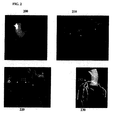
- FIG. 2 illustrates exemplary results for generating a patient-specific anatomical model of the coronary vessel tree.
- Image 200 of Fig. 2 shows coronary CTA data.
- Image 210 shows a centerline tree 212 extracted from the CTA data.
- Image 220 shows a cross-section contours 222 extracted at each point of the centerline tree 212.
- Image 230 shows a 2D surface mesh 232 of the coronary arteries, the aortic root, and the proximal part of the aorta. It is to be understood that the anatomical model of the coronary tree of the patient can be output and displayed, for example on a display screen of the computer system.
- the above described anatomical modeling tasks can be performed automatically or can be user-driven, thereby allowing the user (clinician) to interactively make changes to the anatomical models to analyze the effects of such changes on the subsequent computation of FFR.
- the myocardium may also be segmented (either automatically or manually) in the medical image data to determine an estimate of the left ventricular mass, which in a possible implementation, may be used to estimate the absolute resting flow for the patient which is used to calculate boundary conditions for a computational blood flow and pressure simulation.
- the resting flow could also be computed based on the total volume of the segmented coronary tree, or from the outlet radius of the different coronary vessels.
- a patient-specific anatomical model of the heart that is automatically generated from the image data may be used for this purpose.
- the anatomical heart model is a multi-component model having multiple cardiac components, including the four chambers (left ventricle, left atrium, right ventricle, and right atrium).
- the anatomical heart model may also include components such as the heart valves (aortic valve, mitral valve, tricuspid valve, and pulmonary valve) and the aorta.
- Such a comprehensive model of the heart is used to capture a large variety of morphological, functional, and pathological variations.
- a modular and hierarchical approach can be used to reduce anatomical complexity and facilitate an effective and flexible estimation of individual anatomies.
- the 4D anatomical heart model can be generated by generating individual models of each heart component, for example using marginal space learning (MSL), and then integrating the heart component models by establishing mesh point correspondence. Additional details regarding generation of such a 4D patient-specific heart model are described in United States Published Patent Application No. 2012/0022843 , which is incorporated herein by reference in its entirety.
- MSL marginal space learning
- a plurality of hemodynamically relevant stenotic lesions are identified in the coronary artery.
- all of the lesions can be automatically detected in the medical image data or in the patient-specific anatomical model of the coronary arteries and then a hemodynamic metric, such as FFR, can be computed for each of the detected lesions.
- a hemodynamic metric such as FFR
- Stenosis locations in the coronary tree can be automatically determined by performing coronary centerline extraction from the medical image data, calculating the vessel diameter in cross-sections along the centerline, and comparing the sequences of diameters.
- a stenosis (lesion) is detected wherever the diameter shrinks in the distal direction and then increases again. Additional details regarding a method for automatic detection of coronary artery stenosis are described in United States Published Patent Application No. 2011/0224542 , which is incorporated herein by reference.
- FFR can be automatically calculated for each detected stenosis location by simulating blood flow and pressure in the patient-specific anatomical model of the coronary arteries using a computational model of coronary circulation.
- the blood flow and pressure can be simulated in the anatomical model using CFD computations or any other standard numerical technique, such as finite-element method, finite-difference method, finite volume method, boundary element method, embedded boundary method, immersed boundary method lattice Boltzmann method, etc.
- a multi-scale computational model of coronary circulation can be used to compute the compute the blood flow and pressure in the pre-stenting anatomical model of the coronary arteries over a series of time steps.
- the simulation may be performed for a plurality of time steps corresponding to a fully cardiac cycle or multiple cardiac cycles.
- the computational model of coronary circulation can model the loss of pressure across stenoses or other narrowings in the coronary arteries (e.g., calcification, thrombus, bifurcation, etc.) using pressure-drop models.
- stenosis and lesion are used to generally refer to any type of narrowing in a vessel.
- the pressure drop model for a particular stenosis computes the pressure drop over the stenosis due the narrowing of the vessel without performing an explicit flow computation in that region of the vessel.
- FIG. 3 illustrates an exemplary multi-scale computational model of coronary circulation according to an embodiment of the present invention.
- a heart model 302 is coupled at the root of the aorta.
- the heart model 302 may be implemented as a lumped model parameterized through patient-specific data as shown in FIG. 3 , or may be implemented as a full 3D heart model.
- Large arteries, such as the aorta 304 together with the large arteries supplied by the aorta (e.g., subclavian, brachiocephalic, common carotid, etc.), the left coronary artery (LCA) 306, and the right coronary artery (RCA) 308 can be represented as 1 D blood flow models or full 3D models.
- LCA left coronary artery
- RCA right coronary artery
- semi-analytical circulatory models can be used either separately for certain arterial segments, or embedded within the 1 D or 3D models.
- the vessel walls can be modeled as a purely elastic or visco-elastic material.
- the wall properties may be determined through an empirical relationship fit to measured data or based on patient-specific estimations of wall compliance.
- all microvascular beds are simulated through lumped parameter models 310 which account for the resistance applied to the blood flow and for the compliance of the distal vessels.
- the coronary vascular bed is modeled through such lumped parameter models 310, which are adapted to the coronary circulation in the sense that they take into account the effects of the myocardial contraction on the flow waveform.
- Stenosis segments 312 and 314 are shown in the model of coronary arterial circulation.
- the stenosis segments 312 and 314 cannot be simulated using the 1 D blood flow models since there is a high variation in cross-sectional area and the shape of the stenosis influences the blood flow behavior and especially the trans-stenotic pressure drop which plays a major role in the assessment of the functional importance of such a stenosis.
- reduced-order (as compared to a full 3D model) pressure-drop model can be used for each stenosis segment 312 and 314.
- a pressure-drop model can be used to compute the pressure-drop across each stenosis region (e.g., 312 and 314 of FIG. 3 ) in the pre-stenting anatomical model of the coronary arteries without performing an explicit flow computation in the stenosis region.
- Various pressure-drop models can be used.
- the pressure-drop model for a stenosis may be a fully analytical model or may be a model that includes a combination of analytical and empirical terms.
- a pressure-drop model that includes a combination of analytical and empirical terms is referred to herein as a "semi-empirical pressure-drop model".
- pressure-drop models may be used as well, such as a machine-learning based pressure-drop model that is trained using a machine-learning algorithm to map anatomical and flow features derived from a stenosis to a pressure-drop associated with the stenosis.
- the hemodynamic metric such as FFR
- the hemodynamic metric can be calculated automatically based on patient-specific measurements extracted from the medical image data and patient-specific clinical measurements (e.g., heart rate, blood pressure, etc.) acquired for the patient using a machine learning based technique. Details of such machine learning based techniques for calculating FFR are described in U.S. Published Patent Application 2015/0112182 , entitled “Method and System for Machine Learning Based Assessment of Fractional Flow Reserve", which is incorporated herein by reference in its entirety.
- a FFR value (or other hemodynamic metric) is calculated for each stenotic lesion
- the FFR value is compared to a threshold vale to determine if the stenotic lesion is hemodynamically relevant.
- a threshold of 0.8 may be used for FFR, but the present invention is not limited thereto. If the FFR value at a particular stenotic lesion is less than the threshold value (e.g., ⁇ 0.8), that stenotic lesion is hemodynamically relevant and is included in the plurality of hemodynamically relevant stenotic lesions to be considered for stenting.
- all preceding stenotic lesions i.e., in the proximal direction
- all preceding stenotic lesions in a blood flow path in the coronary artery tree may also be identified as hemodynamically relevant stenotic lesions to be considered for stenting.
- a set of treatment options are generated for stenting the stenotic lesions.
- the set of treatment options can be automatically generated.
- a plurality of treatment options corresponding to a plurality of stent combinations can be generated for each set of hemodynamically relevant stenotic lesions in a particular blood flow path in the coronary artery tree, where each stenting strategy corresponds to stenting a subset of the hemodynamically relevant stenotic lesions.
- the set of stent combinations can include options corresponding stenting each individual lesion and options corresponding to stenting each possible combination of multiple target stenosis regions, up to an option corresponding to stenting all of the target stenosis regions.
- multiple treatment options corresponding to different stent characteristics e.g., implant sizes and/or implant typed, etc.
- a plurality of stenting options is generated and evaluated (step 110) for a set of lesions that are located in a particular blood flow path in the coronary artery tree. Placing a stent in one of those stenotic lesions impacts the blood flow through and hence the pressure drop across the other lesions downstream and upstream of that lesion.
- a possible therapeutic goal can be to restore blood flow to a sufficient level (e.g., FFR greater than the threshold value) with a minimum number of stents.
- the stenting of lesions that are located in separate branches of the coronary tree are treated independently as such lesions have significantly smaller influence. Accordingly, steps 108-114 can be performed independently for each set of lesions in each branch or blood flow path in the coronary arteries.
- the stenting options are (1) stent lesion #1, (2) stent lesion #2, (3) stent both lesions #1 and #2, and (4) don't stent any lesion. Stenting all lesions (option 3) will restore maximal blood flow. Apart from very pathologic cases, this option can be considered to be curative and, in a possible implementation, does not require extra confirmation by simulation.
- the evaluation of the stenting options to predict FFR values is performed to determine whether stenting only a subset of the serial lesions is sufficient to restore blood flow such that the predicted FFR values are above the threshold.
- the evaluation in step 110 can automatically determine whether it is sufficient to stent only lesion #1 or only lesion #2, and if stenting sufficient restores the blood flow, whether lesion #1 or lesion #2 is the preferred lesion to stent.
- Option (4) can be evaluated by assessing the FFR value after the most distal lesion (in this case downstream of lesion #2) without any stenting. If the FFR value is greater than the threshold (e.g., > 0.8), then stenting is not needed for either of the lesions.
- more stenting options are generated and evaluated.
- more stenting options are generated and evaluated.
- the "stent all the lesions" option may not be evaluated and can be accepted as the natural fall back for the case in which none of the stenting options of stenting a subset of lesions restores sufficient blood flow.
- predicted hemodynamic metrics are automatically computed for each of the treatment options.
- a predicted FFR value is computed for each of the stenotic lesions for each of the treatment options.
- the method of FIG. 1 is described using FFR as the hemodynamic metric. However, the method is not limited thereto and other hemodynamic metrics, such as instantaneous wave-free ratio (iFR) and rest Pd/Pa, may be used as well.
- the predicted FFR values for a particular treatment option are calculated by adjusting the pressure drop across the stented lesion(s) for that treatment option and then performing a computational fluid dynamics simulation for the remaining vessel tree.
- the pressure drop for the stented lesion(s) can be set to zero or can be set to a small value to account for the case in which the stenosis may not be opened completely with a stent.
- a small value can be a predetermined value, such as a value derived from results of past therapies, or can be estimated based on a prediction of how successful the stenting of the particular stenosis will be.
- a machine learning based method can be used to detect calcifications at the stenotic lesion and the prediction of how successful the stenting will be can be estimated based on the amount of calcifications detected.
- the pressure drop of the stented lesion(s) can be adjusted by adjusting the pressure drop model using the method described in U.S. Patent Application No. 14/704,233 , entitled “Method and System for Prediction of Post-Stenting Hemodynamic Metrics for Treatment Planning of Arterial Stenosis", filed May 5, 2015, which is incorporated herein by reference in its entirety.
- the blood flow simulation is performed and FFR is calculated for each stenotic lesion based on the simulated blood flow and pressure.
- the blood flow simulation can be performed using CFD using the computational model of coronary circulation as described above in connection with FIG. 3 .
- FFR is defined as the ratio of the maximal blood flow in the stenotic vessel to the maximal blood flow in a normal vessel, and is used to characterize the severity of the stenosis.
- FFR can be approximated for a stenosis by calculating the ratio of the time-averaged pressure distal to the stenosis (Pd) with respect to the average pressure in the aorta (Pa) at the hyperemic state.
- the blow flow simulation simulates hyperemic blood flow, and the computed pressure drop for each stenosis can be averaged over a heart cycle and subsequently used to determine the predicted FFR value for each stenosis.
- FFR can be computed as (Pa - ⁇ P) / Pa, where Pa is the aortic pressure and ⁇ P is the pressure drop over the stenosis.
- the aortic pressure may be assumed at a population average value or may be determined as a function of the non-invasively acquired systolic and diastolic pressures, and the heart rate.
- the patient-specific anatomical model of the coronary arteries can be automatically or interactively adjusted to include a 3D geometric model of the stented lesion(s) to account for the virtual stenting.
- the influence of the stent can be applied to the anatomical model by widening the stenosis region to a healthy vessel radius, or if the stent does not open the vessel completely, by widening the anatomical model to a predetermined percentage (e.g., 90%) of a healthy vessel radius.
- CFD simulations can then be performed using the modified anatomical model of the coronary arteries and FFR values calculated based on the simulations.
- the predicted FFR values for each treatment option can be computed by adjusting patient-specific measurements (e.g., radius measurements) for the stented lesion(s) to reflect full or partial opening due to the virtual stenting and then computed FFR values based on the adjusted patient-specific measurements using a machine learning based technique. Details of such machine learning based techniques for calculating FFR are described in U.S. Published Patent Application 2015/0112182 , entitled “Method and System for Machine Learning Based Assessment of Fractional Flow Reserve", which is incorporated herein by reference in its entirety.
- the treatment options are automatically ranked based on the predicted hemodynamic metrics calculated for each treatment option.
- the ranking is based on the predicted FFR values of the lesions for each treatment option as well as the number of stents used in each treatment option.
- the treatment options can be ranked in order of maximal blood flow restoration (as measured by the FFR values of the lesions), with options with a smaller number of stents ranked ahead of options with a greater number of stents if the options with the smaller number of stents result in predicted FFR values greater than the threshold for all of the lesions. If a lesser number of stents can be deployed to restore sufficient blood flow, this can save money and reduce the risk associated with additional unnecessary stents.
- Some lesions may be difficult or even impossible to stent, as is often the case for a stenosis in the left main coronary artery or an ostial stenosis. If the blood flow can be sufficiently restored by stenting some of all of the other lesions, stenting is a viable therapy option, whereas otherwise a coronary bypass surgery may be necessary.
- the ranking of the treatment options can also consider the difficulty or ease of implementation. For example, the left main coronary artery is short and hence difficult to stent. Even if the best blood flow would result from stenting the left main coronary artery, a clinician may decide to perform a coronary bypass operation instead due to the difficult of stenting the left main coronary artery.
- this option may be the preferred clinical option even if the maximal blood flow restoration is less than the option in which the left main coronary artery is stented. Accordingly, treatment options in which lesions in certain anatomical regions, such as the left main coronary artery, are stented may be penalized in the ranking.
- the predicted results for the treatment options are displayed.
- one or more clinically relevant figure can be generated indicating the merit of the particular treatment option.
- Such figures can show individual pressure drops and/or FFR values across individual stenotic lesions and/or the compound pressure drops across all the lesions in one path and the corresponding FFR values.
- These figures can be presented to the user in a way that supports the user in making a therapy decision selecting one of the stenting options.
- the highest ranked treatment option or top n (e.g., 3) ranked treatment options may be displayed as recommended stenting combinations.
- a table of results for the various stent configurations that displays the stent configurations in the order of the rankings can be displayed.
- the results can be color coded such that stent configurations that lead to a sufficient blood flow restoration (e.g., FFR values greater than the threshold value) are displayed in one color and stent configurations that do not result in sufficient blood flow restoration are displayed in another color.
- a figure that shows the stent locations/configurations on a 3D model or a simplified diagram of the coronary tree can be generated for each treatment option. Pressure drop and/or FFR values for the various lesions can be included in this figure.
- the respective figures for each of the stent configurations can be displayed side by side or only the stent configurations resulting in sufficient blood flow restoration may be displayed.
- the figures or merits for the various treatment options can be generated automatically and presented in an overview of results for decision support.
- a clinical report summarizing all or a subset of the treatment options together with supplemental information, such as images corresponding to the treatment options, may be generated.
- the results can be displayed by displaying an enlarged model of the coronary vessel tree (showing the real patient-specific geometry or a simplified diagram), which can be animated such that in response to the user selection of a table entry from the table of possible treatment options, the corresponding stent configuration and FFR / pressure drop results are displayed in the model of the coronary vessel tree "on demand". It is also possible that the user can select locations on the displayed model of the coronary vessel tree to place stents at those locations and the pressure drop and/or FFR values corresponding to the treatment scenario selected by the user can be displayed on the model or highlighted in the table of results.
- the results for all of the treatment options can be automatically calculated and stored and then displayed to the user "on demand" in response to a user selection.
- the method of FIG. 1 may be partitioned into automatic/off-line calculations and interactive/on-line presentations to the user for review and decision support.
- the "on demand" results may not require pre-calculation and the results can be calculated on demand.
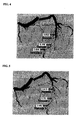
- FIGS. 4-10 illustrate predicted FFR values for a set of serial stenotic lesions (stenosis 1, stenosis 2, and stenosis 3) in the left anterior descending (LAD) artery for different stenting configurations.
- FIG. 4 illustrates predicted FFR values resulting from a stenting configuration in which only stenosis 1 is stented.
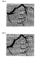
- FIG. 5 illustrates predicted FFR values resulting a stenting configuration in which only stenosis 2 is stented.
- FIG. 6 illustrates predicted FFR values resulting from a stenting configuration in which only stenosis 3 is stented.
- FIG. 4 illustrates predicted FFR values resulting from a stenting configuration in which only stenosis 1 is stented.
- FIG. 5 illustrates predicted FFR values resulting a stenting configuration in which only stenosis 2 is stented.
- FIG. 6 illustrates predicted FFR values resulting from a stenting configuration in which only sten
- FIG. 7 illustrates predicted FFR values resulting from a stenting configuration in which stenosis 1 and stenosis 2 are stented.
- FIG. 8 illustrates predicted FFR values resulting from a stenting configuration in which stenosis 1 and stenosis 3 are stented.
- FIG. 9 illustrates predicted FFR values resulting from a stenting configuration in which stenosis 2 and stenosis 3 are stented.
- FIG. 10 illustrates predicted FFR values resulting from a stenting configuration in which stenosis 1, stenosis 2, and stenosis 3 are all stented.
- Computer 1102 contains a processor 1104, which controls the overall operation of the computer 1102 by executing computer program instructions which define such operation.
- the computer program instructions may be stored in a storage device 1112 (e.g., magnetic disk) and loaded into memory 1110 when execution of the computer program instructions is desired.
- a storage device 1112 e.g., magnetic disk
- the steps of the methods of FIG. 1 may be defined by the computer program instructions stored in the memory 1110 and/or storage 1112 and controlled by the processor 1104 executing the computer program instructions.
- An image acquisition device 1120 such as a CT scanning device, MR scanning device, Ultrasound device, etc., can be connected to the computer 1102 to input image data to the computer 1102. It is possible to implement the image acquisition device 1120 and the computer 1102 as one device. It is also possible that the image acquisition device 1120 and the computer 1102 communicate wirelessly through a network. In a possible embodiment, the computer 1102 may be located remotely with respect to the image acquisition device 1120 and the method steps are performed as part of a server or cloud based service. The computer 1102 also includes one or more network interfaces 1106 for communicating with other devices via a network.
- the computer 1102 also includes other input/output devices 1108 that enable user interaction with the computer 1102 (e.g., display, keyboard, mouse, speakers, buttons, etc.).
- input/output devices 1108 that enable user interaction with the computer 1102 (e.g., display, keyboard, mouse, speakers, buttons, etc.).
- FIG. 11 is a high level representation of some of the components of such a computer for illustrative purposes.
- the above-described methods for medical image synthesis may be implemented using computers operating in a client-server relationship.
- the client computers are located remotely from the server computer and interact via a network.
- the client-server relationship may be defined and controlled by computer programs running on the respective client and server computers.
- a server or another processor that is connected to a network communicates with one or more client computers via a network.
- a client computer may communicate with the server via a network browser application residing and operating on the client computer, for example.
- a client computer may store data on the server and access the data via the network.
- a client computer may transmit requests for data, or requests for online services, to the server via the network.
- the server may perform requested services and provide data to the client computer(s).
- the server may also transmit data adapted to cause a client computer to perform a specified function, e.g., to perform a calculation, to display specified data on a screen, etc.
- the server may transmit a request adapted to cause a client computer to perform one or more of the method steps described herein, including one or more of the steps of FIG. 1 .
- Certain steps of the methods described herein, including one or more of the steps of FIG. 1 may be performed by a server or by another processor in a network-based cloud-computing system.
- Certain steps of the methods described herein, including one or more of the steps of FIG. 1 may be performed by a client computer in a network-based cloud computing system.
- the steps of the methods described herein, including one or more of the steps of FIG. 1 may be performed by a server and/or by a client computer in a network-based cloud computing system, in any combination.
Landscapes
- Health & Medical Sciences (AREA)
- Engineering & Computer Science (AREA)
- Medical Informatics (AREA)
- Life Sciences & Earth Sciences (AREA)
- Public Health (AREA)
- General Health & Medical Sciences (AREA)
- Biomedical Technology (AREA)
- Nuclear Medicine, Radiotherapy & Molecular Imaging (AREA)
- Pathology (AREA)
- Physics & Mathematics (AREA)
- Radiology & Medical Imaging (AREA)
- Surgery (AREA)
- Epidemiology (AREA)
- Primary Health Care (AREA)
- Animal Behavior & Ethology (AREA)
- Veterinary Medicine (AREA)
- Heart & Thoracic Surgery (AREA)
- Molecular Biology (AREA)
- Biophysics (AREA)
- Optics & Photonics (AREA)
- High Energy & Nuclear Physics (AREA)
- Theoretical Computer Science (AREA)
- Data Mining & Analysis (AREA)
- Databases & Information Systems (AREA)
- General Physics & Mathematics (AREA)
- Computer Vision & Pattern Recognition (AREA)
- Pulmonology (AREA)
- Vascular Medicine (AREA)
- Dentistry (AREA)
- Oral & Maxillofacial Surgery (AREA)
- Multimedia (AREA)
- Quality & Reliability (AREA)
- Robotics (AREA)
- Apparatus For Radiation Diagnosis (AREA)
Applications Claiming Priority (2)
| Application Number | Priority Date | Filing Date | Title |
|---|---|---|---|
| US201462027347P | 2014-07-22 | 2014-07-22 | |
| US14/801,987 US9888968B2 (en) | 2014-07-22 | 2015-07-17 | Method and system for automated therapy planning for arterial stenosis |
Publications (3)
| Publication Number | Publication Date |
|---|---|
| EP2977922A2 true EP2977922A2 (fr) | 2016-01-27 |
| EP2977922A3 EP2977922A3 (fr) | 2016-04-27 |
| EP2977922B1 EP2977922B1 (fr) | 2019-03-27 |
Family
ID=53794010
Family Applications (1)
| Application Number | Title | Priority Date | Filing Date |
|---|---|---|---|
| EP15177855.2A Active EP2977922B1 (fr) | 2014-07-22 | 2015-07-22 | Procédé et système de planification de thérapie automatisée pour la sténose artérielle |
Country Status (3)
| Country | Link |
|---|---|
| US (1) | US9888968B2 (fr) |
| EP (1) | EP2977922B1 (fr) |
| CN (1) | CN105380598B (fr) |
Cited By (12)
| Publication number | Priority date | Publication date | Assignee | Title |
|---|---|---|---|---|
| CN112543618A (zh) * | 2018-06-15 | 2021-03-23 | 帕伊医疗成像有限公司 | 用于进行定量血液动力学流量分析的方法和装置 |
| US11515033B2 (en) * | 2020-04-22 | 2022-11-29 | GE Precision Healthcare LLC | Augmented inspector interface with targeted, context-driven algorithms |
| EP4129196A4 (fr) * | 2020-03-27 | 2023-08-23 | Terumo Kabushiki Kaisha | Programme, procédé de traitement d'informations, dispositif de traitement d'informations et procédé de génération de modèle |
| US12315076B1 (en) | 2021-09-22 | 2025-05-27 | Cathworks Ltd. | Four-dimensional motion analysis of a patient's coronary arteries and myocardial wall |
| IL309861A (en) * | 2023-12-31 | 2025-07-01 | Medhub Ltd | Method and system for determining a treatment strategy for a vessel with multiple or diffuse lesions |
| US12354755B2 (en) | 2012-10-24 | 2025-07-08 | Cathworks Ltd | Creating a vascular tree model |
| US12387325B2 (en) | 2022-02-10 | 2025-08-12 | Cath Works Ltd. | System and method for machine-learning based sensor analysis and vascular tree segmentation |
| US12408885B2 (en) | 2016-05-16 | 2025-09-09 | Cathworks Ltd. | Vascular selection from images |
| US12446965B2 (en) | 2023-08-09 | 2025-10-21 | Cathworks Ltd. | Enhanced user interface and crosstalk analysis for vascular index measurement |
| US12499646B1 (en) | 2024-06-12 | 2025-12-16 | Cathworks Ltd. | Three-dimensional sizing tool for cardiac assessment |
| US12518384B2 (en) | 2022-03-31 | 2026-01-06 | Medipixel, Inc. | Lesion determination method and device |
| US12531159B2 (en) | 2023-08-09 | 2026-01-20 | Cathworks Ltd. | Post-PCI coronary analysis |
Families Citing this family (52)
| Publication number | Priority date | Publication date | Assignee | Title |
|---|---|---|---|---|
| US8200466B2 (en) | 2008-07-21 | 2012-06-12 | The Board Of Trustees Of The Leland Stanford Junior University | Method for tuning patient-specific cardiovascular simulations |
| US9405886B2 (en) | 2009-03-17 | 2016-08-02 | The Board Of Trustees Of The Leland Stanford Junior University | Method for determining cardiovascular information |
| US8315812B2 (en) | 2010-08-12 | 2012-11-20 | Heartflow, Inc. | Method and system for patient-specific modeling of blood flow |
| US10433740B2 (en) | 2012-09-12 | 2019-10-08 | Heartflow, Inc. | Systems and methods for estimating ischemia and blood flow characteristics from vessel geometry and physiology |
| US9042613B2 (en) * | 2013-03-01 | 2015-05-26 | Heartflow, Inc. | Method and system for determining treatments by modifying patient-specific geometrical models |
| US10134129B2 (en) * | 2014-04-22 | 2018-11-20 | Siemens Healthcare Gmbh | Method and system for hemodynamic computation in coronary arteries |
| CN106659399B (zh) * | 2014-05-05 | 2020-06-16 | 西门子保健有限责任公司 | 使用患病和假想正常解剖学模型中的流计算的冠状动脉狭窄的非侵入功能评价的方法和系统 |
| CN107580470B (zh) * | 2015-01-15 | 2021-06-22 | 皇家飞利浦有限公司 | 瞬时流量储备-计算机断层摄影 |
| WO2016132164A1 (fr) * | 2015-02-17 | 2016-08-25 | Siemens Healthcare Gmbh | Procédé et système de personnalisation d'une endoprothèse vasculaire |
| CN121685380A (zh) * | 2015-05-12 | 2026-03-17 | 新加坡保健服务集团有限公司 | 医学图像处理方法和系统 |
| US11094058B2 (en) | 2015-08-14 | 2021-08-17 | Elucid Bioimaging Inc. | Systems and method for computer-aided phenotyping (CAP) using radiologic images |
| US11676359B2 (en) | 2015-08-14 | 2023-06-13 | Elucid Bioimaging Inc. | Non-invasive quantitative imaging biomarkers of atherosclerotic plaque biology |
| US12008751B2 (en) | 2015-08-14 | 2024-06-11 | Elucid Bioimaging Inc. | Quantitative imaging for detecting histopathologically defined plaque fissure non-invasively |
| US11087459B2 (en) | 2015-08-14 | 2021-08-10 | Elucid Bioimaging Inc. | Quantitative imaging for fractional flow reserve (FFR) |
| US10176408B2 (en) | 2015-08-14 | 2019-01-08 | Elucid Bioimaging Inc. | Systems and methods for analyzing pathologies utilizing quantitative imaging |
| US11071501B2 (en) | 2015-08-14 | 2021-07-27 | Elucid Bioiwaging Inc. | Quantitative imaging for determining time to adverse event (TTE) |
| US11113812B2 (en) | 2015-08-14 | 2021-09-07 | Elucid Bioimaging Inc. | Quantitative imaging for detecting vulnerable plaque |
| US12026868B2 (en) | 2015-08-14 | 2024-07-02 | Elucid Bioimaging Inc. | Quantitative imaging for detecting histopathologically defined plaque erosion non-invasively |
| JP6918484B2 (ja) * | 2016-02-05 | 2021-08-11 | キヤノンメディカルシステムズ株式会社 | 画像処理装置及び医用画像診断装置 |
| DE102016203860A1 (de) | 2016-03-09 | 2017-09-14 | Siemens Healthcare Gmbh | Vorrichtung und Verfahren zum Ermitteln zumindest eines individuellen fluiddynamischen Kennwerts einer Stenose in einem mehrere serielle Stenosen aufweisenden Gefäßsegment |
| JP6608110B2 (ja) * | 2016-04-13 | 2019-11-20 | 富士フイルム株式会社 | 画像位置合わせ装置および方法並びにプログラム |
| JP7036742B2 (ja) | 2016-05-16 | 2022-03-15 | キャスワークス リミテッド | 血管評価システム |
| CN109716446B (zh) * | 2016-09-28 | 2023-10-03 | 光学实验室成像公司 | 利用血管表象的支架规划系统及方法 |
| CN110494893B (zh) * | 2017-03-31 | 2024-01-23 | 皇家飞利浦有限公司 | 基于ffr的对非侵入性成像的交互监测 |
| WO2019025270A1 (fr) * | 2017-08-01 | 2019-02-07 | Siemens Healthcare Gmbh | Évaluation non invasive et guidage de thérapie pour une coronaropathie dans des lésions diffuses et en tandem |
| CN110998744B (zh) * | 2017-08-01 | 2024-04-05 | 西门子医疗有限公司 | 针对弥漫性和串联性病变中冠状动脉疾病的非侵入性评估和治疗指导 |
| WO2019042789A1 (fr) | 2017-08-30 | 2019-03-07 | Koninklijke Philips N.V. | Prédiction d'état de santé d'artère coronaire sur la base d'un modèle et de données d'imagerie |
| JP2019076526A (ja) | 2017-10-25 | 2019-05-23 | テルモ株式会社 | 治療方法 |
| JP2019080749A (ja) * | 2017-10-30 | 2019-05-30 | テルモ株式会社 | 治療方法 |
| JP2019080876A (ja) * | 2017-10-31 | 2019-05-30 | テルモ株式会社 | 治療方法 |
| JP7008522B2 (ja) * | 2018-02-01 | 2022-01-25 | テルモ株式会社 | 医療システム |
| CN108564568A (zh) * | 2018-03-23 | 2018-09-21 | 沈阳东软医疗系统有限公司 | 冠脉的显示方法、装置、设备及存储介质 |
| US11127138B2 (en) * | 2018-11-20 | 2021-09-21 | Siemens Healthcare Gmbh | Automatic detection and quantification of the aorta from medical images |
| CN109886953B (zh) * | 2019-02-27 | 2024-06-28 | 数坤科技股份有限公司 | 一种血管异常检测方法、装置及计算机可读存储介质 |
| JP7532402B2 (ja) | 2019-04-01 | 2024-08-13 | キャスワークス リミテッド | 血管造影画像選択のための方法および装置 |
| US10861157B2 (en) | 2019-04-04 | 2020-12-08 | Medtronic Vascular, Inc. | System and methods for determining modified fractional flow reserve values |
| CN121707942A (zh) | 2019-08-05 | 2026-03-20 | 易鲁希德生物成像公司 | 形态学和血管周疾病标志物的联合评估 |
| CN114340481B (zh) * | 2019-09-09 | 2025-08-29 | 美德胡布有限公司 | 用于确定流量储备分数的对图像数据的自动分析 |
| EP4033964B1 (fr) | 2019-09-23 | 2025-04-09 | Cathworks Ltd. | Procédés, appareil et système de synchronisation entre un modèle vasculaire tridimensionnel et un dispositif d'imagerie |
| EP4114285A4 (fr) | 2020-03-04 | 2024-03-06 | Shifamed Holdings, LLC | Systèmes de retrait de trhombus et procédés associés |
| KR102546291B1 (ko) | 2020-12-02 | 2023-06-26 | 재단법인 아산사회복지재단 | 초음파 영상 기반의 딥 러닝을 통한 관상동맥 스텐트 예측 방법, 장치 및 기록매체 |
| CN112971979B (zh) * | 2021-02-03 | 2022-08-16 | 上海友脉科技有限责任公司 | 仿真系统、仿真方法及装置 |
| KR102550631B1 (ko) * | 2021-03-16 | 2023-07-03 | (주)파인헬스케어 | 인공지능을 이용한 욕창 단계 평가 및 치료 추천을 제공하는 장치 및 방법 |
| JP7749929B2 (ja) * | 2021-03-23 | 2025-10-07 | コニカミノルタ株式会社 | 動態解析装置及びプログラム |
| EP4340751A4 (fr) | 2021-05-19 | 2025-03-26 | Shifamed Holdings, LLC | Systèmes de retrait de thrombus et procédés associés |
| CN113397579B (zh) * | 2021-07-23 | 2024-12-10 | 上海友脉科技有限责任公司 | 血流动力学分析装置、方法、介质及电子设备 |
| CN113827199B (zh) * | 2021-10-29 | 2024-01-23 | 苏州润迈德医疗科技有限公司 | 基于造影图像调节血管评定参数的方法、系统及存储介质 |
| WO2025032546A1 (fr) * | 2023-08-09 | 2025-02-13 | Cathworks Ltd. | Interface utilisateur améliorée et analyse de la diaphonie pour mesure d'index vasculaire |
| CN116919375A (zh) * | 2023-08-22 | 2023-10-24 | 上海博动医疗科技股份有限公司 | 虚拟支架ffr血流储备分数计算方法、装置、设备及介质 |
| WO2025129125A1 (fr) * | 2023-12-13 | 2025-06-19 | Shifamed Holdings, Llc | Systèmes d'élimination de thrombus et procédés associés |
| JP7852674B2 (ja) * | 2024-06-26 | 2026-04-28 | コニカミノルタ株式会社 | 動態解析装置、動態解析システム、プログラム及び分類方法 |
| GB202411474D0 (en) * | 2024-08-05 | 2024-09-18 | Univ London Queen Mary | Medical support method |
Citations (8)
| Publication number | Priority date | Publication date | Assignee | Title |
|---|---|---|---|---|
| US20100067760A1 (en) | 2008-09-15 | 2010-03-18 | Siemens Corporate Research, Inc. | Method and System for Automatic Coronary Artery Detection |
| US7860290B2 (en) | 2006-04-21 | 2010-12-28 | Siemens Medical Solutions Usa, Inc. | Three-dimensional (3D) modeling of coronary arteries |
| US7953266B2 (en) | 2007-02-06 | 2011-05-31 | Siemens Medical Solutions Usa, Inc. | Robust vessel tree modeling |
| US20110224542A1 (en) | 2010-03-12 | 2011-09-15 | Sushil Mittal | Method and System for Automatic Detection and Classification of Coronary Stenoses in Cardiac CT Volumes |
| US20120022843A1 (en) | 2010-07-21 | 2012-01-26 | Razvan Ioan Ionasec | Method and System for Comprehensive Patient-Specific Modeling of the Heart |
| US20130132054A1 (en) | 2011-11-10 | 2013-05-23 | Puneet Sharma | Method and System for Multi-Scale Anatomical and Functional Modeling of Coronary Circulation |
| US20130246034A1 (en) | 2012-03-13 | 2013-09-19 | Siemens Aktiengesellschaft | Method and System for Non-Invasive Functional Assessment of Coronary Artery Stenosis |
| US20150112182A1 (en) | 2013-10-17 | 2015-04-23 | Siemens Aktiengesellschaft | Method and System for Machine Learning Based Assessment of Fractional Flow Reserve |
Family Cites Families (18)
| Publication number | Priority date | Publication date | Assignee | Title |
|---|---|---|---|---|
| US6236878B1 (en) | 1998-05-22 | 2001-05-22 | Charles A. Taylor | Method for predictive modeling for planning medical interventions and simulating physiological conditions |
| US6643533B2 (en) * | 2000-11-28 | 2003-11-04 | Ge Medical Systems Global Technology Company, Llc | Method and apparatus for displaying images of tubular structures |
| US7379062B2 (en) * | 2005-08-01 | 2008-05-27 | Barco Nv | Method for determining a path along a biological object with a lumen |
| US8014561B2 (en) * | 2006-09-07 | 2011-09-06 | University Of Louisville Research Foundation, Inc. | Virtual fly over of complex tubular anatomical structures |
| DE102006045423B4 (de) * | 2006-09-26 | 2016-07-14 | Siemens Healthcare Gmbh | 07.09.07Verfahren zur Nachbearbeitung eines dreidimensionalen Bilddatensatzes einer Gefäßstruktur |
| US8060186B2 (en) * | 2007-02-15 | 2011-11-15 | Siemens Aktiengesellschaft | System and method for intraoperative guidance of stent placement during endovascular interventions |
| US8098918B2 (en) | 2007-09-21 | 2012-01-17 | Siemens Corporation | Method and system for measuring left ventricle volume |
| US8200466B2 (en) | 2008-07-21 | 2012-06-12 | The Board Of Trustees Of The Leland Stanford Junior University | Method for tuning patient-specific cardiovascular simulations |
| US8315812B2 (en) | 2010-08-12 | 2012-11-20 | Heartflow, Inc. | Method and system for patient-specific modeling of blood flow |
| US8157742B2 (en) | 2010-08-12 | 2012-04-17 | Heartflow, Inc. | Method and system for patient-specific modeling of blood flow |
| DE102010039312B4 (de) | 2010-08-13 | 2020-02-13 | Siemens Healthcare Gmbh | Verfahren zur Simulation eines Blutflusses |
| US9119540B2 (en) | 2010-09-16 | 2015-09-01 | Siemens Aktiengesellschaft | Method and system for non-invasive assessment of coronary artery disease |
| DE102010043849B3 (de) | 2010-11-12 | 2012-02-16 | Siemens Aktiengesellschaft | Vorrichtung und Computertomograph zur Bestimmung und Darstellung der Durchblutung des Herzmuskels |
| US9141763B2 (en) | 2011-02-07 | 2015-09-22 | Siemens Aktiengesellschaft | Method and system for patient-specific computational modeling and simulation for coupled hemodynamic analysis of cerebral vessels |
| US10186056B2 (en) | 2011-03-21 | 2019-01-22 | General Electric Company | System and method for estimating vascular flow using CT imaging |
| CN103300820A (zh) | 2012-03-13 | 2013-09-18 | 西门子公司 | 用于冠状动脉狭窄的非侵入性功能评估的方法和系统 |
| JP6173438B2 (ja) * | 2012-05-14 | 2017-08-02 | コーニンクレッカ フィリップス エヌ ヴェKoninklijke Philips N.V. | 血管の狭窄のための冠血流予備量比(ffr)値の決定 |
| US9858387B2 (en) | 2013-01-15 | 2018-01-02 | CathWorks, LTD. | Vascular flow assessment |
-
2015
- 2015-07-17 US US14/801,987 patent/US9888968B2/en active Active
- 2015-07-22 EP EP15177855.2A patent/EP2977922B1/fr active Active
- 2015-07-22 CN CN201510602460.6A patent/CN105380598B/zh active Active
Patent Citations (9)
| Publication number | Priority date | Publication date | Assignee | Title |
|---|---|---|---|---|
| US7860290B2 (en) | 2006-04-21 | 2010-12-28 | Siemens Medical Solutions Usa, Inc. | Three-dimensional (3D) modeling of coronary arteries |
| US7953266B2 (en) | 2007-02-06 | 2011-05-31 | Siemens Medical Solutions Usa, Inc. | Robust vessel tree modeling |
| US20100067760A1 (en) | 2008-09-15 | 2010-03-18 | Siemens Corporate Research, Inc. | Method and System for Automatic Coronary Artery Detection |
| US20110224542A1 (en) | 2010-03-12 | 2011-09-15 | Sushil Mittal | Method and System for Automatic Detection and Classification of Coronary Stenoses in Cardiac CT Volumes |
| US20120022843A1 (en) | 2010-07-21 | 2012-01-26 | Razvan Ioan Ionasec | Method and System for Comprehensive Patient-Specific Modeling of the Heart |
| US20130132054A1 (en) | 2011-11-10 | 2013-05-23 | Puneet Sharma | Method and System for Multi-Scale Anatomical and Functional Modeling of Coronary Circulation |
| US20130246034A1 (en) | 2012-03-13 | 2013-09-19 | Siemens Aktiengesellschaft | Method and System for Non-Invasive Functional Assessment of Coronary Artery Stenosis |
| US20140058715A1 (en) | 2012-03-13 | 2014-02-27 | Siemens Aktiengesellschaft | Method and System for Non-Invasive Functional Assessment of Coronary Artery Stenosis |
| US20150112182A1 (en) | 2013-10-17 | 2015-04-23 | Siemens Aktiengesellschaft | Method and System for Machine Learning Based Assessment of Fractional Flow Reserve |
Cited By (16)
| Publication number | Priority date | Publication date | Assignee | Title |
|---|---|---|---|---|
| US12354755B2 (en) | 2012-10-24 | 2025-07-08 | Cathworks Ltd | Creating a vascular tree model |
| US12408885B2 (en) | 2016-05-16 | 2025-09-09 | Cathworks Ltd. | Vascular selection from images |
| CN112543618B (zh) * | 2018-06-15 | 2024-05-07 | 帕伊医疗成像有限公司 | 用于进行定量血液动力学流量分析的方法和装置 |
| CN112543618A (zh) * | 2018-06-15 | 2021-03-23 | 帕伊医疗成像有限公司 | 用于进行定量血液动力学流量分析的方法和装置 |
| EP4129196A4 (fr) * | 2020-03-27 | 2023-08-23 | Terumo Kabushiki Kaisha | Programme, procédé de traitement d'informations, dispositif de traitement d'informations et procédé de génération de modèle |
| US11515033B2 (en) * | 2020-04-22 | 2022-11-29 | GE Precision Healthcare LLC | Augmented inspector interface with targeted, context-driven algorithms |
| US12315076B1 (en) | 2021-09-22 | 2025-05-27 | Cathworks Ltd. | Four-dimensional motion analysis of a patient's coronary arteries and myocardial wall |
| US12387325B2 (en) | 2022-02-10 | 2025-08-12 | Cath Works Ltd. | System and method for machine-learning based sensor analysis and vascular tree segmentation |
| US12423813B2 (en) | 2022-02-10 | 2025-09-23 | Cathworks Ltd. | System and method for machine-learning based sensor analysis and vascular tree segmentation |
| US12518384B2 (en) | 2022-03-31 | 2026-01-06 | Medipixel, Inc. | Lesion determination method and device |
| US12446965B2 (en) | 2023-08-09 | 2025-10-21 | Cathworks Ltd. | Enhanced user interface and crosstalk analysis for vascular index measurement |
| US12531159B2 (en) | 2023-08-09 | 2026-01-20 | Cathworks Ltd. | Post-PCI coronary analysis |
| IL309861A (en) * | 2023-12-31 | 2025-07-01 | Medhub Ltd | Method and system for determining a treatment strategy for a vessel with multiple or diffuse lesions |
| US12499646B1 (en) | 2024-06-12 | 2025-12-16 | Cathworks Ltd. | Three-dimensional sizing tool for cardiac assessment |
| US12512196B2 (en) | 2024-06-12 | 2025-12-30 | Cathworks Ltd. | Systems and methods for secure sharing of cardiac assessments using QR codes |
| US12567489B2 (en) | 2024-06-12 | 2026-03-03 | Cathworks Ltd. | Systems and methods for displaying distal fractional flow reserve values in vascular analysis |
Also Published As
| Publication number | Publication date |
|---|---|
| CN105380598A (zh) | 2016-03-09 |
| EP2977922A3 (fr) | 2016-04-27 |
| US9888968B2 (en) | 2018-02-13 |
| US20160022371A1 (en) | 2016-01-28 |
| EP2977922B1 (fr) | 2019-03-27 |
| CN105380598B (zh) | 2018-11-13 |
Similar Documents
| Publication | Publication Date | Title |
|---|---|---|
| EP2977922B1 (fr) | Procédé et système de planification de thérapie automatisée pour la sténose artérielle | |
| US10130266B2 (en) | Method and system for prediction of post-stenting hemodynamic metrics for treatment planning of arterial stenosis | |
| US10134129B2 (en) | Method and system for hemodynamic computation in coronary arteries | |
| JP6409114B2 (ja) | 患者固有の幾何学的形状モデルを変更することによって治療を決定する方法及びシステム | |
| US10803995B2 (en) | Method and system for non-invasive functional assessment of coronary artery stenosis using flow computations in diseased and hypothetical normal anatomical models | |
| KR101910233B1 (ko) | 맥관구조를 수치 평가하는 시스템들 및 방법들 | |
| US10872698B2 (en) | Method and system for enhancing medical image-based blood flow computations using physiological measurements | |
| CN105555195A (zh) | 用于从患者特定的解剖数据识别个性化的血管植入物的系统和方法 | |
| EP3218872A2 (fr) | Procédé et système pour évaluation à base d'apprentissage automatique purement géométrique du flux de réserve coronaire | |
| WO2016075331A2 (fr) | Procédé et système pour évaluation à base d'apprentissage automatique purement géométrique du flux de réserve coronaire | |
| CN112754449B (zh) | 侧支冠状动脉的评估 |
Legal Events
| Date | Code | Title | Description |
|---|---|---|---|
| PUAI | Public reference made under article 153(3) epc to a published international application that has entered the european phase |
Free format text: ORIGINAL CODE: 0009012 |
|
| AK | Designated contracting states |
Kind code of ref document: A2 Designated state(s): AL AT BE BG CH CY CZ DE DK EE ES FI FR GB GR HR HU IE IS IT LI LT LU LV MC MK MT NL NO PL PT RO RS SE SI SK SM TR |
|
| AX | Request for extension of the european patent |
Extension state: BA ME |
|
| PUAL | Search report despatched |
Free format text: ORIGINAL CODE: 0009013 |
|
| AK | Designated contracting states |
Kind code of ref document: A3 Designated state(s): AL AT BE BG CH CY CZ DE DK EE ES FI FR GB GR HR HU IE IS IT LI LT LU LV MC MK MT NL NO PL PT RO RS SE SI SK SM TR |
|
| AX | Request for extension of the european patent |
Extension state: BA ME |
|
| RIC1 | Information provided on ipc code assigned before grant |
Ipc: G06T 7/00 20060101ALI20160318BHEP Ipc: G06F 19/00 20110101AFI20160318BHEP |
|
| RAP1 | Party data changed (applicant data changed or rights of an application transferred) |
Owner name: SIEMENS HEALTHCARE GMBH |
|
| STAA | Information on the status of an ep patent application or granted ep patent |
Free format text: STATUS: REQUEST FOR EXAMINATION WAS MADE |
|
| 17P | Request for examination filed |
Effective date: 20161026 |
|
| RBV | Designated contracting states (corrected) |
Designated state(s): AL AT BE BG CH CY CZ DE DK EE ES FI FR GB GR HR HU IE IS IT LI LT LU LV MC MK MT NL NO PL PT RO RS SE SI SK SM TR |
|
| STAA | Information on the status of an ep patent application or granted ep patent |
Free format text: STATUS: EXAMINATION IS IN PROGRESS |
|
| 17Q | First examination report despatched |
Effective date: 20171206 |
|
| GRAP | Despatch of communication of intention to grant a patent |
Free format text: ORIGINAL CODE: EPIDOSNIGR1 |
|
| STAA | Information on the status of an ep patent application or granted ep patent |
Free format text: STATUS: GRANT OF PATENT IS INTENDED |
|
| INTG | Intention to grant announced |
Effective date: 20181205 |
|
| GRAS | Grant fee paid |
Free format text: ORIGINAL CODE: EPIDOSNIGR3 |
|
| GRAA | (expected) grant |
Free format text: ORIGINAL CODE: 0009210 |
|
| STAA | Information on the status of an ep patent application or granted ep patent |
Free format text: STATUS: THE PATENT HAS BEEN GRANTED |
|
| AK | Designated contracting states |
Kind code of ref document: B1 Designated state(s): AL AT BE BG CH CY CZ DE DK EE ES FI FR GB GR HR HU IE IS IT LI LT LU LV MC MK MT NL NO PL PT RO RS SE SI SK SM TR |
|
| REG | Reference to a national code |
Ref country code: GB Ref legal event code: FG4D |
|
| REG | Reference to a national code |
Ref country code: CH Ref legal event code: EP |
|
| REG | Reference to a national code |
Ref country code: AT Ref legal event code: REF Ref document number: 1113897 Country of ref document: AT Kind code of ref document: T Effective date: 20190415 |
|
| REG | Reference to a national code |
Ref country code: IE Ref legal event code: FG4D |
|
| REG | Reference to a national code |
Ref country code: DE Ref legal event code: R096 Ref document number: 602015027028 Country of ref document: DE |
|
| PG25 | Lapsed in a contracting state [announced via postgrant information from national office to epo] |
Ref country code: SE Free format text: LAPSE BECAUSE OF FAILURE TO SUBMIT A TRANSLATION OF THE DESCRIPTION OR TO PAY THE FEE WITHIN THE PRESCRIBED TIME-LIMIT Effective date: 20190327 Ref country code: NO Free format text: LAPSE BECAUSE OF FAILURE TO SUBMIT A TRANSLATION OF THE DESCRIPTION OR TO PAY THE FEE WITHIN THE PRESCRIBED TIME-LIMIT Effective date: 20190627 Ref country code: FI Free format text: LAPSE BECAUSE OF FAILURE TO SUBMIT A TRANSLATION OF THE DESCRIPTION OR TO PAY THE FEE WITHIN THE PRESCRIBED TIME-LIMIT Effective date: 20190327 Ref country code: LT Free format text: LAPSE BECAUSE OF FAILURE TO SUBMIT A TRANSLATION OF THE DESCRIPTION OR TO PAY THE FEE WITHIN THE PRESCRIBED TIME-LIMIT Effective date: 20190327 |
|
| REG | Reference to a national code |
Ref country code: NL Ref legal event code: MP Effective date: 20190327 |
|
| PG25 | Lapsed in a contracting state [announced via postgrant information from national office to epo] |
Ref country code: BG Free format text: LAPSE BECAUSE OF FAILURE TO SUBMIT A TRANSLATION OF THE DESCRIPTION OR TO PAY THE FEE WITHIN THE PRESCRIBED TIME-LIMIT Effective date: 20190627 Ref country code: GR Free format text: LAPSE BECAUSE OF FAILURE TO SUBMIT A TRANSLATION OF THE DESCRIPTION OR TO PAY THE FEE WITHIN THE PRESCRIBED TIME-LIMIT Effective date: 20190628 Ref country code: HR Free format text: LAPSE BECAUSE OF FAILURE TO SUBMIT A TRANSLATION OF THE DESCRIPTION OR TO PAY THE FEE WITHIN THE PRESCRIBED TIME-LIMIT Effective date: 20190327 Ref country code: NL Free format text: LAPSE BECAUSE OF FAILURE TO SUBMIT A TRANSLATION OF THE DESCRIPTION OR TO PAY THE FEE WITHIN THE PRESCRIBED TIME-LIMIT Effective date: 20190327 Ref country code: LV Free format text: LAPSE BECAUSE OF FAILURE TO SUBMIT A TRANSLATION OF THE DESCRIPTION OR TO PAY THE FEE WITHIN THE PRESCRIBED TIME-LIMIT Effective date: 20190327 Ref country code: RS Free format text: LAPSE BECAUSE OF FAILURE TO SUBMIT A TRANSLATION OF THE DESCRIPTION OR TO PAY THE FEE WITHIN THE PRESCRIBED TIME-LIMIT Effective date: 20190327 |
|
| REG | Reference to a national code |
Ref country code: AT Ref legal event code: MK05 Ref document number: 1113897 Country of ref document: AT Kind code of ref document: T Effective date: 20190327 |
|
| PG25 | Lapsed in a contracting state [announced via postgrant information from national office to epo] |
Ref country code: IT Free format text: LAPSE BECAUSE OF FAILURE TO SUBMIT A TRANSLATION OF THE DESCRIPTION OR TO PAY THE FEE WITHIN THE PRESCRIBED TIME-LIMIT Effective date: 20190327 Ref country code: RO Free format text: LAPSE BECAUSE OF FAILURE TO SUBMIT A TRANSLATION OF THE DESCRIPTION OR TO PAY THE FEE WITHIN THE PRESCRIBED TIME-LIMIT Effective date: 20190327 Ref country code: CZ Free format text: LAPSE BECAUSE OF FAILURE TO SUBMIT A TRANSLATION OF THE DESCRIPTION OR TO PAY THE FEE WITHIN THE PRESCRIBED TIME-LIMIT Effective date: 20190327 Ref country code: EE Free format text: LAPSE BECAUSE OF FAILURE TO SUBMIT A TRANSLATION OF THE DESCRIPTION OR TO PAY THE FEE WITHIN THE PRESCRIBED TIME-LIMIT Effective date: 20190327 Ref country code: ES Free format text: LAPSE BECAUSE OF FAILURE TO SUBMIT A TRANSLATION OF THE DESCRIPTION OR TO PAY THE FEE WITHIN THE PRESCRIBED TIME-LIMIT Effective date: 20190327 Ref country code: AL Free format text: LAPSE BECAUSE OF FAILURE TO SUBMIT A TRANSLATION OF THE DESCRIPTION OR TO PAY THE FEE WITHIN THE PRESCRIBED TIME-LIMIT Effective date: 20190327 Ref country code: SK Free format text: LAPSE BECAUSE OF FAILURE TO SUBMIT A TRANSLATION OF THE DESCRIPTION OR TO PAY THE FEE WITHIN THE PRESCRIBED TIME-LIMIT Effective date: 20190327 Ref country code: PT Free format text: LAPSE BECAUSE OF FAILURE TO SUBMIT A TRANSLATION OF THE DESCRIPTION OR TO PAY THE FEE WITHIN THE PRESCRIBED TIME-LIMIT Effective date: 20190727 |
|
| PG25 | Lapsed in a contracting state [announced via postgrant information from national office to epo] |
Ref country code: PL Free format text: LAPSE BECAUSE OF FAILURE TO SUBMIT A TRANSLATION OF THE DESCRIPTION OR TO PAY THE FEE WITHIN THE PRESCRIBED TIME-LIMIT Effective date: 20190327 Ref country code: SM Free format text: LAPSE BECAUSE OF FAILURE TO SUBMIT A TRANSLATION OF THE DESCRIPTION OR TO PAY THE FEE WITHIN THE PRESCRIBED TIME-LIMIT Effective date: 20190327 |
|
| PG25 | Lapsed in a contracting state [announced via postgrant information from national office to epo] |
Ref country code: AT Free format text: LAPSE BECAUSE OF FAILURE TO SUBMIT A TRANSLATION OF THE DESCRIPTION OR TO PAY THE FEE WITHIN THE PRESCRIBED TIME-LIMIT Effective date: 20190327 Ref country code: IS Free format text: LAPSE BECAUSE OF FAILURE TO SUBMIT A TRANSLATION OF THE DESCRIPTION OR TO PAY THE FEE WITHIN THE PRESCRIBED TIME-LIMIT Effective date: 20190727 |
|
| REG | Reference to a national code |
Ref country code: DE Ref legal event code: R097 Ref document number: 602015027028 Country of ref document: DE |
|
| PG25 | Lapsed in a contracting state [announced via postgrant information from national office to epo] |
Ref country code: DK Free format text: LAPSE BECAUSE OF FAILURE TO SUBMIT A TRANSLATION OF THE DESCRIPTION OR TO PAY THE FEE WITHIN THE PRESCRIBED TIME-LIMIT Effective date: 20190327 |
|
| PLBE | No opposition filed within time limit |
Free format text: ORIGINAL CODE: 0009261 |
|
| STAA | Information on the status of an ep patent application or granted ep patent |
Free format text: STATUS: NO OPPOSITION FILED WITHIN TIME LIMIT |
|
| PG25 | Lapsed in a contracting state [announced via postgrant information from national office to epo] |
Ref country code: SI Free format text: LAPSE BECAUSE OF FAILURE TO SUBMIT A TRANSLATION OF THE DESCRIPTION OR TO PAY THE FEE WITHIN THE PRESCRIBED TIME-LIMIT Effective date: 20190327 Ref country code: MC Free format text: LAPSE BECAUSE OF FAILURE TO SUBMIT A TRANSLATION OF THE DESCRIPTION OR TO PAY THE FEE WITHIN THE PRESCRIBED TIME-LIMIT Effective date: 20190327 |
|
| REG | Reference to a national code |
Ref country code: CH Ref legal event code: PL |
|
| 26N | No opposition filed |
Effective date: 20200103 |
|
| PG25 | Lapsed in a contracting state [announced via postgrant information from national office to epo] |
Ref country code: TR Free format text: LAPSE BECAUSE OF FAILURE TO SUBMIT A TRANSLATION OF THE DESCRIPTION OR TO PAY THE FEE WITHIN THE PRESCRIBED TIME-LIMIT Effective date: 20190327 |
|
| REG | Reference to a national code |
Ref country code: BE Ref legal event code: MM Effective date: 20190731 |
|
| PG25 | Lapsed in a contracting state [announced via postgrant information from national office to epo] |
Ref country code: BE Free format text: LAPSE BECAUSE OF NON-PAYMENT OF DUE FEES Effective date: 20190731 Ref country code: LU Free format text: LAPSE BECAUSE OF NON-PAYMENT OF DUE FEES Effective date: 20190722 Ref country code: CH Free format text: LAPSE BECAUSE OF NON-PAYMENT OF DUE FEES Effective date: 20190731 Ref country code: LI Free format text: LAPSE BECAUSE OF NON-PAYMENT OF DUE FEES Effective date: 20190731 |
|
| PG25 | Lapsed in a contracting state [announced via postgrant information from national office to epo] |
Ref country code: IE Free format text: LAPSE BECAUSE OF NON-PAYMENT OF DUE FEES Effective date: 20190722 |
|
| PG25 | Lapsed in a contracting state [announced via postgrant information from national office to epo] |
Ref country code: CY Free format text: LAPSE BECAUSE OF FAILURE TO SUBMIT A TRANSLATION OF THE DESCRIPTION OR TO PAY THE FEE WITHIN THE PRESCRIBED TIME-LIMIT Effective date: 20190327 |
|
| PG25 | Lapsed in a contracting state [announced via postgrant information from national office to epo] |
Ref country code: HU Free format text: LAPSE BECAUSE OF FAILURE TO SUBMIT A TRANSLATION OF THE DESCRIPTION OR TO PAY THE FEE WITHIN THE PRESCRIBED TIME-LIMIT; INVALID AB INITIO Effective date: 20150722 Ref country code: MT Free format text: LAPSE BECAUSE OF FAILURE TO SUBMIT A TRANSLATION OF THE DESCRIPTION OR TO PAY THE FEE WITHIN THE PRESCRIBED TIME-LIMIT Effective date: 20190327 |
|
| PG25 | Lapsed in a contracting state [announced via postgrant information from national office to epo] |
Ref country code: MK Free format text: LAPSE BECAUSE OF FAILURE TO SUBMIT A TRANSLATION OF THE DESCRIPTION OR TO PAY THE FEE WITHIN THE PRESCRIBED TIME-LIMIT Effective date: 20190327 |
|
| REG | Reference to a national code |
Ref country code: DE Ref legal event code: R081 Ref document number: 602015027028 Country of ref document: DE Owner name: SIEMENS HEALTHINEERS AG, DE Free format text: FORMER OWNER: SIEMENS HEALTHCARE GMBH, 91052 ERLANGEN, DE |
|
| PGFP | Annual fee paid to national office [announced via postgrant information from national office to epo] |
Ref country code: DE Payment date: 20250919 Year of fee payment: 11 |
|
| PGFP | Annual fee paid to national office [announced via postgrant information from national office to epo] |
Ref country code: GB Payment date: 20250811 Year of fee payment: 11 |
|
| PGFP | Annual fee paid to national office [announced via postgrant information from national office to epo] |
Ref country code: FR Payment date: 20250716 Year of fee payment: 11 |