EP2985342A1 - Antigènes immunogènes provenant d'aspergillus fumigatus - Google Patents
Antigènes immunogènes provenant d'aspergillus fumigatus Download PDFInfo
- Publication number
- EP2985342A1 EP2985342A1 EP14181113.3A EP14181113A EP2985342A1 EP 2985342 A1 EP2985342 A1 EP 2985342A1 EP 14181113 A EP14181113 A EP 14181113A EP 2985342 A1 EP2985342 A1 EP 2985342A1
- Authority
- EP
- European Patent Office
- Prior art keywords
- cells
- vivo
- specific
- antigen
- aspergillus fumigatus
- Prior art date
- Legal status (The legal status is an assumption and is not a legal conclusion. Google has not performed a legal analysis and makes no representation as to the accuracy of the status listed.)
- Withdrawn
Links
- 239000000427 antigen Substances 0.000 title claims abstract description 145
- 108091007433 antigens Proteins 0.000 title claims abstract description 140
- 102000036639 antigens Human genes 0.000 title claims abstract description 140
- 241001225321 Aspergillus fumigatus Species 0.000 title claims abstract description 132
- 229940091771 aspergillus fumigatus Drugs 0.000 title claims abstract description 57
- 230000002163 immunogen Effects 0.000 title description 28
- 210000001744 T-lymphocyte Anatomy 0.000 claims abstract description 208
- 210000004027 cell Anatomy 0.000 claims abstract description 105
- 238000000034 method Methods 0.000 claims abstract description 37
- 230000004913 activation Effects 0.000 claims abstract description 34
- 101100148606 Caenorhabditis elegans pst-1 gene Proteins 0.000 claims abstract description 31
- 238000004458 analytical method Methods 0.000 claims abstract description 20
- 238000001514 detection method Methods 0.000 claims abstract description 17
- 238000000338 in vitro Methods 0.000 claims abstract description 13
- 239000008194 pharmaceutical composition Substances 0.000 claims abstract description 10
- 108090000623 proteins and genes Proteins 0.000 claims description 127
- 102000004169 proteins and genes Human genes 0.000 claims description 124
- 102100032937 CD40 ligand Human genes 0.000 claims description 68
- 101000868215 Homo sapiens CD40 ligand Proteins 0.000 claims description 68
- 108090000765 processed proteins & peptides Proteins 0.000 claims description 48
- 101000851370 Homo sapiens Tumor necrosis factor receptor superfamily member 9 Proteins 0.000 claims description 23
- 102100036856 Tumor necrosis factor receptor superfamily member 9 Human genes 0.000 claims description 23
- 239000012634 fragment Substances 0.000 claims description 22
- 108010074328 Interferon-gamma Proteins 0.000 claims description 20
- 102100037850 Interferon gamma Human genes 0.000 claims description 19
- 230000005291 magnetic effect Effects 0.000 claims description 17
- 108010002350 Interleukin-2 Proteins 0.000 claims description 13
- MZOFCQQQCNRIBI-VMXHOPILSA-N (3s)-4-[[(2s)-1-[[(2s)-1-[[(1s)-1-carboxy-2-hydroxyethyl]amino]-4-methyl-1-oxopentan-2-yl]amino]-5-(diaminomethylideneamino)-1-oxopentan-2-yl]amino]-3-[[2-[[(2s)-2,6-diaminohexanoyl]amino]acetyl]amino]-4-oxobutanoic acid Chemical compound OC[C@@H](C(O)=O)NC(=O)[C@H](CC(C)C)NC(=O)[C@H](CCCN=C(N)N)NC(=O)[C@H](CC(O)=O)NC(=O)CNC(=O)[C@@H](N)CCCCN MZOFCQQQCNRIBI-VMXHOPILSA-N 0.000 claims description 12
- 108060008682 Tumor Necrosis Factor Proteins 0.000 claims description 12
- 239000006249 magnetic particle Substances 0.000 claims description 11
- 210000003289 regulatory T cell Anatomy 0.000 claims description 11
- 238000002955 isolation Methods 0.000 claims description 10
- 238000000684 flow cytometry Methods 0.000 claims description 9
- 208000036641 Aspergillus infections Diseases 0.000 claims description 8
- 210000003071 memory t lymphocyte Anatomy 0.000 claims description 8
- 201000002909 Aspergillosis Diseases 0.000 claims description 7
- 102000003816 Interleukin-13 Human genes 0.000 claims description 7
- 108090000176 Interleukin-13 Proteins 0.000 claims description 7
- 108090000978 Interleukin-4 Proteins 0.000 claims description 7
- 108010002616 Interleukin-5 Proteins 0.000 claims description 7
- -1 Il-17 Proteins 0.000 claims description 6
- 102100030703 Interleukin-22 Human genes 0.000 claims description 6
- 101800001155 Latency-associated peptide Proteins 0.000 claims description 6
- 102400000401 Latency-associated peptide Human genes 0.000 claims description 6
- 108010074109 interleukin-22 Proteins 0.000 claims description 6
- 102100025137 Early activation antigen CD69 Human genes 0.000 claims description 5
- 101000934374 Homo sapiens Early activation antigen CD69 Proteins 0.000 claims description 5
- 101000679851 Homo sapiens Tumor necrosis factor receptor superfamily member 4 Proteins 0.000 claims description 5
- 108010024699 TGF-beta latency-associated propeptide Proteins 0.000 claims description 5
- 102100022153 Tumor necrosis factor receptor superfamily member 4 Human genes 0.000 claims description 5
- 238000009169 immunotherapy Methods 0.000 claims description 5
- 108010002335 Interleukin-9 Proteins 0.000 claims description 4
- 229960005486 vaccine Drugs 0.000 claims description 4
- 102000043129 MHC class I family Human genes 0.000 claims description 3
- 108091054437 MHC class I family Proteins 0.000 claims description 3
- 238000003320 cell separation method Methods 0.000 claims description 3
- 238000002372 labelling Methods 0.000 claims description 3
- 239000002131 composite material Substances 0.000 claims description 2
- 238000011068 loading method Methods 0.000 claims description 2
- 102000000852 Tumor Necrosis Factor-alpha Human genes 0.000 claims 2
- 108020004707 nucleic acids Proteins 0.000 claims 1
- 102000039446 nucleic acids Human genes 0.000 claims 1
- 150000007523 nucleic acids Chemical class 0.000 claims 1
- 239000006166 lysate Substances 0.000 description 48
- 102000004127 Cytokines Human genes 0.000 description 39
- 108090000695 Cytokines Proteins 0.000 description 39
- 238000001727 in vivo Methods 0.000 description 27
- 230000000638 stimulation Effects 0.000 description 21
- 230000005867 T cell response Effects 0.000 description 20
- 210000003819 peripheral blood mononuclear cell Anatomy 0.000 description 18
- 230000014509 gene expression Effects 0.000 description 17
- 241000282414 Homo sapiens Species 0.000 description 15
- 230000016396 cytokine production Effects 0.000 description 15
- 230000009257 reactivity Effects 0.000 description 15
- 108091008874 T cell receptors Proteins 0.000 description 14
- 102000016266 T-Cell Antigen Receptors Human genes 0.000 description 14
- 239000002245 particle Substances 0.000 description 14
- 102000004196 processed proteins & peptides Human genes 0.000 description 14
- 102000013691 Interleukin-17 Human genes 0.000 description 13
- 108050003558 Interleukin-17 Proteins 0.000 description 13
- 150000001413 amino acids Chemical group 0.000 description 13
- 239000012636 effector Substances 0.000 description 13
- 238000012512 characterization method Methods 0.000 description 12
- 230000000284 resting effect Effects 0.000 description 12
- 101150013553 CD40 gene Proteins 0.000 description 11
- 102000000588 Interleukin-2 Human genes 0.000 description 11
- 201000009085 invasive aspergillosis Diseases 0.000 description 11
- 102100040247 Tumor necrosis factor Human genes 0.000 description 10
- 102100040245 Tumor necrosis factor receptor superfamily member 5 Human genes 0.000 description 10
- 238000005516 engineering process Methods 0.000 description 9
- 238000007898 magnetic cell sorting Methods 0.000 description 9
- 206010020751 Hypersensitivity Diseases 0.000 description 8
- 239000011325 microbead Substances 0.000 description 8
- 230000004044 response Effects 0.000 description 8
- 238000000926 separation method Methods 0.000 description 8
- 230000007815 allergy Effects 0.000 description 7
- 210000004369 blood Anatomy 0.000 description 7
- 239000008280 blood Substances 0.000 description 7
- 201000010099 disease Diseases 0.000 description 7
- 208000037265 diseases, disorders, signs and symptoms Diseases 0.000 description 7
- 230000002538 fungal effect Effects 0.000 description 7
- 208000015181 infectious disease Diseases 0.000 description 7
- 238000004519 manufacturing process Methods 0.000 description 7
- 239000003550 marker Substances 0.000 description 7
- 241000233866 Fungi Species 0.000 description 6
- 241000282412 Homo Species 0.000 description 6
- TWRXJAOTZQYOKJ-UHFFFAOYSA-L Magnesium chloride Chemical compound [Mg+2].[Cl-].[Cl-] TWRXJAOTZQYOKJ-UHFFFAOYSA-L 0.000 description 6
- 108010008281 Recombinant Fusion Proteins Proteins 0.000 description 6
- 102000007056 Recombinant Fusion Proteins Human genes 0.000 description 6
- FAPWRFPIFSIZLT-UHFFFAOYSA-M Sodium chloride Chemical compound [Na+].[Cl-] FAPWRFPIFSIZLT-UHFFFAOYSA-M 0.000 description 6
- 210000002421 cell wall Anatomy 0.000 description 6
- 230000001684 chronic effect Effects 0.000 description 6
- 238000001943 fluorescence-activated cell sorting Methods 0.000 description 6
- 210000004698 lymphocyte Anatomy 0.000 description 6
- 230000001225 therapeutic effect Effects 0.000 description 6
- 241000228212 Aspergillus Species 0.000 description 5
- 241000222122 Candida albicans Species 0.000 description 5
- 101100291915 Candida albicans (strain SC5314 / ATCC MYA-2876) MP65 gene Proteins 0.000 description 5
- 101100005713 Homo sapiens CD4 gene Proteins 0.000 description 5
- 102000004388 Interleukin-4 Human genes 0.000 description 5
- 108010026552 Proteome Proteins 0.000 description 5
- 230000000890 antigenic effect Effects 0.000 description 5
- 239000011324 bead Substances 0.000 description 5
- KQNZDYYTLMIZCT-KQPMLPITSA-N brefeldin A Chemical compound O[C@@H]1\C=C\C(=O)O[C@@H](C)CCC\C=C\[C@@H]2C[C@H](O)C[C@H]21 KQNZDYYTLMIZCT-KQPMLPITSA-N 0.000 description 5
- JUMGSHROWPPKFX-UHFFFAOYSA-N brefeldin-A Natural products CC1CCCC=CC2(C)CC(O)CC2(C)C(O)C=CC(=O)O1 JUMGSHROWPPKFX-UHFFFAOYSA-N 0.000 description 5
- 230000003993 interaction Effects 0.000 description 5
- 238000010186 staining Methods 0.000 description 5
- YBJHBAHKTGYVGT-ZKWXMUAHSA-N (+)-Biotin Chemical compound N1C(=O)N[C@@H]2[C@H](CCCCC(=O)O)SC[C@@H]21 YBJHBAHKTGYVGT-ZKWXMUAHSA-N 0.000 description 4
- 208000031888 Mycoses Diseases 0.000 description 4
- NWIBSHFKIJFRCO-WUDYKRTCSA-N Mytomycin Chemical compound C1N2C(C(C(C)=C(N)C3=O)=O)=C3[C@@H](COC(N)=O)[C@@]2(OC)[C@@H]2[C@H]1N2 NWIBSHFKIJFRCO-WUDYKRTCSA-N 0.000 description 4
- 102100025946 Transforming growth factor beta activator LRRC32 Human genes 0.000 description 4
- 230000000843 anti-fungal effect Effects 0.000 description 4
- 238000013459 approach Methods 0.000 description 4
- 238000005119 centrifugation Methods 0.000 description 4
- 230000008878 coupling Effects 0.000 description 4
- 238000010168 coupling process Methods 0.000 description 4
- 238000005859 coupling reaction Methods 0.000 description 4
- 239000000284 extract Substances 0.000 description 4
- 230000006870 function Effects 0.000 description 4
- 230000028993 immune response Effects 0.000 description 4
- 239000000463 material Substances 0.000 description 4
- 239000012528 membrane Substances 0.000 description 4
- 239000000203 mixture Substances 0.000 description 4
- 239000008188 pellet Substances 0.000 description 4
- 230000028327 secretion Effects 0.000 description 4
- 239000000725 suspension Substances 0.000 description 4
- 210000001519 tissue Anatomy 0.000 description 4
- 206010006474 Bronchopulmonary aspergillosis allergic Diseases 0.000 description 3
- 101710093674 Cyclic nucleotide-gated cation channel beta-1 Proteins 0.000 description 3
- 206010011968 Decreased immune responsiveness Diseases 0.000 description 3
- 108010058643 Fungal Proteins Proteins 0.000 description 3
- 206010017533 Fungal infection Diseases 0.000 description 3
- 206010061598 Immunodeficiency Diseases 0.000 description 3
- 102000008394 Immunoglobulin Fragments Human genes 0.000 description 3
- 108010021625 Immunoglobulin Fragments Proteins 0.000 description 3
- 206010061218 Inflammation Diseases 0.000 description 3
- 102000003814 Interleukin-10 Human genes 0.000 description 3
- 108090000174 Interleukin-10 Proteins 0.000 description 3
- 208000037026 Invasive Fungal Infections Diseases 0.000 description 3
- 241000124008 Mammalia Species 0.000 description 3
- 238000000692 Student's t-test Methods 0.000 description 3
- 101710169732 Transforming growth factor beta activator LRRC32 Proteins 0.000 description 3
- 208000006778 allergic bronchopulmonary aspergillosis Diseases 0.000 description 3
- 208000026935 allergic disease Diseases 0.000 description 3
- 210000000612 antigen-presenting cell Anatomy 0.000 description 3
- 238000003556 assay Methods 0.000 description 3
- 229960002685 biotin Drugs 0.000 description 3
- 239000011616 biotin Substances 0.000 description 3
- 238000012217 deletion Methods 0.000 description 3
- 230000037430 deletion Effects 0.000 description 3
- 238000009826 distribution Methods 0.000 description 3
- 238000003881 globally optimized alternating phase rectangular pulse Methods 0.000 description 3
- RAXXELZNTBOGNW-UHFFFAOYSA-N imidazole Natural products C1=CNC=N1 RAXXELZNTBOGNW-UHFFFAOYSA-N 0.000 description 3
- 230000004054 inflammatory process Effects 0.000 description 3
- 239000003112 inhibitor Substances 0.000 description 3
- 230000002401 inhibitory effect Effects 0.000 description 3
- 230000003834 intracellular effect Effects 0.000 description 3
- 229910001629 magnesium chloride Inorganic materials 0.000 description 3
- 230000035755 proliferation Effects 0.000 description 3
- 239000011780 sodium chloride Substances 0.000 description 3
- 239000000243 solution Substances 0.000 description 3
- 238000007619 statistical method Methods 0.000 description 3
- 238000002255 vaccination Methods 0.000 description 3
- QKNYBSVHEMOAJP-UHFFFAOYSA-N 2-amino-2-(hydroxymethyl)propane-1,3-diol;hydron;chloride Chemical compound Cl.OCC(N)(CO)CO QKNYBSVHEMOAJP-UHFFFAOYSA-N 0.000 description 2
- 102000006306 Antigen Receptors Human genes 0.000 description 2
- 108010083359 Antigen Receptors Proteins 0.000 description 2
- IJGRMHOSHXDMSA-UHFFFAOYSA-N Atomic nitrogen Chemical compound N#N IJGRMHOSHXDMSA-UHFFFAOYSA-N 0.000 description 2
- 108090001008 Avidin Proteins 0.000 description 2
- 102100022005 B-lymphocyte antigen CD20 Human genes 0.000 description 2
- 201000003883 Cystic fibrosis Diseases 0.000 description 2
- 238000002965 ELISA Methods 0.000 description 2
- 101000897405 Homo sapiens B-lymphocyte antigen CD20 Proteins 0.000 description 2
- 101000946889 Homo sapiens Monocyte differentiation antigen CD14 Proteins 0.000 description 2
- 101000914514 Homo sapiens T-cell-specific surface glycoprotein CD28 Proteins 0.000 description 2
- 101000759926 Homo sapiens Ubiquitin carboxyl-terminal hydrolase isozyme L1 Proteins 0.000 description 2
- 102000000743 Interleukin-5 Human genes 0.000 description 2
- 102000000585 Interleukin-9 Human genes 0.000 description 2
- 102100035877 Monocyte differentiation antigen CD14 Human genes 0.000 description 2
- 241000699670 Mus sp. Species 0.000 description 2
- 241001494479 Pecora Species 0.000 description 2
- 241000700159 Rattus Species 0.000 description 2
- 229920002684 Sepharose Polymers 0.000 description 2
- 230000006052 T cell proliferation Effects 0.000 description 2
- 102100027213 T-cell-specific surface glycoprotein CD28 Human genes 0.000 description 2
- DTQVDTLACAAQTR-UHFFFAOYSA-N Trifluoroacetic acid Chemical compound OC(=O)C(F)(F)F DTQVDTLACAAQTR-UHFFFAOYSA-N 0.000 description 2
- 102100025038 Ubiquitin carboxyl-terminal hydrolase isozyme L1 Human genes 0.000 description 2
- 238000001042 affinity chromatography Methods 0.000 description 2
- 230000000172 allergic effect Effects 0.000 description 2
- 230000003698 anagen phase Effects 0.000 description 2
- 208000006673 asthma Diseases 0.000 description 2
- 208000010668 atopic eczema Diseases 0.000 description 2
- 235000020958 biotin Nutrition 0.000 description 2
- 210000000170 cell membrane Anatomy 0.000 description 2
- 239000003795 chemical substances by application Substances 0.000 description 2
- 238000010367 cloning Methods 0.000 description 2
- 230000000875 corresponding effect Effects 0.000 description 2
- 230000001086 cytosolic effect Effects 0.000 description 2
- 230000001419 dependent effect Effects 0.000 description 2
- 238000011161 development Methods 0.000 description 2
- 230000018109 developmental process Effects 0.000 description 2
- 229940079593 drug Drugs 0.000 description 2
- 239000003814 drug Substances 0.000 description 2
- 210000003162 effector t lymphocyte Anatomy 0.000 description 2
- 230000005294 ferromagnetic effect Effects 0.000 description 2
- 238000000799 fluorescence microscopy Methods 0.000 description 2
- 244000000008 fungal human pathogen Species 0.000 description 2
- 150000004676 glycans Chemical class 0.000 description 2
- 230000007124 immune defense Effects 0.000 description 2
- 230000006698 induction Effects 0.000 description 2
- 230000024949 interleukin-17 production Effects 0.000 description 2
- 210000004072 lung Anatomy 0.000 description 2
- 230000005389 magnetism Effects 0.000 description 2
- 229960004857 mitomycin Drugs 0.000 description 2
- 210000000440 neutrophil Anatomy 0.000 description 2
- 230000005298 paramagnetic effect Effects 0.000 description 2
- 230000008823 permeabilization Effects 0.000 description 2
- 229920001282 polysaccharide Polymers 0.000 description 2
- 239000005017 polysaccharide Substances 0.000 description 2
- 230000000770 proinflammatory effect Effects 0.000 description 2
- 230000001681 protective effect Effects 0.000 description 2
- 238000000746 purification Methods 0.000 description 2
- 238000011002 quantification Methods 0.000 description 2
- 230000001105 regulatory effect Effects 0.000 description 2
- 101150082727 sod-3 gene Proteins 0.000 description 2
- UCSJYZPVAKXKNQ-HZYVHMACSA-N streptomycin Chemical compound CN[C@H]1[C@H](O)[C@@H](O)[C@H](CO)O[C@H]1O[C@@H]1[C@](C=O)(O)[C@H](C)O[C@H]1O[C@@H]1[C@@H](NC(N)=N)[C@H](O)[C@@H](NC(N)=N)[C@H](O)[C@H]1O UCSJYZPVAKXKNQ-HZYVHMACSA-N 0.000 description 2
- 239000000126 substance Substances 0.000 description 2
- 238000012546 transfer Methods 0.000 description 2
- OWEGMIWEEQEYGQ-UHFFFAOYSA-N 100676-05-9 Natural products OC1C(O)C(O)C(CO)OC1OCC1C(O)C(O)C(O)C(OC2C(OC(O)C(O)C2O)CO)O1 OWEGMIWEEQEYGQ-UHFFFAOYSA-N 0.000 description 1
- 238000010600 3H thymidine incorporation assay Methods 0.000 description 1
- WEVYAHXRMPXWCK-UHFFFAOYSA-N Acetonitrile Chemical compound CC#N WEVYAHXRMPXWCK-UHFFFAOYSA-N 0.000 description 1
- APKFDSVGJQXUKY-KKGHZKTASA-N Amphotericin-B Natural products O[C@H]1[C@@H](N)[C@H](O)[C@@H](C)O[C@H]1O[C@H]1C=CC=CC=CC=CC=CC=CC=C[C@H](C)[C@@H](O)[C@@H](C)[C@H](C)OC(=O)C[C@H](O)C[C@H](O)CC[C@@H](O)[C@H](O)C[C@H](O)C[C@](O)(C[C@H](O)[C@H]2C(O)=O)O[C@H]2C1 APKFDSVGJQXUKY-KKGHZKTASA-N 0.000 description 1
- 229920000856 Amylose Polymers 0.000 description 1
- 108091023037 Aptamer Proteins 0.000 description 1
- 101100156338 Caenorhabditis elegans vit-4 gene Proteins 0.000 description 1
- 241000282472 Canis lupus familiaris Species 0.000 description 1
- 208000006545 Chronic Obstructive Pulmonary Disease Diseases 0.000 description 1
- 208000035473 Communicable disease Diseases 0.000 description 1
- 102100038018 Corticotropin-releasing factor receptor 1 Human genes 0.000 description 1
- 102100038019 Corticotropin-releasing factor receptor 2 Human genes 0.000 description 1
- SHIBSTMRCDJXLN-UHFFFAOYSA-N Digoxigenin Natural products C1CC(C2C(C3(C)CCC(O)CC3CC2)CC2O)(O)C2(C)C1C1=CC(=O)OC1 SHIBSTMRCDJXLN-UHFFFAOYSA-N 0.000 description 1
- 241000588724 Escherichia coli Species 0.000 description 1
- 108050001049 Extracellular proteins Proteins 0.000 description 1
- 108010043428 Glycine hydroxymethyltransferase Proteins 0.000 description 1
- 108091006054 His-tagged proteins Proteins 0.000 description 1
- 101000878678 Homo sapiens Corticotropin-releasing factor receptor 1 Proteins 0.000 description 1
- 101000878664 Homo sapiens Corticotropin-releasing factor receptor 2 Proteins 0.000 description 1
- 101001057504 Homo sapiens Interferon-stimulated gene 20 kDa protein Proteins 0.000 description 1
- 101001055144 Homo sapiens Interleukin-2 receptor subunit alpha Proteins 0.000 description 1
- 101001004924 Homo sapiens Transforming growth factor beta activator LRRC32 Proteins 0.000 description 1
- 102100026878 Interleukin-2 receptor subunit alpha Human genes 0.000 description 1
- 241000235058 Komagataella pastoris Species 0.000 description 1
- ZDXPYRJPNDTMRX-VKHMYHEASA-N L-glutamine Chemical compound OC(=O)[C@@H](N)CCC(N)=O ZDXPYRJPNDTMRX-VKHMYHEASA-N 0.000 description 1
- 229930182816 L-glutamine Natural products 0.000 description 1
- HBBGRARXTFLTSG-UHFFFAOYSA-N Lithium ion Chemical compound [Li+] HBBGRARXTFLTSG-UHFFFAOYSA-N 0.000 description 1
- 108091006025 MBP-tagged proteins Proteins 0.000 description 1
- 101710175243 Major antigen Proteins 0.000 description 1
- GUBGYTABKSRVRQ-PICCSMPSSA-N Maltose Natural products O[C@@H]1[C@@H](O)[C@H](O)[C@@H](CO)O[C@@H]1O[C@@H]1[C@@H](CO)OC(O)[C@H](O)[C@H]1O GUBGYTABKSRVRQ-PICCSMPSSA-N 0.000 description 1
- 102000018697 Membrane Proteins Human genes 0.000 description 1
- 108010052285 Membrane Proteins Proteins 0.000 description 1
- 241001465754 Metazoa Species 0.000 description 1
- 241001529936 Murinae Species 0.000 description 1
- 241000699666 Mus <mouse, genus> Species 0.000 description 1
- 108091034117 Oligonucleotide Proteins 0.000 description 1
- 108700026244 Open Reading Frames Proteins 0.000 description 1
- 102000004316 Oxidoreductases Human genes 0.000 description 1
- 108090000854 Oxidoreductases Proteins 0.000 description 1
- 229930182555 Penicillin Natural products 0.000 description 1
- JGSARLDLIJGVTE-MBNYWOFBSA-N Penicillin G Chemical compound N([C@H]1[C@H]2SC([C@@H](N2C1=O)C(O)=O)(C)C)C(=O)CC1=CC=CC=C1 JGSARLDLIJGVTE-MBNYWOFBSA-N 0.000 description 1
- 102000012288 Phosphopyruvate Hydratase Human genes 0.000 description 1
- 108010022181 Phosphopyruvate Hydratase Proteins 0.000 description 1
- 239000004793 Polystyrene Substances 0.000 description 1
- 108010076504 Protein Sorting Signals Proteins 0.000 description 1
- 239000012980 RPMI-1640 medium Substances 0.000 description 1
- 101000948733 Saccharomyces cerevisiae (strain ATCC 204508 / S288c) Probable phospholipid translocase non-catalytic subunit CRF1 Proteins 0.000 description 1
- 206010070834 Sensitisation Diseases 0.000 description 1
- 101710084636 Serine hydroxymethyltransferase Proteins 0.000 description 1
- 102100021225 Serine hydroxymethyltransferase, cytosolic Human genes 0.000 description 1
- 101710099809 Serine hydroxymethyltransferase, cytosolic Proteins 0.000 description 1
- 101710087362 Serine hydroxymethyltransferase, mitochondrial Proteins 0.000 description 1
- 102000019394 Serine hydroxymethyltransferases Human genes 0.000 description 1
- 230000006044 T cell activation Effects 0.000 description 1
- 108010092262 T-Cell Antigen Receptors Proteins 0.000 description 1
- 108010076818 TEV protease Proteins 0.000 description 1
- 101150033527 TNF gene Proteins 0.000 description 1
- 101150032817 TPI1 gene Proteins 0.000 description 1
- 101710127826 Thioredoxin reductase gliT Proteins 0.000 description 1
- 108090001012 Transforming Growth Factor beta Proteins 0.000 description 1
- 102000004887 Transforming Growth Factor beta Human genes 0.000 description 1
- 102000005924 Triose-Phosphate Isomerase Human genes 0.000 description 1
- 108700015934 Triose-phosphate isomerases Proteins 0.000 description 1
- XSQUKJJJFZCRTK-UHFFFAOYSA-N Urea Chemical compound NC(N)=O XSQUKJJJFZCRTK-UHFFFAOYSA-N 0.000 description 1
- JLCPHMBAVCMARE-UHFFFAOYSA-N [3-[[3-[[3-[[3-[[3-[[3-[[3-[[3-[[3-[[3-[[3-[[5-(2-amino-6-oxo-1H-purin-9-yl)-3-[[3-[[3-[[3-[[3-[[3-[[5-(2-amino-6-oxo-1H-purin-9-yl)-3-[[5-(2-amino-6-oxo-1H-purin-9-yl)-3-hydroxyoxolan-2-yl]methoxy-hydroxyphosphoryl]oxyoxolan-2-yl]methoxy-hydroxyphosphoryl]oxy-5-(5-methyl-2,4-dioxopyrimidin-1-yl)oxolan-2-yl]methoxy-hydroxyphosphoryl]oxy-5-(6-aminopurin-9-yl)oxolan-2-yl]methoxy-hydroxyphosphoryl]oxy-5-(6-aminopurin-9-yl)oxolan-2-yl]methoxy-hydroxyphosphoryl]oxy-5-(6-aminopurin-9-yl)oxolan-2-yl]methoxy-hydroxyphosphoryl]oxy-5-(6-aminopurin-9-yl)oxolan-2-yl]methoxy-hydroxyphosphoryl]oxyoxolan-2-yl]methoxy-hydroxyphosphoryl]oxy-5-(5-methyl-2,4-dioxopyrimidin-1-yl)oxolan-2-yl]methoxy-hydroxyphosphoryl]oxy-5-(4-amino-2-oxopyrimidin-1-yl)oxolan-2-yl]methoxy-hydroxyphosphoryl]oxy-5-(5-methyl-2,4-dioxopyrimidin-1-yl)oxolan-2-yl]methoxy-hydroxyphosphoryl]oxy-5-(5-methyl-2,4-dioxopyrimidin-1-yl)oxolan-2-yl]methoxy-hydroxyphosphoryl]oxy-5-(6-aminopurin-9-yl)oxolan-2-yl]methoxy-hydroxyphosphoryl]oxy-5-(6-aminopurin-9-yl)oxolan-2-yl]methoxy-hydroxyphosphoryl]oxy-5-(4-amino-2-oxopyrimidin-1-yl)oxolan-2-yl]methoxy-hydroxyphosphoryl]oxy-5-(4-amino-2-oxopyrimidin-1-yl)oxolan-2-yl]methoxy-hydroxyphosphoryl]oxy-5-(4-amino-2-oxopyrimidin-1-yl)oxolan-2-yl]methoxy-hydroxyphosphoryl]oxy-5-(6-aminopurin-9-yl)oxolan-2-yl]methoxy-hydroxyphosphoryl]oxy-5-(4-amino-2-oxopyrimidin-1-yl)oxolan-2-yl]methyl [5-(6-aminopurin-9-yl)-2-(hydroxymethyl)oxolan-3-yl] hydrogen phosphate Polymers Cc1cn(C2CC(OP(O)(=O)OCC3OC(CC3OP(O)(=O)OCC3OC(CC3O)n3cnc4c3nc(N)[nH]c4=O)n3cnc4c3nc(N)[nH]c4=O)C(COP(O)(=O)OC3CC(OC3COP(O)(=O)OC3CC(OC3COP(O)(=O)OC3CC(OC3COP(O)(=O)OC3CC(OC3COP(O)(=O)OC3CC(OC3COP(O)(=O)OC3CC(OC3COP(O)(=O)OC3CC(OC3COP(O)(=O)OC3CC(OC3COP(O)(=O)OC3CC(OC3COP(O)(=O)OC3CC(OC3COP(O)(=O)OC3CC(OC3COP(O)(=O)OC3CC(OC3COP(O)(=O)OC3CC(OC3COP(O)(=O)OC3CC(OC3COP(O)(=O)OC3CC(OC3COP(O)(=O)OC3CC(OC3COP(O)(=O)OC3CC(OC3CO)n3cnc4c(N)ncnc34)n3ccc(N)nc3=O)n3cnc4c(N)ncnc34)n3ccc(N)nc3=O)n3ccc(N)nc3=O)n3ccc(N)nc3=O)n3cnc4c(N)ncnc34)n3cnc4c(N)ncnc34)n3cc(C)c(=O)[nH]c3=O)n3cc(C)c(=O)[nH]c3=O)n3ccc(N)nc3=O)n3cc(C)c(=O)[nH]c3=O)n3cnc4c3nc(N)[nH]c4=O)n3cnc4c(N)ncnc34)n3cnc4c(N)ncnc34)n3cnc4c(N)ncnc34)n3cnc4c(N)ncnc34)O2)c(=O)[nH]c1=O JLCPHMBAVCMARE-UHFFFAOYSA-N 0.000 description 1
- DPXJVFZANSGRMM-UHFFFAOYSA-N acetic acid;2,3,4,5,6-pentahydroxyhexanal;sodium Chemical compound [Na].CC(O)=O.OCC(O)C(O)C(O)C(O)C=O DPXJVFZANSGRMM-UHFFFAOYSA-N 0.000 description 1
- 230000003213 activating effect Effects 0.000 description 1
- 230000001154 acute effect Effects 0.000 description 1
- 230000003044 adaptive effect Effects 0.000 description 1
- 238000011467 adoptive cell therapy Methods 0.000 description 1
- 108700025316 aldesleukin Proteins 0.000 description 1
- 230000004075 alteration Effects 0.000 description 1
- 210000001132 alveolar macrophage Anatomy 0.000 description 1
- APKFDSVGJQXUKY-INPOYWNPSA-N amphotericin B Chemical compound O[C@H]1[C@@H](N)[C@H](O)[C@@H](C)O[C@H]1O[C@H]1/C=C/C=C/C=C/C=C/C=C/C=C/C=C/[C@H](C)[C@@H](O)[C@@H](C)[C@H](C)OC(=O)C[C@H](O)C[C@H](O)CC[C@@H](O)[C@H](O)C[C@H](O)C[C@](O)(C[C@H](O)[C@H]2C(O)=O)O[C@H]2C1 APKFDSVGJQXUKY-INPOYWNPSA-N 0.000 description 1
- 229960003942 amphotericin b Drugs 0.000 description 1
- 230000001745 anti-biotin effect Effects 0.000 description 1
- 230000001857 anti-mycotic effect Effects 0.000 description 1
- 229940121375 antifungal agent Drugs 0.000 description 1
- 239000002543 antimycotic Substances 0.000 description 1
- FIVPIPIDMRVLAY-UHFFFAOYSA-N aspergillin Natural products C1C2=CC=CC(O)C2N2C1(SS1)C(=O)N(C)C1(CO)C2=O FIVPIPIDMRVLAY-UHFFFAOYSA-N 0.000 description 1
- 230000003115 biocidal effect Effects 0.000 description 1
- 230000008827 biological function Effects 0.000 description 1
- 230000000903 blocking effect Effects 0.000 description 1
- 210000000601 blood cell Anatomy 0.000 description 1
- 238000010322 bone marrow transplantation Methods 0.000 description 1
- 230000000981 bystander Effects 0.000 description 1
- TVFDJXOCXUVLDH-UHFFFAOYSA-N caesium atom Chemical compound [Cs] TVFDJXOCXUVLDH-UHFFFAOYSA-N 0.000 description 1
- 229910001417 caesium ion Inorganic materials 0.000 description 1
- 239000004202 carbamide Substances 0.000 description 1
- 230000015556 catabolic process Effects 0.000 description 1
- 230000004663 cell proliferation Effects 0.000 description 1
- 238000002659 cell therapy Methods 0.000 description 1
- 230000030570 cellular localization Effects 0.000 description 1
- 238000002512 chemotherapy Methods 0.000 description 1
- 230000004186 co-expression Effects 0.000 description 1
- 239000011248 coating agent Substances 0.000 description 1
- 238000000576 coating method Methods 0.000 description 1
- 230000002596 correlated effect Effects 0.000 description 1
- 230000000139 costimulatory effect Effects 0.000 description 1
- 230000009260 cross reactivity Effects 0.000 description 1
- 238000012258 culturing Methods 0.000 description 1
- 238000003568 cytokine secretion assay Methods 0.000 description 1
- 238000004163 cytometry Methods 0.000 description 1
- 230000007547 defect Effects 0.000 description 1
- 230000007812 deficiency Effects 0.000 description 1
- 238000006731 degradation reaction Methods 0.000 description 1
- 210000004443 dendritic cell Anatomy 0.000 description 1
- 238000000432 density-gradient centrifugation Methods 0.000 description 1
- 238000011033 desalting Methods 0.000 description 1
- 238000013461 design Methods 0.000 description 1
- 238000003745 diagnosis Methods 0.000 description 1
- QONQRTHLHBTMGP-UHFFFAOYSA-N digitoxigenin Natural products CC12CCC(C3(CCC(O)CC3CC3)C)C3C11OC1CC2C1=CC(=O)OC1 QONQRTHLHBTMGP-UHFFFAOYSA-N 0.000 description 1
- SHIBSTMRCDJXLN-KCZCNTNESA-N digoxigenin Chemical compound C1([C@@H]2[C@@]3([C@@](CC2)(O)[C@H]2[C@@H]([C@@]4(C)CC[C@H](O)C[C@H]4CC2)C[C@H]3O)C)=CC(=O)OC1 SHIBSTMRCDJXLN-KCZCNTNESA-N 0.000 description 1
- 238000010790 dilution Methods 0.000 description 1
- 239000012895 dilution Substances 0.000 description 1
- 230000006806 disease prevention Effects 0.000 description 1
- BFMYDTVEBKDAKJ-UHFFFAOYSA-L disodium;(2',7'-dibromo-3',6'-dioxido-3-oxospiro[2-benzofuran-1,9'-xanthene]-4'-yl)mercury;hydrate Chemical compound O.[Na+].[Na+].O1C(=O)C2=CC=CC=C2C21C1=CC(Br)=C([O-])C([Hg])=C1OC1=C2C=C(Br)C([O-])=C1 BFMYDTVEBKDAKJ-UHFFFAOYSA-L 0.000 description 1
- 230000000694 effects Effects 0.000 description 1
- 238000010828 elution Methods 0.000 description 1
- 230000012202 endocytosis Effects 0.000 description 1
- 230000007613 environmental effect Effects 0.000 description 1
- 238000003114 enzyme-linked immunosorbent spot assay Methods 0.000 description 1
- 238000002474 experimental method Methods 0.000 description 1
- 239000013604 expression vector Substances 0.000 description 1
- 239000012997 ficoll-paque Substances 0.000 description 1
- 238000001914 filtration Methods 0.000 description 1
- 239000012530 fluid Substances 0.000 description 1
- 210000004475 gamma-delta t lymphocyte Anatomy 0.000 description 1
- 230000009395 genetic defect Effects 0.000 description 1
- 229940103893 gliotoxin Drugs 0.000 description 1
- 229930190252 gliotoxin Natural products 0.000 description 1
- 210000002288 golgi apparatus Anatomy 0.000 description 1
- 239000003102 growth factor Substances 0.000 description 1
- 210000003958 hematopoietic stem cell Anatomy 0.000 description 1
- 230000009610 hypersensitivity Effects 0.000 description 1
- 230000005934 immune activation Effects 0.000 description 1
- 210000002865 immune cell Anatomy 0.000 description 1
- 230000008076 immune mechanism Effects 0.000 description 1
- 230000008105 immune reaction Effects 0.000 description 1
- 210000000987 immune system Anatomy 0.000 description 1
- 238000003119 immunoblot Methods 0.000 description 1
- 230000001024 immunotherapeutic effect Effects 0.000 description 1
- 238000011534 incubation Methods 0.000 description 1
- 230000002757 inflammatory effect Effects 0.000 description 1
- 238000001802 infusion Methods 0.000 description 1
- 239000002198 insoluble material Substances 0.000 description 1
- 230000010354 integration Effects 0.000 description 1
- 238000011835 investigation Methods 0.000 description 1
- 238000005342 ion exchange Methods 0.000 description 1
- 238000004255 ion exchange chromatography Methods 0.000 description 1
- 210000000265 leukocyte Anatomy 0.000 description 1
- 239000003446 ligand Substances 0.000 description 1
- 239000007788 liquid Substances 0.000 description 1
- 229910001416 lithium ion Inorganic materials 0.000 description 1
- 238000007885 magnetic separation Methods 0.000 description 1
- 238000002826 magnetic-activated cell sorting Methods 0.000 description 1
- 230000001404 mediated effect Effects 0.000 description 1
- 239000002609 medium Substances 0.000 description 1
- 230000002503 metabolic effect Effects 0.000 description 1
- 239000011859 microparticle Substances 0.000 description 1
- 239000004005 microsphere Substances 0.000 description 1
- 210000001616 monocyte Anatomy 0.000 description 1
- 239000000178 monomer Substances 0.000 description 1
- 239000004570 mortar (masonry) Substances 0.000 description 1
- 238000010172 mouse model Methods 0.000 description 1
- 238000011527 multiparameter analysis Methods 0.000 description 1
- 239000002105 nanoparticle Substances 0.000 description 1
- 210000002569 neuron Anatomy 0.000 description 1
- 230000007935 neutral effect Effects 0.000 description 1
- 229910052757 nitrogen Inorganic materials 0.000 description 1
- 239000002907 paramagnetic material Substances 0.000 description 1
- 244000052769 pathogen Species 0.000 description 1
- 230000001717 pathogenic effect Effects 0.000 description 1
- 229940049954 penicillin Drugs 0.000 description 1
- 230000002093 peripheral effect Effects 0.000 description 1
- 239000012071 phase Substances 0.000 description 1
- 230000000704 physical effect Effects 0.000 description 1
- 229920002223 polystyrene Polymers 0.000 description 1
- 238000011176 pooling Methods 0.000 description 1
- 239000013641 positive control Substances 0.000 description 1
- 230000003389 potentiating effect Effects 0.000 description 1
- 238000002360 preparation method Methods 0.000 description 1
- 230000008569 process Effects 0.000 description 1
- 238000012545 processing Methods 0.000 description 1
- 229940087463 proleukin Drugs 0.000 description 1
- 230000002035 prolonged effect Effects 0.000 description 1
- 239000003531 protein hydrolysate Substances 0.000 description 1
- 230000026447 protein localization Effects 0.000 description 1
- 238000001742 protein purification Methods 0.000 description 1
- 108020003175 receptors Proteins 0.000 description 1
- 102000005962 receptors Human genes 0.000 description 1
- 230000002829 reductive effect Effects 0.000 description 1
- 230000000717 retained effect Effects 0.000 description 1
- 238000012216 screening Methods 0.000 description 1
- 230000035945 sensitivity Effects 0.000 description 1
- 230000008313 sensitization Effects 0.000 description 1
- 238000000822 sequential centrifugation Methods 0.000 description 1
- 210000002966 serum Anatomy 0.000 description 1
- 230000011664 signaling Effects 0.000 description 1
- 239000007790 solid phase Substances 0.000 description 1
- 241000894007 species Species 0.000 description 1
- 238000001228 spectrum Methods 0.000 description 1
- 238000012289 standard assay Methods 0.000 description 1
- 238000000528 statistical test Methods 0.000 description 1
- 210000000130 stem cell Anatomy 0.000 description 1
- 238000011476 stem cell transplantation Methods 0.000 description 1
- 230000004936 stimulating effect Effects 0.000 description 1
- 238000003860 storage Methods 0.000 description 1
- 229960005322 streptomycin Drugs 0.000 description 1
- 230000004083 survival effect Effects 0.000 description 1
- ZRKFYGHZFMAOKI-QMGMOQQFSA-N tgfbeta Chemical compound C([C@H](NC(=O)[C@H](C(C)C)NC(=O)CNC(=O)[C@H](CCC(O)=O)NC(=O)[C@H](CCCNC(N)=N)NC(=O)[C@H](CC(N)=O)NC(=O)[C@H](CC(C)C)NC(=O)[C@H]([C@@H](C)O)NC(=O)[C@H](CCC(O)=O)NC(=O)[C@H]([C@@H](C)O)NC(=O)[C@H](CC(C)C)NC(=O)CNC(=O)[C@H](C)NC(=O)[C@H](CO)NC(=O)[C@H](CCC(N)=O)NC(=O)[C@@H](NC(=O)[C@H](C)NC(=O)[C@H](C)NC(=O)[C@@H](NC(=O)[C@H](CC(C)C)NC(=O)[C@@H](N)CCSC)C(C)C)[C@@H](C)CC)C(=O)N[C@@H]([C@@H](C)O)C(=O)N[C@@H](C(C)C)C(=O)N[C@@H](CC=1C=CC=CC=1)C(=O)N[C@@H](C)C(=O)N1[C@@H](CCC1)C(=O)N[C@@H]([C@@H](C)O)C(=O)N[C@@H](CC(N)=O)C(=O)N[C@@H](CCC(O)=O)C(=O)N[C@@H](C)C(=O)N[C@@H](CC=1C=CC=CC=1)C(=O)N[C@@H](CCCNC(N)=N)C(=O)N[C@@H](C)C(=O)N[C@@H](CC(C)C)C(=O)N1[C@@H](CCC1)C(=O)N1[C@@H](CCC1)C(=O)N[C@@H](CCCNC(N)=N)C(=O)N[C@@H](CCC(O)=O)C(=O)N[C@@H](CCCNC(N)=N)C(=O)N[C@@H](CO)C(=O)N[C@@H](CCCNC(N)=N)C(=O)N[C@@H](CC(C)C)C(=O)N[C@@H](CC(C)C)C(O)=O)C1=CC=C(O)C=C1 ZRKFYGHZFMAOKI-QMGMOQQFSA-N 0.000 description 1
- 230000009258 tissue cross reactivity Effects 0.000 description 1
- 238000004448 titration Methods 0.000 description 1
- 230000032258 transport Effects 0.000 description 1
- 238000011282 treatment Methods 0.000 description 1
- XLYOFNOQVPJJNP-UHFFFAOYSA-N water Substances O XLYOFNOQVPJJNP-UHFFFAOYSA-N 0.000 description 1
- 239000007222 ypd medium Substances 0.000 description 1
Images





Classifications
-
- A—HUMAN NECESSITIES
- A61—MEDICAL OR VETERINARY SCIENCE; HYGIENE
- A61K—PREPARATIONS FOR MEDICAL, DENTAL OR TOILETRY PURPOSES
- A61K39/00—Medicinal preparations containing antigens or antibodies
- A61K39/0002—Fungal antigens, e.g. Trichophyton, Aspergillus, Candida
-
- G—PHYSICS
- G01—MEASURING; TESTING
- G01N—INVESTIGATING OR ANALYSING MATERIALS BY DETERMINING THEIR CHEMICAL OR PHYSICAL PROPERTIES
- G01N33/00—Investigating or analysing materials by specific methods not covered by groups G01N1/00 - G01N31/00
- G01N33/48—Biological material, e.g. blood, urine; Haemocytometers
- G01N33/50—Chemical analysis of biological material, e.g. blood, urine; Testing involving biospecific ligand binding methods; Immunological testing
- G01N33/5005—Chemical analysis of biological material, e.g. blood, urine; Testing involving biospecific ligand binding methods; Immunological testing involving human or animal cells
- G01N33/5008—Chemical analysis of biological material, e.g. blood, urine; Testing involving biospecific ligand binding methods; Immunological testing involving human or animal cells for testing or evaluating the effect of chemical or biological compounds, e.g. drugs, cosmetics
- G01N33/5044—Chemical analysis of biological material, e.g. blood, urine; Testing involving biospecific ligand binding methods; Immunological testing involving human or animal cells for testing or evaluating the effect of chemical or biological compounds, e.g. drugs, cosmetics involving specific cell types
- G01N33/5047—Cells of the immune system
- G01N33/505—Cells of the immune system involving T-cells
-
- A—HUMAN NECESSITIES
- A61—MEDICAL OR VETERINARY SCIENCE; HYGIENE
- A61K—PREPARATIONS FOR MEDICAL, DENTAL OR TOILETRY PURPOSES
- A61K39/00—Medicinal preparations containing antigens or antibodies
- A61K2039/57—Medicinal preparations containing antigens or antibodies characterised by the type of response, e.g. Th1, Th2
-
- G—PHYSICS
- G01—MEASURING; TESTING
- G01N—INVESTIGATING OR ANALYSING MATERIALS BY DETERMINING THEIR CHEMICAL OR PHYSICAL PROPERTIES
- G01N2333/00—Assays involving biological materials from specific organisms or of a specific nature
- G01N2333/37—Assays involving biological materials from specific organisms or of a specific nature from fungi
- G01N2333/38—Assays involving biological materials from specific organisms or of a specific nature from fungi from Aspergillus
-
- G—PHYSICS
- G01—MEASURING; TESTING
- G01N—INVESTIGATING OR ANALYSING MATERIALS BY DETERMINING THEIR CHEMICAL OR PHYSICAL PROPERTIES
- G01N2333/00—Assays involving biological materials from specific organisms or of a specific nature
- G01N2333/435—Assays involving biological materials from specific organisms or of a specific nature from animals; from humans
- G01N2333/705—Assays involving receptors, cell surface antigens or cell surface determinants
Definitions
- the present invention relates generally to in-vivo-target antigens from Aspergillus fumigatus, in particular to the use of these antigens for detecting, isolating and/or analyzing Aspergillus fumigatus-specific CD4 + T cells; and the use of these antigens as vaccines against Aspergillus fumigatus infection or allergy.
- Aspergillus fumigatus (A. fumigatus) is a ubiquitous spore-producing mold that can cause a diverse spectrum of human diseases, ranging from allergic hypersensitivity and non-invasive colonization to life-threatening invasive infections. Invasive aspergillosis (IA) is the most devastating disease caused by this fungus in immunocompromised patients. Despite new anti-fungal drugs, morbidity and mortality continue to be unacceptable high and invasive aspergillosis has become a major cause of infection-related mortality in hematopoietic stem cell recipients.
- IA Invasive aspergillosis
- Aspergillus fumigatus-specific CD4 + T cells in a blood or tissue sample or via MHC multimers loaded with peptides derived from the in vivo target proteins.
- they can be used in a method for detecting, isolating and/or analyzing Aspergillus fumigatus-specific CD4 + T cells.
- the frequency, phenotype or functional characteristics of the specific T cells may be used as a read-out to identify disease associated changes, which might have diagnostic or therapeutic relevance.
- compositions comprising Aspergillus-specific CD4+ T cells obtainable by the method of the present invention or comprising an in-vivo-target antigen of Aspergillus fumigatus may be used in adoptive cell therapy.
- Pharmaceutical compositions comprising one or more of the identified proteins or fragments thereof may also be used for vaccination against Aspergillus infections or allergies.
- A. fumigatus-specific CD4 + T cells in human blood has been described in several studies, using in vitro stimulation assays with whole conidia and hyphae, crude lysates, single proteins or epitopes. However, it has not been defined yet, which developmental stage (resting, swollen, germinating conidia or mycelia) and which subcellular protein fraction prime A. fumigatus-specific T cells in healthy human donors. Here, we demonstrate that the activated developmental stages of the fungus (swollen, germinating conidia and hyphae) contain the largest reservoir of potential T cell epitopes. Furthermore, T cell antigens in the metabolic active A.
- fumigatus morphotypes were largely overlapping, which is in line with recent results on the proteomic signature of A. fumigatus during early development. These studies show that the majority of mycelial proteins are also present in all early, metabolically active morphotypes and only the abundance varies.
- ARTE Antigen-Reactive T cell Enrichment; Bacher et al J Immunol 2013 ) was used for the direct quantification and multi-parameter characterization of rare human CD4 + T cells specific for various antigens of the important human pathogenic fungus Aspergillus fumigatus.
- ARTE can be used for the direct quantification and multi-parameter characterization of rare human CD4 + T cells specific for various antigens of the important human-pathogenic fungus A. fumigatus.
- the sensitivity and flexibility of the method enabled the analysis of T cells specific for various developmental stages, subcellular compartments as well as a large set of selected single recombinant proteins.
- the multi-parameter characterization of T cells reactive against single A was used for the direct quantification and multi-parameter characterization of rare human CD4 + T cells specific for various antigens of the important human pathogenic fungus Aspergillus fumigatus.
- the sensitivity and flexibility of the method enabled the analysis of T cells specific for various developmental stages, sub
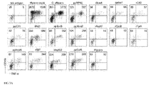
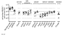
- fumigatus proteins i.e. the combination of frequencies, naive/memory distribution and effector cytokine production of specific T cells allowed the classification of proteins/antigens of A.fumigatus into two groups: the in-vivo-target antigens and the non-target antigens.
- One group revealed a picture similar to the whole protein lysate, characterized by high frequencies of naive T cells (about 50%) and low frequencies of effector cytokine producers ( ⁇ 20% IFN-gamma, ⁇ 5% IL-17) in most donors, suggesting that these proteins are no in vivo targets of T cells in healthy donors.
- the other group revealed a surprisingly high frequency of memory T cells (preferentially more than 60%) and only few naive T cells (preferentially less than 40%), suggesting that proteins of this group are in vivo targets of the T cell response in healthy donors.
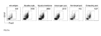
- This group of in vivo targets can further be split up into two subgroups: "Immunogenic" proteins are characterized by high overall T cell frequencies (preferentially more than 1 in 10E3, more preferentially more than 1 in 10E4, most preferentially more than 1 in 10E5), mainly memory type cells (60-100%) and high IFN- ⁇ (15-80%) and/or IL-17 (5-30%) production.
- Exhausting proteins were classified due to their low to intermediate overall frequencies (preferentially less than 1 in 10E4, more preferentially less than 1 in 10E5, most preferentially less than 1 in 10E6) and lack of effector cytokine production ( ⁇ 15% IFN-gamma, ⁇ 5% IL-17), although the majority of cells had a clear memory phenotype (60-100%). These properties may indicate deletion and/or anergy of specific T cells due to overstimulation in vivo again supporting the idea that these proteins are in vivo recognized during in vivo contact with A. fumigatus. Thus the "immunogenic" and “exhausting” antigens are defined as "in-vivo-target antigens”.
- non-target proteins since they induce high overall T cell frequencies, but strikingly a large proportion of the cells is still in the naive state and also lacks effector cytokine production. This indicates that no immune reactivity is induced in vivo despite the fact that these proteins can stimulate T cells when present during in vitro stimulation. This suggests that non-target proteins are not relevant for T cell responses against A. fumigatus infections in healthy donors. The fact that the T cells reacting against the total A. fumigatus lysate also contain many naive T cells shows that indeed a large part of the A.
- fumigatus proteome actually belongs to the non-target protein group, i.e. is immunologically neutral.
- identification of true in vivo target proteins is an important step to identify proteins which possess relevance during in vivo A. fumigatus infections and thus possess potential value as diagnostic or therapeutic tools.
- single proteins might be classified into different groups in different donors, indicating that donor-specific features may influence the reactivity against single proteins, e.g. MHC restriction elements.
- ARTE In addition to the phenotypic characteristics, ARTE also allows to determine the functionally important production of effector cytokines, such as IFN- ⁇ or IL-17.
- effector cytokines such as IFN- ⁇ or IL-17.
- IL-17 is frequently claimed as an important cytokine for anti-fungal immune responses, the importance for protection against fungal infections versus immunopathology is currently under debate. In this study we observed a predominant IFN- ⁇ production and only low IL-17, which is in line with previous reports suggesting that A. fumigatus elicits predominantly Th1 responses in vivo.
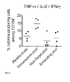
- Th2 cytokines (IL-4, IL-5, IL-13) were only marginal produced against the defined pools of single proteins (data not shown) and have previously been shown to be typically below 5% of all reactive CD154 + cells against the total soluble lysate ( Bacher et al J. Immunol 2013 ). However, when single proteins or pools thereof were analyzed, polyfunctional cytokine production (co-expression of TNF- ⁇ , IL-2 and IFN- ⁇ ) and strong IL-17 cytokine production was only observed against proteins classified as "immunogenic". Also against the immunogenic proteins, IFN- ⁇ was the dominating lineage defining cytokine confirming also on the level of single proteins that the in vivo response against A.
- Treg are not selectively generated against a subgroup of proteins but their expansion seems to be coupled to the expansion of conventional T cells, which may be mediated via growth factor supply, such as IL-2.
- growth factor supply such as IL-2.
- Treg the T cell response against all A. fumigatus proteins is controlled by Treg and therefore the depletion of Treg might be a promising strategy for releasing full T cell responses, e.g. for immunotherapeutic approaches.
- Treg can be used directly ex vivo or following in vitro expansion to treat patients with overshooting or chronic inflammatory or allergic responses against A . fumigatus.
- the present invention provides the in-vitro use of at least one in-vivo-target antigen of Aspergillus fumigatus for selective activation, detection and/or analysis of Aspergillus fumigatus-specific CD4 + cells in a sample comprising cells, wherein said at least one in-vivo-target-antigen is selected from the group consisting of antigens Scw4, Pst1, Shm2, GliT and TpiA.
- the present invention provides a method for detecting, isolating and/or analyzing Aspergillus fumigatus-specific CD4 + T cells, the method comprising:
- Detection and analysis of said Aspergillus fumigatus-specific CD4 + T cells may be performed by standard assays known in the field to analyse activated T cells, e.g. by detection of activation markers by flow cytometry or fluorescence microscopy, by quantitating secreted cytokines via ELISA, quantitation of proliferation ( 3 H thymidine incorporation, CFSE dilution or comparable assays for cellular proliferation), intracellular cytokine staining, cytokine secretion assay, cytokine ELISPOT.
- standard assays known in the field to analyse activated T cells e.g. by detection of activation markers by flow cytometry or fluorescence microscopy, by quantitating secreted cytokines via ELISA, quantitation of proliferation ( 3 H thymidine incorporation, CFSE dilution or comparable assays for cellular proliferation), intracellular cytokine staining, cytokine secretion assay,
- Isolation of said Aspergillus fumigatus-specific CD4 + T cells may be performed e.g. by flow-cytometry methods such as FACS ® or by magnetic cell separation methods such as MACS ® .
- the method may comprise the additional step of enrichment of reactive T cells from a sample comprising T cells before adding the at least one in-vivo-target antigen. Enrichment of said reactive T cells may be performed by
- the method may comprise the additional step of expansion of reactive T cells.
- Exemplary, enrichment of reactive CD154 + or CD137 + T cells from a sample comprising cells may be performed by using magnetic cell separation technologies such as MACS ® (Magnetic-activated cell sorting) or flow cytometric technologies such as FACS ® (Fluorescence activated cell sorting) using an antigen-binding fragment such as an antibody against the marker CD154 or any other specific marker accessible on the cell surface.
- magnetic cell separation technologies such as MACS ® (Magnetic-activated cell sorting) or flow cytometric technologies
- FACS ® Fluorescence activated cell sorting
- an antigen-binding fragment such as an antibody against the marker CD154 or any other specific marker accessible on the cell surface.
- Exemplary, expansion of said enriched reactive T cells can be performed with methods well known in the art such as culturing them alone or together with irradiated or mitomycin C treated autologous feeder cells and cytokines, such as IL-2.
- specific T cells may be expanded by simply adding the antigens to a mixture of cells comprising the specific T cells.
- the mixture may also comprise antigen-presenting cells.
- Cytokines such as IL-2 may be added to enhance T cell proliferation.
- the present invention provides the use of peptides derived from at least one in-vivo-target antigen of A.fumigatus, for loading recombinant MHC class I or class II (HLA) proteins with said peptides to generate functional MHC/peptide complexes or multimeric composites thereof, such as tetramers, pentamers or other higher multimeric structures suitable to bind and/or activate peptide specific T cells, wherein said at least one in-vivo-target antigen is selected from the group consisting of antigens Scw4, Pst1, Shm2, GliT and TpiA
- Such multimers would also be suitable to label and enrich specific T cells, if the multimers are also labelled, e.g. fluorescently, magnetically or by defined isotopes, similar as described above.
- the present invention provides a pharmaceutical composition
- a pharmaceutical composition comprising at least one in-vivo-target antigen of Aspergillus fumigatus for use as vaccine against aspergillosis, wherein said at least one in-vivo-target antigen is selected from the group consisting of antigens Scw4, Pst1, Shm2, GliT and TpiA.
- the present invention provides a pharmaceutical composition comprising Aspergillus fumigatus -specific CD4 + T cells for use in immunotherapy of aspergillosis, wherein said Aspergillus fumigatus-specific CD4 + T cells are obtained by the present method.
- the present invention provides a kit for detecting, isolating and/or analyzing Aspergillus fumigatus-specific CD4 + T cells comprising
- the tag may be a magnetic particle or fluorophore.
- in-vivo-target antigen has a further specific meaning as described herein. Since the method ARTE allows multi-parameter characterization of very rare single A. fumigatus protein-specific T cells, the combination of frequencies, naive/memory distribution and effector cytokine production allowed classification of the fungal antigens/proteins into three subgroups: "Immunogenic" antigens/proteins are characterized by high overall T cell frequencies, mainly memory type cells and high IFN- ⁇ and/or IL-17 production. In contrast, "exhausting" antigens/proteins were classified due to their low to intermediate overall frequencies, and lack of effector cytokine production, although the majority of cells had a clear memory phenotype.
- the amino acid sequences of Scw4, Pst1, Shm2, GliT and TpiA are given in SEQ ID NO:1, SEQ ID NO:2, SEQ ID NO:3, SEQ ID NO:4 and SEQ ID NO:5, respectively (in the one-letter code of amino acids).
- the gene/protein names ShmB or SHMT may also be used for Shm2.
- the gene/protein name Tpi1 may also be used for TpiA.
- the term "in-vivo-target antigen" Scw4, Pst1, Shm2, GliT and TpiA as used herein refers to all constellations of the respective antigen which retains the intended function of being an in-vivo-target antigen of Aspergillus fumigatus as defined herein.
- the divergences to the SEQ ID Nos:1, SEQ ID NO:2, SEQ ID NO:3, SEQ ID NO:4 and SEQ ID NO:5, respectively should not affect their immunogenic potential as in-vivo-target antigen, respectively, of Aspergillus fumigatus as disclosed herein. Therefore, the in-vivo-target antigens Scw4, Pst1, Shm2, GliT and TpiA, respectively, can be the full length protein of SEQ ID NO:1, SEQ ID NO:2, SEQ ID NO:3, SEQ ID NO:4 and SEQ ID NO:5, respectively.
- sequence identity may be determined using pairwise alignments using alignments programs for amino acid sequences well known to the art.
- the in-vivo-target antigen of Scw4, Pst1, Shm2, GliT and TpiA, respectively, can also be a fragment of the protein Scw4, Pst1, Shm2, GliT and TpiA, respectively, e.g. a minimal peptide of 6-15 amino acids in length.
- the in-vivo-target antigen Scw4, Pst1, Shm2, GliT and TpiA, respectively is a peptide pool.
- the peptide pool may comprise several peptides covering the complete or part of the sequences of Scw4, Pst1, Shm2, GliT and TpiA, respectively, peptides with may or may have not overlapping amino acid sequences.
- the in-vivo-peptide antigen is a peptide pool comprising 15meric overlapping peptides (e.g. 11 amino acids overlap) and spanning the whole protein sequence.
- specific T cells refers to a T cell expressing a certain T cell antigen receptor with the capacity to recognize only a specific peptide bound to a certain MHC molecule but not to the same MHC molecule complexed with other irrelevant peptides. "Specific” does not exclude that several possible peptides may exist which can bind to the MHC molecule and which are then recognized by the same TCR. This can be peptides with high homology, i.e. similar amino acid sequences, but even complete unrelated peptides may have this capacity.
- TCR does rather mean that a TCR does only react to small selection of peptide/MHC complexes out of large reservoire of possible combinations, and in this way the particular T cell is specific for the antigen, the peptide is derived of but it may not exclude the possibility that cross-reactivity against other peptide derived from similar or unrelated antigen occur. That a certain TCR recognized a certain MHC/peptide combination can be tested with various technologies. Direct binding of the TCR can be demonstrated by using multimeric complexes (tetramers, pentamers) of the specific MHC/peptide (antigen) complex labelled with a fluorochrome. Such multimeric MHC molecules are well known in the art.
- MHC class I or class II in humans HLA class I or class II, Human Leukocyte Antigen
- MHC class II in humans HLA class I or class II, Human Leukocyte Antigen
- antigen binding "groove" of the MHC molecule antigenic peptides e.g. 9-15mers
- a functional MHC/peptide monomer is generated which can bind to the specific TCR recognizing the combination of MHC + peptide.
- this binding affinity is low. Therefore multimeric structures are generated via various technologies known in the art to fuse several MHC/peptide complexes (e.g. 4, 5, 6 or more) to generate a high avidity protein which can bind simultaneously to multiple TCRs on the T cell surface and generate stable interaction. Binding of such multimeric complexes may also induce specific T cell activation.
- recognition can be demonstrated by measuring the response of the T cells to activation by the specific peptide/MHC molecule, e.g. by adding the peptide to a mixture of autologous antigen presenting cells and the T cells for several hours or days and measuring cytokine expression, expression of surface markers such as CD154 or CD137 or proliferation of the T cells.
- T cell is used herein mostly overlapping with the term "antigen-specific" since T cells which can specifically recognize a specific peptide/MHC combination as described above become activated by this stimulus via their TCR, i.e. they express certain cytokines, activation markers or start to proliferate. This sign of activation can then be used to identify the cells as described above.
- Antigen-reactive refers to T cells which are activated via there TCR but not to T cells which are activated independently of the TCR, e.g. via cytokines produced by other activated cells in the culture.
- the groups of antigen-specific and antigen-reactive T cells do large overlap, however anergic or inactivated T cells may exist which still have the specific TCR but lost their potential to become activated, i.e. they are excluded from the group of antigen-reactive T cells.
- Antigen-specificity and reactivity can be determined in all types of T cells.
- conventional (Tcon) and regulatory T cells (Treg) can be distinguished.
- Treg are defined here as Foxp3 + CD4 + T cells which typically do also express CD25 and lack expression of CD127. They represent a separate T cell lineage required to maintain tolerance against auto-antigens and harmless foreign antigens.
- Tcon cells as defined here comprise all T cells which are not Treg.
- Treg naive (no previous contact to antigen) and antigen-experienced or memory T cells (Tmem) can be found, which substantially differ in many phenotypical and functional aspects.
- Each T cell have a specific antigen receptor (T cell receptor, TCR) for one particular antigen.
- T cells with specificity for a particular antigen are typically rare, i.e. about 1 in 1 million for naive T cells and typically ⁇ 1% for memory T cells, except in certain acute infectious diseases. Therefore it requires analysis methods able to identify these rare cells to study the immune status of a certain patient against a defined antigen or pathogen.
- antibody refers to polyclonal or monoclonal antibodies which can be generated by methods well known to the person skilled in the art.
- the antibody may be of any species, e.g. murine, rat, sheep, human.
- non-human antigen binding fragments are to be used, these can be humanized by any method known in the art.
- the antibodies may also be modified antibodies (e.g. oligomers, reduced, oxidized and labelled antibodies).
- antibody comprises both intact molecules and antibody fragments, such as Fab, Fab', F(ab')2, Fv and single-chain antibodies.
- antigen-binding fragment includes any moiety other than antibodies or antibody fragments that binds preferentially to the desired target molecule of the cell. Suitable moieties include, without limitation, oligonucleotides known as aptamers that bind to desired target molecules
- tag refers to the coupling of the antigen-binding fragment, e.g. an antibody or fragment thereof, to other molecules, e.g. particles, fluorophores, haptens like biotin, or larger surfaces such as culture dishes and microtiter plates.
- the coupling results in direct immobilization of the antigen-binding fragment, e.g. if the antigen-binding fragment is coupled to a larger surface of a culture dish.
- this coupling results in indirect immobilization, e.g. an antigen-binding fragment coupled directly or indirectly (via e.g. biotin) to a magnetic bead is immobilized if said bead is retained in a magnetic field.
- the coupling of the antigen-binding fragment to other molecules results not in a direct or indirect immobilization but allows for enrichment, separation, isolation, and detection of cells according to the present invention, e.g. if the antigen-binding fragment is coupled to a fluorophore which then allows discrimination of stronger labeled cells, weaker labeled cells, and non-labeled cells, e.g. via flow cytometry methods, like FACSorting, or fluorescence microscopy.
- particle refers to a solid phase such as colloidal particles, microspheres, nanoparticles, or beads. Methods for generation of such particles are well known in the field of the art.
- the particles may be magnetic particles.
- the particles may be in a solution or suspension or they may be in a lyophilized state prior to use in the present invention. The lyophilized particle is then reconstituted in convenient buffer before contacting the sample to be processed regarding the present invention.
- magnetic in “magnetic particle” as used herein refers to all subtypes of magnetic particles which can be prepared with methods well known to the skilled person in the art, especially ferromagnetic particles, superparamagnetic particles and paramagnetic particles.
- “Ferromagnetic” materials are strongly susceptible to magnetic fields and are capable of retaining magnetic properties when the field is removed.
- Paramagnetic materials have only a weak magnetic susceptibility and when the field is removed quickly lose their weak magnetism.
- Superparamagnetic” materials are highly magnetically susceptible, i.e. they become strongly magnetic when placed in a magnetic field, but, like paramagnetic materials, rapidly lose their magnetism.
- activation marker refers to proteins specifically expressed on the cell surface of T cells following their activation via their specific antigen-receptor, usually within 1-48 hours after activation.
- Activated T cells may comprise CD4 T cells, CD8 T cells, naive T cells, memory T cells, gamma-delta T cells or regulatory T-cells.
- activation markers are, for example, CD154, CD137, secreted or membrane-anchored cytokines, e.g. IL-4, IL-5, IL-13, IFN-gamma, IL-10, IL-2, IL-22, TNF-alpha or "latent TGF-beta" (LAP), GARP (LRRC32), CD121a/b.
- CD154 is a member of the TNF gene family and, inter alia, is expressed by various cells, particularly by T lymphocytes. CD154 is rapidly (within minutes) downregulated by the stimulated T lymphocytes upon contact with its receptor CD40. CD154 can be used to detect T lymphocytes independently of their functional potential. The use of CD 154 in the detection and separation of T lymphocytes leads to reliable detection and isolation of T lymphocytes, possible independently of their function, i.e. all antigen-specific T lymphocytes in a sample can be determined and separated.
- a method is disclosed in WO2004/027428 for the detection and/or isolation of antigen-specific T lymphocytes in a suspension following activation with an antigen, in which method the suspension is contacted with a CD40/CD154 system inhibitor, intra- and/or extracellular determination of CD154 is effected, and the cells having CD154 are detected and/or isolated.
- Addition of a CD40/CD154 system inhibitor impairs or inhibits the interaction and signalling between CD40 and CD154.
- CD40/CD154 system inhibitors can be any of molecules or even physical exposures capable of blocking or inhibiting the interaction between CD40 and CD154. Accordingly, the inhibiting agent can be an antibody, e.g.
- said agent can also be a substance inhibiting the secretion or endocytosis in the cell, such as brefeldin A (Bref-A).
- Bref-A inhibits the Golgi apparatus and the secretion of a variety of cytokines.
- EP2306191B1 disclosed that the use of CD154 as a negative selection marker greatly increases the window of time for the use of the marker CD 137 for the positive selection of regulatory T cells (Treg) and that 2 to 24 hours after activation, regulatory T-cells can be isolated with great purity and yield. Therefore it is possible with the present invention to analyse rare antigen-specific Treg cells, i.e. Aspergillus fumigatus-specific CD4+ T cells, using the combination of activation markers CD137 and CD154.
- any sorting technology can be used. This includes for example affinity chromatography or any other antibody-dependent separation technique known in the art, which allows processing. Any ligand-dependent separation technique known in the art may be used in conjunction with both positive and negative separation techniques that rely on the physical properties of the cells.
- Magnetic cell sorting or “magnetic cell sorting process” is used herein to refer to procedures for cell separation (cell sorting) including, but are not limited to, magnetic separation using antibodies linked to colloidal magnetic particles or micron-sized magnetic particles (e.g. 1-10 ⁇ m). Methods to separate cells magnetically are commercially available e.g. from Invitrogen, Stem cell Technologies, in Cellpro, Seattle or Advanced Magnetics, Boston. For example, autologous monoclonal antibodies can be directly coupled to magnetic polystyrene particles like Dynal M 450 or similar magnetic particles and used e.g. for cell separation.
- antibodies can be biotinylated or conjugated with digoxigenin and used in conjunction with avidin or anti-digoxigenin coated affinity columns.
- monoclonal antibodies are used in conjunction with colloidal superparamagnetic microparticles having an organic coating by e.g. polysaccharides ( Miltenyi et al., 1990, Cytometry 11:231-8 ). These particles can be used having a size of 10 to 200 nm, preferably between 40 and 100 nm, and can be either directly conjugated to autologous antibodies or used in combination with anti-immunoglobulin, avidin or antihapten-specific microbeads. Polysaccharide-coated superparamagnetic particles are commercially available from Miltenyi Biotec GmbH, Germany.
- the cells can be analyzed (characterized) after selective activation with one or more of the in-vivo-target antigens of Aspergillus fumigatus as disclosed herein according to all methods known to the person skilled in the art.
- Preferred for the characterization of cells are in particular cell sorting (e.g. further magnetic cell sorting (MACS)), fluorescence activated cell sorting (FACS), ELISA, PCR and/or all fluorescence microscopes known in the art.
- the sample comprising T cells can be any sample comprising T cells.
- the sample is directly derived from blood, peripheral mononuclear blood cells (PBMC), body tissue or cells from tissue fluids of animals, preferentially mammals such as humans, mouse, rat, sheep or dogs.
- the sample comprising T cells may also be a sample in which T cells are enriched from e.g. a blood sample.
- the T cells may be the only subtype of cells in the sample, or in addition to the T cells may be present antigen presenting cells, or further cells of the blood.
- the sample comprising T cells may encompass expanded T cells or non-expanded T cells.
- the separated Aspergillus fumigatus-specific CD4 + T cells can be used before and/or after cloning and/or growing and/or concentrated in cell mixtures as pharmaceutical composition in the immunotherapy of aspergillosis. It is additionally possible for the coding gene sequences of the TCR (T-cell receptor) to be isolated from the separated Aspergillus fumigatus -specific CD4 + T cells and be used for further therapeutic purposes such as, for example, for cellular therapies. It is additionally possible to employ the Aspergillus fumigatus-specific CD4 + T cells in the form mentioned in further investigations and/or analyses.
- the pharmaceutical composition can be used for the treatment and/or prevention of diseases in mammals, possibly including administration of a pharmaceutically effective amount of the pharmaceutical composition to the mammal.
- a human sample e.g. blood or tissue from a patient suffering from IA or at risk for an A. fumigatus infection is obtained.
- Patients at risk for fungal infections are immunocompromised patients, e.g. following chemotherapy, hematopoetic stem cell transplantation, HIV patients, or patients with intrinsic genetic defects leading to immune deficiency.
- the Aspergillus fumigatus-specific CD4+ T cells are detected via expression of activation markers such as CD154, CD137, cytokines or proliferation and quantitated and/or characterized, e.g. by flow-cytometry or purified via FACS sorting.
- the Aspergillus fumigatus-specific CD4 + T cells are isolated by labeling the cells of the sample with an antibody against the marker molecules mentioned above coupled to a magnetic bead and separated magnetically by applying a magnetic field.
- the Aspergillus fumigatus-specific CD4 + T cells are directly labelled with an MHC multimer loaded with one or several peptides derived from the in vivo target antigens identified in the present invention and the labelled cells are similarly characterized or isolated as described above by well known methods in the art.
- the information about the frequency, phenotype and functional properties of the A. fumigatus specific cells identified as described above may be used to diagnose invasive fungal infection or to identify sensitization of the patient's immune system against the fungus.
- A. fumigatus related diseases may be diagnosed or analysed via characterization of the A. fumigatus specific T cells from the patients.
- diseases include cystic fibrosis, allergies, asthma, chronic obstructive pulmonary disease (COPD).
- COPD chronic obstructive pulmonary disease
- Analysing the specific T cell response against the in vivo target proteins can be used to identify the type of immune response and help to select the appropriate therapeutic concept.
- Increased frequencies identify invasive aspergillosis.
- Alterations in the Treg / effector T cell ratio may identify not properly regulated immune reactions, e.g.
- cytokine patterns identify patient subgroups: allergic or asthmatic patients have increased Th2 cytokines alone or together with Th17 cytokines, patients with strong neutrophil contribution to the inflammation in the lung have increased Th17 cytokines, or patients with a defect in the immune response (no/low effector cytokine production), e.g. T cell exhaustion as a result of in vivo overstimulation.
- T cells specific for one or more in-vivo-target antigens of Aspergillus fumigatus wherein said at least one in-vivo-target antigen is selected from the group consisting of antigens Scw4, Pst1, Shm2, GliT and TpiA, are isolated as described above, e.g. using CD154 as a marker for activated effector T cells.
- the cell can be isolated from the patient or from the donor in case of bone marrow transplantation or be isolated from a third party donor with related MHC molecules.
- the isolated T cells can then be directly infused into a patient with an A. fumigatus infection, e.g. invasive fungal infection.
- the T cells can be expanded in vitro and infused into the patient with the aim to enhance the immune defense against the fungal infection and cure the patient.
- the antigens can also be used to isolate and expand A . fumigatus specific Foxp3+ regulatory T cells (Treg), e.g. by sorting specifically CD137 expressing T cells or CD154- CD137+ T cells after a short 6-12 hours stimulation with the antigen in vitro.
- Treg can similarly be expanded in vitro or be directly used for infusion into patients, e.g. with chronic inflammatory reactions or allergies against A . fumigatus.
- one or several in-vivo-target antigens of Aspergillus fumigatus which are selected from the group consisting of antigens Scw4, Pst1, Shm2, GliT and TpiA are used to vaccinate people at risk for getting A. fumigatus infections or allergies or chronic inflammatory reactions.
- kits contains the components necessary to perform selective activation of Aspergillus fumigatus specific CD4 + T cells in a sample comprising T cells and detection, isolation and/or analysis of said Aspergillus fumigatus-specific CD4 + T cells.
- a kit for detecting, isolating and/or analyzing Aspergillus fumigatus-specific CD4 + T cells comprises
- the tag is a magnetic particle such as a magnetic bead or a fluorophore.
- the antigen binding fragments are coupled to magnetic particles as described herein.
- the magnetic particles e.g. MicroBeads (Miltenyi Biotec GmbH)
- the kit may be in a solution or suspension or they may be in a lyophilized state prior to use in a method of the present invention.
- the lyophilized particle is then reconstituted in convenient buffer before contacting with the sample containing neuronal cells to be processed regarding the present invention.
- PBMCs Peripheral blood mononuclear cells
- a . fumigatus ATCC46645 protein extracts, except for the lysate of resting conidia (RC), 2x10E8 conidia were inoculated in 200 ml YPD medium and cultured at 37 °C and 200 rpm. Swollen conidia (SC) were harvested after 6 h, germinated conidia (GC) after 8 h, and mycelium after 20 h of cultivation. Cells were recovered by centrifugation (RC, SC, GC) or filtration (mycelium) and washed with water before storage at -80 °C.
- SC Swollen conidia
- GC germinated conidia
- mycelium after 20 h of cultivation.
- Cells were recovered by centrifugation (RC, SC, GC) or filtration (mycelium) and washed with water before storage at -80 °C.
- Total RC, SC, or GC lysates were generated by disruption of frozen cells in saline [0.9 % (w/v) NaCl] using a Micro-Dismembrator (Sartorius).
- frozen mycelium was ground in liquid nitrogen by using a mortar and pestle and resuspendend in PBS supplemented with 2 mM MgCl 2 .
- Total soluble protein fractions of the lysates were obtained after removal of insoluble material (cell wall pellet) by centrifugation for 15 min at 10,000 ⁇ g. Fractionated mycelial protein extracts were obtained by sequential centrifugation of total mycelial lysate.
- the cell wall protein fraction was processed by resuspension of the cell wall pellet (15 min of centrifugation at 10000 xg) in PBS/2 mM MgCl 2 .
- the cell membrane-enriched protein fraction pellet
- Cell membrane extract was generated by resuspension of the membrane pellet in PBS/2 mM MgCl 2 .
- the recombinant proteins used were generated by standard technologies known in the field, i.e. cloning of the coding sequences into a suitable expression vector adding a suitable tag for subsequent protein purification (e.g. 6 his tag or MBP-HIS-tagged).
- the proteins were expressed in E. coli, namely Crf2, GliT, Scw4, Aspf3, Shm2, CpcB, Aspf22, Pst1 and TpiA or in Pichia pastoris FG-GAP repeat protein, [devoid of the sequence encoding the 25 amino acids N-terminal secretion signal peptide ( ⁇ 25FG-GAP)] and of AspF2.
- SEQ ID NO:1 SEQ ID NO:2, SEQ ID NO:3, SEQ ID NO:4 and SEQ ID NO:5.
- All proteins were purified by affinity chromatography using an ⁇ kta explorer purification system (GE Healthcare). If necessary, an ion exchange column (SOURCE 15Q, GE Healthcare) was used for further purification. Generally, all buffer exchanges were conducted using HiPrep 26/10 desalting columns (GE Healthcare). All HIS-tagged proteins were applied to a Ni Sepharose 6 Fast Flow (GE Healthcare) column, and eluted with 250 mM imidazole.
- MPP-tagged proteins were loaded onto an Amylose Resin HF (New England Biolabs) column and eluted with 10 mM maltose.
- the MBP-HIS-tag of MBP-tagged proteins and the HIS-tag of AspF3 were cleaved using TEV-protease and removed by its binding to Ni Sepharose.
- Scw4 was further purified by reversed phase chromatopraphy (Source 15RPC, GE Healthcare) after exchanging the buffer to 0.05% (v/v) trifluoroacetic acid, 10 % (v/v) acetonitril. Purified Scw4 was lyophilized and resolved in PBS. For all other purified proteins, the buffer was exchanged to PBS (FG-GAP, Pst1, AspF22, AspF2, GliT) or 0.9 % NaCl (Shm2, CpcB, AspF3, TpiA)
- PBMCs peripheral blood mononuclear cells
- PBMCs peripheral blood mononuclear cells
- RPMI-1640 Miltenyi Biotec, Bergisch Gladbach, Germany
- 5 % (v/v) human AB-serum BioWhittaker/Lonza, Walkersville, MD, USA
- 2 mM L-glutamine PAA Laboratories, Pasching, Austria.
- Cells were stimulated for 7 hours with the following antigens: A. fumigatus lysates (each 40 ⁇ g/ml), C.
- albicans -lysate (20 ⁇ g/ml; Greer Laboratories, Lenoir, NC, USA), CMV-lysate (10 ⁇ g/ml, Siemens Healthcare Diagnostics, Marburg, Germany), recombinant A. fumigatus proteins (Crf2, Pst1, Aspf2, Aspf3, Shm2, FG-GAP, GliT, Aspf22, CpcB, TpiA, Scw4; each 20 ⁇ g/ml) or peptide pools ( C.
- Enrichment of reactive CD154 + T cells or combined enrichment of CD154 + /CD137 + T cells was performed using the CD154 MicroBead Kit alone, or in combination with the CD137 MicroBead Kit (both Miltenyi Biotec).
- cells were indirectly magnetically labeled with CD154-Biotin and CD137-PE followed by anti-Biotin Microbeads and anti-PE Microbeads and enriched by two sequential MS MACS columns (Miltenyi Biotec).
- 1 ⁇ g/ml Brefeldin A Sigma Aldrich
- Magnetically enriched CD154 + T cells were expanded with 1:100 mitomycin C (Sigma Aldrich) treated autologous feeder cells in X-VivoTM15 (BioWhittaker/Lonza), supplemented with 5 % (v/v) AB-serum (BioWhittaker/Lonza), 200 U/ml IL-2 (Proleukin ® ; Novartis, Laubis, Germany) and 100 U/ml penicillin, 100 ⁇ g/ml streptomycin, 0.25 ⁇ g/ml amphotericin B (Antibiotic Antimycotic Solution, Sigma Aldrich) at a density of 2.5x10E6 cells/cm 2 . During expansion for 2-3 weeks, medium was replenished and cells were split as needed.
- 5 ⁇ 10E5 expanded T cells were re-stimulated with autologous CD3-depleted (CD3 MicroBeads; Miltenyi Biotec) PBMC in a ratio of 1:1 in 96-well flat bottom plates with different antigens in presence of 1 ⁇ g/ml CD28 functional grade pure Ab for 2h plus additional 4h with 1 ⁇ g/ml Brefeldin A (Sigma Aldrich). After fixation and permeabilization cells were stained intracellularly for CD 154 and cytokines.
- CD3-depleted CD3 MicroBeads; Miltenyi Biotec
- CD4-VioBlue CD4-FITC
- CD4-APC-Vio770 VIT4
- CD3-APC BW264/56
- CD14-VioGreen CD14-PerCP
- CD20-VioGreen CD20-PerCP
- CD8-VioGreen BW135/80
- CD45RO-FITC CD45RO-PerCP
- CD45RO-PerCP UCHL-1
- CCR7-PE REA108
- CD45RA-APC CD45RA-FITC
- T6D11 anti-Biotin-PE, anti-Biotin-VioBlue (Bio3-18E7)
- CD154-PE CD154-APC
- CD154-VioBlue 5C8
- TNF- ⁇ -FITC TNF- ⁇ -FITC
- TNF- ⁇ -PE-Vio770 cA2
- IFN- ⁇ -FITC IFN- ⁇ -FITC
- IFN- ⁇ -APC IFN- ⁇ -PE (45-15)
- Example 2 Human CD4 + T cell response against lysates of different A. fumigatus morphotypes
- CD4 + T cells specifically reacting against A . fumigatus can be identified using CD154 expression as a specific read-out for antigen activated CD4 + T cells after short in vitro stimulation with fungal lysate ( Bacher et al, J Immunol 2013 ; Frentsch et al, Nat Med 2005 )
- PBMC peripheral blood mononuclear cells
- Reactive CD154-expressing CD4 + T cells were identified by flow-cytometry.
- cytokine production and phenotype revealed no major differences of T cells stimulated with the different A . fumigatus morphotype lysates: as shown in Fig. 1D , against all lysates a high frequency of TNF- ⁇ and IL-2 producers could be detected, whereas the production of the lineage defining cytokines IFN- ⁇ and IL-17 was only low, although IFN- ⁇ production was clearly predominant over IL-17, as already described before ( Bacher et al J Immunol 2013 , Schönbrunn et al J Immunol 2012 ). Similarly, irrespective of the A. fumigatus lysate used for stimulation a comparable amount of reactive memory T cells was detected ( Fig. 1E ).
- T cells specific for a panel of 15 selected A. fumigatus proteins with different biological functions and cellular localization. Either recombinant proteins or synthesized 15mer peptide pools covering the complete protein sequence were used for stimulation.
- the analyzed proteins included cell wall, GPI-anchored, secreted, as well as cytosolic proteins and were chosen based on their high abundance within the conidial, mycelia or secreted proteome and/or their previous description as being immunogenic, based on T cell or serum reactivity.
- a concentration of 20 ⁇ g/ml of recombinant proteins for the stimulation of 1 ⁇ 10E7 PBMCs was determined based on titration of the single proteins on expanded total mycelia-reactive T cell lines (data not shown).
- the C. albicans protein MP65 had previously been described as a major antigen target of human T cell responses and served as a positive control.
- ppMP65 is a control peptide pool of the protein MP65 of C . albicans.
- CD154 + cells were isolated, subsequently expanded for 3 weeks and tested for specificity via antigen re-stimulation (MP65, whole Aspergillus lysate, CMV lysate or the A. fumigatus single protein used for initial isolation of the cells). Percentage of reactive cells among CD4 + lymphocytes are shown for several donors, as determined by CD154 and TNF- ⁇ expression.
- the T cell responses against the different proteins were quite variable with frequencies ranging from 1.2 ⁇ 10E-6 to 3.1 ⁇ 10E-4 ( Fig. 3C ) and showed strong intra- and inter-donor variability ( Fig. 3A , C ).
- the subcellular location of the proteins did not result in a clear-cut phenotypic/functional characteristic of the resulting T cell response, although a trend towards a strong reactivity against membrane-associated proteins was observed.
- Example 5 Integration of phenotypic and functional markers of specific T cells allows classification of antigenic proteins
- non-target proteins since they induce high overall T cell frequencies, but strikingly a large proportion of the cells is still in the naive state and also lacks effector cytokine production. This indicates that no immune reactivity is induced in vivo.
- the reactivity against the mycelia crude lysate as shown in figure 4 is also characterized by a high frequency of naive T cells and rather low effector cell frequencies, when compared to the immunogenic protein group, suggesting that a large fraction of the fungal proteins actually belong to the exhausting or non-target group.
- Example 6 A. fumigatus -specific Tcon and Treg recognize the same antigens
- the single proteins were pooled according to our previous classification into a immunogenic group, containing proteins with the highest reactivity (Scw4, Crf1, Crf2, Pst1 and Shm2) as well as a non-target (Gel1, CatB) and exhausting (Aspf2, Aspf3, CpcB, Fg-Gap) group and used for stimulation in comparison to the mycelia crude lysate or the mycelia membrane fraction.
- CD137 which is expressed by Treg after 6 hours of stimulation was used together with CD154 enrichment to differentiate between Treg (CD137 + CD154 - ) and Tcon (CD137 - CD154 + ) ( Bacher, P. et al, Mucosal Immunol.
Landscapes
- Health & Medical Sciences (AREA)
- Life Sciences & Earth Sciences (AREA)
- Immunology (AREA)
- Engineering & Computer Science (AREA)
- Cell Biology (AREA)
- Chemical & Material Sciences (AREA)
- Biomedical Technology (AREA)
- Hematology (AREA)
- Molecular Biology (AREA)
- Urology & Nephrology (AREA)
- General Health & Medical Sciences (AREA)
- Microbiology (AREA)
- Medicinal Chemistry (AREA)
- Mycology (AREA)
- Tropical Medicine & Parasitology (AREA)
- Analytical Chemistry (AREA)
- Veterinary Medicine (AREA)
- Pathology (AREA)
- Public Health (AREA)
- Biotechnology (AREA)
- Animal Behavior & Ethology (AREA)
- Epidemiology (AREA)
- Pharmacology & Pharmacy (AREA)
- Food Science & Technology (AREA)
- Physics & Mathematics (AREA)
- General Physics & Mathematics (AREA)
- Biochemistry (AREA)
- Bioinformatics & Cheminformatics (AREA)
- Toxicology (AREA)
- Peptides Or Proteins (AREA)
- Medicines Containing Antibodies Or Antigens For Use As Internal Diagnostic Agents (AREA)
- Measuring Or Testing Involving Enzymes Or Micro-Organisms (AREA)
- Investigating Or Analysing Biological Materials (AREA)
- Medicines Containing Material From Animals Or Micro-Organisms (AREA)
- Zoology (AREA)
- Virology (AREA)
Priority Applications (3)
| Application Number | Priority Date | Filing Date | Title |
|---|---|---|---|
| EP14181113.3A EP2985342A1 (fr) | 2014-08-15 | 2014-08-15 | Antigènes immunogènes provenant d'aspergillus fumigatus |
| EP15180326.9A EP2993227A3 (fr) | 2014-08-15 | 2015-08-10 | Antigènes immunogènes provenant d'aspergillus fumigatus |
| US14/824,957 US9770495B2 (en) | 2014-08-15 | 2015-08-12 | Immunogenic antigens from Aspergillus fumigatus |
Applications Claiming Priority (1)
| Application Number | Priority Date | Filing Date | Title |
|---|---|---|---|
| EP14181113.3A EP2985342A1 (fr) | 2014-08-15 | 2014-08-15 | Antigènes immunogènes provenant d'aspergillus fumigatus |
Publications (1)
| Publication Number | Publication Date |
|---|---|
| EP2985342A1 true EP2985342A1 (fr) | 2016-02-17 |
Family
ID=51302937
Family Applications (2)
| Application Number | Title | Priority Date | Filing Date |
|---|---|---|---|
| EP14181113.3A Withdrawn EP2985342A1 (fr) | 2014-08-15 | 2014-08-15 | Antigènes immunogènes provenant d'aspergillus fumigatus |
| EP15180326.9A Ceased EP2993227A3 (fr) | 2014-08-15 | 2015-08-10 | Antigènes immunogènes provenant d'aspergillus fumigatus |
Family Applications After (1)
| Application Number | Title | Priority Date | Filing Date |
|---|---|---|---|
| EP15180326.9A Ceased EP2993227A3 (fr) | 2014-08-15 | 2015-08-10 | Antigènes immunogènes provenant d'aspergillus fumigatus |
Country Status (2)
| Country | Link |
|---|---|
| US (1) | US9770495B2 (fr) |
| EP (2) | EP2985342A1 (fr) |
Families Citing this family (1)
| Publication number | Priority date | Publication date | Assignee | Title |
|---|---|---|---|---|
| GB201204687D0 (en) * | 2012-03-16 | 2012-05-02 | Microsoft Corp | Communication privacy |
Citations (5)
| Publication number | Priority date | Publication date | Assignee | Title |
|---|---|---|---|---|
| WO2002086090A2 (fr) * | 2001-04-23 | 2002-10-31 | Elitra Pharmaceuticals, Inc. | Identification de genes essentiels d'aspergillus fumigatus, et procedes d'utilisation |
| WO2004027428A1 (fr) | 2002-08-23 | 2004-04-01 | Deutsches Rheuma-Forschungszentrum Berlin | Procede permettant de detecter et d'isoler des lymphocytes t qui reconnaissent un antigene defini |
| US7504490B1 (en) * | 1998-10-16 | 2009-03-17 | Oscient Pharmaceuticals Corporation | Nucleic acid and amino acid sequences relating to Apergillus fumigatus for diagnostics and therapeutics |
| EP2518504A1 (fr) * | 2011-04-29 | 2012-10-31 | Miltenyi Biotec GmbH | Procédé pour la caractérisation quantitative et qualitative de lymphocytes T spécifiques à l'antigène reconnaissant l'antigène spécifique |
| EP2306191B1 (fr) | 2009-09-10 | 2012-12-26 | Miltenyi Biotec GmbH | Utilisation de CD154 pour l'identification et la séparation de lymphocytes T ne participant pas à la régulation à partir d'un mélange de lymphocytes T de régulation |
-
2014
- 2014-08-15 EP EP14181113.3A patent/EP2985342A1/fr not_active Withdrawn
-
2015
- 2015-08-10 EP EP15180326.9A patent/EP2993227A3/fr not_active Ceased
- 2015-08-12 US US14/824,957 patent/US9770495B2/en active Active
Patent Citations (5)
| Publication number | Priority date | Publication date | Assignee | Title |
|---|---|---|---|---|
| US7504490B1 (en) * | 1998-10-16 | 2009-03-17 | Oscient Pharmaceuticals Corporation | Nucleic acid and amino acid sequences relating to Apergillus fumigatus for diagnostics and therapeutics |
| WO2002086090A2 (fr) * | 2001-04-23 | 2002-10-31 | Elitra Pharmaceuticals, Inc. | Identification de genes essentiels d'aspergillus fumigatus, et procedes d'utilisation |
| WO2004027428A1 (fr) | 2002-08-23 | 2004-04-01 | Deutsches Rheuma-Forschungszentrum Berlin | Procede permettant de detecter et d'isoler des lymphocytes t qui reconnaissent un antigene defini |
| EP2306191B1 (fr) | 2009-09-10 | 2012-12-26 | Miltenyi Biotec GmbH | Utilisation de CD154 pour l'identification et la séparation de lymphocytes T ne participant pas à la régulation à partir d'un mélange de lymphocytes T de régulation |
| EP2518504A1 (fr) * | 2011-04-29 | 2012-10-31 | Miltenyi Biotec GmbH | Procédé pour la caractérisation quantitative et qualitative de lymphocytes T spécifiques à l'antigène reconnaissant l'antigène spécifique |
Non-Patent Citations (21)
| Title |
|---|
| BACHER ET AL., J IMMUNOL, 2013 |
| BACHER ET AL., J. IMMUNOL, 2013 |
| BACHER ET AL., MUCOSAL IMMUNOL, 2013 |
| BACHER, P. ET AL., MUCOSAL IMMUNOL., 2013 |
| BACHER, P.; C. SCHINK; J. TEUTSCHBEIN; O. KNIEMEYER; M. ASSENMACHER; A. A. BRAKHAGE; A. SCHEFFOLD: "Antigen-reactive T cell enrichment for direct, high-resolution analysis of the human naive and memory Th cell repertoire", J IMMUNOL, vol. 190, 2013, pages 3967 - 3976 |
| BACHER, P.; O. KNIEMEYER; A. SCHONBRUNN; B. SAWITZKI; M. ASSENMACHER; E. RIETSCHEL; A. STEINBACH; O. A. CORNELY; A. A. BRAKHAGE; A: "Antigen-specific expansion of human regulatory T cells as a major tolerance mechanism against mucosal fungi", MUCOSAL IMMUNOL, 2013 |
| FRENTSCH ET AL., NAT MED, 2005 |
| FRENTSCH, M.; O. ARBACH; D. KIRCHHOFF; B. MOEWES; M. WORM; M. ROTHE; A. SCHEFFOLD; A. THIEL: "Direct access to CD4+ T cells specific for defined antigens according to CD154 expression", NAT MED, vol. 11, 2005, pages 1118 - 1124, XP055004658, DOI: doi:10.1038/nm1292 |
| H. HEBART: "Analysis of T-cell responses to Aspergillus fumigatus antigens in healthy individuals and patients with hematologic malignancies", BLOOD, vol. 100, no. 13, 1 August 2002 (2002-08-01), pages 4521 - 4528, XP055169964, ISSN: 0006-4971, DOI: 10.1182/blood-2002-01-0265 * |
| H. JOLINK ET AL: "Characterization of the T-Cell-Mediated Immune Response Against the Aspergillus fumigatus Proteins Crf1 and Catalase 1 in Healthy Individuals", THE JOURNAL OF INFECTIOUS DISEASES, vol. 208, no. 5, 22 May 2013 (2013-05-22), pages 847 - 856, XP055169961, ISSN: 0022-1899, DOI: 10.1093/infdis/jit237 * |
| H. JOLINK ET AL: "Induction of A. fumigatus-specific CD4-positive T cells in patients recovering from invasive aspergillosis", HAEMATOLOGICA, vol. 99, no. 7, 18 April 2014 (2014-04-18), pages 1255 - 1263, XP055169962, ISSN: 0390-6078, DOI: 10.3324/haematol.2013.098830 * |
| MILTENYI ET AL., CYTOMETRY, vol. 11, 1990, pages 231 - 8 |
| MUCOSAL IMMUNOL, 2013 |
| P. BACHER ET AL: "Identification of Immunogenic Antigens from Aspergillus fumigatus by Direct Multiparameter Characterization of Specific Conventional and Regulatory CD4+ T Cells", THE JOURNAL OF IMMUNOLOGY, vol. 193, no. 7, 29 August 2014 (2014-08-29), pages 3332 - 3343, XP055169965, ISSN: 0022-1767, DOI: 10.4049/jimmunol.1400776 * |
| SCHOENBRUNN, A. ET AL., J IMMUNOL, 2012 |
| SCHOENBRUNN, A.; M. FRENTSCH; S. KOHLER; J. KEYE; H. DOOMS; B. MOEWES; J. DONG; C. LODDENKEMPER; J. SIEPER; P. WU: "A converse 4-1BB and CD40 ligand expression pattern delineates activated regulatory T cells (Treg) and conventional T cells enabling direct isolation of alloantigen-reactive natural Foxp3+ Treg", J IMMUNOL, vol. 189, 2012, pages 5985 - 5994 |
| SCHONBRUNN ET AL., J IMMUNOL, 2012 |
| SHI ET AL., BMC MICROBIOL, 2012 |
| SHI, L. N.; F. Q. LI; M. HUANG; J. F. LU; X. X. KONG; S. Q. WANG; H. F. SHAO: "Immunoproteomics based identification of thioredoxin reductase GliT and novel Aspergillus fumigatus antigens for serologic diagnosis of invasive aspergillosis", BMC MICROBIOL, vol. 12, 2012, pages 11, XP021117883, DOI: doi:10.1186/1471-2180-12-11 |
| VODISCH ET AL., PROTEOMICS, 2009 |
| VODISCH, M.; D. ALBRECHT; F. LESSING; A. D. SCHMIDT; R. WINKLER; R. GUTHKE; A. A. BRAKHAGE; O. KNIEMEYER: "Two-dimensional proteome reference maps for the human pathogenic filamentous fungus Aspergillus fumigatus", PROTEOMICS, vol. 9, 2009, pages 1407 - 1415 |
Also Published As
| Publication number | Publication date |
|---|---|
| US20160047809A1 (en) | 2016-02-18 |
| US9770495B2 (en) | 2017-09-26 |
| EP2993227A3 (fr) | 2016-06-01 |
| EP2993227A2 (fr) | 2016-03-09 |
Similar Documents
| Publication | Publication Date | Title |
|---|---|---|
| Naghavian et al. | Microbial peptides activate tumour-infiltrating lymphocytes in glioblastoma | |
| Bacher et al. | Identification of immunogenic antigens from Aspergillus fumigatus by direct multiparameter characterization of specific conventional and regulatory CD4+ T cells | |
| DeLong et al. | Ara h 1–reactive T cells in individuals with peanut allergy | |
| JP7315478B2 (ja) | エピトープを特定するための方法および組成物 | |
| JP5161798B2 (ja) | T細胞アッセイ | |
| CN102277331A (zh) | 快速分离人Foxp3+ Treg细胞的方法和试剂盒 | |
| TW201245224A (en) | Cytotoxic t cell inducing composition | |
| Ross et al. | The canine MHC class Ia allele DLA-88* 508: 01 presents diverse self-and canine distemper virus-origin peptides of varying length that have a conserved binding motif | |
| Archila et al. | Tetramer-guided epitope mapping: a rapid approach to identify HLA-restricted T-cell epitopes from composite allergens | |
| US9770495B2 (en) | Immunogenic antigens from Aspergillus fumigatus | |
| Tischer-Zimmermann et al. | Rapid and sustained T cell-based immunotherapy against invasive fungal disease via a combined two step procedure | |
| Kinnunen et al. | Allergen‐specific naïve and memory CD4+ T cells exhibit functional and phenotypic differences between individuals with or without allergy | |
| US20120276557A1 (en) | Method for the quantitative and qualitative characterization of antigen-specific t cells recognizing a specific antigen | |
| Class et al. | Patent application title: IMMUNOGENIC ANTIGENS FROM ASPERGILLUS FUMIGATUS Inventors: Alexander Scheffold (Berlin, DE) Petra Bacher (Berlin, DE) Olaf Kniemeyer (Jena, DE) Janka Teutschbein (Jena, DE) Axel Brakhage (Weimar, DE) Assignees: Miltenyi Biotec GmbH Leibniz Institute for Natural Product Research and Infection Biology-Hans Knoll Institute | |
| McMurchy et al. | In vitro generation of human T regulatory cells: generation, culture, and analysis of FOXP3-transduced T cells | |
| WO2024013330A1 (fr) | Vaccins immunogènes contre le cancer personnalisés | |
| AU2021312796A1 (en) | Compositions and methods for identifying epitopes | |
| US20150309048A1 (en) | Methods and kits for detecting antigen-induced memory cd8+ t cells | |
| WO2019099788A1 (fr) | Dosage de lymphocytes t régulateurs spécifiques d'un antigène | |
| Camisaschi et al. | Monitoring the frequency and function of regulatory T cells and summary of the approaches currently used to inhibit regulatory T cells in cancer patients | |
| Thiel et al. | Identification of immunodominant CD4+ T cell epitopes in patients with Yersinia‐induced reactive arthritis by cytometric cytokine secretion assay | |
| CN110753752B (zh) | 稳定的调节t细胞的分离及其用途 | |
| Lozano Vigario | Molenaar | |
| Vigario | Molenaar | |
| Holiar et al. | Epithelial MHC II antigen presentation dynamically informs intestinal homeostasis and injury |
Legal Events
| Date | Code | Title | Description |
|---|---|---|---|
| PUAI | Public reference made under article 153(3) epc to a published international application that has entered the european phase |
Free format text: ORIGINAL CODE: 0009012 |
|
| AK | Designated contracting states |
Kind code of ref document: A1 Designated state(s): AL AT BE BG CH CY CZ DE DK EE ES FI FR GB GR HR HU IE IS IT LI LT LU LV MC MK MT NL NO PL PT RO RS SE SI SK SM TR |
|
| AX | Request for extension of the european patent |
Extension state: BA ME |
|
| 17P | Request for examination filed |
Effective date: 20160810 |
|
| RBV | Designated contracting states (corrected) |
Designated state(s): AL AT BE BG CH CY CZ DE DK EE ES FI FR GB GR HR HU IE IS IT LI LT LU LV MC MK MT NL NO PL PT RO RS SE SI SK SM TR |
|
| 17Q | First examination report despatched |
Effective date: 20170606 |
|
| STAA | Information on the status of an ep patent application or granted ep patent |
Free format text: STATUS: THE APPLICATION IS DEEMED TO BE WITHDRAWN |
|
| 18D | Application deemed to be withdrawn |
Effective date: 20171017 |