EP2985605A1 - Méthode de détection du cancer du colon - Google Patents
Méthode de détection du cancer du colon Download PDFInfo
- Publication number
- EP2985605A1 EP2985605A1 EP14782540.0A EP14782540A EP2985605A1 EP 2985605 A1 EP2985605 A1 EP 2985605A1 EP 14782540 A EP14782540 A EP 14782540A EP 2985605 A1 EP2985605 A1 EP 2985605A1
- Authority
- EP
- European Patent Office
- Prior art keywords
- monoclonal antibody
- fragment
- exosomes
- colorectal cancer
- signal intensity
- Prior art date
- Legal status (The legal status is an assumption and is not a legal conclusion. Google has not performed a legal analysis and makes no representation as to the accuracy of the status listed.)
- Withdrawn
Links
Images


Classifications
-
- G—PHYSICS
- G01—MEASURING; TESTING
- G01N—INVESTIGATING OR ANALYSING MATERIALS BY DETERMINING THEIR CHEMICAL OR PHYSICAL PROPERTIES
- G01N33/00—Investigating or analysing materials by specific methods not covered by groups G01N1/00 - G01N31/00
- G01N33/48—Biological material, e.g. blood, urine; Haemocytometers
- G01N33/50—Chemical analysis of biological material, e.g. blood, urine; Testing involving biospecific ligand binding methods; Immunological testing
- G01N33/53—Immunoassay; Biospecific binding assay; Materials therefor
- G01N33/575—Immunoassay; Biospecific binding assay; Materials therefor for cancer
- G01N33/57535—Immunoassay; Biospecific binding assay; Materials therefor for cancer of the large intestine, e.g. colon, rectum or anus
-
- C—CHEMISTRY; METALLURGY
- C07—ORGANIC CHEMISTRY
- C07K—PEPTIDES
- C07K16/00—Immunoglobulins [IG], e.g. monoclonal or polyclonal antibodies
- C07K16/18—Immunoglobulins [IG], e.g. monoclonal or polyclonal antibodies against material from animals or humans
- C07K16/28—Immunoglobulins [IG], e.g. monoclonal or polyclonal antibodies against material from animals or humans against receptors, cell surface antigens or cell surface determinants
- C07K16/2803—Immunoglobulins [IG], e.g. monoclonal or polyclonal antibodies against material from animals or humans against receptors, cell surface antigens or cell surface determinants against the immunoglobulin superfamily
-
- G—PHYSICS
- G01—MEASURING; TESTING
- G01N—INVESTIGATING OR ANALYSING MATERIALS BY DETERMINING THEIR CHEMICAL OR PHYSICAL PROPERTIES
- G01N2333/00—Assays involving biological materials from specific organisms or of a specific nature
- G01N2333/435—Assays involving biological materials from specific organisms or of a specific nature from animals; from humans
- G01N2333/705—Assays involving receptors, cell surface antigens or cell surface determinants
- G01N2333/70596—Molecules with a "CD"-designation not provided for elsewhere in G01N2333/705
Definitions
- the present invention relates to a method for detecting a colorectal cancer. More particularly, the present invention relates to a method for detecting the presence of a colorectal cancer with a monoclonal antibody or an antibody fragment thereof against a particular antigen (CD9, CD63, or CD 147) on an exosomal surface in samples, a method for evaluating a therapeutic effect to a colorectal cancer, and a kit used in these methods.
- exosome is a granular vesicle existing in a body fluid in a living body. It has been known that a wide variety of membrane proteins exist on an exosomal surface, as in the case with a general cell surface. In addition, it has been reported that exosomes are secreted from various kinds of cells, for example, cells of the immune system and various cancer cells, and the function as an intermediary in intercellular communication in a living body to be associated with physiological phenomena and association with a disease such as cancer have been remarked.
- Non-Patent Publication 1 it has been reported in Non-Patent Publication 1 that exosomes are isolated from ascites or blood of ovarian carcinoma patients, and exosomal uptake by immune cells results in suppression of immunization system, leading to augmented tumor growth.
- antibodies against CD24, ADAM10, and CD9 have been used.
- Patent Publication 1 discloses that various cancers are diagnosed with CD9, CD31, CD63, CD81, CD82, CD37, CD53 or the like as a surface marker for vesicles existing in live bodies.
- Non-Patent Publication 2 discloses that HAb18G/CD147 is expressed in a higher level in 28 kinds of carcinoma cells than normal cells, so that a monoclonal antibody against the protein can be used as a tumor biomarker.
- Non-Patent Publication 3 has reported that expression of MAGE-1 and HER-2/neu is increased in microvesicles existing in plasma of patients with gastric cancer.
- Non-Patent Publication 4 discloses a kit for diagnosing cancer antigen by measuring CA19-9 in sera.
- Patent Publication 1 WO 2012/115885
- An object of the present invention is to provide a method for detecting a colorectal cancer including measuring exosomes, and a kit used in the method.
- the present invention relates to the following [1] to [7]:
- a colorectal cancer can be detected by measuring exosomes without the concerns of being pseudo-positive, whereby the presence or absence of the onset of the colorectal cancer or the progressive degree can be judged.
- the present invention is a method for detecting a colorectal cancer in a test individual, and the invention has a great feature in that the method includes measuring a signal derived from exosomes in a body fluid sample with a specific monoclonal antibody, and judging that it is possible that an individual is suffering from a colorectal cancer in a case where the value is greater than that of a normal individual.
- the method includes measuring an amount of exosomes expressing a specific antigen in a body fluid sample derived from a test individual with a monoclonal antibody or a fragment thereof against the antigen (hereinafter also referred to as "step A"); and comparing a signal intensity of exosomes in the above step with a signal intensity in a control individual (hereinafter also referred to as "step B"), wherein a case where the above signal intensity in the test individual is found to be stronger than the signal intensity in the control individual serves as an index of the presence of the colorectal cancer.
- the detection of a colorectal cancer as used herein embraces the detection of the presence or absence of onset of a colorectal cancer, or a progressive degree of pathology.
- the present inventors have previously found that according to a measurement system composed of a combination of an anti-CD9 antibody and an anti-CD63 antibody, a stronger signal intensity is obtained in the blood of patients with a colorectal cancer than the blood of normal individuals.
- a stronger signal intensity is obtained in the blood of patients with a colorectal cancer than the blood of normal individuals.
- strong signals are detected to some extent even in normal individuals, so that it is disadvantageous as a diagnostic method of a colorectal cancer.
- identification of CD 147 as an antigen specific to exosomes secreted from colorectal cancer cells having high malignancy was succeeded.
- the present inventors have found that the fluctuations of the measurement values of the normal individuals are small without detecting false positive values by combining an antibody against this CD 147 antigen with an anti-CD9 antibody or an anti-CD63 antibody, whereby obtaining excellent results that the signals are detected only from the patients with a colorectal cancer.
- the present invention is perfected thereby.
- the step A is a step of measuring an amount of exosomes expressing a specific antigen in a body fluid sample derived from a test individual with a monoclonal antibody or a fragment thereof against the antigen.
- the monoclonal antibody or a fragment thereof used in the step A include 3 kinds of monoclonal antibodies or fragments thereof. Concretely, an anti-CD 147 monoclonal antibody or a fragment thereof, an anti-CD9 monoclonal antibody or a fragment thereof, and an anti-CD63 monoclonal antibody or a fragment thereof are used, and at least an anti-CD 147 monoclonal antibody or a fragment thereof is used.
- Each of the monoclonal antibody or a fragment thereof used in the present invention may be one recognizing a specific antigen, and can be prepared in accordance with a known method.
- an anti-CD 147 monoclonal antibody or a fragment thereof recognizes CD 147
- an anti-CD9 monoclonal antibody or a fragment thereof recognizes CD9
- an anti-CD63 monoclonal antibody or a fragment thereof recognizes CD63, which may be prepared in accordance with immune responses of mammals, or may be prepared on the basis of the sequence information of each antigen.
- the anti-CD9 monoclonal antibody and the anti-CD63 monoclonal antibody ones obtained from cells deposited at International Patent Organism Depositary, National Institute of Technology and Evaluation, Incorporated Administrative Agency (Tsukuba Central 6, 1-1-1 Higashi, Tsukuba, Ibaraki-ken, Japan) under the accession numbers given hereinbelow can also be used as a hybridoma producing the above monoclonal antibodies:
- a "monoclonal antibody fragment” means a part of a monoclonal antibody mentioned above, the fragment having a specific binding property to CD9, CD63 or CD 147 in the same manner as in the monoclonal antibody.
- the fragment having a specific binding property to CD9, CD63 or CD147 concretely includes Fab, F(ab') 2 , Fab', a single-chain antibody (scFv), a disulfide-stabilized antibody (dsFv), a dimerized V region fragment (Diabody), peptides including CDR, and the like ( Expert Opinion on Therapeutic Patents, 6(5), 441-456, 1996 ).
- Embodiment 1 an embodiment using an anti-CD 147 monoclonal antibody or a fragment thereof
- Embodiment 2 an embodiment using in combination
- CD 147 on exosomes can be recognized with a labeled anti-CD 147 monoclonal antibody or a fragment thereof to quantify exosomes expressing CD147.
- the labeling of the anti-CD 147 monoclonal antibody or a fragment thereof is not particularly limited, and the labeling can be carried out in accordance with a known method.
- the monoclonal antibody or a fragment thereof of Embodiment 1 is suitably used in a Western blot method described later.
- CD9 and/or CD63 and CD 147 on exosomes can be recognized with monoclonal antibodies or fragments against these antigens to quantify exosomes expressing CD9 and/or CD63 and CD147.
- the anti-CD 147 monoclonal antibody or a fragment thereof may be used as an immobilized antibody, and the anti-CD9 monoclonal antibody or a fragment thereof may be used as a labeled antibody.
- the anti-CD9 monoclonal antibody or a fragment thereof may be used as an immobilized antibody, and the anti-CD 147 monoclonal antibody or a fragment thereof may be used as a labeled antibody.
- the anti-CD63 monoclonal antibody or a fragment thereof may be used as an immobilized antibody, and the anti-CD 147 monoclonal antibody or a fragment thereof may be used as a labeled antibody.
- the anti-CD63 monoclonal antibody or a fragment thereof may be used as a labeled antibody, and the anti-CD 147 monoclonal antibody or a fragment thereof may be used as an immobilized antibody.
- the anti-CD9 monoclonal antibody or a fragment thereof and the anti-CD63 monoclonal antibody or a fragment thereof may be used as an immobilized antibodies, and the anti-CD 147 monoclonal antibody or a fragment thereof may be used as a labeled antibody.
- the anti-CD9 monoclonal antibody or a fragment thereof and the anti-CD63 monoclonal antibody or a fragment thereof may be used as labeled antibodies, and the anti-CD 147 monoclonal antibody or a fragment thereof may be used as an immobilized antibody.
- the preparation of the an immobilized antibodies and the labeled antibodies are not particularly limited, and the antibodies can be prepared in accordance with known methods.
- the monoclonal antibody or fragment thereof of Embodiment 2 is suitably used in a sandwich ELISA method, or an ExoScreen method described later.
- Samples to be used for measuring the amount of exosomes in the present invention are not particularly limited so long as the samples are body fluid samples, and, for example, exemplified by ones selected from the group consisting of blood, sera, plasma, urine, saliva, milk, nasal discharge, and cerebrospinal fluid.
- the measurement of the amount of exosomes may be any methods so long as the methods use the monoclonal antibody or a fragment thereof mentioned above, and the measurement can be carried out, for example, in accordance with a Western blot method, a sandwich ELISA method, and an ExoScreen method.
- the monoclonal antibody or a fragment thereof of Embodiment 1 can be used.
- CD 147 existing on exosomes derived from a colorectal cancer cell can be detected by analyzing blood of patients with a colorectal cancer in accordance with a Western blot method with an anti-CD 147 monoclonal antibody or a fragment thereof.
- the monoclonal antibody or a fragment thereof of Embodiment 2 can be used. Concretely, first, one kind of a monoclonal antibody or a fragment thereof is used as a solid phase antibody, and the solid phase antibody is contacted with a sample containing exosomes to form a complex. Thereafter, another monoclonal antibody or a fragment thereof previously labeled is added thereto, to form a further complex, whereby an amount of exosome expressing antigens recognized by both the antibodies can be measured by detecting a label.
- the ExoScreen method is a method to which AlphaLISA developed by PerkinElmer is applied.
- the present method uses two kinds of antibodies having different epitopes, wherein one antibody is biotinylated, and the other is conjugated to AlphaLISA acceptor beads to react with an analyte sample. Thereafter, streptavidin-conjugated donor beads are added thereto to conjugate the biotinylated antibody with the donor beads via streptavidin, so that acceptor beads are adjoining to donor beads.
- the donor beads are excited at 680 nm, resulting in the release of singlet oxygen from the donor beads, and light at 615 nm is emitted when the singlet oxygen reaches the acceptor beads, which can be detected as a signal.
- exosomes having sizes about 100 nm can be measured as analyte samples.
- the amount of exosomes containing a target protein in a body fluid sample can be measured.
- the subsequent step B is carried out using the amount of exosomes obtained.
- the step B is a step of comparing a signal intensity of the exosomes obtained in the step A with a signal intensity in a control individual, wherein a case where the above signal intensity in the test individual is found to be stronger than the signal intensity in the control individual serves as an index of the presence of the colorectal cancer.
- the control individual as used herein may be any individuals not developing a colorectal cancer, and include normal individuals.
- the signal intensity in the control individual is an amount of exosomes in a body fluid sample derived from the control individual, and the signal intensity may be measured together when the amount of exosomes of the test individuals is measured in the step A, or measured separately.
- the amounts of exosomes of a plurality of control individuals are measured, and the amounts of exosomes of the control individual may be set from the statistics thereof.
- the body fluid sample derived from the control individuals is preferably the same kinds of samples as the body fluid sample derived from test individuals.
- a body fluid sample derived from a test individual is blood
- a body fluid sample derived from a control individual is also blood.
- the amount of exosomes of the test individual and the amount of exosomes of the control individual are compared and analyzed.
- the method for comparison is not particularly limited, and a known method (a Steel method, a t-test, a Wilcoxon test or the like) can be used.
- one embodiment of the present invention includes a method for providing information of a colorectal cancer or suspect of a colorectal cancer, characterized in that the method includes detecting exosomes recognized by at least one member selected from the group consisting of an anti-CD9 monoclonal antibody and a fragment thereof and an anti-CD63 monoclonal antibody and a fragment thereof, and an anti-CD 147 monoclonal antibody or a fragment thereof from a body fluid sample derived from a test individual.
- the present invention also can provide a method for evaluation characterized in that the method includes measuring signals derived from exosomes with the above monoclonal antibody or a fragment thereof before and after receiving the treatment of a colorectal cancer, and judging the treatment to have effects in a case where the values after the treatment are smaller than those before the treatment.
- another embodiment of the present invention provides a kit for detecting a colorectal cancer.
- the kit of the present invention includes all sorts, so long as exosomes in a body fluid sample can be detected.
- the kit includes a kit containing an antibody that can recognize an antigen existing on the above exosomal surface, in other words, an anti-CD 147 monoclonal antibody or a fragment thereof, an anti-CD9 monoclonal antibody or a fragment thereof, and an anti-CD63 monoclonal antibody or a fragment thereof.
- an anti-CD 147 monoclonal antibody or a fragment thereof an anti-CD9 monoclonal antibody or a fragment thereof
- an anti-CD63 monoclonal antibody or a fragment thereof an antigen existing on the above exosomal surface
- the embodiments using these antibody or fragments thereof include:
- kits can be used in any detection methods so long as the detection methods use an antibody when detecting exosomes in a body fluid sample (for example, a Western blot method, an ELISA method, an ExoScreen method, or the like).
- a protein other than exosomes may be simultaneously detected with the above antibody.
- kits of the present invention in a case where for example, amounts of exosomes existing in blood samples of normal individuals and test individuals are measured and caused to have a significant difference in the expression levels in both, the decision and/or diagnosis of onset of the colorectal cancer in the test individuals can be performed.
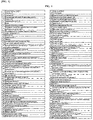
- FIG. 1 The results of proteomic analysis of exosomes derived from human colorectal cancer cell line HCT116 (American Type Culture Collection) are shown in FIG. 1 .
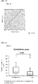
- an antigen CD 147 extracted from the above results expression of CD 147 in the exosomes derived from the colorectal cancer cell line was evaluated by Western blot method using a mouse antihuman CD 147 monoclonal antibody (manufactured by Novus Biologicals, Clone MEM-M 6/1). A comparison was made using HCT116 cells having very high malignancy and Caco2 cells (American Type Culture Collection) having low malignancy as the human colorectal cancer cell line. As a result, the presence of CD 147 could be confirmed with exosomes derived from HCT116 cell line having very high malignancy, but could not be detected with the exosomes derived from the Caco2 cells ( FIG. 2 ).
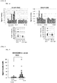
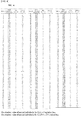
- Signals derived from exosomes expressing CD9 and CD 147 were measured in the same manner as in Test Example 2, using sera derived from even more colorectal cancer patients and sera derived from normal individuals ( FIG. 5 ). Concretely, 194 cases of sera derived from colorectal cancer patients and 191 cases of sera derived from normal individuals were used.
- FIGs. 7A, 7B , and 7C show that ROC analysis was carried out to compare the detection abilities of the colorectal cancer in each of the measurement methods, where high detection ability is exhibited as the AUC approximates 1.
- the AUC was 0.820, which was found to be far larger than those of CEA or CA19-9 (0.669 and 0.622, respectively). It was shown that the examination of colorectal cancer using the signals derived from exosomes expressing CD9 and CD147 was more excellent than that of CEA or CA19-9.
- Test Example 4 Analysis of Changes of Signal Intensities Before and After Operation
- the method of the present invention determines whether or not a sample provider has a high possibility of developing a colorectal cancer can be judged. Therefore, the method is useful because the sample provider can take a means of inhibiting the progression of cancer.
Landscapes
- Health & Medical Sciences (AREA)
- Immunology (AREA)
- Chemical & Material Sciences (AREA)
- Life Sciences & Earth Sciences (AREA)
- Molecular Biology (AREA)
- Engineering & Computer Science (AREA)
- Organic Chemistry (AREA)
- General Health & Medical Sciences (AREA)
- Hematology (AREA)
- Biochemistry (AREA)
- Medicinal Chemistry (AREA)
- Urology & Nephrology (AREA)
- Biomedical Technology (AREA)
- Food Science & Technology (AREA)
- Physics & Mathematics (AREA)
- Analytical Chemistry (AREA)
- Microbiology (AREA)
- Cell Biology (AREA)
- General Physics & Mathematics (AREA)
- Pathology (AREA)
- Biophysics (AREA)
- Genetics & Genomics (AREA)
- Proteomics, Peptides & Aminoacids (AREA)
- Biotechnology (AREA)
- Peptides Or Proteins (AREA)
- Preparation Of Compounds By Using Micro-Organisms (AREA)
Applications Claiming Priority (2)
| Application Number | Priority Date | Filing Date | Title |
|---|---|---|---|
| JP2013080303 | 2013-04-08 | ||
| PCT/JP2014/057588 WO2014167969A1 (fr) | 2013-04-08 | 2014-03-19 | Méthode de détection du cancer du colon |
Publications (2)
| Publication Number | Publication Date |
|---|---|
| EP2985605A1 true EP2985605A1 (fr) | 2016-02-17 |
| EP2985605A4 EP2985605A4 (fr) | 2017-02-15 |
Family
ID=51689381
Family Applications (1)
| Application Number | Title | Priority Date | Filing Date |
|---|---|---|---|
| EP14782540.0A Withdrawn EP2985605A4 (fr) | 2013-04-08 | 2014-03-19 | Méthode de détection du cancer du colon |
Country Status (6)
| Country | Link |
|---|---|
| US (1) | US20160047812A1 (fr) |
| EP (1) | EP2985605A4 (fr) |
| JP (1) | JP6386995B2 (fr) |
| CN (1) | CN105074468A (fr) |
| CA (1) | CA2907606A1 (fr) |
| WO (1) | WO2014167969A1 (fr) |
Families Citing this family (11)
| Publication number | Priority date | Publication date | Assignee | Title |
|---|---|---|---|---|
| WO2017124000A1 (fr) * | 2016-01-14 | 2017-07-20 | The Regents Of The University Of California | Procédé exoquant 3d pour analyse de molécules de surface et quantification d'exosomes spécifiques à un tissu dans des fluides biologiques |
| EP3417877A4 (fr) * | 2016-02-18 | 2019-10-02 | Kyoto University | Complexe capable d'inhiber la fonction génétique dans un exosome, et suppresseur de la prolifération et/ou de la métastase cancéreuses |
| WO2017223186A1 (fr) * | 2016-06-21 | 2017-12-28 | Nant Holdings Ip, Llc | Traitement du cancer guidé par exosome |
| JP2019168223A (ja) * | 2016-08-12 | 2019-10-03 | 公立大学法人和歌山県立医科大学 | エクソソームの膜に存在するタンパク質の検出方法 |
| IL266652B2 (en) * | 2016-11-16 | 2024-11-01 | Nanosomix Inc | Quantification of subpopulations of exosomes and diagnosis of neurodegenerative disorders |
| JP6797427B2 (ja) | 2017-01-12 | 2020-12-09 | テオリアサイエンス株式会社 | 被検者が膵臓癌に罹患している可能性を試験する方法 |
| JP7512292B2 (ja) * | 2019-09-05 | 2024-07-08 | 公益財団法人がん研究会 | 大腸がんマーカー、及びこれを用いた検査方法 |
| US12392776B2 (en) * | 2020-06-01 | 2025-08-19 | City University Of Hong Kong | Methods and kits for detecting exosomal protein |
| CN116256521B (zh) * | 2022-11-23 | 2025-12-02 | 南开大学 | 一种用于肠癌诊断的粪便外泌体蛋白的标志物及其应用 |
| JPWO2024262608A1 (fr) | 2023-06-22 | 2024-12-26 | ||
| CN120971738B (zh) * | 2025-08-14 | 2026-02-03 | 北京国浩宇医疗科技有限公司 | 一种用于外泌体cd82蛋白含量检测的试剂盒 |
Family Cites Families (10)
| Publication number | Priority date | Publication date | Assignee | Title |
|---|---|---|---|---|
| US6924114B2 (en) * | 2000-09-20 | 2005-08-02 | Surromed, Inc. | Method for monitoring resting and activated platelets in unfixed blood samples |
| EP1481249A1 (fr) * | 2002-03-07 | 2004-12-01 | Cambridge University Technical Services Limited | Empreintes digitales de scd |
| WO2010056337A2 (fr) * | 2008-11-12 | 2010-05-20 | Caris Mpi, Inc. | Procédés et systèmes d'utilisation d'exosomes pour déterminer des phénotypes |
| US20140113310A9 (en) * | 2008-12-05 | 2014-04-24 | Myriad Genetics, Incorporated | Cancer detection markers |
| US20120231970A1 (en) * | 2009-09-30 | 2012-09-13 | Japan Health Sciences Foundation | Colon cancer marker and method for testing for colon cancer |
| EP2308897A1 (fr) * | 2009-10-09 | 2011-04-13 | Pierre Fabre Medicament | Anticorps chimères spécifiques de CD151 et leurs utilisations pour le traitement du cancer |
| EP2542696B1 (fr) * | 2010-03-01 | 2016-09-28 | Caris Life Sciences Switzerland Holdings GmbH | Biomarqueurs pour théranostique |
| ES2624284T3 (es) * | 2010-07-07 | 2017-07-13 | Aethlon Medical Inc | Métodos para cuantificar exosomas |
| AU2011316477A1 (en) * | 2010-10-11 | 2013-05-02 | Medsaic Pty Ltd | Assay for disease detection |
| EP2678448A4 (fr) | 2011-02-22 | 2014-10-01 | Caris Life Sciences Luxembourg Holdings S A R L | Biomarqueurs circulants |
-
2014
- 2014-03-19 CA CA2907606A patent/CA2907606A1/fr not_active Abandoned
- 2014-03-19 EP EP14782540.0A patent/EP2985605A4/fr not_active Withdrawn
- 2014-03-19 JP JP2015511175A patent/JP6386995B2/ja active Active
- 2014-03-19 US US14/778,831 patent/US20160047812A1/en not_active Abandoned
- 2014-03-19 CN CN201480019268.8A patent/CN105074468A/zh active Pending
- 2014-03-19 WO PCT/JP2014/057588 patent/WO2014167969A1/fr not_active Ceased
Also Published As
| Publication number | Publication date |
|---|---|
| JP6386995B2 (ja) | 2018-09-05 |
| JPWO2014167969A1 (ja) | 2017-02-16 |
| CN105074468A (zh) | 2015-11-18 |
| EP2985605A4 (fr) | 2017-02-15 |
| US20160047812A1 (en) | 2016-02-18 |
| CA2907606A1 (fr) | 2014-10-16 |
| WO2014167969A1 (fr) | 2014-10-16 |
Similar Documents
| Publication | Publication Date | Title |
|---|---|---|
| EP2985605A1 (fr) | Méthode de détection du cancer du colon | |
| EP2472264B1 (fr) | Détection multiplex de cellules tumorales utilisant plusieurs agents se fixant à des marqueurs extracellulaires | |
| Chen et al. | Elevated level of anterior gradient-2 in pancreatic juice from patients with pre-malignant pancreatic neoplasia | |
| EP3130605B1 (fr) | Récepteur alpha de folate à titre de marqueur diagnostique et pronostique des cancers exprimant un récepteur alpha de folate | |
| CA3029926C (fr) | Biomarqueur diagnostique pour cancer des voies biliaires extra-hepatique ou cancer de la vesicule biliaire | |
| Jou et al. | Salivary zinc finger protein 510 peptide as a novel biomarker for detection of oral squamous cell carcinoma in early stages | |
| US20130040849A1 (en) | Method and kit for cancer diagnosis | |
| CN105745543B (zh) | 卵巢癌的自身抗体生物标志物 | |
| CA2786005A1 (fr) | Procedes de diagnostic du potentiel de malignite de lesions kystiques pancreatiques | |
| Luka et al. | Development of a serum biomarker assay that differentiates tumor‐associated MUC5AC (NPC‐1C ANTIGEN) from normal MUC5AC | |
| EP2863224A1 (fr) | Kit permettant de diagnostiquer un mélanome malin | |
| US20160341733A1 (en) | Detection and treatment of cancer | |
| WO2011148669A1 (fr) | La vitronectine, marqueur du cancer colorectal et procédé d'analyse de la concentration en vitronectine d'un échantillon de sang | |
| WO2016143805A1 (fr) | Procédé de détection du cancer de la vessie | |
| US11391737B2 (en) | Methods to predict progression of Barrett's Esophagus to high grade dysplasia esophageal adenocarcinoma | |
| Kojima et al. | Titration of serum CEA, p53 antibodies and CEA-IgM complexes in patients with colorectal cancer | |
| KR102735773B1 (ko) | 혈액 내 단백질 바이오마커를 포함하는 암 진단용 키트 | |
| KR102128251B1 (ko) | 아르기닌이 메틸화된 drd2에 특이적으로 결합하는 대장암 진단용 바이오마커 조성물 | |
| EP3364188B1 (fr) | Biomarqueur sanguin pour utilisation dans l'évaluation de l'effet d'une pharmacothérapie sur le cancer du rein | |
| WO2025083329A1 (fr) | Liant d'anticorps en tant que biomarqueur pour le cancer | |
| WO2025197993A1 (fr) | Procédé de concentration de vésicules extracellulaires positives à la tétraspanine 10, et procédé de détection du cancer ou de prédiction du pronostic du cancer | |
| WO2021045189A1 (fr) | Marqueur du cancer colo-rectal et procédé de test l'utilisant | |
| Chen et al. | Research Elevated level of anterior gradient-2 in pancreatic juice from patients with pre-malignant pancreatic neoplasia | |
| HK1208884B (en) | Methods to predict progression of barrett ' s esophagus to high grade dysplasia or esophageal adenocarcinoma | |
| KR20080092490A (ko) | 대장암 진단용 단백질 마커 켈그라눌린 a와 켈그라눌린 b및 이들 각각에 대한 항체를 포함하는 대장암 진단키트 |
Legal Events
| Date | Code | Title | Description |
|---|---|---|---|
| PUAI | Public reference made under article 153(3) epc to a published international application that has entered the european phase |
Free format text: ORIGINAL CODE: 0009012 |
|
| 17P | Request for examination filed |
Effective date: 20151006 |
|
| AK | Designated contracting states |
Kind code of ref document: A1 Designated state(s): AL AT BE BG CH CY CZ DE DK EE ES FI FR GB GR HR HU IE IS IT LI LT LU LV MC MK MT NL NO PL PT RO RS SE SI SK SM TR |
|
| AX | Request for extension of the european patent |
Extension state: BA ME |
|
| DAX | Request for extension of the european patent (deleted) | ||
| RAP1 | Party data changed (applicant data changed or rights of an application transferred) |
Owner name: THEORIA SCIENCE INC. |
|
| RIC1 | Information provided on ipc code assigned before grant |
Ipc: G01N 33/53 20060101ALI20161010BHEP Ipc: C12P 21/08 20060101ALI20161010BHEP Ipc: G01N 33/574 20060101AFI20161010BHEP Ipc: C07K 16/28 20060101ALI20161010BHEP |
|
| A4 | Supplementary search report drawn up and despatched |
Effective date: 20170118 |
|
| RIC1 | Information provided on ipc code assigned before grant |
Ipc: C07K 16/28 20060101ALI20170112BHEP Ipc: G01N 33/574 20060101AFI20170112BHEP Ipc: C12P 21/08 20060101ALI20170112BHEP Ipc: G01N 33/53 20060101ALI20170112BHEP |
|
| STAA | Information on the status of an ep patent application or granted ep patent |
Free format text: STATUS: THE APPLICATION IS DEEMED TO BE WITHDRAWN |
|
| 18D | Application deemed to be withdrawn |
Effective date: 20170815 |